Embed Size (px)
Citation preview
Marquette Law ReviewVolume 41Issue 2 Fall 1957 Article 2
The Heart and CirculationJoseph Kuzma
Follow this and additional works at: http://scholarship.law.marquette.edu/mulr
Part of the Law Commons
This Article is brought to you for free and open access by the Journals at Marquette Law Scholarly Commons. It has been accepted for inclusion inMarquette Law Review by an authorized administrator of Marquette Law Scholarly Commons. For more information, please [email protected].
Repository CitationJoseph Kuzma, The Heart and Circulation, 41 Marq. L. Rev. 121 (1957).Available at: http://scholarship.law.marquette.edu/mulr/vol41/iss2/2
SYMPOSIUM ON TRAUMA AND HEART DISEASE
The following article is one of three planned as a symposium deal-ing with the medical and legal problems arising from the effect of in-jury on the heart. The purpose here is to acquaint the attorney withthe medical background necessary to the preparation of scientificproof required in personal injury and compensation cases. Later ar-ticles will deal with the legal aspects, particularly proof and causation,and finally with the problems confronting the expert witness.
THE HEART AND CIRCULATION
JOSEPH F. KUZmA, M.D.*
I. NORMAL ANATOMY
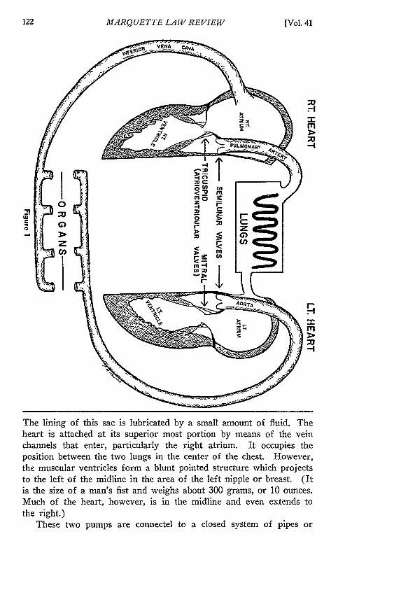
The human heart is a dual pump- right heart and left heart -
arranged in series, so designed that it looks and acts as a single organ.It consists of four chambers, each "heart" having two: one, a thinmuscular reservoir sitting most superiorly, which is called the atrium;and the other, which si-s inferiorly is the thick muscular actively con-tractile part of the pump, called the ventricle. The two "hearts" arejoined into one unit by having a common vertical partition whichseparates right from left.1 Each atrium is marked by a number ofopenings through which blood enters. The atrium is connected to theventricle by a single opening which is guarded by a valve called theatrio-ventricular valve; in the right heart known as the tricuspid valve,and in the left as the mitral valve. Each ventricle, by contraction,forces the blood (stroke volume) into the circulation. The outflow-of each ventricle is guarded by a semilunar valve which consists ofthree leaflets. These prevent the blood from returning into the ven-tricle during the filling phase. The blood from the right ventricle ispropelled into the lungs (pulmonary circulation), and then is deliveredto the left atrium through four openings. From the left atrium, itpasses through the mitral valve into the left ventricle. The output ofthe left ventricle is propelled into the great vessel known as aorta andthere is distributed to all parts of the body in which the various organsare hooked up in "parallel." Blood returns to the right atrium throughtwo large veins, superior and inferior vena cava. The right and leftheart, therefore, are "in series."
The "right" heart has the smaller capacity, has a thinner wall,
creates less pressure than the "left," and is known as the pump for
the pulmonary circulation. The "left" heart is a part of the systemiccirculation. The heart is encased in a fibrous sac called the pericardium.
*Professor of Pathology, Marquette University School of Medicine.1 Figures 1 and 2.
MARQUETTE LAW REVIEW
The lining of this sac is lubricated by a small amount of fluid. Theheart is attached at its superior most portion by means of the veinchannels that enter, particularly the right atrium. It occupies theposition between the two lungs in the center of the chest. However,the muscular ventricles form a blunt pointed structure which projectsto the left of the midline in the area of the left nipple or breast. (Itis the size of a man's fist and weighs about 300 grams, or 10 ounces.Much of the heart, however, is in the midline and even extends tothe right.)
These two pumps are connectel to a closed system of pipes or
[Vol. 41
THE HEART
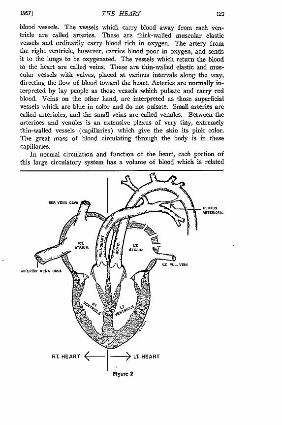
blood vessels. The vessels which carry blood away from each ven-tricle are called arteries. These are thick-walled muscular elasticvessels and ordinarily carry blood rich in oxygen. The artery fromthe right ventricle, however, carries blood poor in oxygen, and sendsit to the lungs to be oxygenated. The vessels which return the bloodto the heart are called veins. These are thin-walled elastic and mus-cular vessels with valves, placed at various intervals along the way,directing the flow of blood toward the heart. Arteries are normally in-terpreted by lay people as those vessels which pulsate and carry redblood. Veins on the other hand, are interpreted as those superficialvessels which are blue in color and do not pulsate. Small arteries arecalled arterioles, and the small veins are called venules. Between thearterioes and venules is an extensive plexus of very tiny, extremelythin-walled vessels (capillaries) which give the skin its pink color.The great mass of blood circulating through the body is in thesecapillaries.
In normal circulation and function of the heart, each portion ofthis large circulatory system has a volume of blood which is related
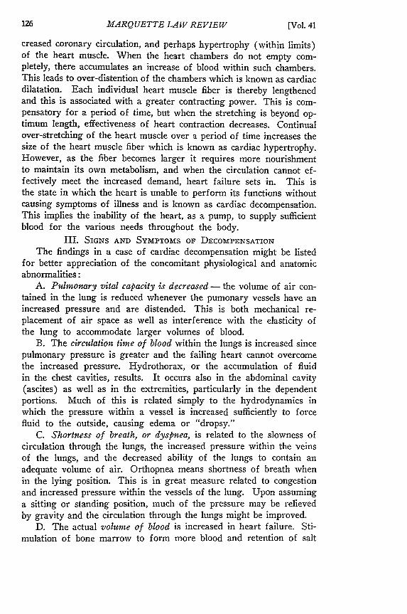
DUCTUSARTERIOSIS
LT. PUL. VEIN
RT HEART E
1957]
HEART
MARQUETTE LAW REVIEW
to that in other portions; that is, the volume within the chambers ofthe heart, in the arteries, in the veins, and in the capillaries, is allcorrelated so that there is a uniform circulation of blood withoutabnormal accumulation in any particular area.
Each organ has its own artery from the main aortic circulation,and is hooked-up to the feed-line aorta in "parallel" arrangement.Nourishment of all tissues comes from the blood supply. Althoughthe heart is bathed with blood on its internal surfaces, it does notreceive sufficient nourishment just by contact on the lining of itschambers. Accordingly, it has its own arterial blood supply throughtwo vessels, called coronary arteries, right and left. These originatefrom the aorta immediately above the left ventricle of the heart andencompass the base of the heart as a crown might fit on a head;therefore, they are referred to as "coronary" arteries. From thecrown type of arrangement, branches extend longitudinally along thechambers of the heart towards its very point or apex.
II. NORMAL PHYSIOLOGY
The actual contracting and functioning actions of the heart areexceedingly intricate. There is innate natural contractility of heartmuscle and it may show muscular action for some time after it isseparated from the body. However, the normal intact heart has abuilt-in "communications" system which fundamentally controls itsrate and force of contraction, and coordinates the action of all parts.This is called the conduction system, fibers of which run in all fourchambers but begin in the right atrial chamber. The heart is alsogoverned by the nervous system through the parasympathetic vagusnerve, and by outside regulators such as the carotid sinus in the neck,and the adrenal glands which produce adrenalin. In normal action,all chambers of the heart are synchronized and effectively propelblood along the way. Force of contraction is governed in part bythe tension on the heart muscle itself. When a chamber (ventricle)is slightly over-distended, it will contract with greater force to expelthe blood which it had acquired. Under usual circumstances, theheart will contract approximately 72 times each minute and will haveejected several liters of blood.
The pathway of the circulation is somewhat as follows: blood fromvarious portions of the body is brought to the right atrium by twoprincipal veins called the superior and inferior vena cava. The right
atrium will by contraction, by gravity, and by increasing load, pass the
blood to the right ventricle through the tricuspid valve. The right
ventricle, upon contraction, propels the blood into the pulmonary artery
through the semilunar valve, and the blood is thereby distributed
through the ramifications of the pulmonary artery within the lung.
After passing through the capillaries of the lung, the blood is re-
[Vol. 41
THE HEART
collected into larger channels called veins and four such veins emptyinto the left atrium. The blood is then passed on to the left ventricle,through the mitral valve, and on contraction of the left ventricle, it issent into the aorta through the aortic semilunar valve. The aorta hasmany branches and each major organ has a principal artery coming offdirectly from the aorta. After passing through the capillary bed ofthe various organs, the blood is recollected into the venous system andbrought back to the right atrium and the cycle is repeated. Within thiscircuit of propelled fluid, a certain level of hydrostatic pressure ismaintained. The pressure created by the contraction of the ventriclesin propelling blood into the arteries is known as the systolic pressureand ordinarily is in the neighborhood of 120 mam. of mercury. Im-mediately after contraction, the pressure in the vessel falls. A certainlevel is then maintained until the ventricle contracts again and createsa systolic pressure. This sustained pressure within the blood vesselsbetween contractions of the ventricles is known as diastolic bloodpressure and is roughly in the neighborhood of 70 mm. of mercury.It is rather clear, therefore, that systolic pressure in great measureresults from the force of the contraction of the ventricle, and that thediastolic pressure depends in the main on the state of the blood vessels,namely, how much resistance the vessels present to the circulation ofthe blood (constriction increases resistance), and how effective theyare in dissipating the systolic pressure by their elasticity. Exerciseand effort increase blood pressure; rest and sleep are associated witha decrease.
The pressure in the outgoing vessels from the heart is graduallydiminshed as the finer ramifications are reached so that in capillaries,the blood is propelled through this network with a very small head ofpressure. On the other side of the capillary bed where the blood isbeing collected into veins to be returned to the heart, the pressure islow and at times is a negative pressure; particularly as created byactions of respiration so that blood is, in effect, "lifted" to the heartfrom the lower extremities rather than forced by intraluminal positivepressure.
It should be relatively clear at this point, that variations in thenumber of contractions that the heart makes in a period of time, theresistance against which the heart must work to propel the blood, andabnormal accumulations of blood in various systems within the body,may adversely influnece the effectiveness of the circulation. Never-theless, the heart has a very marked ability to compensate for stressesplaced upon it, but the compensation may not be effective over anindefinite period of time. The ability of the heart to perform effectivelyduring states of increased demand is called cardiac reserve. Reservedepends on increase of heart rate, increased filling of ventricles, in-
1957]
MARQUETTE LAW REVIEW
creased coronary circulation, and perhaps hypertrophy (within limits)of the heart muscle. When the heart chambers do not empty com-pletely, there accumulates an increase of blood within such chambers.This leads to over-distention of the chambers which is known as cardiacdilatation. Each individual heart muscle fiber is thereby lengthenedand this is associated with a greater contracting power. This is com-pensatory for a period of time, but when the stretching is beyond op-timum length, effectiveness of heart contraction decreases. Continualover-stretching of the heart muscle over a period of time increases thesize of the heart muscle fiber which is known as cardiac hypertrophy.However, as the fiber becomes larger it requires more nourishmentto maintain its own metabolism, and when the circulation cannot ef-fectively meet the increased demand, heart failure sets in. This isthe state in which the heart is unable to perform its functions withoutcausing symptoms of illness and is known as cardiac decompensation.This implies the inability of the heart, as a pump, to supply sufficientblood for the various needs throughout the body.
III. SIGNS AND SYMPTOMS OF DECOMPENSATION
The findings in a case of cardiac decompensation might be listedfor better appreciation of the concomitant physiological and anatomicabnormalities:
A. Pulmonary vital capacity is decreased - the volume of air con-tained in the lung is reduced whenever the pumonary vessels have anincreased pressure and are distended. This is both mechanical re-placement of air space as well as interference with the elasticity ofthe lung to accommodate larger volumes of blood.
B. The circulation time of blood within the lungs is increased sincepulmonary pressure is greater and the failing heart cannot overcomethe increased pressure. Hydrothorax, or the accumulation of fluidin the chest cavities, results. It occurs also in the abdominal cavity(ascites) as well as in the extremities, particularly in the dependentportions. Much of this is related simply to the hydrodynamics inwhich the pressure within a vessel is increased sufficiently to forcefluid to the outside, causing edema or "dropsy."
C. Shortness of breath, or dyspnea, is related to the slowness ofcirculation through the lungs, the increased pressure within the veinsof the lungs, and the decreased ability of the lungs to contain anadequate volume of air. Orthopnea means shortness of breath whenin the lying position. This is in great measure related to congestionand increased pressure within the vessels of the lung. Upon assuminga sitting or standing position, much of the pressure may be relievedby gravity and the circulation through the lungs might be improved.
D. The actual volume of blood is increased in heart failure. Sti-mulation of bone marrow to form more blood and retention of salt
[Vol. 41
THE HEART
which attracts water is the explanation. Any organ of the body maybecome enlarged as its venous pressure is increased and the conditionof "passive congestion" develops.
E. Blueness of color, or cyanosis, is related to the presence of bloodwith insufficient oxygen. This may be due to stagnation of circulationin heart failure, or may be related to disease of the lungs whereinnormal oxygen exchange cannot take place.
IV. DISEASE STATES OF THE HEART
Disturbances of the heart and blood vessel system, kmown aspathological states, are basically of one of three types. These will begiven below and a short explanation of the various important categorieswithin each type will be mentioned:
A. Principally physiological disorders of the heart which leadto failure: Irregularities of heart function, principally those of rateand rhythm of the pump action are significant. There is a wide rangeof normal or physiological cardiac activity within which range therate of contraction (number of beats per minute) of the heart as wellas the output of each contraction sustain life without symptoms ofdistress. Increased rate of heart action takes place with exercise andemotional disturbances. Such ranges, of course, are considered to bewithin physiological normals. However, if one speeds the rate ofheart contraction beyond 150 times per minute, then incomplete fillingof the ventricles may take place and this, of course, would be reflectedin reduced output of each contraction, and therefore reduced coronarycirculation. However, the heart in most instances requires morenourishment. A very rapid heart rate (tachycardia) will seriouslyreduce the cardiac output and thereby cause increased venous pressureand distress, or evidence of heart failure. Serious abnormalities inrate leading to distress and even death may develop from intoxication,such as belladonna poisoning.
There are also irregularities of cardiac rhythm (relationship of re-currence of heart contraction) which may adversely influence the heartin doing its work. In auricular fibrillation, instead of a uniform syn-chronized contraction of the atrial heart muscle, there are irregularconvulsive movements which are not effective in propelling blood.This timing disorder is frequently associated with other diseases ofthe heart that are anatomic in nature, such as rheumatic fever. Aheart suffering from auricular fibrillation is at a disadvntage sincethe actions of the atrium are not synchronized and fully effective.Great energy may be expended with little work accomplished. Muralthrombosis (blood clot on atrial walls) develops in arrythmias andpresents a threat for infarction of other organs by embolic occlusionof vessels. (Clots loosened and carried in the blood stream plug avessel and cause death of the tissue supplied by that vessel).
1957]
MARQUETTE LA W REVIEW
A condition similar to auricular fibrillation is called ventricularfibrillation. In this instance, the heart muscle of the ventricle itselfis undergoing tremulous or convulsive movements without effectivecontraction. This condition is not compatible with life and, unless cor-rected immediately, death usually results in a matter of a few minutes.Occurrence of ventricular fibrillation and cardiac arrest during anes-thesia and surgical procedures has recently been emphasized.
A decreased rate of heart contraction is called bradycardia. Somedegree of slowing of the rate occurs with rest and sleep, however, anextreme degree may be brought on by various disturbances, such asjaundice, typhoid fever, and tumors within the skull. In a greaternumber of patients, slow heart rate develops from interference withthe conduction system within the heart itself. This may be producedby drugs or by scar tissue that involves the conduction system. Asheart rate is decreased, a point is reached wherein the brain receivesinsufficient blood supply and the individual suffers from fainting, orsyncope. In any instance of sudden decrease of blood volume orpressure, syncope may develop. Many of the disorders which relateto consciousness or syncope have much to do with nerve innervationand emotional disturbances. The vagus nerve and vaso-vagal reflexeswhich govern heart action are intimately tied into this particularphenomenon. It is important to note that a normal cardiovascularsystem recovers quickly after such syncope and the individual is re-stored to his full health in a matter of minutes. However, in diseasestates of the cardiovascular system, the return to normal may beslower than usual and, in fact, death may occur from cardiac standstillor ventricular fibrillation.
Closely related to the condition of syncope is the problem of shock.This is frequently defined as an altered state of health characterized bya markedly decreased blood pressure, decreased circulating blood
volume, decrease of cardiac output, rapid weak pulse, pale, cold, moist
skin, and loss of consciousness. Such a state may result from nervousinfluence, severe trauma, and loss of blood. From a teleological stand-
point, the reduction in blood volume, the drop in blood pressure, and
the syncope which attends the state of shock, are beneficial. An
organism suffering blood loss, on becoming unconscious, assumes a
horizontal position which improves cerebral circulation, and at the
same time reduces the demands of other organs. As blood pressure
and blood volume are decreased, there is a tendency for hemorrhageto stop. Shock, therefore, may be effective in saving life; however,
sometimes shock is an irreversible phenomenon. In these instances,
the continued decrease in cardiac output leads to oxygen starvation
in many tissues, and the brain being extremely sensitive to oxygen want,
may suffer irreparable damage.
[Vol. 41
THE HEART
B. Chiefly anatomic conditions which place an undue work loadon the heart: The outstanding example of this is the disease ofhypertension, or high blood pressure. This is characterized by an in-crease in both systolic and diastolic pressure and is usually related toconstriction and inelasticity of the small vessels called arterioles. Thedisease frequently begins with a disorder in the kidney characterizedby decreased kidney circulation. Under such circumstances, a sub-stance is produced in the kidney which upsets the humoral balancecontrolling arteriolar size and causes the blood vessels to constrict.Continued constriction of the arterioles, leads to degenerative changeswithin the vessel walls. Degenerative changes consist of loss of elastictissue and muscle fibers, and replacement by a rather dense uniforminelastic material. The channel of the vessel is thereby greatly reduced.Reduction in the calibre of the small vessels throughout the body placesa severe load on the heart. The heart finds it difficult to empty itschambers against an increased resistance interposed by constrictedarterioles. The heart, first, stretches (dilates) to accommodate the in-creased volume of blood which accumulates as the chambers cannotcompletely empty; and as a result, the muscle fibers eventually enlarge(cardiac hypertrophy). After a time, the dilatation and hypertrophycannot cope with the increased work load, and gradually the heartaccumulates more and more blood within its chambers, and more andmore of it backs up into the venous side of the heart, elevating peri-pheral venous pressure. All signs and symptoms of heart failure thenbecome evident (left heart failure). Similarly, diseases of the lungwhich reduce the blood vessel volume within the lung, such as pro-gressive fibrosis, place an undue load on the right ventricle, leadingto decompensation (right heart failure). Terminally, total failureoccurs and one does not distinguish between right and left failure.
A congenital narrowing of the aorta, called coarctation, also pro-duces a severe load on the heart. In such instances, the heart maydilate and hypertrophy over a long period of time and years mayelapse before heart failure, "decompensation," becomes evident.
Ability of the heart to empty itself may be seriously hindered bydiseases of the heart valves. These commonly are the result of rheu-matic fever, syphillis, and bacterial inflammations of the lining of theheart (endocarditis). Such diseases produce two types of valvular
dysfunction: one is called stenosis or narrowing of the valve opening.This is especially true of the mitral valve with the disease of rheumaticfever. The other is enlargement of the valve opening to such an extentthat it is incompetent. This is called valvular insufficiency and is bestdemonstrated in the disease of syphillis. Acute inflammation (Aschoffnodules- collections of cells of inflammation about degenerating sup-
porting connective tissue) of the heart valve in rheumatic fever is
1957]
MARQUETTE LAW REVIEW
followed by adhesions between the leaflets of the valve, thickening andstiffening of the leaflets, and severe distortion produced by gradualshrinkage of the scar tissue. This takes place over the course ofmany years.
In syphillis, on the other hand, there is a degeneration of theelastic and muscular tissue oi the aorta at its origin. This weakensthe wall to the point of a blowout. However, weakening at the heartvalve causes stretching and pulling apart of the leaflets so that theycannot make contact and close.
A chamber which must empty itself against a narrowed valveopening undergoes dilatation and hypertrophy and it is only a matterof time before this will no longer compensate for the increased loadand, thereafter, the heart will be in "failure." Some of the largesthearts (greatest hypertrophy) are seen with stenosis of the aorticvalve. When the mitral valve is narrowed as by rheumatic fever, theinability of the left atrium fully to empty itself through the narrowopening into the ventricle is reflected backwards into the lungs, intothe pulmonary artery, and into the right ventricle, which becomesenlarged, since it is the chamber that must overcome the load. Valveswhich are incompetent (allow blood to go in retrograde direction)are accompanied by dilatation and hypertrophy of the chamber whichdoesn't empty itself fully since blood returns to the chamber ratherthan being directed forward during the resting phase (diastole) of thepump action.
A condition quite similar to overloading of a chamber by an in-competent valve may also result from malformations of the heartduring its development. Such are known as congenital anomalies, andthe important ones are associated with the abnormal shunting of bloodto chambers and vessels where the added volume creates extra load.A disorder of this kind is known as the persistent patent ductusarteriosus which means a channel communicating between the pul-monary artery and the aorta. It serves as a physiological shunt(pulmonary artery to aorta) during intrauterine life. However, afterbirth this shunt is no longer needed and spontaneously closes sincethe lungs are expanded with air and can take all the blood which theright heart expels. In postnatal life, the pressure in the aorta isgreater than in the pulmonary artery. If the ductus remains open,then with each contraction of the heart, the blood will be sent fromthe aorta through this ductus, into the pulmonary artery. Eventuallythe pulmonary artery will dilate and may even rupture.
An opening between the two ventricles through the interventricularseptum is called Rogers disease, (patent interventricular septal defect)and it too is associated with increased volume and load in the rightheart since after birth the pressure of the left chamber is greater
[Vol. 41
THE HEART
than the right.Pulmonary stenosis is a congenital anomaly with narrowing of the
outlet of the right heart at the pulmonary artery. This, of course,places a severe work load on the, right ventricle each time the ventricleattempts to empty itself against the narrowed opening. This causesdilatation and severe hypertrophy of the right ventricle. Cardiac dilata-tion and hypertrophy compensate for the above difficulties for a periodof time. However, decompensation is the final chapter.
Another serious obstacle to health and life in individuals withrheumatic valvular disease and congenital anomalies is bacterial endo-carditis. Erosions of heart lining membrane (endocardium) on dam-aged valves and at congenital defects permit bacteria to grow therein.Unless the bacteria are controlled, death is certain from septicemia(blood stream poisoning by bacteria). However, even eradicatedbacterial disease of heart valves has left its injurious mark on theprevious disorder and life is frequently shortened.
C. Ineffective pump musculature: In this category is a group ofconditions which are characterized primarily by heart muscle diseaseswhich render contraction and pump action ineffective. Some of theseare related to nutritional disturbances, such as the disease of beriberi,or Vitamin B1 deficiency. Likewise, in chronic alcoholism, the de-velopment of a heart muscle filled with fat may be attended by lossof vigorous contraction. The heart muscle in diseases of high fever(typhoid, meningitis, etc.) frequently is at a disadvantage in itsability to contract, (toxic myocarditis). There are unusual instanceswherein the heart muscle itself is the seat of infectious disease, suchas Fiedler's myocarditis. The severe infiltration of the heart muscleby cells of inflammation precludes its function as a contracting, work-performing organ. Serious disturbances of the heart action may alsoresult from changes in the concentrations of certain essential sub-stances in the blood and tissues, namely the elements of calcium andpotassium.
The most important disease affecting the ability of the heart muscleto contract and propel blood is that which is known as myocardialinfarction. This is a condition in which a portion of the heart muscleundergoes death and eventual replacement by connective tissue scar.Infarction is caused by sudden loss of blood supply to the heart muscleas occurs in occlusions of the hearts' vessels (coronary thrombosis).The sudden loss of the blood supply to the heart muscle may causeventricular fibrillation or cardiac standstill and sudden death- with-out anatomic change in the heart muscle. If, however, such an in-dividual survives, the heart muscle goes through a chain of events inthe process of dying and being removed. The muscle is severelychanged as evidenced by its appearance in a matter of 24 hours. At
1957]
MARQUETTE LAW REVIEW
this time, cells of inflammation and hemorrhage are prominent withinthe muscle which is undergoing death. Towards the end of the firstweek, the heart muscle is completely dead, it is being digested in theprocess of removal, and at this time it is extremely weak. The heart,in contracting, may actually break the wall and propel blood into thepericardial sac. Such an occurrence is called cardiac tamponade. Ina matter of just a few minutes, the pressure within the unyieldingfibrous pericardial sac may equal that within the chamber of the ven-tricle and as soon as this happens, the ventricle is no longer an effectivepump and cannot propel blood to the organs of the body. Suddendeath is the result of this. However, if the heart wall does not rupture,then it is gradually replaced by scar tissue over a period of weeks.While this is going on, however, the lining of the heart at the pointof the muscle damage, may develop a blood clot. This is called amural thrombosis. The clot which develops may be broken off by theactions of the heart and be carried in the blood stream to any otherorgan of the body where it occludes a blood vessel and causes aninfarction just like that of the heart muscle itself. In the brain, thisis evidenced by loss of motor function (paralysis), loss of speech, lossof consciousness, etc. Heart failure may occur at any time aftermyocardial infarction, but many months or years after such an attack,the scar tissue of the heart wall may begin to bulge externally. Thepressure within the chamber causes the bulge which is called ananeurysm.
The story of myocardial infarction is not at all complete withoutdiscussion of its cause. The coronary arteries must be patent and carrya sufficient amount of blood at a sufficient pressure to maintain thenourishment of the heart muscle. The coronary arteries are subjectto the pressure which is quite the same as that of the aorta and undergoa degenerative disease called arteriosclerosis. This means that theelastic and muscle fibers of these arteries are lost and replaced byscar tissue, by fatty deposits, and by calcium. Such calcium and fattydeposits may actually build up to a mass in the wall and project intothe channel itself. This may reach the proportions of occluding theentire lumen of the vessel. More commonly, however, the bulgingpart of such a mass suffers loss of the lining of the vessel and ablood clot suddenly develops on such an area. This is called coronarythrombosis. Hemorrhage within the wall of the coronary artery, whenit is diseased and replaced by fatty materials, causes a lifting of thelining and pushing of it to the opposite side; thereby, effectively oc-cluding the circulation at that point. The result of coronary occlusion,irrespective of how it develops, is myocardial infarction.
A condition, frequently confused bV the clinician as myocardialinfarction, is a tearing of the aorta, called dissecting aneurysm. It
[Vol. 41
THE HEART
begins as a weakening and destruction of the elastic and muscle fibersof its wall (cystic medial necrosis). Hemorrhage develops in this siteand then tears apart the inner and outer layers of the aortic wall.A double-barrel aorta is thereby produced; one channel (the normalone) with moving blood, and the other within the wall with a trappedcollection of blood. Eventually, the blood collected within the aorticwall may tear an opening through the inner wall into the normal lumenor rupture the outer wall and produce a fatal hemorrhage into thechest cavity or into the pericardial sac (cardiac tamponade).
V. FoRENsic CONSIDERATIONSIn general, there are only a few situations in which trauma or
stress have dire consequences in the absence of preexistent cardiacdisorder of some kind or other. Certainly a direct injury, notablypenetrating wounds, such as stab or bullet, need not be related topreexistent cardiac disease. At times, nevertheless, one must weighthe results of such injury in a normal heart versus one with pre-existent disease; for example, a stab or bullet wound which does notcompletely penetrate the heart chamber, may be tolerated without evi-dence of decompensation or permanent illness by a normal heart.However, a wound of like depth on the surface of a thinned myo-cardium following infarction, may cause perforation and cardiactamponade with sudden death. Likewise, the ability to repair a woundof the myocardium, may be related to the coronary artery supply andits efficiency. In such instances, healing of the myocardium mightbe retarded in the cases where interstitial fibrosis has resulted fromcoronary insufficiency.
In Group A, of part IV, the disturbances which may be aggravatedor may result in fatality (the broad consideration of stress and trauma)have to do with ventricular fibrillation, shock, and cardiac standstill.These may be called physiological death with very poor, if any, ana-tomic correlation. Notable in this might be the example of the individualwho collapses and dies while being interrogated by a police officerfollowing a minor automobile accident. He may have been only awitness of the accident. There are also the instances of witchcraft andvoodooism which have caused sudden death in presumably normalindividuals. This must be related to severe nervous influence of theheart that may produce cardiac standstill or ventricular fibrillation.The effect may be mediated through chemical substances such asadrenalin which is sharply elevated by severe fright. Such people un-doubtedly have an unduly excitable heart as the basic disorder.
In Group B, any situation which has produced an exaggeratedwork load on the heart, may be further augmented by effort or stress.This, of course, may be reflected in the usual signs and symptoms ofcardiac decompensation, or may be associated with sudden death.
1957]
MARQUETTE LA W REVIEW
Sudden death may be caused by rupture of a blood vessel in hyper-tension, (cerebral hemorrhage or stroke) by rupture of an aorta withcoarctation, by rupture of a dissecting aneurysm and cardiac tampon-ade, by sudden dilatation of the heart and by ventricular fibrillation indiseases of valvular character.
In Group C, the general disorder of diseased myocardium andits ability to act as an effective pump, may be seriously hampered byeffort. Certainly, physical effort may produce an increased bloodpressure, increased coronary artery pressure, rupture and hemorrhageof a fatty deposit in the coronary artery and injury of the endotheliumcovering such a plaque followed by the development of a blood clot(thrombus). On the other hand, sudden drop in blood pressure inthe state of shock or even the physiological lowering during sleep mayprecipitate myocardial infarction in people with coronray artery dis-ease. (These need a certain "head of pressure" to maintain adequatecirculation through diseased coronary vessels). Blunt injury to thechest wall may fracture a brittle coronary artery with its fatty andcalcified plaques and lead to obstruction of the coronary artery. Injurymay also cause a severe bruise of the heart muscle. Once a myocardialinfarction has taken place, continued physical exertion may lead toperforation of the wall of the heart at the point of infarction, as isso well evidenced by its development in mentally ill people who donot seek medical help or do not abide by the orders of bed rest duringthis phase of illness. Blunt injury to the chest and back and severephysical exertion have been known to cause either the inception ofa dissecting aneurysm of the aorta or its perforation into the peri-cardial sac. Nevertheless, it must be emphasized that the greater num-ber of myocardial infarctions occur without physical exertion (duringsleep, watching T.V., etc.) and that the overwhelming majority ofdissecting aneurysm cases are unrelated to effort or injury.
It must be clearly understood, in conclusion, that all diseases ofthe heart, except those of external physical violence, occur as "natural"phenomenon. Coronary thrombosis, myocardial infarction, dissectinganeurysms, rupture of cerebral vessels associated with hypertension,congestive heart failure associated with valvular disease - all of theseproduce invalidism and death without any consideration of "injury,"external influence, work or effort. It follows, therefore, that any
external influence which adversely affects this system, may exaggerate
or aggravate a naturally existing disease and hasten demise. Seriousconsideration, therefore, must relate to quantitative relationships be-
tween natural disease and its aggravation by external influence. Except
for direct physical injuries and intoxications, practically all other
influences of forensic consideration do not adversely affect the ability
of the normal heart to carry on its work.
[Vol. 41


