Embed Size (px)
Citation preview
BRAINA JOURNAL OF NEUROLOGY
REVIEW ARTICLE
The genetics of dystonia: new twists in an old taleGavin Charlesworth,1 Kailash P. Bhatia2 and Nicholas W. Wood1
1 Department of Molecular Neuroscience, UCL Institute of Neurology, Queen Square, London, WC1N 3BG, UK
2 Sobell Department of Motor Neuroscience and Movement Disorders, UCL Institute of Neurology, Queen Square, London, WC1N 3BG, UK
Correspondence to: Professor Nicholas W. Wood.
Department of Molecular Neuroscience,
UCL Institute of Neurology, Queen Square,
London. WC1N 3BG
E-mail: [email protected]
Dystonia is a common movement disorder seen by neurologists in clinic. Genetic forms of the disease are important to recognize
clinically and also provide valuable information about possible pathogenic mechanisms within the wider disorder. In the past
few years, with the advent of new sequencing technologies, there has been a step change in the pace of discovery in the field of
dystonia genetics. In just over a year, four new genes have been shown to cause primary dystonia (CIZ1, ANO3, TUBB4A and
GNAL), PRRT2 has been identified as the cause of paroxysmal kinesigenic dystonia and other genes, such as SLC30A10 and
ATP1A3, have been linked to more complicated forms of dystonia or new phenotypes. In this review, we provide an overview of
the current state of knowledge regarding genetic forms of dystonia—related to both new and well-known genes alike—and
incorporating genetic, clinical and molecular information. We discuss the mechanistic insights provided by the study of the
genetic causes of dystonia and provide a helpful clinical algorithm to aid clinicians in correctly predicting the genetic basis of
various forms of dystonia.
Keywords: dystonia; genetics; molecular mechanisms; clinical phenotype
IntroductionNew ‘next generation’ sequencing technologies have massively
increased the speed of genetic discoveries in recent years and
this has made itself felt in the field of dystonia research as in
many others. In just over a year, four new genes have been
shown to cause primary dystonia (CIZ1, ANO3, TUBB4A and
GNAL) (Charlesworth et al., 2012; Fuchs et al., 2012;
Hersheson et al., 2012; Lohmann et al., 2012; Xiao et al.,
2012); PRRT2 has been identified as the cause of paroxysmal
kinesigenic dystonia, and other genes (Wang et al., 2011), such
as SLC30A10 and ATP1A3, have been linked to more complicated
forms of dystonia or new phenotypes (Heinzen et al., 2012;
Quadri et al., 2012; Rosewich et al., 2012; Tuschl et al., 2012).
The identification of some of these genes strengthens the evidence
for long-suspected molecular culprits in the pathophysiology of
dystonia (e.g. dopaminergic transmission and transcription
abnormalities), whereas others have highlighted less commonly
implicated mechanisms that can lead to the disease (e.g. ion chan-
nel, microtubular or synaptic dysfunction). In this review, we pro-
vide a comprehensive overview of the major forms of dystonia for
which the genetic cause is now known, with a particular emphasis
on the primary, paroxysmal and major ‘dystonia-plus’ syndromes.
Genetic, clinical and, where possible, molecular information are
given and a helpful clinical algorithm is provided to aid clinicians
in correctly predicting the genetic basis of various forms of
dystonia.
The dystonias are a heterogenous group of hyperkinetic move-
ment disorders, characterized by involuntary sustained muscle con-
tractions affecting one or more sites of the body, which lead to
twisting and repetitive movements or abnormal postures of the
affected body part. It is the third most common movement dis-
order worldwide (Fanh et al., 1988; Geyer and Bressman, 2006;
Defazio et al., 2007; Breakefield et al., 2008). Approximately 70
doi:10.1093/brain/awt138 Brain 2013: 136; 2017–2037 | 2017
Received January 21, 2013. Revised March 27, 2013. Accepted March 28, 2013� The Author (2013). Published by Oxford University Press on behalf of the Guarantors of Brain.This is an Open Access article distributed under the terms of the Creative Commons Attribution License (http://creativecommons.org/licenses/by/3.0/), which permits unrestricted
reuse, distribution, and reproduction in any medium, provided the original work is properly cited.
000 people are affected by dystonia in the UK alone, including
some 8000 children and adolescents (Paudel et al., 2012).
Affected individuals can suffer considerable physical and psycho-
social distress, which has been demonstrated to have a significant
impact on their quality of life (Skogseid et al., 2007; Soeder et al.,
2009; Zoons et al., 2012). The pathophysiology of the disorder is
poorly understood at present. In all probability, it is heterogeneous
and arises from a dysfunction of the various central neural circuits
that control and coordinate voluntary movements, such as those
found in the basal ganglia, the cerebellum, the sensorimotor
cortex, and the interactions between these three regions of the
brain (Fahn, 1988; Quartarone et al., 2008; Argyelan et al., 2009;
Teo et al., 2009).
Classification of dystoniaThe classification of the dystonia is complex and not entirely sat-
isfactory. Several approaches or systems operate in parallel.
Clinically, the dystonias are usually classified according to one of
four major variables: (i) age of onset (early onset versus adult
onset); (ii) distribution of affected body parts (focal, multifocal,
segmental or generalized); (iii) the underlying cause (primary, sec-
ondary or heredodegenerative); or (iv) special clinical features
(paroxysmal, exercise-induced, task-specific or DOPA-responsive)
(Albanese et al., 2011). The current European Federation of
Neurological Societies recommended classification scheme is
based on this approach (Table 1) (Albanese et al., 2011).
From a genetic point of view, hereditary dystonia can be clas-
sified either by the gene causing the condition, where it is known,
or by reference to one of the ever expanding list of dystonia loci,
of which there are currently 23 (Table 2). The system of DYT loci
is particularly unsatisfactory. The system was designed to indicate
genomic regions that had been linked to a specific hereditary dis-
order, but where the actual causative gene was not yet known
(Kramer et al., 1990). DYT loci were assigned in chronological
order based on the appearance of reports in the medical literature.
In theory, once the underlying genetic cause was known, the locus
was supposed to be withdrawn and the disorder merged into the
entry for the cloned gene. However, in practice, this has not hap-
pened and both clinicians and researchers alike tend to use dys-
tonia loci and genes names interchangeably, e.g. DYT1/TOR1A or
DYT6/THAP1. With time, several other problems have arisen. The
designation of some loci has never been replicated and is of ques-
tionable significance (e.g. DYT7 or DYT13), whereas others are
known to be the result of incorrect assignations due to erroneous
linkage (e.g. DYT9, DYT14 and DYT19) (Wider et al., 2008;
Weber et al., 2011). Some DYT loci do not even designate any
chromosomal location, but are based solely on the observation of
a few families with a similar phenotype or mode of inheritance
(e.g. DYT2) (Khan et al., 2003; Zlotogora, 2004). More import-
antly, not all pathogenic mutations causing dystonia have been
assigned to a DYT locus (e.g. mutations in SPR, CIZ1 or GNAL),
whereas some syndromes with prominent dystonic components
have been assigned to loci belonging to other movement disorders
(e.g. PARK13 and PARK14) or vice versa (DYT3 and DYT12).
Investigation of dystoniaThe diagnosis of dystonia is, fundamentally, clinical. It relies on the
presence of repetitive or sustained abnormal postures (with or
without tremor) and the recognition of specific features, such as
a geste antagoniste or overflow and mirror movements. Geste
antagoniste refers to a voluntary manoeuvre (such as touching
the face or an affected body part) that temporarily reduces the
severity of dystonic posture or movements. An overflow move-
ment is an unintentional muscle contraction that accompanies, but
is anatomically distinct, from the primary dystonic movement, i.e.
posturing of a hand normally unaffected by dystonia when per-
forming tasks with the affected hand. Conversely, mirror move-
ments are dystonic postures of a body part normally affected by
dystonia when performing a motor task with a body part that is
not affected by dystonia.
In general, for primary dystonia, few, if any, tests are required.
The main exception to this rule is early-onset (530 years of age)
dystonia of unknown aetiology, which should always prompt con-
sideration of a diagnosis of DOPA-responisve dystonia or Wilson’s
disease, as accurate identification of these diseases at an early
stage will permit the introduction of potentially life-changing treat-
ments. Therefore, many would advocate, at the very least, a
metabolic analysis that includes measurement of serum copper
and caeruloplasmin and, possibly, a trial of L-DOPA in this
group. In practice, an MRI and genetic testing for TOR1A and
THAP1 mutations are often also performed. Finally, given the
recent identification of a new form of treatable dystonia caused
by brain manganese deposition secondary to mutations in
SLC30A10 (Quadri et al., 2012; Tuschl et al., 2012), serum man-
ganese measurement should at least be considered.
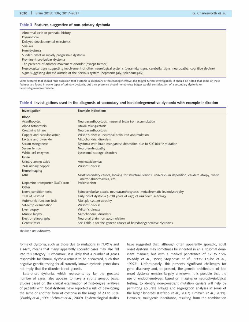
The features listed in Table 3 are those that might raise suspicion
that the dystonia is not primary and trigger further investigation. The
purpose of such investigations is to identify a secondary cause for the
dystonia or to further elucidate the cause of dystonia presenting as
part of a heredodegenerative condition. In practice, a combination of
blood tests, structural imaging and selected secondary investigations
are usually required to secure the diagnosis and Table 4 gives an
indication of some of the investigations that may be appropriate
given the aetiology under consideration. As regards neuroimaging,
MRI is generally the modality of choice, although secondary CT may
be required to accurately distinguish calcium from iron deposition in
the basal ganglia. A dopamine transporter (DaT) scan may be useful
to distinguish DOPA-responsive dystonia or rapid-onset dystonia-
parkinsonism (where it will be normal) from other causes of parkin-
sonism with secondary dystonia (Romero-Lopez et al., 2008;
Zanotti-Fregonara et al., 2008).
Genetic burden in dystonia andgenetic testingCurrent evidence suggests that there is a significant genetic contri-
bution to many forms of dystonia. Monogenic inheritance is most
often seen in early-onset cases, where a family history can often be
elicited. However, the reduced penetrance of some monogenic
2018 | Brain 2013: 136; 2017–2037 G. Charlesworth et al.
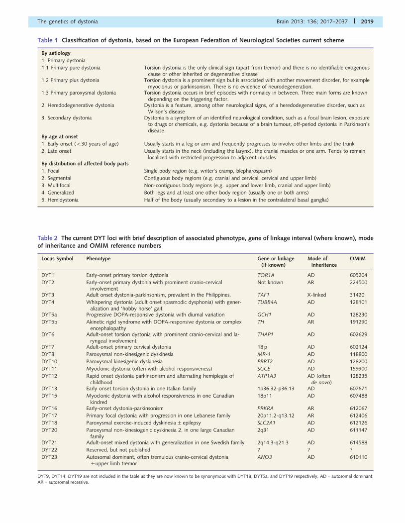
Table 2 The current DYT loci with brief description of associated phenotype, gene of linkage interval (where known), modeof inheritance and OMIM reference numbers
Locus Symbol Phenotype Gene or linkage(if known)
Mode ofinheritence
OMIM
DYT1 Early-onset primary torsion dystonia TOR1A AD 605204
DYT2 Early-onset primary dystonia with prominent cranio-cervicalinvolvement
Not known AR 224500
DYT3 Adult onset dystonia-parkinsonism, prevalent in the Philippines. TAF1 X-linked 31420
DYT4 Whispering dystonia (adult onset spasmodic dysphonia) with gener-alization and ‘hobby horse’ gait
TUBB4A AD 128101
DYT5a Progressive DOPA-responsive dystonia with diurnal variation GCH1 AD 128230
DYT5b Akinetic rigid syndrome with DOPA-responsive dystonia or complexencephalopathy
TH AR 191290
DYT6 Adult-onset torsion dystonia with prominent cranio-cervical and la-ryngeal involvement
THAP1 AD 602629
DYT7 Adult-onset primary cervical dystonia 18 p AD 602124
DYT8 Paroxysmal non-kinesigenic dyskinesia MR-1 AD 118800
DYT10 Paroxysmal kinesigenic dyskinesia PRRT2 AD 128200
DYT11 Myoclonic dystonia (often with alcohol responsiveness) SGCE AD 159900
DYT12 Rapid onset dystonia parkinsonism and alternating hemiplegia ofchildhood
ATP1A3 AD (oftende novo)
128235
DYT13 Early onset torsion dystonia in one Italian family 1p36.32-p36.13 AD 607671
DYT15 Myoclonic dystonia with alcohol responsiveness in one Canadiankindred
18p11 AD 607488
DYT16 Early-onset dystonia-parkinsonism PRKRA AR 612067
DYT17 Primary focal dystonia with progression in one Lebanese family 20p11.2-q13.12 AR 612406
DYT18 Paroxysmal exercise-induced dyskinesia � epilepsy SLC2A1 AD 612126
DYT20 Paroxysmal non-kinesiogenic dyskinesia 2, in one large Canadianfamily
2q31 AD 611147
DYT21 Adult-onset mixed dystonia with generalization in one Swedish family 2q14.3-q21.3 AD 614588
DYT22 Reserved, but not published ? ? ?
DYT23 Autosomal dominant, often tremulous cranio-cervical dystonia�upper limb tremor
ANO3 AD 610110
DYT9, DYT14, DYT19 are not included in the table as they are now known to be synonymous with DYT18, DYT5a, and DYT19 respectively. AD = autosomal dominant;AR = autosomal recessive.
Table 1 Classification of dystonia, based on the European Federation of Neurological Societies current scheme
By aetiology
1. Primary dystonia
1.1 Primary pure dystonia Torsion dystonia is the only clinical sign (apart from tremor) and there is no identifiable exogenouscause or other inherited or degenerative disease
1.2 Primary plus dystonia Torsion dystonia is a prominent sign but is associated with another movement disorder, for examplemyoclonus or parkinsonism. There is no evidence of neurodegeneration.
1.3 Primary paroxysmal dystonia Torsion dystonia occurs in brief episodes with normalcy in between. Three main forms are knowndepending on the triggering factor.
2. Heredodegenerative dystonia Dystonia is a feature, among other neurological signs, of a heredodegenerative disorder, such asWilson’s disease
3. Secondary dystonia Dystonia is a symptom of an identified neurological condition, such as a focal brain lesion, exposureto drugs or chemicals, e.g. dystonia because of a brain tumour, off-period dystonia in Parkinson’sdisease.
By age at onset
1. Early onset (530 years of age) Usually starts in a leg or arm and frequently progresses to involve other limbs and the trunk
2. Late onset Usually starts in the neck (including the larynx), the cranial muscles or one arm. Tends to remainlocalized with restricted progression to adjacent muscles
By distribution of affected body parts
1. Focal Single body region (e.g. writer’s cramp, blepharospasm)
2. Segmental Contiguous body regions (e.g. cranial and cervical, cervical and upper limb)
3. Multifocal Non-contiguous body regions (e.g. upper and lower limb, cranial and upper limb)
4. Generalized Both legs and at least one other body region (usually one or both arms)
5. Hemidystonia Half of the body (usually secondary to a lesion in the contralateral basal ganglia)
The genetics of dystonia Brain 2013: 136; 2017–2037 | 2019
forms of dystonia, such as those due to mutations in TOR1A and
THAP1, means that many apparently sporadic cases may also fall
into this category. Furthermore, it is likely that a number of genes
responsible for familial dystonia remain to be discovered, such that
negative genetic testing for all currently known dystonia genes does
not imply that the disorder is not genetic.
Late-onset dystonia, which represents by far the greatest
number of cases, also appears to have a strong genetic basis.
Studies based on the clinical examination of first-degree relatives
of patients with focal dystonia have reported a risk of developing
the same or another form of dystonia in the range of 23 to 36%
(Waddy et al., 1991; Schmidt et al., 2009). Epidemiological studies
have suggested that, although often apparently sporadic, adult
onset dystonia may sometimes be inherited in an autosomal dom-
inant manner, but with a marked penetrance of 12 to 15%
(Waddy et al., 1991; Stojanovic et al., 1995; Leube et al.,
1997b). Unfortunately, this presents significant challenges for
gene discovery and, at present, the genetic architecture of late
onset dystonia remains largely unknown. It is possible that the
use of endophenotypes, based on imaging or neurophysiological
testing, to identify non-penetrant mutation carriers will help by
permitting accurate linkage and segregation analyses in some of
the larger kindreds (Defazio et al., 2007; Kimmich et al., 2011).
However, multigenic inheritance, resulting from the combination
Table 4 Investigations used in the diagnosis of secondary and heredodegenerative dystonia with example indication
Investigation Example indications
Blood
Acanthocytes Neuroacanthocytosis, neuronal brain iron accumulation
Alpha fetoprotein Ataxia telangiectasia
Creatinine kinase Neuroacanthocytosis
Copper and caeruloplasmin Wilson’s disease, neuronal brain iron accumulation
Lactate and pyruvate Mitochondrial disorders
Serum manganese Dystonia with brain manganese deposition due to SLC30A10 mutation
Serum ferritin Neuroferritinopathy
White cell enzymes Lysosomal storage disorders
Urine
Urinary amino acids Aminoacidaemias
24 h urinary copper Wilson’s disease
Neuroimaging
MRI Most secondary causes, looking for structural lesions, iron/calcium deposition, caudate atropy, whitematter abnormalities, etc.
Dopamine transporter (DaT) scan Parkinsonism
Other
Nerve condition tests Spinocerebellar ataxia, neuroacanthocytosis, metachromatic leukodystrophy
Trial of L-DOPA Early onset dystonia (530 years of age) of unknown aetiology
Autonomic function tests Multiple system atrophy
Slit-lamp examination Wilson’s disease
Liver biopsy Wilson’s disease
Muscle biopsy Mitochondrial disorders
Electro-retinography Neuronal brain iron accumulation
Genetic tests See Table 7 for the genetic causes of heredodegenerative dystonias
This list is not exhaustive.
Table 3 Features suggestive of non-primary dystonia
Abnormal birth or perinatal history
Dysmorphia
Delayed developmental milestones
Seizures
Hemidystonia
Sudden onset or rapidly progressive dystonia
Prominent oro-bulbar dystonia
The presence of another movement disorder (except tremor)
Neurological signs suggesting involvement of other neurological systems (pyramidal signs, cerebellar signs, neuropathy, cognitive decline)
Signs suggesting disease outside of the nervous system (hepatomegaly, splenomegaly)
Some features that should raise suspicion that dystonia is secondary or heredodegenerative and trigger further investigation. It should be noted that some of thesefeatures are found in some types of primary dystonia, but their presence should nonetheless trigger careful consideration of a secondary dystonia or
heredodegenerative disorder.
2020 | Brain 2013: 136; 2017–2037 G. Charlesworth et al.
of two or more genetic changes, each imparting a low to moder-
ately increased risk of developing dystonia and acting in combin-
ation with environmental factors, may also underlie a significant
proportion of the apparent heritability of late-onset dystonia.
Large-scale genome-wide association studies will be helpful in dis-
secting out the genetic contribution in these cases.
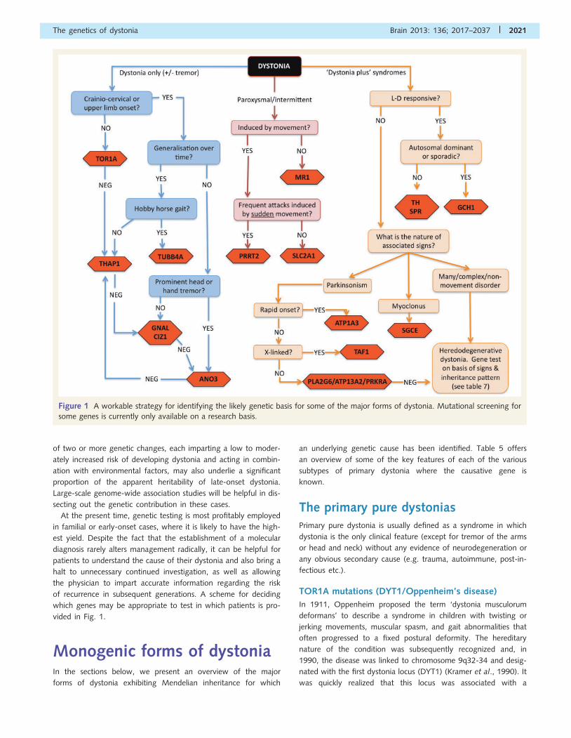
At the present time, genetic testing is most profitably employed
in familial or early-onset cases, where it is likely to have the high-
est yield. Despite the fact that the establishment of a molecular
diagnosis rarely alters management radically, it can be helpful for
patients to understand the cause of their dystonia and also bring a
halt to unnecessary continued investigation, as well as allowing
the physician to impart accurate information regarding the risk
of recurrence in subsequent generations. A scheme for deciding
which genes may be appropriate to test in which patients is pro-
vided in Fig. 1.
Monogenic forms of dystoniaIn the sections below, we present an overview of the major
forms of dystonia exhibiting Mendelian inheritance for which
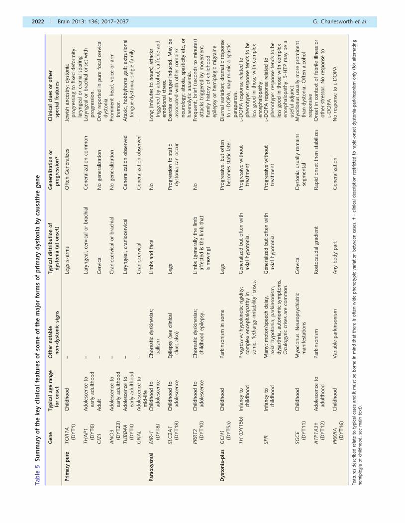
an underlying genetic cause has been identified. Table 5 offers
an overview of some of the key features of each of the various
subtypes of primary dystonia where the causative gene is
known.
The primary pure dystoniasPrimary pure dystonia is usually defined as a syndrome in which
dystonia is the only clinical feature (except for tremor of the arms
or head and neck) without any evidence of neurodegeneration or
any obvious secondary cause (e.g. trauma, autoimmune, post-in-
fectious etc.).
TOR1A mutations (DYT1/Oppenheim’s disease)
In 1911, Oppenheim proposed the term ‘dystonia musculorum
deformans’ to describe a syndrome in children with twisting or
jerking movements, muscular spasm, and gait abnormalities that
often progressed to a fixed postural deformity. The hereditary
nature of the condition was subsequently recognized and, in
1990, the disease was linked to chromosome 9q32-34 and desig-
nated with the first dystonia locus (DYT1) (Kramer et al., 1990). It
was quickly realized that this locus was associated with a
Figure 1 A workable strategy for identifying the likely genetic basis for some of the major forms of dystonia. Mutational screening for
some genes is currently only available on a research basis.
The genetics of dystonia Brain 2013: 136; 2017–2037 | 2021
Tab
le5
Sum
mar
yof
the
key
clin
ical
feat
ure
sof
som
eof
the
maj
or
form
sof
pri
mar
ydys
tonia
by
causa
tive
gen
e
Gen
eTyp
ical
age
range
for
onse
tO
ther
nota
ble
non-d
ysto
nic
signs
Typ
ical
dis
trib
uti
on
of
dys
tonia
(at
onse
t)G
ener
aliz
atio
nor
pro
gre
ssio
n?
Cli
nic
alcl
ues
or
oth
ersp
ecia
lfe
ature
s
Pri
mar
ypure
TO
R1A
(DY
T1)
Child
hood
–Le
gs�
arm
sO
ften
Gen
eral
izes
Jew
ish
ance
stry
;dys
tonia
pro
gre
ssin
gto
fixe
ddef
orm
ity;
lary
ngea
lor
cran
ialsp
arin
gTH
AP1
(DY
T6)
Adole
scen
ceto
early
adulthood
–La
ryngea
l,ce
rvic
alor
bra
chia
lG
ener
aliz
atio
nco
mm
on
Lary
ngea
lor
bra
chia
lonse
tw
ith
pro
gre
ssio
n.
CIZ
1A
dult
–C
ervi
cal
No
gen
eral
izat
ion
Only
report
edin
pure
foca
lce
rvic
aldys
tonia
AN
O3
(DY
T23)
Adole
scen
ceto
early
adulthood
–C
ranio
cerv
ical
or
bra
chia
lN
ogen
eral
izat
ion
Pro
min
ent
hea
d,
voic
eor
arm
trem
or.
TU
BB
4A
(DY
T4)
Adole
scen
ceto
early
adulthood
–La
ryngea
l,cr
anio
cerv
ical
Gen
eral
izat
ion
obse
rved
Ata
xic,
hobbyh
ors
egai
t;ex
trusi
onal
tongue
dys
tonia
;si
ngle
fam
ily
GN
AL
Adole
scen
ceto
mid
-life
–C
ranio
cerv
ical
Gen
eral
izat
ion
obse
rved
–
Par
aoxy
smal
MR
-1(D
YT8)
Child
hood
toad
ole
scen
ceC
hore
atic
dys
kines
ias;
bal
lism
Lim
bs
and
face
No
Long
(min
ute
sto
hours
)at
tack
s,tr
igger
edby
alco
hol,
caff
eine
and
emotional
stre
ss.
SLC
2A
1(D
YT18)
Child
hood
toad
ole
scen
ceEp
ilepsy
(see
clin
ical
clues
also
)Le
gs
Pro
gre
ssio
nto
stat
icdys
tonia
can
occ
ur
Exer
cise
or
hunger
induce
d.
May
be
asso
ciat
edw
ith
oth
erco
mple
xneu
rolo
gy:
atax
ia,
spas
tici
tyet
c,or
hae
moly
tic
anae
mia
.PR
RT2
(DY
T10)
Child
hood
toad
ole
scen
ceC
hore
atic
dys
kines
ias;
child
hood
epile
psy
.Li
mbs
(gen
eral
lyth
elim
baf
fect
edis
the
limb
that
ism
ovi
ng)
No
Freq
uen
t,brief
(sec
onds
tom
inute
s)at
tack
str
igger
edby
move
men
t.Fa
mily
his
tory
of
child
hood
epile
psy
or
hem
iple
gic
mig
rain
e
Dys
tonia
-plu
sG
CH
1(D
YT5a)
Child
hood
Par
kinso
nis
min
som
eLe
gs
Pro
gre
ssiv
e,but
oft
enbec
om
esst
atic
late
r.D
iurn
alva
riat
ion;
dra
mat
icre
sponse
toL-
DO
PA
;m
aym
imic
asp
astic
par
apar
esis
.TH
(DY
T5b)
Infa
ncy
toch
ildhood
Pro
gre
ssiv
ehyp
oki
net
icrigid
ity;
com
ple
xen
cephal
opat
hy
inso
me;
‘leth
argy-
irrita
bili
ty’
cris
es.
Gen
eral
ized
but
oft
enw
ith
axia
lhyp
oto
nia
.Pro
gre
ssiv
ew
ithout
trea
tmen
tL-
DO
PA
resp
onse
rela
ted
tophen
oty
pe:
resp
onse
tends
tobe
less
good
inth
ose
with
com
ple
xen
cephal
opat
hy.
SPR
Infa
ncy
toch
ildhood
Man
y:m
oto
r/sp
eech
del
ay,
axia
lhyp
oto
nia
,par
kinso
nis
m,
dys
arth
ria,
auto
nom
icsy
mpto
ms.
Ocu
logyr
iccr
ises
are
com
mon.
Gen
eral
ized
but
oft
enw
ith
axia
lhyp
oto
nia
.Pro
gre
ssiv
ew
ithout
trea
tmen
tL-
DO
PA
resp
onse
rela
ted
tophen
oty
pe:
resp
onse
tends
tobe
less
good
inth
ose
with
com
ple
xen
cephal
opat
hy.
5-H
TP
may
be
ause
fulad
junct
SG
CE
(DY
T11)
Child
hood
Myo
clonus.
Neu
ropsy
chia
tric
man
ifes
tations
Cer
vica
lD
ysto
nia
usu
ally
rem
ains
segm
enta
lM
yocl
onus
usu
ally
more
pro
min
ent
than
dys
tonia
.O
ften
alco
hol
resp
onsi
veA
TP1A
3†
(DY
T12)
Adole
scen
ceto
adulthood
Par
kinso
nis
mR
ost
oca
udal
gra
die
nt
Rap
idonse
tth
enst
abili
zes
Onse
tin
conte
xtof
febrile
illnes
sor
oth
erst
ress
or.
No
resp
onse
toL-
DO
PA
PR
KR
A(D
YT16)
Child
hood
Var
iable
par
kinso
nis
mA
ny
body
par
tG
ener
aliz
atio
nN
ore
sponse
toL-
DO
PA
Feat
ure
sdes
crib
edre
late
toty
pic
alca
ses
and
itm
ust
be
born
ein
min
dth
atth
ere
isoft
enw
ide
phen
oty
pic
variat
ion
bet
wee
nca
ses.
†=
clin
ical
des
crip
tion
rest
rict
edto
rapid
-onse
tdys
tonia
-par
kinso
nis
monly
(for
alte
rnat
ing
hem
iple
gia
of
child
hood,
see
mai
nte
xt).
2022 | Brain 2013: 136; 2017–2037 G. Charlesworth et al.
significant proportion of all childhood-onset dystonia, particularly
in Ashkenazi Jews (Ozelius et al., 1992; Kramer et al., 1994).
Seven years later the gene responsible for the condition was iden-
tified and named TOR1A (Ozelius et al., 1997). The TOR1A gene
comprises five exons and is widely expressed. In almost all cases of
DTY1-related dystonia, the underlying genetic mutation is an
inframe GAG deletion located in exon 5 of the gene, resulting in
the loss of a single glutamic acid in the final protein product (torsin
A). This mutation has been shown to be responsible for �80% of
all primary, early-onset dystonia in Ashkenazi Jewish populations
and up to 50% of primary, early-onset dystonia in non-Jewish
populations (Bressman, 2006; Jamora et al., 2006). Only two
other missense variants (p.A288G and p.F205I) and one frame-
shift, 4-base pair deletion (c.934_937delAGAG) in this gene have
since been reported to cause dystonia, though their pathogenicity
is by no means certain (Kabakci et al., 2004; Zirn et al., 2008;
Calakos et al., 2010).
The penetrance of the GAG deletion is notably low, with cur-
rent estimates somewhere within the region of 30–40% (Kramer
et al., 1994; Bressman, 2006). Moreover, even when the condi-
tion does manifest, the spectrum of severity of the symptoms is
wide, ranging from mild focal dystonia to severe and disabling
generalized dystonia. The exact mechanism underlying this vari-
ability remains unclear, but is presumed that other genetic or
environmental modifiers must exist that influence the penetrance
and presentation of the disease. To date, only one coding poly-
morphism in exon 4 of TOR1A (rs1801968), which encodes
either aspartic acid (D) or histidine (H), has convincingly been
shown to modify the risk of developing symptoms of dystonia.
It appears that carrying the H allele in trans (that is, on the
opposite allele from that harbouring the GAG deletion) may
reduce the risk of developing symptoms of dystonia by 10-fold,
to as little as 3% (Risch et al., 2007; Kamm et al., 2008a).
Interestingly, in cellular models, the overexpression of TOR1A
containing the H allele was sufficient by itself to induce formation
of inclusions of torsin A in a manner similar to expression of
GAG-deleted TOR1A, though at a much lower level (Kock
et al., 2006). It is thought that this may be the result of a dis-
ruption of protein interactions induced by the presence of the
exposed histidine residue. However, expression of GAG-deleted
TOR1A containing the H allele in cis actually reduced the forma-
tion of inclusions, implying that these two changes act to cancel
each other out to some degree (Kock et al., 2006). It has even
been suggested that the GAG-deletion may need to be carried in
conjunction with a D allele in cis to be penetrant (Risch et al.,
2007), though this has not yet been proven. Yet, although the
effect of the 216H coding polymorphism appears to be robust (in
trans, at least), its relatively low population frequency (�12% in
Europeans and less in other populations) means it cannot fully
explain the reduced penetrance or variable expressivity seen in
this condition; other factors clearly remain to be identified.
In terms of non-genetic factors that might affect the penetrance
of TOR1A mutations, one small study has assessed exposure to
perinatal adversity, childhood infections, general anaesthesia and
trauma in manifesting and non-manifesting carriers as well as non-
carriers from a series of 28 families with DYT1-related dystonia by
means of a retrospective questionnaire (Martino et al., 2012).
Only self-reported perinatal adversity, in particular complications
of vaginal delivery, showed a statistically significant (P = 0.02)
positive correlation with manifestation of the disease.
Clinically, DYT1-related dystonia typically presents in childhood
with dystonic posturing of the foot or leg, though it may begin in
any part of the body, and evolves to generalized dystonia with
fixed deformities. As mentioned above, however, late-onset or
much milder forms of the disorder are recognized (Jamora et al.,
2006). There is generally a family history consistent with auto-
somal dominant inheritance, albeit with reduced penetrance, but
apparently sporadic early and late onset cases are also seen
(Hjermind et al., 2002).
The TOR1A gene encodes a protein torsin A, which is a member
of the AAA + family of ATPases and is believed to be involved in
maintenance of both structural integrity and/or normal function of
protein processing and trafficking. Torsin A is expressed through-
out the CNS in humans, but is found at particularly high levels in
the dopaminergic neurons of substantia nigra pars compacta, locus
coeruleus, Purkinje cells, cerebellar dentate nucleus, basis pontis,
thalamus, hippocampal formation, oculomotor nuclei and frontal
cortex (Shashidharan et al., 2000; Konakova et al., 2001; Rostasy
et al., 2003). The exact mechanisms by which mutant torsin A
causes dystonia are still unclear, but there is accumulating data to
suggest that torsin A is important for cellular compartments and
pathways, including the cytoskeleton, the nuclear envelope, the
secretory pathway and the synaptic vesicle machinery (Goodchild
and Dauer, 2005; Goodchild et al., 2005; Hewett et al., 2006,
2007; Henriksen et al., 2009; Kakazu et al., 2012).
THAP1 mutations (DYT6)
After the discovery of TOR1A and the subsequent uptake of gen-
etic testing for mutations in this gene, it became clear that a
second, distinct form of pure primary dystonia existed. Affected
individuals, who were negative for TOR1A mutations, tended to
have an older age at onset—in their adolescence or adulthood—
and presented predominantly with cranio-cervical or focal dystonia
affecting the upper limbs. In 1997, the DYT6 locus was assigned
to chromosome 8p21-q22 by linkage analysis in two Mennonite
families, but it was not until 2009 that the gene responsible for
the condition was finally identified as THAP1 (thanatos associated
protein domain containing, apoptosis associated protein 1)
(Almasy et al., 1997; Fuchs et al., 2009). Missense, nonsense
and frameshift mutations, spread throughout most of the coding
portion of the gene, have all been associated with disease.
Mutations in this gene have been detected in genetically diverse
populations throughout the world and, unlike TOR1A, do not
seem to be especially prevalent in any one particular population.
Inheritance is autosomal dominant with a reduced penetrance. To
date, penetrance has only been measured in Amish–Mennonite
families, where it appears to be �60%, but this may not be
true of all populations or mutations (Saunders-Pullman et al.,
2007). Most mutations are found in the heterozygous state, but
homozygotes are described (Fuchs et al., 2009; Houlden et al.,
2010).
The clinical spectrum of THAP1 mutations is wide. Presentation
with oromandibular, cranio-cervical or laryngeal dystonia is
common, but presentations with focal dystonia of the limbs,
The genetics of dystonia Brain 2013: 136; 2017–2037 | 2023
segmental or generalized dystonia are all described in the litera-
ture. In a recent analysis by Xiromerisiou et al. (2012) of 100
patients reported to carry THAP1 mutations, the mean age at
onset of dystonia was 24 years, 60% of patients were females
and the distribution was generalized in 37%, segmental in 30%,
multifocal in 6% and focal in 27%.
The THAP1 protein is an atypical zinc finger protein character-
ized by an N-terminal THAP domain, a proline-rich region and a
C-terminal nuclear localization domain (Roussigne et al., 2003).
The THAP domain is conserved in both vertebrates and inverte-
brates (Clouaire et al., 2005). It has DNA binding properties and is
thought to be involved in the regulation of transcription, either on
its own or in concert with other proteins. Thus, one possible
mechanism by which mutations in THAP1 might cause dystonia
is by dysregulated transcription of key genes. THAP1 protein is
also known to interact with the prostate apoptosis response pro-
tein 4 (PAR-4), which is a transcription regulator involved in apop-
tosis (Roussigne et al., 2003). Under conditions of cellular stress or
toxicity, this protein is rapidly upregulated and it has been linked
to neurodegeneration in a number of disease models (Guo et al.,
1998; Duan et al., 1999; Pedersen et al., 2000). One intriguing
finding has been that THAP1 may regulate transcription of TOR1A
by binding to one of two sites in its promoter region (Gavarini
et al., 2010; Kaiser et al., 2010). Mutations in THAP1 have been
shown to disrupt binding to the TOR1A promoter and decrease
TOR1A-driven luciferase expression, thus suggesting that under
some conditions TOR1A is negatively regulated by THAP1 protein
and that mutations in THAP1 may lead to abnormally high levels
of torsin A (Kaiser et al., 2010). Although these data come from
experimental cell lines and thus are of uncertain physiological sig-
nificance, this does at least raise the possibility that the disease
mechanisms in DYT1 and DYT6 dystonia may be linked. However,
if this is the case, the question arises as to how to link this mech-
anistically with the prevailing idea that the GAG-deletion in
TOR1A leads to a loss of function of the protein (Goodchild
et al., 2005). One possibility is that TOR1A expression must be
maintained within a set range in relation to its binding partners
and that dysfunction may result from either increased expression
or loss of function (Bragg et al., 2011). Indeed, transgenic mice
overexpressing wild-type TOR1A have been shown to display evi-
dence of neurohistological, neurochemical and behavioural
abnormalities, which may provide some support for this hypothesis
(Grundmann et al., 2007).
ANO3 mutations (DYT23)
Recently, our laboratory employed a combination of exome
sequencing and linkage analysis in moderately-sized, three gener-
ation Caucasian kindred with autosomal dominant cranio-cervical
dystonia to identify a mutation in ANO3 (p.Arg494Trp) that seg-
regated with the disease in the family (Charlesworth et al., 2012).
Subsequent screening of this gene in 188 probands with cervical
dystonia revealed five further novel variants that were not found
in any databases of sequence variation, scattered throughout the
gene, including one variant in its 5’ UTR. Segregation analysis was
possible for two of these additional variants (p.Trp490Cys and
p.Ser685Gly), which were seen to segregate with disease. The
expression pattern of the gene was supportive of its role in
dystonia, with expression being several folds higher in the striatum
than anywhere else (Charlesworth et al., 2012).
Clinically, patients with mutations in this gene exhibited focal or
segmental dystonia, variably affecting the cranio-cervical, laryn-
geal or brachial regions. There was often dystonic tremor with a
jerky quality affecting the head, voice or upper limbs. The age at
onset ranged from the very early childhood to 40 years old.
Interestingly, despite prolonged follow-up and a gradual increase
in severity, the dystonia was never seen to generalize in any af-
fected individual, remaining confined to the head, neck and upper
limbs. Some individuals manifested upper limb tremor alone and
had been misclassified as essential tremor.
ANO3 encodes a protein called anoctamin 3, about which little
is yet known. It belongs to a family of genes (ANO1–10) that
appear to be closely related in sequence and topology, but with
distinct expression patterns. Several of these genes have already
been linked to various diseases, suggesting they play an important
role within their tissue types (Duran and Hartzell, 2011). ANO1
and ANO2, the best studied members of the family, encode pro-
teins that function as Ca2 +-activated chloride channels and it is
possible anoctamin 3 may function in the same manner (Caputo
et al., 2008). Certainly, hydropathy analysis suggests a similar
topology, with eight hydrophobic helices that are likely to be
transmembrane domains and cytosolic N- and C-termini, but
more recent work has suggested that anoctamin 3 may in fact
be targeted to the endoplasmic reticulum, rather than the cell
surface, like anoctamins 1 and 2 (Duran et al., 2012).
Moreover, patient fibroblasts habouring the p.Trp490Cys mutation
showed evidence of a potential defect in endoplasmic reticulum
related Ca2 + handling. It is believed that Ca2 +-activated chloride
channels have a role to play in the modulation of neuronal excit-
ability (Hartzell et al., 2005; Huang et al., 2012) and, in view of
high expression of ANO3 in the striatum, it is possible that muta-
tions in this gene lead to abnormal excitability of striatal neurons,
which manifests itself clinically in unwanted dystonic movements,
though further functional work will be required to test this
hypothesis.
GNAL mutations
At the same time, mutations in another gene, GNAL, were identi-
fied as further cause of familial primary pure dystonia (Fuchs et al.,
2012). Using whole exome sequencing and linkage in two unre-
lated families, two mutations in GNAL were initially identified, a
nonsense mutation (p.Ser293*) resulting in a premature stop
codon in one family and a missense mutation (p.Val137Met) in
the other. Screening of 39 additional families identified six further
novel mutations, with segregation confirmed in all families for
which DNA from additional affected individuals was available.
Clinically, of the 28 individuals with dystonia resulting from a
mutation in GNAL, 82% had onset in the region of the neck and
93% had cervical involvement when examined for the study.
However, progression to other sites had occurred in at least half
of those affected and generalized dystonia was seen in �10% of
cases. Thus, the clinical phenotype in GNAL-related dystonia ap-
pears to be not dissimilar to that caused by mutations in THAP1.
Though the authors noted that onset in GNAL mutations was
never brachial, this may just be a chance finding given the small
2024 | Brain 2013: 136; 2017–2037 G. Charlesworth et al.
number of cases identified so far, rather than a true distinguishing
factor.
GNAL encodes G�olf, the alpha subunit of triheteromeric G pro-
tein Golf, which is involved in dopamine (D1) signalling.
Experimental evidence exists to show that G�olf is mostly respon-
sible for the coupling of D1 receptors to adenylyl cyclase in striatal
neurons and that G�olf is required for D1-mediated behaviour and
biochemical effects (Herve et al., 1993; Zhuang et al., 2000;
Corvol et al., 2001). Because D1 dopamine receptors have a
known role in mediating locomotor activity, the link between
GNAL and dystonia is biologically plausible. The gene is located
on the short arm of chromosome 18 in a region that has been
linked not only to DYT7 dystonia (though the original DYT7 family
do not appear to carry a mutation in GNAL), but also to bipolar
disorder, schizophrenia and attention deficit hyperactivity disorder
(Leube et al., 1997a; Berrettini, 2000; Segurado et al., 2003;
Laurin et al., 2008; Winter et al., 2012). Interestingly, homozy-
gous knockout mice for this gene are hyposmic and display hyper-
active behaviours (Belluscio et al., 1998).
CIZ1 mutations
In early 2012, Xiao et al. (2012) reported that they had used a
combination of linkage analysis and whole exome sequencing to
identify a mutation in CIZ1 (p.Ser264Gly) as the likely causal vari-
ant in a large Caucasian kindred with primary cervical dystonia
inherited as an autosomal dominant trait. Those affected in the
family exhibited focal cervical dystonia, occasionally with mild
tremor, having its onset in early adulthood to late midlife (18–
66 years of age). However, affectation status was not always
clear cut: five family members were said to have ‘definite’ cervical
dystonia, whereas five family members were said to have ‘pos-
sible’ cervical dystonia, making linkage analysis difficult (Uitti and
Maraganore, 1993).
CIZ1 encodes CDKN1A interacting zinc finger protein 1, a
p21Cip1/Waf1-interacting zinc finger protein expressed in the brain
and involved in DNA synthesis and cell-cycle control. Functional
work suggests that the p.Ser264Gly mutation may alter splicing of
the gene and normal subnuclear localization of CIZ1 protein (Xiao
et al., 2012). Screening in a cohort of patients with adult-onset
dystonia identified two additional missense mutations in three in-
dividuals (p.Pro47Ser and p.Arg672Met), all with focal cervical
dystonia developing in mid-to-late life.
Some researchers feel it may be too early to place full confi-
dence in the association of mutations in CIZ1 and focal cervical
dystonia. Firstly, databases of normal sequence variation reveal
that the CIZ1 gene contains a large number of missense variants
in supposedly healthy individuals (a third of which are predicted to
be pathogenic) and, in the study by Xiao et al. (2012) an equal
number of novel missense variants were found in controls as were
found in patients. Secondly, and more importantly, segregation
was not demonstrated in a second independent family for any
of the purported mutations. Thirdly, there is some question re-
garding the quality of the exome data used in this study: coverage
was only 420 reads for �60% of the exome and the number of
false-positive variants appears unusually high (Klein et al., 2012).
Screening of this gene in further cohorts of cervical dystonia cases
from various populations will be required to decide if these reser-
vations are warranted or not.
TUBB4A mutations (DYT 4)
DYT4 was first described in 1985 by forensic psychiatrist Neville
Parker in a large family with third decade onset of autosomal
dominantly inherited ‘whispering dysphonia’ and generalized dys-
tonia (Parker, 1985). Over 25 affected individuals have been re-
ported, typically presenting with a laryngeal dysphonia progressing
to a generalized dystonia and a peculiar ‘hobby horse’ gait (Wilcox
et al., 2011). Alcohol responsiveness was not uncommon, leading
to severe alcohol abuse in some DYT4 patients (Wilcox et al.,
2011). The family is descended from an affected male who was
born in 1801 in the small rural coastal town of Heacham in
Norfolk and, to date, no other kindred has been described world-
wide with a similar phenotype.
Using a combination of linkage analysis and exome sequencing,
a mutation in the gene TUBB4A (p.Arg2Gly) has recently been
identified as causal in the DYT4 kindred by two groups independ-
ently (Hersheson et al., 2012; Lohmann et al., 2012). TUBB4A
encodes b-tubulin 4 a, a constituent of microtubules, and the mu-
tation results in an arginine to glycine amino acid substitution in
the key, highly conserved autoregulatory MREI (methionine–argin-
ine–glutamic acid–isoleucine) domain of the protein. The gene is
expressed throughout the brain, but at the highest levels in the
cerebellum, which has been linked to the pathogenesis of dystonia
(Hersheson et al., 2012). The MREI tetrapeptide sequence at the
start of the N-terminal domain is known to be necessary for the
autoregulation of the b-tubulin messenger RNA transcript and sep-
arate in vitro studies using site-directed mutagenesis have previ-
ously demonstrated that the p.Arg2Gly mutation abrogates this
autoregulatory ability (Yen et al., 1988a, b). One further possibly
pathogenic variant (p.Ala271Thr) was detected during the screen-
ing of a cohort of 394 unrelated dystonia patients: the individual
concerned exhibited spasmodic dysphonia with oromandibular
dystonia and dyskinesia with an age at onset of 60. Her mother
had been similarly affected, but was now deceased so that segre-
gation analysis was not possible, meaning the pathogenicity of the
variant is uncertain (Lohmann et al., 2012).
The paroxysmal dystoniasThe paroxysmal dystonias are characterized, in theory at least, by
episodes of dystonia or other dyskinesia separated periods of
neurological normality. In practice, there may be other associated
neurological features besides the movement disorder and, espe-
cially in the case of SLC2A1-related disease, both the movement
disorder and the other associated neurological symptoms may
become relatively fixed with time.
MR-1 mutations (DYT8)
Symptoms of paroxysmal non-kinesigenic dyskinesia (PNKD) typ-
ically begin in childhood or adolescence and include dystonic and
choreatic dyskinesias or ballistic movements, lasting from minutes
to hours. There are often premonitory symptoms of an impending
attack (paraesthesia or tension in the affected area) and sleep can
prevent or abort attacks. The frequency of attacks varies between
The genetics of dystonia Brain 2013: 136; 2017–2037 | 2025
individuals, ranging from daily attacks to only a few in a lifetime.
Typically, they are precipitated by alcohol, caffeine or stress, but
less often by exercise, fatigue or cold (Bruno et al., 2007).
Neurological examination is normal between attacks. Treatment
is with carbamazepine or benezodiazepines, particularly
clonazepam.
The condition results from mutations in the myofribillogenesis
regulator gene (MR-1, also known as PNKD) and is inherited as an
autosomal dominant trait. Only three mutations have been
described to date, all clustered in the N-terminus of the protein:
p.Ala7Val, p.Ala9Val and p.Ala33Pro (Lee et al., 2004; Rainier
et al., 2004; Chen et al., 2005; Djarmati et al., 2005;
Hempelmann et al., 2006; Ghezzi et al., 2009). Despite this,
haplotype analysis has failed to reveal a common founder, sug-
gesting that these represent independent mutational events (Chen
et al., 2005). It is currently believed that this region of the protein
may represent a mitochondrial targeting sequence (Ghezzi et al.,
2009). Mutations may act by disrupting protein processing in vivo,
a hypothesis that is supported by evidence from transgenic mice
(Shen et al., 2011). The function of the MR-1 protein is not fully
understood, but it shows close homology to glyoxalase hydroxya-
cylglutathione hydrolase, which is known to detoxify methyl-
glyoxal, a compound found in coffee and alcohol and a by-
product of glycolysis, thus providing a link to the standard triggers
(Lee et al., 2004).
PRRT2 mutations (DYT10)
Mutations in PRRT2 cause paroxysmal kinesigenic dyskinesia
(PKD), which has an estimated prevalence 1 in 150 000 individuals
(Bruno et al., 2004). Affected individuals have short (seconds to
minutes) and frequent (up to 100 times per day) attacks of dys-
tonic or choreiform movements, precipitated by sudden move-
ments or startle. As with paroxysmal non-kinesigenic dyskinesia,
there is often warning of an impending attack—a so-called
‘aura’—consisting of numbness or paraesthesia in the affected
body part. The attacks usually begin in childhood and are highly
responsive to anticonvulsant therapy, such as carbamazepine
(Silveira-Moriyama et al., 2013). Reports of what was, in reteros-
pect, probably this disorder have appeared in the literature from as
early as 1892 and the condition was designated dystonia 10 in
1998 (Smith and Heersema, 1941; Muller et al., 1998). However,
the discovery of the gene underlying the condition, PRRT2, would
wait until exome sequencing approaches could be applied to the
problem (Chen et al., 2011; Wang et al., 2011).
Most mutations are truncating and by far the most common of
these is the c.649dupC mutation, but missense variants (possibly
with reduced penetrance) have also been described (Cao et al.,
2012; Friedman et al., 2012a; Hedera et al., 2012; Steinlein
et al., 2012). The PRRT2 protein is highly expressed in the
CNS and is probably localized to the synapse (Lee et al.,
2012). Using yeast 2-hybrid screening, it has been shown to
interact with synaptosomal protein 25 (SNAP25), suggesting a
role in the fusion of the synaptic vesicles to the plasma mem-
brane (Stelzl et al., 2005). Both PRRT2 and SNAP25 are highly
expressed in the basal ganglia and disrupted neurotransmitter
release has been suggested as a possible pathogenic mechanism
in PRRT2 mutations. Truncating mutations produce a protein
lacking the transmembrane domain, and are thought to result
in altered subcellular localization (Chen et al., 2011). Missense
mutations may cause loss of function or act in a dominant-nega-
tive manner (Wang et al., 2011; Li et al., 2012).
PRRT2-related disease is notable for its varied presentation,
differing not merely between individuals carrying different mu-
tations, but also between individuals carrying the same mutation
and even between affected members within the same family
(Silveira-Moriyama et al., 2013). As well as the classical parox-
ysmal kinesigenic dyskinesia phenotype described above, infantile
convulsions with choreoathetosis (ICCA) with or without parox-
ysmal kinesigenic dyskinesia, benign familial infantile seizures,
episodic ataxia, hemiplegic migraine and even benign paroxysmal
torticollis of infancy all appear to be possible manifestations of
mutations in this gene (Dale et al., 2012; Gardiner et al., 2012;
Heron et al., 2012; Lee et al., 2012). The common thread
would seem to be the paroxysmal nature of all PRRT2-related
disorders.
Finally, it would appear that the family used to define DYT19
also carry the common c.649dup mutation in PRRT2, suggesting
that the initial linkage was incorrect (Gardiner et al., 2012).
SLC2A1 mutations (DYT18)
Dominantly inherited mutations in the SLC2A1 gene are now
known to be the cause of paroxysmal exercise-induced dyskinesia
(PED) (Suls et al., 2008; Weber et al., 2008). In fact, SLC2A1
mutations cause a wide spectrum of disease, resulting from a de-
ficiency of the encoded glucose transporter type 1, which is still
commonly referred to as GLUT1 despite the change in the recom-
mended gene name. GLUT1 is the principal glucose transporter in
the brain and it has generally been assumed that neuronal dys-
function arises from energy failure. Under this assumption, the
prevalence of movement disorders was thought to reflect the
heightened sensitivity of the basal ganglia to energy deficits
(Perez-Duenas et al., 2009). However, recent studies in a mouse
model of GLUT1-deficiency, which recaptures many of the fea-
tures of the disease, demonstrated the preservation of aerobic
metabolism, such that mechanisms other than impaired aerobic
glycolysis must be responsible for the manifestations of the dis-
order (Marin-Valencia et al., 2012). Possible culprits include
reduced transfer of lactate generated through anaerobic glycolysis
from the astrocyte into the neuron and reduced anaerobic ATP
formation in the astrocyte having a negative impact on glutamate-
glutamine recycling, which, if proven, would place the emphasis
on glial dysfunction in the pathogenesis of GLUT1-deficiency
(Magistretti and Pellerin, 1996; Marin-Valencia et al., 2012).
At its most severe, GLUT1 deficiency can present with early
onset psychomotor delay, drug resistant epilepsy, acquired micro-
cephaly and other signs of widespread neurological dysfunction,
such as spasticity, ataxia, tremor, dystonia, choreoathetosis and
ballism, which may all be paroxysmal, static or static with parox-
ysmal worsening. From this extreme, there exists an entire spec-
trum of decreasing disease severity, ranging through
developmental delay and movement disorders without epilepsy,
right down to the mildest cases, in which only minimal phenotypic
abnormalities are detectable (Brockmann, 2009; Leen et al.,
2026 | Brain 2013: 136; 2017–2037 G. Charlesworth et al.
2010). There can be considerable heterogeneity of clinical presen-
tation even within the same family (Leen et al., 2010).
Paroxysmal exercise-induced dyskinesia is, therefore, probably
best regarded as just one possible manifestation of the complex
and variable disorder that is GLUT1-deficiency syndrome. It is
characterized by attacks of combined chorea, athetosis, and dys-
tonia precipitated by exercise—particularly brisk walking or run-
ning—that usually begin in childhood (Suls et al., 2008; Weber
et al., 2008). The legs and feet are by far the most commonly
affected body parts during attacks, which generally last from a
few minutes to an hour (Bruno et al., 2004). Unlike paroxysmal
kinesigenic dyskinesia or paroxysmal non-kinesigenic dyskinesia,
affected individuals do not report aura-like symptoms before an
attack. The symptoms tend to improve with intravenously admin-
istered glucose and with permanent ketogenic diet, though they
can become static with time (Weber et al., 2008). Because par-
oxysmal exercise-induced dyskinesia is simply a manifestation of
GLUT1 deficiency, it may occur alone in minimally affected pa-
tients or may be accompanied by various other manifestations of
the disease, such as epilepsy, ataxia, haemolytic anaemia or spastic
paraplegia. No doubt this wide variation in clinical presentation
contributed to the erroneous assignation of DYT9 (paroxysmal
choreoathetosis with spasticity) and DYT18 (PED with or without
epilepsy) as two separate conditions; it is now known that the
causative gene in both cases was, in fact, SLC2A1 (Weber
et al., 2011).
Given the variable expressivity of SLC2A1 mutations, re-
searchers have sought correlations between genotype and pheno-
type. It has been suggested that multiple exon deletion is
associated with the early-onset, more severe form of the disorder,
whereas missense mutations are associated with less severe cog-
nitive deficits and a lower rate of movement disorders (Leen et al.,
2010).
The dystonia-plus syndromesThe dystonia-plus syndromes represent a heterogeneous group
of diseases, where dystonia is accompanied by other neuro-
logical features but is not part of a complex neurodegenerative
disorder.
SGCE mutations (DYT11)
Mutations in SGCE cause myoclonus-dystonia, the commonest of
the ‘dystonia-plus’ syndromes with a prevalence of about 2 per
million in Europe (Asmus et al., 2007). The condition is inherited in
an autosomal dominant fashion but with a significant role played
by imprinting (see below). It usually begins in childhood, with a
mean age of onset of �6 years, and is characterized by brief
lightning-like myoclonic jerks, most often affecting the neck,
trunk and upper limbs, in combination with focal or segmental
dystonia in around two-thirds of patients (Asmus and Gasser,
2004). Myoclonus usually dominants the clinical picture and can
be reliably provoked by complex motor activities, such as writing
or copying a picture. There is often a dramatic improvement after
alcohol, but this feature is not specific to the condition and is not
present in all cases (Nardocci, 2011).
The SGCE gene encodes the 438 amino acid protein "-sarcogly-
can, which contains a single transmembrane domain. Despite its
ubiquitous expression pattern and close homology to �-sarcogly-
can (68%), mutations in SGCE do not lead to any detectable
deficits in peripheral nerve or muscle function. Over 50 different
mutations have been reported as causing myoclonus-dystonia,
most of them located within the portion of the gene encoding
the extracellular domain of the protein (Kinugawa et al., 2009).
It is thought that mutations lead to mislocalization of the protein
from the plasma membrane to the endoplasmic reticulum and to
the promotion of its degradation by the proteasome (Esapa et al.,
2007). Interestingly, in humans, transcription occurs almost exclu-
sively from the paternal allele, whereas transcription from the ma-
ternal allele is silenced by promoter methylation (Grabowski et al.,
2003). Because of this fact, 95% of those inheriting an SGCE
mutation from their mother will not manifest any symptoms of
the disease.
In patients exhibiting alcohol responsiveness, GABAergic drugs
such as clonazepam or gamma-hydroxybutyrate often produce a
pronounced but temporary improvement in the condition.
However, their short duration of action can lead to overuse and
addiction and this danger is compounded by apparently higher
rates of psychiatric comorbidity—particularly obsessive–compulsive
disorder, anxiety and alcohol dependency—in SGCE mutation car-
riers (Hess et al., 2007; Peall et al., 2013). The myoclonus seen in
this condition is subcortical and does not respond to agents used
to treat cortical myoclonus, such as valproate, levetiractem or
piracetam (Roze et al., 2008). Levodopa responsiveness (of both
the myoclonic and dystonic elements of the condition) has been
reported in two cases and some have suggested a trial of levodopa
is warranted (Luciano et al., 2009).
ATP1A3 mutations (DYT12)
Mutations in ATP1A3 are primarily associated with rapid onset
dystonia-parkinsonism. Familial cases show autosomal dominant
inheritance with reduced penetrance (�90%), but de novo muta-
tions are common so that the absence of a family history should
not preclude consideration of the condition (de Carvalho Aguiar
et al., 2004). Most mutations are missense, affecting highly con-
served transmembrane or N-terminus domains. The mechanism by
which such mutations actually cause rapid onset dystonia parkin-
sonism is not fully understood. ATP1A3 encodes the catalytic unit
of a sodium pump that uses ATP hydrolysis to exchange Na + and
K + across the cell membrane, thus maintaining the ionic gradients
important for electrical excitability, neurotransmitter transport,
volume regulation and other key cellular functions. Studies using
cells transfected with mutant ATP1A3 have suggested mutations
may reduce affinity for Na + and that the resultant dysfunction in
ion transport may impair cell viability, but the relative importance
of these mechanisms to the actual pathophysiology of rapid onset
dystonia-parkinsonism remains uncertain (Rodacker et al., 2006;
Blanco-Arias et al., 2009).
Clinically, the condition is characterized by the onset, over mi-
nutes to days, of dystonia and parkinsonism, usually in the context
of specific physical or psychological stressors, particularly febrile
illness. After this period of rapid development of symptoms, the
condition usually stabilizes and continued progression has only
The genetics of dystonia Brain 2013: 136; 2017–2037 | 2027
been reported once (Brashear et al., 2007). There is often a ros-
trocaudal gradient of symptoms. Despite clinical parkinsonism,
dopamine transporter PET and single-photon emission computed
tomography studies reveal normal dopamine uptake (Brashear
et al., 1999; Zanotti-Fregonara et al., 2008) and, unfortunately,
patients with rapid-onset dystonia parkinsonism do not respond to
levodopa or pallidal deep brain stimulation (Deutschlander et al.,
2005; Kamm et al., 2008b).
Interestingly, de novo heterozygous mutations in ATP1A3 also
appear to underlie a rare neuropaediatric condition termed alter-
nating hemiplegia of childhood (AHC) (Heinzen et al., 2012;
Rosewich et al., 2012). Alternating hemiplegia of childhood is
characterized by the onset before the age of 18 months of
paroxysmal neurological events, such as hemiplegia alternating
in laterality, quadriplegia, dystonic spells, oculomotor abnormal-
ities and autonomic dysfunction, all of which abate during sleep.
Non-paroxysmal manifestations develop after a few months or
years of the disease, comprising developmental delay, intellectual
disability of variable degree, ataxia, dysarthria, choreoathetosis,
and, in some patients, pyramidal tract signs. In one study, a
mutation in ATP1A3 was demonstrated in all 24 patients with
alternating hemiplegia of childhood (Rosewich et al., 2012).
A second study found ATP1A3 mutations is just under three-
quarters of their cases (Heinzen et al., 2012). One mutation,
p.Asp801Asn, was found 30–40% of all cases, suggesting it is
a mutational hot spot.
PRKRA mutations (DYT16)
Mutations in the gene PRKRA appear to be a rare cause of auto-
somal recessive dystonia-parkinsonism. Camargos et al. (2008)
identified two apparently unrelated consanguineous Brazilian
families with a total of six individuals exhibiting young-onset, gen-
eralized dystonia or dystonia-parkinsonism. Autozygosity mapping
revealed a shared segment of homozygosity segregating with the
disease and subsequent mutational screening of the region identi-
fied a homozygous missense mutation (p.Pro222Lys) in all six in-
dividuals (Camargos et al., 2008). PRKRA encodes protein kinase,
interferon-inducible double-stranded RNA-dependent activator,
which, in response to extracellular stress activates the latent pro-
tein kinase PKR, a protein involved in signal transduction, cell dif-
ferentiation, cell proliferation, antiviral response and apoptosis
(Patel et al., 2000). The Pro222Lys mutation alters a conserved
amino acid between the second and third RNA-binding motifs, but
the manner by which it causes disease is remains elusive. As with
patients carrying mutations in ATP1A3, response to standard med-
ical therapy was minimal or absent.
DOPA-responsive dystoniaTechnically, the DOPA-responsive dystonias fall within the cat-
egory of the ‘dystonia-plus’ syndromes, but they have been sepa-
rated out in this discussion because they are bound together
aetiologically by their connection to the endogenous pathway
for the synthesis of dopamine, which accounts for their shared
clinic characteristic of an improvement in symptoms in response
to treatment with oral L-DOPA. To date, three genes have been
convincingly shown to cause DOPA-responsive dystonia: GCH1
(GTP cyclohydrolase 1), TH (tyrosine hydroxylase) and SPR
(sepiapterin reductase) (Ichinose et al., 1994; Brautigam et al.,
1998; Bonafe et al., 2001). All three genes encode enzymes
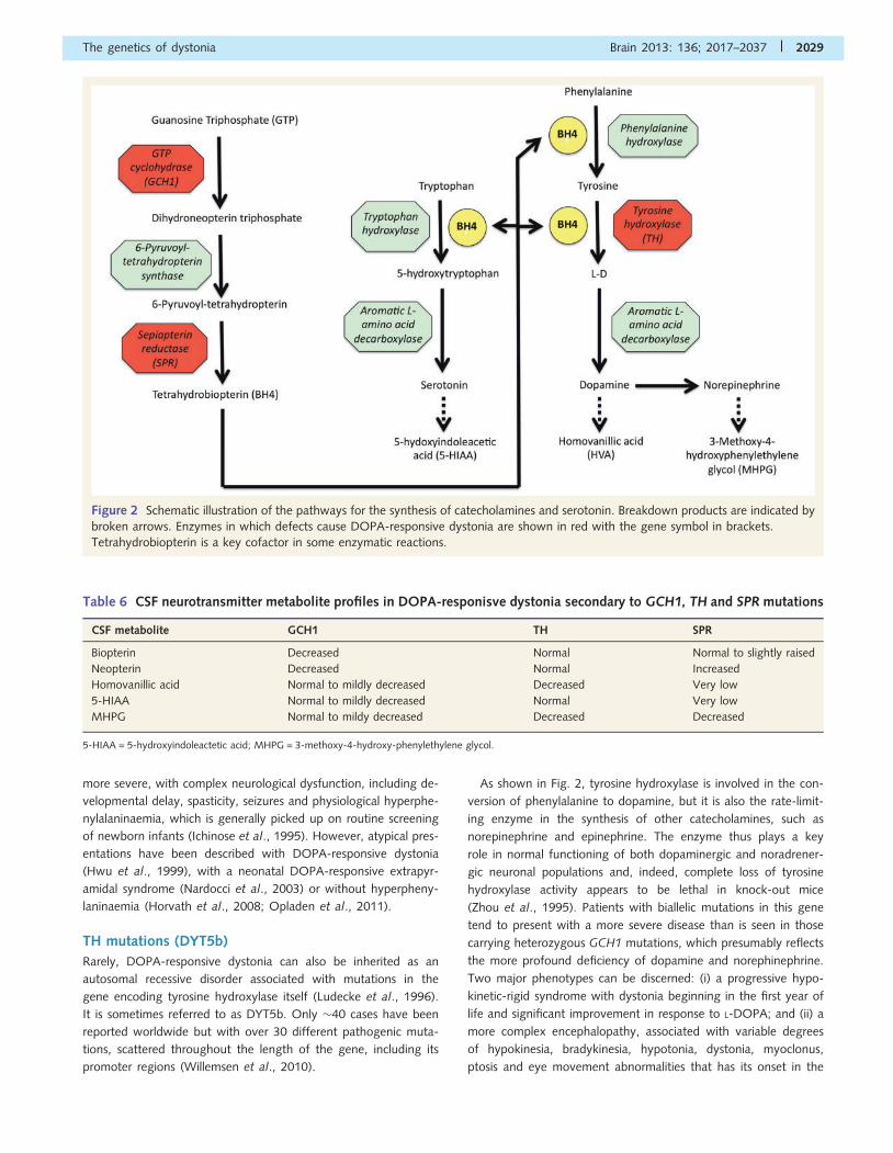
required for the biosynthesis of dopamine (Fig. 2).
GCH1 mutations (DYT5a/Segawa’s disease)
Mutations in this gene account for �60–80% of autosomal dom-
inant DOPA-responsive dystonia (Furukawa, 2004). Penetrance is
incomplete and lower for males (�40–50%) than females (�80%)
(Wider et al., 2008). GCH1 encodes the enzyme GTP cyclohydro-
lase 1, which catalyzes the Erst step in the reaction of tetrahydro-
biopterin neo-synthesis from GTP. Tetrahydrobiopterin is an
essential cofactor for tyrosine hydroxylase, the rate-limiting
enzyme for catecholamine synthesis, as well as for tryptophan
hydroxylase and phenylalanine hydroxylase, which are involved
in the production of serotonin. GCH1 mutations act in a dominant
negative manner, with measurable enzyme activity usually being
520% of normal, resulting in a relative deficiency of dopamine
and serotonin (Ichinose and Nagatsu, 1997; Hwu et al., 2000).
Diagnosis can be confirmed on the basis of reduced CSF levels of
total biopterin (of which tetrahydrobiopterin is the main compo-
nent) and neopterin (a product of the reaction involving GCH1)
(Table 6) or an abnormal phenylalanine loading test (Bandmann
et al., 2003; Furukawa, 2003).
Clinical presentation is typically with lower limb dystonia and
gait disturbance in the first decade of life, which occasionally
leads to misdiagnosis as cerebral palsy or spastic paraparesis
(Nygaard et al., 1990). Diurnal fluctuation in the severity of the
dystonia is common, though this may abate with age. With time
there is usually gradual generalization, and parkinsonism and dys-
tonic tremor are possible complications of the condition (Segawa
et al., 2003; Cobb et al., 2009). In a subgroup of patients, the
dystonia is mild, progresses only very slowly and may require no
treatment (Trender-Gerhard et al., 2009). Infrequently, patients
may present later in life with a dystonic tremor or akinetic-rigid
parkinsonism (Tassin et al., 2000; Trender-Gerhard et al., 2009).
There is some evidence that subtle neuropsychiatric features may
be associated with mutations in this gene. In one recent study of
18 patients, major depressive and sleep disorders occurred in
about half of those over 20 years of age, whereas obsessive–com-
pulsive disorder was found in 25% of cases (Van Hove et al.,
2006). In terms of treatment, the response even to low doses of
L-DOPA is generally excellent (70–100% improvement in clinical
symptoms), sustained and is not generally associated with the
late-onset dyskinesias that often accompany prolonged use of L-
DOPA in other conditions, such as Parkinson’s disease (Clot et al.,
2009).
It should be noted that a previously-proposed novel form of
DOPA-responsive dystonia, DYT14, is now known to be syn-
onymous with DYT5a. Misclassification of one patient from the
‘DYT14’ family had led to incorrect locus assignment on the basis
of erroneous linkage. After removal of this patient, the linkage
peak now included the GCH1 gene, which was found to habour
a causative deletion (Wider et al., 2008).
Finally, autosomal recessive mutations in GCH1 have also been
reported, resulting in no detectable enzyme activity in the liver. As
might be expected the phenotype is generally distinct and much
2028 | Brain 2013: 136; 2017–2037 G. Charlesworth et al.
more severe, with complex neurological dysfunction, including de-
velopmental delay, spasticity, seizures and physiological hyperphe-
nylalaninaemia, which is generally picked up on routine screening
of newborn infants (Ichinose et al., 1995). However, atypical pres-
entations have been described with DOPA-responsive dystonia
(Hwu et al., 1999), with a neonatal DOPA-responsive extrapyr-
amidal syndrome (Nardocci et al., 2003) or without hyperpheny-
laninaemia (Horvath et al., 2008; Opladen et al., 2011).
TH mutations (DYT5b)
Rarely, DOPA-responsive dystonia can also be inherited as an
autosomal recessive disorder associated with mutations in the
gene encoding tyrosine hydroxylase itself (Ludecke et al., 1996).
It is sometimes referred to as DYT5b. Only �40 cases have been
reported worldwide but with over 30 different pathogenic muta-
tions, scattered throughout the length of the gene, including its
promoter regions (Willemsen et al., 2010).
As shown in Fig. 2, tyrosine hydroxylase is involved in the con-
version of phenylalanine to dopamine, but it is also the rate-limit-
ing enzyme in the synthesis of other catecholamines, such as
norepinephrine and epinephrine. The enzyme thus plays a key
role in normal functioning of both dopaminergic and noradrener-
gic neuronal populations and, indeed, complete loss of tyrosine
hydroxylase activity appears to be lethal in knock-out mice
(Zhou et al., 1995). Patients with biallelic mutations in this gene
tend to present with a more severe disease than is seen in those
carrying heterozygous GCH1 mutations, which presumably reflects
the more profound deficiency of dopamine and norephinephrine.
Two major phenotypes can be discerned: (i) a progressive hypo-
kinetic-rigid syndrome with dystonia beginning in the first year of
life and significant improvement in response to L-DOPA; and (ii) a
more complex encephalopathy, associated with variable degrees
of hypokinesia, bradykinesia, hypotonia, dystonia, myoclonus,
ptosis and eye movement abnormalities that has its onset in the
Figure 2 Schematic illustration of the pathways for the synthesis of catecholamines and serotonin. Breakdown products are indicated by
broken arrows. Enzymes in which defects cause DOPA-responsive dystonia are shown in red with the gene symbol in brackets.
Tetrahydrobiopterin is a key cofactor in some enzymatic reactions.
Table 6 CSF neurotransmitter metabolite profiles in DOPA-responisve dystonia secondary to GCH1, TH and SPR mutations
CSF metabolite GCH1 TH SPR
Biopterin Decreased Normal Normal to slightly raised
Neopterin Decreased Normal Increased
Homovanillic acid Normal to mildly decreased Decreased Very low
5-HIAA Normal to mildly decreased Normal Very low
MHPG Normal to mildy decreased Decreased Decreased
5-HIAA = 5-hydroxyindoleactetic acid; MHPG = 3-methoxy-4-hydroxy-phenylethylene glycol.
The genetics of dystonia Brain 2013: 136; 2017–2037 | 2029
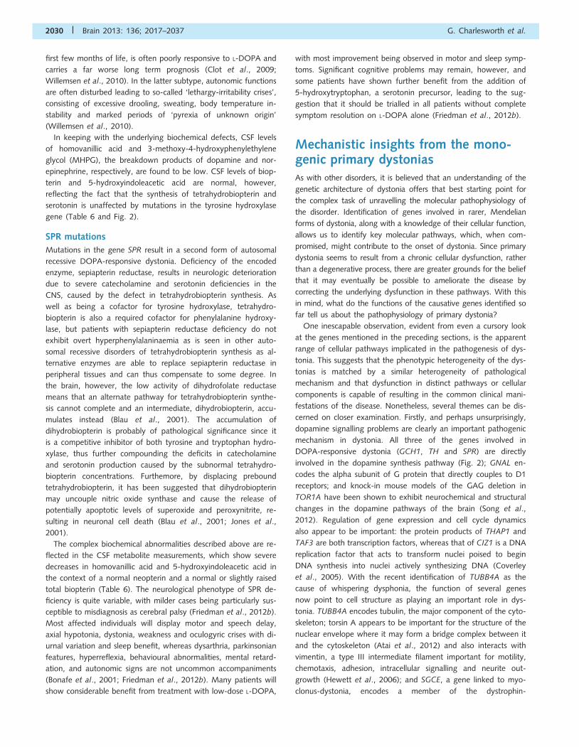
first few months of life, is often poorly responsive to L-DOPA and
carries a far worse long term prognosis (Clot et al., 2009;
Willemsen et al., 2010). In the latter subtype, autonomic functions
are often disturbed leading to so-called ‘lethargy-irritability crises’,
consisting of excessive drooling, sweating, body temperature in-
stability and marked periods of ‘pyrexia of unknown origin’
(Willemsen et al., 2010).
In keeping with the underlying biochemical defects, CSF levels
of homovanillic acid and 3-methoxy-4-hydroxyphenylethylene
glycol (MHPG), the breakdown products of dopamine and nor-
epinephrine, respectively, are found to be low. CSF levels of biop-
terin and 5-hydroxyindoleacetic acid are normal, however,
reflecting the fact that the synthesis of tetrahydrobiopterin and
serotonin is unaffected by mutations in the tyrosine hydroxylase
gene (Table 6 and Fig. 2).
SPR mutations
Mutations in the gene SPR result in a second form of autosomal
recessive DOPA-responsive dystonia. Deficiency of the encoded
enzyme, sepiapterin reductase, results in neurologic deterioration
due to severe catecholamine and serotonin deficiencies in the
CNS, caused by the defect in tetrahydrobiopterin synthesis. As
well as being a cofactor for tyrosine hydroxylase, tetrahydro-
biopterin is also a required cofactor for phenylalanine hydroxy-
lase, but patients with sepiapterin reductase deficiency do not
exhibit overt hyperphenylalaninaemia as is seen in other auto-
somal recessive disorders of tetrahydrobiopterin synthesis as al-
ternative enzymes are able to replace sepiapterin reductase in
peripheral tissues and can thus compensate to some degree. In
the brain, however, the low activity of dihydrofolate reductase
means that an alternate pathway for tetrahydrobiopterin synthe-
sis cannot complete and an intermediate, dihydrobiopterin, accu-
mulates instead (Blau et al., 2001). The accumulation of
dihydrobiopterin is probably of pathological significance since it
is a competitive inhibitor of both tyrosine and tryptophan hydro-
xylase, thus further compounding the deficits in catecholamine
and serotonin production caused by the subnormal tetrahydro-
biopterin concentrations. Furthemore, by displacing prebound
tetrahydrobiopterin, it has been suggested that dihydrobiopterin
may uncouple nitric oxide synthase and cause the release of
potentially apoptotic levels of superoxide and peroxynitrite, re-
sulting in neuronal cell death (Blau et al., 2001; Jones et al.,
2001).
The complex biochemical abnormalities described above are re-
flected in the CSF metabolite measurements, which show severe
decreases in homovanillic acid and 5-hydroxyindoleacetic acid in
the context of a normal neopterin and a normal or slightly raised
total biopterin (Table 6). The neurological phenotype of SPR de-
ficiency is quite variable, with milder cases being particularly sus-
ceptible to misdiagnosis as cerebral palsy (Friedman et al., 2012b).
Most affected individuals will display motor and speech delay,
axial hypotonia, dystonia, weakness and oculogyric crises with di-
urnal variation and sleep benefit, whereas dysarthria, parkinsonian
features, hyperreflexia, behavioural abnormalities, mental retard-
ation, and autonomic signs are not uncommon accompaniments
(Bonafe et al., 2001; Friedman et al., 2012b). Many patients will
show considerable benefit from treatment with low-dose L-DOPA,
with most improvement being observed in motor and sleep symp-
toms. Significant cognitive problems may remain, however, and
some patients have shown further benefit from the addition of
5-hydroxytryptophan, a serotonin precursor, leading to the sug-
gestion that it should be trialled in all patients without complete
symptom resolution on L-DOPA alone (Friedman et al., 2012b).
Mechanistic insights from the mono-genic primary dystoniasAs with other disorders, it is believed that an understanding of the
genetic architecture of dystonia offers that best starting point for
the complex task of unravelling the molecular pathophysiology of
the disorder. Identification of genes involved in rarer, Mendelian
forms of dystonia, along with a knowledge of their cellular function,
allows us to identify key molecular pathways, which, when com-
promised, might contribute to the onset of dystonia. Since primary
dystonia seems to result from a chronic cellular dysfunction, rather
than a degenerative process, there are greater grounds for the belief
that it may eventually be possible to ameliorate the disease by
correcting the underlying dysfunction in these pathways. With this
in mind, what do the functions of the causative genes identified so
far tell us about the pathophysiology of primary dystonia?
One inescapable observation, evident from even a cursory look
at the genes mentioned in the preceding sections, is the apparent
range of cellular pathways implicated in the pathogenesis of dys-
tonia. This suggests that the phenotypic heterogeneity of the dys-
tonias is matched by a similar heterogeneity of pathological
mechanism and that dysfunction in distinct pathways or cellular
components is capable of resulting in the common clinical mani-
festations of the disease. Nonetheless, several themes can be dis-
cerned on closer examination. Firstly, and perhaps unsurprisingly,
dopamine signalling problems are clearly an important pathogenic
mechanism in dystonia. All three of the genes involved in
DOPA-responsive dystonia (GCH1, TH and SPR) are directly
involved in the dopamine synthesis pathway (Fig. 2); GNAL en-
codes the alpha subunit of G protein that directly couples to D1
receptors; and knock-in mouse models of the GAG deletion in
TOR1A have been shown to exhibit neurochemical and structural
changes in the dopamine pathways of the brain (Song et al.,
2012). Regulation of gene expression and cell cycle dynamics
also appear to be important: the protein products of THAP1 and
TAF3 are both transcription factors, whereas that of CIZ1 is a DNA
replication factor that acts to transform nuclei poised to begin
DNA synthesis into nuclei actively synthesizing DNA (Coverley
et al., 2005). With the recent identification of TUBB4A as the
cause of whispering dysphonia, the function of several genes
now point to cell structure as playing an important role in dys-
tonia. TUBB4A encodes tubulin, the major component of the cyto-
skeleton; torsin A appears to be important for the structure of the
nuclear envelope where it may form a bridge complex between it
and the cytoskeleton (Atai et al., 2012) and also interacts with
vimentin, a type III intermediate filament important for motility,
chemotaxis, adhesion, intracellular signalling and neurite out-
growth (Hewett et al., 2006); and SGCE, a gene linked to myo-
clonus-dystonia, encodes a member of the dystrophin-
2030 | Brain 2013: 136; 2017–2037 G. Charlesworth et al.
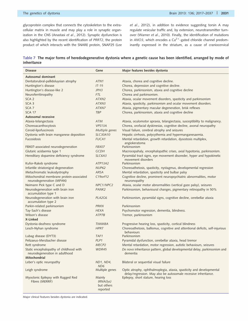
glycoprotein complex that connects the cytoskeleton to the extra-
cellular matrix in muscle and may play a role in synaptic organ-
ization in the CNS (Anastasi et al., 2012). Synaptic dysfunction is
also highlighted by the recent identification of PRRT2, the protein
product of which interacts with the SNARE protein, SNAP25 (Lee
et al., 2012), in addition to evidence suggesting torsin A may
regulate vesicular traffic and, by extension, neurotransmitter turn-
over (Warner et al., 2010). Finally, the identification of mutations
in ANO3, which encodes a Ca2 +-gated chloride channel predom-
inantly expressed in the striatum, as a cause of craniocervical
Table 7 The major forms of heredodegenerative dystonia where a genetic cause has been identified, arranged by mode ofinheritance
Disease Gene Major features besides dystonia
Autosomal dominant
Dentatorubral-pallidoluysian atrophy ATN1 Ataxia, chorea and cognitive decline.
Huntington’s disease IT-15 Chorea, depression and cognitive decline.
Huntington’s disease-like 2 JPH3 Chorea, parkinsonism, ataxia and cognitive decline
Neuroferritinopathy FTL Chorea and parkinsonism.
SCA 2 ATXN2 Ataxia, ocular movement disorders, spasticity and parkinsonism
SCA 3 ATXN3 Ataxia, spasticity, parkinsonism and ocular movement disorders.
SCA 7 ATXN7 Ataxia, pigmentary macular degeneration, brisk reflexes
SCA 17 TBP Chorea, parkinsonism, ataxia and cognitive decline
Autosomal recessive
Ataxia-telangectasia ATM Ataxia, oculomotor apraxia, telangiectasia, susceptibility to malignancy.
Choreoacanthocytosis VPS13A Chorea, orofacial dyskinesias, cognitive decline, axonal neuropathy
Ceroid-lipofuscinosis Multiple genes Visual failure, cerebral atrophy and seizures
Dystonia with brain manganese deposition SLC30A10 Hepatic cirrhosis, polycythemia and hypermanganesaemia.
Fucosidosis FUCA1 Mental retardation, growth retardation, dysostosis multiplex,angiokeratoma
FBX07-associated neurodegeneration FBX07 Parkinsonism
Glutaric acidaemia type 1 GCDH Macrocepahaly, encephalopathic crises, axial hypotonia, parkinsonism.
Hereditary dopamine deficiency syndrome SLC6A3 Pyramidal tract signs, eye movement disoreder, hyper and hypokineticmovement disorders
Kufor-Rakeb syndrome ATP13A2 Parkinsonism
Infantile striatonigral degeneration NUP62 Choreoathetosis, spasticity, nystagmus, developmental regression
Metachromatic leukodystrophy ARSA Mental retardation, spasticity and bulbar palsy
Mitochondrial membrane protein-associatedneurodegeneration (MPAN)
C19orf12 Cognitive decline, prominent neuropsychiatric abnormalities, motorneuronopathy
Neimann Pick type C and D NPC1/NPC2 Ataxia, ocular motor abnormalities (vertical gaze palsy), seizures
Neurodegeneration with brain ironaccumulation type 1
PANK2 Parkinsonism, behavioural changes, pigmentary retinopathy in 50%
Neurodegeneration with brain ironaccumulation type 2
PLA2G6 Parkinsonism, pyramidal signs, cognitive decline, cerebellar ataxia
Parkin-related parkinsonism PRKN Parkinsonism
Tay-Sach’s disease HEXA Psychomotor regression, dementia, blindness.
Wilson’s disease ATP7B Tremor, parkinsonism
X-Linked
Dystonia-deafness syndrome TIMM8A Progressive hearing loss, spasticity, cortical blindness
Lesch-Nyhan syndrome HPRT Choreoathetosis, ballismus, cognitive and attentional deficits, self-injuriousbehaviours
Lubag disease (DYT3) TAF1 Parkinsonism
Pelizaeus-Merzbacher disease PLP1 Pyramidal dysfunction, cerebellar ataxia, head tremor
Rett syndrome MECP2 Mental retardation, motor regression, autistic behaviours, seizures
Static encephalopathy of childhood withneurodegeneration in adulthood
WDR45 De novo inheritance pattern, global developmental delay, parkinsonism anddementia
Mitochondrial
Leber’s optic neuropathy ND1, ND4,ND6
Bilateral or sequential visual failure
Leigh syndrome Multiple genes Optic atrophy, ophthalmoplegia, ataxia, spasticity and developmentaldelay/regression. May also be autosomale recessive inheritance.
Myoclonic Epilepsy with Ragged RedFibres (MERRF)
MainlytRNA(lys)but othersreported
Epilepsy, short stature, hearing loss
Major clinical features besides dystonia are indicated.
The genetics of dystonia Brain 2013: 136; 2017–2037 | 2031
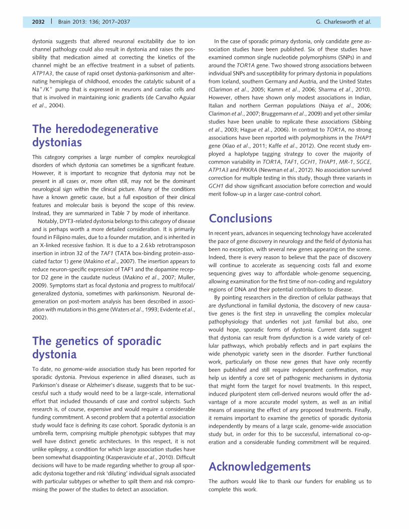
dystonia suggests that altered neuronal excitability due to ion
channel pathology could also result in dystonia and raises the pos-
sibility that medication aimed at correcting the kinetics of the
channel might be an effective treatment in a subset of patients.
ATP1A3, the cause of rapid onset dystonia-parkinsonism and alter-
nating hemiplegia of childhood, encodes the catalytic subunit of a
Na +/K + pump that is expressed in neurons and cardiac cells and
that is involved in maintaining ionic gradients (de Carvalho Aguiar
et al., 2004).
The heredodegenerativedystoniasThis category comprises a large number of complex neurological
disorders of which dystonia can sometimes be a significant feature.
However, it is important to recognize that dystonia may not be
present in all cases or, more often still, may not be the dominant
neurological sign within the clinical picture. Many of the conditions
have a known genetic cause, but a full exposition of their clinical
features and molecular basis is beyond the scope of this review.
Instead, they are summarized in Table 7 by mode of inheritance.
Notably, DYT3-related dystonia belongs to this category of disease
and is perhaps worth a more detailed consideration. It is primarily
found in Filipino males, due to a founder mutation, and is inherited in
an X-linked recessive fashion. It is due to a 2.6 kb retrotransposon
insertion in intron 32 of the TAF1 (TATA box-binding protein-asso-
ciated factor 1) gene (Makino et al., 2007). The insertion appears to
reduce neuron-specific expression of TAF1 and the dopamine recep-
tor D2 gene in the caudate nucleus (Makino et al., 2007; Muller,
2009). Symptoms start as focal dystonia and progress to multifocal/
generalized dystonia, sometimes with parkinsonism. Neuronal de-
generation on post-mortem analysis has been described in associ-
ation with mutations in this gene (Waters et al., 1993; Evidente et al.,
2002).
The genetics of sporadicdystoniaTo date, no genome-wide association study has been reported for
sporadic dystonia. Previous experience in allied diseases, such as
Parkinson’s disease or Alzheimer’s disease, suggests that to be suc-
cessful such a study would need to be a large-scale, international
effort that included thousands of case and control subjects. Such
research is, of course, expensive and would require a considerable
funding commitment. A second problem that a potential association
study would face is defining its case cohort. Sporadic dystonia is an
umbrella term, comprising multiple phenotypic subtypes that may
well have distinct genetic architectures. In this respect, it is not
unlike epilepsy, a condition for which large association studies have
been somewhat disappointing (Kasperaviciute et al., 2010). Difficult
decisions will have to be made regarding whether to group all spor-
adic dystonia together and risk ‘diluting’ individual signals associated
with particular subtypes or whether to spilt them and risk compro-
mising the power of the studies to detect an association.
In the case of sporadic primary dystonia, only candidate gene as-
sociation studies have been published. Six of these studies have
examined common single nucleotide polymorphisms (SNPs) in and
around the TOR1A gene. Two showed strong associations between
individual SNPs and susceptibility for primary dystonia in populations
from Iceland, southern Germany and Austria, and the United States
(Clarimon et al., 2005; Kamm et al., 2006; Sharma et al., 2010).
However, others have shown only modest associations in Indian,
Italian and northern German populations (Naiya et al., 2006;
Clarimon et al., 2007; Bruggemann et al., 2009) and yet other similar
studies have been unable to replicate these associations (Sibbing
et al., 2003; Hague et al., 2006). In contrast to TOR1A, no strong
associations have been reported with polymorphisms in the THAP1
gene (Xiao et al., 2011; Kaffe et al., 2012). One recent study em-
ployed a haplotype tagging strategy to cover the majority of
common variability in TOR1A, TAF1, GCH1, THAP1, MR-1, SGCE,
ATP1A3 and PRKRA (Newman et al., 2012). No association survived
correction for multiple testing in this study, though three variants in
GCH1 did show significant association before correction and would
merit follow-up in a larger case-control cohort.
ConclusionsIn recent years, advances in sequencing technology have accelerated
the pace of gene discovery in neurology and the field of dystonia has
been no exception, with several new genes appearing on the scene.
Indeed, there is every reason to believe that the pace of discovery
will continue to accelerate as sequencing costs fall and exome
sequencing gives way to affordable whole-genome sequencing,
allowing examination for the first time of non-coding and regulatory
regions of DNA and their potential contributions to disease.
By pointing researchers in the direction of cellular pathways that
are dysfunctional in familial dystonia, the discovery of new causa-
tive genes is the first step in unravelling the complex molecular
pathophysiology that underlies not just familial but also, one
would hope, sporadic forms of dystonia. Current data suggest
that dystonia can result from dysfunction is a wide variety of cel-
lular pathways, which probably reflects and in part explains the
wide phenotypic variety seen in the disorder. Further functional
work, particularly on those new genes that have only recently
been published and still require independent confirmation, may
help us identify a core set of pathogenic mechanisms in dystonia
that might form the target for novel treatments. In this respect,
induced pluripotent stem cell-derived neurons would offer the ad-
vantage of a more accurate model system, as well as an initial
means of assessing the effect of any proposed treatments. Finally,
it remains important to examine the genetics of sporadic dystonia
independently by means of a large scale, genome-wide association
study but, in order for this to be successful, international co-op-
eration and a considerable funding commitment will be required.
AcknowledgementsThe authors would like to thank our funders for enabling us to
complete this work.
2032 | Brain 2013: 136; 2017–2037 G. Charlesworth et al.
FundingThis work was supported by the Medical Research council and the
Wellcome Trust [grant number WT089698/Z/09/Z] and the
Bachmann-Strauss Dystonia and Parkinson Foundation.
ReferencesAlbanese A, Asmus F, Bhatia KP, Elia AE, Elibol B, Filippini G, et al. EFNS
guidelines on diagnosis and treatment of primary dystonias. Eur J
Neurol 2011; 18: 5–18.Almasy L, Bressman SB, Raymond D, Kramer PL, Greene PE, Heiman GA,
et al. Idiopathic torsion dystonia linked to chromosome 8 in two
Mennonite families. Ann Neurol 1997; 42: 670–3.
Anastasi G, Tomasello F, Di Mauro D, Cutroneo G, Favaloro A, Conti A,
et al. Expression of sarcoglycans in the human cerebral cortex: an
immunohistochemical and molecular study. Cells Tissues Organs
2012; 196: 470–80.
Argyelan M, Carbon M, Niethammer M, Ulug AM, Voss HU,
Bressman SB, et al. Cerebellothalamocortical connectivity regulates
penetrance in dystonia. J Neurosci 2009; 29: 9740–7.Asmus F, Devlin A, Munz M, Zimprich A, Gasser T, Chinnery PF. Clinical
differentiation of genetically proven benign hereditary chorea and
myoclonus-dystonia. Mov Disord 2007; 22: 2104–9.
Asmus F, Gasser T. Inherited myoclonus-dystonia. Adv Neurol 2004; 94:
113–19.
Atai NA, Ryan SD, Kothary R, Breakefield XO, Nery FC. Untethering the
nuclear envelope and cytoskeleton: biologically distinct dystonias
arising from a common cellular dysfunction. Int J Cell Biol 2012;
2012: 634214.
Bandmann O, Goertz M, Zschocke J, Deuschl G, Jost W, Hefter H, et al.
The phenylalanine loading test in the differential diagnosis of dystonia.
Neurology 2003; 60: 700–2.Belluscio L, Gold GH, Nemes A, Axel R. Mice deficient in G(olf) are
anosmic. Neuron 1998; 20: 69–81.
Berrettini WH. Susceptibility loci for bipolar disorder: overlap with in-
herited vulnerability to schizophrenia. Biol Psychiatry 2000; 47:
245–51.
Blanco-Arias P, Einholm AP, Mamsa H, Concheiro C, Gutierrez-de-
Teran H, Romero J, et al. A C-terminal mutation of ATP1A3 under-
scores the crucial role of sodium affinity in the pathophysiology of
rapid-onset dystonia-parkinsonism. Hum Mol Genet 2009; 18:
2370–7.Blau N, Bonafe L, Thony B. Tetrahydrobiopterin deficiencies without
hyperphenylalaninemia: diagnosis and genetics of dopa-responsive
dystonia and sepiapterin reductase deficiency. Mol Genet Metab
2001; 74: 172–85.
Bonafe L, Thony B, Penzien JM, Czarnecki B, Blau N. Mutations in the
sepiapterin reductase gene cause a novel tetrahydrobiopterin-depend-
ent monoamine-neurotransmitter deficiency without hyperphenylalani-
nemia. Am J Hum Genet 2001; 69: 269–77.
Bragg DC, Armata IA, Nery FC, Breakefield XO, Sharma N. Molecular
pathways in dystonia. Neurobiol Dis 2011; 42: 136–47.Brashear A, Dobyns WB, de Carvalho Aguiar P, Borg M, Frijns CJ,
Gollamudi S, et al. The phenotypic spectrum of rapid-onset dys-
tonia-parkinsonism (RDP) and mutations in the ATP1A3 gene. Brain
2007; 130 (Pt 3): 828–35.
Brashear A, Mulholland GK, Zheng QH, Farlow MR, Siemers ER,
Hutchins GD. PET imaging of the pre-synaptic dopamine uptake
sites in rapid-onset dystonia-parkinsonism (RDP). Mov Disord 1999;
14: 132–7.
Brautigam C, Wevers RA, Jansen RJ, Smeitink JA, de Rijk-van Andel JF,
Gabreels FJ, et al. Biochemical hallmarks of tyrosine hydroxylase defi-
ciency. Clin Chem 1998; 44: 1897–904.
Breakefield XO, Blood AJ, Li Y, Hallett M, Hanson PI, Standaert DG. The
pathophysiological basis of dystonias. Nat Rev Neurosci 2008; 9:
222–34.
Bressman S. Genetics of dystonia. J Neural Transm Suppl 2006; 70:
489–95.
Brockmann K. The expanding phenotype of GLUT1-deficiency syndrome.
Brain Dev 2009; 31: 545–52.
Bruggemann N, Kock N, Lohmann K, Konig IR, Rakovic A, Hagenah J,
et al. The D216H variant in the DYT1 gene: a susceptibility factor for
dystonia in familial cases? Neurology 2009; 72: 1441–3.
Bruno MK, Hallett M, Gwinn-Hardy K, Sorensen B, Considine E,
Tucker S, et al. Clinical evaluation of idiopathic paroxysmal kinesigenic
dyskinesia: new diagnostic criteria. Neurology 2004; 63: 2280–7.
Bruno MK, Lee HY, Auburger GW, Friedman A, Nielsen JE, Lang AE,
et al. Genotype-phenotype correlation of paroxysmal nonkinesigenic
dyskinesia. Neurology 2007; 68: 1782–9.Calakos N, Patel VD, Gottron M, Wang G, Tran-Viet KN, Brewington D,
et al. Functional evidence implicating a novel TOR1A mutation in idio-
pathic, late-onset focal dystonia. J Med Genet 2010; 47: 646–50.
Camargos S, Scholz S, Simon-Sanchez J, Paisan-Ruiz C, Lewis P,
Hernandez D, et al. DYT16, a novel young-onset dystonia-parkinson-
ism disorder: identification of a segregating mutation in the stress-re-
sponse protein PRKRA. Lancet Neurol 2008; 7: 207–15.
Cao L, Huang XJ, Zheng L, Xiao Q, Wang XJ, Chen SD. Identification of
a novel PRRT2 mutation in patients with paroxysmal kinesigenic dys-
kinesias and c.649dupC as a mutation hot-spot. Parkinsonism Relat
Disord 2012; 18: 704–6.Caputo A, Caci E, Ferrera L, Pedemonte N, Barsanti C, Sondo E, et al.
TMEM16A, a membrane protein associated with calcium-dependent
chloride channel activity. Science 2008; 322: 590–4.
Charlesworth G, Plagnol V, Holmstrom KM, Bras J, Sheerin UM, Preza E,
et al. Mutations in ANO3 cause dominant craniocervical dystonia: ion
channel implicated in pathogenesis. Am J Hum Genet 2012; 91:
1041–50.
Chen DH, Matsushita M, Rainier S, Meaney B, Tisch L, Feleke A, et al.
Presence of alanine-to-valine substitutions in myofibrillogenesis
regulator 1 in paroxysmal nonkinesigenic dyskinesia: confirmation in
2 kindreds. Arch Neurol 2005; 62: 597–600.Chen WJ, Lin Y, Xiong ZQ, Wei W, Ni W, Tan GH, et al. Exome sequen-
cing identifies truncating mutations in PRRT2 that cause paroxysmal
kinesigenic dyskinesia. Nat Genet 2011; 43: 1252–5.
Clarimon J, Asgeirsson H, Singleton A, Jakobsson F, Hjaltason H, Hardy J,
et al. Torsin a haplotype predisposes to idiopathic dystonia. Ann
Neurol 2005; 57: 765–7.
Clarimon J, Brancati F, Peckham E, Valente EM, Dallapiccola B,
Abruzzese G, et al. Assessing the role of DRD5 and DYT1 in two
different case-control series with primary blepharospasm. Mov
Disord 2007; 22: 162–6.
Clot F, Grabli D, Cazeneuve C, Roze E, Castelnau P, Chabrol B, et al.
Exhaustive analysis of BH4 and dopamine biosynthesis genes in pa-
tients with Dopa-responsive dystonia. Brain 2009; 132 (Pt 7):
1753–63.
Clouaire T, Roussigne M, Ecochard V, Mathe C, Amalric F, Girard JP. The
THAP domain of THAP1 is a large C2CH module with zinc-dependent
sequence-specific DNA-binding activity. Proc Natl Acad Sci USA 2005;
102: 6907–12.
Cobb SA, Wider C, Ross OA, Mata IF, Adler CH, Rajput A, et al.
GCH1 in early-onset Parkinson’s disease. Mov Disord 2009; 24:
2070–5.
Corvol JC, Studler JM, Schonn JS, Girault JA, Herve D. Galpha(olf) is
necessary for coupling D1 and A2a receptors to adenylyl cyclase in
the striatum. J Neurochem 2001; 76: 1585–8.Coverley D, Marr J, Ainscough J. Ciz1 promotes mammalian DNA rep-
lication. J Cell Sci 2005; 118 (Pt 1): 101–12.Dale RC, Gardiner A, Antony J, Houlden H. Familial PRRT2 mutation
with heterogeneous paroxysmal disorders including paroxysmal torti-
collis and hemiplegic migraine. Dev Med Child Neurol 2012; 54:
958–60.
The genetics of dystonia Brain 2013: 136; 2017–2037 | 2033
de Carvalho Aguiar P, Sweadner KJ, Penniston JT, Zaremba J, Liu L,
Caton M, et al. Mutations in the Na + /K + -ATPase Œ�3 Gene
ATP1A3 are associated with rapid-onset dystonia parkinsonism.
Neuron 2004; 43: 169–75.
Defazio G, Berardelli A, Hallett M. Do primary adult-onset
focal dystonias share aetiological factors? Brain 2007; 130 (Pt 5):
1183–93.Deutschlander A, Asmus F, Gasser T, Steude U, Botzel K. Sporadic rapid-
onset dystonia-parkinsonism syndrome: failure of bilateral pallidal
stimulation. Mov Disord 2005; 20: 254–7.
Djarmati A, Svetel M, Momcilovic D, Kostic V, Klein C. Significance of
recurrent mutations in the myofibrillogenesis regulator 1 gene. Arch
Neurol 2005; 62: 1641.
Duan W, Zhang Z, Gash DM, Mattson MP. Participation of prostate
apoptosis response-4 in degeneration of dopaminergic neurons in
models of Parkinson’s disease. Ann Neurol 1999; 46: 587–97.
Duran C, Hartzell HC. Physiological roles and diseases of Tmem16/
Anoctamin proteins: are they all chloride channels? Acta Pharmacol
Sin 2011; 32: 685–92.Duran C, Qu Z, Osunkoya AO, Cui Y, Hartzell HC. ANOs 3-7 in the
anoctamin/Tmem16 Cl- channel family are intracellular proteins. Am J
Physiol Cell Physiol 2012; 302: C482–93.
Esapa CT, Waite A, Locke M, Benson MA, Kraus M, McIlhinney RA, et al.
SGCE missense mutations that cause myoclonus-dystonia syndrome
impair epsilon-sarcoglycan trafficking to the plasma membrane: modu-
lation by ubiquitination and torsinA. Hum Mol Genet 2007; 16: 327–42.
Evidente VG, Advincula J, Esteban R, Pasco P, Alfon JA, Natividad FF,
et al. Phenomenology of “Lubag” or X-linked dystonia-parkinsonism.
Mov Disord 2002; 17: 1271–7.Fahn S. Concept and classification of dystonia. Adv Neurol 1988; 50:
1–8.Fanh S, Marsden CD, Caine DB. Dystonia 2. Proceedings of the Second
International Symposium on Torsion Dystonia. Harriman, New York,
1986. Adv Neurol 1988; 50: 1–705.
Friedman J, Olvera J, Silhavy JL, Gabriel SB, Gleeson JG. Mild paroxysmal
kinesigenic dyskinesia caused by PRRT2 missense mutation with
reduced penetrance. Neurology 2012a; 79: 946–8.
Friedman J, Roze E, Abdenur JE, Chang R, Gasperini S, Saletti V, et al.
Sepiapterin reductase deficiency: a treatable mimic of cerebral palsy.
Ann Neurol 2012b; 71: 520–30.
Fuchs T, Gavarini S, Saunders-Pullman R, Raymond D, Ehrlich ME,
Bressman SB, et al. Mutations in the THAP1 gene are
responsible for DYT6 primary torsion dystonia. Nat Genet 2009; 41:
286–8.
Fuchs T, Saunders-Pullman R, Masuho I, Luciano MS, Raymond D,
Factor S, et al. Mutations in GNAL cause primary torsion dystonia.
Nat Genet 2012; 45: 88–92.
Furukawa Y. Genetics and biochemistry of dopa-responsive dystonia:
significance of striatal tyrosine hydroxylase protein loss. Adv Neurol
2003; 91: 401–10.
Furukawa Y. Update on dopa-responsive dystonia: locus heterogeneity
and biochemical features. Adv Neurol 2004; 94: 127–38.
Gardiner AR, Bhatia KP, Stamelou M, Dale RC, Kurian MA,
Schneider SA, et al. PRRT2 gene mutations: From paroxysmal dyskin-
esia to episodic ataxia and hemiplegic migraine. Neurology 2012; 79:
2115–21.
Gavarini S, Cayrol C, Fuchs T, Lyons N, Ehrlich ME, Girard JP, et al.
Direct interaction between causative genes of DYT1 and DYT6 primary
dystonia. Ann Neurol 2010; 68: 549–53.
Geyer HL, Bressman SB. The diagnosis of dystonia. Lancet Neurol 2006;
5: 780–90.
Ghezzi D, Viscomi C, Ferlini A, Gualandi F, Mereghetti P, DeGrandis D,
et al. Paroxysmal non-kinesigenic dyskinesia is caused by mutations of
the MR-1 mitochondrial targeting sequence. Hum Mol Genet 2009;
18: 1058–64.Goodchild RE, Dauer WT. The AAA + protein torsinA interacts with a
conserved domain present in LAP1 and a novel ER protein. J Cell Biol
2005; 168: 855–62.
Goodchild RE, Kim CE, Dauer WT. Loss of the dystonia-associated pro-
tein torsinA selectively disrupts the neuronal nuclear envelope. Neuron
2005; 48: 923–32.
Grabowski M, Zimprich A, Lorenz-Depiereux B, Kalscheuer V, Asmus F,
Gasser T, et al. The epsilon-sarcoglycan gene (SGCE), mutated in myo-
clonus-dystonia syndrome, is maternally imprinted. Eur J Hum Genet
2003; 11: 138–44.
Grundmann K, Reischmann B, Vanhoutte G, Hubener J, Teismann P,
Hauser TK, et al. Overexpression of human wildtype torsinA and
human DeltaGAG torsinA in a transgenic mouse model causes pheno-
typic abnormalities. Neurobiol Dis 2007; 27: 190–206.Guo Q, Fu W, Xie J, Luo H, Sells SF, Geddes JW, et al. Par-4 is a
mediator of neuronal degeneration associated with the pathogenesis
of Alzheimer disease. Nat Med 1998; 4: 957–62.
Hague S, Klaffke S, Clarimon J, Hemmer B, Singleton A, Kupsch A, et al.
Lack of association with TorsinA haplotype in German patients with
sporadic dystonia. Neurology 2006; 66: 951–2.
Hartzell C, Putzier I, Arreola J. Calcium-activated chloride channels. Annu
Rev Physiol 2005; 67: 719–58.
Hedera P, Xiao J, Puschmann A, Momcilovic D, Wu SW, Ledoux MS.
Novel PRRT2 mutation in an African-American family with paroxysmal
kinesigenic dyskinesia. BMC Neurol 2012; 12: 93.
Heinzen EL, Swoboda KJ, Hitomi Y, Gurrieri F, Nicole S, de Vries B, et al.
De novo mutations in ATP1A3 cause alternating hemiplegia of child-
hood. Nat Genet 2012; 44: 1030–4.
Hempelmann A, Kumar S, Muralitharan S, Sander T. Myofibrillogenesis
regulator 1 gene (MR-1) mutation in an Omani family with
paroxysmal nonkinesigenic dyskinesia. Neurosci Lett 2006; 402:
118–20.
Henriksen C, Madsen LB, Bendixen C, Larsen K. Characterization of the
porcine TOR1A gene: The first step towards generation of a pig model
for dystonia. Gene 2009; 430: 105–15.
Heron SE, Grinton BE, Kivity S, Afawi Z, Zuberi SM, Hughes JN, et al.
PRRT2 mutations cause benign familial infantile epilepsy and infantile
convulsions with choreoathetosis syndrome. Am J Hum Genet 2012;
90: 152–60.
Hersheson J, Mencacci NE, Davis M, MacDoanld HN, Trabzuni D,
Ryten M, et al. Mutations in the autoregulatory domain of b-tubulin
4a cause hereditary dystonia. Ann Neurol 2013. doi: 10.1002/
ana.23832.
Herve D, Levi-Strauss M, Marey-Semper I, Verney C, Tassin JP,
Glowinski J, et al. G(olf) and Gs in rat basal ganglia: possible involve-
ment of G(olf) in the coupling of dopamine D1 receptor with adenylyl
cyclase. J Neurosci 1993; 13: 2237–48.
Hess CW, Raymond D, Aguiar Pde C, Frucht S, Shriberg J, Heiman GA,
et al. Myoclonus-dystonia, obsessive-compulsive disorder, and alcohol
dependence in SGCE mutation carriers. Neurology 2007; 68: 522–4.
Hewett JW, Tannous B, Niland BP, Nery FC, Zeng J, Li Y, et al. Mutant
torsinA interferes with protein processing through the secretory pathway
in DYT1 dystonia cells. Proc Natl Acad Sci USA 2007; 104: 7271–6.Hewett JW, Zeng J, Niland BP, Bragg DC, Breakefield XO. Dystonia-
causing mutant torsinA inhibits cell adhesion and neurite extension
through interference with cytoskeletal dynamics. Neurobiol Dis 2006;
22: 98–111.
Hjermind LE, Werdelin LM, Sorensen SA. Inherited and de novo muta-
tions in sporadic cases of DYT1-dystonia. Eur J Hum Genet 2002; 10:
213–6.
Horvath GA, Stockler-Ipsiroglu SG, Salvarinova-Zivkovic R, Lillquist YP,
Connolly M, Hyland K, et al. Autosomal recessive GTP cyclohydrolase I
deficiency without hyperphenylalaninemia: evidence of a phenotypic
continuum between dominant and recessive forms. Mol Genet Metab
2008; 94: 127–31.
Houlden H, Schneider SA, Paudel R, Melchers A, Schwingenschuh P,
Edwards M, et al. THAP1 mutations (DYT6) are an additional cause
of early-onset dystonia. Neurology 2010; 74: 846–50.Huang WC, Xiao S, Huang F, Harfe BD, Jan YN, Jan LY. Calcium-acti-
vated chloride channels (CaCCs) regulate action potential and synaptic
response in hippocampal neurons. Neuron 2012; 74: 179–92.
2034 | Brain 2013: 136; 2017–2037 G. Charlesworth et al.
Hwu WL, Chiou YW, Lai SY, Lee YM. Dopa-responsive dystonia is
induced by a dominant-negative mechanism. Ann Neurol 2000; 48:
609–13.
Hwu WL, Wang PJ, Hsiao KJ, Wang TR, Chiou YW, Lee YM. Dopa-
responsive dystonia induced by a recessive GTP cyclohydrolase I mu-
tation. Hum Genet 1999; 105: 226–30.
Ichinose H, Nagatsu T. Molecular genetics of hereditary dystonia–
mutations in the GTP cyclohydrolase I gene. Brain Res Bull 1997;
43: 35–8.Ichinose H, Ohye T, Matsuda Y, Hori T, Blau N, Burlina A, et al.
Characterization of mouse and human GTP cyclohydrolase I genes.
Mutations in patients with GTP cyclohydrolase I deficiency. J Biol
Chem 1995; 270: 10062–71.
Ichinose H, Ohye T, Takahashi E, Seki N, Hori T, Segawa M, et al.
Hereditary progressive dystonia with marked diurnal fluctuation
caused by mutations in the GTP cyclohydrolase I gene. Nat Genet
1994; 8: 236–42.Jamora RD, Tan EK, Liu CP, Kathirvel P, Burgunder JM, Tan LC. DYT1
mutations amongst adult primary dystonia patients in Singapore with
review of literature comparing East and West. J Neurol Sci 2006; 247:
35–7.
Jones CL, Vasquez-Vivar J, Kalyanaraman B, Griscavage-Ennis M,
Gross SS. Tetrahydropterins but not dihydropterins attenuate the re-
duction of superoxide from eNOS. Pteridines 2001; 12: 52–3.
Kabakci K, Hedrich K, Leung JC, Mitterer M, Vieregge P, Lencer R, et al.
Mutations in DYT1: extension of the phenotypic and mutational spec-
trum. Neurology 2004; 62: 395–400.Kaffe M, Gross N, Castrop F, Dresel C, Gieger C, Lichtner P, et al.
Mutational screening of THAP1 in a German population with primary
dystonia. Parkinsonism Relat Disord 2012; 18: 104–6.
Kaiser FJ, Osmanoric A, Rakovic A, Erogullari A, Uflacker N, Braunholz D,
et al. The dystonia gene DYT1 is repressed by the transcription factor
THAP1 (DYT6). Ann Neurol 2010; 68: 554–9.
Kakazu Y, Koh JY, Ho KW, Gonzalez-Alegre P, Harata NC. Synaptic
vesicle recycling is enhanced by torsinA that harbors the DYT1 dys-
tonia mutation. Synapse 2012; 66: 453–64.
Kamm C, Asmus F, Mueller J, Mayer P, Sharma M, Muller UJ, et al.
Strong genetic evidence for association of TOR1A/TOR1B with idio-
pathic dystonia. Neurology 2006; 67: 1857–9.Kamm C, Fischer H, Garavaglia B, Kullmann S, Sharma M, Schrader C,
et al. Susceptibility to DYT1 dystonia in European patients is modified
by the D216H polymorphism. Neurology 2008a; 70: 2261–2.
Kamm C, Fogel W, Wachter T, Schweitzer K, Berg D, Kruger R, et al. Novel
ATP1A3 mutation in a sporadic RDP patient with minimal benefit from
deep brain stimulation. Neurology 2008b; 70 (Pt 2): 1501–3.
Kasperaviciute D, Catarino CB, Heinzen EL, Depondt C, Cavalleri GL,
Caboclo LO, et al. Common genetic variation and susceptibility to
partial epilepsies: a genome-wide association study. Brain 2010; 133
(Pt 7): 2136–47.Khan NL, Wood NW, Bhatia KP. Autosomal recessive, DYT2-like primary
torsion dystonia: a new family. Neurology 2003; 61: 1801–3.Kimmich O, Bradley D, Whelan R, Mulrooney N, Reilly RB, Hutchinson S,
et al. Sporadic adult onset primary torsion dystonia is a genetic disorder
by the temporal discrimination test. Brain 2011; 134 (Pt 9): 2656–63.
Kinugawa K, Vidailhet M, Clot F, Apartis E, Grabli D, Roze E.
Myoclonus-dystonia: an update. Mov Disord 2009; 24: 479–89.
Klein C, Konig IR, Lohmann K. Exome sequencing for gene discovery:
time to set standard criteria. Ann Neurol 2012; 72: 627–8.
Kock N, Naismith TV, Boston HE, Ozelius LJ, Corey DP, Breakefield XO,
et al. Effects of genetic variations in the dystonia protein torsinA: iden-
tification of polymorphism at residue 216 as protein modifier. Hum
Mol Genet 2006; 15: 1355–64.
Konakova M, Huynh DP, Yong W, Pulst SM. Cellular distribution of
torsin A and torsin B in normal human brain. Arch Neurol 2001; 58:
921–7.Kramer PL, de Leon D, Ozelius L, Risch N, Bressman SB, Brin MF, et al.
Dystonia gene in Ashkenazi Jewish population is located on chromo-
some 9q32-34. Ann Neurol 1990; 27: 114–20.
Kramer PL, Heiman GA, Gasser T, Ozelius LJ, de Leon D, Brin MF, et al.
The DYT1 gene on 9q34 is responsible for most cases of early limb-
onset idiopathic torsion dystonia in non-Jews. Am J Hum Genet 1994;
55: 468–75.
Laurin N, Ickowicz A, Pathare T, Malone M, Tannock R, Schachar R,
et al. Investigation of the G protein subunit Galphaolf gene (GNAL) in
attention deficit/hyperactivity disorder. J Psychiatr Res 2008; 42:
117–24.
Lee HY, Huang Y, Bruneau N, Roll P, Roberson ED, Hermann M, et al.
Mutations in the novel protein PRRT2 cause paroxysmal kinesigenic
dyskinesia with infantile convulsions. Cell Rep 2012; 1: 2–12.
Lee HY, Xu Y, Huang Y, Ahn AH, Auburger GW, Pandolfo M, et al. The
gene for paroxysmal non-kinesigenic dyskinesia encodes an en-
zyme in a stress response pathway. Hum Mol Genet 2004; 13:
3161–70.
Leen WG, Klepper J, Verbeek MM, Leferink M, Hofste T, van
Engelen BG, et al. Glucose transporter-1 deficiency syndrome: the ex-
panding clinical and genetic spectrum of a treatable disorder. Brain
2010; 133 (Pt 3): 655–70.
Leube B, Hendgen T, Kessler KR, Knapp M, Benecke R, Auburger G.
Evidence for DYT7 being a common cause of cervical dystonia (torti-
collis) in Central Europe. Am J Med Genet 1997a; 74: 529–32.
Leube B, Kessler KR, Goecke T, Auburger G, Benecke R. Frequency of
familial inheritance among 488 index patients with idiopathic focal
dystonia and clinical variability in a large family. Mov Disord 1997b;
12: 1000–6.
Li J, Zhu X, Wang X, Sun W, Feng B, Du T, et al. Targeted genomic
sequencing identifies PRRT2 mutations as a cause of paroxysmal kine-
sigenic choreoathetosis. J Med Genet 2012; 49: 76–8.Lohmann K, Wilcox RA, Winkler S, Ramirez A, Rakovic A, Park JS, et al.
Whispering dysphonia (DYT4 dystonia) is caused by a mutation in the
TUBB4 gene. Ann Neurol 2013. doi: 10.1002/ana.23829.Luciano MS, Ozelius L, Sims K, Raymond D, Liu L, Saunders-Pullman R.
Responsiveness to levodopa in epsilon-sarcoglycan deletions. Mov
Disord 2009; 24: 425–8.
Ludecke B, Knappskog PM, Clayton PT, Surtees RA, Clelland JD,
Heales SJ, et al. Recessively inherited L-DOPA-responsive parkinsonism
in infancy caused by a point mutation (L205P) in the tyrosine hydro-
xylase gene. Hum Mol Genet 1996; 5: 1023–8.
Magistretti PJ, Pellerin L. Cellular mechanisms of brain energy metabol-
ism. Relevance to functional brain imaging and to neurodegenerative
disorders. Ann N Y Acad Sci 1996; 777: 380–7.Makino S, Kaji R, Ando S, Tomizawa M, Yasuno K, Goto S, et al. Reduced
neuron-specific expression of the TAF1 gene is associated with X-linked
dystonia-parkinsonism. Am J Hum Genet 2007; 80: 393–406.Marin-Valencia I, Good LB, Ma Q, Duarte J, Bottiglieri T, Sinton CM,
et al. Glut1 deficiency (G1D): epilepsy and metabolic dysfunction in a
mouse model of the most common human phenotype. Neurobiol Dis
2012; 48: 92–101.
Martino D, Gajos A, Gallo V, Cif L, Coubes P, Tinazzi M, et al.
Extragenetic factors and clinical penetrance of DYT1 dystonia: an ex-
ploratory study. J Neurol 2012; 260: 1081–6.
Muller U. The monogenic primary dystonias. Brain 2009; 132 (Pt 8):
2005–25.
Muller U, Steinberger D, Nemeth AH. Clinical and molecular genetics of
primary dystonias. Neurogenetics 1998; 1: 165–77.
Naiya T, Biswas A, Neogi R, Datta S, Misra AK, Das SK, et al. Clinical
characterization and evaluation of DYT1 gene in Indian primary dys-
tonia patients. Acta Neurol Scand 2006; 114: 210–5.
Nardocci N. Myoclonus-dystonia syndrome. Handb Clin Neurol 2011;
100: 563–75.
Nardocci N, Zorzi G, Blau N, Fernandez Alvarez E, Sesta M, Angelini L,
et al. Neonatal dopa-responsive extrapyramidal syndrome in twins
with recessive GTPCH deficiency. Neurology 2003; 60: 335–7.Newman JR, Sutherland GT, Boyle RS, Limberg N, Blum S, O’Sullivan JD,
et al. Common polymorphisms in dystonia-linked genes and suscepti-
bility to the sporadic primary dystonias. Parkinsonism Relat Disord
2012; 18: 351–7.
The genetics of dystonia Brain 2013: 136; 2017–2037 | 2035
Nygaard TG, Trugman JM, de Yebenes JG, Fahn S. Dopa-responsive
dystonia: the spectrum of clinical manifestations in a large North
American family. Neurology 1990; 40: 66–9.
Opladen T, Hoffmann G, Horster F, Hinz AB, Neidhardt K, Klein C, et al.
Clinical and biochemical characterization of patients with early infantile
onset of autosomal recessive GTP cyclohydrolase I deficiency without
hyperphenylalaninemia. Mov Disord 2011; 26: 157–61.Ozelius LJ, Hewett JW, Page CE, Bressman SB, Kramer PL, Shalish C,
et al. The early-onset torsion dystonia gene (DYT1) encodes an ATP-
binding protein. Nat Genet 1997; 17: 40–8.Ozelius LJ, Kramer PL, de Leon D, Risch N, Bressman SB, Schuback DE,
et al. Strong allelic association between the torsion dystonia gene
(DYT1) andloci on chromosome 9q34 in Ashkenazi Jews. Am J Hum
Genet 1992; 50: 619–28.
Parker N. Hereditary whispering dysphonia. J Neurol Neurosurg
Psychiatry 1985; 48: 218–24.
Patel CV, Handy I, Goldsmith T, Patel RC. PACT, a stress-modulated
cellular activator of interferon-induced double-stranded RNA-activated
protein kinase, PKR. J Biol Chem 2000; 275: 37993–8.
Paudel R, Hardy J, Revesz T, Holton JL, Houlden H. Review: Genetics
and neuropathology of primary pure dystonia. Neuropathol Appl
Neurobiol 2012; 38: 520–34.
Peall KJ, Smith DJ, Kurian MA, Wardle M, Waite AJ, Hedderly T, et al.
SGCE mutations cause psychiatric disorders: clinical and genetic char-
acterization. Brain 2013; 136 (Pt 1): 294–303.Pedersen WA, Luo H, Kruman I, Kasarskis E, Mattson MP. The prostate
apoptosis response-4 protein participates in motor neuron degener-
ation in amyotrophic lateral sclerosis. FASEB J 2000; 14: 913–24.
Perez-Duenas B, Prior C, Ma Q, Fernandez-Alvarez E, Setoain X,
Artuch R, et al. Childhood chorea with cerebral hypotrophy: a treat-
able GLUT1 energy failure syndrome. Arch Neurol 2009; 66: 1410–4.
Quadri M, Federico A, Zhao T, Breedveld GJ, Battisti C, Delnooz C, et al.
Mutations in SLC30A10 cause parkinsonism and dystonia with hyper-
manganesemia, polycythemia, and chronic liver disease. Am J Hum
Genet 2012; 90: 467–77.
Quartarone A, Morgante F, Sant’angelo A, Rizzo V, Bagnato S,
Terranova C, et al. Abnormal plasticity of sensorimotor circuits extends
beyond the affected body part in focal dystonia. J Neurol Neurosurg
Psychiatry 2008; 79: 985–90.
Rainier S, Thomas D, Tokarz D, Ming L, Bui M, Plein E, et al.
Myofibrillogenesis regulator 1 gene mutations cause paroxysmal dys-
tonic choreoathetosis. Arch Neurol 2004; 61: 1025–9.
Risch NJ, Bressman SB, Senthil G, Ozelius LJ. Intragenic Cis and Trans
modification of genetic susceptibility in DYT1 torsion dystonia. Am J
Hum Genet 2007; 80: 1188–93.
Rodacker V, Toustrup-Jensen M, Vilsen B. Mutations Phe785Leu and
Thr618Met in Na + , K + -ATPase, associated with familial rapid-onset
dystonia parkinsonism, interfere with Na + interaction by distinct
mechanisms. J Biol Chem 2006; 281: 18539–48.Romero-Lopez J, Moreno-Carretero MJ, Escriche-Jaime D, Corredera-
Garcia E. Rapid-onset dystonia-parkinsonism: sporadic form. Rev
Neurologia 2008; 47: 638–40.
Rosewich H, Thiele H, Ohlenbusch A, Maschke U, Altmuller J,
Frommolt P, et al. Heterozygous de-novo mutations in ATP1A3 in
patients with alternating hemiplegia of childhood: a whole-exome
sequencing gene-identification study. Lancet Neurol 2012; 11:
764–73.
Rostasy K, Augood SJ, Hewett JW, Leung JC, Sasaki H, Ozelius LJ, et al.
TorsinA protein and neuropathology in early onset generalized dys-
tonia with GAG deletion. Neurobiol Dis 2003; 12: 11–24.Roussigne M, Cayrol C, Clouaire T, Amalric F, Girard JP. THAP1 is a
nuclear proapoptotic factor that links prostate-apoptosis-response-4
(Par-4) to PML nuclear bodies. Oncogene 2003; 22: 2432–42.Roze E, Apartis E, Clot F, Dorison N, Thobois S, Guyant-Marechal L,
et al. Myoclonus-dystonia: clinical and electrophysiologic pattern
related to SGCE mutations. Neurology 2008; 70: 1010–6.
Saunders-Pullman R, Raymond D, Senthil G, Kramer P, Ohmann E,
Deligtisch A, et al. Narrowing the DYT6 dystonia region and evidence
for locus heterogeneity in the Amish-Mennonites. Am J Med Genet A
2007; 143A: 2098–105.
Schmidt A, Jabusch HC, Altenmuller E, Hagenah J, Bruggemann N,
Lohmann K, et al. Etiology of musician’s dystonia: familial or environ-
mental? Neurology 2009; 72: 1248–54.Segawa M, Nomura Y, Nishiyama N. Autosomal dominant guanosine
triphosphate cyclohydrolase I deficiency (Segawa disease). Ann
Neurol 2003; 54 (Suppl 6): S32–45.Segurado R, Detera-Wadleigh SD, Levinson DF, Lewis CM, Gill M,
Nurnberger JI Jr, et al. Genome scan meta-analysis of schizophrenia
and bipolar disorder, part III: Bipolar disorder. Am J Hum Genet 2003;
73: 49–62.Sharma N, Franco RA Jr, Kuster JK, Mitchell AA, Fuchs T, Saunders-
Pullman R, et al. Genetic evidence for an association of the
TOR1A locus with segmental/focal dystonia. Mov Disord 2010; 25:
2183–7.Shashidharan P, Kramer BC, Walker RH, Olanow CW, Brin MF.
Immunohistochemical localization and distribution of torsinA in
normal human and rat brain. Brain Res 2000; 853: 197–206.Shen Y, Lee HY, Rawson J, Ojha S, Babbitt P, Fu YH, et al. Mutations in
PNKD causing paroxysmal dyskinesia alters protein cleavage and sta-
bility. Hum Mol Genet 2011; 20: 2322–32.
Sibbing D, Asmus F, Konig IR, Tezenas du Montcel S, Vidailhet M,
Sangla S, et al. Candidate gene studies in focal dystonia. Neurology
2003; 61: 1097–101.
Silveira-Moriyama L, Gardiner AR, Meyer E, King MD, Smith M,
Rakshi K, et al. Clinical features of childhood-onset paroxysmal kinesi-
genic dyskinesia with PRRT2 gene mutations. Dev Med Child Neurol
2013; 55: 327–34.
Skogseid IM, Malt UF, Roislien J, Kerty E. Determinants and status of
quality of life after long-term botulinum toxin therapy for cervical
dystonia. Eur J Neurol 2007; 14: 1129–37.
Smith LA, Heersema PH. Periodic Dystonia. Mayo Clin Proc 1941; 16: 4.
Soeder A, Kluger BM, Okun MS, Garvan CW, Soeder T, Jacobson CE,
et al. Mood and energy determinants of quality of life in dystonia. J
Neurol 2009; 256: 996–1001.
Song CH, Fan X, Exeter CJ, Hess EJ, Jinnah HA. Functional analysis of
dopaminergic systems in a DYT1 knock-in mouse model of dystonia.
Neurobiol Dis 2012; 48: 66–78.Steinlein OK, Villain M, Korenke C. The PRRT2 mutation c.649dupC is
the so far most frequent cause of benign familial infantile convulsions.
Seizure 2012; 21: 740–2.Stelzl U, Worm U, Lalowski M, Haenig C, Brembeck FH, Goehler H,
et al. A human protein-protein interaction network: a resource for
annotating the proteome. Cell 2005; 122: 957–68.
Stojanovic M, Cvetkovic D, Kostic VS. A genetic study of idiopathic focal
dystonias. J Neurol 1995; 242: 508–11.
Suls A, Dedeken P, Goffin K, Van Esch H, Dupont P, Cassiman D, et al.
Paroxysmal exercise-induced dyskinesia and epilepsy is due to muta-
tions in SLC2A1, encoding the glucose transporter GLUT1. Brain 2008;
131 (Pt 7): 1831–44.
Tassin J, Durr A, Bonnet AM, Gil R, Vidailhet M, Lucking CB, et al.
Levodopa-responsive dystonia. GTP cyclohydrolase I or parkin muta-
tions? Brain 2000; 123 (Pt 6): 1112–21.
Teo JT, van de Warrenburg BP, Schneider SA, Rothwell JC, Bhatia KP.
Neurophysiological evidence for cerebellar dysfunction in primary focal
dystonia. J Neurol Neurosurg Psychiatry 2009; 80: 80–3.
Trender-Gerhard I, Sweeney MG, Schwingenschuh P, Mir P,
Edwards MJ, Gerhard A, et al. Autosomal-dominant GTPCH1-deficient
DRD: clinical characteristics and long-term outcome of 34 patients.
J Neurol Neurosurg Psychiatry 2009; 80: 839–45.
Tuschl K, Clayton PT, Gospe SM Jr, Gulab S, Ibrahim S, Singhi P, et al.
Syndrome of hepatic cirrhosis, dystonia, polycythemia, and hyperman-
ganesemia caused by mutations in SLC30A10, a manganese trans-
porter in man. Am J Hum Genet 2012; 90: 457–66.
Uitti RJ, Maraganore DM. Adult onset familial cervical dystonia: re-
port of a family including monozygotic twins. Mov Disord 1993; 8:
489–94.
2036 | Brain 2013: 136; 2017–2037 G. Charlesworth et al.
Van Hove JL, Steyaert J, Matthijs G, Legius E, Theys P, Wevers R, et al.Expanded motor and psychiatric phenotype in autosomal dominant
Segawa syndrome due to GTP cyclohydrolase deficiency. J Neurol
Neurosurg Psychiatry 2006; 77: 18–23.
Waddy HM, Fletcher NA, Harding AE, Marsden CD. A genetic study ofidiopathic focal dystonias. Ann Neurol 1991; 29: 320–4.
Wang JL, Cao L, Li XH, Hu ZM, Li JD, Zhang JG, et al. Identification of
PRRT2 as the causative gene of paroxysmal kinesigenic dyskinesias.
Brain 2011; 134 (Pt 12): 3493–501.Warner TT, Granata A, Schiavo G. TorsinA and DYT1 dystonia: a synap-
topathy? Biochem Soc Trans 2010; 38: 452–6.
Waters CH, Faust PL, Powers J, Vinters H, Moskowitz C, Nygaard T,et al. Neuropathology of lubag (x-linked dystonia parkinsonism). Mov
Disord 1993; 8: 387–90.
Weber YG, Kamm C, Suls A, Kempfle J, Kotschet K, Schule R, et al.
Paroxysmal choreoathetosis/spasticity (DYT9) is caused by a GLUT1defect. Neurology 2011; 77: 959–64.
Weber YG, Storch A, Wuttke TV, Brockmann K, Kempfle J, Maljevic S,
et al. GLUT1 mutations are a cause of paroxysmal exertion-induced
dyskinesias and induce hemolytic anemia by a cation leak. J Clin Invest2008; 118: 2157–68.
Wider C, Melquist S, Hauf M, Solida A, Cobb SA, Kachergus JM, et al.
Study of a Swiss dopa-responsive dystonia family with a deletion in
GCH1: redefining DYT14 as DYT5. Neurology 2008; 70 (Pt 2):1377–83.
Wilcox RA, Winkler S, Lohmann K, Klein C. Whispering dysphonia in an
Australian family (DYT4): a clinical and genetic reappraisal. MovDisord 2011; 26: 2404–8.
Willemsen MA, Verbeek MM, Kamsteeg EJ, de Rijk-van Andel JF, Aeby A,
Blau N, et al. Tyrosine hydroxylase deficiency: a treatable disorder of
brain catecholamine biosynthesis. Brain 2010; 133 (Pt 6): 1810–22.Winter P, Kamm C, Biskup S, Kohler A, Leube B, Auburger G, et al.
DYT7 gene locus for cervical dystonia on chromosome 18p is ques-
tionable. Mov Disord 2012; 27: 1819–21.
Xiao J, Uitti RJ, Zhao Y, Vemula SR, Perlmutter JS, Wszolek ZK, et al.Mutations in CIZ1 cause adult onset primary cervical dystonia. Ann
Neurol 2012; 71: 458–69.
Xiao J, Zhao Y, Bastian RW, Perlmutter JS, Racette BA, Tabbal SD, et al.
The c.-237_236GA4TT THAP1 sequence variant does not increaserisk for primary dystonia. Mov Disord 2011; 26: 549–52.
Xiromerisiou G, Houlden H, Scarmeas N, Stamelou M, Kara E, Hardy J,
et al. THAP1 mutations and dystonia phenotypes: Genotype pheno-
type correlations. Mov Disord 2012; 27: 1290–4.Yen TJ, Gay DA, Pachter JS, Cleveland DW. Autoregulated changes
in stability of polyribosome-bound beta-tubulin mRNAs are
specified by the first 13 translated nucleotides. Mol Cell Biol 1988a;8: 1224–35.
Yen TJ, Machlin PS, Cleveland DW. Autoregulated instability of beta-
tubulin mRNAs by recognition of the nascent amino terminus of beta-
tubulin. Nature 1988b; 334: 580–5.Zanotti-Fregonara P, Vidailhet M, Kas A, Ozelius LJ, Clot F, Hindie E,
et al. [123I]-FP-CIT and [99mTc]-HMPAO single photon emission
computed tomography in a new sporadic case of rapid-onset dys-
tonia-parkinsonism. J Neurol Sci 2008; 273: 148–51.Zhou QY, Quaife CJ, Palmiter RD. Targeted disruption of the tyrosine
hydroxylase gene reveals that catecholamines are required for mouse
fetal development. Nature 1995; 374: 640–3.
Zhuang X, Belluscio L, Hen R. G(olf)alpha mediates dopamine D1 recep-tor signaling. J Neurosci 2000; 20: RC91.
Zirn B, Grundmann K, Huppke P, Puthenparampil J, Wolburg H, Riess O,
et al. Novel TOR1A mutation p.Arg288Gln in early-onset dystonia(DYT1). J Neurol Neurosurg Psychiatry 2008; 79: 1327–30.
Zlotogora J. Autosomal recessive, DYT2-like primary torsion dystonia: a
new family. Neurology 2004; 63: 1340.
Zoons E, Dijkgraaf MG, Dijk JM, van Schaik IN, Tijssen MA. Botulinumtoxin as treatment for focal dystonia: a systematic review of the phar-
maco-therapeutic and pharmaco-economic value. J Neurol 2012; 259:
2519–26.
The genetics of dystonia Brain 2013: 136; 2017–2037 | 2037


![[Contemporary views on dystonia] Съвременно схващане за дистониите](https://img.dokumen.tips/doc/110x75/6333385bce61be0ae50e8a64/contemporary-views-on-dystonia-svremenno-skhvashchane-za-distoniite.jpg)


