Embed Size (px)
Citation preview

The Crystal Structure of Streptococcus pyogenes UridinePhosphorylase Reveals a Distinct Subfamily of NucleosidePhosphorylases†,‡
Timothy H. Trana, S. Christoffersenb, Paula W. Allanc, William B. Parkerc, Jure Piskurb, I.Serrad, M. Terrenid, and Steven E. Ealicka,*
aDepartment of Chemistry and Chemical Biology, Cornell University, Ithaca, NY 14853-1301,USAbDepartment of Cell and Organism Biology, Lund University, SwedencSouthern Research Institute, Birmingham, AL 35205, USAdDepartment of Drug Sciences, University of Pavia, via Taramelli 12, 27100 Pavia, Italy
AbstractUridine phosphorylase (UP), a key enzyme in the pyrimidine salvage pathway, catalyzes thereversible phosphorolysis of uridine or 2′-deoxyuridine to uracil and ribose 1-phosphate or 2′-deoxyribose 1-phosphate. This enzyme belongs to the nucleoside phosphorylase I superfamilywhose members show diverse specificity for nucleoside substrates. Phylogenetic analysis showsStreptococcus pyogenes uridine phosphorylase (SpUP) is found in a distinct branch of thepyrimidine subfamily of nucleoside phosphorylases. To further characterize SpUP, we determinedthe crystal structure in complex with the products, ribose 1-phosphate and uracil, at 1.8 Åresolution. Like Escherichia coli UP (EcUP), the biological unit of SpUP is a hexamer with an α/βmonomeric fold. A novel feature of the active site is the presence of His169, which structurallyaligns with Arg168 of the EcUP structure. A second active site residue, Lys162, is not present inpreviously determined UP structures and interacts with O2 of uracil. Biochemical studies of wildtype SpUP showed that substrate specificity is similar to that of EcUP, while EcUP is aboutsevenfold more efficient than SpUP. Biochemical studies on active site mutant SpUP showed thatmutations of His169 reduced activity, while mutation of Lys162 abolished all activity, suggestingthat negative charge in the transition state resides mostly on uracil O2. This is in contrast to EcUPfor which transition state stabilization occurs mostly at O4.
Uridine phosphorylase (UP) (EC 2.4.2.3) is a key enzyme in the pyrimidine salvage pathwayand is found in most prokaryotes and eukaryotes. The salvage pathway is an alternative tothe energetically expensive de novo biosynthetic pathway, which requires six biochemical
†This work was supported by NIH grant GM73220. This work is based upon research conducted at the Advanced Photon Source onthe Northeastern Collaborative Access Team beamlines, which are supported by award RR-15301 from the National Center forResearch Resources at the National Institutes of Health. Use of the Advanced Photon Source is supported by the U.S. Department ofEnergy, Office of Basic Energy Sciences, under Contract No. DE-AC02-06CH11357.‡The coordinates of the SpUP/R1P/Ura complex have been deposited in the Protein Data Bank under accession code 3QPB.*To whom correspondence should be addressed at the Department of Chemistry and Chemical Biology, Cornell University, Ithaca, NY14853. Telephone: (607) 255-7961. Fax: (607) 255-1227. [email protected] INFORMATIONTable S1, which shows the primers used in site-directed mutagenesis, and phylogenetic trees showing the relationship of orthologs ofSpUp (Figure S1) and EcUP (Figure S2), are shown in Supporting Information. This material is available free of charge via theInternet at http://pubs.acs.org.
NIH Public AccessAuthor ManuscriptBiochemistry. Author manuscript; available in PMC 2012 August 2.
Published in final edited form as:Biochemistry. 2011 August 2; 50(30): 6549–6558. doi:10.1021/bi200707z.
NIH
-PA Author Manuscript
NIH
-PA Author Manuscript
NIH
-PA Author Manuscript

transformations to make precursors for DNA and RNA biosynthesis (1). Specifically, UPuses free phosphate to catalyze the reversible phosphorolysis of ribonucleosides or 2′-deoxynucleosides of uracil and their analogues to the corresponding nucleobases and (2′-deoxy)ribose 1-phosphate (R1P). In addition, UP is essential for utilizing nucleosides as acarbon source during pyrimidine catabolism (2). Several structures of UPs have beenreported and roles for active site residues have been proposed (3–7). All known UPs belongto the nucleoside phosphorylase I (NP-I) superfamily (8); however, mammalian UPs aredimers, while bacterial UPs are hexamers comprised of a trimer of dimers. Other NP-Ifamilies include purine nucleoside phosphorylase (PNP), methylthioadenosinephosphorylase, adenosine 5′-monophosphate nucleosidase (AMN) and 5′-methylthioadenosine/S-adenosylhomocysteine nucleosidase (MTAN).
Nucleoside phosphorylases are also known to inactivate certain pyrimidine and purinenucleoside analogues with potential antitumor properties (7, 9). For example, the pyrimidineanalogue 5-fluorouracil is used as a chemotherapeutic agent for the treatment of advancedstage colorectal cancer (10). However, the nucleoside analogue 5-fluorouridine is cleaved byUP and consequently, no more effective than 5-fluorouracil itself. Thus, inhibitors forhuman UP enzymes might enhance the efficacy of certain pyrimidine nucleoside analogues.Mechanisms of inhibition of UPs by analogues have been proposed (7, 11). Despite thesestudies, the details of the chemical mechanism and the role of active site residues are not aswell understood as those of PNP (12–16). For example, in the presence of the sulfate, whichhas been used previously as an unreactive mimic of phosphate (14), some pyrimidinenucleosides cleave to form a ribosyl intermediate and the free base. Interestingly, theproposed ribosyl intermediate exists as a glycal, a phenomenon that has not been observedpreviously in nucleoside phosphorylases (6).
The catalytic mechanism of PNPs, which is expected to be similar to that of the Ups, hasbeen extensively explored by enzyme kinetics. The deuterium kinetic isotope effect thatoccurred during the phosphorolytic reaction supports an SN1 mechanism (17, 18). Thismechanism was further validated by studies on the arsenolysis reaction that demonstratedthe existence of oxocarbenium ion character during the transition state (15). The resultinghigh-energy oxocarbenium ion is stabilized by a continuum of electrostatic interactionsamong the substrate intermediates and conserved residues in the active site (12, 14). Thescheme for the proposed transition state stabilization is shown in Figure 1.
Here we report the X-ray structure of Streptococcus pyogenes UP (SpUP) in complex withthe products ribose 1-phosphate and uracil (SpUP/R1P/Ura) at 1.8 Å resolution. The three-dimensional structure shows that the overall monomeric fold is similar to the NP-Isuperfamily; however, unlike previously reported UP structures, SpUP lacks the two criticalactive site arginine residues that are proposed to stabilize the negative charge thataccumulates on the pyrimidine ring during formation of the oxocarbenium-like transitionstate (3, 6). Instead SpUP utilizes one histidine residue and one lysine residue for thispurpose. These residues are conserved within a group of UPs and thus establish a previouslyunrecognized subfamily. We also report biochemical studies of wild-type and mutant SpUPto probe substrate specificity, mechanism and catalytic efficiency. Lys162 was found to beessential through electrostatic interactions with uracil O2. As a result of the differences inactive site residues and differences in transition state stabilization, Escherichia coli UP(EcUP) is about seven times more efficient than SpUP.
Tran et al. Page 2
Biochemistry. Author manuscript; available in PMC 2012 August 2.
NIH
-PA Author Manuscript
NIH
-PA Author Manuscript
NIH
-PA Author Manuscript

MATERIALS AND METHODSCloning of Native SpUP
The up gene was PCR amplified from Streptococcus pyogenes (ATCC 12344) genomicDNA using the following primers: upstream primer 5′-GGG TAG CAT ATG CAA AATTAT TCA GGT GAA GTC GG-3′ (inserts an NdeI site at the start codon of the up openreading frame); downstream primer 5′-CCC TAC TCG AGT TAT TGT GAT TTA TCATTT TCA ATA AG-3′ (inserts an XhoI site after the end of the up open reading frame). Thepurified PCR product was digested with NdeI and XhoI, purified and ligated into similarlydigested pTHT (a pET-28 derived vector that incorporates a modified 6×HisTag followed bya TEV protease cleavage site onto the N-terminus of the expressed protein). Kanamycin-resistant colonies were screened for presence of the insert and a representative plasmid wasdesignated pSpUP.THT. The PCR-derived DNA was sequenced and shown to contain noerrors.
Mutagenesis of SpUPStandard methods were used for DNA manipulations (19, 20). Plasmid DNA was purifiedwith the Fermentas GeneJet Miniprep kit. E. coli strain MachI (Invitrogen) was used as arecipient for transformations during plasmid construction and for plasmid propagation andstorage.
Site-directed mutagenesis was performed on pSpUP.THT by a standard PCR protocol usingPfu Turbo DNA polymerase per the manufacturer’s instructions (Invitrogen) and DpnI (NewEngland Biolabs) to digest the methylated parental DNA prior to transformation.
In addition to the forward and reverse primers required to introduce the mutation, a thirdprimer was designed to screen for the presence of the mutation by colony PCR (SupportingTable S1).
Expression and Purification of SpUPThe plasmid described above was transformed into expression strain BL21(DE3) E. colicells. An overnight culture of 10 mL was grown in LB media at 37 °C supplemented with 50μg/mL kanamycin, and then introduced into 1 L culture containing 50 μg/mL kanamycin.The culture was grown at 37 °C with shaking until an OD600 of 0.6 was reached, at whichpoint the temperature was reduced to 15 °C and isopropyl-1-β-D-galactopyranoside wasadded to a final concentration of 1 mM. Cells were harvested by centrifugation at 7459 g for20 min after approximately 16 h growth. The pellet was stored at −80 °C until purification.
The frozen cell pellet was thawed overnight at 4 °C and resuspended in approximately 30mL of lysis buffer (20 mM Tris (pH 8), 10 mM imidazole, and 300 mM NaCl). The cellsuspension was sonicated and then centrifuged at 47, 488 g for 1 h to remove the cellulardebris. All the steps after cell lysis were performed at 4 °C. The clarified lysate was loadedonto a pre-equilibrated Ni-NTA gravity column with a volume of 1 mL, after which thecolumn was rinsed with 20 column volumes of lysis buffer. SpUP was then eluted with 10mL of elution buffer (20 mM Tris (pH 8), 300 mM imidazole, 10% glycerol, and 300 mMNaCl). The eluted protein was loaded directly onto a size exclusion column (Hiload 26/60Superdex 200 pg, GE Healthcare) for further purification. The protein fractions from thecolumn were pooled together and, using an Amicon Ultra centrifugal filter, wereconcentrated to 25–30 mg/mL as determined by the method of Bradford (20). The proteinwas confirmed to be at least 95% pure by SDS-PAGE analysis. The pure protein was bufferexchanged into 20 mM Tris (pH 8), 50 mM NaCl, and 1 mM DTT, flash frozen in liquidnitrogen and stored at −80 °C until use.
Tran et al. Page 3
Biochemistry. Author manuscript; available in PMC 2012 August 2.
NIH
-PA Author Manuscript
NIH
-PA Author Manuscript
NIH
-PA Author Manuscript
Crystallization of SpUP with ProductsFrozen SpUP was thawed at 4 °C and incubated for 12 h with 8 mM R1P and 8 mM uracil inTris buffer pH 8. Cocrystallization trials were initially carried out using the vapor diffusionhanging drop method at 22 °C with sparse matrix screening solutions (Hampton Research,Emerald Biosystems). For each drop, 1 μL of protein solution was combined with an equalvolume of well solution. The SpUP/R1P/Ura complex crystallized in 0.1 M sodium citrate(pH 5.2), 18% (w/v) PEG 4000, and 16% isopropanol. The plate-like crystals took 3–7 daysto grow and grew to a maximum size of 0.1 mm × 0.2 mm × 0.45 mm. The crystalsbelonged to space group P1, with a unit cell volume consistent with three completehexamers per asymmetric unit (Table 1). No additional cryoprotectant was used for crystalfreezing.
Data Collection and ProcessingData for SpUP/R1P/Ura were collected at 100 K using NE-CAT beamline 24-ID-C at theAdvanced Photon Source (APS) at Argonne National Laboratory using a Quantum 315 X-ray detector (Area Detector Systems Corporation). The data were collected at a wavelengthof 0.9785 Å over 360° using a 0.5° oscillation range. All data were indexed, integrated, andscaled using the HKL2000 program suite (21). The data collection statistics are shown inTable 1.
Structure DeterminationThe SpUP structure was determined by molecular replacement with EcUP (PDB ID: 1K3F)(5, 22) as the search model, using MolRep in the CCP4 program suite (23). CHAINSAW inthe CCP4 suite was used to prune the side chains of the search model to the last commonatom. The relatively large unit cell and predicted solvent content suggested three completehexamers per asymmetric unit. The three hexamers showed pseudo translational symmetryexcept that one hexamer was significantly tilted with respect to the other two. After onearound of tight rigid body refinement followed by a round of restrained refinement inRefmac5 of the CCP4 program suites, the Rfactor and Rfree dropped to 32.4% and 34.7%,respectively. In subsequent rounds of refinements, the restraints were gradually relaxed.Most of the side chains were built during these rounds of refinement. Further rounds ofrefinement were performed in CNS (24) starting with rigid body refinement, simulatedannealing, and finally B-factor refinement. Difference Fourier maps (Fo-Fc and 2Fo-Fc) andcomposite omit maps were calculated from models after each round of refinement. Manualmodel building was done using Coot (25) and the noncrystallographic symmetry (NCS)-averaged composite omit map to minimize model bias. Water molecules were added afterconvergence of Rfactor and Rfree. The ligands were modeled into all 18 active sites of SpUP.The ligands were generated using PRODRG (26). The geometry of SpUP was validatedusing PROCHECK (27). The final Rfactor and Rfree converged to 17.7% and 20.3%,respectively. The complete refinement statistics are given in Table 1.
Determination of Substrate SpecificityIn 1 mL total reaction volumes containing 100 mM HEPES and 50 mM phosphate buffer(K2HPO4) with a final pH of 7.4, an appropriate amount of SpUP was incubated with 500μM substrate. Samples of 150 μL were removed from the reaction after incubating for 0,0.25, 0.5, 1 and 2 hours respectively, and immediately mixed with 150 μL of water, andquenched by boiling. The precipitated protein was removed by filtration using a 0.2 μmsyringe filter, and the sample was injected onto a 5 μm BDS Hypersil C-18 column (150 ×4.6 mm) (Keystone Scientific Inc., State College, PA). The mobile phase was a 12.5 mMammonium dihydrogen phosphate buffer (pH 4.5) containing 1.25% acetonitrile (flow rate
Tran et al. Page 4
Biochemistry. Author manuscript; available in PMC 2012 August 2.
NIH
-PA Author Manuscript
NIH
-PA Author Manuscript
NIH
-PA Author Manuscript

of 1 mL/min). Substrates and products were detected by their absorbance at 260 nm as theyeluted from the column.
Steady State Kinetic Measurements for SpUP and EcUPAll uridine phosphorolysis reactions were carried out in 500 μL total volume containing 50mM phosphate buffer (K2HPO4) and 50 mM HEPES with a final pH of 7.5. In the abovereaction buffer containing a known amount of uridine substrate, the reaction was initiated bythe addition of SpUP or EcUP to final concentrations of 36 nM and 9 nM, respectively. Theconcentrations are calculated for the monomers. At these concentrations, the enzymes werecompletely quenched by the addition of 10 μL of 10 M NaOH, which allowed theappearance of the absorbance peak of the uracil product to be monitored at 290 nm (28). Theinitial rate for each uridine concentration was measured when the reaction had procededapproximately 10% toward completion. All the initial rates were then plotted and fitted tothe Michaelis-Menten equation using the Kaleidagraph software package (SynergySoftware). In two duplicate sets of experiments, the reaction was quenched with acetic acidand heat (100°C) before NaOH was added to ensure that the measurements were consistent.
Figure PreparationFigures were prepared using PyMOL (29) and ChemBioDraw (CambridgeSoft).
RESULTSQuaternary Structure of SpUP
As expected from the relatively high sequence identity with EcUP (40%) and recentclassification (8), the biological unit of SpUP is a hexamer having 32 symmetry (Figure 2a).The toroidal hexamer has a diameter of approximately 100 Å and a thickness of 40 Å. Thehexamer of SpUP also has a central channel with a diameter of 18 Å. The oligomeric statewas also confirmed by the size exclusion chromatography data, which estimated themolecular weight of the SpUP oligomer to be approximately 167 kDa. While each protomercontains an active site, the active site is located at a dimer interface and contains tworesidues, His13 and Arg51, from the adjacent monomer. Thus, the minimal functional unit isa dimer. The distance between two active sites, which are located on opposite faces of thehexamer, is about 21 Å. In the crystal structure of SpUP/R1P/Ura, three hexamers are foundin the asymmetric unit complex.
Structure of SpUP ProtomerThe SpUP protomer shown in Figure 2b adopts an α/β fold. The core contains a mixed eight-stranded β-sheet with a sharp twist in the following orientation:β2↑β3↓β4↑β1↑β5↑β10↑β8↓β6↓. The topology diagram for SpUP is shown in Figure 2c. The β-sheet is flanked by six α-helices, with α1, α4 and α9 on one side and α2, α3 and α7 on theother. β2, β5 and β10 are longer than the rest of the β-strands, containing nine, ten, and eightresidues, respectively. Alpha helices, α5, α6 and α8 as well as a 310 helix (η1) are clusteredon one side of the active site. Four α helices, α2, α4, α6, and α9, are much longer than otherhelices. Helix α9, capping the C-terminus, is the longest with 17 residues, whereas helicesα2, α4, and α6 contain 11, 13, and 13 residues, respectively. The loop between α8 and α9,consisting of residues 234–238, generally has weak electron density and high B-factors.
SpUP Active SiteFigure 3 illustrates the active site for the SpUP/R1P/Ura complex. The most significantfeature of the phosphate binding site is the presence of three highly conserved arginineresidues, Arg33, Arg51*, and Arg94 (* denotes a residue from a neighboring monomer).
Tran et al. Page 5
Biochemistry. Author manuscript; available in PMC 2012 August 2.
NIH
-PA Author Manuscript
NIH
-PA Author Manuscript
NIH
-PA Author Manuscript

The three arginine residues position the phosphate moiety of R1P with a strong ionicnetwork. Two other conserved residues also hydrogen bond to the phosphate group: Gly29via the amide nitrogen atom and Thr97 via its side chain. In addition, the oxygen atom of thephosphate ion closest to the ribose also forms a hydrogen bond to O3′ atom of the sugarmoiety.
Figure 3b shows the ribose binding site. The 2′- and 3′-hydroxyl groups form a pair ofhydrogen bonds with Glu198. The C2′-hydroxyl forms a second hydrogen bond with Arg94.His13*, which is conserved in all UPs, hydrogen bonds to the hydroxyl group of C5′. Thr97forms a weak hydrogen bond with O4′ of the ribose ring. The sulfur atom of the highlyconserved Met197 interacts with the hydrophobic face of the ribose and could be involved inpositioning the substrate within the active site (12). In addition, the main-chain nitrogenatom of Met197 hydrogen bonds to the 2′-hydroxyl group. This methionine residue isconserved throughout the entire NP-I family. A conserved water molecule also hydrogenbonds to the O4′ atom of the sugar group.
While the phosphate and ribose binding sites are both similar to those of EcUP (3), the uracilbinding site shows significant differences. Nδ of His169 is located approximately 3 Å awayfrom the O4 atom of the uracil base; however, the N-H…O angle is 120°, suggesting a weakhydrogen bond. Gln168 is strictly conserved among all the known UP structures and makeshydrogen bonds with the N3 and the O4 atoms of the uracil base. Lys162 is coplanar withthe uracil and hydrogen bonds to O2 as well as to Gln168. Phe165 forms a herringboneinteraction with the aromatic ring of the uracil. Val220 interacts with the hydrophobicportion of the uracil ring.
Substrate SpecificityBecause of the variation in the active site residues compared to EcUP, we tested the abilityof SpUP to cleave other purine and pyrimidine nucleosides. SpUP cleaves uridine,deoxyuridine and thymidine, but not cytidine, deoxycytidine, adenosine, inosine, orguanosine. Results are shown in Table 2.
Uridine Phosphorolysis AssayUsing the crystal structure, several mutant SpUPs were designed to probe the roles of activesite residues in the base binding region. Lys162 was mutated to alanine, and His169 wasmutated to alanine, asparagine, and aspartate. In addition, Val220 was mutated to aspartateand glutamate to determine if the enzyme could accommodate a purine base in the presenceof these polar, charged groups. Mutation of Lys162 abolished activity, while the othermutations showed various levels of reduction of activity. The results for all mutant SpUPsare shown in Table 3.
Steady State Kinetic Measurements for SpUP and EcUPThe UV spectra of uridine and uracil are very similar. Therefore, to spectrophotometricallymonitor the formation of the uracil product, an aliquot of 10 μL of 10 M NaOH (0.2 M) wasadded to the reaction. This amount of base was sufficient to quench the reaction and todeprotonate the free uracil, and thus to change the aromaticity of the uracil ring. This gaverise to a large absorption peak at 290 nm for the free uracil (28). The same amount of basehas no effect on uridine. The robustness of this assay was confirmed by duplicateexperiments, in which the reaction was quenched with acetic acid or heat (100 °C) beforeNaOH was added. Table 4 shows the resulting steady state parameters for SpUP and EcUP.
Tran et al. Page 6
Biochemistry. Author manuscript; available in PMC 2012 August 2.
NIH
-PA Author Manuscript
NIH
-PA Author Manuscript
NIH
-PA Author Manuscript

DISCUSSIONOverall Structure of SpUP
The biological unit of SpUP is a hexamer, as is the case for other prokaryotic UPs (Figure2a) (3, 30). The asymmetric unit of the SpUP/R1P/Ura complex contains three hexamers.While two of the hexamers show good translational pseudosymmetry, with the center tocenter separation of about 55 Å, the threefold axis of the third hexamer is tilted 37° withrespect to the other two resulting in space group P1 (Table 1). The SpUP protomerssuperimpose well on the EcUP protomer, especially in the active site, with a root meansquare deviation of the Cα carbons ranging from 0.99 to 1.05 Å (Figure 4). SpUP adopts thecanonical α/β fold of the NP-I superfamily (8). Residues 170–182, including the 310 helix(η1) and part of α6, comprise an insertion characteristic of UPs (3), which allows pyrimidinenucleosides such as uridine and thymidine, to bind to the active site, while excluding purinenucleosides. The primary sequence alignment demonstrating this insertion is shown inFigure 5. As shown in the alignment, SpUP has two fewer residues in this specificity regioncompared to EcUP; however, this insertion is absent in E. coli purine nucleosidephosphorylase (EcPNP), and thus allows EcPNP to accommodate purine bases.
Comparison of SpUP with Other NP-I EnzymesSpUP was compared with related but divergent NP-I enzymes including EcPNP (PDB ID,1PR0) (31), E. coli AMN (EcAMN, PDB ID 1T8S) (32), and E. coli MTAN (EcMTAN,PDB ID 1NC1) (33). Examination of the biological unit of SpUP and those of the selectedNP-I enzymes reveals that although these enzymes have different substrate specificities anduse different nucleophiles for the cleavage reactions, they all share the same monomericfold. Three of the enzymes are hexamers, while EcMTAN is a dimer.
The secondary structure of these enzymes around the active sites and the geometry ofsubstrate binding are highly conserved among these enzymes. Several critical active siteresidues are conserved among the four structures: (1) a histidine residue from a neighboringprotomer that forms a hydrogen bond with the ribose 5′-hydroxyl group, (2) an aromaticresidue that stacks against the nucleobase, (3) a strictly conserved glutamate residue thatforms hydrogen bonds with both the 2′ and 3′-hydroxyl groups of the ribose, and (4) amethionine residue that packs against the hydrophobic β-face of the ribose to position it inthe active site. Residues in the phosphate binding site of SpUP and EcPNP (as well asEcUP) are also highly conserved but are different from those in human PNP (34) or thenucleosidase (32), which utilize water as the nucleophile.
Substrate Specificity for SpUPEnzymes in the NP-I family are generally specific for either a purine nucleoside orpyrimidine nucleoside and the nucleophile can be either phosphate or water. An exception isthe PNP from Plasmodium falciparum, which can also cleave uridine, but at much lowerlevels (35). Nucleoside specificity within the NP-I family results from the size of the basebinding site and complementarity of hydrogen bonding at the periphery of the purine orpyrimidine base. Two active site residues, Lys162 and His169, that were not previouslyobserved in the active sites of NP-I family members and a slight variation in the size of theloop that normally discriminates between purine and pyrimidine nucleosides led us todetermine the SpUP substrate specificity. The results shown in Table 4 indicate that thepreferred substrate specificity is deoxyuridine (138%) > uridine (100%) > thymidine (73%).Cleavage of cytidine, deoxcytidine, adenosine, inosine, or guanosine was below detectablelevels.
Tran et al. Page 7
Biochemistry. Author manuscript; available in PMC 2012 August 2.
NIH
-PA Author Manuscript
NIH
-PA Author Manuscript
NIH
-PA Author Manuscript

Comparison of the Steady State Kinetic Parameters for SpUP and EcUPThe discovery of SpUP, which catalyzes the same reaction as EcUP but uses different activesite residues compared to EcUP, led us to determine if the two UPs had different catalyticefficiencies. Steady state kinetics showed that the turnover number (kcat) of SpUP has avalue of 15 s−1 and its apparent substrate binding affinity (Km) of 0.158 mM, which give anoverall catalytic efficiency of 95 mM−1 s−1 (Table 4). The corresponding kcat value forEcUP is 23 s−1, which is slightly higher than that of SpUP, whereas its Km value (0.036mM) is fourfold lower than that of SpUP giving a catalytic efficiency of 639 mM−1 s−1 forEcUP. The previously reported value for the Km of EcUP performed at 25 °C and pH 7.4 is0.091 mM (36), which is more than twofold higher than the value measured under similarconditions in our base-quench assay. Thus, the catalytic efficiency of EcUP is nearly seventimes higher than that of SpUP.
Substrate Specificity and Mechanistic ImplicationsMutation of active site residues provided insight into the roles of key residues (Table 3). Themost striking result was the complete loss of activity for the K162A mutant. The enzymestill maintains low but detectable levels of activity with the H169A (8%), H169N (3%) andH169D (1%) mutants. In the SpUP complex, Lys162 hydrogen bonds to the O2 of uracilsuggesting that this is the major site for accumulation of negative charge. His169 is near O4but the hydrogen bonding geometry is not optimum, although it is possible that tilting of thebase in the substrate complex might provide more favorable geometry. In the H169Nmutant, hydrogen bonding to O4 might still be possible and, in the H169A mutant, a watermolecule might occupy the vacant side chain space and provide a hydrogen bond. Assumingthat the side chain in the H169D mutant is mostly ionized, hydrogen bonding is not favored.These results suggest that a small amount of negative charge accumulates on O4 duringcatalysis; however, most of the negative charge would be on O2 and stabilized by Lys162.Val220 was mutated to aspartate and glutamate because acidic side chains are sometimesfound in this position in the purine nucleoside phosphorylases; however, these mutants hadvery low activity. In addition, the active site loop insertion is consistent with exclusion ofpurine nucleosides.
While enzymes in the NP-I family are generally specific for either a purine nucleoside or apyrimidine nucleoside, and the nucleophile can be either phosphate or water, all familymembers are believed to function through a high-energy oxycarbenium ion intermediate andrequire acidic residues or strong hydrogen bond donors to stabilize the negative charge thataccumulates on the purine or pyrimidine nucleobase during glycosidic bond cleavage.Various NP-I family members have evolved to promote glycosidic bond cleavage (Figure 6).In the case of purine nucleosides, activation of the leaving group is usually provided by ahydrogen bond to the purine N(7) atom using either an asparagine or protonated asparticacid residue. SpUP and EcUP both catalyze the phosphorolytic cleavage of the glycosidicbond of uridine and related nucleosides; however, differences in active site residues suggestdifferent mechanisms for activation of the leaving group and stabilization of the negativecharge resulting from oxycarbenium ion formation (Figure 7). EcUP and most other UPsutilize two arginine residues (Arg168 and Arg223) to stabilize negative charge and activatethe leaving group (3). Arg168 hydrogen bonds directly to O4, while Arg168 hydrogen bondsto O4 through a tightly bound water molecule. Conserved Gln166 donates a hydrogen bondto O2 and accepts a hydrogen bond from N3. In SpUP, Arg168 is replaced by His169 butArg223 is completely absent. Instead, Gln166 is replaced by Gln168, which shifts to accepta hydrogen bond from N3 and donate a hydrogen bond to O4. The shift of Gln168 makesroom for Lys162, which donates a hydrogen bond to O2. Our studies suggest that thishydrogen bond is the primary source of charge stabilization and leaving group activation.
Tran et al. Page 8
Biochemistry. Author manuscript; available in PMC 2012 August 2.
NIH
-PA Author Manuscript
NIH
-PA Author Manuscript
NIH
-PA Author Manuscript

Implication for Evolution of the UPsComparison of the active sites of SpUP and EcUP suggests that the two enzymes havesimilar transition states; however, SpUP utilizes different residues to stabilize the negativecharge on the base. To determine the distribution of the two subfamilies, a multiplesequence alignment of SpUP and EcUP with sequences found using a BLAST search againstthe nonredundant database was performed using ClustalW (37). After removal of partialsequences and sequences showing more than 95% sequence identity with an includedsequence, 106 UP sequences remained. Among these sequences, 49 UPs were found tocontain conserved lysine and histidine residues equivalent to Lys162 and His169 of SpUP.None of these UPs have residues equivalent to Arg168 or Arg223 in EcUP. On the otherhand, 57 of the sequences possess the two arginine residues equivalent to Arg168 or Arg223found in EcUP, but none of these have the residues equivalent to Lys162 and His169 inSpUP. These 106 sequences, including SpUP and EcUP, were placed on a phylogenetic treeusing the same ClustalW server. All the SpUP-like species are found in one cluster(Supporting Figure S1), while the EcUP-like species are found in a different cluster(Supporting Figure S2). Further examination of the alignment indicates that most of theSpUP-like species are from Gram-positive bacteria, while most of the EcUP-like species arefrom Gram-negative bacteria. Two exceptions are the Thermococcus and Fusobacteriaspecies, which fall in the SpUP-like branch.
Interestingly, while UP orthologs are found in both prokaryotes and eukaryotes, no UPothologs were found in archaebacteria. Further examination of available archaebacterialgenomes revealed species lacking UP orthologs contained pyrimidine nucleosidephosphorylase (PyNP), which is a member of the nucleoside phosphorylase II superfamily(38). PyNP is structurally homologous to thymidine phosphorylase; however the formeraccepts both uridine and thymidine as substrates, while the latter is found in mostprokaryotes and eukaryotes and is highly specific for thymidine (8).
Our results show clear evidence for evolution of two separate UP subfamilies. Both EcUPand SpUP have similar structures and similar substrate specificities; however, there arecritical differences in active site residues and EcUP is nearly seven times as efficient as thatof SpUP. The improved catalytic efficiency of EcUP compared to SpUP may provide acompetitive advantage for purine metabolism; however, evolution of two distinct UPsubfamilies may result in competitive advantages yet to be discovered.
Supplementary MaterialRefer to Web version on PubMed Central for supplementary material.
AcknowledgmentsWe would like to thank the National Institutes of Health for the funding support. JP would like to thankCancerfonden (Sweden) for the funding support. The molecular cloning was provided by Cynthia Kinsland at theCornell University Protein Production Facility. We are grateful to the staff scientists at the NortheasternCollaborative Access Team beamlines of the Advanced Photon Source for their help with the data collection.Finally, we thank Leslie Kinsland for her assistance during manuscript preparation.
ABBREVIATIONS
UP uridine phosphorylase
PNP purine nucleoside phosphorylase
SpUP Streptococcus pyogenes uridine phosphorylase
Tran et al. Page 9
Biochemistry. Author manuscript; available in PMC 2012 August 2.
NIH
-PA Author Manuscript
NIH
-PA Author Manuscript
NIH
-PA Author Manuscript

EcUP Escherichia coli uridine phosphorylase
EcPNP Escherichia coli purine nucleoside phosphorylase
EcAMN Escherichia coli adenosine 5′-monophosphate nucleosidase
EcMTAN Escherichia coli 5′-methylthioadenosine/S-adenosylhomocysteinenucleosidase
bPNP bovine purine nucleoside phosphorylase
APS Advanced Photon Source
NP-I nucleoside phosphorylase I
Ura uracil
R1P ribose 1-phosphate
NCS noncrystallographic symmetry
References1. Zhang Y, Morar M, Ealick SE. Structural biology of the purine biosynthetic pathway. Cell Mol Life
Sci. 2008; 65:3699–3724. [PubMed: 18712276]2. Leer JC, Hammer-Jespersen K, Schwartz M. Uridine phosphorylase from Escherichia coli. Physical
and chemical characterization. Eur J Biochem. 1977; 75:217–224. [PubMed: 16751]3. Caradoc-Davies TT, Cutfield SM, Lamont IL, Cutfield JF. Crystal structures of Escherichia coli
uridine phosphorylase in two native and three complexed forms reveal basis of substrate specificity,induced conformational changes and influence of potassium. J Mol Biol. 2004; 337:337–354.[PubMed: 15003451]
4. Lashkov AA, Zhukhlistova NE, Gabdoulkhakov AH, Shtil AA, Efremov RG, Betzel C, MikhailovAM. The X-ray structure of Salmonella typhimurium uridine nucleoside phosphorylase complexedwith 2,2′-anhydrouridine, phosphate and potassium ions at 1.86 Å resolution. Acta Crystallogr D.2010; 66:51–60. [PubMed: 20057049]
5. Morgunova E, Mikhailov AM, Popov AN, Blagova EV, Smirnova EA, Vainshtein BK, Mao C,Armstrong Sh R, Ealick SE, Komissarov AA, et al. Atomic structure at 2.5 Å resolution of uridinephosphorylase from E. coli as refined in the monoclinic crystal lattice. FEBS Lett. 1995; 367:183–187. [PubMed: 7796917]
6. Paul D, O’Leary SE, Rajashankar K, Bu W, Toms A, Settembre EC, Sanders JM, Begley TP, EalickSE. Glycal formation in crystals of uridine phosphorylase. Biochemistry. 2010; 49:3499–3509.[PubMed: 20364833]
7. Roosild TP, Castronovo S, Fabbiani M, Pizzorno G. Implications of the structure of human uridinephosphorylase 1 on the development of novel inhibitors for improving the therapeutic window offluoropyrimidine chemotherapy. BMC Struct Biol. 2009; 9:14. [PubMed: 19291308]
8. Pugmire MJ, Ealick SE. Structural analyses reveal two distinct families of nucleosidephosphorylases. Biochem J. 2002; 361:1–25. [PubMed: 11743878]
9. Renck D, Ducati RG, Palma MS, Santos DS, Basso LA. The kinetic mechanism of human uridinephosphorylase 1: Towards the development of enzyme inhibitors for cancer chemotherapy. ArchBiochem Biophys. 2010; 497:35–42. [PubMed: 20226755]
10. Longley DB, Harkin DP, Johnston PG. 5-fluorouracil: mechanisms of action and clinical strategies.Nat Rev Cancer. 2003; 3:330–338. [PubMed: 12724731]
11. Bu W, Settembre EC, el Kouni MH, Ealick SE. Structural basis for inhibition of Escherichia coliuridine phosphorylase by 5-substituted acyclouridines. Acta Crystallogr D. 2005; 61:863–872.[PubMed: 15983408]
12. Erion MD, Takabayashi K, Smith HB, Kessi J, Wagner S, Honger S, Shames SL, Ealick SE.Purine nucleoside phosphorylase. 1 Structure-function studies. Biochemistry. 1997; 36:11725–11734. [PubMed: 9305962]
Tran et al. Page 10
Biochemistry. Author manuscript; available in PMC 2012 August 2.
NIH
-PA Author Manuscript
NIH
-PA Author Manuscript
NIH
-PA Author Manuscript
13. Erion MD, Stoeckler JD, Guida WC, Walter RL, Ealick SE. Purine nucleoside phosphorylase. 2Catalytic mechanism. Biochemistry. 1997; 36:11735–11748. [PubMed: 9305963]
14. Federov A, Shi W, Kicska G, Tyler PC, Furneaux RH, Hanson JC, Gainsford GJ, Larese JZ,Schramm VL, Almo SC. Transition State Structure of Purine Nucleoside Phosphorylase andPrinciples of Atomic Motion in Enzymatic Catalysis. Biochemistry. 2001; 40:853–860. [PubMed:11170405]
15. Kline PC, Schramm VL. Purine nucleoside phosphorylase. Catalytic mechanism and transition-state analysis of the arsenolysis reaction. Biochemistry. 1993; 32:13212–13219. [PubMed:8241176]
16. Li L, Luo M, Ghanem M, Taylor EA, Schramm VL. Second-sphere amino acids contribute totransition-state structure in bovine purine nucleoside phosphorylase. Biochemistry. 2008;47:2577–2583. [PubMed: 18281958]
17. Lehikoinen PK, Sinnott ML, Krenitsky TA. Investigation of a-deuterium kinetic isotope effects onthe purine nucleoside phosphorylase reaction by the equilibrium-perturbation technique. BiochemJ. 1989; 257:355–359. [PubMed: 2494984]
18. Stein RL, Cordes EH. Kinetic ⟨-deuterium isotope effects for Escherichia coli purine nucleosidephosphorylase-catalyzed phosphorolysis of adenosine and inosine. J Biol Chem. 1981; 256:767–772. [PubMed: 6778874]
19. Ausubel, FM.; Brent, F. Current Protocols in Molecular Biology. John Wiley and Sons; New York:1987.
20. Sambrook, J.; Fritsch, EF.; Maniatis, T. Molecular Cloning: A Laboratory Manual. Vol. 3. ColdSpring Harbor Laboratory Press; Plainview, New York: 1989.
21. Otwinowski Z, Minor W. Processing of x-ray diffraction data collected in oscillation mode.Methods Enzymol. 1997; 276:307–326.
22. Berman HM, Westbrook J, Feng Z, Gilliland G, Bhat TN, Weissig H, Shindyalov IN, Bourne PE.The Protein Data Bank. Nucleic Acids Res. 2000; 28:235–242. [PubMed: 10592235]
23. Collaborative Computational Project-Number 4. The CCP-4 suite: programs for proteincrystallography. Acta Crystallogr D. 1994; 50:760–763. [PubMed: 15299374]
24. Brünger AT, Adams PD, Clore GM, DeLano WL, Gros P, Grosse-Kunstleve RW, Jiang JS,Kuszewski J, Nilges M, Pannu NS, Read RJ, Rice LM, Simonson T, Warren GL. Crystallography& NMR system: A new software suite for macromolecular structure determination. ActaCrystallogr D. 1998; 54:905–921. [PubMed: 9757107]
25. Emsley P, Cowtan K. Coot: model-building tools for molecular graphics. Acta Crystallogr D.2004; 60:2126–2132. [PubMed: 15572765]
26. Schuettelkopf AW, van Aalten DMF. PRODRG: a Tool for High-Throughput Crystallography ofProtein-Ligand Complexes. Acta Crystallogr D. 2004; 60:1355–1363. [PubMed: 15272157]
27. Laskowski RA, MacArthur MW, Moss DS, Thornton JM. PROCHECK: a program to check thestereochemical quality of protein structures. J Appl Crystallogr. 1993; 26:283–291.
28. Ploeser JM, Loring HS. The ultraviolet absorption spectra of the pyrimidine ribonucleosides andribonucleotides. J Biol Chem. 1949; 178:431–437. [PubMed: 18112127]
29. DeLano, WL. The PyMOL Molecular Graphics System. DeLano Scientific; San Carlos, CA: 2002.30. Dontsova MV, Gabdoulkhakov AG, Molchan OK, Lashkov AA, Garber MB, Mironov AS,
Zhukhlistova NE, Morgunova EY, Voelter W, Betzel C, Zhang Y, Ealick SE, Mikhailov AM.Preliminary investigation of the three-dimensional structure of Salmonella typhimurium uridinephosphorylase in the crystalline state. Acta Crystallogr F. 2005; 61:337–340.
31. Bennett EM, Li C, Allan PW, Parker WB, Ealick SE. Structural basis for substrate specificity ofEscherichia coli purine nucleoside phosphorylase. J Biol Chem. 2003; 278:47110–47118.[PubMed: 12937174]
32. Zhang Y, Cottet SE, Ealick SE. Structure of Escherichia coli AMP nucleosidase reveals similarityto nucleoside phosphorylases. Structure. 2004; 12:1383–1394. [PubMed: 15296732]
33. Lee JE, Cornell KA, Riscoe MK, Howell PL. Structure of Escherichia coli 5′-methylthioadenosine/S-adenosylhomocysteine nucleosidase inhibitor complexes provide insight into the conformationalchanges required for substrate binding and catalysis. J Biol Chem. 2003; 278:8761–8770.[PubMed: 12496243]
Tran et al. Page 11
Biochemistry. Author manuscript; available in PMC 2012 August 2.
NIH
-PA Author Manuscript
NIH
-PA Author Manuscript
NIH
-PA Author Manuscript

34. Ealick SE, Rule SA, Carter DC, Greenhough TJ, Babu YS, Cook WJ, Habash J, Helliwell JR,Stoeckler JD, Parks RE Jr, et al. Three-dimensional structure of human erythrocytic purinenucleoside phosphorylase at 3.2 Å resolution. J Biol Chem. 1990; 265:1812–1820. [PubMed:2104852]
35. Kicska GA, Tyler PC, Evans GB, Furneaux RH, Kim K, Schramm VL. Transition state analogueinhibitors of purine nucleoside phosphorylase from Plasmodium falciparum. J Biol Chem. 2002;277:3219–3225. [PubMed: 11707439]
36. Krenitsky TA. Uridine phosphorylase from Escherichia coli. Kinetic properties and mechanism.Biochim Biophys Acta. 1976; 429:352–358. [PubMed: 769833]
37. Thompson JD, Higgins DG, Gibson TJ. CLUSTAL W: improving the sensitivity of progressivemultiple sequence alignment through sequence weighting, position-specific gap penalties andweight matrix choice. Nucleic Acids Res. 1994; 22:4673–4680. [PubMed: 7984417]
38. Pugmire MJ, Ealick SE. The crystal structure of pyrimidine nucleoside phosphorylase in a closedconformation. Structure. 1998; 6:1467–1479. [PubMed: 9817849]
Tran et al. Page 12
Biochemistry. Author manuscript; available in PMC 2012 August 2.
NIH
-PA Author Manuscript
NIH
-PA Author Manuscript
NIH
-PA Author Manuscript

Figure 1.Proposed transition state for PNP from human blood (12).
Tran et al. Page 13
Biochemistry. Author manuscript; available in PMC 2012 August 2.
NIH
-PA Author Manuscript
NIH
-PA Author Manuscript
NIH
-PA Author Manuscript
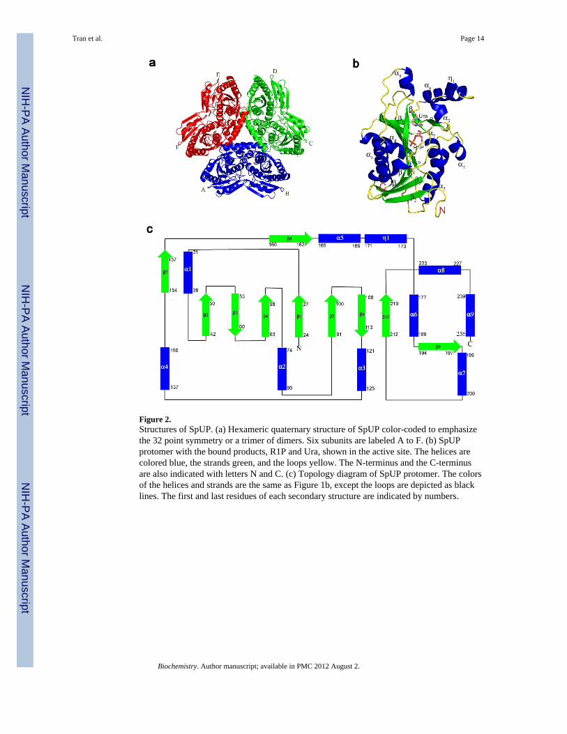
Figure 2.Structures of SpUP. (a) Hexameric quaternary structure of SpUP color-coded to emphasizethe 32 point symmetry or a trimer of dimers. Six subunits are labeled A to F. (b) SpUPprotomer with the bound products, R1P and Ura, shown in the active site. The helices arecolored blue, the strands green, and the loops yellow. The N-terminus and the C-terminusare also indicated with letters N and C. (c) Topology diagram of SpUP protomer. The colorsof the helices and strands are the same as Figure 1b, except the loops are depicted as blacklines. The first and last residues of each secondary structure are indicated by numbers.
Tran et al. Page 14
Biochemistry. Author manuscript; available in PMC 2012 August 2.
NIH
-PA Author Manuscript
NIH
-PA Author Manuscript
NIH
-PA Author Manuscript
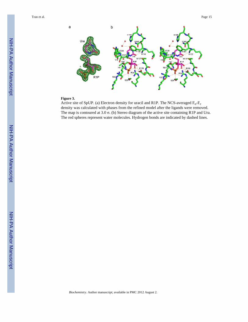
Figure 3.Active site of SpUP. (a) Electron density for uracil and R1P. The NCS-averaged Fo-Fcdensity was calculated with phases from the refined model after the ligands were removed.The map is contoured at 3.0 σ. (b) Stereo diagram of the active site containing R1P and Ura.The red spheres represent water molecules. Hydrogen bonds are indicated by dashed lines.
Tran et al. Page 15
Biochemistry. Author manuscript; available in PMC 2012 August 2.
NIH
-PA Author Manuscript
NIH
-PA Author Manuscript
NIH
-PA Author Manuscript

Figure 4.Superposition of the active sites of SpUP (green) and EcUP (magenta) in stereo. The spheresare water molecules found in the active site.
Tran et al. Page 16
Biochemistry. Author manuscript; available in PMC 2012 August 2.
NIH
-PA Author Manuscript
NIH
-PA Author Manuscript
NIH
-PA Author Manuscript
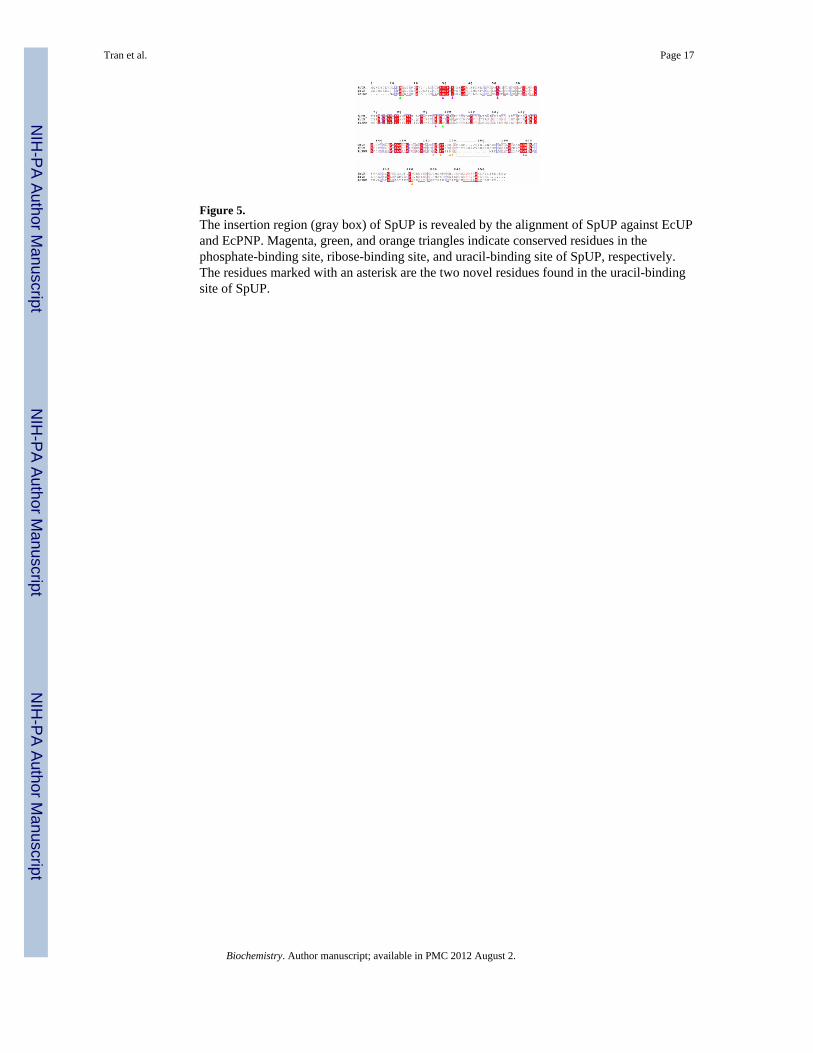
Figure 5.The insertion region (gray box) of SpUP is revealed by the alignment of SpUP against EcUPand EcPNP. Magenta, green, and orange triangles indicate conserved residues in thephosphate-binding site, ribose-binding site, and uracil-binding site of SpUP, respectively.The residues marked with an asterisk are the two novel residues found in the uracil-bindingsite of SpUP.
Tran et al. Page 17
Biochemistry. Author manuscript; available in PMC 2012 August 2.
NIH
-PA Author Manuscript
NIH
-PA Author Manuscript
NIH
-PA Author Manuscript

Figure 6.Active sites of SpUP with uridine and phosphate manually positioned in the active site (a),EcPNP/inosine/PO4 complex (b), EcMTAN/5′-methylthiotubercidin (c), and EcAMN/formycin 5′-monophosphate (d), respectively (14, 31–33). ‘W’ stands for water. Residuesmarked with an asterisk come from an adjacent monomer.
Tran et al. Page 18
Biochemistry. Author manuscript; available in PMC 2012 August 2.
NIH
-PA Author Manuscript
NIH
-PA Author Manuscript
NIH
-PA Author Manuscript

Figure 7.Transition state stabilization of high energy intermediates of the phosphorolysis reaction inEcUP (a) and SpUP (b) by a continuum of electrostatic and hydrogen bond interactions asillustrated by the dotted line.
Tran et al. Page 19
Biochemistry. Author manuscript; available in PMC 2012 August 2.
NIH
-PA Author Manuscript
NIH
-PA Author Manuscript
NIH
-PA Author Manuscript
NIH
-PA Author Manuscript
NIH
-PA Author Manuscript
NIH
-PA Author Manuscript
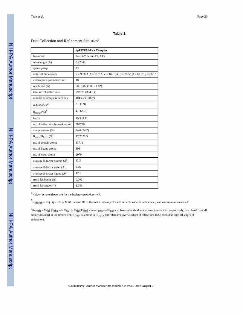
Tran et al. Page 20
Table 1
Data Collection and Refinement Statisticsa
SpUP/R1P/Ura Complex
beamline 24-ID-C, NE-CAT, APS
wavelength (Å) 0.97849
space group P1
unit cell dimensions a = 90.0 Å, b = 91.7 Å, c = 169.3 Å, α = 78.5°, β = 82.3°, γ = 60.1°
chains per asymmetric unit 18
resolution (Å) 50 - 1.82 (1.85 - 1.82)
total no. of reflections 793731 (30451)
number of unique reflections 404352 (16027)
redundancya 2.0 (1.9)
Rmerge (%)b 4.9 (20.5)
I/σ(I) 19.3 (4.1)
no. of reflections in working set 383726
completeness (%) 94.6 (74.7)
Rwork/ Rfreeb (%) 17.7/ 20.3
no. of protein atoms 33711
no. of ligand atoms 396
no. of water atoms 2979
average B-factor protein (Å2) 13.3
average B-factor water (Å2) 23.6
average B-factor ligand (Å2) 17.1
rmsd for bonds (Å) 0.005
rmsd for angles (°) 1.283
aValues in parentheses are for the highest-resolution shell.
bRmerge = ΣΣi | Ii – <I> | / Σ <I>, where <I> is the mean intensity of the N reflections with intensities Ii and common indices h,k,l.
cRwork = Σhkl| |Fobs| – k |Fcal| | / Σhkl |Fobs| where Fobs and Fcal are observed and calculated structure factors, respectively, calculated over all
reflections used in the refinement. Rfree, is similar to Rwork but calculated over a subset of reflections (5%) excluded from all stages ofrefinement.
Biochemistry. Author manuscript; available in PMC 2012 August 2.

NIH
-PA Author Manuscript
NIH
-PA Author Manuscript
NIH
-PA Author Manuscript
Tran et al. Page 21
Table 2
SpUP substrate specificity
substrate cleavage activity (nmol/mg/h)
uridine 485,000
deoxyuridine 673,000
thymidine 352,000
cytidine < 10,000
deoxycytidine < 10,000
adenosine < 10,000
guanosine < 10,000
inosine < 10,000
Biochemistry. Author manuscript; available in PMC 2012 August 2.

NIH
-PA Author Manuscript
NIH
-PA Author Manuscript
NIH
-PA Author Manuscript
Tran et al. Page 22
Table 3
Uridine phosphorolysis assay results for SpUP and mutants
SpUP uridine cleavage activities (nmol/mg/h)
wild-type 1,500,000
K162A < 20
H169A 112,000
H169D 9,000
H169N 42,000
V220D 550
V220E 3,400
Biochemistry. Author manuscript; available in PMC 2012 August 2.

NIH
-PA Author Manuscript
NIH
-PA Author Manuscript
NIH
-PA Author Manuscript
Tran et al. Page 23
Table 4
Steady State Kinetic Parameters for SpUP and EcUPa
Enzyme kcat (s−1) Km (mM) kcat/Km (mM−1 s−1)
SpUP 15 ± 3 0.158 ± 0.025 95 ± 24
EcUP 23 ± 1 0.036 ± 0.004 639 ± 76
SpUP-H169A 1.7 ± 0.2 0.171 ± 0.005 9.9 ± 1
SpUP-K162Ab - - N/A
aThe concentration of uridine was varied and the production of uracil was monitored.
bNo activity was observed at the level of detection of the assay.
Biochemistry. Author manuscript; available in PMC 2012 August 2.