Embed Size (px)
Citation preview
p s sapplications and materials science
a
statu
s
soli
di
www.pss-a.comph
ysi
ca
REPRINT
The Cd(1–x)
Mn(x)
In2S
4(0.5 ≤ x ≤ 1.0) spinel system:
an X-ray powder diffraction study
G. E. Delgado1
, L. Betancourt2
, V. Sagredo2
, and M. C. Morón3
1
Laboratorio de Cristalografía, Departamento de Química, Facultad de Ciencias,
Universidad de Los Andes, Mérida 5101, Venezuela
2
Laboratorio de Magnetismo, Departamento de Física, Facultad de Ciencias, Universidad de Los Andes,
Mérida 5101, Venezuela
3
Instituto de Ciencia de Materiales de Aragón, C.S.I.C. – Universidad de Zaragoza, 50009, Zaragoza,
Spain
Received 19 February 2006, revised 22 June 2006, accepted 10 July 2006
Published online 1 December 2006
PACS 61.10.Nz, 61.66.Fn, 75.50.Lk, 75.50.Pp
Compositions of the Cd(1 – x)
Mn(x)
In2S
4system (0.5 ≤ x ≤ 1.0) were synthesized by the melt and annealing
technique and grown by the chemical vapor transport method. The magnetic cation distributions have
been estimated by X-ray powder diffraction structure refinements using the Rietveld method. These alloys
form a solid solution in all the range of compositions and crystallize with cubic symmetry in the space
group Fd 3m. All phases show a spinel structure with a random arrangement of cations.
phys. stat. sol. (a) 203, No. 15, 3627–3632 (2006) / DOI 10.1002/pssa.200622332
phys. stat. sol. (a) 203, No. 15, 3627–3632 (2006) / DOI 10.1002/pssa.200622332
© 2006 WILEY-VCH Verlag GmbH & Co. KGaA, Weinheim
Original
Paper
The Cd(1–x)Mn(x)In2S4 (0.5 ≤ x ≤ 1.0) spinel system:
an X-ray powder diffraction study
G. E. Delgado*, 1, L. Betancourt2, V. Sagredo2, and M. C. Morón3
1 Laboratorio de Cristalografía, Departamento de Química, Facultad de Ciencias,
Universidad de Los Andes, Mérida 5101, Venezuela 2 Laboratorio de Magnetismo, Departamento de Física, Facultad de Ciencias, Universidad de Los Andes,
Mérida 5101, Venezuela 3 Instituto de Ciencia de Materiales de Aragón, C.S.I.C. – Universidad de Zaragoza, 50009, Zaragoza,
Spain
Received 19 February 2006, revised 22 June 2006, accepted 10 July 2006
Published online 1 December 2006
PACS 61.10.Nz, 61.66.Fn, 75.50.Lk, 75.50.Pp
Compositions of the Cd(1 – x)Mn(x)In2S4 system (0.5 ≤ x ≤ 1.0) were synthesized by the melt and annealing
technique and grown by the chemical vapor transport method. The magnetic cation distributions have
been estimated by X-ray powder diffraction structure refinements using the Rietveld method. These alloys
form a solid solution in all the range of compositions and crystallize with cubic symmetry in the space
group Fd 3m. All phases show a spinel structure with a random arrangement of cations.
© 2006 WILEY-VCH Verlag GmbH & Co. KGaA, Weinheim
1 Introduction
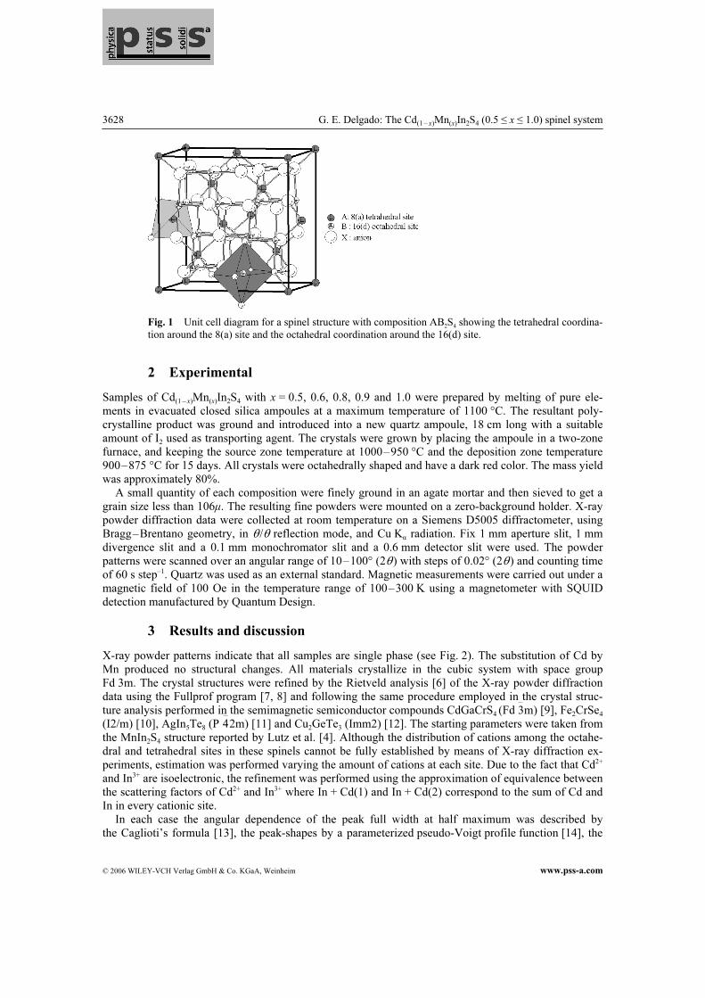
Magnetic semiconducting compounds with spinel structure are of continuing interest from theoretical and experimental point of view because these materials can exhibit spin-glass behavior [1, 2]. Spin-glass behavior has been found in magnetic diluted A spinels as a consequence of topological frustration. The spinel-type structure for AB2X4 compounds is ideally described as based on cubic close-packed large anions with smaller cations occupying tetrahedral and octahedral sites. This arrangement is shown in Fig. 1. They may be described by the (A1–iBi
)tetr(AiB2–i)
octX4 structural formula, where A and B are the cations, X the anion and i the inversion parameter. Depending of i value they can be classified as normal (i = 0) or inverse (i = 1) spinels. As the cations show different site preferences, by choosing the appropriate atoms it is possible to performe a selective magnetic dilution in one of the two sublattices. The magnetic system Cd(1 – x)Mn(x)In2S4 is a particular example of that dilution. The parent phases of this system are the sulphides CdIn2S4 and MnIn2S4. They both belong to the spinel family, crystallizing with partially in-verted spinel structure. The unit cell dimensions reported are: for CdIn2S4, a = 10.843(1) Å [3] and for MnIn2S4, a = 10.720(1) Å [4]. Although the optical properties of Cd(1 – x)Mn(x)In2S4 have been studied in some detail [5], the literature contains scarce information on magnetic and crystal diffraction investi-gations. This paper details the X-ray powder diffraction characterization of the Cd(1 – x)Mn(x)In2S4 system (0.5 ≤ x ≤ 1.0), where the diamagnetic Cd2+ cations are substituted by the magnetic Mn2+ cations.
* Corresponding author: e-mail: [email protected], Phone: +58 274 2401372, Fax: +58 274 2401286
3628 G. E. Delgado: The Cd(1 – x)Mn(x)In2S4 (0.5 ≤ x ≤ 1.0) spinel system
© 2006 WILEY-VCH Verlag GmbH & Co. KGaA, Weinheim www.pss-a.com
Fig. 1 Unit cell diagram for a spinel structure with composition AB2S4 showing the tetrahedral coordina-
tion around the 8(a) site and the octahedral coordination around the 16(d) site.
2 Experimental
Samples of Cd(1 – x)Mn(x)In2S4 with x = 0.5, 0.6, 0.8, 0.9 and 1.0 were prepared by melting of pure ele-ments in evacuated closed silica ampoules at a maximum temperature of 1100 °C. The resultant poly-crystalline product was ground and introduced into a new quartz ampoule, 18 cm long with a suitable amount of I2 used as transporting agent. The crystals were grown by placing the ampoule in a two-zone furnace, and keeping the source zone temperature at 1000–950 °C and the deposition zone temperature 900–875 °C for 15 days. All crystals were octahedrally shaped and have a dark red color. The mass yield was approximately 80%. A small quantity of each composition were finely ground in an agate mortar and then sieved to get a grain size less than 106µ. The resulting fine powders were mounted on a zero-background holder. X-ray powder diffraction data were collected at room temperature on a Siemens D5005 diffractometer, using Bragg–Brentano geometry, in θ /θ reflection mode, and Cu Kα radiation. Fix 1 mm aperture slit, 1 mm divergence slit and a 0.1 mm monochromator slit and a 0.6 mm detector slit were used. The powder patterns were scanned over an angular range of 10–100° (2θ) with steps of 0.02° (2θ) and counting time of 60 s step–1. Quartz was used as an external standard. Magnetic measurements were carried out under a magnetic field of 100 Oe in the temperature range of 100–300 K using a magnetometer with SQUID detection manufactured by Quantum Design.
3 Results and discussion
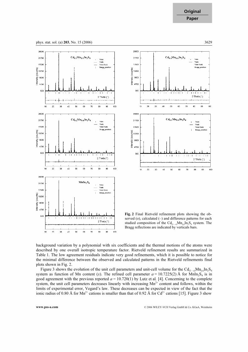
X-ray powder patterns indicate that all samples are single phase (see Fig. 2). The substitution of Cd by Mn produced no structural changes. All materials crystallize in the cubic system with space group Fd 3m. The crystal structures were refined by the Rietveld analysis [6] of the X-ray powder diffraction data using the Fullprof program [7, 8] and following the same procedure employed in the crystal struc-ture analysis performed in the semimagnetic semiconductor compounds CdGaCrS4 (Fd 3m) [9], Fe2CrSe4
(I2/m) [10], AgIn5Te8 (P 42m) [11] and Cu2GeTe3 (Imm2) [12]. The starting parameters were taken from the MnIn2S4 structure reported by Lutz et al. [4]. Although the distribution of cations among the octahe-dral and tetrahedral sites in these spinels cannot be fully established by means of X-ray diffraction ex-periments, estimation was performed varying the amount of cations at each site. Due to the fact that Cd2+ and In3+ are isoelectronic, the refinement was performed using the approximation of equivalence between the scattering factors of Cd2+ and In3+ where In + Cd(1) and In + Cd(2) correspond to the sum of Cd and In in every cationic site. In each case the angular dependence of the peak full width at half maximum was described by the Caglioti’s formula [13], the peak-shapes by a parameterized pseudo-Voigt profile function [14], the
phys. stat. sol. (a) 203, No. 15 (2006) 3629
www.pss-a.com © 2006 WILEY-VCH Verlag GmbH & Co. KGaA, Weinheim
Original
Paper
background variation by a polynomial with six coefficients and the thermal motions of the atoms were described by one overall isotropic temperature factor. Rietveld refinement results are summarized in Table 1. The low agreement residuals indicate very good refinements, which it is possible to notice for the minimal difference between the observed and calculated patterns in the Rietveld refinements final plots shown in Fig. 2. Figure 3 shows the evolution of the unit cell parameters and unit-cell volume for the Cd(1 – x)Mn(x)In2S4 system as function of Mn content (x). The refined cell parameter a = 10.7225(2) Å for MnIn2S4 is in good agreement with the previous reported a = 10.720(1) by Lutz et al. [4]. Concerning to the complete system, the unit cell parameters decreases linearly with increasing Mn2+ content and follows, within the limits of experimental error, Vegard’s law. These decreases can be expected in view of the fact that the ionic radius of 0.80 Å for Mn2+ cations is smaller than that of 0.92 Å for Cd2+ cations [15]. Figure 3 show
Fig. 2 Final Rietveld refinement plots showing the ob-
served (o), calculated (–) and difference patterns for each
studied composition of the Cd(1 – x)Mn(x)In2S4 system. The
Bragg reflections are indicated by verticals bars.
3630 G. E. Delgado: The Cd(1 – x)Mn(x)In2S4 (0.5 ≤ x ≤ 1.0) spinel system
© 2006 WILEY-VCH Verlag GmbH & Co. KGaA, Weinheim www.pss-a.com
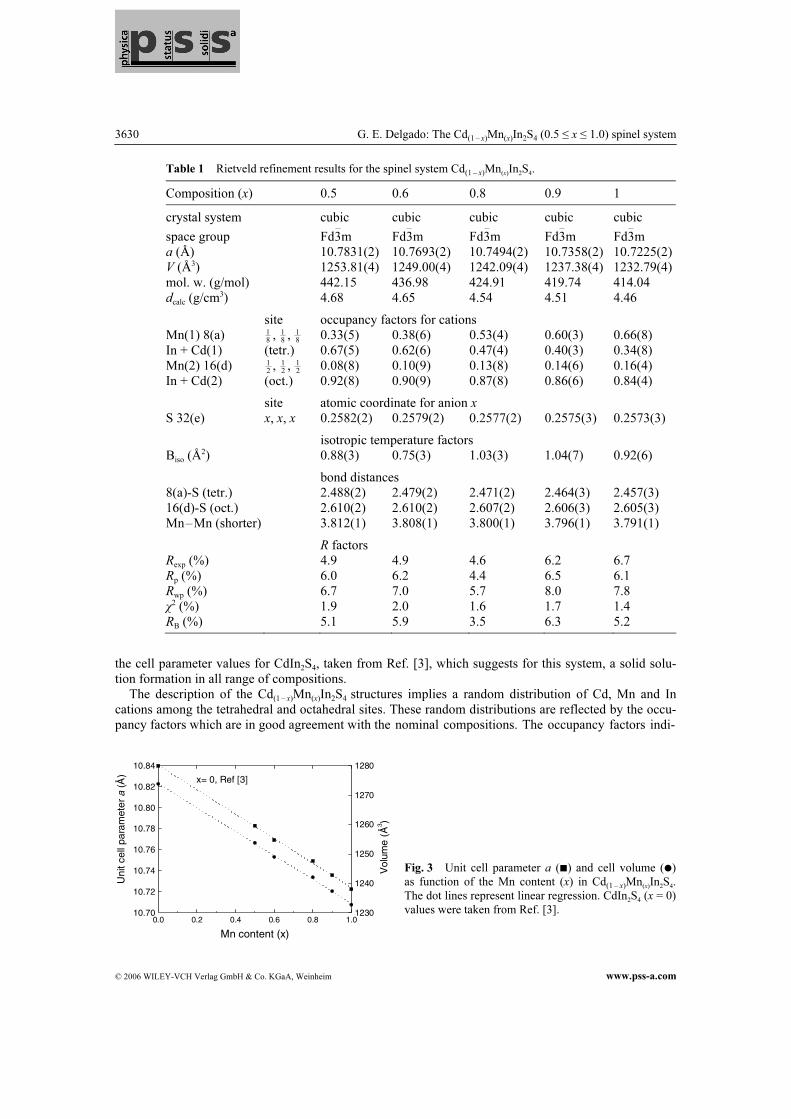
Table 1 Rietveld refinement results for the spinel system Cd(1 – x)Mn(x)In2S4.
Composition (x) 0.5 0.6 0.8 0.9 1
crystal system cubic cubic cubic cubic cubic
space group Fd3m Fd3m Fd3m Fd3m Fd3m a (Å) 10.7831(2) 10.7693(2) 10.7494(2) 10.7358(2) 10.7225(2) V (Å3) 1253.81(4) 1249.00(4) 1242.09(4) 1237.38(4) 1232.79(4) mol. w. (g/mol) 442.15 436.98 424.91 419.74 414.04 dcalc (g/cm3) 4.68 4.65 4.54 4.51 4.46
site occupancy factors for cations Mn(1) 8(a) 1
8, 1
8, 1
8 0.33(5) 0.38(6) 0.53(4) 0.60(3) 0.66(8)
In + Cd(1) (tetr.) 0.67(5) 0.62(6) 0.47(4) 0.40(3) 0.34(8) Mn(2) 16(d) 1
2, 1
2, 1
2 0.08(8) 0.10(9) 0.13(8) 0.14(6) 0.16(4)
In + Cd(2) (oct.) 0.92(8) 0.90(9) 0.87(8) 0.86(6) 0.84(4)
site atomic coordinate for anion x S 32(e) x, x, x 0.2582(2) 0.2579(2) 0.2577(2) 0.2575(3) 0.2573(3)
isotropic temperature factors Biso (Å
2) 0.88(3) 0.75(3) 1.03(3) 1.04(7) 0.92(6)
bond distances 8(a)-S (tetr.) 2.488(2) 2.479(2) 2.471(2) 2.464(3) 2.457(3) 16(d)-S (oct.) 2.610(2) 2.610(2) 2.607(2) 2.606(3) 2.605(3) Mn–Mn (shorter) 3.812(1) 3.808(1) 3.800(1) 3.796(1) 3.791(1)
R factors Rexp (%) 4.9 4.9 4.6 6.2 6.7 Rp (%) 6.0 6.2 4.4 6.5 6.1 Rwp (%) 6.7 7.0 5.7 8.0 7.8 χ2 (%) 1.9 2.0 1.6 1.7 1.4 RB (%) 5.1 5.9 3.5 6.3 5.2
the cell parameter values for CdIn2S4, taken from Ref. [3], which suggests for this system, a solid solu-tion formation in all range of compositions. The description of the Cd(1 – x)Mn(x)In2S4 structures implies a random distribution of Cd, Mn and In cations among the tetrahedral and octahedral sites. These random distributions are reflected by the occu-pancy factors which are in good agreement with the nominal compositions. The occupancy factors indi-
0.0 0.2 0.4 0.6 0.8 1.010.70
10.72
10.74
10.76
10.78
10.80
10.82
10.84
x= 0, Ref [3]
Mn content (x)
Uni
tcel
lpar
amet
era
(Å)
1230
1240
1250
1260
1270
1280
Vol
ume
(Å3 )
Fig. 3 Unit cell parameter a (�) and cell volume (�)
as function of the Mn content (x) in Cd(1 – x)Mn(x)In2S4.
The dot lines represent linear regression. CdIn2S4 (x = 0)
values were taken from Ref. [3].
phys. stat. sol. (a) 203, No. 15 (2006) 3631
www.pss-a.com © 2006 WILEY-VCH Verlag GmbH & Co. KGaA, Weinheim
Original
Paper
0.5 0.6 0.7 0.8 0.9 1.02.420
2.440
2.460
2.480
2.500
Mn content (x)
8(a
)-S
dist
ance
(Å)
2.600
2.605
2.610
2.615
2.620
16(d
)-S
dist
ance
(Å
0.1
0.3
0.5
0.7
0 0.2 0.4 0.6 0.8 1
Occ
upa
ncy
fact
ors
ofM
n
Mn content (x) cate the molecular formulas: Mn0.49(Cd,In)2.51S4, Mn0.58(Cd,In)2.42S4, Mn0.79(Cd,In)2.21S4, Mn0.88(Cd,In)2.12S4 and Mn0.98In2.02S4 for the phases with x = 0.5, 0.6, 0.8, 0.9 and 1, respectively. The deviation of the anions from the ideal x = 1
4 position is in each case around 0.008 Å, which repre-
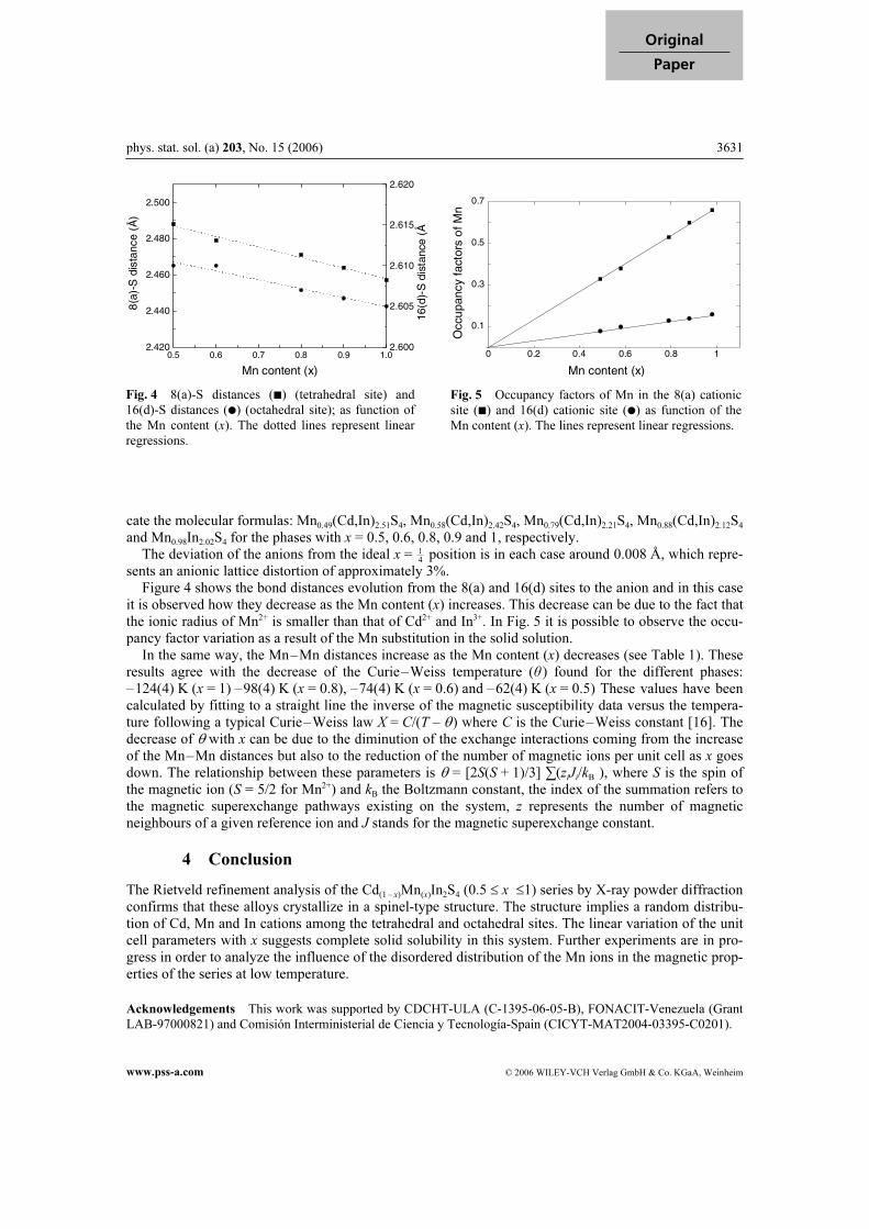
sents an anionic lattice distortion of approximately 3%. Figure 4 shows the bond distances evolution from the 8(a) and 16(d) sites to the anion and in this case it is observed how they decrease as the Mn content (x) increases. This decrease can be due to the fact that the ionic radius of Mn2+ is smaller than that of Cd2+ and In3+. In Fig. 5 it is possible to observe the occu-pancy factor variation as a result of the Mn substitution in the solid solution. In the same way, the Mn–Mn distances increase as the Mn content (x) decreases (see Table 1). These results agree with the decrease of the Curie–Weiss temperature (θ) found for the different phases: –124(4) K (x = 1) –98(4) K (x = 0.8), –74(4) K (x = 0.6) and –62(4) K (x = 0.5) These values have been calculated by fitting to a straight line the inverse of the magnetic susceptibility data versus the tempera-ture following a typical Curie–Weiss law X = C/(T – θ) where C is the Curie–Weiss constant [16]. The decrease of θ with x can be due to the diminution of the exchange interactions coming from the increase of the Mn–Mn distances but also to the reduction of the number of magnetic ions per unit cell as x goes down. The relationship between these parameters is θ = [2S(S + 1)/3] ∑(z
iJi/kB ), where S is the spin of
the magnetic ion (S = 5/2 for Mn2+) and kB the Boltzmann constant, the index of the summation refers to the magnetic superexchange pathways existing on the system, z represents the number of magnetic neighbours of a given reference ion and J stands for the magnetic superexchange constant.
4 Conclusion
The Rietveld refinement analysis of the Cd(1 – x)Mn(x)In2S4 (0.5 ≤ x ≤1) series by X-ray powder diffraction confirms that these alloys crystallize in a spinel-type structure. The structure implies a random distribu-tion of Cd, Mn and In cations among the tetrahedral and octahedral sites. The linear variation of the unit cell parameters with x suggests complete solid solubility in this system. Further experiments are in pro-gress in order to analyze the influence of the disordered distribution of the Mn ions in the magnetic prop-erties of the series at low temperature.
Acknowledgements This work was supported by CDCHT-ULA (C-1395-06-05-B), FONACIT-Venezuela (Grant
LAB-97000821) and Comisión Interministerial de Ciencia y Tecnología-Spain (CICYT-MAT2004-03395-C0201).
Fig. 4 8(a)-S distances (�) (tetrahedral site) and
16(d)-S distances (�) (octahedral site); as function of
the Mn content (x). The dotted lines represent linear
regressions.
Fig. 5 Occupancy factors of Mn in the 8(a) cationic
site (�) and 16(d) cationic site (�) as function of the
Mn content (x). The lines represent linear regressions.
3632 G. E. Delgado: The Cd(1 – x)Mn(x)In2S4 (0.5 ≤ x ≤ 1.0) spinel system
© 2006 WILEY-VCH Verlag GmbH & Co. KGaA, Weinheim www.pss-a.com
References
[1] G. F. Goya, H. R. Rechenberg, and V. Sagredo, J. Magn. Magn. Matter. 226–230, 1298 (2001).
[2] E. Agostinelli, P. Filaci, D. Fiorani, and A. M. Tesla, J. Phys. C 8, Supp. 12, 1061 (1988).
[3] H. Haeuseler, J. Solid State Chem. 29, 121 (1979).
[4] H. D. Lutz and M. Jung, Z. Anorg. Allg. Chem. 579, 57 (1989).
[5] L. Betancourt, V. Sagredo, G. E. Delgado, and C. Rincón, Rev. Mex. Fis., in press (2006).
[6] H. M. Rietveld, J. Appl. Cryst. 2, 65 (1969).
[7] J. Rodriguez-Carvajal, Fullprof (version 3.20, Feb. 2005), Laboratoire Léon Brillouin (CEA-CNRS), France.
[8] T. Roisnel and J. Rodriguez-Carvajal, Mater. Sci. Forum 378–381, 118 (2001).
[9] G. E. Delgado, A. J. Mora, L. Betancourt, and V. Sagredo, phys. stat. sol. (a) 199(3), 373 (2003).
[10] G. E. Delgado and V. Sagredo, phys. stat. sol. (a) 201(3), 421 (2004).
[11] A. J. Mora, G. E. Delgado, C. Pineda, and T. Tinoco, phys. stat. sol. (a) 201(7), 1477 (2004).
[12] G. E. Delgado, A. J. Mora, M. Pirela, M. A. Villarreal, A. Velásquez, and B. J. Fernández, phys. stat. sol. (a)
201(12), 2900 (2004).
[13] G. Cagliotti, A. Paoletti, and F. P. Ricci, Nucl. Instrum. 3, 223 (1958).
[14] P. Thompson, D. E. Cox, and J. B. Hastings, J. Appl. Crystallogr. 20, 79 (1987).
[15] S. D. Shannon, Acta Crystallogr. A 32, 751 (1976).
[16] R. L. Carlin, Magnetochemistry (Springer-Verlag, New York, 1986).
![Stress deformations and structural quenching in charge-ordered Sm[sub 0.5]Ca[sub 0.5]MnO[sub 3] thin films](https://img.dokumen.tips/doc/110x75/635a01f49e39cba4b605eefd/stress-deformations-and-structural-quenching-in-charge-ordered-smsub-05casub.jpg)


![Microstructure, phase transition, and electrical properties of (K[sub 0.5]Na[sub 0.5])[sub 1−x]Li[sub x](Nb[sub 1−y]Ta[sub y])O[sub 3] lead-free piezoelectric ceramics](https://img.dokumen.tips/doc/110x75/6336572db5f91cb18a0bc82e/microstructure-phase-transition-and-electrical-properties-of-ksub-05nasub.jpg)


