Embed Size (px)
Citation preview
1
1st NYU BIOMEDICAL & BIOSYSTEMS CONFERENCE
APRIL 9-11, 2017
ABU DHABI, UNITED ARAB EMIRATES
2 3
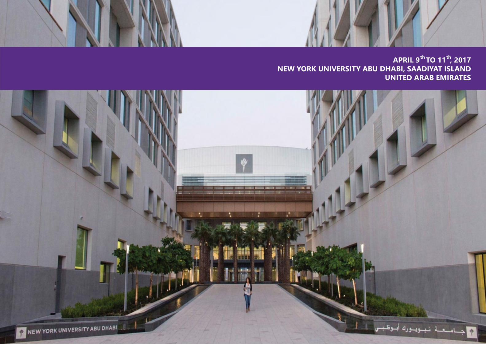
APRIL 9 TO 11 , 2017NEW YORK UNIVERSITY ABU DHABI, SAADIYAT ISLAND
UNITED ARAB EMIRATES
th th
4 5
CONFERENCE ORGANIZERS
CHAIRS COMMITTEE MEMBERS
CONFERENCE ORGANIZERSADMINISTRATION AND VOLUNTEERS
MOHAMMAD QASAIMEH ALESHA CASTILLO ALI TRABOLSI DIPESH CHAUDHURY EMILIE DRESSAIRE
JIN MONTCLARE KALLE LEVON MARY COWMAN MAZIN MAGZOUB
RAFAEL SONG RICHARD THORSEN SACHIN KHAPLI SOHMYUNG HA
VITTORIA FLAMINI YOUSSEF IDAGHDOUR
SUNIL KUMAR WEIQIANG CHEN
AARON WATSON TAMARA GJORGJIEVA
6 7
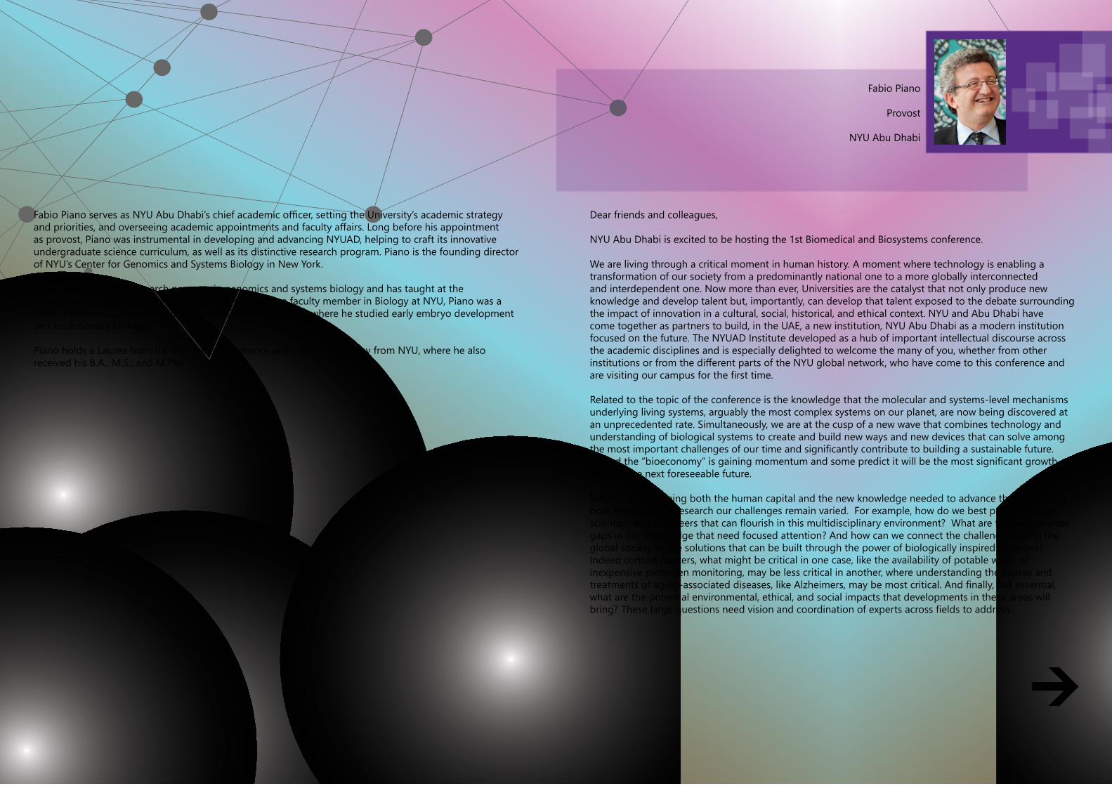
Fabio Piano serves as NYU Abu Dhabi’s chief academic officer, setting the University’s academic strategy and priorities, and overseeing academic appointments and faculty affairs. Long before his appointment as provost, Piano was instrumental in developing and advancing NYUAD, helping to craft its innovative undergraduate science curriculum, as well as its distinctive research program. Piano is the founding director of NYU’s Center for Genomics and Systems Biology in New York.
Piano leads an active research program in genomics and systems biology and has taught at the undergraduate and graduate levels. Prior to serving as a faculty member in Biology at NYU, Piano was a Damon Runyon Cancer Research Fellow at Cornell University, where he studied early embryo development and evolutionary biology.
Piano holds a Laurea from the University of Florence and a Ph.D. in Biology from NYU, where he also received his B.A., M.S., and M.Phil.
Dear friends and colleagues,
NYU Abu Dhabi is excited to be hosting the 1st Biomedical and Biosystems conference.
We are living through a critical moment in human history. A moment where technology is enabling a transformation of our society from a predominantly national one to a more globally interconnected and interdependent one. Now more than ever, Universities are the catalyst that not only produce new knowledge and develop talent but, importantly, can develop that talent exposed to the debate surrounding the impact of innovation in a cultural, social, historical, and ethical context. NYU and Abu Dhabi have come together as partners to build, in the UAE, a new institution, NYU Abu Dhabi as a modern institution focused on the future. The NYUAD Institute developed as a hub of important intellectual discourse across the academic disciplines and is especially delighted to welcome the many of you, whether from other institutions or from the different parts of the NYU global network, who have come to this conference and are visiting our campus for the first time.
Related to the topic of the conference is the knowledge that the molecular and systems-level mechanisms underlying living systems, arguably the most complex systems on our planet, are now being discovered at an unprecedented rate. Simultaneously, we are at the cusp of a new wave that combines technology and understanding of biological systems to create and build new ways and new devices that can solve among the most important challenges of our time and significantly contribute to building a sustainable future. Indeed the “bioeconomy” is gaining momentum and some predict it will be the most significant growth area for the next foreseeable future.
NYUAD is developing both the human capital and the new knowledge needed to advance these areas. In both teaching and research our challenges remain varied. For example, how do we best prepare future scientists and engineers that can flourish in this multidisciplinary environment? What are the fundamental gaps in our knowledge that need focused attention? And how can we connect the challenges facing the global society to the solutions that can be built through the power of biologically inspired solutions? Indeed context matters, what might be critical in one case, like the availability of potable water or inexpensive pathogen monitoring, may be less critical in another, where understanding the causes and treatments of aging-associated diseases, like Alzheimers, may be most critical. And finally, but essential, what are the potential environmental, ethical, and social impacts that developments in these areas will bring? These large questions need vision and coordination of experts across fields to address.
Fabio Piano
Provost
NYU Abu Dhabi
8 9
Three features of NYUAD make it especially well positioned to not only host this inaugural conference but to use its discussions to inspire us to build on this interdisciplinary field. One, NYUAD is an intellectual hub connected to all of NYU, the largest private university in the US, with deep roots across disciplines that include the sciences, medicine, engineering, social sciences, humanities and the arts. Indeed, over the last few years over 200 of our distinguished New York based colleagues have joined the now more than 175 highly selected residential faculty and over 160 full time researchers here in Abu Dhabi to work and teach the over 1000 undergraduate and over 70 graduate students, resulting in a vibrant scholarly environment. The result has been remarkable, for example our undergraduate college attracts, retain, and graduates among the most talented students in the world coming from over 110 countries and speaking more than 100 languages. Indeed, to name only two examples, among out first graduates, eight have received the prestigious Rhodes Scholarship, more than doubling our historical record, and one of our graduates has already become a minister, the youngest of any government.
Second, we are young, agile, and able to build in ideal ways facing the future without needing to dismantle the past. Within NYUAD, our organizational structure is designed to prevent barriers and engineers and scientists, to cite two examples, work in a co-located open environment and the undergraduate students start out in the same classes.
Finally, we are in and of Abu Dhabi, geographically located in one of the most increasingly important regions of the world. We are at a crossroads of cultures and peoples that are welcomed to contribute to the global society being developed in the UAE. The support, wisdom and innovative sprit of the Abu Dhabi Government have helped propel our progress.
So I welcome you to exchange openly your most visionary ideas and to also think of how we can help shape this discipline for a future that will require new approaches, new thinking, new training, in the hope that NYUAD can become one of the models on how to build the ideal foundation for the future in this field.
Fabio PianoProvostNYU Abu Dhabi
Fabio Piano
(continued)
10 11
Samer M Madanat is the Dean of Engineering at NYU Abu Dhabi, Xenel Distinguished Professor of Engineering and former Chair of the Department of Civil & Environmental Engineering (from 2012 to 2015) at the University of California at Berkeley. From 2005 to 2014, he served as the Director of the Institute of Transportation Studies. Prior to that, he served as Director of the PATH program at UC Berkeley. He received a B.Sc. in Civil Engineering from the University of Jordan in 1986, and a M.S and Ph.D. in Transportation Systems from MIT in 1988 and 1991, respectively.
He is Editor of the Journal of Transport Policy and Associate Editor of the European Journal of Transportation and Logistics. He is also a member of the Editorial Boards of Transportation Research Part C, Transportation Research Part D, and Computer-Aided Civil and Infrastructure Engineering.
Dear Participants in the 1st NYU Biomedical and Biosystems Conference,
On behalf of my NYUAD Engineering colleagues, I am pleased to welcome you to our campus, and to the first NYU Biomedical and Biosystems Conference.
Bio-Innovation is one of the five strategic research themes for NYUAD, with major activity in both Sciences and Engineering. In the context of the Engineering strategic plan, Bioengineering is one of our emerging research clusters.
There is increasing demand for Bioengineering majors worldwide. Nationally, healthcare systems represent one of the six national priorities of the United Arab Emirates’ Vision 2021, is recognized as a key focus sector by the UAE National Innovation Strategy, and signify an important component of the Economic Vision 2030 of the Emirate of Abu Dhabi. Technologies in medicine and healthcare systems clearly represent a vital component of the vision of the UAE and the emirate of Abu Dhabi.
Our vision for the future of Bioengineering at NYUAD includes a new Bioengineering Major, and a research center focused on Bioengineering and Bio-Innovation, located on our campus with participation from faculty in Engineering, Biology, Chemistry, Public Health, Economics, and others. This proposed center would engage in collaborative research with NYU’s Tandon School of Engineering and the NYU Medical School, thus taking advantage of the vast array of talent in the NYU Global Network.
I wish a productive Conference and, for those visiting us from outside Abu Dhabi, an enjoyable stay in our beautiful city.
Samer Michel MadanatDean of Engineering, NYUADGlobal Network Professor of Engineering, NYU Tandon School of Engineering
Samer Michel Madanat
Dean of Engineering, NYUAD
Global Network Professor of Engineering
12 13
Katepalli R. Sreenivasan is Dean and President of the NYU Tandon School of Engineering, the Eugene Kleiner Professor of Innovation in Mechanical Engineering (School of Engineering), and Professor of Physics (Faculty of Arts and Science) and Mathematics (Courant Institute of Mathematical Sciences). He also holds an affiliate appointment in the department of biomaterials at the NYU College of Dentistry.
Dr. Sreenivasan came to NYU from the International Centre for Theoretical Physics (Trieste, Italy) where he was Director and Abdus Salam Professor, and the University of Maryland, where he was Distinguished University Professor and Professor of Physics, the Glenn Martin Professor of Engineering, and Director of the Institute for Physical Science and Technology. Prior to that he was the Harold W. Cheel Professor of Mechanical Engineering at Yale University, and held joint appointments in applied physics, physics, and mathematics. Dr. Sreenivasan is a member of the National Academy of Sciences and the National Academy of Engineering, and is a Fellow of the American Academy of Arts and Sciences. An active researcher, Dr. Sreenivasan’s research is in the areas of turbulence, cryogenic helium, and nonlinear dynamics.
Bioengineering has been successful in bridging quantitative and analytical skills with the medical and biological areas---for example, in the design of equipment, instruments and surgical tools, in providing scientific understanding of central biological processes, etc. The need for strong bioengineering is becoming more acute in the 21st century because biological advances are accelerating continually and a lot of that basic research has to translate to the practice of medicine and health care. We are well on our way to making a reasonably complete description of the genetic code of human life; we might know the genetic predisposition of a person for a particular disease; we will get a better understanding of the human mind and the relation between network configurations of the brain and functional aspects such as intentions, talents and propensities. Bioengineering will be deeply engaged in this seemingly limitless transformation. In particular, advancing human health is of paramount importance because larger and larger fraction of people are aging, want to live in cities where the population density is high, and depend on technology for maintaining and enhancing the quality of their lives. We need teams of researchers who have mastered the relevant technologies, on the one hand, and are skilled at interpreting the latest scientific discoveries, on the other, while evolving and bringing the beneficial technologies to market.
These goals prompted us to start the bioengineering institute in Tandon. In particular, the existence of a first-rate Medical School in NYU, and other schools such as Dentistry, Public Health and Nursing has been a great motivator. Quite aside from research opportunities, we are very keen to provide students an education that is vital to system design, scientific creativity and communications, and some understanding of how transformational research works all the way to the market. NYU’s global campuses in Abu Dhabi and Shanghai have only enriched the scope of our efforts: they have added different types of research emergencies to our slate of possibilities. This is the context for our excitement about this meeting in Abu Dhabi. It brings together a number of experts from within NYU, and without, to discuss frontier aspects in bioengineering and neighboring fields. I am thrilled to be part of it, and greatly look forward to the meeting itself.
Katepalli SreenivasanDean, NYU Tandon School of EngineeringUniversity Professor and Eugene Kleiner Chair for Innovation
Katepalli Sreenivasan
Dean, NYU Tandon School of Engineering
University Professor and Eugene Kleiner Chair for Innovation

14 15
Sunil Kumar is a Global Professor of Engineering at New York University (NYU) and Vice Provost for Graduate and Postdoctoral Programs at NYU Abu Dhabi. Previously, from 2009 until 2015, he was the Dean of Engineering at NYU Abu Dhabi. Kumar’s research interests are in the areas of radiation and optics, laser-material interactions, imaging, and applied mathematics. He received a PhD in mechanical engineering from the University of California at Berkeley, master’s degrees in mechanical engineering and mathematics from the State University of New York at Buffalo, and an undergraduate degree in mechanical engineering from the Indian Institute of Technology at Kharagpur, India.
Dear Conference Speakers and Participants,
On behalf of the organizing committee, I welcome you to the 1st NYU BioMedical and BioSystems Conference, and thank you for taking the time from your busy schedules to present your research and engage in scientific discussions.
Advances in the diagnosis and treatment of disease increasingly require a holistic integration of technology with science and clinical practice. It is the goal of the Biomedical and Biosystems Conference to bring together engineers, scientists, and medical practitioners from the different schools of New York University’s Abu Dhabi and New York campuses, from universities across the world, from regional government agencies, and from hospitals to discuss this exciting biomedical frontier. Addresses by world-renowned pioneers will also present first-hand narratives from around the globe.
This is the first in a series of annual conferences with the same title planned to address topics at the transdisciplinary interface of engineering, sciences, and medicine, spanning the spectrum from fundamental research to application and translation into practice.
I trust that you will be stimulated by the presentations by investigators from different disciplines who are creating new conceptual, theoretical, methodological, and translational frameworks and innovations beyond discipline-specific approaches.
I hope that you will also participate in next year’s conference, and I will look forward to welcoming you again to our campus in Abu Dhabi.
Sunil KumarCo-Chair, NYU Biomedical and Biosystems ConferenceGlobal Professor of Mechanical EngineeringNYU Abu Dhabi and NYU Tandon School of Engineering
Sunil Kumar
Co-Chair, 1 NYU Biomedical and Biosystems Conference
Global Professor of Mechanical Engineering
st
16 17
TABLE OF CONTENT
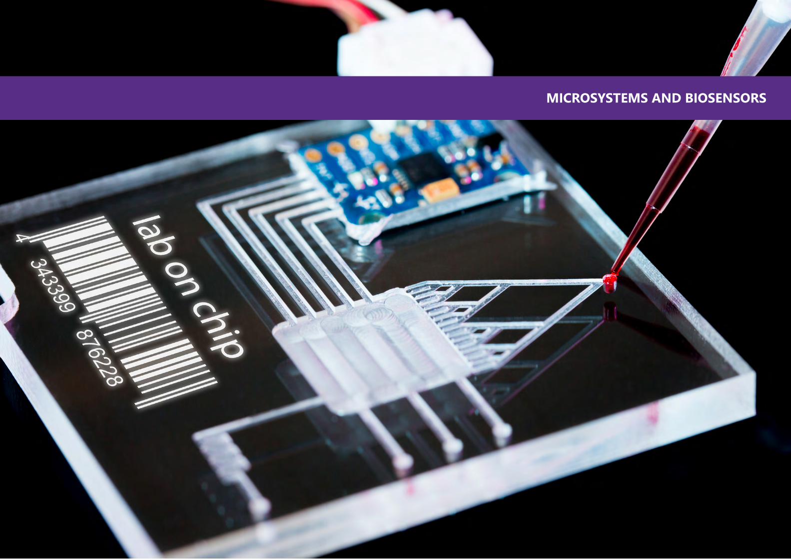
MICROSYSTEMS AND BIOSENSORS
Chwee Teck LimMicrofluidic Technologies for Cancer Diagnosis & Personalized Medicine
Rohit KarnikLabel-Free Sorting and Analysis of Cells Mediated by Weak Molecular Interactions in Microfluidic Devices
Chia-Hung ChenSingle Cell Analysis for Precision Medicine by Using Continuous Flow Microfluidics
Mohammed ZourobRapid and Low-cost biosensors for Pathogens Detection
Ryan L. HartmanChemical Reaction Engineering New Tools for Discovery and Development in Flow
Ruba KhnoufMicrofluidics Based Immunoassays: Advances and Challenges
Thomas Gervais Transport in capillary and open microfluidics: from theory to life science applications
Kalle LevonExtended Gate FET array with a Coating of Organic Electronics for Monitoring Enzymatic Reaction
28
30
32
34
36
38
40
42
Mohamed Abdelgawad Microfluidic Tools for Assisted Reproduction
Abraham Mansouri Streaming Current Magnetic Fields In A Charged Nanopore
Anastasios Hantzakos State of the Art Laryngeal Imaging: Current and future Trends
John T. W. Yeow Nanodevices for Biomedical Instruments
Ala’aldeen Al-HalhouliRecent Advances in Microfluidics at the NanoLab
Khaled Mohamed Al-Aribe Optical Lab-on-a-chip Via Biological Proton Pumps
Rafael (Yong-Ak) SongAccelerating the Mass Transport of DNA Molecules onto Morpholino Microarray by a Nanofluidic Concentrator Chip for Enhanced Surface Hybridization
44
46
48
50
52
54
56
18 19
TABLE OF CONTENT
Katsuo Kurabayashi Motor Protein Micro Powering, Molecular Sorting, and Biosensing
Suzanne GaudetThe Quieting Role Of Caspases In Myoblast Differentiation
Piergiorgio Percipalle Actin In Genome Organization
Jeroen Adema Unravelling Single Cell functional Genomics – Study the Behavior of Cells At The Single-cell Level
Shady AminPhytoplankton-bacteria Relationships: How Microscale Interactions Influence the Macroscale Environment
George Shubeita Moving In A Crowd: Motors, Forces, And Disease
Wen-Biao Gan In Vivo Imaging Of Synaptic Structural Plasticity And Stability In The Cortex
Susanna NarkilahtiNeuronal Tissue Engineering- From Cells To Grafts
60
62
64
66
68
70
72
74
Jeremy Teo Choon Meng Non-Invasive Image-Based Tool For Single-Cell Biomechanical Evaluation
Matteo Chiesa Dependence Of Surface Aging On DNA Topography
Kourosh Salehi-Ashtiani Intracellular Spectral Recompositioning Of Light: A Design-Based Approach To Increase Photosynthetic Efficiency In Diatoms
Florian Roser The Neurosurgeons Perspective On Biomedical Engineering
Sohmyung HaA 3x3x0.3 mm Fully Integrated Modular Electrocorticographic Microsystem
Hasan Al-Nashash Cognitive Vigilance Assessment And Enhancement
Kartik Sreenivasan Using Neural Oscillations To Decode Human Cognition
Dipesh Chaudhury Neural Circuits Linking Circadian Rhythms, Sleep and Mood Disorders
76
78
80
82
84
86
88
90
CELL BIOLOGY, BIOENGINEERING, AND NEUROENGINEERING
3
20 21
TABLE OF CONTENT
Andrew D. Hamilton Synthetic Mimics of Protein Secondary Structure And Function
Youssef Zaim Wadghiri Nanobiomaterials in Preclinical Imaging And Diagnostic Radiology: MRI A Versatile Imaging Modality
Ghaleb Husseini Drug Delivery And Ultrasound
Seiichi Yamano The Potential Of Non-Viral Gene Transfer For Clinical Applications
Rihab Nasr Anti-Tumor Efficacy Of Arsenic/Interferon In Preclinical Models Of Chronic Myeloid Leukemia Resistant To Tyrosine Kinase Inhibitors
Mazin Magzoub Hexokinase II-Derived Cell-Penetrating Peptide Targets Mitochondria And Triggers Apoptosis In Cancer Cells
94
96
98
100
102
104
DRUG DELIVERY AND DISCOVERY
Kevin E. HealyOrgans On A Chip: The Future Of Precision Medicine?
Kalle Levon Well-Being Of Cells In 3D Nano-Environment
Richard A. BlackModelling Blood And Lymphatic Vessels In Vitro: Challenges And Opportunities
Amitabha Chattopadhyay A Membrane Cholesterol-Based Strategy To Tackle Entry Of Intracellular Pathogens: Evading Drug Resistance
Yaser E. Greish Gastric Stem Loaded Biodegradable Microfibers For The Regeneration Of Gastric Mucosa
Panče NaumovThe Mystery Of Firefly Bioluminescence
Laising Yen Building Designer RNA Nanostructures
Nikhil Gupta Mechanical Characterization Of Hierarchical Biomaterial Across Length Scales And Strain Rates
108
110
112
114
116
118
120
122
BIOMATERIALS AND TISSUE ENGINEERING
22 23
TABLE OF CONTENT
Will Wenmiao Shu 3D Bioprinting Of Human Pluripotent Stem Cells And Hydrogels For Tissue Engineering
Ryo JimboInfluential Factors For Bone Loss During Implant Surgical Procedures. Interaction Between Instrumentation And Geometry. How Can Science Solve A Clinical Problem?
Sachin Khapli Hierarchically Porous Calcium Carbonate Scaffolds For Bone Tissue Engineering
Mohamed Al-Sayegh The Linkage Role Of Actin Cytoskeleton And Adipocyte Differentiation: A Molecular Scaffold Modifier Potential
Bryan A. Chin Rapid And Direct Detection Of Pathogens On Fresh Produce
Pengyu Chen Unravelling Intercellular Communication Using Nanoplasmon Ruler
124
126
128
130
132
134
Anwarul HasanPhotocrosslinked Hydrogels And Electrospun Scaffolds In Tissue Engineering And Regenerative Medicine
Chuanju Liu Targeting Progranulin To Treat OA & RA
136
138
BIOMECHANICS AND MECHANOBIOLOGY
Edward Guo Muscle-Like “Beating” Osteocytes Under Loading Enhance Vesicle Release To Mediate Bone Formation
Joo H. Kim State Estimation For Balance Stability: Towards Dynamic Walking Of Wearable Robots
Asimina Kazakidi Computational Hemodynamics Research Across The Extremes Of Age
Huseyin C. YalcinMechanobiology Of Cardiovascular Diseases
Vittoria Flamini Computational Methods For Patient-Specific Decision-Making And Medical Device Design Optimization
142
144
146
148
150
24 25
TABLE OF CONTENT
Sylvie Coupaud Characterization Of Patterns Of Disuse-Related Osteoporosis
Weiqiang Chen Microfluidic Vascularized Microsystem For Probing Inflammation-Biased Angiogenesis
152
154
TRANSLATIONAL MEDICINE
Joel S. Schuman Optical Coherence Tomography
Francesco SerinoIn Vivo, Non-Invasive, Vascular Tissue Engineering Is Possible: The Grail Project Achievements
Mary K. CowmanHyaluronan And Hyaluronan Fragments: Diagnostic And Therapeutic Use
Matija SnuderlDissecting Genetic And Epigenetic Heterogeneity In Cancer Using Dielectrophoretic Capture And Digital Sorting Of Single Cells
158
160
162
164
Thorsten KirschA Multidisciplinary Translational Research Approach To Improve Cartilage Repair
John DoyleNew Methods Of Respiratory Monitoring: Embracing Technical and Clinical Challenges
Mohammad A. QasaimehMicrofluidics For Clinical Applications: Isolating Circulating Tumor Cells From Peripheral Blood Samples
Index
166
168
170
172
26 27
MICROSYSTEMS AND BIOSENSORS

28 29
MICROSYSTEMS AND BIOSENSORS
Professor
Biomedical Engineering
Mechanobiology Institute, National University of Singapore
Professor Lim is a Provost’s Chair Professor at the Department of Biomedical Engineering as well as a Principal Investigator at the Mechanobiology Institute at the National University of Singapore. His research interests include the development of nano/micro technologies for disease detection, diagnosis and therapy. He has authored more than 310 peer-reviewed papers and delivered more than 300 plenary/keynote/invited talks. He is an elected Fellow of the American Institute for Medical and Biological Engineering as well as International Academy of Medical and Biological Engineering. He is also an elected Council Member of the World Council of Biomechanics. He currently sits on the editorial boards of more than 12 international journals.
Prof Lim has co-founded five startups which are commercializing technologies developed in his lab. He and his team have garnered more than 60 research awards and honors including the International Precision Medicine Conference Prize (2017), Asian Scientist 100 (2016), University’s Outstanding Researcher Award & Outstanding Innovator (2014), Credit Suisse Technopreneur of the Year Award and Wall Street Journal Asian Innovation Award (Gold) (2012), TechVenture Most Disruptive Innovation Award and Asian Entrepreneurship Award (First Prize) (2012), President’s Technology Award (2011) and the IES Prestigious Engineering Achievement Award (2010, 2016).
BIO
Chwee Teck Lim
The presence of Circulating Tumor Cells (CTCs) in bloodstream of patients with epithelial cancers is an important intermediate step in cancer metastasis and can provide valuable insights into disease detection, staging and personalized treatment. As compared to obtaining a tissue biopsy which is invasive and painful, ‘‘liquid biopsy’’ for CTCs detection can be easily performed via a routine blood draw. The presence and number of CTCs in peripheral blood has been associated with the severity of the disease and have potential use for early detection, diagnosis, prognosis and treatment monitoring purposes. The isolation of CTCs using microfluidics is attractive as the flow conditions can be accurately manipulated to achieve an efficient separation. Here, we demonstrate several effective separation methods by utilizing the unique differences in the mechanical properties such as size and deformability of cancer cells from that of blood cells. By exploiting the fluid dynamics in specially designed microfluidic channels, CTCs which are generally stiffer and larger can be physically separated from the more deformable blood constituents. Using this label-free approach, we are able to retrieve viable CTCs that are not only suitable for downstream molecular analysis such as genetic or RNA sequencing, but also for expansion and culture. With blood specimens from cancer patients, we confirmed successful detection, isolation and retrieval of CTCs. Identification of CTCs via their mechanical signatures will not only aid in the determination of malignancy and disease, but also enable personalized treatment by the possible detection of any actionable mutation unique to individual patient. The microfluidic technology has since been commercialized and being used in the USA, Europe and Asia.
MICROFLUIDIC TECHNOLOGIES FOR CANCER DIAGNOSIS AND PERSONALIZED MEDICINE
ABSTRACT

30 31
Associate Professor
Mechanical Engineering
Massachusetts Institute of Technology
Rohit Karnik is Associate Professor of Mechanical Engineering at the Massachusetts Institute of Technology, where he leads the Microfluidics and Nanofluidics Research Group. His research focuses on the physics of micro- and nanofluidic flows and design of micro- and nanofluidic devices for applications in healthcare, energy systems, and bioseparation and analysis. He obtained his B. Tech. degree from the Indian Institute of Technology at Bombay in 2002, and his PhD from the University of California at Berkeley in 2006 under the guidance of Prof. Arun Majumdar. After postdoctoral work with Prof. Robert Langer at MIT, he joined the Department of Mechanical Engineering at MIT in 2007. Among other honors, he is a recipient of the Institute Silver Medal (IIT Bombay, 2002), NSF Career Award (2010), Keenan Award for Innovation in Undergraduate Education (2011), DOE Early Career Award (2012), and IIT Bombay Young Alumnus Achiever Award (2014).
BIO
Rohit Karnik
LABEL-FREE SORTING AND ANALYSIS OF CELLS MEDIATED BY WEAK MOLECULAR INTERACTIONS IN MICROFLUIDIC DEVICES
Multiple sample-processing steps present a challenge for the development of low-complexity devices for laboratory or point-of-care separation and analysis of cells. In this talk, I will discuss a new approach that can directly separate, enrich, or analyze cells with minimal or no sample processing requirements. We show that transient cell-surface adhesive molecular interactions can exert forces on the n cell rolling, a physiological phenomenon involved in cell trafficking where transient molecular bonds are continuously formed and broken as the cell rolls on a surface under the action of hydrodynamic forces. Using this approach, we demonstrate separation of cells with high purity and efficiency in parallel microchannel devices, and direct separation of neutrophils from blood with ultrahigh enrichment in a neutrophil activation-dependent manner. We extend this approach to controllably contact mesenchymal stem cells with receptor-coated surfaces to quantify cell adhesion behavior by visualization of their trajectories in a “cell adhesion cytometer”, which can track changes in the cell phenotype. The results demonstrate the potential of the emerging technology of using transient cell-surface molecular interactions to directly separate and analyze cells for point-of-care diagnostics, isolation of rare cells, quality control of stem cells, and other applications.
ABSTRACT

32 33
Assistant Professor
Biomedical Engineering
National University of Singapore
Chia-Hung Chen is an Assistant Professor in Biomedical Engineering at the National University of Singapore. His current research is focused on developing a continuous flow microfluidic device as a functional flow-cytometer for applications in systems biology, drug screening, bio-fabrication, clinical detections and precision medicine. As a principal investigator at NUS, Chia-Hung has successfully obtained external (government/industry) funding to support my research team, with ~3M USD of research expenditures over ~5 years, and his lab has delivered research outcomes. Moreover, Chia-Hung has effectively interacted with a diverse community of students and faculty to address challenges in device development and healthcare to fabricate novel products. Given his expertise in systems engineering and fluidic devices, he has collaborated with bioinformatics researchers and clinicians at the National University Hospital of Singapore (NUHS) and Massachusetts General Hospital (MGH) to develop diagnostic tools for use in translational medicine and a real-time monitoring system for individual therapeutics.
BIO
Chia-Hung Chen
SINGLE CELL ANALYSIS FOR PRECISION MEDICINE BY USING CONTINUOUS FLOW MICROFLUIDICS
Precision medicine refers to giving the right therapeutics, to the right patient, at the right time. In the context of cancer, successful implementation of precision medicine, requires treatment individualization not only taking into account patient and tumor factors, but also tumor heterogeneity and tumor evolution over time. In this study, a continuous flow microfluidic device was developed as a functional flow cytometer (micro-FACS) to detect secreted multiplexed protease activities at single cell resolution. The individual cells from patient samples are encapsulated within water-in-oil droplets for single cell multiplexed protease assay. We modified FRET (fluorescence resonance energy transfer)-based substrates to accommodate different fluorescent pairs with distinct excitation and emission wavelengths to obtain multiple signals from droplets containing single cells. Four substrate-protease reactions in a droplet were simultaneously monitored at three distinct pairs of fluorescent excitation (UV: 400nm, B: 470nm, G: 546nm, R: 635nm) and emission (B: 520nm, G: 580nm, R: 670nm) wavelengths. To infer a quantitative profile of multiple proteolytic activities from single cells, we applied the computational method Proteolytic Activity Matrix Analysis (PrAMA). The capability to determine multiple protease activities at single cell resolution has the potential to characterize tumor progress of individual patients.
ABSTRACT
34 35
Professor
Chemistry
Alfaisal University
BIO Mohammed Zourob got his Ph.D from the Department of Instrumentation and Analytical Science (DIAS) at the University of Manchester in 2003. He did a postdoctoral scientist at DIAS, working in chemical/biosensors and lab-on-a-chip for biomedical and environmental applications. Then he moved to the Department of Materials Science, University of Manchester, to work on developing high-throughput screening platforms for “Omics” applications. At the end of 2005, he moved to the Institute of Biotechnology, University of Cambridge, where his research focused on optical sensing and biomemetic materials.
Dr. Zourob headed the biosensors division at Biophage Inc, a biotech company based in Montreal. In 2009 he joined GDG Environment Ltd as Director of R&D. In 2010 he joint INRS-University of Quebec as associate professor and then he moved to Cranfield University-UK. Now Dr Zourob is holding professor of Biosensors at Alfaisal University-KSA. Prof. Zourob now is leading the Biosensors BioMEMS and Bionanotechnology Lab (BBBL). Dr. Zourob has published many scientific papers in peer-reviewed journals, more than thirteen book chapters, and thirteen patents. He edited 6 books in chemical/biosensors, microarray sand lab on a chip. His current theme of research focusing on developing; 1) novel diagnostic tools; 2) highly specific recognition receptors for diagnostic and imaging applications; and 3) lab on a chip systems for sample processing.
Mohammed Zourob
RAPID AND LOW-COST BIOSENSORS FOR PATHOGENS DETECTION
In recent years, outbreaks of infectious diseases have widely spread and grown as a public health problem. Traditional and current detection techniques of pathogens are time-consuming and require expensive instrumentation and are labor intensive. To conquer this limitation, a number of novel, ultra-rapid colorimetric biosensors were developed to detect the presence or absence of bacteria and viruses rapidly and at low-cost. The designed colorimetric biosensing platforms used paper, cotton sabs and plastic substrates. This technology was applied for the detection of various bacteria and viruses such as E. Coli, Salmonella, Listeria, pseudomonas, staphylococcus aureus, bacillus anthracis and human papilloma viruses, adenoviruses and noroviruses. The developed biosensor demonstrated tremendous sensitivity and applicability with a lower limit of detection of 10 CFU mL for bacteria in real food and body fluids samples in less than a minute analysis time. In conclusion, this approach permits the use of a disposable biosensor chips that can be mass-produced at low-cost and can also be used by regulatory agencies for better control potential health risks associated with pathogens contamination.
ABSTRACT
-1
36 37
Assistant Professor
Chemical and Biomolecular Engineering
NYU Tandon School of Engineering
Ryan L. Hartman is Assistant Professor and Faculty Engineer in Residence in the Department of Chemical and Biomolecular Engineering at New York University. Prof. Hartman completed his postdoctoral research in the Department of Chemical Engineering at the Massachusetts Institute of Technology (Cambridge), his Ph.D. in Chemical Engineering from the University of Michigan (Ann Arbor), and his B.S. in Chemical Engineering from Michigan Technological University (Houghton). He is the Catalysis and Reaction Engineering Programming Chair of the American Institute of Chemical Engineers. He was recently honored as Visiting Assistant Professor of the Institute of Condensed Matter Chemistry of Bordeaux (ICMCB) CNRS. Previously, Hartman was Assistant Professor and Reichhold-Shumaker Fellow in the Department of Chemical and Biological Engineering at The University of Alabama (Tuscaloosa). He is also a winner of the NSF CAREER Award and member of the National Academy of Inventors. Hartman returned to academia following his private sector career with Schlumberger Limited.
BIO
Ryan L. Hartman
Engineering novel tools for the discovery of science, and translation of the new knowledge from the laboratory to application are societal challenges. Our laboratory helps to address these challenges by applying catalysis and reaction engineering (CRE) principles. By applying CRE principles, we are able to design novel reactors and catalysts for the discovery and the development of chemical methods in flow. Performing chemical reactions in flow (or sometimes referred to as “flow chemistry” in the synthetic chemistry community) has the potential to reduce the amount of chemical waste generated, minimize the building space and energy requirements, expedite information, yield more accurate predictive mathematical models, and enable safer handling of hazardous compounds during scientific discovery, development, and manufacture. This so called “process intensification” has merit to revolutionize the way we currently discover, develop, and manufacture fine chemicals, materials, natural products, and pharmaceuticals that have global markets. This brief overview will summarize our research on flow chemistry with microsystems for i) green chemical reaction engineering for kinetics discovery and sustainable manufacturing and ii) novel laboratory-scale reactors for the online discovery of multiphase reactions. Finally, emerging trends in catalysis and reaction engineering will be highlighted.
CHEMICAL REACTION ENGINEERING NEW TOOLS FOR DISCOVERY AND DEVELOPMENT IN FLOW
ABSTRACT

38 39
Microfluidic devices have been investigated as tools for the analysis of nucleic acids, proteins, cells, and tissues, but they have yet to replace traditional, bench-top laboratory techniques and equipment. One of those heavily used techniques for protein analysis is the immunoassay; western blotting and Enzyme Linked Immunoassay (ELISA) being the most common. This talk will describe the advances and advantages of using microfluidic devices in immunoassays, it will address methods of the enhancement of the sensitivity and limit of detection of the assay, and reducing assay time, cost and effort required to implement them.
Particularly this talk will discuss binding methods of antibodies to the surface of microfluidic devices to reduce signal to noise ratio, using electrokinetics in the preconcentration of the analytes to reduce the duration of the reaction and to automate sample injection, and finally using gold nanoparticles in the improvement of electrochemical immunoassays through signal amplification.
40 41
Assistant Professor
Engineering Physics and Biomedical Engineering
Polytechnique Montreal
Thomas Gervais is assistant professor of engineering physics and biomedical engineering at Polytechnique Montréal since 2013. His research focuses on fundamental fluid mechanics and mass transfer in microstructures, with an emphasis on developing miniature systems for the interrogation of cancer tissues. He holds a bachelor degree in engineering physics from Polytechnique Montreal and a Ph.D. in bioengineering from MIT. Beyond research and teaching, he is also a seasoned populariser of science, having written over 100 popular science articles, 35 short TV documentaries, and appeared in over 75 episodes of various French Canadian Science TV shows in the past decade. He also works as consultant in the field of scientific communications (pro bono) and to solve challenging transport problems in the natural resources and high tech industries.
BIO
Thomas Gervais
In the past decade, microfluidics has grown to take new, freer, more application-driven forms. These include, capillary-driven, open microfluidics, and hanging drop microfluidic systems. Understanding fundamental fluid mechanics and transport phenomena in these new systems gives rise to new functionalities not hitherto possible with conventional channel networks. In this talk, we will discuss how fluid mechanics and mass transfer modelling plays a key role in the design, optimization, and operation of several emerging microfluidic-based technologies. We will provide examples of theory-guided technological development from our laboratory and from our collaborators in the field of on-chip 3D tissue culture, microfluidic probes, and capillary-driven microfluidics.
TRANSPORT IN CAPILLARY AND OPEN MICROFLUIDICS: FROM THEORY TO LIFE SCIENCE APPLICATIONS
ABSTRACT

42 43
Professor
Chemical and Biomolecular Engineering
NYU Tandon School of Engineering
Kalle Levon, professor and entrepreneur, specialist on charged surfaces and surface interactions with applications in monitoring biological binding events with functionalized transistors and building nanoscale 3D scaffolding for the well-being of cells.
NIH Center for Scientific Review: Biomaterials and Biointerfaces 2007- 2011
Honorary member of the Editorial Board: International Journal of Nanomedicine
BIO
Kalle Levon
Our custom disposable platform exploits ion-sensitive FET (ISFET) technology. Via simple surface modifications the design allows a broad range of analytes to be tested with low cost. We have compared our read-out device to a commercial potentiometer using K as an example species analyte. The developed sensing system has a slightly better limit of detection and is notably less susceptible to external noise, which is commonly observed with potentiometers. The designed platform is fabricated using standard electronic processes with gold surface and we used commercial discrete transistors as the transducing element. It can be mass produced with high yield and low cost. To circumvent the drift that typically occurs with modified solid state electrodes we incorporated a transducing layer between the electric conductor (gold pad) and the ionically conducting ion-selective membrane. The polyaniline doped with dinonylnaphtalene sulfonic acid (PANI- DNNSA) was used as a transducing layer for the first time. The PANI-DNNSA layer significantly reduces the drift of the electrodes compared to a configuration without the transducing layer. In addition, it allows convenient organic chemistry for the immobilization of antibodies or enzymes for monitoring biological binding events. Such a hand-held sensing system using a transistor based multiplexed platform allows coupling the electrochemical information wirelessly to a smartphone.
EXTENDED GATE FET ARRAY WITH A COATING OF ORGANIC ELECTRONICS FOR MONITORING ENZYMATIC REACTION
ABSTRACT
+

44 45
Assistant Professor
Mechanical Engineering
Assiut University
Dr. Mohamed Abdelgawad received his B.Sc. in mechanical engineering from Assiut University, Egypt, in 1998, M.Sc from Concordia University, Canada, in 2003, and PhD from University of Toronto, Canada, in 2009. After receiving his PhD, Dr. Abdelgawad worked as a post-doctoral fellow with the department of Surgical Oncology at Princess Margaret Hospital in Toronto. In 2010, he returned to Egypt and joined Assiut University as an assistant professor with the mechanical engineering department where he established Assiut Microfluidics Lab. Dr. Abdelgawad’s research interests focus on using microfluidics to improve medical and biological applications and studying fundamentals of fluid flow on the microscale. Current projects running in his lab include mechanical characterization of biological cells, studying sperm motion in different environments, nanoparticle synthesis using Microfluidics, and studying physics of droplet motion in digital microfluidic devices.
BIO
Mohamed Abdelgawad
During the past two decades, microfluidics emerged as an important tool in assisted reproduction applications such as in-vitro fertilization, embryo culture, sperm selection, and cryopreservation. In the first part of this talk, I will describe a microfluidic device we developed to characterize rheotaxis (tendency of sperm cells to swim against the flow) as one of the mechanisms that guide spermatozoa to the oocytes by following secretions from the female genital tracts after mating. We studied the effect of flow velocity and channel geometry on sperm motion and how they progress to the oocyte. In the second part of the talk, I will describe a complete digital microfluidic platform for automated vitrification of mammalian embryos. Individual mouse embryos were encapsulated inside micro droplets that were manipulated on a digital microfluidic device to take the embryos through the whole vitrification protocol. This platform enables full automation of the vitrification process including embryo loading and retrieval, generating the required concentrations of the cryoprotectant, and processing the embryo through steps of the protocol. Survival and developmental rates of embryos vitrified on the developed platform were comparable to those of manually vetrified embryos suggesting that digital microfluidics has a great potential for automating vitrification processes.
MICROFLUIDIC TOOLS FOR ASSISTED REPRODUCTION
ABSTRACT

46 47
Associate Professor
Mechanical Engineering
American University in Dubai
Dr. Abraham Mansouri is an associate professor of Mechanical Engineering in American University in Dubai, he joined the Department of Mechanical Engineering in Fall 2011. His research spans a wide range of topics in pure and applied science including interfacial phenomena (contact angle measurements, interpretations and surface characterization), microfluidics, electrokinetics, and energy conversion. His research interests have broadened during his doctoral studies through collaboration with the National Institute of Nanotechnology (NINT) in Edmonton, Canada, and the Institute of Analytical Science in Dortmund, Germany.
Prior to joining AUD, Dr. Mansouri worked in NOVA Chemicals (Calgary – Canada), the largest producer of polymer in North America, as a research engineer in the area of advanced fluid dynamics. He also worked for the HVAC division of Mitsubishi Electric in Vancouver, on various energy modeling and design projects in support of their City Multi VRF heat pump and heat recovery systems. He obtained his PhD from University of Alberta in Canada in micro/nanofluidics.
BIO
Abraham Mansouri
Magnetic fields induced by currents created in pressure driven flows inside a solid-state charged nanopore were modeled by numerically solving a system of steady state continuum partial differential equations, i.e., Poisson, Nernst-Planck, Ampere and Navier-Stokes equations (PNPANS). This analysis was based on non-dimensional transport governing equations that were scaled using Debye length as the characteristic length scale, and applied to a finite length cylindrical nano-channel. The comparison of numerical and analytical studies shows an excellent agreement and verified the magnetic fields density both inside and outside the nanopore. The radially non-uniform currents resulted in highly non-uniform magnetic fields within the nanopore that decay as 1/r outside the nanopore. It is worth noting that for either streaming currents or streaming potential cases, the maximum magnetic field occurred inside the pore in the vicinity of nanopore wall, as opposed to a cylindrical conductor that carries a steady electric current where the maximum magnetic fields occur at the perimeter of conductor. Based on these results, it is suggested and envisaged that non-invasive external magnetic fields readouts generated by streaming/ionic currents may be viewed as secondary electronic signatures of biomolecules to complement and enhance current DNA nanopore sequencing techniques.
STREAMING CURRENT MAGNETIC FIELDS IN A CHARGED NANOPORE
ABSTRACT
48 49
Consultant
Otorhinolaryngology
Cleveland Clinic Abu Dhabi
Anastasios Hantzakos, MD, Ph.D, MHA, FEBORL: Dr. Hantzakos joined Cleveland Clinic Abu Dhabi as a Consultant Otorhinolaryngologist from its opening in 2014. He served as a Consultant at the 1st Department of Otorhinolaryngology – Head & Neck Surgery (AORL) of the National & Kapodistrian University of Athens (NKUA) in Hippocration General Hospital of Athens since 2007. He received his Medical Degree in 1991 from the NKUA. He completed his basic surgical training at Emory University in Atlanta, USA, and received his Specialist Certification in Otorhinolaryngology from the AORL. He received additional training as a Senior Registrar in the East Anglia Deanery Training Scheme in the UK with a special interest in Rhinology and Otology. He received his PhD from the NKUA in 2003 and a Master’s Degree in Health Administration (MHA) at the Hellenic Open University in 2016. He was employed as a Consultant in the Hellenic National Health System since 2004. He has obtained a fellowship in Phonosurgery and Laryngeal Laser Surgery in 2006 from the Department of Otorhinolaryngology – Head & Neck Surgery of the University of Louvain at Mont-Godinne University Hospital in Belgium. Since 2008, he serves as an active member of the Scientific Council of the European Laryngological Society (ELS) and Chairman of the Phonosurgery Committee. He is an examiner and a fellow of the European Board Examinations in Otorhinolaryngology - Head & Neck Surgery since their onset in 2010, and an examiner for Health Authority Abu Dhabi (HAAD) since 2016. He is also a member of the Voice Foundation and the European Society for Swallowing Disorders (ESSD).
Dr. Hantzakos is certified to practice in Greece, UK and Belgium. He has authored numerous papers in peer – reviewed international journals and has written chapters in national and international textbooks and books. He is a member of various study groups and committees within the ELS.
BIO
Anastasios Hantzakos
The purpose of the presentation is to provide a review of the current and latest advances in laryngeal imaging, most specifically videostroboscopy, videokymography and high-speed videoendoscopy, narrow-bad imaging, laser depth-kymography, magnetic resonance imaging, and optical coherence tomography. Videostroboscopy and Videokymography are applications widely available and well recognized for their clinical value. High-speed videoendoscopy has improved the diagnostic accuracy in benign voice disorders. Narrow band imaging and similar software applications have significantly improved the visualization of premalignant laryngeal conditions. Laser Depth-kymography is a 3D display of the vertical movements of the vocal folds during phonation in calibrated spatial values by using a specially designed 3D endoscope. Newest laryngeal magnetic resonance modalities allow for high-resolution imaging of laryngeal tissue microstructure, or measuring of dynamic laryngeal structures during phonation. Optical coherence tomography allows for the capture of dynamic high resolution cross-sectional images of the vibrating vocal fold mucosa during phonation.
STATE OF THE ART LARYNGEAL IMAGING: CURRENT AND FUTURE TRENDS
ABSTRACT

50 51
Professor and Canada Research Chair
Systems Design Engineering
University of Waterloo
John T. W. Yeow received his PhD degree in mechanical and industrial engineering from the University of Toronto. He is currently a Professor and Canada Research Chair in Micro/Nanodevices in the Department of Systems Design Engineering at University of Waterloo, Canada. His current research interests are in the field of developing miniaturized biomedical instruments. He is a recipient of the Professional Engineering Ontario Engineering Excellence Award, Natural Science & Engineering Research Canada Innovation Challenge Award, Douglas R. Colton’s Medal of Research Excellence, Micralyne Microsystems Design Award. He is currently a Canada Research Chair in Micro/Nanodevices. He serves as the Editor-in-Chief of the IEEE Nanotechnology Magazine. He is a Fellow of the Engineering Institute of Canada, and a Member of College of New Scholars, Artists and Scientists of the Royal Society of Canada. He is also a Distinguisher Lecturer of the IEEE Nanotechnology Technical Council.
BIO
John T. W. Yeow
The emergence of minimally invasive diagnostics and therapeutics in modern high-tech medicine has generated an unmet demand in miniaturized biomedical devices. There exist a definite need for clinical diagnostic and treatment instruments that are based on micro and nanotechnologies. In the past decade, micromachining technology and nanomaterials are making big impacts in many fields, especially in the field of biomedical engineering. The small size and low mass provided by micro/nanodevices make medical instruments portable, power efficient, and, in many cases, more effective. This talk will focus on the current development of the state-of-the-art miniaturized X-ray CT machines, endoscopic imaging devices, and MEMS-based ultrasound transducers.
NANODEVICES FOR BIOMEDICAL INSTRUMENTS
ABSTRACT

52 53
Associate Professor
Mechatronics Engineering
German Jordanian Univeristy
Ala’aldeen Al-Halhouli is a Professor at the mechatronics engineering department at the German Jordanian University (GJU). He joined the department in February 2013 and became the Dean of the school of Applied Technical Sciences in Mar. 2016. He obtained a Ph.D. degree from the University of Jordan in 2007 and a habilitation degree with Venia Legendi on microfluidics from the Technische Universität Braunschweig (TU BS)” in Germany in 2013. Between 2007 and 2013, he was working as a research associate and lecturer at Institute of Microtechnology (IMT) of TU BS. During the summer of 2014, he was a visiting scientist at Micro/Nanofluidic BioMEMS group, Massachusetts Institute of Technology (MIT), USA.
Dr. Al-Halhouli has special interest in microfluidic systems for biomedical applications. He published more than 60 papers in international Journals and conference proceedings. He received several Awards such as the GJU distinguished researcher Award and the Cray Award for the best published paper in Microsystem technology, Braunschweig, Germany, 2007.
BIO
Ala’aldeen Al-Halhouli
The demand for compact, inexpensive, disposable, and high throughput devices that can be implemented in various biomedical, biotechnological and chemical fields has led to novel multifunctional microfluidic platforms. The ability of such platforms to manipulate small amounts of fluid (on the scale of microliters and below) in miniaturized channels has significantly decreased the required samples and reagent volumes. This enabled the production of commercial chips and devices that are widely used by non-trained people at low cost. In this talk, the recent advances in microfluidic research at the NanoLab @ GJU including Lab-on-chip and Lab-on-disc for mixing and separation of particles/cells, and micro-sized robots manipulation in microfluidic channels will be presented. Furthermore, it will cover the recent results in the field of multi-stage inertial microfluidics for mixing and separation processes. Finally, the talk will conclude with an outlook that highlights potential development and applications of the presented microfluidic platforms.
As for the Nanolab @ GJU (http://nanolab.gju.edu.jo/), it is a newly established research lab at the German Jordanian University in Jordan that focuses on developing and investigating novel micro- and nano devices, circuits, sensors, and systems for life science, biomedical and energy applications.
RECENT ADVANCES IN MICROFLUIDICS AT THE NANOLAB - GERMAN JORDANIAN UNIVERSITY (GJU)
ABSTRACT
“
54 55
Assistant Professor
Mechanical Engineering
Abu Dhabi University
Khaled Mohamed Al-Aribe received the B.Sc degree in mechanical engineering in 1992, the M.Sc. in 1999 from Garyounis University, Benghazi-Libya and Ph.D. degrees in 2012 from the University of Western Ontario, London, Ontario, Canada. He is an Assistant Professor in the Department of Mechanical Engineering, Abu Dhabi University, Abu Dhabi, United Arab Emirates. In 2015 and 2016, he was a visiting research Professor at the University of Western Ontario, London, Ontario, Canada.
He has recently authored and co-authored several publications including book chapters, journal papers, conference papers and conference presentations. His research activities involve opto-fluidic actuation, photonic materials, bio-materials and optical sensing. This research has resulted in the development of an innovative opto-fluidic actuators. Professor Al-Aribe has acted as a technical reviewer for numerous academic journals, conferences, and granting agencies.
BIO
Khaled Mohamed Al-Aribe
Optically driven transducers in a microchannel provides a mechanism for controlling a variety of biological and chemical processes on Lab-on-a-Chip (LOC). A microscale photoelectrochemical transducer that exploits the photon energy inherent in focused laser beams to generate pH gradients in micro channels is constructed from monolayers of the biologically synthesized bR proton pumps known by bacteriorhodopsin. The bacteriorhodopsin (bR) is a light sensitive protein found in Halobacterium salinarium. In nature these bR molecules employ the sun’s energy to transport hydrogen ions across the cell membrane thereby generating the potential difference necessary for life activities.
The constructed photoelectrochemical transducer is an ultrathin layer (~13nm) of molecularly oriented PM patches self-assembled on an Au-coated porous substrate. The movement of ions across the porous membrane creates a pH gradient that causes the hydroxyethyl methacrylate–acrylic acid (HEMA–AA) gel to swell. Experimental studies show that a 13 nm self-assembled photoelectric layer can generate approximately 1.3 mV/(mWcm ), and pH change of 0.42 when exposed to an 18 mW, 568 nm light source. The small change in pH around the phase transition point pK enabled the hydrogel microactuator to swell by more than 80% of its original volume in less than 85 min.
OPTICAL LAB-ON-A-CHIP VIA BIOLOGICAL PROTON PUMPS
ABSTRACT
2

56 57
Assistant Professor
Division of Engineering - Bioengineering
NYU Abu Dhabi
Rafael Y. Song’s research and teaching interests are interdisciplinary in both mechanical engineering disciplines such as design and manufacturing of MEMS devices, fluid mechanics, and micro/nanofabrication, as well as in biological engineering areas such as BioMEMS for point-of-care diagnostics, neuroprosthetic implants, high-throughput screening and optogenetics. He received his B.S., M.S., and Ph.D. in mechanical engineering from RWTH Aachen University, Germany and worked at the Korea Institute of Science and Technology (KIST) as a Senior Research Scientist. He joined the Micro/Nanofluidic BioMEMS Group in the Department of Electrical Engineering and Computer Science at MIT as a Research Scientist. He held an appointment as a Research Fellow at the Beth Israel Deaconess Medical Center/Harvard Medical School in Boston until he joined NYUAD in September 2012. He currently holds a joint appointment in the Department of Chemical and Biomolecular Engineering at the New York University Tandon School of Engineering.
BIO
Rafael (Yong-Ak) Song
Micro- and Nanotechnology open up new exciting possibilities to manipulate biological entities and detect biological signals at such unprecedented precision that we can gain deeper insights into the complex biological processes and find new approaches to solve biological problems. In this talk, I will present a new microchip technology for enhancing the reaction speed and sensitivity of biosensors such as DNA microarrays. Starting from only a small amount of samples in the range of few microliters for assays, we can reduce the detection time drastically by increasing the concentration of an analyte locally and reducing its diffusion length through electrokinetic preconcentration. Due to its simplicity, the proposed nanofluidic chip platform can easily be extended to other types of biosensors such as Whispering Gallery Mode (WGM) sensors, mass spectrometry and other highly multiplexed biosensors. Once fully developed, the concentrator chip will become an indispensable high-throughput tool for ultrasensitive biosensing as well as for “Liquid Biopsy”.
ACCELERATING THE MASS TRANSPORT OF DNA MOLECULES ONTO MORPHOLINO MICROARRAY BY A NANOFLUIDIC CONCENTRATOR CHIP FOR ENHANCED SURFACE HYBRIDIZATION
ABSTRACT

58 59
CELL BIOLOGY, BIOENGINEERING, AND NEUROENGINEERING

60 61
Professor
Mechanical Engineering, Electrical Engineering and Computer Science
University of Michigan
Katsuo Kurabayashi is Professor of Mechanical Engineering and Electrical Engineering and Computer Science at the University of Michigan, Ann Arbor. He received his BS in Precision Engineering from the University of Tokyo in 1992, and his MS and PhD in Materials Science and Engineering from Stanford University, CA, in 1994 and 1998, respectively. His current research focuses on optofluidics, nanoplasmonic and biomolecular biosensing, and microsystems for immunology, clinical diagnosis, and analytical chemistry. He received the 2001 NSF Early Faculty Career Development (CAREER) Award, and the Robert Caddell Memorial Award in 2005, the Pi Tau Sigma Outstanding Professor Award in 2007, the Mechanical Engineering Outstanding Achievement Award in 2013 from the University of Michigan, and the Ted Kennedy Family Team Excellence Award in 2015 from the College of Engineering at the University of Michigan.
BIO
Katsuo Kurabayashi
Motor proteins are nanometer-scale biomaterials that convert chemical energy stored in ATP into mechanical work. They are responsible for mass transport and cell movement in biological systems. The bionanotechnology research community has been exploring the development of new hybrid devices that incorporate motor proteins in a non-biological engineered material structure. Manipulation of nanomechanical structures using these motors is an excellent example of utilizing life’s natural resources to power man-made systems at high fuel-to-power conversion efficiency. This talk discusses our research on nanometer-scale mechanical power extraction, mass transport, and biosensing in biomolecular motor-integrated micro/nano materials and devices with a fundamental understanding of nanomechanical processes to control their stochastic movements. Expected advantages and technological challenges accompanying the hybrid devices are also discussed.
MOTOR PROTEIN MICRO POWERING, MOLECULAR SORTING, AND BIOSENSING
ABSTRACT

62 63
Assistant Professor
Cancer Biology
Dana-Farber Cancer Institute at Harvard Medical School
Suzanne Gaudet is an assistant professor at the Department of Cancer Biology and the Center for Cancer Systems Biology at the Dana-Farber Cancer Institute and the Department Genetics at Harvard Medical School. Her research focuses on the quantitative understanding of how cells, in particular cancer cells, dynamically respond to cytokine signals from the immune system. She received a B.Sc. in Biology from Université de Montréal and earned a Ph.D. in Biochemistry from Harvard University working with Daniel Branton. She then joined Peter Sorger’s laboratory at MIT as a postdoctoral associate for the launch of an interdisciplinary research collaborative at the interface of biology, informatics and microdevice engineering and from 2003 to 2008, worked as a research scientist and scientific coordinator at the Cell Decision Processes Center at MIT and Harvard. In addition to her scientific endeavors, she is committed to promoting diversity in science through better mentorship and leadership.
BIO
Suzanne Gaudet
The apoptotic caspases are proteases that mediate a programmed cell death and canonically, their activation is a point of no return on the path to apoptosis. However, it has been argued that these caspases play a non-apoptotic role in skeletal muscle differentiation because caspase inhibition reduces myoblast differentiation efficiency. We have found that caspase inhibition during myoblast differentiation leads to survival a large population of cells that would have died had caspases been allowed to activate. Here, I will discuss how, by combining single-cell RNA-seq as well as live- and fixed-cell imaging, we have characterized non-cell autonomous signaling mechanisms by which these surviving cells inhibit the differentiation of neighboring myoblasts.
THE QUIETING ROLE OF CASPASES IN MYOBLAST DIFFERENTIATION
ABSTRACT
64 65
Associate Professor
Division of Science - Biology
NYU Abu Dhabi
Dr. Percipalle has a degree in chemistry and a PhD in Molecular Genetics obtained from the International School for Advanced Studies, Trieste, Italy. As PhD student he trained at the International Centre for Genetic Engineering and Biotechnology (ICGEB), Trieste. For postdoctoral training he worked at the Medical Research Council Laboratory of Molecular Biology, Cambridge, UK, and at the Karolinska Institute, Stockholm, in the Department of Cell and Molecular Biology. Dr. Percipalle joined the same Department as Group Leader and, later on, as Associate Professor of Cell Biology with grants from the Swedish Research Council and Cancer Society (Cancerfonden). His research activities focus on transcriptional and posttranscriptional control of gene expression and their impact on cell fate and identity. In 2015 he moved to New York University Abu Dhabi as Associate Professor of Biology. He currently holds an appointment as Guest Professor in the Department of Molecular Biosciences, The Wenner Gren Institute, Stockholm University.
BIO
Piergiorgio Percipalle
During differentiation and development, cell fate and identity are established by waves of genetic reprogramming. Although the mechanisms are largely unknown, during these events dynamic chromatin reorganization is likely to ensure that genes involved in the same cellular functions are co-regulated depending on the nuclear environment. Here, using high content screening of embryonic fibroblasts from a β-actin knockout mouse, we report major chromatin rearrangements and changes in histone modifications such as methylated H3K9 and methylated H3K4. Transcriptome profiling showed that actin-dependent chromatin reorganization was concomitant with global changes in the pattern of gene expression. Specifically gene programs involved in angiogenesis, cytoskeletal organization and myofibroblast features were up-regulated in β-actin knockouts. Compatible with these observations, β-actin knockout cells acquired both angiogenic features and myofibroblast phenotypes. Some of these features were gained in a β-actin dosage-dependent manner. Importantly, reintroducing an NLS-containing β-actin in the knockout cells affected nuclear features and gene expression. These results suggest that, by primarily affecting the spatial organization of heterochromatin, β-actin has a unique nuclear role that controls global transcriptional changes of genetic programs and plays a role in the determination of cell fate and identity during cell differentiation and development.
ACTIN IN GENOME ORGANIZATION
ABSTRACT

66 67
Field Applications Specialist
Fluidigm Corporation
Jeroen Adema studied at the University of Applied Sciences in Leiden, The Netherlands, specializing in both life science and molecular biology. His thesis work focused on Hantavirus in the Dutch rodent population at the Dutch national institute for public health and the environment (RIVM). After graduation, he continued working at the RIVM for a while on various zoonotic diseases before joining a start-up company called RnAssays developing flow-cytometry based immunoassays for the detection of Salmonella in pig serum and meat drip. Jeroen joined GenDx, a Dutch company specializing in HLA-typing for stem-cell transplantation purposes, and worked on sequencing-based detection methods for HLA. In 2015, Jeroen joined Fluidigm taking a lead role in supporting all genomic products including single-cell solutions in the distribution territory Europe, Middle-East and Africa and focusing on facilitating research studies in various academic settings.
BIO
Jeroen Adema
The Fluidigm Polaris™ system enables researchers to zero in on a particular cell subpopulationa of interest and obtain details on which genes and/or pathways drive these cells. The innovative Polaris™ system is combined with the most complex microfluidic device Fluidigm has developed. This allows users to sort out 48 specific single cells of interest, representing as little as 3% of the total population of input cells. The sorted cells will be placed in a capture chamber containing culture media and are environmentally controlled; temperature, humidity, atmosphere. These individual cells can be grown up to 24 hours and challenged with various different components during this time. After the required incubation time the system will proceed creating cDNA libraries that can be used for preparing single-cell RNA sequencing libraries. We will illustrate the complexity of the instrument and microfluidic device and illustrate the use of the instrument in biological studies.
UNRAVELLING SINGLE CELL FUNCTIONAL GENOMICS - STUDY THE BEHAVIOR OF CELLS AT THE SINGLE-CELL LEVEL
ABSTRACT

68 69
Assistant Professor
Division of Science - Biology
NYU Abu Dhabi
During his doctoral studies, Dr. Amin studied iron acquisition mechanisms in marine bacteria. He showed that iron-binding ligands (a.k.a. siderophores) produced by algal-associatedbacteria provided algae with bioavailable ironthrough photochemical reactions in exchangefor organic carbon. After his Ph.D., Dr. Amin continued his research on microbial interactions at the University of Washington’s School of Oceanography. His research identified a widespread mode of signaling between a group of phytoplankton and associated bacteria, whereby some bacteria produced a hormone that stimulated algal cell division, photosynthesis and carbon fixation. In 2015, he joined NYU Abu Dhabi as an Assistant Professor in Biology with an affiliation in Chemistry.
BIO
Shady Amin
Interactions between phytoplankton and bacteria are arguably the most important relationships in the marine environment. Although typically studied over large spatiotemporal scales, emerging evidence indicates that this relationship is often governed by microscale interactions played out within the region immediately surrounding individual phytoplankton cells. This microenvironment, known as the phycosphere, is the planktonic analogue to the rhizosphere in plants. This talk will highlight how recently discovered exchanges of chemical currencies between algae and bacteria within the phycosphere leads to interference in phytoplankton cell cycles and the rise of mutualistic or antagonistic behavior, which ultimately shape the oceanic ecosystem.
PHYTOPLANKTON-BACTERIA RELATIONSHIPS: HOW MICROSCALE INTERACTIONS INFLUENCE THE MACROSCALE ENVIRONMENT
ABSTRACT
70 71
Assistant Professor
Department of Science - Physics
NYU Abu Dhabi
George Shubeita is assistant Professor of Physics at New York University Abu Dhabi. He graduated with a BSc degree in physics from Birzeit University in 1995 and completed his PhD work in physics from the University of Lausanne in Switzerland in 2002. It was during his postdoctoral work at the University of California Irvine that he started working in the field of biological physics studying molecular motor-based intracellular transport. In 2015 he moved his laboratory to NYUAD from the University of Texas at Austin where he had been since 2007. The general theme of research in his laboratory is in the area of cell physics, where the synergy between physics and biology leads to concurrent advancement of our understanding of biological function and the physical principles governing it.
BIO
George Shubeita
To position vesicles and organelles inside the cell at the right place and in a timely fashion the cell relies on a set of molecular motor proteins -kinesins, dynein and myosins- that shuttle cargo along the network of cytoskeletal filaments. These motor proteins use the energy released by ATP hydrolysis to generate the force they need to haul the cargos; thus, measuring that force amounts to directly probing their function. I will briefly introduce the method we developed to measure molecular motor forces inside living cells, and highlight an example where measuring forces allowed us to understand how motor function is regulated in a model of Alzheimer’s disease. In addition to biochemical regulation, the physical environment imposed by the extremely crowded cytosol can modulate motor function as I will demonstrate using experiments both in cells and in vitro. Finally, I will outline a method we developed to measure the degree of macromolecular crowding within cells using a fluorescence sensor that can report on the heterogeneity of the cytosol.
MOVING IN A CROWD: MOTORS, FORCES, AND DISEASE
ABSTRACT

72 73
Professor
Physiology and Neuroscience
NYU School of Medicine
Dr. Wen-Biao Gan is a professor at New York University School of Medicine. His research focuses on understanding how the brain integrates new information continuously while stably maintaining previously stored memories. Using transcranial two-photon microscopy to study changes in postsynaptic dendritic spines in living mouse cerebral cortex, his laboratory has investigated how motor learning, fear learning and extinction, stress hormone glucocorticoids, microglia, and sleep affect synaptic plasticity. More recently, his laboratory has developed new behavioral paradigms to image activity of dendrites and dendritic spines in the cortex of awake behaving mice and identified an important role of dendritic calcium spikes in learning-dependent synaptic plasticity.
BIO
Wen-Biao Gan
Dendritic spines are the postsynaptic components of most excitatory synapses in the mammalian brain. In vivo two-photon imaging of dendritic spines in the cerebral cortex indicates that spines are highly plastic during development and become remarkably stable in adulthood. In my talk, I will discuss how learning experiences regulate the development and plasticity of dendritic spines in the living mouse cortex. I will also discuss the role of sleep in dendritic spine remodeling during development and learning. Because dendritic spines are the key elements for information acquisition and retention, understanding how spines are formed and maintained in the living brain provides important insights into the structural basis of learning and memory.
IN VIVO IMAGING OF SYNAPTIC STRUCTURAL PLASTICITY AND STABILITY IN THE CORTEX
ABSTRACT
74 75
Senior Researcher
Institute of Biomedical Technology
University of Tampere
Susanna Narkilahti received Master degree in biochemistry from University of Oulu in 2000 and PhD in Neurobiology from University of Kuopio in 2005 and Adjunct Professor ship in Stem cells and Tissue Engineering from University of Tampere in 2010. She initiated the NeuroGroup in 2006 and has thereafter been group leader. SN has extensive and internationally acknowledged knowhow in neurologic field including epilepsy research and pluripotent stem cell applications. She has previously participated several national and international projects. In BioMediTech, she is a leader of core facilities; BMT Imaging Core and BMT Electrophysiology Core. The research of SN has focused on human pluripotent stem cells and in vitro modeling and her main interest/activities are: production and characterization of human pluripotent stem cell derived neural cells, controlled/guided 2D and 3D cultures, disease modeling, in vitro modeling, functionality of neuronal networks in vitro, and data analysis.
BIO
Susanna Narkilahti
Neural tissue engineering (TE) has emerged as a promising strategy for neural regeneration, both for the central nervous system (CNS) and the peripheral nervous system (PNS), which suffer from limited regenerative capacity. For functional neural TE graft, it is important to combine neural tissue mimicking material e.g. a hydrogel and clinically relevant human cell type and study graft properties already in vitro stage prior to animal studies.
We have worked with human pluripotent stem cells derived neural cells, that is, neurons, astrocytes and oligodendrocytes for over a decade. These cells has been tested with various hydrogel matrices in order to find optimal supporting scaffolds for cell grafts. Also, we have focused on integrating additional features to these scaffolds such as controlled orientation and functionalization of hydrogels for improved cell survival. From studies conducted so far, it is obvious that both cell origin (rodent vs human) and well as cell production method has high impact on neural cell behavior, survival and formation of neuronal networks on 3D environment.
NEURONAL TISSUE ENGINEERING- FROM CELLS TO GRAFTS
ABSTRACT

76 77
Assistant Professor
Bioengineering
Khalifa University
Jeremy Teo received his B.Eng (Mechanical), M.Eng (Bioengineering), and Ph.D (Medicine) degrees from the National University of Singapore in 2001, 2003, and 2008 respectively. He was subsequently a postdoctoral fellow at the Institute of Bioengineering and Nanotechnology - A*STAR labs, contributing to the design and development of a patented wearable bioartificial kidney from 2007 to 2009. In addition, between 2009 and 2011, he spent time in the Swartz Lab at the Institute of Bioengineering - Swiss Federal Institute of Technology Lausanne (EPFL), pursuing knowledge in lymphatic and cancer bioengineering using insilco, invitro and invivo platforms. He is currently an Assistant Professor at Khalifa University, UAE; part of the original cohort of faculty which built and progress the curriculum and research capabilities at the Department of Bioengineering.
BIO
Jeremy Teo Choon Meng
The physiological state of a cell is governed by a multitude of processes and can be described by a combination of mechanical, spatial and temporal properties. Quantifying cell dynamics at multiple scales is essential for comprehensive studies of cellular function, and remains a challenge for traditional end-point assays. We introduce an efficient, non-invasive computational tool that takes time-lapse images as input to automatically detect, segment and analyze unlabeled live cells; the program then outputs kinematic cellular shape and migration parameters, while simultaneously measuring cellular stiffness and viscosity. We demonstrate the capabilities of the program by testing it on human mesenchymal stem cells (huMSCs) induced to differentiate towards the osteoblastic (huOB) lineage, and T-lymphocyte cells (T cells) of naïve and stimulated phenotypes. The program detected relative cellular stiffness differences in huMSCs and huOBs that were comparable to those obtained with studies that utilize atomic force microscopy; it further distinguished naïve from stimulated T cells, based on characteristics necessary to invoke an immune response. In summary, we introduce an integrated tool to decipher spatiotemporal and intracellular dynamics of cells, providing a new and alternative approach for cell characterization.
NON-INVASIVE IMAGE-BASED TOOL FOR SINGLE-CELL BIOMECHANICAL EVALUATION
ABSTRACT
78 79
Professor
Mechanical Engineering
Masdar Institute of Science and Technology
Prof. Matteo Chiesa is the head of the LENS (Laboratory for Energy and NanoScience) at the Material and Mechanical Engineering at the Khalifa University of Science and Technology (former Masdar Institute). Prof. Chiesa scholarly contribution has focused on understanding, controlling and manipulating matter at the nanoscale where chemical reactions, nucleation processes, corrosion and phase transitions originate. Specifically the effects that atmospheric moisture and the presence of nanoscale water films on surfaces have on such processes have been consistently investigated by means of purposely developed experimental techniques. Prior to joining Masdar Institute faculty in 2007, Prof. Matteo Chiesa was a Post Doctoral Research Fellow at the Massachusetts Institute of Technology where he investigated the properties of nano-engineered insulating material that may play a key role in addressing the technological challenges faced by the oil industry in enhancing oil recovery while minimizing its environmental impact. This research was performed in close collaboration with the R&D department at Aibel (former ABB Offshore System) for which he previously served as a technical advisor. He was also employed at SINTEF Petroleum and Energy, one of the largest European research institutions.
BIO
Matteo Chiesa
Bimodal atomic force microscopy AFM is employed to investigate the relevance of the aging of the surface and accumulation of adsorbates on the resolved topography of biomolecules. We produce raw bimodal images and a set of contrast channels derived from these to show that the imaging of DNA molecules on hydrophilic model substrates such as mica should be performed immediately after the sample is prepared. Days after preparation, i.e. 48 hours, adsorbates shield the forces arising from the true substrate and molecule and the molecule might become “invisible” in the images. We employ dsDNA molecules on mica as a model system since the nominal height of dsDNA is comparable to the height of the adsorbed films. With this set up, the molecules can fully disappear under attractive imaging due to the shielding effects of the adsorbates. We further transform the images obtained at once after cleaving the mica surface and show that the data is then suitable to be transformed into more physically meaningful maps such as Hamaker maps.
DEPENDENCE OF SURFACE AGING ON DNA TOPOGRAPHY
ABSTRACT

80 81
Associate Professor
Division of Science - Biology
NYU Abu Dhabi
Kourosh Salehi-Ashtiani received his Ph.D. from Northwestern University (Illinois, USA) in Cell and Molecular Biology after which he joined the research group of Prof. Jack Szostak at Harvard Medical School. He subsequently moved to Center for Cancer Systems Biology (CCSB) at Dana-Farber Cancer Institute (a teaching affiliate of Harvard Medical School) as a Group Leader. Salehi-Ashtiani joined NYU Abu Dhabi in 2011 as an Associate Professor of Biology; his group carries out both basic and translational research in the fields of synthetic and algal systems biology.
BIO
Kourosh Salehi-Ashtiani
Diatoms as photosynthetic cell factories can provide the means to reach a sustainable production of petrochemical substitutes and bioactive compounds; however, a requisite to achieve this goal is to increase the species photosynthetic efficiency, which generally remains less than five percent. We have developed and implemented a strategy, herein referred to as Intracellular Spectral Recompositioning of light (or ISR), which through absorption of excess blue light and its intracellular emission in green spectral band, can improve light utilization. We demonstrate that ISR can be employed chemogenically, by using lipophilic fluorophores, or biogenically, through expression of an enhanced green fluorescent protein (eGFP) in the model diatom Phaeodactylum tricornutum. Engineered P. tricornutum cells expressing eGFP achieved twenty-eight percent higher efficiency in photosynthesis than the parental strain under a mixed red and blue light condition. Transcriptome analysis of the engineered strain identified up-regulation of genes, such as the Light Harvesting Complex Protein X (LHCX) genes including LHCX1, LHCX3, and LHCX4, along with down regulation of non-photochemical quenching (NPQ) genes involved in response to light stress. The developed ISR approach is applicable toward improving cultivation of diatomaceous production strains under high light intensity cultivation in indoor reactors, or in open outdoor ponds.
INTRACELLULAR SPECTRAL RECOMPOSITIONING OF LIGHT: A DESIGN-BASED APPROACH TO INCEASE PHOTOSYNTHETIC EFFICIENCY IN DIATOMS
ABSTRACT
82 83
Chief of Neurosurgery
Neurological Institute
Cleveland Clinic Abu Dhabi
Florian Roser, MD, is Chief of Neurosurgery in the Neurological Institute at Cleveland Clinic Abu Dhabi. Prior to joining Cleveland Clinic Abu Dhabi, Dr. Roser served as the Vice-Chairman and Chief of Staff in the Department of Neurosurgery and is holding the position of Full Professor of Neurosurgery at the Eberhard-Karls University of Tübingen in Germany. Additionally, he serves as Clinical Professor of Surgery at the Cleveland Clinic Lerner College of Medicine at the Case Western Reserve University in Cleveland, Ohio, US.
Dr. Roser is Associate Editor of Clinical Neurology and Neurosurgery (Elsevier Journal) and serves as an ad-hoc reviewer of numerous prestigious Neurosurgery Journals, including Spine, Spinal Cord, Neurosurgery, Journal of Neurological Science A and B, Journal of Neurosurgery,. He has authored over 85 peer reviewed articles and has presented more than 140 lectures worldwide. Furthermore, Dr. Roser has received several research grants from prestigious research institutions, for basic molecular research on meningioma tissue, clinical research on diagnostics in Syringomyelia and technical innovations in surgical anatomy.
Dr. Roser is a graduate of the Johannes-Gutenberg-University in Mainz, Germany, and studied Medicine in Morgantown, West Virginia, US, as well as in Johannesburg, South Africa. He completed his residency in the prestigious Department of Neurosurgery, Klinikum Hannover Nordstadt. Dr. Roser resides in Abu Dhabi with his wife and two kids, and enjoys contemporary literature and various sports activities.
BIO
Florian Roser
Innovation in neurosurgery frequently address issues of precision or optics to enhance safety and subsequently outcome of surgery. Ergonomic design to reduce surgeons fatigue, increase dexterity and reduce intraoperative failure are not accounted for in the same way. Complex surgical procedures frequently use several equipment requiring different interfaces, no streamline between those tools exist. There are research attempts to integrate solutions and to overcome restrictive us of equipment. Multidisicplinary teams of surgeons, biomedical engineers and designer might identify intuitive interactions to enhance usability of intraoperative equipment. The lecture will demonstrate neurosurgical examples of present situation and try to provide solutions.
THE NEUROSURGEONS PERSPECTIVE ON BIOMEDICAL ENGINEERING
ABSTRACT
84 85
Assistant Professor
Division of Engineering - Bioengineering
NYU Abu Dhabi
Sohmyung Ha is an Assistant Professor of Electrical and Computer Engineering at New York University Abu Dhabi, UAE. He received the B.S degree summa cum laude and the M.S. degree in electrical engineering from the Korea Advanced Institute of Science and Technology (KAIST), Daejeon, Korea, in 2004 and 2006, respectively. From 2006 to 2010, he worked as an analog and mixed-signal circuit designer at Samsung Electronics Inc., Yongin, Korea, where he was a part of the engineering team responsible for several of the world best-selling multimedia devices, smartphones and TVs. After the extended career in industry, he returned to academia as a Fulbright Scholar pursuing a Ph.D. degree in the Department of Bioengineering, University of California San Diego, La Jolla, CA, USA. In 2016, he received the Ph.D. degree in Bioengineering with the Engelson Best Ph.D. Thesis Award for Biomedical Engineering and joined New York University Abu Dhabi.
BIO
Sohmyung Ha
Recent demand and initiatives in brain research have driven significant interest towards developing chronically implantable neural interface systems with high spatiotemporal resolution and spatial coverage extending to the whole brain. Electrocorticography (ECoG) promises a minimally invasive, chronically implantable neural interface with resolution and spatial coverage capabilities that, with future technology scaling, may meet the needs of recently proposed brain initiatives. In this presentation, I will introduce a modular micro-ECoG system concept based on a fully encapsulated neural interfacing acquisition chip (ENIAC). Multiple ENIACs can be placed across the cortical surface, enabling dense coverage over wide area with high spatiotemporal resolution. The circuit and system level details of ENIAC will be presented, along with measurement results.
A 3X3X0.3 MM FULLY INTEGRATED MODULAR ELECTROCORTICOGRAPHIC MICROSYSTEM
ABSTRACT
3

86 87
Professor
Electrical Engineering
American University of Sharjah
Hasan Al-Nashash is a professor and former chair of the department of Electrical Engineering at the American University of Sharjah. The main themes of Dr. Al-Nashash’s research work are in the areas of neuroengineering and analog microelectronics. Dr. Al-Nashash has received a number of awards. In addition, he designed and developed several electronic instruments to measure various biodynamic parameters. He is the author of more than 120 refereed journal and conference papers, 5 book chapters and 2 issued US patents. He was involved in establishing several electrical engineering departments and biomedical engineering specialization in Jordan, Oman and the UAE. Dr. Al-Nashash played an active role in organizing several biomedical and electrical engineering conferences.
Dr. Al-Nashash is a senior member of the IEEE and the Middle East and Africa representative on the IEEE-EMBS Administrative Committee. He worked closely with several biomedical engineering departments and hospitals at the National University of Singapore, Johns Hopkins University, the American Hospital in Dubai, Khlifa Hospital in Ajman and Jordan University Hospital. Dr. Al-Nashash was the keynote and invited speaker at several conferences.
BIO
Hasan Al-Nashash
Cognitive vigilance enhancement is of paramount importance in applications that demand continuous monitoring with varying levels of cognitive workload. Examples include security and military surveillance, industrial control rooms and air traffic control. Such tasks require a certain level of attention in order to maintain an acceptable level of cognitive efficiency. Extreme high or low cognitive workload can lead to reduction in cognitive efficiency. Hence, the grand challenge is to develop a method for continuous and accurate assessment of vigilance level and use neurofeedback to reach optimum cognitive efficiency by engaging the subject with challenging stimuli or task sharing. In this presentation, we will address the meaning of cognitive workload, vigilance and efficiency and discuss various subjective, behavioral and objective methods for measuring workload and vigilance levels. Alternative digital signal processing algorithms are used to analyze the collected data and transform it into a quantitative index that reflects the level cognitive vigilance. In addition, we will show results obtained from human subjects demonstrating the ability to enhance vigilance by manipulating cognitive workload.
COGNITIVE VIGILANCE ASSESSMENT AND ENHANCEMENT
ABSTRACT

88 89
Assistant Professor
Division of Science - Psychology
NYU Abu Dhabi
Kartik is an assistant professor of psychology with an affiliation in biology at NYU Abu Dhabi. His overarching research aim is to understand the neurobiological mechanisms that support the ability to form and carry out goals. His work focuses on the dynamic and flexible neural coding of short-term memory representations as well as the networks involved in keeping memory representations robust to interference. To study these phenomena, Kartik employs a multimodal approach that includes methods such as functional magnetic resonance imaging (fMRI), human electrophysiology, and transcranial magnetic stimulation (TMS).
BIO
Kartik Sreenivasan
Oscillations in the electrical activity of the brain are ubiquitous and are thought to underlie key brain functions at a variety of spatial scales. In humans, it is relatively straightforward to measure neural oscillations from electrodes placed on the scalp. A key endeavor in human neuroscience is to map the properties of these oscillations onto discrete brain states. In this talk, I outline some attempts to use neural oscillations to infer elements of cognition. First, I will demonstrate how the spatial pattern of oscillations in the alpha-band (9-13 Hz) can be used to decode memory for location. Next, I will discuss how oscillations can be used to predict where an individual’s attention is focused at a given moment in time. Finally, I will outline some practical considerations and potential applications of this research.
USING NEURAL OSCILLATIONS TO DECODE HUMAN COGNITION
ABSTRACT

90 91
Assistant Professor
Division of Science - Biology
NYU Abu Dhabi
Dipesh Chaudhury is interested in understanding pathophysiological changes in neural circuit functions that lead to mood disorders and memory loss. He received his bachelor’s degree from the University of London (UK) and PhD from the Open University (UK) under the supervision of Prof. Steven Rose. He has held various post-doctoral and associate scientist positions at UCLA (USA), CNRS in Marseille (France), Cornell University (USA) and Mount Sinai School of Medicine (USA). In the spring of 2015 he joined the Biology faculty of NYU Abu Dhabi. The Chaudhury Lab uses a multidisciplinary approach combining rodent behavioural models of depression, optogenetics, in vitro and in vivo electrophysiology, viral tracing, pharmacological and imaging techniques to systematically investigate functional changes in cellular and molecular mechanism in neural circuits connecting the circadian and sleep/wake centers of the brain to regions associated with mood disorders such as depression.
BIO
Dipesh Chaudhury
Though it has been known since the 1950’s that daily rhythms are disrupted in patients suffering from mood disorders, the molecular mechanisms linking aberration in circadian / sleep rhythms and mood disorders is still not well understood. Observations that brain regions associated with mood regulation have robust neural connections with regions that regulate circadian and sleep/wake rhythms allow us to investigate changes in neural dynamics between these regions following the expression of depression-like behaviour. In my talk I will discuss changes in neural dynamics between brain regions that encode mood, circadian and sleep/wake rhythmicity in mice that are resilient (non-depressed) and susceptible (depressed) to social defeat stress.
NEURAL CIRCUITS LINKING CIRCADIAN RHYTHMS, SLEEP AND MOOD DISORDERS
ABSTRACT
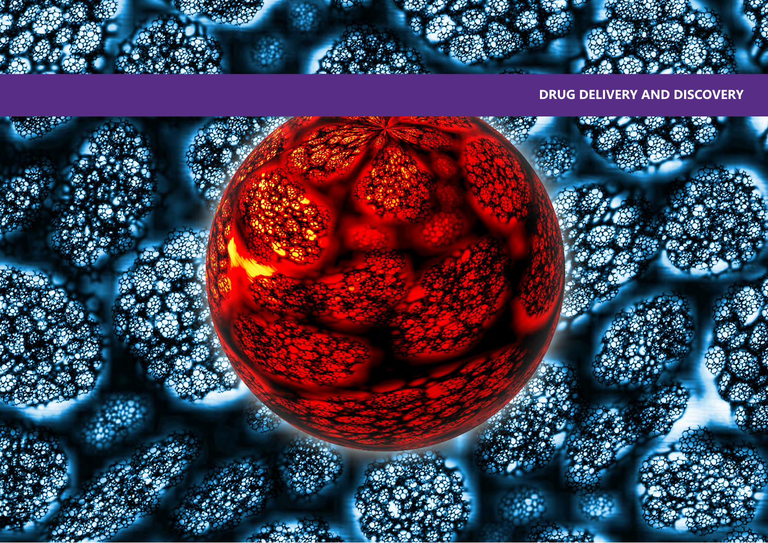
92 93
DRUG DELIVERY AND DISCOVERY

94 95
Professor and President
New York University
Andrew Hamilton was named the 16th president of New York University in March 2015. He most recently served as the vice chancellor of Oxford University, the university’s senior officer, after an academic career that took him from Princeton to the University of Pittsburgh, and then to Yale, where he was named provost. Throughout his time in academic leadership positions, he has maintained his scholarly work, including an active research laboratory, and is continuing to do so at NYU.
A distinguished chemist and a Fellow of the Royal Society, Dr. Hamilton’s scholarly work lies at the intersection of organic and biologic chemistry. He received his PhD from Cambridge University, his master’s degree from the University of British Columbia, and his undergraduate degree from Exeter University.
BIO
Andrew D. Hamilton
In this lecture we will describe a program aimed at the design of synthetic agents that can recognize the exterior surface of proteins and block protein-protein interactions involved in different cell signaling pathways. The unique distribution of charged, hydrophobic and hydrophilic groups on the surface of proteins offers the potential that well-designed artificial receptors will bind strongly and selectively. Our principal strategy involves the synthesis of molecules that mimic the side chain distribution and recognition properties of, often non-contiguous, surface domains involved in the protein-protein contact. The strategy will be exemplified with three examples. The first involves the design of synthetic mimics of extended peptide strands that make contact with well-defined clefts or cavities on protein surfaces. In a second example we will describe synthetic mimics of alpha-helical domains involved in protein-protein interactions. In particular we have designed terphenyl-based mimics of the BH3 helix of Bak and shown by fluorescence polarization and NMR that they bind to BclxL with a Kd of 100nM and disrupt the Bak/BclxL complex. We further extend this strategy with the use of helix mimetics to target proteins involved amyloid diseases such as Islet Amyloid Polypeptide (IAPP) implicated in type II diabetes. The third and most challenging example will involve the development of synthetic agents that can recognize a large area on the exterior surface of proteins and in doing so block critical protein-protein interactions. In particular we have developed synthetic scaffolds that mimic the large surface area of an extended beta-sheet region by exploiting the stabilising properties of an appropriately functionalised diarylacetylene unit.
SYNTHETIC MIMICS OF PROTEIN SECONDARY STRUCTURE AND FUNCTION
ABSTRACT

96 97
Associate Professor
Radiology
NYU School Of Medicine
Dr. Youssef Z. Wadghiri, Ph.D.is an Associate Professor of Radiology and the founding Director of the Preclinical Imaging Core at the NYU School of Medicine. His overall research goals are to develop non-invasive methods and molecular biomarkers to better understand human disease. His lab uses extensive interdisciplinary collaborations and MRI in combination with cellular and molecular targeting techniques to validate small animal imaging protocols that would be transferable to humans. Dr. Wadghiri’s latest work has led to two significant contributions using targeted labeling techniques: the first in vivo observation of Alzheimer’s plaques in mouse models and first in vivo imaging establishing neuronal transport impairment in direct correlation with Tau pathology. Dr. Wadghiri has authored over 40 peer-reviewed publications, 5 book chapters, and 119 proceedings at international conferences. He has served as a reviewer on many scientific journals and funding agencies. He holds multiple international honors, awards, and 4 patents.
BIO
Youssef Zaim Wadghiri
The use of new imaging technologies with innovations in chemistry has reduced significantly the need of exploratory surgeries to find a diagnosis for a disease. Imaging is currently routinely used clinically for the prognosis as well as for the monitoring of disease activity and treatment efficacy. It plays a similar role in biomedical research where animal models are especially required to prove the existence of a therapeutic effect, before clinical trials can be considered. In this case, the noninvasive examination of experimental models enables the monitoring of individual subject longitudinally. These studies would otherwise rely on conventional techniques that either may interfere with the disease mechanism studied or result in terminal studies.
To this effect, MRI has particularly played an important role thanks its intrinsic soft tissue contrast and penetration resulting in three dimensional detailed anatomical images without the use of damaging radiation. Exogenous molecules were quickly introduced with MRI to selectively improve the sensitivity and specificity of pathological tissues that would otherwise go undetected. This talk will review the various exogenous contrast agents (CAs) commonly employed in MRI, their physico-chemical characteristics and biocompatibility. Clinical and biomedical applications will illustrate their use either individually or through high payloads.
NANOBIOMATERIALS IN PRECLINICAL IMAGING AND DIAGNOSTIC RADIOLOGY: MRI A VERSATILE IMAGING MODALITY
ABSTRACT
98 99
Professor
Chemical Engineering
American University of Sharjah
Dr. Ghaleb A. Husseini graduated with a PhD in Chemical Engineering (Biomedical Engineering emphasis) from Brigham Young University in 2001 and joined the American University of Sharjah (AUS in the United Arab Emirates) as an Assistant Professor in the Chemical Engineering Department January 2004. He was promoted to Associate Professor and Professor in 2008 and 2013, respectively. Two years ago, Dr. Husseini took a sabbatical leave which enabled him to travel to Ecole Polytechnique Fédérale de Lausanne (EPFL, and work in Dr. Jeffrey Hubbell’s laboratory).
He works is in the area of ultrasound activated drug delivery. His research involves sequestering chemotherapeutic agents in liposomes, micelles and other nanoparticles. The contents of this drug delivery system can then be released using ultrasound (US). This way the drug has minimal interactions with the healthy cells in the host body, and it can carry out its therapeutic effect at the sonicated cancerous region only and reduce the undesirable side effects associated with chemotherapy. He has recently established a Drug Delivery laboratory at AUS using an internal grant.
Dr. Husseini has published 84 journal articles (in addition to 1 book chapter and 1 patent) and 45 conference papers/abstracts. In addition, he was a Theme Editor for a special issue in Advanced Drug Delivery Reviews and is currently serving on the Editorial Board of the International Review of Applied Sciences and Engineering (IRASE) and the European Journal of Nanomedicine. He served as a Distinguished Lecturer IEEE-EMBS (Jan 2014- Dec 2015).
BIO
Ghaleb A. Husseini
Chemotherapy is the most extensively used treatment in the fight against malignant neoplasms. Unfortunately, chemotherapy use is plagued with numerous side effects. These side effects are caused primarily because of the non-specific nature of the treatment as the drug is capable of killing normal and cancerous cells alike. Several drug delivery systems have been investigated to reduce these side effects by encapsulating the chemotherapeutic agent in a nano-sized carrier until it reaches the tumor site. These carriers include: solid nanoparticles, micelles, liposomes and e-liposomes. Once the nanoparticle reaches the desired location, ultrasound is applied to release the chemotherapy drug directly to the cancer site, thus avoiding any interaction with the healthy cells in the body. This way the adverse side effects of chemotherapy are minimized. This presentation will discuss two novel chemotherapy carriers (micelles and emulsion-Liposomes) used in conjunction with acoustic radiation to treat malignancies.
DRUG DELIVERY AND ULTRASOUND
ABSTRACT

100 101
Associate Professor
Prosthodontics
NYU College of Dentistry
Dr. Seiichi Yamano currently is an Associate Professor of Prosthodontics at New York University College of Dentistry. Dr. Yamano first earned his dental degree from Nihon University and then a PhD in Immunology/Microbiology from Tokyo Medical University in Japan. Then, he came to the U.S. as a Research Fellow at NIDCR/NIH. Also, he received his DMD from the University of Pennsylvania, and a Prosthodontics certificate and MMSc in Oral Biology from Harvard University. Currently, Dr. Yamano maintains his research laboratory at NYU to investigate three major areas: (1) utilization of efficient non-viral vectors for gene therapy, (2) growth factor protein delivery for bone regeneration, and (3) molecular analysis of osseointegration for novel implant surfaces as well as teaches prosthodontics for predoctoral and postdoctoral students. He has been given many major research and teaching awards including NIDCR/NIH grant awards. Dr. Yamano has contributed to over 150 scientific publications, abstracts, and books.
BIO
Seiichi Yamano
Gene therapy is emerging as an exciting prospect for the treatment of many diseases. Synthetic, non-viral vectors are potential alternatives to viral vectors that preclude several obstacles such as immune response, limited gene carrying capacity, recombination and high cost. To overcome these barriers, we developed two non-viral hybrid vectors. The first is composed of a cell permeable peptide (HIV-1 Tat peptide sequence modified with histidine and cysteine residues) combined with a cationic lipid. The second vector substitutes cationic polymer for the lipid. These non-viral vectors have excellent transfection efficiency with little cytotoxicity across a range of cell lines including different types of cancer cells. Moreover, transfection efficiency using the vector in oral cancer cells has a significantly higher expression (~45-fold) than normal cells and has a higher expression (~65%) than an adenoviral vector (~50%). In vivo transfection with the non-viral vectors leads to high and long-term transgene expression (~7 months) after intramuscular injection of the vectors. Using the vector, we recently demonstrated that ex vivo non-viral gene transfer of μ-opioid receptor significantly reduced pain in a paw cancer model (oral cancer cell inoculated into the paw). Our long-term goal is to move our method of non-viral transfection to the clinic.
THE POTENTIAL OF NON-VIRAL GENE FOR CLINICAL APPLICATIONS
ABSTRACT

102 103
Associate Professor
Anatomy, Cell Biology and Physiology
American University of Beirut
Dr. Rihab Nasr is an Associate Professor in the department of Anatomy, Cell Biology and Physiology and the director of Cancer Prevention and Control Program at the Faculty of Medicine at the American University of Beirut. Dr. Nasr received her PhD in Fundamental basis of oncogenesis from the University of Paris VII in France. Her major research activities in basic and translational research focus on developing targeted therapies for human leukemias. Dr. Nasr is also interested in microRNAs and their implication in cancer.
Dr. Nasr received the Best Biomedical research award from Qatar Foundation in 2011, was selected as one of the seven top Arab Women by Sayidati magazine for the year 2013, was the recipient of UNESCO L’Oreal “For women in Science” Levant and Egypt fellowship in 2014, and the Franco-Lebanese Scientific Excellence award by the Legion of Honor in Lebanon and the French Institute in 2015.
BIO
Rihab Nasr
Tyrosine Kinase Inhibitors (TKI) have been established for the treatment of Chronic Myeloid Leukemia (CML). However, patients can develop resistance against TKI mainly due to kinase domain mutations. T315I mutation confers resistance to almost all TKI. Although some evidence suggested the potential efficacy of ponatinib against T315I mutation, its use is limited by its toxicity. Our study aims to test the anti-tumor efficacy of Arsenic trioxide (ATO) and Interferon alpha (IFN) in preclinical models of CML resistant to TKI. We found that ATO/IFN inhibited the proliferation and induced apoptosis of Imatinib-resistant CML cells. The in vivo effect of ATO/IFN was studied using a retroviral BCR-ABL T315I murine CML model. Leukemic mice treated with ATO/IFN showed a significant prolonged survival as compared to untreated controls. As expected, imatinib had no effect on the survival of leukemic mice harboring BCR-ABL with T315I mutation. Importantly, ATO/IFN severely impaired engraftment into untreated secondary recipients, with some recipients never developing the disease, demonstrating a dramatic decrease in CML Leukemia Initiating Cells activity. These findings provide clear evidence of the preclinical efficacy of ATO/IFN in resistant CML models and accordingly open the perspective of investigating this combination in the therapeutic strategy of resistant CML.
ANTI-TUMOR EFFICACY OF ARSENIC/INTERFERON IN PRECLINICAL MODELS OF CHRONIC MYELOID LEUKEMIA RESISTANT TO TYROSINE KINASE INHIBITORS
ABSTRACT
104 105
Assistant Professor
Division of Science - Biology
NYU Abu Dhabi
Mazin Magzoub is a biophysicist specializing in the development of novel methods for the delivery of antitumor agents and therapeutics for amyloid diseases (e.g. Alzheimer’s and prion diseases). Prior to joining NYUAD, Dr. Magzoub was a postdoctoral scholar at the University of California, San Francisco, where he developed novel biophysical methods for measurement of macromolecule diffusion deep in tissues such as tumors and brain. Subsequently, as an Associate Research Scientist in Andrew Miranker’s laboratory at Yale University, he worked on elucidating the molecular mechanisms underlying type 2 diabetes.
Dr. Magzoub received his Ph.D. in biophysics from Stockholm University in 2004, where he studied the biophysical properties of cell-penetrating peptides, a class of peptides with the ability to mediate the cellular import of therapeutic compounds with high efficiency and low toxicity.
BIO
Mazin Magzoub
Overexpression of mitochondria-bound hexokinase II (HKII) in cancer cells plays a paramount role in their metabolic reprogramming and protects them against apoptosis, thereby facilitating their growth and proliferation. Here, we show that covalently coupling a peptide corresponding to the mitochondrial membrane-binding N-terminal domain of HKII (pHK) to a short penetration accelerating sequence (PAS) enhanced the peptide’s cellular uptake, mitochondrial localization and cytotoxicity in HeLa cells. Further analysis revealed that pHK-PAS depolarized the mitochondrial membrane potential, inhibited mitochondrial respiration and glycolysis, and depleted intracellular ATP levels. The effects of pHK-PAS were correlated with dissociation of endogenous full-length HKII from mitochondria and release of cytochrome c. Significantly, pHK-PAS treatment of non-cancerous HEK-293 cells resulted in substantially lower cytotoxicity. Thus, pHK-PAS effectively disrupts the mitochondria-HKII association in cancer cells, leading to mitochondrial dysfunction and finally apoptosis. Our results demonstrate the potential of the pHK-PAS cell-penetrating peptide (CPP) as a novel cancer therapeutic strategy.
HEXOKINASE II-DERIVED CELL-PENETRATING PEPTIDE TARGETS MITOCHONDRIA AND TRIGGERS APOPTOSIS IN CANCER CELLS
ABSTRACT
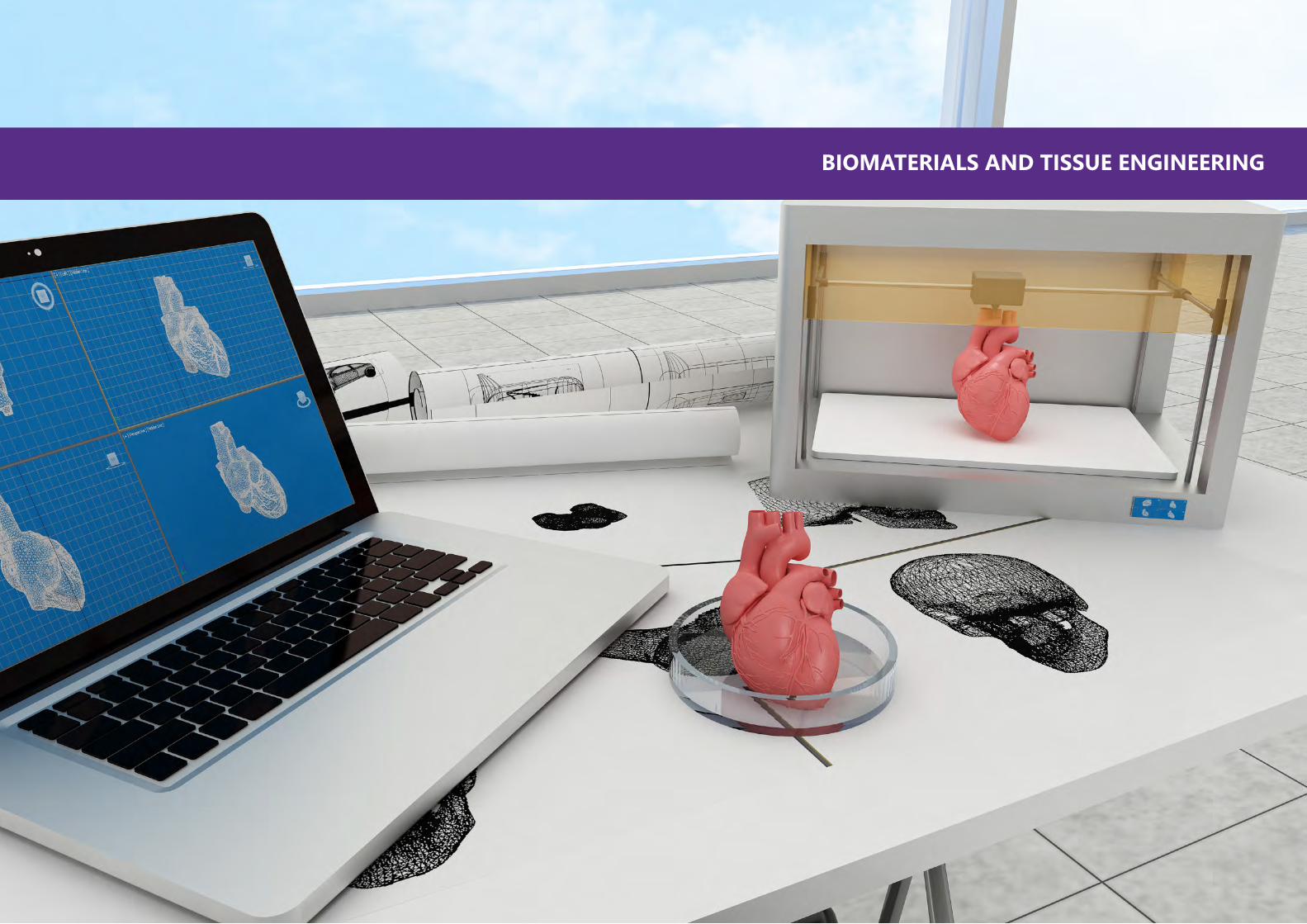
106 107
BIOMATERIALS AND TISSUE ENGINEERING

108 109
Professor
Bioengineering
University of California at Berkeley
Kevin E. Healy, Ph.D. is the Jan Fandrianto and Selfia Halim Distinguished Professor in Engineering at the University of California at Berkeley in the Departments of Bioengineering, and Materials Science and Engineering. He served as Chair of the Department of Bioengineering from 2011 to 2015. He is a thought leader and innovator working at the interface between stem cells and materials science to develop dynamic engineered systems to explore both fundamental biological phenomena and new applications in translational medicine. His group currently conducts research in the areas of: bioinspired stem cell microenvironments to control stem cell lineage specification and self-organization into microtissues or organs; bioinspired systems for regenerative medicine; biological interfaces; and, microphysiological systems for drug toxicity screening. He is an elected Fellow of the American Institute of Medical and Biological Engineering (AIMBE), Fellow in Biomaterials Science and Engineering (FBSE), and the American Association for the Advancement of Science (AAAS).
BIO
Kevin E. Healy
Drug discovery and development are hampered by high failure rates attributed to reliance on non-human animal models that poorly recapitulate human disease states employed during safety and efficacy testing. With the discovery of human induced pluripotent stem cells (hiPSCs), bioengineers can now develop in vitro disease specific tissue models to be used for high content drug screening and patient-specific medicine. Combining the genetic background of human cells with appropriate biophysical tissue architecture and “tissue-like” drug gradients recapitulates a minimal organoid of the human myocardium sufficiently to allow accurate prediction of the toxicity of drugs. This presentation will discuss our progress in developing integrated in vitro models of human cardiac and liver tissue based on populations of normal and patient specific hiPSCs differentiated into cardiomyocytes, hepatocytes, or supporting cells. The benefits of our approach include: 1) robust microengineering platforms that control microtissue organization and function; 2) precise delivery of molecules (e.g., drugs) in a computationally predictable manner; 3) ability to model human disease; and, 4) cost efficient and high content characterization of an integrated multi-organ drug response.
ORGANS ON A CHIP: THE FUTURE OF PRECISION MEDICINE
ABSTRACT
110 111
Professor
Chemical and Biomolecular Engineering
NYU Tandon School of Engineering
Kalle Levon, professor and entrepreneuer, Specialist on charged surfaces and surface interactions with applications in monitoring biological binding events with functionalized transistors and building nanoscale 3D scaffolding for the well-being of cells.
NIH Center for Scientific Review: Biomaterials and Biointerfaces 2007- 2011
Honorary member of the Editorial Board: International Journal of Nanomedicine
BIO
Kalle Levon
Autologous stem cell transplantation has given enormous promise for tissue engineering due to decreased inflammation, as one example. The complex, multifunctional cellular events are ensured by the controlled well-being of cells with undisturbed cell-cell interactions. Carefully building the correct, “homey” scaffolding with nanofibers, preferably biological, assures adequate mechanical growth support but at the same time the needed high entropy environment. Additionally, 3-dimensionality removes the impact of denaturing 2D surfaces and allows the progress of the multifunctional events proceed in all directions. We shall present the importance of the change in fiber diameter when building a functional thyroid using stem cell transplantation. In addition, we present our novel 3D scaffolding with encapsulated cells in the nanofiber network.
WELL-BEING OF CELLS IN 3D NANO-ENVIRONMENT
ABSTRACT
112 113
Senior Lecturer
Bioengineering
University of Strathclyde
Richard Black (BSc, PhD, PGCert, CSci, CEng, FIMechE, FIPEM, FHEA) is a biomedical engineer with over 25 years’ experience in the field. He graduated BSc (Hons) Engineering Science from the University of Edinburgh, and gained his PhD from the Faculty of Medicine at the University of Liverpool, where he was lecturer in Medical Device Design. He is currently academic director of the UK Engineering & Physical Sciences Research Council (EPSRC) Centre for Doctoral Training in Medical Devices and Health Technologies at the University of Strathclyde. A Chartered Scientist and Engineer, Dr Black is Fellow of the Institution of Mechanical Engineers and the Institute of Physics and Engineering in Medicine, and Editor-in-Chief of Medical Engineering & Physics, one of three journal publications of the Institute (www.medengphys.com).
BIO
Richard A. Black
The impact on tissue perfusion of cardiovascular disease, and the removal of lymph nodes and vessels following surgical treatment for cancer, for example, can have a profound effect on the circulation, leading to fluid imbalances and oedema, which are debilitating for the patients affected; and whereas many groups are active in developing novel strategies for blood vessel replacement, less attention has been paid to research to develop protocols and devices that are able to replicate the functional characteristics of intact blood and lymphatic vessels. Tissue Engineering offers the means to generate in vitro models of blood and lymphatic vessels for drug testing in the short term, and produce wholly biological vessels that surgeons may use to bypass or replace these vessels in patients. In order to achieve these goals, however, the engineered vessels must recapitulate the structure and function of the vessels they replace, yet be able to resist the remodelling pathologies to which those grafts are prone. In this talk I shall review work to develop models of vascular and lymphatic vessels, and how research of this kind may suggest ways to treat conditions associated with inadequate tissue perfusion, oedema, and disorders of the vascular and lymphatic systems.
MODELLING BLOOD AND LYMPHATIC VESSELS IN VITRO: CHALLENGES AND OPPORTUNITIES
ABSTRACT

114 115
J.C. Bose Fellow
Membrane and Receptor Biology
Center for Cellular and Molecular Biology
Prof. Amitabha Chattopadhyay obtained his Ph.D. from the Stony Brook University (New York), and was a Postdoctoral Fellow at UC Davis. His work is focused on monitoring organization, dynamics and function of biological membranes in healthy and diseased conditions. A seminal contribution of Prof. Chattopadhyay is the role of membrane cholesterol in regulating organization, dynamics and function of G protein-coupled receptors. His work has provided novel insight in the role of membrane cholesterol in the entry of pathogens into host cells. Prof. Chattopadhyay was awarded the prestigious TWAS Prize, Bhatnagar Award, Ranbaxy Research Award, Prof. G.N. Ramachandran 60th Birthday Medal, and is a J.C. Bose Fellow. He is an elected Fellow of the Royal Society of Chemistry, and all the Indian Academies of Science. He has served on the editorial boards of a large number of reputed journals. Prof. Chattopadhyay has authored >250 research papers and delivered >500 invited lectures.
BIO
Amitabha Chattopadhyay
Diseases caused by intracellular pathogens represent a major public health problem worldwide, including the Middle East. The cellular plasma membrane acts as a portal for the entry of intracellular pathogens. An essential step for an intracellular pathogen to gain entry into a host cell therefore is to be able to cross the cell membrane. In this talk, I will highlight the role of host membrane cholesterol in regulating the entry of intracellular pathogens such as Leishmania and Mycobacterium. Our results show that pathogen entry is inhibited either upon depletion (or complexation) of membrane cholesterol. We propose a general mechanism, based on cholesterol-induced conformational changes, involving cholesterol binding sites in host cell surface receptors that are implicated in pathogen entry. A therapeutic strategy targeting modulation of membrane cholesterol has the advantage of avoiding the commonly encountered problem of drug resistance in tackling infection by intracellular pathogens. Interestingly, our results show that an optimum level of host membrane cholesterol is necessary for efficient infection by pathogens. We envision that insights into the role of host membrane cholesterol in pathogen entry would be instrumental in the development of novel therapeutic strategies to effectively tackle intracellular pathogenesis.
A MEMBRANE CHOLESTEROL-BASED STRATEGY TO TACKLE ENTRY OF INTRACELLULAR PATHOGENS: EVADING DRUG RESISTANCE
ABSTRACT
116 117
Associate Professor
Chemistry
United Arab Emirates University
Dr. Yaser Greish is currently serving as an associate professor of Materials Chemistry at the Department of Chemistry, College of Science, UAE University. He received his PhD in Materials Science and Engineering from the Pennsylvania State University, USA in 2001. He worked as a PostDoc scholar in a NIH project at Penn State university until 2004. In 2005, he joined the UAE University as an assistant professor of Materials Chemistry, then promoted to an associate professor in 2011. He teaches at both the undergraduate and graduate levels in the university. He is also affiliated with the MSc programs of Chemistry, Materials Science and Environmental Science at the UAEU. His has been working in the area of biomaterials since 1991, specially in the development of bone replacement and bone fixation materials. In the last 15 years, he started his research in the area of drug delivery and tissue engineering using nanoparticles, nanofibers and nanocomposites. He has 45 publications in peer-reviewed journals and more than 80 presentations in international conferences.
BIO
Yaser E. Greish
In tissue engineering and tissue regeneration, biodegradable polymeric scaffolds are usually used. The main concept of using these materials is the need for their initial support of cells until a new tissue is formed or regenerated. Biodegradable polymers are biocompatible by default but they differ in their chemical structure and the degree of degradation. In the current study, two types of biodegradable polymers were used as scaffolds for the regeneration of gastric mucosa using gastric stem cells. Upon optimization and preliminary in vitro experiments, nano-/microfibers of one of the polymer was used for in detailed vitro and in vivo experiments. This was based on the finding that higher surface are micro-/nanofibers have higher surface area and interconnected porosity. Therefore, a 3D fibrous scaffold that mimics the extracellular matrix in nature was made to better suit the culture and differentiation of gastric stem cells into gastric functional cells. In the current presentation, highlights of our most recent findings will be shown, based on the work that we have been doing in our laboratories at the UAE University.
GASTRIC STEM LOADED BIODEGRADABLE MICROFIBERS FOR THE REGENERATION OF GASTRIC MUCOSA
ABSTRACT

118 119
Associate Professor
Division of Science - Chemistry
NYU Abu Dhabi
After acquiring his PhD in chemistry and materials science from Tokyo Institute of Technology in 2004, Dr. Naumov continued his research as an independent research fellow of the National Institute for Materials Science (NIMS) in Japan. Since 2007 he was Associate Professor at Osaka University, and after a short stint at Kyoto University in 2011, in 2012 he joined New York University Abu Dhabi. His publication portfolio includes over 170 publications that have been cited more than 2500 times, with an h-index of 25. He serves as a member of the review panels with the NSF, ERC, ACS, and several national funding agencies. He is active reviewer for more than 40 journals. Dr Naumov is Alexander von Humboldt fellow, JSPS fellow, and recipient of a number of international awards.
BIO
Panče Naumov
The process of biochemical production of light, bioluminescence, which is utilized by a number of lower organisms to attract prey, or for mating or defense, is a visually fascinating and practically important, yet mechanistically enigmatic phenomenon. This talk will summarize our contributions that have advanced this burgeoning field in the past years. The crystal structure of the unstable emitter (oxyluciferin) was determined, and its spectrochemistry investigated. By using mathematical methods (MCR-ALS), the absorption end emission spectra and the lifetimes of the chemical forms of the emitter were disentangled. Action spectroscopy of the lumophore in vacuo revealed that its electronic states are altered by microenvironmental perturbations. The results also provided evidence of excited-state tautomerization, which could be important in resolving the enol−keto conundrum related to the color-tuning mechanism. The reasons for the apparent instability of oxyluciferin were also explained, which is now ascribed to autodimerization of the enol and keto forms. Finally, the intricate spectrochemistry of the emitter inside the enzyme was disentangled.
THE MYSTERY OF FIREFLY BIOLUMINESCENE
ABSTRACT

120 121
Associate Professor
Pathology and Immunology
Baylor College of Medicine
Dr. Laising Yen, Associate Professor, Baylor College of Medicine, Houston, USA. Dr. Yen received his Ph.D. degree from Yale University and trained as a post-doctoral fellow at Harvard Medical School. The main focus of Yen lab is to harness the power of RNA to create new instruments for medicine and biological studies. The current research areas include: (1) investigation of RNA mutations in human cancer for diagnosis and therapy, (2) development of RNA-based molecular switches that can be turned on/off by FDA-approved drugs for gene regulation, and (3) building designer RNA nanostructures in mammalian cells for synthetic biology applications. The Yen lab uses a variety of chemical and molecular biology strategies, in combination with mammalian cell cultures and animal models in pursuit of the goals.
BIO
Laising Yen
RNAs are highly programmable polymers due to their ability to form specific Watson-Crick base pairing, a property that can be exploited to create well-defined 2D and 3D structures. These structures are thermodynamically stable, and formed via spontaneous self-assembly, a process that requires no catalytic co-factors. Because single-stranded RNA can be efficiently expressed at high levels in live cells, it offers the opportunity to program cells to assemble designer nanostructures. Here we designed and constructed short single-stranded RNA monomers that self-assembled into higher order architectures, reaching a size range of micrometers. When these RNA monomers were expressed in human cells, they appeared to form stable structures. The results point to the possibility that designer RNA can be used to program mammalian cells to assemble nanostructures for specific cellular functions useful for synthetic biology applications.
BUILDING DESIGNER RNA NANOSTRUCTURES
ABSTRACT

122 123
Associate Professor
Mechanical and Aerospace Engineering
NYU Tandon School of Engineering
Dr. Nikhil Gupta is an Associate Professor in the Mechanical and Aerospace Engineering Department at the New York University Tandon School of Engineering. He directs the Composite Materials and Mechanics Laboratory. His research interests include developing lightweight advanced composite materials for dynamic loading conditions. His laboratory is also interested in developing dynamic test methods. His research is supported by the by The Office of Naval Research, National Science Foundation, Army Research Laboratory, Department of Energy and industry. Dr. Gupta has two issued patents, authored over 140 journal papers and book chapters, four books, and presented over 50 keynote and invited lectures.
BIO
Nikhil Gupta
Mechanical properties of bones depend on the length scale at which they are characterized due to their hierarchical structure. There is also a significant effect of test conditions on the test results for bones due to the viscoelastic nature of the material and presence of pores and fluid. Hierarchical structure of bones requires imaging and characterization from nanoscale to macroscale in order to understand their properties. In this work, effect of strain rate at the measured mechanical properties of bones is explored. The tests are conducted in low strain rate regime at nanoscale and in low and high strain rate regimes at macroscale. Results show that the strength and modulus depend on the test strain rate and the strain rate sensitivity coefficient depends on the stiffness of the material. Cortical bones show positive strain rate sensitivity while cancellous bones show negative strain rate sensitivity. Challenges related to testing bones in an environment that mimics the actual physiological conditions are also discussed.
MECHANICAL CHARACTERIZATION OF HIERARCHICAL BIOMATERIAL ACROSS LENGTH SCALES AND STRAIN RATES
ABSTRACT

124 125
Professor
Biomedical Engineering
University of Strathclyde
Will Wenmiao Shu is the Hay Professor in Biomedical Engineering at the University of Strathclyde (Glasgow). He obtained his PhD at the Engineering Department from University of Cambridge, UK.
His research interests cover a range of biomedical engineering topics including 3D biofabrication, biosensors, microsystems and their applications for regenerative medicine. He led the research to demonstrate the first bioprinting of human embryonic stem cells (h-ESCs) and human induced pluripotent stem cells (h-iPSCs), paving the way for their applications on animal-free drug testing and 3D printed organs. He held a visiting position at Stanford University. He is an editorial board member for IOP Biofabrication Journal and serves as a board director of the International Society for Biofabrication (ISBF).
BIO
Will Wenmiao Shu
Biofabrication is an emerging field that encompasses a wide range of enabling technologies including 3D bioprinting, bioassembly and additive manufacturing for the applications in organ printing, tissue engineering and regenerative medicine. This talk will give a brief overview of the rapidly developing field of 3D biofabrication. I will present the development of 3D stem cell printer that has been validated to print highly viable and functional human embryonic stem cells (h-ESCs) and induced pluripotent stem cells (h-iPSCs). 3D bioprinting based on both scaffold based using biomaterials and scaffold-free approaches will be discussed. In particular, novel strategies are developed to print smart biomaterials based on alginate, DNA and peptide hydrogels for 3D stem cell constructs. Our recent work on stem cell-derived liver and heart tissues in 3D for in vitro drug testing applications will be presented.
3D BIOPRINTING OF HUMAN PLURIPOTENT STEM CELLS AND HYDROGELS FOR TISSUE ENGINEERING
ABSTRACT

126 127
Associate Professor
Oral and Maxillofacial Surgery and Oral Medicine
Malmö University
Dr Ryo Jimbo received DDS at Nagasaki University, Japan, in 2004 and then at Gothenburg University, Sweden, in 2014. He defended his thesis in 2007 at Nagasaki and received specialist training in Prosthodontics and in Oral & Maxillofacial Surgery. From 2009, he worked as visiting researcher at the Department of Biomaterials, Gothenburg University. At present, he is Associate Professor at the Departmentof Oral and Maxillofacial Surgery and Oral Medicine at Malmö University, and is dedicated to education, clinical practice and implant, biomaterial research. He has published more than 150 articles and lectures internationally. He is in the editorial board of CIDRR and JOMR.
BIO
Ryo Jimbo
From an engineering or a biomaterial science point of view, dental implants may be regarded as a simple and a straightforward device, since today, many of the organs that possess further complex architectures can be regenerated and the technology regarding the world of regenerative medicine is far beyond dental implants. However, when we look at the clinical reality, the global dental implant market is still growing at a rapid rate annually, and ironically, more complications are emerging, as some would like to call it periimplantitis. It seems although in the early phases of osseointgration, the cause for bone loss around the implant cannot be explained just by infection. One of the major reasons for the marginal bone loss at this very early stage is caused supposedly by the surgical procedures. Even if a medical device is designed or calculated to be used in a certain way, it is often up to the clinician to follow the recommendations. Obviously, if not used in the intended manner, there will be biological or biomechanical consequences that may lead to bone loss around the implant or in worst cases, the loss of the implant. In this lecture, the clinical problems related to surgical procedures will be introduced, and how this may have occurred will be discussed. How can science solve a clinical problem? The answer to this question could lead to further developments that may solve a clinical problem.
INFLUENTIAL FACTORS FOR BONE LOSS DURING IMPLANT SURGICAL PROCEDURE. INTERACTION BETWEEN INTRUMENTATION AND GEOMETRY. HOW CAN SCIENCE SOLVE A CLINICAL PROBLEM?
ABSTRACT

128 129
Assistant Professor
Division of Engineering
NYU Abu Dhabi
Dr. Sachin Khapli is an Assistant Professor of Engineering at NYU Abu Dhabi. His research is focused on the development of novel biomaterials and their applications in tissue engineering. He is fascinated by various surface and interfacial phenomena that occur at the interfaces between artificial materials and living systems. Current projects in his group deal with the development of novel methods for the fabrication of nanostructured materials and thin films using supercritical fluid technology. Using these methods, his group recently demonstrated the fabrication of hierarchically porous scaffolds composed of bioceramics and endowed with osteoconductive properties. He is also interested in the development of smart, mechanochromic hydrogels and their applications in bio-sensing.
Sachin holds a B. S. from the Indian Institute of Technology, Kharagpur and Ph.D. from Rice University, Houston, TX.
BIO
Sachin Khapli
The development of artificial materials for applications in bone tissue engineering is challenging due to the functional diversity, hierarchical structure, and extraordinary mechanical properties of the bone tissue. Porous metals, in particular Titanium and its alloys, are the most-used implant materials. However, the lack of any specific interactions with the cells limits the ability of these implants to bond with the host tissue.
We report the development of a novel, supercritical CO based process for coating titanium alloys with bioactive ceramic coatings that exhibit hierarchical porosity on the macro, micro, and nano-scales. In preliminary investigations, we have demonstrated the utility of this technique for coating planar silicon substrates and screw shaped Ti-implants with porous scaffolds of CaCO bioceramic. The performance of these coatings was evaluated in in vitro andin vivo preclinical studies, respectively. Our porous coating promotes bone healing, as observed by in vivo investigation of titanium implants in sheep subjects. A significant increase in the amount of bone in proximity with the implant surface was observed from 1-to-6 weeks in vivo, and all surfaces presented biocompatible and osseoconductive properties. In vitro studies using cells from mouse osteoblast precursor cell line, MC3T3, also demonstrated the cytocompatibility of hierarchical porous CaCO scaffolds.
HIERARCHICALLY POROUS CALCIUM CARBONATE SCAFFOLDS FOR BONE TISSUE ENGINEERING
ABSTRACT
2
3
3

130 131
Research Assistant Professor
Division of Science - Biology
NYU Abu Dhabi
Dr Mohamed Al-Sayegh is a Research Assistant Professor (Research Fellow) at the Biology Division who manages the Stem Cell Biology laboratory at New York University Abu Dhabi (NYUAD). Prior to joining NYUAD, Mohamed held a Postdoctoral position in the Mazzoni lab at Biology Department at NYU Washington Square campus. During his Postdoctorate training, Mohamed developed an interest into next generation sequencing applications, specifically transcriptomics, by studying the affects of direct programming of embryonic stem cell of transcription factors into specific subtypes of motor neurons. In addition to this, he also is interested in understanding cell differentiation of various cell types including adipocyte differentiation. Mohamed received his BA in Biology from Valparaiso University (Valparaiso, Indiana, USA); MSc in Molecular Genetics from the University of Leicester (Leicester, UK); and his PhD in Molecular and Cell Biology from the University of London (St Georges Medical School, London, UK).
BIO
Mohamed Al-Sayegh
The role of actin cytoskeleton dynamics has been implicated in various biological functions specifically in the content of cell differentiation both at the cellular (i.e. cell migration) and transcription (i.e. serum response factor) levels. However, little is known about the functionality of actin in relation to adipocyte differentiation. Here, we report initial insights into the affects of actin on adipogenesis. Generation of mouse embryonic fibroblast (MEF) cell lines that consist of wild type (WT), Heterogeneous (HET) and Knockout (KO) alleles of actin show altered cell morphology. Adipocyte differentiation of MEF cell lines with HET and KO alleles show elevated levels of triglyceride lipid droplet formation as opposed to the WT cell line according to Oil Red O staining and its corresponding optical density readings. These data suggest that changes into actin morphology at the genetic level can either enhance or restrict the progression of adipocyte functional formation. We propose further validation approaches to demonstrate the role actin cytoskeleton on adipocyte differentiation.
THE LINKAGE ROLE OF ACTIN CYTOSKELETON AND ADIPOCYTE DIFFERENTIATION: A MOLECULAR SCAFFOLD MODIFIER POTENTIAL
ABSTRACT
132 133
Professor and Chairman
Materials Engineering
Auburn University
Bryan A. Chin received his M.S. and Ph.D., with distinction, in Materials Science and Engineering from Stanford University. Dr. Chin currently serves as Professor and Chairman of Materials Engineering at Auburn University. In the year 2003, Dr. Chin was named Director of the Auburn University Detection and Food Safety Center (AUDFS), a University Peak of Excellence. AUDFS has been granted more than 40 patents, commercialized 8 products and spun off two companies and two other Centers. As Director, Dr. Chin oversees research dedicated to the protection of our nation’s food supply from natural or deliberate acts of contamination. Among his many professional recognitions, Dr. Chin has published over 300 refereed articles, is a foreign member of the Russian Academy of Engineering Sciences, Breeden Professor of the Samuel Ginn College of Engineering, Honorary Professor of the Chinese Academy of Sciences and is a Fellow of ASM, ANS, AWS and ECS.
BIO
Bryan A. Chin
Every year over 8.9 million Americans become ill due to foodborne illness resulting in a cost of more than $15.6 billion dollars. Over 95% of these foodborne illnesses were due to fifteen pathogens. This paper presents a method of bacterial detection that directly detects and quantifies the presence of specific bacteria on the surfaces of fresh produce without sample preparation (water rinse, soak, stomaching, etc.) and/or enrichment. The method combines wireless magnetoelastic (ME) biosensors and a surface-scanning detector for the swift screening of foods at ports of entry, food processing facilities and in agriculture fields. Tests were conducted on tomatoes spiked with difference concentrations of Salmonella Typhimurium as a model pathogen. The speed of detection was from 2 to 10 minutes with a limit of detection (LOD) of 500 cfu/mm . Specificity of detection was better than 200 cells of Salmonella in 10 background cells. This technology was the $20,000 prize winner in the U.S. Food and Drug Administration’s first Food Safety Challenge in 2015.
RAPID AND DIRECT DETECTION OF PATHOGENS ON FRESH PRODUCE
ABSTRACT
2
8

134 135
Assistant Professor
Materials Engineering
Auburn University
Pengyu Chen received his B.S. in Materials Science and Engineering from Nanjing University in 2006. He obtained M.S. in Materials Science and Engineering (2009) and Ph.D. in nanomaterials and biophysics (2012) from Clemson University, and joined Mechanical Engineering at the University of Michigan as a postdoc fellow in 2013. He is now an Assistant Professor in Materials Engineering at Auburn University. His research focuses on advanced nanomaterial based biosensors for biomarker detection, precision medicine, and environmental & food safety. He was the recipient of US Environmental Protection Agency, National Research Council Fellow and won several presentation awards (oral and poster) at national and international conferences.
BIO
Pengyu Chen
Unravelling the underlying mechanism of how cells communicate upon external stimulation offers critical knowledge for modern biology and clinical science in a wide range of fields, such as fundamental biology, drug discovery, disease diagnosis, and cancer/stem cell research, to name a few. However, the complexity and heterogeneity of cellular interactions make their characterization by currently available technique difficult. In this talk, we would like to present a novel synthetic hybrid biomaterial – aptamer linked nanoplasmon ruler - which can potentially allow accurate, real-time mapping of multiple cytokine secretion profile from a single immune cell upon external stimulation. Such a novel approach will establish a new paradigm that permits, for the first time, the direct visualization of the dynamic intercellular communication process in the immune system. The knowledge obtained from this study will facilitate a more comprehensive understanding of the immune intercellular network, unlocking the potential to transform the experimental studies into an information-rich science not only in immunology but beyond.
UNRAVELLING INTERCELLULAR COMMUNICATION USING NANOPLASMON RULER
ABSTRACT
136 137
Assistant Professor
Mechanical and Industrial Engineering
Qatar University
Anwarul Hasan, PhD, PEng, is an Assistant Professor in the Department of Mechanical and Industrial Engineering at Qatar University, Qatar. He is also a Visiting Assistant Professor at Harvard Medical School, USA, an Assistant Professor of Biomedical Engineering and the Department of Mechanical Engineering at the American University of Beirut, Lebanon. Earlier, Dr Hasan obtained his PhD from University of Alberta, Canada in 2010. He worked in industry during 2010-2011, and was a National Science and Engineering Research Council of Canada (NSERC) Research Fellow at Harvard and MIT during 2012-2013. Prof Hasan has more than 60 peer reviewed papers including about 40 journal articles and 20 conference proceeding papers. His current research interests are in the areas of Biomaterials, Biomechanics and Tissue Engineering, particularly for cardiovascular, bone and neural applications as well as diabetic wound healing and microfluidic models of engineered tissues.
BIO
Anwarul Hasan
Tissue engineering and regenerative medicine have emerged as potential solutions for many diseases including some cardiovascular, bone and neural diseases Examples include development of implantable tissue engineered vascular grafts for bypass heart surgeries and regeneration of cardiac tissue after tissue-damage or tissue-death resulting from cardiac arrests, to mention just a few. A major obstacle for the widespread use of tissue engineering in clinical applications is the lack of suitable biomaterials with required combination of biomechanical and biological properties. Photocrosslinkable micro-porous hydrogels and electrospun nano-microfiber scaffolds have been widely investigated for application in tissue engineering and regenerative medicine. This talk will present some interesting results from our recent studies on potential applications of Photocrosslinkable methacrylated gelatin hydrogel and electrospun-airjet-sprayed composite scaffolds in cardiovascular tissue engineering and regenerative medicine as well as a number of other Biomedical Engineering applications.
PHOTOCROSSLINKED HYDROGELS AND ELECTROSPUN SCAFFOLDS IN TISSUE ENGINEERING AND REGENERATIVE MEDICINE
ABSTRACT

138 139
Professor
Orthopaedic Surgery and Cell Biology
NYU School of Medicine
Dr. Chuanju Liu is a Professor of Orthopaedic Surgery and Cell Biology at NYU School of Medicine. He is Editor-in-Chief of two journals (Clinical Medicine Insights: Arthritis and Musculoskeletal Disorders; Rheumatology Research Reviews) and serves on a number of Editorial Boards, including JBC and Scientific Reports. He is also a highly active participant in peer review of manuscripts, grant applications to government agencies (NIH, DOD) and private foundations. His current research focuses on arthritis, musculoskeletal regeneration, and lysosomal storage diseases. He has published over 130 papers in prestigious journals, including Science, PNAS, and Annals of the Rheumatic Diseases. In addition, he is the recipient of numerous awards, including the American Society for Bone and Mineral Research’s Harold M. Frost Award, the Arthritis Foundation’s Dorothy W. Goldstein Award, the Kappa Delta Award from the American Academy of Orthopaedic Surgeons, and the Innovative Research Award from the American College of Rheumatology.
BIO
Chuanju Liu
Progranulin (PGRN) is a growth factor with multiple functions, including anti-inflammation and tissue repair. PGRN was originally isolated as an osteoarthritis (OA)-associated growth factor in our genome-wide screen for differentially expressed genes in OA (Guo, et al, Arthritis Rheum. 2010). Our subsequent functional genetic screen for PGRN-binding partners identified TNFR as the PGRN-binding receptor (Tang, et al, Science, 2011). Armed with this knowledge, we further focused on the therapeutic potential of PGRN by identifying the domains that interacted with TNFRs, which led to the development of an engineered protein, termed “Atsttrin”, for short. Interestingly, we found that Atsttrin exhibits more efficacy than currently approved anti-TNF drugs in animal models of rheumatoid arthritis (RA). We reported recently that PGRN is also protective against OA (Zhao, et al, Ann Rheum Dis. 2015). In addition, we and other groups demonstrated the chondroprotective role of Atsttrin in mouse Post-Traumatic OsteoArthritis (PTOA) model. I am going to talk about the discovery of a new biologic drug that may have potential for treating both OA and RA through targeting PGRN growth factor. Additionally, I will present a collaborative drug delivery effort concerning development and testing of an injectable PGRN-loaded protein engineered hydrogel for PTOA.
TARGETING PROGRANULIN TO TREAT OA & RA
ABSTRACT
140 141
BIOMECHANICS AND MECHANOBIOLOGY

142 143
Professor and Vice Chair
Biomedical Engineering
Columbia University
Dr. Guo received his B.S. in 1984 in Applied Mechanics from Peking University, M.S. in 1990 and Ph.D. in 1994 in Medical Engineering and Medical Physics from Harvard University-MIT, and postdoctoral training at the University of Michigan at Ann Arbor 1993-1996. His research interests in micromechanics of bone tissue, computational biomechanics, and mechanobiology of bone. His past honors include Young Investigator Recognition Award from the Orthopaedic Research Society, National Research Service Award from the US National Institutes of Health (NIH), a CAREER award from the US NSF, Distinguished Young Scholars (Overseas) from the National Natural Science Foundation of China. He was elected as a fellow to the American Institute for Medical and Biological Engineering. He was one of the founders and served as co-Editor-in-Chief of Cellular and Molecular Bioengineering (CMBE). He also founded the Special Interest Group (SIG) in CMBE in the BMES and served as its founding Chair.
BIO
X. Edward Guo
Osteocytes, the long-lived, mature bone cells embedded in the mineralized bone matrix, have recently been receiving much-deserved attention in both the research and clinical communities. They are now widely acknowledged as the orchestrators of mechanotransduction in bone, producing the bulk of three proteins critical to bone metabolism: sclerostin, receptor activator of nuclear factor kappa-B ligand (RANKL), and osteoprotegerin (OPG). However, the specifics of the mechanotransduction mechanisms and the associated intercellular communication remain poorly understood.
We report that unique mechanosensitive Ca oscillations in osteocytes under mechanical loading induce smooth muscle like contractions in osteocytes. These Ca -dependent contractions facilitate release of extracellular vesicles (EVs) containing key bone-regulatory proteins (sclerostin, RANKL, and OPG). Furthermore, blocking these Ca -dependent contractions and EV releases significantly attenuate mechanoadaptation to mechanical loading in vivo. These results suggest a novel downstream functional consequence of osteocyte intracellular dynamics, uncovering a mechanistic link between osteocyte mechanosensitivity and orchestration of bone formation and resorption events.
MUSCLE-LIKE “BEATING” OSTEOCYTES UNDER LOADING ENHANCE VESICLE RELEASE TO MEDIATE BONE FORMATION
ABSTRACT
2+
2+
2+
144 145
Associate Professor
Mechanical and Aerospace Engineering
NYU Tandon School of Engineering
Dr. Joo H. Kim is an Associate Professor in the Department of Mechanical and Aerospace Engineering at New York University (NYU). Dr. Kim directs the Applied Dynamics and Optimization Laboratory with fundamental disciplinary areas in multibody system dynamics, optimization theory and algorithms, and design and control of engineering and biological systems. His group’s research for application includes robots and machines, biomechanical systems, and their intersections such as powered exoskeletons and prostheses, with particular interest in nonlinear programming, contact optimization, machine and human energetics, and balance and gait stability. Dr. Kim’s research has been sponsored by NSF, NASA, NYU, and industry. He received a Ph.D. degree in mechanical engineering in 2006, M.S. degrees in mathematics, mechanical engineering, and biomedical engineering, all from the University of Iowa, and a B.S. degree in mechanical engineering from Korea University, Seoul, South Korea. Before joining NYU in 2009, he was an Adjunct Assistant Professor of Mechanical Engineering and Postdoctoral Research Scholar in the Center for Computer-Aided Design at the University of Iowa. Dr. Kim is a member of ASME and IEEE, and has organized numerous symposia and sessions in international conferences. He is currently serving as an Associate Editor for the Conference Editorial Board of the IEEE-RAS International Conference on Humanoid Robots. Dr. Kim is the recipient of several awards and honors, including the 2007 Top Government Technology of the Year Award from the State of Iowa, the 2014 Advanced Modeling and Simulation Best Paper Award from the ASME Computers and Information in Engineering Division, and the 2015 Freudenstein/General Motors Young Investigator Award from the ASME Design Engineering Division.
BIO
Joo H. Kim
Biped walking robots and humans are generally unstable. While existing criteria from state-space dynamical systems approach or ground reference points are useful in some domains, complete system models and constraints have not been taken into account for prediction and indication of fall for general legged systems. In this talk, a numerical framework that estimates the balanced and falling states is introduced. The overall approach is based on the integration of joint-space and Cartesian-space dynamics of a legged system model. The full-body constrained joint-space dynamics includes the forces and moments from both the current contacts and additional fallen contacts. According to the refined notions of balanced, falling, and fallen, the system parameters and physical constraints for balancing are incorporated into an optimization-based algorithm to solve for the velocity extrema (i.e., the maximum perturbation allowed to maintain balance without changing contacts) in the Cartesian space at each center-of-mass (COM) position within the COM workspace. The algorithm constructs the stability boundary as a COM state-space partition between balanced and falling states. Inclusion in the resulting six-dimensional manifold is a necessary condition for a state of the given system to be balanced under the given contact configuration, while exclusion is a sufficient condition for falling. The framework is used to analyze the balance stability of legged systems with various degrees of complexities. Evaluation of the balance stability for a wearable robot and the implementation for its dynamic walking will be discussed. The talk will be concluded with discussion on the trade-off between balance stability and energetic efficiency of biped walking, along with on-going work on robotic energy consumption modeling.
STATE ESTIMATION FOR BALANCE STABILITY: TOWARDS DYNAMIC WALKING OF WEARABLE ROBOTS
ABSTRACT
146 147
Lecturer
Biomedical Engineering
University of Strathclyde
Dr Asimina Kazakidi is a Lecturer in Biofluid Mechanics at the University of Strathclyde, UK, and, as of January 2017, an awarded Marie Skłodowska-Curie Fellow. As a Research Associate and Research Fellow, she worked at Imperial College London, UK, and the Foundation for Research & Technology Hellas, Greece, participating in highly-innovative UK and EU projects and leading an individual Fellowship. She has a PhD from the Department of Aeronautics at Imperial College and an MSc from the Department of Bioengineering, also at Imperial College. Her BSc was in Physics. Asimina has an extended experience in Computational Fluid Dynamics (CFD), focusing on complex biofluid mechanics problems and utilising a range of CFD methods, from finite volume and immersed boundary to spectral/hp element methods. She has specialized knowledge in all aspects of numerical simulation technology processes and she is passionate about problem solving in the fields of biofluid mechanics and cardiovascular hemodynamics.
BIO
Asimina Kazakidi
Hemodynamics problems often possess complex and multifaceted attributes that make real-world experimentation challenging. Computational modelling has been the gold standard for the assessment of such problems, highlighting key aspects of the underlying blood flow mechanisms in discrete conditions, while conducing to the development of novel prediction tools. In this talk, we will present three compelling cardiovascular topics that we address with the use of high-fidelity numerical approaches: a) multi-scale computational methods for obesity-altered hemodynamics in children and adolescents; b) blood flow dynamics in the surviving adult congenital heart patient; and c) simulation of cerebral aneurysm by flow diversion. The role of haemodynamic factors, particularly wall shear stress, and the use of state-of-the-art simulation methodologies will be presented. Cross-disciplinary perspectives between these topics and future efforts will be discussed.
COMPUTATIONAL HEMODYNAMICS RESEARCH ACROSS THE EXTREMES OF AGE
ABSTRACT

148 149
Assistant Professor
Biomedical Research Center
Qatar University
Dr. Yalcin is currently an assistant professor at Qatar University Biomedical Research Center, and a joint faculty at Mechanical Engineering and Biomedical Science programs at the same university. He is a former member of Qatar Cardiovascular Research Center as a senior scientist, under directorate of Sir Prof Maghdi Yacoub. Dr. Yalcin has received his PhD in Mechanical Engineering/Bioengineering in 2007 from Lehigh University, USA. He then worked as a postdoc at cardiovascular developmental bioengineering lab (PI: Jonathan Butcher) at Cornell University USA till 2010. His current research focus includes in vitro/in vivo CVD disease models, patient specific computational modeling for CVDs, and development of tissue engineered heart valve substitutes. He has authored 20+ scientific papers and secured a total fund of about 1.5M USD for his research.
BIO
Huseyin C. Yalcin
Cardiovascular diseases (CVDs) are major heart problems in entire gulf region with higher incidence rates compared to other countries. Both genetic and epigenetic factors contribute to these diseases. Disturbed hemodynamics is accepted as an important mechanical stimuli for the development of both congenital and adult CVDs. In our research, we investigate specific mechanobiological factors associated with disturbed hemodynamics that influence the progression of CVDs. For this purpose, we generate clinically relevant defects on zebrafish and chick embryos via surgical and reverse genetics approaches. Micro-CT, time lapse microscopy and echocardiography are used to perform structural and functional analyses on resulting phenotypes. Disturbed hemodynamics is further characterized via computational models. These models are generated from micro-CT geometries, and Doppler velocities are used as velocity boundary conditions in these models. Immunohistochemistry and RT-PCR are used to identify up/down regulations in cardiac genes. Clinically, we generate patient-specific computational models to simulate disturbed hemodynamics through diseased heart valves. Therefore, in this study, by a combination of animal and clinical works, we aim to identify characteristics of the disturbed hemodynamics as well as hemodynamically driven gene pathways that contribute to CVDs. This information will provide a critical basis for future therapies aiming to rescue these defects.
MECHANOBIOLOGY OF CARDIOVASCULAR DISEASES
ABSTRACT

150 151
Assistant Professor
Mechanical and Aerospace Engineering
NYU Tandon School of Engineering
Dr. Vittoria Flamini is an Industry Assistant Professor in the Department of Mechanical and Aerospace Engineering at New York University Tandon School of Engineering. Her research interests include cardiovascular biomechanics, multi-scale modeling of biological tissues, fluid-structure interactions, medical devices, clinical imaging, and patient-specific modeling. For her work on Diffusion Tensor Imaging, she earned recognitions in several competitions, most notably the ASME Summer Bioengineering Conference Ph.D. Level Best Paper Award in 2009 and the IMechE Republic of Ireland Young Engineers Research Paper in 2012. For her current research, she received the BME Innovation and Career Development Award at the 2014 BMES Annual Meeting. Dr. Flamini received her BSc. and MSc. degrees in Medical Engineering from the Università di Roma “Tor Vergata”, Rome, Italy, in 2003 and 2006, respectively, and her Ph.D. in Biomedical Engineering from Dublin City University, Dublin, Ireland in 2011.
BIO
Vittoria Flamini
Advances in medical imaging technologies have greatly enhanced the amount of information that can be obtained non-invasively from clinical images. For example, by using Magnetic Resonance Imaging (MRI), it is possible to recover flow profiles, tissue strain, and fiber architecture at organ and tissue scales. Simultaneously, advances in numerical methods have enabled simulation-based computational models of increasingly complex systems. These methods are becoming more commonly used in medicine, biomechanics, and the medical industry, in as much as the FDA is considering using computational simulations as part of pre-clinical testing for medical devices.
In this talk, I describe the application of computational methods to several biomedical problems: from simulating the interaction between the aortic valve and blood flow, to analyzing the optimal design of a medical device. I also outline a framework that couples patient-specific models and computational analysis to simulate the outcome of transcatheter valve repair in pediatric patients. This framework serves as an example of how I envision the future of healthcare: patient-specific, non-invasive, technologically advanced, seamlessly integrating engineering in clinical practice and in surgical decision making.
COMPUTATIONAL METHODS FOR PATIENT-SPECIFIC DECISION-MAKING AND MEDICAL DEVICE DESIGN OPTIMIZATION
ABSTRACT

152 153
Lecturer
Biomedical Engineering
University of Strathclyde
Dr Sylvie Coupaud obtained a BA in Biological Sciences (University of Oxford, 1999), MRes in Biomedical Engineering (University of Strathclyde, 2001), and PhD in Rehabilitation Engineering (University of Glasgow, 2005). Upon completing her postdoctoral research program, she was appointed as Faculty staff in the Department of Biomedical Engineering at the University of Strathclyde, in November 2015. Dr Coupaud’s research focus is on disuse osteoporosis and other long-term health consequences of spinal cord injury, with clear implications for aging research. She is a state-registered Clinical Scientist, and maintains strong clinical collaborations through patient-based studies at the Queen Elizabeth National Spinal Injuries Unit (Glasgow, U.K.). She holds a Solomons Award (2015-18) from the International Spinal Research Trust, to support her clinical research activity. A doctoral student from Dr Coupaud’s group was recently welcomed in the Castillo lab at NYU (summer 2016), initiating collaborative research activity on the relationship between angiogenesis and osteogenesis.
BIO
Sylvie Coupaud
The rapid deconditioning of body systems following spinal cord injury (SCI) has been likened to accelerated aging. Specifically, in the musculoskeletal system, disuse-related bone loss follows muscle atrophy, and is manifested as a decrease in bone mineral density (of up to 50% in the first year post-SCI) and a thinning of the cortical shaft, rendering the bones more susceptible to fracture from everyday activities. Osteoporotic fractures are common in chronic SCI, especially around the lower limb joints. Coupaud and her team at the University of Strathclyde, alongside clinical collaborators at the Queen Elizabeth National Spinal Injuries Unit (Glasgow, U.K.), perform longitudinal, cross-sectional and interventional patient studies, using multi-scale imaging and modelling to characterize bone loss after SCI. Individualized finite-element models of the long bones provide predictions of the osteoporotic bones’ susceptibility to fracture in simulated loading scenarios. Animal models are used to investigate factors affecting spatial and temporal patterns of bone loss associated with paralysis, but in the absence of the confounding co-morbidities affecting patients. A rodent model of SCI is providing a comprehensive characterization of changes in the microarchitecture and mechanical properties of cancellous and cortical bone compartments of the long bones, to quantify their effects on fracture susceptibility.
CHARACTERIZATION OF PATTERNS OF DISUSE-RELATED OSTEOPOROSIS
ABSTRACT

154 155
Assistant Professor
Mechanical and Aerospace Engineering
NYU Tandon School of Engineering
Weiqiang Chen is an Assistant Professor in the Department of Mechanical and Aerospace Engineering. He received his B.S. in Physics from Nanjing University in 2005 and M.S. degrees from Shanghai Jiao Tong University in 2008 and Purdue University in 2009, both in Electrical Engineering. He earned his Ph.D. in Mechanical Engineering from the University of Michigan in 2014. He is the receipt of American Heart Association Scientist Development Award, the 2013 Baxter Young Investigator Award for distinguished research for critical care therapies, the University of Michigan Richard F. & Eleanor A. Towner Prize for Outstanding PhD Research, and the ProQuest Distinguished Dissertation Award. Dr. Chen’s research interests focus on microfluidics, lab-on-a-chip, biomaterials, mechanobiology, cancer and stem cell biology.
BIO
Weiqiang Chen
As the essential parts of circulatory systems, vascular networks have appealed numerous attention in current clinical and translational researches. Engineering vascularized microenvironment models with tunable, multifunctional, and controllable capabilities are important for mimicking in vivo tissue conditions and buildup in vitro disease models. While current in vitro vascular models can make study simpler, the results can’t be used for solutions immediately, due to the over-simplifying of in vivo vascular systems, as well as the lack of controllability in vascularized three-dimensional microenvironment including biochemical (e.g. growth factors, cytokines), biophysical (e.g. flow stress, substrate stiffness), intercellular communications (e.g. immune cells, cancer cells) and cell-matrix interactions. Microfluidics provides one promising technique with the capability of controlling, tuning and multifunctioning, due to its inherent features of large-scale integration and controllable flow patterns in microscale. We developed a three-dimensional microfluidic vascularized microenvironment integrated with tunable matrix mechanical property, adhesive signature, and biochemical conditions. Using such platform to mimic more-like in vivo conditions, inflammation-biased angiogenesis has been specially investigated under different cell-matrix interactions, which imply the interdependent roles of biochemical and biophysical factors in regulating vascular reorganizations. Our work can provide an integrated, tunable and multifunctional microfluidic platform for studying interactions of inflammation and angiogenesis in vascular-implicated disease models.
MICROFLUIDIC VASCULARIZED MICROSYSTEM FOR PROBING INFLAMMATION-BIASED ANGIOGENESIS
ABSTRACT
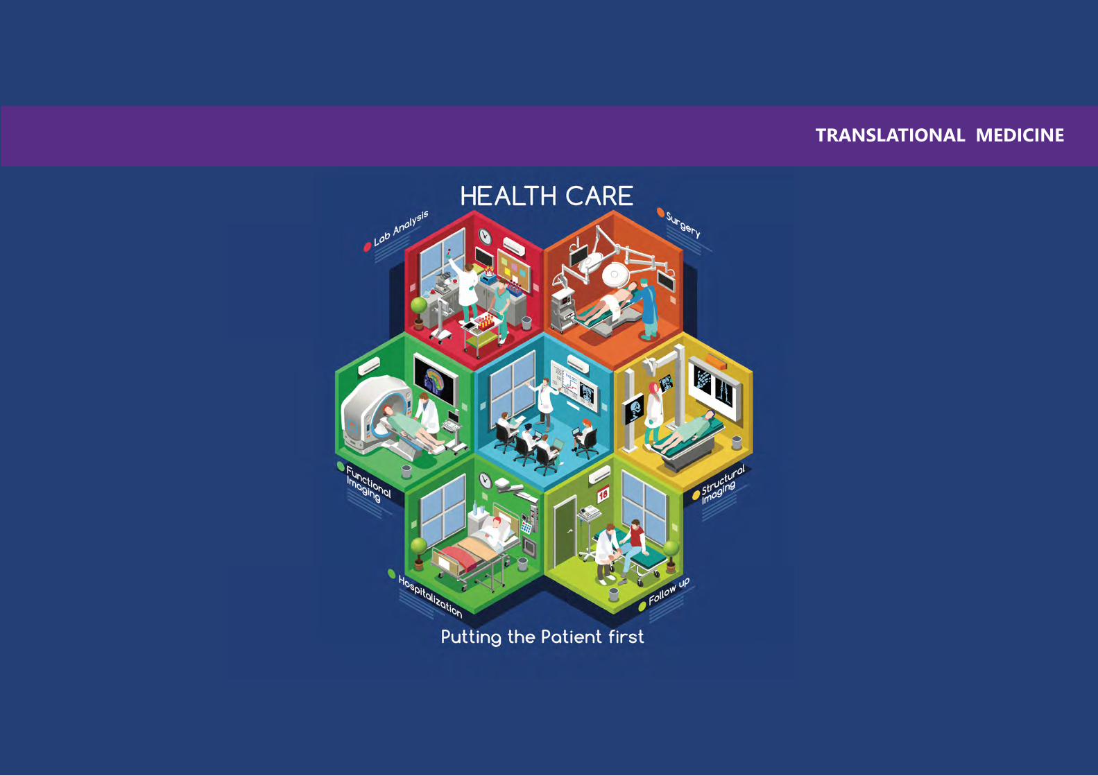
156 157
TRANSLATIONAL MEDICINE
158 159
Professor and Chairman
Ophthalmology
NYU School of Medicine
BIO Joel S. Schuman, MD, FACS is Professor and Chairman of Ophthalmology at New York University (NYU) Langone Medical Center, Professor of Electrical and Computer Engineering at the NYU Tandon School of Engineering and Professor of Professor of Neuroscience and Physiology at the NYU Neuroscience Institute. Prior to arriving at NYU in 2016, he was Distinguished Professor and Chairman of Ophthalmology, Eye and Ear Foundation Endowed Chair in Ophthalmology, Director of UPMC Eye Center (2003-2016) and before that was at Tufts University in 1991, where he was Residency Director and Glaucoma and Cataract Service Chief. In 1998 he became Professor of Ophthalmology, and Vice Chair in 2001.
Dr. Schuman and his colleagues were first to identify a molecular marker for human glaucoma, published in Nature Medicine in 2001. Continuously funded by the National Eye Institute as a principal investigator since 1995, he is an inventor of optical coherence tomography (OCT), used world-wide for ocular diagnostics. Dr. Schuman published more than 300 peer-reviewed scientific journal articles.
In addition, in 2002 Dr. Schuman received the Alcon Research Institute Award and the Lewis Rudin Glaucoma Prize, in 2006 the ARVO Translational Research Award, and in 2012 the Carnegie Science Center Award as well as sharing the Champalimaud Award (a 1 million Euro cash prize). He was elected to the American Ophthalmological Society in 2008. In 2011 Dr. Schuman was the Clinician-Scientist Lecturer of the American Glaucoma Society. In 2013 he gave the Robert N. Shaffer Lecture at the American Academy of Ophthalmology (AAO) Annual Meeting, and received the AAO Lifetime Achievement Award. In 2014 he became a Gold Fellow of ARVO. He was elected to the American Association of Physicians in 2016. He is named in Who’s Who in America, Who’s Who in Medical Sciences Education, America’s Top Doctors and Best Doctors in America, and has been named a Top Doctor by Pittsburgh Magazine 2006 – 2016.
Joel S. Schuman
OPTICAL COHERENCE TOMOGRAPHY
ABSTRACT Optical Coherence Tomography (OCT) has matured in less than twenty-five years from a single A-scan taking more than 20 minutes to perform to a technology capable of imaging at more than 1,000,000 A-scans per second. It is the most broadly and rapidly adopted technology ever used in ophthalmology. There have been multiple technical advances since OCT’s introduction in 1991, including cross-sectional imaging of ocular tissues, segmentation of optical cross-sections into relevant tissue structures and layers, the advance to commercial time domain OCT (TD-OCT) at 400 A-scans per second, the introduction of normative OCT data, the advance from time domain to spectral domain OCT (SD-OCT), with commercially available SD-OCT at 25,000 to 64,000 A-scans per second, to ever faster imaging with swept source OCT (SS-OCT) and SD-OCT with ultra-fast cameras, with 3 dimensional OCT imaging and 3D image reconstruction. Laboratory work demonstrates even great potential in terms of image acquisition speed, as well as the capability for adaptive optics combined with OCT, spectroscopic OCT imaging, Doppler OCT, OCT oximetry, and multiple other potential applications. OCT in 2017 is faster, more reproducible and enables more detailed imaging than ever before.
OCT software allows clinicians to measure neural tissue in great detail, including the optic nerve, peripapillary retinal nerve fiber layer (RNFL) and the macular ganglion cell complex (GCC). These tissues can be segmented from OCT images and analyzed in three dimensions. Further, the measurements can be compared to normative data, allowing the discrimination between health and disease (in this case, glaucoma) and assessment of the degree of glaucomatous abnormality. Clinicians now have available to them software for assessing statistically significant change over time, allowing the evaluation of glaucoma progression.
The question that arises is, “How does statistically significant glaucomatous progression as detected by OCT relate to clinically significant glaucomatous change?” There is no easy answer to this question. It is possible to equate degree of RNFL abnormality (structural glaucoma damage) with the amount of visual field abnormality (functional glaucoma damage). It is clear that a dead or absent neuron cannot function, and that when enough neurons are lost this must correspond to lost visual field. Several studies have shown that progressive loss of RNFL as measured by OCT corresponds to progressive visual field loss, and further, that OCT detects more change events in a given period of time, and earlier change, than what is detectable by conventional standard achromatic perimetry. The relationship between OCT measured RNFL thinning and visual fields should be interpretable in the same context that visual field loss relates to clinically significant change in visual function. This parallelism may permit the understanding of OCT identified structural change and clinically significant glaucomatous change.

160 161
Consultant
Vascular Surgery
Cleveland Clinic Abu Dhabi
Dr Serino MD graduated with honors in 1983 by the Catholic University School of Medicine in Rome and completed his residency in general Surgery in 1987 by the Department of Surgery of the University of Rome “la Sapienza”. He became in 1987 Assistant professor of Surgery at the Catholic University, working in the department of Substitutive and Transplantation Surgery of the University Hospital A. Gemelli in Rome, where he started one of the first diabetic foot surgery programs in Italy.
In 1989 he was invited as Visiting Assistant Professor at the University of Texas of Houston, focused in a research and clinical transplantation program. He regained his position of Assistant professor of Surgery at The Catholic University Hospital in Rome where he focused on transplant and vascular surgery, founding the first endovascular program in Rome. He joined in 1997 the Vascular Surgery department of the IDI Research Hospital in Rome and set up the endovascular program which lately directed : the center was pioneer in several fields, as the aortic endograft surgery , implanting the first AneuRX graft in the country , the hybrid endo by pass surgery and gene therapy, launching the first clinical research program on therapeutic angiogenesis. He accounted more than 3000 vascular interventions, was site principal Investigator of 12 clinical research projects, completed 212 publications and presented as faculty in 128 national or international meetings.
On research side, he is Scientific Director of the Explora Research Laboratory in Rome since 2010, where he has inspired and directed several European Funded International Projects on Cardio Vascular Tissue Engineering and regenerative medicine ( total fund raising of 17 mil euro) . He was elected Associate Professor, Biomaterials and Tissue Engineering Institute, Composite and Biomedical Materials CNR National Center for Research in 2013 and served as Adjunct Professor and Visiting Professor at NTU ,School of Material Science and Engineering of Singapore in 2008-2009 and 2013-2014 . In July 2014 he joined the Cleveland Clinic Abu Dhabi as Consultant Vascular Surgeon.
BIO
Francesco Serino
The Grail Project is focused on an innovative approach to the therapy for atherosclerosis: current therapy is based on dilatation (angioplasty) of by passing obstructed segments of diseased vessels with the aim to restore distal organs flow, while there was never an attempt for regenerate the diseased vessel. The atherosclerotic disease originate and progress in the Intimal layer, even when the extension of it will occlude the arterial segment involved. Consequently, it is possible to question if the therapy of all atherosclerotic vessels could be limited to the removal of the intimal layer only and we have seen as this concept has been widely applied in vascular surgery when applied to extra coronary vessels (so called Endarterectomy). The newest technology implementation, as the cool laser energy, allow today to extend the concept of “vessel de-bulking”, removing most of the material occluding it, with endoluminal technology, with means intervention conducted by a simple arterial puncture in the groin with patient fully awaken and able to be dismissed on the same day. This technique can be applied to coronary arteries as well as peripheral vessels. The Grail project and consortium is working to a revolutionary approach. The in vivo engineering of the inner arterial layer after plaque removal. To this aim “smart polymers”, which are able to respond to stimuli and may present active biological properties, represent an invaluable tool in biotechnology and medicine, where they can act as drug carriers or scaffold for tissue engineering. Some of those present properties which render them suitable for in vivo tissue engineering approach, as the temperature sensitivity or Inverse temperature Transition (ITT), bio degradability, inject ability, for which they can be delivered in liquid status by specially designed catheters and left to shift in a solid form at body temperature. The Grail Project is based on the use of recombinant Elastin Like Polymers that seems a versatile scaffold with properties the can be used in this innovative application, the repair or engineering of a blood vessel.
IN VIVO, NON-INVASIVE, VASCULAR TISSUE ENGINEERING IS POSSIBLE: THE GRAIL PROJECT ACHIEVEMENTS
ABSTRACT

162 163
Professor and Associate Dean for Bioengineering
Chemical and Biomolecular Engineering
NYU Tandon School of Engineering
Mary K. Cowman, Ph.D., is Professor of Biochemistry, Bioengineering, and Orthopaedic Surgery, and Associate Dean for Bioengineering in the New York University Tandon School of Engineering. She received her Ph.D. in Chemistry from Case Western Reserve University, and completed postdoctoral training in Biochemistry at Brandeis University, and in Ophthalmology Research at Columbia University College of Physicians and Surgeons. Dr. Cowman’s research concerns the structure, function, and medical applications of the biomatrix surrounding cells, with special expertise in hyaluronan polysaccharide. Dr. Cowman is President of the International Society for Hyaluronan Sciences.
BIO
Mary K. Cowman
The glycosaminoglycan hyaluronan (HA) is a key component of the microenvironment surrounding cells. In healthy tissues, HA molecules have extremely high molecular mass, and consequently large hydrodynamic volumes. Tethered to the cell surface by clustered receptor proteins, HA molecules crowd each other, as well as other macromolecular species. This leads to severe non-ideality, because steric exclusion leads to an increase in effective concentration of the macromolecules. The excluded volume depends on both polymer concentration and hydrodynamic volume / molecular mass. The biomechanical properties of the extracellular matrix, tissue hydration, receptor clustering, and receptor-ligand interactions are strongly affected by the presence of HA, and its molecular mass. In inflammation, the HA chains can be degraded. Depending on the rate of chain degradation relative to the rates of new synthesis and removal of damaged chains, short fragments of the HA molecules can be present at significant levels. The physical properties of the extracellular matrix are affected, and the HA fragments de-cluster their primary receptors and act as endogenous danger signals. Analysis of the extent of HA fragmentation can be utilized in medical diagnostic tests. In addition, control of receptor interactions with HA fragments is leading to new therapeutic approaches to tissue repair.
HYALURONAN AND HYALURONAN FRAGMENTS: DIAGNOSTIC AND THERAPEUTIC USE
ABSTRACT

164 165
Assistant Professor
Pathology and Neurology
NYU School of Medicine
Dr. Snuderl is Assistant Professor of Pathology and Neurology and Director of the Molecular Pathology and Diagnostics at NYU Langone Medical Center. He obtained his MD degree from Charles University, Prague and completed his residency in Anatomic Pathology and Neuropathology at Massachusetts General Hospital. He then completed fellowship in Molecular Genetic Pathology at Harvard Medical School and post-doctoral fellowship at the Edwin L .Steele Laboratory for Tumor Biology at Massachusetts General Hospital.
Dr. Snuderl joined the faculty at NYU Department of Pathology in January 2013 and has been actively involved in designing novel molecular tests and platforms particularly in molecular neuropathology and epigenetics. Dr. Snuderl has strong interest in tumor genetics, heterogeneity and microenvironment. The American Association of Neuropathologists awarded his work with the Lucien J. Rubinstein Award for the Best Paper on Neuro-oncology in 2009 and 2015 and Honorable Mention in the same category in 2008 and 2010.
BIO
Matija Snuderl
Cancer heterogeneity represents a critical obstacle to efficient therapy. We have previously shown that mutually exclusive genetic subclones can coexist and play different roles in growth of brain tumors and that metabolism of tumor cells depends on their microenvironment. Cancer cells can also manipulate normal stromal cells to provide necessary growth factors. In addition to genetic heterogeneity of cancer cells, molecular profiling of the whole tumor can be skewed by the presence of variable stromal and inflammatory cell subpopulations. To separate cancer cell, stromal cell and inflammatory subclones and dissect their genetic and epigenetic heterogeneity, we implemented DEPArray system, a microchip-based digital sorter, which enables us to distinguish cells by fluorescently labeled antibodies and DNA content and isolate pure, single cells or homogenous subpopulations from fresh or archival pathology samples. Downstream analysis includes DNA and RNA next-generation sequencing and methylation profiling on a single cell level. Using dielectrophoretic capture and digital sorting, we can investigate genetic and epigenetic heterogeneity of cancer cells with unambiguous determination of sequence variants, loss-of-heterozygosity, copy number variants and methylation. We can further investigate epigenetic and transcriptional changes in tumor-associated stromal and inflammatory cells and dissect how cancer cell growth and spread depends on non-neoplastic cells.
DISSECTING GENETIC AND EPIGENETIC HETEROGENEITY IN CANCER USING DIELECTROPHORETIC CAPTURE AND DIGITAL SORTINGOF SINGLE CELLS
ABSTRACT

166 167
Professor
Orthopaedic Surgery and Cell Biology
NYU School of Medicine
Thorsten Kirsch, Ph.D., is a Professor of Orthopaedic Surgery and Cell Biology at NYU School of Medicine. In addition, he is Vice Chair for Research in the Department of Orthopaedic Surgery and Director of the Musculoskeletal Research Center. Dr. Kirsch joined NYU School of Medicine in 2008 coming from the University of Maryland School of Medicine, where he was a Professor and Director of Orthopaedic Research. Dr. Kirsch studies mechanisms regulating skeletal cell differentiation and repair during development and pathology.
Dr. Kirsch earned his Ph.D. from the Albrecht-Ludwig-University, Erlangen, Germany with summa cum laude in 1992, and completed his postdoctoral training at the University of South Carolina and University of Pennsylvania. Dr. Kirsch has spoken at many national and international meetings, has received numerous awards, and is a reviewer for various National Institutes of Health Study Sections.
BIO
Thorsten Kirsch
A special challenge is the repair or regeneration of articular cartilage after injury or in osteoarthritis, since it is an avascular tissue with no intrinsic repair mechanisms. To define novel mechanisms to improve cartilage repair or regeneration I established a collaborative research program involving bioengineers, biologists, imaging researchers, and clinicians. Specifically, we are interested in identifying novel mechanisms that are involved cartilage destruction after injury or pathology in order to develop novel therapeutic strategies to interfere with these mechanisms and support tissue regeneration. Recently, we have identified together with Dr. Cowman from NYU Tandon School of Engineering and our collaborators from Canada a 15mer peptide that interacts with hyaluronan and greatly improved cartilage repair in rabbits after microfracture surgery. Our findings indicate that this peptide improves cartilage repair by enabling hyaluronan to form a stem cell niche at the injury site that allows stem cells to attach and undergo chondrogenic differentiation, and by interfering with catabolic signaling of low molecular hyaluronan fragments in the joint and suppressing inflammatory events that slow down or prevent cartilage repair. Current studies are underway to develop this peptide together with stem cells and high molecular hyaluronan into a novel therapeutic strategy for cartilage repair.
A MULTIDISCIPLIARY TRANSATIONAL RESEARCH APPROACH TO IMPROVE CARTILAGE REPAIR
ABSTRACT

168 169
Chief
Anesthesiology
Cleveland Clinic Abu Dhabi
Dr. Doyle is the Chief of General Anesthesiology at Cleveland Clinic Abu Dhabi. He is on secondment from Cleveland Clinic, Cleveland, USA, and also serves as Professor of Anesthesiology at the Cleveland Clinic Lerner College of Medicine of Case Western Reserve University. He received his MD degree in 1982 and his PhD degree in Biomedical Engineering in 1986, both from the University of Toronto, Canada.
Dr. Doyle has a long-standing interest in ENT anesthesia and difficult airway management as well as an interest in the use of technology in medicine. He is past president of both the Society for Airway Management and the Society for Technology in Anesthesia. Dr. Doyle is widely published. His most recent book, coedited with Dr. Basem Abdelmalak, is entitled Anesthesia for Otolaryngologic Surgery (Cambridge University Press, 2012). Another book, entitled Clinical Airway Management: An Illustrated Case-Based Approach, was published by Cambridge University Press this year.
BIO
John Doyle
The need for simple and reliable means of respiratory monitoring has existed since the time of Hippocrates. This need has become especially strong in recent years with the increased use of opioids such as morphine or fentanyl for acute pain management, as these drugs depress respiration. Despite this important need, no simple and reliable method of continuous respiratory monitoring has come into wide-spread clinical use. In this presentation I describe the potential of advanced acoustic analysis of breath sounds as one means to meet this need, as well as review the technical and clinical challenges associated with classical methods of respiratory monitoring (capnography, pulse oximetry, arterial blood gas analysis, spirometry, thermistor-based methods, methods based on photoplethysmography, and methods based on electrical impedance). As an example of a novel method of respiratory sound analysis I show how color spectrographic analysis of breath sounds recorded from the external ear canal might serve as a simple, reliable and inexpensive candidate solution to the respiratory monitoring problem. I hypothesize that the real-time display of color spectrogram breathing patterns locally or at a central monitoring station may turn out to be a useful means of respiratory monitoring in patients at increased risk of respiratory depression.
NEW METHODS OF RESPIRATORY MONITORING: EMBRACING TECHNICAL CHALLENGES
ABSTRACT

170 171
Assistant Professor
Division of Engineering - Mechanical and Biomedical Engineering
NYU Abu Dhabi
Dr. Mohammad Qasaimeh is an Assistant Professor of Mechanical and Biomedical Engineering at NYU Abu Dhabi (NYUAD), and with the Mechanical and Aerospace Engineering Department at Tandon School of Engineering, New York University, Brooklyn, USA. He established the Advanced Microfluidics and Microdevices Laboratory (AMMLab), and his current research interests include developing microfluidic and MEMS devices for biomedical applications and point of care diagnostics. Prior to joining NYUAD, he was a Postdoctoral Research Associate at Massachusetts Institute of Technology and Harvard Medical School. Dr. Qasaimeh completed his PhD degree in Biomedical Engineering in 2013 from McGill University, where he received several prestigious fellowships and awards including the NSERC Postdoctoral Fellowship, the Alexander Graham Bell Graduate Scholarship (CGSD3), and the FQRNT Students-Researchers Stars Award. Dr. Qasaimeh research has been published in several peer-reviewed journals including Nature Communications, PLOS Biology, Lab on a Chip, Scientific Reports, among others. Dr. Qasaimeh delivered more than 15 keynote and invited speeches at national and international conferences, and is involved with several local and international conferences as an organizing committee member.
BIO
Mohammad A. Qasaimeh
Over the last decade, there has been great interest in utilizing the peripheral blood circulating tumor cells (CTCs) to predict response to therapy and overall survival of cancer patients. CTCs shed from the tumor are thought to contribute to the hematogenous spread of cancer to distant sites. Despite the clinical importance of CTCs, current knowledge of these cells is extremely poor, largely owing to the fact that their isolation and enumeration is very challenging. CTCs are very rare, typically ranging from 1 to 10 cells per milliliter of blood. One important advantage of using a CTC blood test is that it possesses a significantly lower risk to the patient compared to surgically obtaining a fresh tissue biopsy.
Recently, several groups started developing microfluidic platforms for CTCs isolation. Microfluidics, first introduced two decades ago, present an attractive approach for engineering and miniaturizing bioassays by accurately controlling biological samples and their microenvironments. Microfluidics have the potential to achieve high throughputs, sensitivity and selectivity for innovative single cell analysis platforms.
This talk will highlight our efforts in developing a microfluidic device for isolating plasma CTCs from multiple myeloma (MM) blood samples. MM is a cancer caused by malignant plasma cells in bone marrow. The device was able to detect a low (<10 cell/mL) level of plasma cells in normal blood, which increased significantly in MM patients. The device showed superior sensitivity and was able to detect an elevated CTCs count in MM patients that have otherwise normal peripheral blood indicators of MM.
MICROFLUIDICS FOR CLINICAL APPLICATIONS: ISOLATING CIRCULATING TUMOR CELLS FROM PERIPHERAL BLOOD SAMPLES
ABSTRACT
172 173
INDEX
1- Mohamed Abdelgawad
2- Rohit Karnik
3- Thomas Gervais
4-John Yeow
5- Mohammed Zourob
6- Ruba Khnouf
7- Alaaldeen Al-Halhouli
8- Anwarul Hasan
9- Suzanne Gaudet
10-Kevin E. Healy
11- Huseyin Yalcin
[email protected] UniversityEgypt
[email protected] Institute of TechnologyUSA
[email protected] Polytechnique de MontrealCanada
[email protected] of WaterlooCanada
[email protected] UniversityKSA
[email protected] University of Science and TechnologyUSA
[email protected] Jordanian UniversityJordan
[email protected] UniversityQatar
[email protected] Medical SchoolUSA
[email protected] of California, BerkeleyUSA
[email protected] UniversityQatar
12- Nikhil Gupta
13- Mary Cowman
14- Will Shu
15- Richard Black
16- Sylvie Coupaud
17- Asimina Kazakidi
18- Seiichi Yamano
19- Thorsten Kirsh
20- Chuanjiu Liu
21- Youssef Wadghiri
22- Wenbiao Gan
[email protected] York University Tandon School of EngineeringUSA
[email protected] York University Tandon School of EngineeringUSA
[email protected] of StrathclydeUK
[email protected] of StrathclydeUK
[email protected] of StrathclydeUK
[email protected] of StrathclydeUK
[email protected] York University College of DentistryUSA
[email protected] York University School of MedicineUSA
[email protected] York University School of MedicineUSA
[email protected] York University School of MedicineUSA
[email protected] York University School of MedicineUSA
174 175
INDEX
23- Weiqiang Chen
24-Ryan Hartman
25- Kalle Levon
26-Vittoria Flamini
27- Matija Snuderl
28- Joel S. Schuman
29-Bryan Allen Chin
30- Pengyu Chen
31- Katsuo Kurabayashi
32- Edward Guo
33- Joo Kim
[email protected] York University Tandon School of EngineeringUSA
[email protected] York University Tandon School of EngineeringUSA
[email protected] York University Tandon School of EngineeringUSA
[email protected] York University Tandon School of EngineeringUSA
[email protected] York University School of MedicineUSA
[email protected] York University School of MedicineUSA
[email protected] UniversityUSA
[email protected] UniversityUSA
[email protected] of MichiganUSA
[email protected] UniversityUSA
[email protected] York University Tandon School of EngineeringUSA
34- Andy Hamilton
35- Rihab Nasr
36- Laising Yen
37-Ryo Jimbo
38- Amitabha Chattopadhyay
39- Jeroen Adema
40- Susanna Narkilahti
41- Chwee-Teck Lim
42- Chen Chia-Hung
43- Abraham Mansouri
44- Ghaleb Husseini
[email protected] York UniversityUSA
[email protected] University of BeirutLebanon
[email protected] College of MedicineUSA
[email protected] UniversitySweden
[email protected] for Cellular and Molecular BiologyIndia
[email protected] CorporationNetherlands
[email protected] of TampereFinland
[email protected] University of SingaporeSingapore
[email protected] University of SingaporeSingapore
[email protected] University in DubaiDubai, UAE
[email protected] University of SharjahSharjah, UAE

176 177
INDEX
45- Hasan Al-Nashash
46- Yaser greish
47- Florian Roser
48-Jeremy Teo
49- Francesco Serino
50- Khaled M. Al-Aribe
51- Matteo Chiesa
52- Anastasios Hantzakos
53- John Doyle
54- Mohammad Qasaimeh
55- Pance Naumov
[email protected] University of SharjahSharjah, UAE
[email protected] Arab Emirates UniversityAl Ain, UAE
[email protected] Clinic Abu DhabiAbu Dhabi, UAE
[email protected] UniversityAbu Dhabi, UAE
[email protected] Clinic Abu DhabiAbu Dhabi, UAE
[email protected] Dhabi UniversityAbu Dhabi, UAE
[email protected] Institute of Science and TechnologyAbu Dhabi, UAE
[email protected] Clinic Abu DhabiAbu Dhabi, UAE
[email protected] Clinic Abu DhabiAbu Dhabi, UAE
[email protected] York University Abu DhabiAbu Dhabi, UAE
[email protected] York University Abu DhabiAbu Dhabi, UAE
56- Sachin Khapli
57- Sohmyung Ha
58- Mazin Magzoub
59- Dipesh Chaudhury
60- Rafael Song
61- Kourosh Salehi-Ashtiani
62-Piergiorgio Percipalle
63- Shady Amin
64-George Shubeita
65- Kartik Sreenivasan
66-Mohamed Al-Sayegh
[email protected] york University Abu DhabiAbu Dhabi, UAE
[email protected] York University Abu DhabiAbu Dhabi, UAE
[email protected] York University Abu DhabiAbu Dhabi, UAE
[email protected] York University Abu DhabiAbu Dhabi, UAE
[email protected] York University Abu DhabiAbu Dhabi, UAE
[email protected] York University Abu DhabiAbu Dhabi, UAE
[email protected] York University Abu DhabiAbu Dhabi, UAE
[email protected] York University Abu DhabiAbu Dhabi, UAE
[email protected] York University Abu DhabiAbu Dhabi, UAE
[email protected] York University Abu DhabiAbu Dhabi, UAE
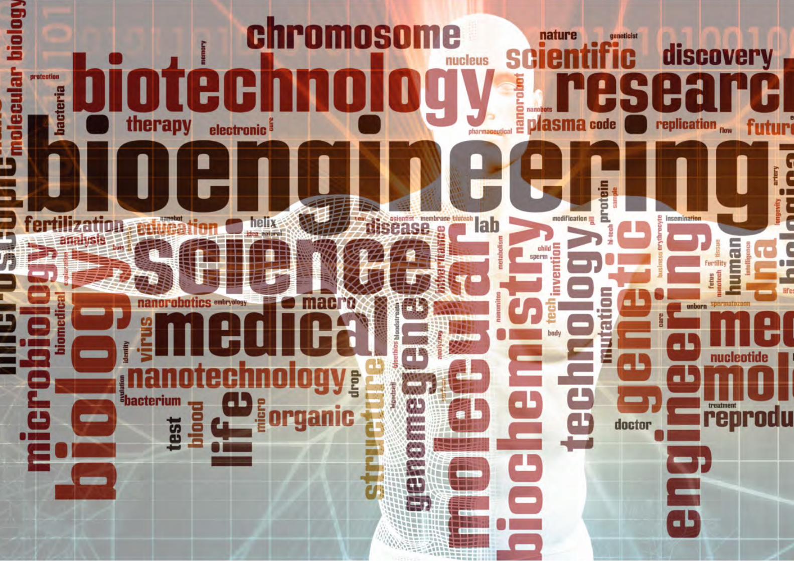
178 179
180


