Embed Size (px)
Citation preview
Surgical management ofmedically refractory epilepsy in
patients with polymicrogyria*Doris D.Wang, †Renatta Knox, *JohnD. Rolston, *Dario J. Englot, ‡A. James Barkovich,
§Tarik Tihan, *¶Kurtis I. Auguste, †Robert C. Knowlton, †Susannah B. Cornes, and
*Edward F. Chang
Epilepsia, 57(1):151–161, 2016doi: 10.1111/epi.13264
Dr. Doris Wang is aneurosurgery residentat the University ofCalifornia, SanFrancisco.
SUMMARY
Objective: Polymicrogyria (PMG) is a malformation of cortical development charac-
terized by formation of an excessive number of small gyri. Sixty percent to 85% of
patients with PMG have epilepsy that is refractory to medication, but surgical options
are usually limited. We characterize a cohort of patient with polymicrogyria who
underwent epilepsy surgery and document seizure outcomes.
Methods: A retrospective study of all patients with PMG who underwent epilepsy
surgery (focal seizure foci resection and/or hemispherectomy) at our center was
performed by review of all clinical data related to their treatment.
Results: We identified 12 patients (7 males and 5 female) withmean age of 18 (ranging
from 3 months to 44 years) at time of surgery. Mean age at seizure onset was 8 years,
with themajority (83%) having childhood onset. Six patients had focal, five hadmultifo-
cal, and one patient had diffuse PMG. Perisylvian PMG was the most common pattern
seen on magnetic resonance imaging (MRI). Eight patients had other cortical malfor-
mations including hemimegalencephaly and cortical dysplasia. Scalp electroen-
cephalography (EEG) often showed diffuse epileptic discharges that poorly lateralized
but were focal on intracranial electrocorticography (ECoG). Eight patients underwent
seizure foci resection and four underwent hemispherectomy. Mean follow-up was
7 years (ranging from one to 19 years). Six patients (50%) were seizure-free at last fol-
low-up. One patient had rare seizures (Engel class II). Three patients were Engel class
III, having either decreased seizure frequency or severity, and two patients were Engel
class IV. Gross total resection of the PMG cortex trended toward good seizure control.
Significance: Our study shows that even in patients with extensive or bilateral PMG
malformations, some may still be good candidates for surgery because the epilepto-
genic zonemay involve only a portion of themalformation. Intracranial ECoG can pro-
vide additional localizing information compared to scalp EEG in guiding resection of
epileptogenic foci.
KEY WORDS: Polymicrogyria, Cortical malformation, Cortical dysplasia, Malforma-
tion of cortical development, Schizencephaly, Hemimegalencephaly, Hemispherec-
tomy, Refractory epilepsy, Surgical treatment, Seizure outcome.
Polymicrogyria (PMG) is a cortical malformation charac-terized by an excessive number of small irregular gyri lead-ing to the appearance of an abnormal cortical surface. Itarises from disturbances during cerebral cortical develop-ment, most likely during the late stages of neuronal migra-tion and early stages of cortical organization.1 Causes ofPMG are diverse and include congenital infection (in partic-ular cytomegalovirus infection),2 in utero ischemia, andgenetic mutations (e.g.,WDR62,NDE1, and TBR2).3
Accepted October 30, 2015; Early View publication December 9, 2015.*Department of Neurological Surgery, UCSF Comprehensive Epilepsy
Center, University of California, San Francisco, California, U.S.A.;Departments of †Neurology, ‡Radiology, and §Pathology, University ofCalifornia, San Francisco, California, U.S.A.; and ¶Benioff Children’sHospital, University of California, San Francisco, California, U.S.A.
Address correspondence to Edward F. Chang, Department of Neurologi-cal Surgery, University of California San Francisco, 505 Parnassus Avenue,M779, San Francisco, CA 94143, U.S.A. E-mail: [email protected]
Wiley Periodicals, Inc.© 2015 International League Against Epilepsy
151
FULL-LENGTHORIGINALRESEARCH
PMG can be focal, multifocal, or diffuse, affecting differ-ent areas and portions of the cerebral cortex in a unilateralor bilateral pattern. The most common locations are theinsula and the perisylvian cortex. Patients with PMG mayhave a variety of clinical presentations including hemipare-sis, intractable epilepsy, as well as speech and developmen-tal delays. Although the incidence of PMG is unknown, it isone of the most common malformations of cortical develop-ment and an important contributor to medically refractoryepilepsy,4 as 60–85% of all patients with the diagnosis ofPMG have epilepsy.5 Onset is typically during childhoodbut can present in the second decade. Seizure types are mul-tiple and include focal with or without alteration of aware-ness, secondary generalized, epileptic spasms, and atonicseizures, among others. Associated epileptic syndromes aregenerally defined as those symptomatic of malformations ofcortical development, and status epilepticus of sleep may bea feature.6,7 Most patients with PMG are refractory to medi-cation.8
Clinical management of epilepsy patients with PMGcan be difficult. Often, they are not good surgical candi-dates given the extent and often multifocal involvementof the lesion. There are limited data on surgical outcomesof patients with PMG.9–12 A few case reports havedescribed successful surgical treatment of patients withfocal PMG by resection of epileptic foci,5,13 but there arefew systematic studies to date that examine surgical out-comes for patients with PMG who have undergone epi-lepsy surgery.
In this study, we characterize 12 cases of PMGwith medi-cally refractory epilepsy that underwent surgical resectionfor management of seizures. Surgical interventions lead to areduction in seizure frequency or severity, and in somecases, complete seizure freedom.
MethodsPatient selection
Patients were queried from neuroimaging and pathologydatabases at University of California San Francisco (UCSF)between 1987 and 2014. Patients were selected based oneither radiographic or pathologic findings of PMG. We alsoincluded patients with PMG with other malformations ofcortical development including cortical dysplasia, schizen-cephaly, heterotopia, and hemimegalencephaly. We identi-fied 34 patients and included 19 who underwent surgicalresection for management of their epilepsy. Only 12 patientshad sufficient clinical data to be in the study. All researchprotocols were approved by the UCSF institutional reviewboard for human research (Committee for HumanResearch).
Clinical data reviewPreoperative data included date of seizure onset, type of
seizure (focal with or without alteration of awareness,secondary generalized, atonic, infantile spasms, and so on),seizure frequency, seizure duration, medications, and otherassociated developmental symptoms. Other preoperativetests included scalp electroencephalography (EEG) studies,magnetic resonance (MR) imaging, and neuropsychologicaltesting when available. Data pertaining to patient demo-graphics, seizure-related history, neuroimaging, surgicaldetails, pathology reports, and seizure outcome wereobtained from hospital and departmental records. Availablescalp EEG was independently reviewed by an experiencedepileptologist (SBC). Available preoperative MRI was inde-pendently reviewed by an experienced neuroradiologist(AJB). PMG lesions were considered to be focal if they wererestricted to a single lobe or well-circumscribed adjacentgyri (i.e., perisylvian), multifocal if they were multiplediscrete areas (i.e., bilateral hemispheric involvement), anddiffuse if the abnormal cortices expanded to multiple lobesor involved the whole hemisphere.
Resection of epileptogenic fociOperative data, including the use of cortical and subcorti-
cal language and motor mapping and intracranial electro-corticography (ECoG), were reviewed. Most cases utilizedStealth (Medtronic, Minneapolis, MN, U.S.A.) or BrainLab(BrainLab North America, Westchester, IL, U.S.A.) frame-less neuronavigation to assist with intraoperative localiza-tion. Intraoperative electrocorticography (ECoG) wasobtained with use of a customized crown of 16 ball-tippedcarbon electrodes referred to a common cerebral reference.When necessary, additional coverage was obtained by usingsubdural electrode strips and depth electrodes. Patientsunderwent implantation of chronic subdural electrodearrays for extraoperative seizure monitoring and electricalstimulation mapping when there is no clear localization of
Key Points• In our cohort of 12 patients with polymicrogyria whounderwent epilepsy surgery, 83% of patients hadchildhood onset of epilepsy secondary to polymicro-gyria
• Perisylvian polymicrogyria was the most common pat-tern seen, and most patients had unilateral polymicro-gyria
• Despite extensive abnormalities on MRI and broadepileptiform abnormalities on scalp EEG, intracranialelectrocorticography can provide additional localizinginformation for guiding surgical planning
• Surgical intervention led to significant seizure reduc-tion in 33% of patients, and 50% of patients becameseizure-free
Epilepsia, 57(1):151–161, 2016doi: 10.1111/epi.13264
152
D. D.Wang et al.
epileptic foci on scalp EEG and/or the lesion was adjacentto eloquent cortical areas. Extraoperative ECoG was used inpatients 4, 6, 10, and 12 to guide resection, and intraopera-tive ECoG was typically not repeated prior to resection. Theanatomic resection was tailored according to the extent ofthe intracranial ECoG abnormalities and the functionalanatomy. Epileptogenic tissue was defined by those con-tacts with active epileptiform discharges on intracranialECoG during the interictal or ictal period from either intra-operative or extraoperative recordings. Epileptic tissue wasconsidered to be focal if epileptiform discharges arose fromcontacts within a well-circumscribed region of the cortex orwithin a lobe, and diffuse if discharges were seen through-out contacts spanning multiple lobes.
Patients with defined seizure foci on scalp EEG and/orintracranial ECoG underwent focal resection of their elec-trographically active regions as first line of treatment. Theexceptions are patients with hemimegalencephaly or withnonfocal ECoG findings, who received hemispherectomiesas their initial surgery. Extent of resection was designated asgross-total or subtotal according to review of available pre-and postoperative MR images and the pre- and postexcisionintracranial ECoG recordings. Resection of epileptogenictissue determined by ECoG was considered complete if alltissue exhibiting critical patterns was resected. Gross totalresection was designated for patients who underwent func-tional hemispherectomy given complete disconnection ofcortical and subcortical structures.
Postoperative outcome and statistical analysisClinic visits and phone interviews were conducted for fol-
low-up. Each patient’s outcome was assessed using theEngel Seizure Outcome Classification Scheme14: Engelclass I, free from disabling seizures (Ia, completely seizure-free since surgery; Ib, nondisabling simple partial seizuresonly since surgery; Ic, some disabling seizures since sur-gery, but free from disabling seizures for ≥2 years; Id, gen-eralized convulsions with discontinuation of antiepilepticdrugs only); Engel class II, rare disabling seizures (IIa, ini-tially free from disabling seizures, but still has rare seizures;IIb, rare disabling seizures since surgery; IIc, occasional dis-abling seizures since surgery, but rare seizures for the past2 years; IId, nocturnal seizures only); Engel class III, worth-while improvement (IIIa, worthwhile seizure reduction;IIIb, prolonged seizure-free intervals amounting to >50% offollow-up period, but not <2 years); and Engel class IV, noworthwhile improvement (IVa, significant seizure reduc-tion; IVb, no appreciable change, IVc, seizures worse).Postoperative motor and cognitive deficits were extractedfrom clinical notes, and when available, from outpatientphysical/occupational therapy visits and school perfor-mance notes.
For univariate analysis for predictors of seizure control,we used chi-square test for categorical variables and t-testsfor continuous variables.
ResultsPatient demographic and seizure characteristics
Our study included 12 patients (7 male and 5 female) withages ranging from 3 months to 44 years at time of surgery(mean age 18.7 years, median 13.1). Seizure onset rangedfrom birth to 26 years of age (mean age 8.5 years, median5.5) with the majority (10 of 12 patients, 83%) having child-hood onset. Mean epilepsy duration until surgery was10.2 years. Seizure types included focal with or withoutalteration in awareness, secondarily generalized tonic–clonic, epileptic spasms, and atypical absence seizures. Allpatients had poor response to antiepileptic drugs (AED)after a trial of multiple (three to six) medications. Afterreview of clinical semiology, scalp EEG, and imaging fea-tures, nine patients were classified as having medicallyrefractory epilepsy symptomatic of malformation of corticaldevelopment, and one patient had electrical status epilepti-cus during sleep (ESES). Two patients were uncategorizedgiven either normal or unavailable scalp EEG. In addition toseizures, three patients were found to have congenitalhydrocephalus (patients 3, 8, and 9), and one had infantilespasms (patient 11). Of note, patient 2 had a genetic syn-drome known as CLOVES-like syndrome, which is congen-ital disorder characterized by lipomatous overgrowth,vascular malformations, epidermal nevus, and spinalanomalies. Patient demographics are summarized inTable 1.
Preoperative imaging characteristicsAs demonstrated in our patient population, the location of
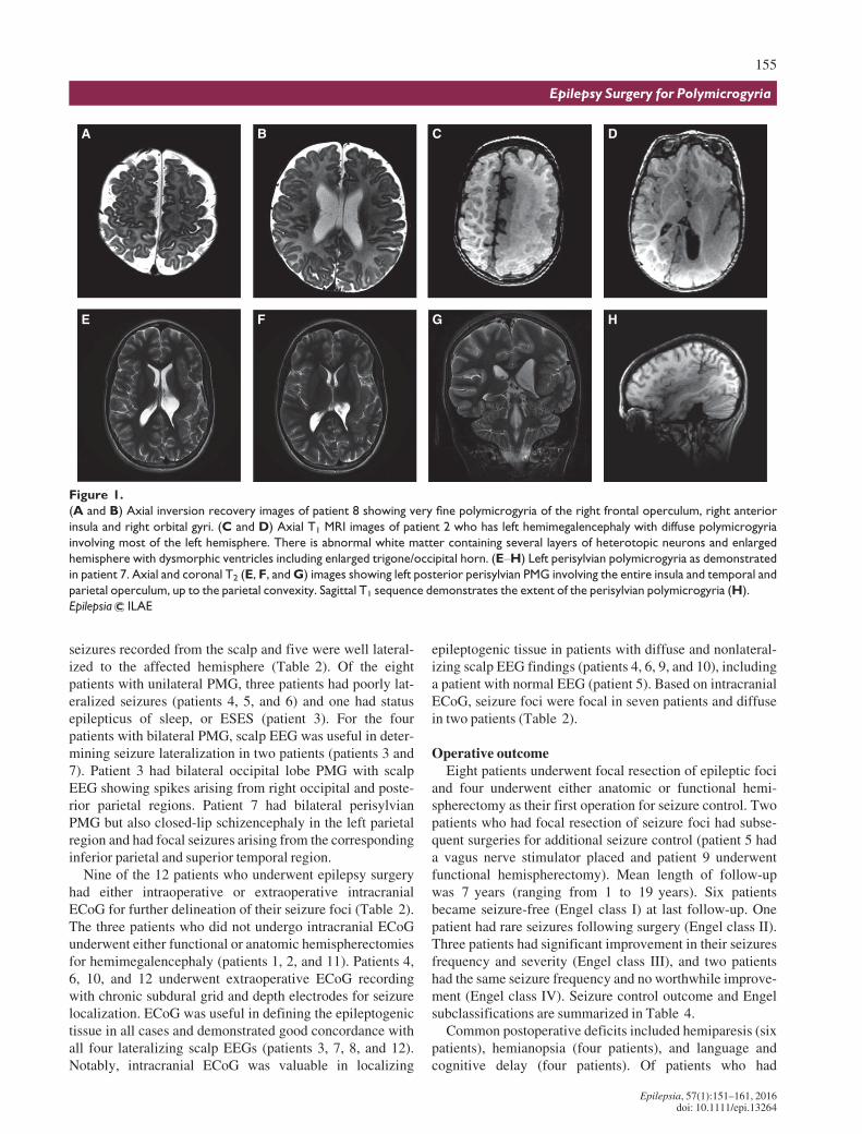
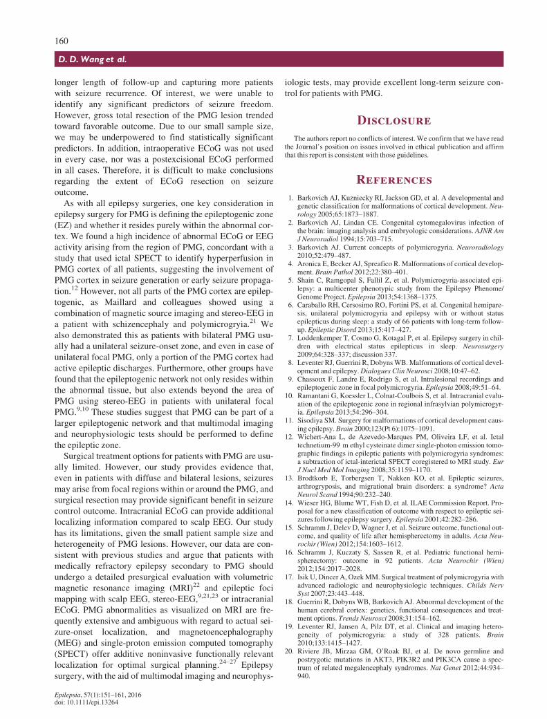
PMG can be unilateral or bilateral, and associated with othercentral nervous system (CNS) cortical development abnor-malities. The PMG lesion was found in one or more of thefollowing lobes: frontal lobe (two patients) (Fig. 1A,B),temporal lobe (three patients), and occipital lobe (threepatients). The most common location was the perisylvianregion, which was the site in six patients (50%) (Fig. 1E–H). Eight patients had unilateral PMG (three lefthemispheric, five right hemispheric), and four patients hadbilateral involvement. Those patients with bilateral involve-ment included two with bilateral perisylvian and two withbilateral occipital lobe PMG. In addition to PMG, eightpatients had other cortical malformations, including threepatients with hemimegalencephaly, two patients with corti-cal dysplasia, two patients with heterotopia/architecturaldistortion, and one patient with closed-lip schizencephaly.Of the three patients with hemimegalencephaly, PMG wasin the affected hemisphere in all cases (Fig. 1C,D). Thepreoperative MRI findings are described in Table 1.
Seizure localizationAll patients underwent video scalp electroencephalogra-
phy (EEG) for preoperative seizure localization. Of the 11available scalp EEG reports prior to surgery, eight had focal
Epilepsia, 57(1):151–161, 2016doi: 10.1111/epi.13264
153
Epilepsy Surgery for Polymicrogyria
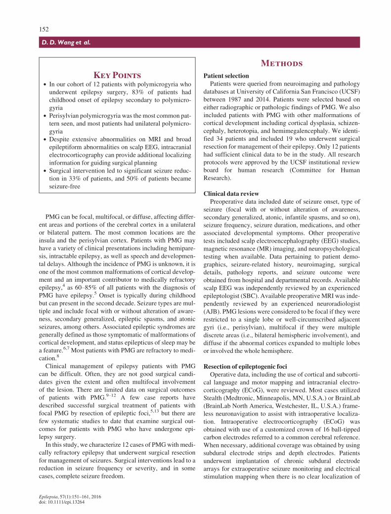
Table
1.Patientpreoperativeepilepsy
syndro
me,PMG
characteristics,su
rgery
description,andseizure
controloutcome
Pt
Age,sex
Age
onset
Preopseizure
type
PMGtypeandassociatedimagingfindings
Epilepsy
syndrome
Surgery
perform
ed
Engel
13month,F
Birth
Focaldyscognitive
Multifo
calleftPMGoffrontalpole,convexity
andfrontalandparietaloperculawith
transm
antlecorticaldysplasiaand
hemimegalencephaly
Notavailable
Lefthemispherectomy
Ia
21year,F
Birth
ES,focalm
otor
Diffuse
leftPMGwithhemimegalencephaly,
pachygyriaandsubependym
alheterotopia
Focalrefractory
withES,
symptomaticofM
CD
Leftfunctional
hemispherectomy
Ia
39year,M
6year
Focalm
otoranddyscognitive,
SGTCS,SE,atypical
absences,andESES
Bilateraltem
poraloccipitalpolymicrogyria,
bilateralparietalandoccipitalwhile
matter
volumelossandthinningoftheposterior
corpuscallosum
ESES
Rightoccipitalseizure
foci
resection
Ia
434year,M
26year
Focal�
dyscognitive,m
otor
orSG
TCS
Rightoccipitalpolymicrogyriaextending
fromoccipitalpoleto
calcarinesulcusand
toposteromedialaspectofoccipitalhorn
of
lateralventricle
Focalrefractory,sym
ptomatic
ofM
CD
Rightoccipitalseizure
foci
resection
Ia
530year,F
11year
Focal�
dyscognitive,m
otor
orSG
TCS
Focalrightperisylvianpolymicrogyriawith
involvementofthefrontal,parietaland
temporallobes
Notavailable
1.R
ightfrontalseizure
foci
resection
2.V
NS
Ic
638year,M
26year
Focaldyscognitive�
SGTCS
Righttemporallobepolymicrogyriawith
corticaldysplasia
Focalrefractory,sym
ptomatic
ofM
CD
Rightfrontoparietaland
temporalseizure
foci
resection
Ic
736year,F
Child
hood
Focaldyscognitivewith
aphasia
andmotor
symptoms�
SGTCS
Bilateralperisylvianpolymicrogyriawithleft
closed-lipschizencephaly
Focalrefractory,sym
ptomatic
ofM
CD
Leftfrontotemporalseizure
fociresection
IIc
84month,M
4month
Focalm
otor�
SGTCS
Focalrightfrontalpolymicrogyriainvolving
operculum,anteriorinsula,orbitalgyri,
reducedwhitemattervolume
Focalrefractory,sym
ptomatic
ofM
CD
1.R
ightfrontalseizure
foci
resection
2.R
ightfunctional
hemispherectomy
IIIa
910year,M
5year
Focalsensory
and
motor�
SGTCS
Bilateraloccipitallobepolymicrogyriawith
leftparietalgrossarchitecturaldistortion
likelyfrominutero
insult
Focalrefractory,sym
ptomatic
ofM
CD
Leftfunctional
hemispherectomy
IIIa
10
44year,F
18year
Focaldyscognitivewith
aphasia
�SG
TCS
Leftgreater
than
rightbilateralposterior
perisylvianpolymicrogyriaextendingto
parietaloperculum
Focalrefractory,sym
ptomatic
ofM
CD
Rightanteriortemporal,
inferiorfrontalandparietal
seizure
fociresection
IIIa
11
2year,M
2month
ES,focalm
otor
FocalrightperisylvianPMGand
hemimegalencephaly
Focalrefractory
withES,
symptomaticofM
CD
Rightfunctional
hemispherectomy
IVb
12
16year,M
3.5
year
Focalm
otor�
dyscognitive,
�SGTCS
Focalleftperisylvianpolymicrogyriawithleft
hippocampalatrophyandnodular
heterotopiaoftheoccipitalhorn
Focalrefractory,sym
ptomatic
ofM
CD
Leftsuperiortemporal,peri-
insula,andparietalseizure
fociresection
IVb
PMG,polymicrogyria;FU
,follow-up;SG
TCS,secondarygeneralizedtonic–clonicseizures;ES,epilepticspasm;SE,statusepilepticus;ESES,electricalstatusepilepticusduringsleep;MCD,malform
ationofcortical
development;VNS,vagusnervestimulator.
Epilepsia, 57(1):151–161, 2016doi: 10.1111/epi.13264
154
D. D.Wang et al.
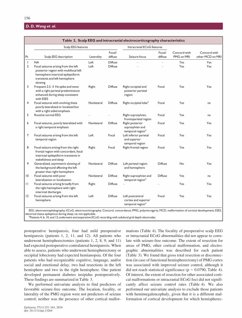
seizures recorded from the scalp and five were well lateral-ized to the affected hemisphere (Table 2). Of the eightpatients with unilateral PMG, three patients had poorly lat-eralized seizures (patients 4, 5, and 6) and one had statusepilepticus of sleep, or ESES (patient 3). For the fourpatients with bilateral PMG, scalp EEG was useful in deter-mining seizure lateralization in two patients (patients 3 and7). Patient 3 had bilateral occipital lobe PMG with scalpEEG showing spikes arising from right occipital and poste-rior parietal regions. Patient 7 had bilateral perisylvianPMG but also closed-lip schizencephaly in the left parietalregion and had focal seizures arising from the correspondinginferior parietal and superior temporal region.
Nine of the 12 patients who underwent epilepsy surgeryhad either intraoperative or extraoperative intracranialECoG for further delineation of their seizure foci (Table 2).The three patients who did not undergo intracranial ECoGunderwent either functional or anatomic hemispherectomiesfor hemimegalencephaly (patients 1, 2, and 11). Patients 4,6, 10, and 12 underwent extraoperative ECoG recordingwith chronic subdural grid and depth electrodes for seizurelocalization. ECoG was useful in defining the epileptogenictissue in all cases and demonstrated good concordance withall four lateralizing scalp EEGs (patients 3, 7, 8, and 12).Notably, intracranial ECoG was valuable in localizing
epileptogenic tissue in patients with diffuse and nonlateral-izing scalp EEG findings (patients 4, 6, 9, and 10), includinga patient with normal EEG (patient 5). Based on intracranialECoG, seizure foci were focal in seven patients and diffusein two patients (Table 2).
Operative outcomeEight patients underwent focal resection of epileptic foci
and four underwent either anatomic or functional hemi-spherectomy as their first operation for seizure control. Twopatients who had focal resection of seizure foci had subse-quent surgeries for additional seizure control (patient 5 hada vagus nerve stimulator placed and patient 9 underwentfunctional hemispherectomy). Mean length of follow-upwas 7 years (ranging from 1 to 19 years). Six patientsbecame seizure-free (Engel class I) at last follow-up. Onepatient had rare seizures following surgery (Engel class II).Three patients had significant improvement in their seizuresfrequency and severity (Engel class III), and two patientshad the same seizure frequency and no worthwhile improve-ment (Engel class IV). Seizure control outcome and Engelsubclassifications are summarized in Table 4.
Common postoperative deficits included hemiparesis (sixpatients), hemianopsia (four patients), and language andcognitive delay (four patients). Of patients who had
A B C D
E F G H
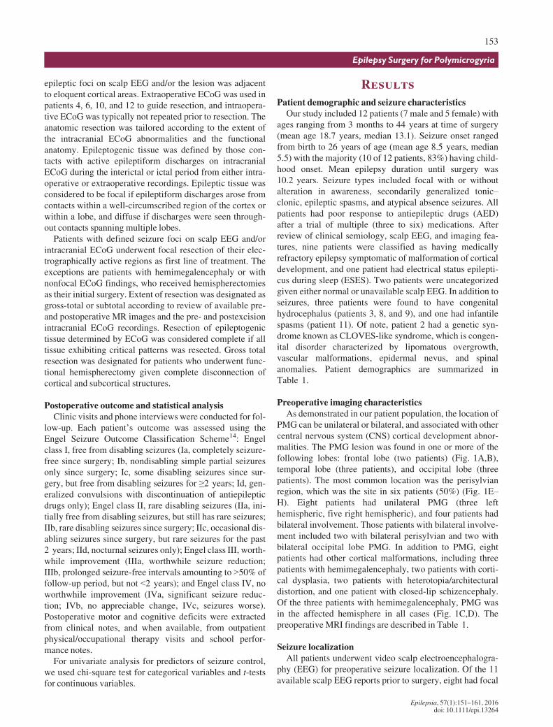
Figure 1.
(A and B) Axial inversion recovery images of patient 8 showing very fine polymicrogyria of the right frontal operculum, right anterior
insula and right orbital gyri. (C and D) Axial T1 MRI images of patient 2 who has left hemimegalencephaly with diffuse polymicrogyria
involving most of the left hemisphere. There is abnormal white matter containing several layers of heterotopic neurons and enlarged
hemisphere with dysmorphic ventricles including enlarged trigone/occipital horn. (E–H) Left perisylvian polymicrogyria as demonstrated
in patient 7. Axial and coronal T2 (E, F, andG) images showing left posterior perisylvian PMG involving the entire insula and temporal and
parietal operculum, up to the parietal convexity. Sagittal T1 sequence demonstrates the extent of the perisylvian polymicrogyria (H).
Epilepsia ILAE
Epilepsia, 57(1):151–161, 2016doi: 10.1111/epi.13264
155
Epilepsy Surgery for Polymicrogyria
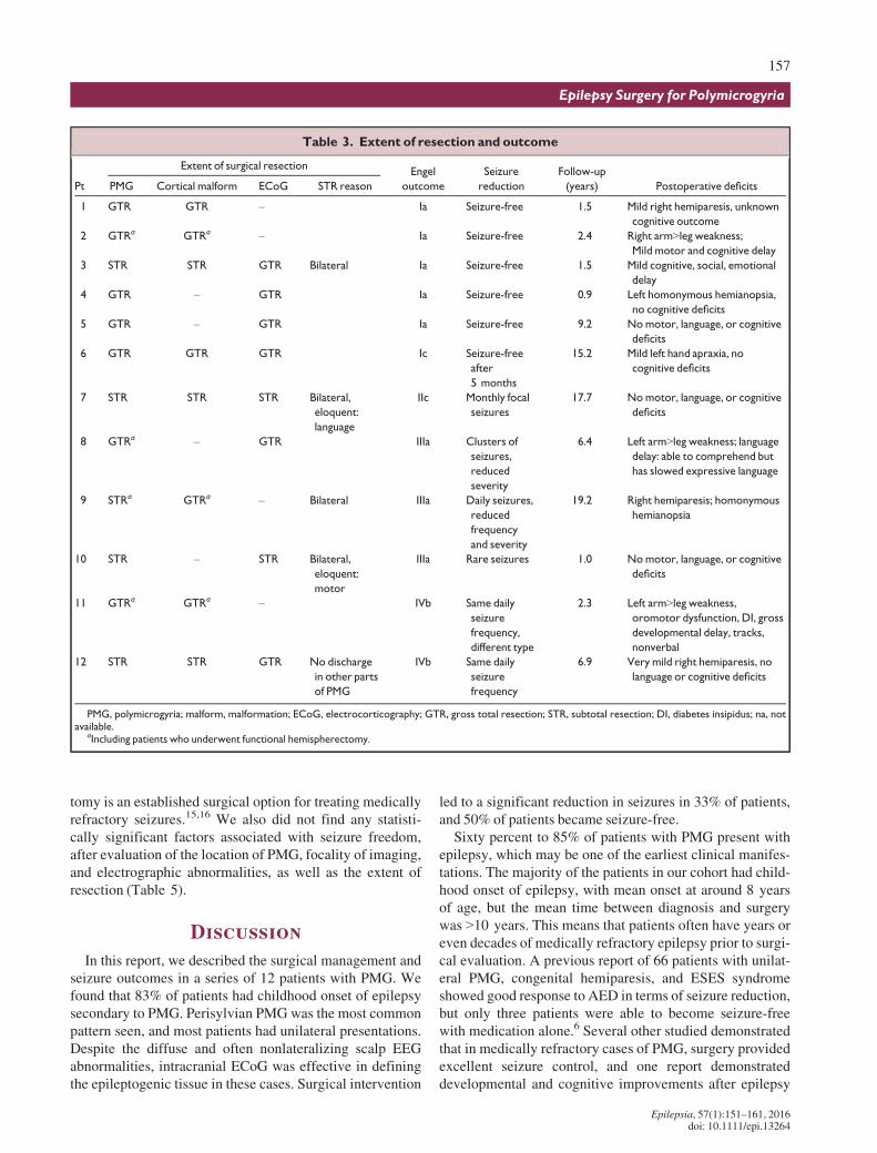
postoperative hemiparesis, four had mild preoperativehemiparesis (patients 1, 2, 11, and 12). All patients whounderwent hemispherectomies (patients 1, 2, 8, 9, and 11)had expected postoperative contralateral hemiparesis. Whenable to assess, patients who underwent hemispherectomy oroccipital lobectomy had expected hemianopsia. Of the fourpatients who had recognizable cognitive, language, and/orsocial and emotional delay, two had resections in the lefthemisphere and two in the right hemisphere. One patientdeveloped permanent diabetes insipidus postoperatively.These findings are summarized in Table 3.
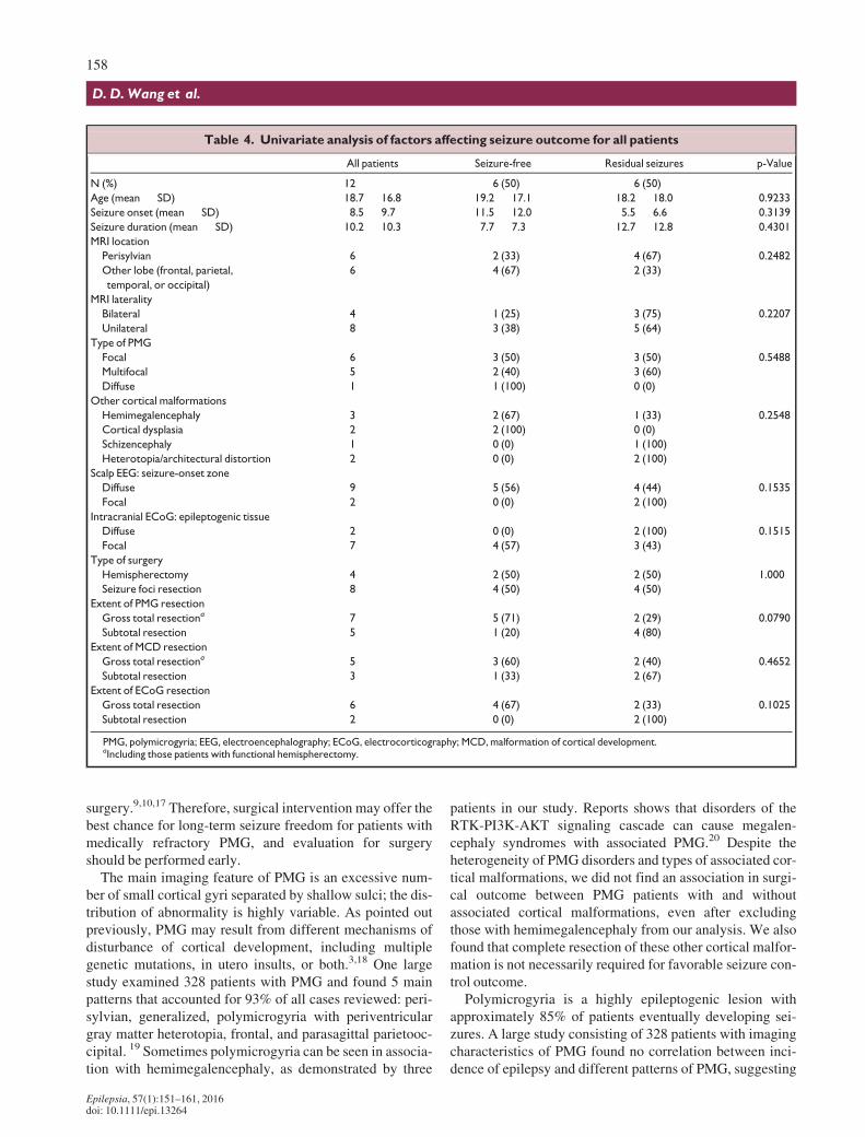
We performed univariate analysis to find predictors offavorable seizure-free outcome. The location, focality, orlaterality of the PMG region were not predictors of seizurecontrol; neither was the presence of other cortical malfor-
mations (Table 4). The focality of preoperative scalp EEGor intracranial ECoG abnormalities did not appear to corre-late with seizure-free outcome. The extent of resection forareas of PMG, other cortical malformation, and electro-graphic abnormalities was described for each patient(Table 3). We found that gross total resection or disconnec-tion (in case of functional hemispherectomy) of PMG cortexwas associated with improved seizure control, although itdid not reach statistical significance (p = 0.0790, Table 4).Of interest, the extent of resection for other associated corti-cal malformations or intracranial ECoG foci did not signifi-cantly affect seizure control rates (Table 4). We alsoperformed our univariate analysis to exclude those patientswith hemimegalencephaly, given that it is a different mal-formation of cortical development for which hemispherec-
Table 2. Scalp EEG and intracranial electrocorticography characteristics
Pt
Scalp EEG features Intracranial ECoG features
Concord with
PMG onMRI
Concord with
other MCD onMRIScalp EEG description Laterality
Focal/
diffuse Seizure focus
Focal/
diffuse
1 NA Left Diffuse – – Yes Yes
2 Focal seizures arising from the left
posterior region with multifocal left
hemisphere interictal epileptiform
transients and left hemisphere
slowing
Left Diffuse – – Yes Yes
3 Frequent 2.5–3 Hz spike and wave
with a right parietal predominance
enhanced during sleep consistent
with ESES
Right Diffuse Right occipital and
posterior parietal
region
Focal Yes Yes
4 Focal seizures with evolving theta
poorly lateralized or localized but
with a right sided emphasis
Nonlateral Diffuse Right occipital lobea Focal Yes na
5 Routine normal EEG – – Right suprasylvian,
frontoparietal region
Focal Yes na
6 Focal seizures, poorly lateralized with
a right temporal emphasis
Nonlateral Diffuse Right posterior
suprasylvian and
temporal regiona
Focal Yes Yes
7 Focal seizures arising from the left
temporal region
Left Focal Left inferior parietal
and superior
temporal region
Focal Yes Yes
8 Focal seizure arising from the right
frontal region with concordant, focal
interictal epileptiform transients in
wakefulness and sleep
Right Focal Right frontal region Focal Yes Yes
9 Generalized, asymmetric slowing of
the background affecting the left
greater than right hemisphere
Nonlateral Diffuse Left parietal region
and hemisphere
Diffuse No Yes
10 Focal seizures with poor
lateralization or localization
Nonlateral Diffuse Right suprasylvian and
temporal regionaDiffuse Yes na
11 Focal seizures arising broadly from
the right hemisphere with right
interictal discharges
Right Diffuse – – Yes Yes
12 Focal seizures arising from the left
hemisphere
Left Diffuse Left postcentral
cortex and superior
temporal regiona
Focal Yes Yes
EEG, electroencephalography; ECoG, electrocorticography; Concord, concordance; PMG, polymicrogyria; MCD, malformation of cortical development; ESES,electrical status epilepticus during sleep; na; not applicable.
aPatients 4, 6, 10, and 12 underwent extraoperative ECoG recording with subdural grid depth electrodes.
Epilepsia, 57(1):151–161, 2016doi: 10.1111/epi.13264
156
D. D.Wang et al.
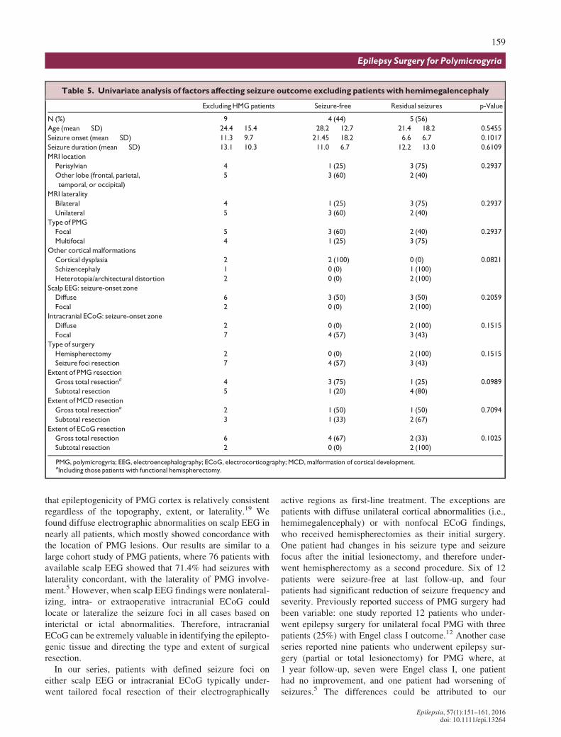
tomy is an established surgical option for treating medicallyrefractory seizures.15,16 We also did not find any statisti-cally significant factors associated with seizure freedom,after evaluation of the location of PMG, focality of imaging,and electrographic abnormalities, as well as the extent ofresection (Table 5).
DiscussionIn this report, we described the surgical management and
seizure outcomes in a series of 12 patients with PMG. Wefound that 83% of patients had childhood onset of epilepsysecondary to PMG. Perisylvian PMG was the most commonpattern seen, and most patients had unilateral presentations.Despite the diffuse and often nonlateralizing scalp EEGabnormalities, intracranial ECoG was effective in definingthe epileptogenic tissue in these cases. Surgical intervention
led to a significant reduction in seizures in 33% of patients,and 50% of patients became seizure-free.
Sixty percent to 85% of patients with PMG present withepilepsy, which may be one of the earliest clinical manifes-tations. The majority of the patients in our cohort had child-hood onset of epilepsy, with mean onset at around 8 yearsof age, but the mean time between diagnosis and surgerywas >10 years. This means that patients often have years oreven decades of medically refractory epilepsy prior to surgi-cal evaluation. A previous report of 66 patients with unilat-eral PMG, congenital hemiparesis, and ESES syndromeshowed good response to AED in terms of seizure reduction,but only three patients were able to become seizure-freewith medication alone.6 Several other studied demonstratedthat in medically refractory cases of PMG, surgery providedexcellent seizure control, and one report demonstrateddevelopmental and cognitive improvements after epilepsy
Table 3. Extent of resection and outcome
Pt
Extent of surgical resectionEngel
outcome
Seizure
reduction
Follow-up
(years) Postoperative deficitsPMG Cortical malform ECoG STR reason
1 GTR GTR – Ia Seizure-free 1.5 Mild right hemiparesis, unknown
cognitive outcome
2 GTRa GTRa – Ia Seizure-free 2.4 Right arm>leg weakness;Mild motor and cognitive delay
3 STR STR GTR Bilateral Ia Seizure-free 1.5 Mild cognitive, social, emotional
delay
4 GTR – GTR Ia Seizure-free 0.9 Left homonymous hemianopsia,
no cognitive deficits
5 GTR – GTR Ia Seizure-free 9.2 Nomotor, language, or cognitive
deficits
6 GTR GTR GTR Ic Seizure-free
after
5 months
15.2 Mild left hand apraxia, no
cognitive deficits
7 STR STR STR Bilateral,
eloquent:
language
IIc Monthly focal
seizures
17.7 Nomotor, language, or cognitive
deficits
8 GTRa – GTR IIIa Clusters of
seizures,
reduced
severity
6.4 Left arm>leg weakness; languagedelay: able to comprehend but
has slowed expressive language
9 STRa GTRa – Bilateral IIIa Daily seizures,
reduced
frequency
and severity
19.2 Right hemiparesis; homonymous
hemianopsia
10 STR – STR Bilateral,
eloquent:
motor
IIIa Rare seizures 1.0 Nomotor, language, or cognitive
deficits
11 GTRa GTRa – IVb Same daily
seizure
frequency,
different type
2.3 Left arm>leg weakness,oromotor dysfunction, DI, gross
developmental delay, tracks,
nonverbal
12 STR STR GTR No discharge
in other parts
of PMG
IVb Same daily
seizure
frequency
6.9 Very mild right hemiparesis, no
language or cognitive deficits
PMG, polymicrogyria; malform, malformation; ECoG, electrocorticography; GTR, gross total resection; STR, subtotal resection; DI, diabetes insipidus; na, notavailable.
aIncluding patients who underwent functional hemispherectomy.
Epilepsia, 57(1):151–161, 2016doi: 10.1111/epi.13264
157
Epilepsy Surgery for Polymicrogyria
surgery.9,10,17 Therefore, surgical intervention may offer thebest chance for long-term seizure freedom for patients withmedically refractory PMG, and evaluation for surgeryshould be performed early.
The main imaging feature of PMG is an excessive num-ber of small cortical gyri separated by shallow sulci; the dis-tribution of abnormality is highly variable. As pointed outpreviously, PMG may result from different mechanisms ofdisturbance of cortical development, including multiplegenetic mutations, in utero insults, or both.3,18 One largestudy examined 328 patients with PMG and found 5 mainpatterns that accounted for 93% of all cases reviewed: peri-sylvian, generalized, polymicrogyria with periventriculargray matter heterotopia, frontal, and parasagittal parietooc-cipital. 19 Sometimes polymicrogyria can be seen in associa-tion with hemimegalencephaly, as demonstrated by three
patients in our study. Reports shows that disorders of theRTK-PI3K-AKT signaling cascade can cause megalen-cephaly syndromes with associated PMG.20 Despite theheterogeneity of PMG disorders and types of associated cor-tical malformations, we did not find an association in surgi-cal outcome between PMG patients with and withoutassociated cortical malformations, even after excludingthose with hemimegalencephaly from our analysis. We alsofound that complete resection of these other cortical malfor-mation is not necessarily required for favorable seizure con-trol outcome.
Polymicrogyria is a highly epileptogenic lesion withapproximately 85% of patients eventually developing sei-zures. A large study consisting of 328 patients with imagingcharacteristics of PMG found no correlation between inci-dence of epilepsy and different patterns of PMG, suggesting
Table 4. Univariate analysis of factors affecting seizure outcome for all patients
All patients Seizure-free Residual seizures p-Value
N (%) 12 6 (50) 6 (50)
Age (mean � SD) 18.7 � 16.8 19.2 � 17.1 18.2 � 18.0 0.9233
Seizure onset (mean � SD) 8.5 � 9.7 11.5 � 12.0 5.5 � 6.6 0.3139
Seizure duration (mean � SD) 10.2 � 10.3 7.7 � 7.3 12.7 � 12.8 0.4301
MRI location
Perisylvian 6 2 (33) 4 (67) 0.2482
Other lobe (frontal, parietal,
temporal, or occipital)
6 4 (67) 2 (33)
MRI laterality
Bilateral 4 1 (25) 3 (75) 0.2207
Unilateral 8 3 (38) 5 (64)
Type of PMG
Focal 6 3 (50) 3 (50) 0.5488
Multifocal 5 2 (40) 3 (60)
Diffuse 1 1 (100) 0 (0)
Other cortical malformations
Hemimegalencephaly 3 2 (67) 1 (33) 0.2548
Cortical dysplasia 2 2 (100) 0 (0)
Schizencephaly 1 0 (0) 1 (100)
Heterotopia/architectural distortion 2 0 (0) 2 (100)
Scalp EEG: seizure-onset zone
Diffuse 9 5 (56) 4 (44) 0.1535
Focal 2 0 (0) 2 (100)
Intracranial ECoG: epileptogenic tissue
Diffuse 2 0 (0) 2 (100) 0.1515
Focal 7 4 (57) 3 (43)
Type of surgery
Hemispherectomy 4 2 (50) 2 (50) 1.000
Seizure foci resection 8 4 (50) 4 (50)
Extent of PMG resection
Gross total resectiona 7 5 (71) 2 (29) 0.0790
Subtotal resection 5 1 (20) 4 (80)
Extent of MCD resection
Gross total resectiona 5 3 (60) 2 (40) 0.4652
Subtotal resection 3 1 (33) 2 (67)
Extent of ECoG resection
Gross total resection 6 4 (67) 2 (33) 0.1025
Subtotal resection 2 0 (0) 2 (100)
PMG, polymicrogyria; EEG, electroencephalography; ECoG, electrocorticography; MCD, malformation of cortical development.aIncluding those patients with functional hemispherectomy.
Epilepsia, 57(1):151–161, 2016doi: 10.1111/epi.13264
158
D. D.Wang et al.
that epileptogenicity of PMG cortex is relatively consistentregardless of the topography, extent, or laterality.19 Wefound diffuse electrographic abnormalities on scalp EEG innearly all patients, which mostly showed concordance withthe location of PMG lesions. Our results are similar to alarge cohort study of PMG patients, where 76 patients withavailable scalp EEG showed that 71.4% had seizures withlaterality concordant, with the laterality of PMG involve-ment.5 However, when scalp EEG findings were nonlateral-izing, intra- or extraoperative intracranial ECoG couldlocate or lateralize the seizure foci in all cases based oninterictal or ictal abnormalities. Therefore, intracranialECoG can be extremely valuable in identifying the epilepto-genic tissue and directing the type and extent of surgicalresection.
In our series, patients with defined seizure foci oneither scalp EEG or intracranial ECoG typically under-went tailored focal resection of their electrographically
active regions as first-line treatment. The exceptions arepatients with diffuse unilateral cortical abnormalities (i.e.,hemimegalencephaly) or with nonfocal ECoG findings,who received hemispherectomies as their initial surgery.One patient had changes in his seizure type and seizurefocus after the initial lesionectomy, and therefore under-went hemispherectomy as a second procedure. Six of 12patients were seizure-free at last follow-up, and fourpatients had significant reduction of seizure frequency andseverity. Previously reported success of PMG surgery hadbeen variable: one study reported 12 patients who under-went epilepsy surgery for unilateral focal PMG with threepatients (25%) with Engel class I outcome.12 Another caseseries reported nine patients who underwent epilepsy sur-gery (partial or total lesionectomy) for PMG where, at1 year follow-up, seven were Engel class I, one patienthad no improvement, and one patient had worsening ofseizures.5 The differences could be attributed to our
Table 5. Univariate analysis of factors affecting seizure outcome excluding patients with hemimegalencephaly
Excluding HMG patients Seizure-free Residual seizures p-Value
N (%) 9 4 (44) 5 (56)
Age (mean � SD) 24.4 � 15.4 28.2 � 12.7 21.4 � 18.2 0.5455
Seizure onset (mean � SD) 11.3 � 9.7 21.45 � 18.2 6.6 � 6.7 0.1017
Seizure duration (mean � SD) 13.1 � 10.3 11.0 � 6.7 12.2 � 13.0 0.6109
MRI location
Perisylvian 4 1 (25) 3 (75) 0.2937
Other lobe (frontal, parietal,
temporal, or occipital)
5 3 (60) 2 (40)
MRI laterality
Bilateral 4 1 (25) 3 (75) 0.2937
Unilateral 5 3 (60) 2 (40)
Type of PMG
Focal 5 3 (60) 2 (40) 0.2937
Multifocal 4 1 (25) 3 (75)
Other cortical malformations
Cortical dysplasia 2 2 (100) 0 (0) 0.0821
Schizencephaly 1 0 (0) 1 (100)
Heterotopia/architectural distortion 2 0 (0) 2 (100)
Scalp EEG: seizure-onset zone
Diffuse 6 3 (50) 3 (50) 0.2059
Focal 2 0 (0) 2 (100)
Intracranial ECoG: seizure-onset zone
Diffuse 2 0 (0) 2 (100) 0.1515
Focal 7 4 (57) 3 (43)
Type of surgery
Hemispherectomy 2 0 (0) 2 (100) 0.1515
Seizure foci resection 7 4 (57) 3 (43)
Extent of PMG resection
Gross total resectiona 4 3 (75) 1 (25) 0.0989
Subtotal resection 5 1 (20) 4 (80)
Extent of MCD resection
Gross total resectiona 2 1 (50) 1 (50) 0.7094
Subtotal resection 3 1 (33) 2 (67)
Extent of ECoG resection
Gross total resection 6 4 (67) 2 (33) 0.1025
Subtotal resection 2 0 (0) 2 (100)
PMG, polymicrogyria; EEG, electroencephalography; ECoG, electrocorticography; MCD, malformation of cortical development.aIncluding those patients with functional hemispherectomy.
Epilepsia, 57(1):151–161, 2016doi: 10.1111/epi.13264
159
Epilepsy Surgery for Polymicrogyria
longer length of follow-up and capturing more patientswith seizure recurrence. Of interest, we were unable toidentify any significant predictors of seizure freedom.However, gross total resection of the PMG lesion trendedtoward favorable outcome. Due to our small sample size,we may be underpowered to find statistically significantpredictors. In addition, intraoperative ECoG was not usedin every case, nor was a postexcisional ECoG performedin all cases. Therefore, it is difficult to make conclusionsregarding the extent of ECoG resection on seizureoutcome.
As with all epilepsy surgeries, one key consideration inepilepsy surgery for PMG is defining the epileptogenic zone(EZ) and whether it resides purely within the abnormal cor-tex. We found a high incidence of abnormal ECoG or EEGactivity arising from the region of PMG, concordant with astudy that used ictal SPECT to identify hyperperfusion inPMG cortex of all patients, suggesting the involvement ofPMG cortex in seizure generation or early seizure propaga-tion.12 However, not all parts of the PMG cortex are epilep-togenic, as Maillard and colleagues showed using acombination of magnetic source imaging and stereo-EEG ina patient with schizencephaly and polymicrogryia.21 Wealso demonstrated this as patients with bilateral PMG usu-ally had a unilateral seizure-onset zone, and even in case ofunilateral focal PMG, only a portion of the PMG cortex hadactive epileptic discharges. Furthermore, other groups havefound that the epileptogenic network not only resides withinthe abnormal tissue, but also extends beyond the area ofPMG using stereo-EEG in patients with unilateral focalPMG.9,10 These studies suggest that PMG can be part of alarger epileptogenic network and that multimodal imagingand neurophysiologic tests should be performed to definethe epileptic zone.
Surgical treatment options for patients with PMG are usu-ally limited. However, our study provides evidence that,even in patients with diffuse and bilateral lesions, seizuresmay arise from focal regions within or around the PMG, andsurgical resection may provide significant benefit in seizurecontrol outcome. Intracranial ECoG can provide additionallocalizing information compared to scalp EEG. Our studyhas its limitations, given the small patient sample size andheterogeneity of PMG lesions. However, our data are con-sistent with previous studies and argue that patients withmedically refractory epilepsy secondary to PMG shouldundergo a detailed presurgical evaluation with volumetricmagnetic resonance imaging (MRI)22 and epileptic focimapping with scalp EEG, stereo-EEG,9,21,23 or intracranialECoG. PMG abnormalities as visualized on MRI are fre-quently extensive and ambiguous with regard to actual sei-zure-onset localization, and magnetoencephalography(MEG) and single-proton emission computed tomography(SPECT) offer additive noninvasive functionally relevantlocalization for optimal surgical planning.24–27 Epilepsysurgery, with the aid of multimodal imaging and neurophys-
iologic tests, may provide excellent long-term seizure con-trol for patients with PMG.
DisclosureThe authors report no conflicts of interest. We confirm that we have read
the Journal’s position on issues involved in ethical publication and affirmthat this report is consistent with those guidelines.
References1. Barkovich AJ, Kuzniecky RI, Jackson GD, et al. A developmental and
genetic classification for malformations of cortical development. Neu-rology 2005;65:1873–1887.
2. Barkovich AJ, Lindan CE. Congenital cytomegalovirus infection ofthe brain: imaging analysis and embryologic considerations. AJNR AmJ Neuroradiol 1994;15:703–715.
3. Barkovich AJ. Current concepts of polymicrogyria. Neuroradiology2010;52:479–487.
4. Aronica E, Becker AJ, Spreafico R. Malformations of cortical develop-ment. Brain Pathol 2012;22:380–401.
5. Shain C, Ramgopal S, Fallil Z, et al. Polymicrogyria-associated epi-lepsy: a multicenter phenotypic study from the Epilepsy Phenome/Genome Project. Epilepsia 2013;54:1368–1375.
6. Caraballo RH, Cersosimo RO, Fortini PS, et al. Congenital hemipare-sis, unilateral polymicrogyria and epilepsy with or without statusepilepticus during sleep: a study of 66 patients with long-term follow-up. Epileptic Disord 2013;15:417–427.
7. Loddenkemper T, Cosmo G, Kotagal P, et al. Epilepsy surgery in chil-dren with electrical status epilepticus in sleep. Neurosurgery2009;64:328–337; discussion 337.
8. Leventer RJ, Guerrini R, DobynsWB.Malformations of cortical devel-opment and epilepsy.Dialogues Clin Neurosci 2008;10:47–62.
9. Chassoux F, Landre E, Rodrigo S, et al. Intralesional recordings andepileptogenic zone in focal polymicrogyria. Epilepsia 2008;49:51–64.
10. Ramantani G, Koessler L, Colnat-Coulbois S, et al. Intracranial evalu-ation of the epileptogenic zone in regional infrasylvian polymicrogyr-ia. Epilepsia 2013;54:296–304.
11. Sisodiya SM. Surgery for malformations of cortical development caus-ing epilepsy. Brain 2000;123(Pt 6):1075–1091.
12. Wichert-Ana L, de Azevedo-Marques PM, Oliveira LF, et al. Ictaltechnetium-99 m ethyl cysteinate dimer single-photon emission tomo-graphic findings in epileptic patients with polymicrogyria syndromes:a subtraction of ictal-interictal SPECT coregistered to MRI study. EurJ Nucl MedMol Imaging 2008;35:1159–1170.
13. Brodtkorb E, Torbergsen T, Nakken KO, et al. Epileptic seizures,arthrogryposis, and migrational brain disorders: a syndrome? ActaNeurol Scand 1994;90:232–240.
14. Wieser HG, Blume WT, Fish D, et al. ILAE Commission Report. Pro-posal for a new classification of outcome with respect to epileptic sei-zures following epilepsy surgery. Epilepsia 2001;42:282–286.
15. Schramm J, Delev D,Wagner J, et al. Seizure outcome, functional out-come, and quality of life after hemispherectomy in adults. Acta Neu-rochir (Wien) 2012;154:1603–1612.
16. Schramm J, Kuczaty S, Sassen R, et al. Pediatric functional hemi-spherectomy: outcome in 92 patients. Acta Neurochir (Wien)2012;154:2017–2028.
17. Isik U, Dincer A, OzekMM. Surgical treatment of polymicrogyria withadvanced radiologic and neurophysiologic techniques. Childs NervSyst 2007;23:443–448.
18. Guerrini R, Dobyns WB, Barkovich AJ. Abnormal development of thehuman cerebral cortex: genetics, functional consequences and treat-ment options. Trends Neurosci 2008;31:154–162.
19. Leventer RJ, Jansen A, Pilz DT, et al. Clinical and imaging hetero-geneity of polymicrogyria: a study of 328 patients. Brain2010;133:1415–1427.
20. Riviere JB, Mirzaa GM, O’Roak BJ, et al. De novo germline andpostzygotic mutations in AKT3, PIK3R2 and PIK3CA cause a spec-trum of related megalencephaly syndromes. Nat Genet 2012;44:934–940.
Epilepsia, 57(1):151–161, 2016doi: 10.1111/epi.13264
160
D. D.Wang et al.
21. Maillard L, Koessler L, Colnat-Coulbois S, et al. Combined SEEG andsource localisation study of temporal lobe schizencephaly and polymi-crogyria.Clin Neurophysiol 2009;120:1628–1636.
22. De Ciantis A, Barkovich AJ, Cosottini M, et al. Ultra-high-field MRimaging in polymicrogyria and epilepsy. AJNR Am J Neuroradiol2015;36:309–316.
23. Rikir E, Koessler L, Gavaret M, et al. Electrical source imaging in cor-tical malformation-related epilepsy: a prospective EEG-SEEG concor-dance study. Epilepsia 2014;55:918–932.
24. Burneo JG, Bebin M, Kuzniecky RI, et al. Electroclinical and magne-toencephalographic studies in epilepsy patients with polymicrogyria.Epilepsy Res 2004;62:125–133.
25. Burneo JG, Kuzniecky RI, Bebin M, et al. Cortical reorganization inmalformations of cortical development: a magnetoencephalographicstudy.Neurology 2004;63:1818–1824.
26. Knowlton RC, Elgavish RA, Bartolucci A, et al. Functional imaging:II. Prediction of epilepsy surgery outcome. Ann Neurol 2008;64:35–41.
27. Knowlton RC, Elgavish RA, Limdi N, et al. Functional imaging: I.Relative predictive value of intracranial electroencephalography. AnnNeurol 2008;64:25–34.
Epilepsia, 57(1):151–161, 2016doi: 10.1111/epi.13264
161
Epilepsy Surgery for Polymicrogyria


