Embed Size (px)
Citation preview
BioMed CentralBMC Cancer
ss
Open AcceResearch articleSuppression of local invasion of ameloblastoma by inhibition of matrix metalloproteinase-2 in vitroAnxun Wang†1, Bin Zhang†2, Hongzhang Huang*3, Leitao Zhang2, Donglin Zeng3, Qian Tao3, Jianguang Wang2 and Chaobin Pan2Address: 1Department of Oral and Maxillofacial Surgery, The First Affiliated Hospital, Sun Yat-sen University, Guangzhou, Guangdong, 510080, PR China, 2Department of Oral and Maxillofacial Surgery, The Second Affiliated Hospital, Sun Yat-sen University, 107 Yan-jiang Road West, Guangzhou, Guangdong, 510120, PR China and 3Department of Oral and Maxillofacial Surgery, Guanghua College of Stomatology, Sun Yat-sen University, Guangzhou, Guangdong, 510055, PR China
Email: Anxun Wang - [email protected]; Bin Zhang - [email protected]; Hongzhang Huang* - [email protected]; Leitao Zhang - [email protected]; Donglin Zeng - [email protected]; Qian Tao - [email protected]; Jianguang Wang - [email protected]; Chaobin Pan - [email protected]
* Corresponding author †Equal contributors
AbstractBackground: Ameloblastomas are odontogenic neoplasms characterized by local invasiveness.This study was conducted to address the role of matrix metalloproteinase-2 (MMP-2) in theinvasiveness of ameloblastomas.
Methods: Plasmids containing either MMP-2 siRNA or tissue inhibitor of metalloproteinase-2(TIMP-2) cDNA were created and subsequently transfected into primary ameloblastoma cells.Zymography, RT-PCR, and Western blots were used to assess MMP-2 activity and expression ofMMP-2 and TIMP-2, as well as protein levels.
Results: Primary cultures of ameloblastoma cells expressed cytokeratin (CK) 14 and 16, and MMP-2, but only weakly expressed CK18 and vimentin. MMP-2 mRNA and protein levels weresignificantly inhibited by RNA interference (P < 0.05). Both MMP-2 siRNA and TIMP-2overexpression inhibited MMP-2 activity and the in vitro invasiveness of ameloblastoma.
Conclusion: These results indicate that inhibition of MMP-2 activity suppresses the localinvasiveness of ameloblastoma cells. This mechanism may serve as a novel therapeutic target inameloblastomas pursuant to additional research.
BackgroundAmeloblastomas are the most frequently encounteredtumors arising from odontogenic epithelium [1,2].Although characterized as a benign neoplasm, ameloblas-tomas are locally invasive and frequently recrudescenttumors of the jaw [2]. Numerous studies have identifiedboth genetic and molecular alterations in these odon-togenic tumors of the epithelium [3,4], but the mecha-
nisms underlying the local invasiveness of this neoplasmhave yet to be clarified.
Matrix metalloproteinases (MMPs) are a family of zinc-and calcium-dependent proteolytic enzymes [4]. Theseenzymes play central roles in the regulation of the extra-cellular matrix during embryonic development and tissueremodeling. MMPs also participate in tumor invasion and
Published: 30 June 2008
BMC Cancer 2008, 8:182 doi:10.1186/1471-2407-8-182
Received: 13 January 2008Accepted: 30 June 2008
This article is available from: http://www.biomedcentral.com/1471-2407/8/182
© 2008 Wang et al; licensee BioMed Central Ltd. This is an Open Access article distributed under the terms of the Creative Commons Attribution License (http://creativecommons.org/licenses/by/2.0), which permits unrestricted use, distribution, and reproduction in any medium, provided the original work is properly cited.
Page 1 of 9(page number not for citation purposes)
BMC Cancer 2008, 8:182 http://www.biomedcentral.com/1471-2407/8/182
metastasis [5,6]. The major function of tissue inhibitors ofmatrix metalloproteinases (TIMPs) is to inhibit the activeforms of MMPs in a 1:1 stoichiometric ratio via non-cov-alent mechanisms [7,8]. Aberrant MMP activity in tumorcells and the surrounding stromal tissues has been impli-cated in tumor invasion and metastasis [9,10]. Previousstudies have shown that ameloblastomas have an elevatedexpression of MMP-2, MMP-9, and vascular endothelialgrowth factor (VEGF), and are void of or have an abnor-mal expression of E-cadherin and TIMP-2 [11-14].
Therapeutic interventions that inhibit MMP activityappear to be promising based on a number of in vitro andin vivo tumor invasiveness studies [15-17]. To determinewhether inhibition of MMP-2 activity is capable of sup-pressing the invasiveness of ameloblastomas, plasmidswere constructed and subsequently transfected into amel-oblastoma cells to cause the overexpression of, or toknockdown, MMP-2. This study was designed to test thehypothesis that MMP-2 activity is involved in the invasive-ness of ameloblastomas and that inhibition of MMP-2 isa useful approach for treating ameloblastomas. The datacollected in this study indicate that siRNA targeting ofMMP-2 mRNA or TIMP-2 overxpression inhibits the activ-ity of MMP-2 in ameloblastoma cells, which results inreduced ameloblastoma cell invasiveness in vitro, indicat-ing that inhibition of MMP-2 activity may serve as a noveltherapeutic target in the management of ameloblastomas.
MethodsPrimary cell cultures and identificationThe samples used in this study were obtained after obtain-ing informed consent of each patient and with theapproval of the Sun Yet-sen University Ethics Committee.Briefly, ameloblastoma tissues were minced and incu-bated overnight in Dulbecco's modified Eagle medium(DMEM, Invitrogen, CA, USA) containing 1 mg/mL colla-genase I (Invitrogen) at 37°C. Collagenase-digested tis-sues were plated onto 35 mm dishes coated with collagenI (Invitrogen) in DMEM containing 10% fetal calf serum,200 μg/ml streptomycin, and 200 IU/ml penicillin, andincubated at 37°C with 5% CO2. When the cells were con-fluent, they were divided again and used for the ensuingexperiments.
Immunocytochemistry was used to confirm the epithelialorigin of the ameloblastoma cells using the SP method asdescribed by the manufacturer (Maixin, Fuzhou, China).The primary antibodies were anti-cytokeratin 14, 16, and18 and anti-vimentin (Maixin). Immunofluorescence wasused to detect the expression of MMP-2, as described bythe product's manufacturer.
Plasmid construction and transient transfectionTo generate the plasmid vector, pRNA-MMP-2, pRNA-U6.1/neovector (Genscript, NJ, USA) containing a cGFPsequence was used. MMP-2 shRNA contains a comple-ment of a 21-nucleotide sequence (tgtgctgaaggacacactaaa,GenBank NM-004530), which was separated by a 7-nucleotide non-complementary spacer (CCACACC). Acontrol vector (pRNA-neg) was constructed in the sameway using a 21-nucleotide sequence (gattcaggtgtagaac-gagca). These sequences were confirmed using nucleotideBLAST to ensure that there was no homology with anyother known human gene. These annealed sequenceswere inserted into the pRNA-U6.1/neo backbone afterdigestion with BamH1 and HindIII. After amplification,all vector constructs were verified by sequencing.
The pcDNA3 vector (Genscript) containing an enhancedgreen fluorescence protein (EGFP) sequence wasemployed to generate the plasmid vector, pcDNA-TIMP-2.The cDNA encoding TIMP-2 was obtained via RT-PCR.The primer sequence for TIMP-2 (723 bp, GenBankNM003522) containing EcoRI and XhoI was as follows:forward 5'cgatgaattcatgggcgccgcggcccgc3'; reverse5'cgatctcgagttattatgggtcctcgatgagaaac3'. TIMP-2 cDNA wassubcloned into the clone site of the pcDNA3. Followingamplification, vector constructs were verified by sequenc-ing.
The constructed plasmids were transiently transfected intothe cultured ameloblastoma cells using LipofectaminePlus reagent (Invitrogen), according to the manufacturer'sinstructions. Transfected cells were subsequently used inthe following experiment.
Detection of MMP-2/TIMP-2 activityMMP-2/TIMP-2 activity in the culture medium of theameloblastoma cells was detected by zymography accord-ing to the method reported by Kleiner et al. [18]. Briefly,10 μl of the medium from serum-free ameloblastoma cellcultures was mixed with the same volume of samplebuffer and applied to a 10% (wt/vol) polyacrylamide gelcontaining 1 mg/ml gelatin. Gels were incubated in 2.5%Triton X-100 for 45 min after electrophoresis, then incu-bated at 37°C overnight in a digestion buffer. Gels werestained and destained. The bands were analyzed by anauto-imaging analysis system (Kontron IBAS2.0, Ger-many). All densitometry measurements were madebetween samples in the same gel to ensure comparability.
For detection of TIMP-2 activity, all procedures were sim-ilar to the detection of MMP-2 activity (described above),except that the gel contained 1% (wt/vol) MMP-2 (Sigma,St. Louis, MO, USA).
Page 2 of 9(page number not for citation purposes)
BMC Cancer 2008, 8:182 http://www.biomedcentral.com/1471-2407/8/182
RNA preparation and RT-PCRTotal RNA was extracted using an RNeasy mini kit (Qia-gen), according to the manufacturer's instructions. RT-PCR was performed using one-step RT-PCR assays (Qia-gen). Specific primers for detecting mRNA transcripts ofthe MMP-2 or TIMP-2 gene were as follows: MMP-2(NM004530), 5'-AGCCACCCCTAAAGAGATCC-3' and3'GTTCTAAGGCAGCCAGCAGT-5'; TIMP-2 (NM003255), 5'-ATTTGACCCAGAGTGGAACG-3' and 3'-TCCT-TCGGCGAGTTTATGGA-5'; and GAPDH (NM008084), 5'GGTCGGAGTCAACGGATTTGGTCG-3' and 3'-CCTC-CGACGCCTGCTTCACCAC-5'.
Transcript levels were normalized according to GAPDHtranscripts and the products were resolved by agarose elec-trophoresis. The intensity was quantified by image-analy-sis computer software (NIH Image).
Western blotsWestern blotting was performed to detect MMP-2 andTIMP-2 proteins (Santa Cruz Biotechnology, Santa Cruz,CA, USA). Cells were harvested by trypsinization andlysed with a radio-immune precipitation assay (RIPA)lysis buffer (Santa Cruz Biotechnology). Total proteinconcentrations were measured by the Bradford method(Bio-Rad). Aliquots (30–50 μg) of cellular proteins wereresolved by SDS-PAGE (10%), then electrotransferredonto PVDF membranes and immunoprobed. The protein-antibody complexes were detected by chemiluminescence(CSPD; Tropix, Bedford, MA, USA), according to the man-ufacturer's protocol (Applied Biosystems, MA, USA). TheGAPDH gene was used as an internal control and theband intensity was quantified.
In vitro cell invasionThe invasive ability of ameloblastoma cells was assayed intranswell cell chambers (Costa, Cambrige, MA, USA),according to the method reported by Kido et al. [19].Briefly, polycarbonate filters with an 8.0 μm pore sizewere precoated with fibronectin on the lower surface.Matrigel was applied to the upper surface of the filters (5μg/filter). Ameloblastoma cell suspensions (100 μl with 2× 10⁶ cells/ml) that had or had not been transfected wereadded to the upper compartment and incubated for 72 hat 37°C at 5% CO2. The filters were fixed with methanoland stained with Giemsa stain. The cells invading thelower surface through the Matrigel were manuallycounted under a microscope. The rate of invasion was cal-culated by the following equation: (number of cellsinvading the lower surface in the control group – numberof cells invading the lower surface in the treated group)/number of cells invading the lower surface in the controlgroup × 100%.
Statistical analysisAll experiments were performed in triplicate. Data areexpressed as the mean ± standard deviation (SD). Analysisof variance (ANOVA) was used to compare differencesbetween treatment and control cells. A p < 0.05 was con-sidered significant.
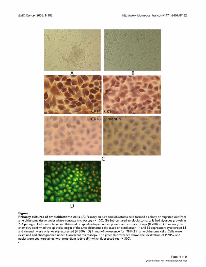
ResultsCells in primary cultureAfter ameloblastoma tissues were cultured for 18–36hours, ameloblastoma cells either formed a colony ormigrated out from the ameloblastoma tissue and under-went membraniform growth in which cells were arrangedlike a slabstone (Figure 1A). Secondary cell culturesshowed large, flattened, or spindle-shaped morphology(Figure 1B). Immunocytochemistry revealed that the cul-tured ameloblastoma cells expressed normal levels ofcytokeratins 14 and 16 and only weakly expressed cytok-eratin 18 and vimentin (Figure 1C), indicating that thesecultures were primarily comprised of ameloblastoma cellsof epithelial origin.
To investigate whether MMP-2 was expressed in the pri-mary ameloblastoma cell cultures, immunofluorescencewas used. Ameloblastoma cells that were immunopositivefor MMP-2 showed green fluorescence in the cytoplasmand red fluorescence in the cell nucleus under a fluores-cent microscope (Figure 1D).
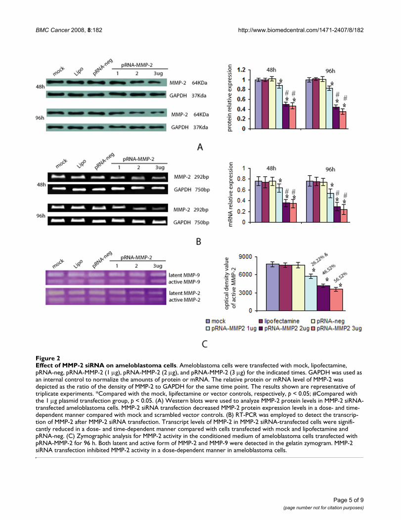
MMP-2 siRNA transfection decreased MMP-2 expression and activityTo investigate whether MMP-2 siRNA transfectiondecreases MMP-2 expression and activity, ameloblastomacells were transfected with various doses of pRNA-MMP-2(1–3 μg) and the protein level of MMP-2 was analyzed byWestern blots. MMP-2 siRNA transfection decreasedMMP-2 protein expression in both a dose- and time-dependent manner compared with mock and scrambledvector controls (Figure 2A). There were no differencesbetween the three control groups (p > 0.05), but a signifi-cantly lower protein level of MMP-2 after transfectionwith various doses of pRNA-MMP-2 (p < 0.05) were notedcompared with the three control groups. This MMP-2inhibition was more pronounced in the cells transfectedwith 2 or 3 μg of plasmid than in the 1 μg plasmid trans-fection group (p < 0.05). No difference between the 2 and3 μg plasmid transfection groups was noted (p > 0.05).
To determine whether the decreased production of MMP-2 was due to a decrease in gene transcription, MMP-2 tran-scripts were assessed via RT-PCR. As shown in Figure 2B,the transcript levels of MMP-2 in MMP-2 transfected cellswere significantly lower than MMP-2 RNA levels in themock, lipidosome, and pRNA-negative transfected cells(p< 0.05).
Page 3 of 9(page number not for citation purposes)
BMC Cancer 2008, 8:182 http://www.biomedcentral.com/1471-2407/8/182
Page 4 of 9(page number not for citation purposes)
Primary cultures of ameloblastoma cellsFigure 1Primary cultures of ameloblastoma cells. (A) Primary culture ameloblastoma cells formed a colony or migrated out from ameloblastoma tissue under phase-contrast microscopy (× 100). (B) Sub-cultured ameloblastoma cells had vigorous growth in 2–4 passages. Cells were large and flattened or spindle-shaped under phase-contrast microscopy (× 200). (C) Immunocyto-chemistry confirmed the epithelial origin of the ameloblastoma cells based on cytokeratin 14 and 16 expression; cytokeratin 18 and vimentin were only weakly expressed (× 300). (D) Immunofluorescence for MMP-2 in ameloblastoma cells. Cells were examined and photographed under fluorescent microscopy. The green fluorescence shows the localization of MMP-2 and nuclei were counterstained with propidium iodine (PI) which fluoresced red (× 300).
BMC Cancer 2008, 8:182 http://www.biomedcentral.com/1471-2407/8/182
Page 5 of 9(page number not for citation purposes)
Effect of MMP-2 siRNA on ameloblastoma cellsFigure 2Effect of MMP-2 siRNA on ameloblastoma cells. Ameloblastoma cells were transfected with mock, lipofectamine, pRNA-neg, pRNA-MMP-2 (1 μg), pRNA-MMP-2 (2 μg), and pRNA-MMP-2 (3 μg) for the indicated times. GAPDH was used as an internal control to normalize the amounts of protein or mRNA. The relative protein or mRNA level of MMP-2 was depicted as the ratio of the density of MMP-2 to GAPDH for the same time point. The results shown are representative of triplicate experiments. *Compared with the mock, lipifectamine or vector controls, respectively, p < 0.05; #Compared with the 1 μg plasmid transfection group, p < 0.05. (A) Western blots were used to analyze MMP-2 protein levels in MMP-2 siRNA-transfected ameloblastoma cells. MMP-2 siRNA transfection decreased MMP-2 protein expression levels in a dose- and time-dependent manner compared with mock and scrambled vector controls. (B) RT-PCR was employed to detect the transcrip-tion of MMP-2 after MMP-2 siRNA transfection. Transcript levels of MMP-2 in MMP-2 siRNA-transfected cells were signifi-cantly reduced in a dose- and time-dependent manner compared with cells transfected with mock and lipofectamine and pRNA-neg. (C) Zymographic analysis for MMP-2 activity in the conditioned medium of ameloblastoma cells transfected with pRNA-MMP-2 for 96 h. Both latent and active form of MMP-2 and MMP-9 were detected in the gelatin zymogram. MMP-2 siRNA transfection inhibited MMP-2 activity in a dose-dependent manner in ameloblastoma cells.
BMC Cancer 2008, 8:182 http://www.biomedcentral.com/1471-2407/8/182
Page 6 of 9(page number not for citation purposes)
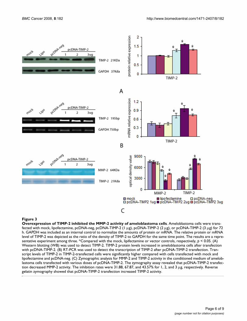
Overexpression of TIMP-2 inhibited the MMP-2 activity of ameloblastoma cellsFigure 3Overexpression of TIMP-2 inhibited the MMP-2 activity of ameloblastoma cells. Ameloblastoma cells were trans-fected with mock, lipofectamine, pcDNA-neg, pcDNA-TIMP-2 (1 μg), pcDNA-TIMP-2 (2 μg), or pcDNA-TIMP-2 (3 μg) for 72 h. GAPDH was included as an internal control to normalize the amounts of protein or mRNA. The relative protein or mRNA level of TIMP-2 was depicted as the ratio of the density of TIMP-2 to GAPDH for the same time point. The results are a repre-sentative experiment among three. *Compared with the mock, lipifectamine or vector controls, respectively, p < 0.05. (A) Western blotting (WB) was used to detect TIMP-2. TIMP-2 protein levels increased in ameloblastoma cells after transfection with pcDNA-TIMP-2. (B) RT-PCR was used to detect the transcription of TIMP-2 after pcDNA-TIMP-2 transfection. Tran-script levels of TIMP-2 in TIMP-2-transfected cells were significantly higher compared with cells transfected with mock and lipofectamine and pcDNA-neg. (C) Zymographic analysis for MMP-2 and TIMP-2 activity in the conditioned medium of amelob-lastoma cells transfected with various doses of pcDNA-TIMP-2. The zymography assay revealed that pcDNA-TIMP-2 transfec-tion decreased MMP-2 activity. The inhibition rates were 31.88, 67.87, and 43.57% for 1, 2, and 3 μg, respectively. Reverse gelatin zymography showed that pcDNA-TIMP-2 transfection increased TIMP-2 activity.
BMC Cancer 2008, 8:182 http://www.biomedcentral.com/1471-2407/8/182
To investigate whether MMP-2 activity was associatedwith MMP-2 knockdown, a zymogram assay was used todetect enzyme activity in the culture medium followingtransfection with various doses of pRNA-MMP-2 for 96 h(Figure 2C). Both latent and active forms of MMP-2 andMMP-9 were detected in the gelatin zymogram. The MMP-9 was not different after MMP-2 knockdown compared topre-knockdown values. MMP-2 siRNA transfection inhib-ited MMP-2 activity in a dose-dependent manner whereasno difference in MMP-2 activity was identified in the threecontrol groups (p > 0.05). After transfection of ameloblas-toma cells with various doses of pRNA-MMP-2, the activ-ity of MMP-2 was significantly decreased compared withthe three control groups (p < 0.05).
Overexpression of TIMP-2 inhibited MMP-2 activityTo determine the effect of TIMP-2 overexpression onMMP-2 activity, ameloblastoma cells were transfectedwith various doses of pcDNA-TIMP-2 (1–3 μg). Westernblots and RT-PCR showed that the levels of both TIMP-2mRNA and TIMP-2 protein in the TIMP-2 transfected cellswere significantly higher than that of cells transfected withmock, lipofectamine, and pcDNA-neg (p < 0.05; Figures3A and 3B). Further, zymography and reverse gelatinzymography assays revealed that pcDNA-TIMP-2 transfec-tion decreased MMP-2 activity and increased TIMP-2activity in ameloblastoma cells (Figure 3C). While no dif-ference between the three control groups were noted (p >0.05), significantly lower MMP-2 activity and significantlyhigher TIMP-2 activity were noted following transfectionwith various doses of pcDNA-TIMP-2 (p < 0.05).
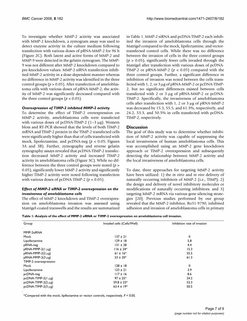
Effect of MMP-2 siRNA or TIMP-2 overexpression on the invasiveness of ameloblastoma cellsThe effect of MMP-2 knockdown and TIMP-2 overexpres-sion on ameloblastoma invasion was assessed usingMatrigel-coated transwells and the results are summarized
in Table 1. MMP-2 siRNA and pcDNA-TIMP-2 each inhib-ited the invasion of ameloblastoma cells through theMatrigel compared to the mock, lipifectamine, and vector-transfected control cells. While there was no differencebetween the invasion of cells in the three control groups(p > 0.05), significantly fewer cells invaded through theMatrigel after transfection with various doses of pcDNA-TIMP-2 or pRNA-MMP-2 (p < 0.05) compared with thethree control groups. Further, a significant difference ininhibition of invasion was noted between the cells trans-fected with 1, 2, or 3 μg of pRNA-MMP-2 or pcDNA-TIMP-2, but no significant differences existed between cellstransfected with 2 or 3 μg of pRNA-MMP-2 or pcDNA-TIMP-2. Specifically, the invasiveness of ameloblastomacells after transfection with 1, 2 or 3 μg of pRNA-MMP-2was decreased by 15.3, 55.5, and 61.3%, respectively, and24.2, 53.3, and 50.5% in cells transfected with pcDNA-TIMP-2, respectively.
DiscussionThe goal of this study was to determine whether inhibi-tion of MMP-2 activity was capable of suppressing thelocal invasiveness of human ameloblastoma cells. Thiswas accomplished using an MMP-2 gene knockdownapproach or TIMP-2 overexpression and subsequentlydetecting the relationship between MMP-2 activity andthe local invasiveness of ameloblastoma cells.
To date, three approaches for targeting MMP-2 activityhave been utilized: 1) the in vitro and in vivo delivery ofnaturally occurring inhibitors of MMP-2 (i.e., TIMP); 2)the design and delivery of novel inhibitory molecules ormodifications of naturally occurring inhibitors; and 3)targeting MMP-2 mRNA via various gene silencing strate-gies [20]. Previous studies performed by our grouprevealed that the MMP-2 inhibitor, Ro31-9790, inhibitedadhesion and invasion of ameloblastoma cells in primary
Table 1: Analysis of the effect of MMP-2 siRNA or TIMP-2 overexpression on ameloblastoma cell invasion.
Group Invaded cells (Cells/Well) Inhibition rate of invasion
MMP-2siRNAMock 137 ± 21 0Lipofectamine 129 ± 18 5.8pRNA-neg 131 ± 28 4.4pRNA-MMP-2(1 μg) 116 ± 24* 15.3pRNA-MMP-2(2 μg) 61 ± 16* 55.5pRNA-MMP-2(3 μg) 53 ± 20* 61.3TIMP-2 overexpressionMock 128 ± 18 0Lipofectamine 123 ± 21 3.9pcDNA-neg 117 ± 16 8.6pcDNA-TIMP-2(1 μg) 97 ± 25* 24.2pcDNA-TIMP-2(2 μg) 59.8 ± 23* 53.3pcDNA-TIMP-2(3 μg) 63.4 ± 19* 50.5
*Compared with the mock, lipifectamine or vector controls, respectively, P < 0.05.
Page 7 of 9(page number not for citation purposes)
BMC Cancer 2008, 8:182 http://www.biomedcentral.com/1471-2407/8/182
cell cultures [21]. While Ro31-9790 did not alter theexpression of either MMP-2 or TIMP-2, Ro31-9790 didinhibit the activity of MMP-2. This led to the suppositionthat the suppression of the local invasiveness of amelob-lastoma cells may be related to inhibition of MMP-2 activ-ity.
Utilization of siRNAs is one of the most effective genesilencing methods and is a promising new approach in theanalysis of gene function and gene therapy [22-25].Numerous studies have used siRNAs to analyze the func-tion of MMP-2 [26-28], but to date no studies have uti-lized this technique with ameloblastomas. Therefore, weinvestigated whether siRNAs targeted at MMP-2 are capa-ble of inhibiting the activity of MMP-2 in ameloblastomacells. siRNAs against MMP-2 significantly inhibited MMP-2 mRNA expression and MMP-2 protein levels in primaryameloblastoma cell cultures. Furthermore, MMP-2siRNAtransfection decreased the activity of MMP-2.
The conversion of MMP proenzymes to the activatedforms is controlled by the stoichiometric binding ofTIMPs which are synthesized by cells, such as fibroblasts,endothelial cells, and tumor cells [29]. The ability ofTIMPs to inhibit the activity of MMPs is known to signifi-cantly suppress tumor invasion and metastasis [5,30,31].To determine whether TIMP-2 inhibited the activity ofMMP-2 in ameloblastoma cells, plasmids were con-structed to overexpress TIMP-2. TIMP-2 overexpressioninhibited MMP-2 activity in ameloblastoma cells. Thisoutcome suggested that TIMP-2 might suppress invasive-ness in ameloblastomas in humans.
The underlying molecular mechanisms resulting in localinvasion by ameloblastomas are closely related to the pro-teolytic degradation of the basement membrane. Amongthe proteases thought to be involved in ameloblastomainvasion, attention has focused on MMP-2 [11,12,14,21].To investigate whether inhibition of MMP-2 activity willsuppress the invasiveness of ameloblastoma cells, weknocked down MMP-2 by RNA interference or overex-pressed TIMP-2. Both MMP-2 knockdown and TIMP-2overexpression inhibited the activity of MMP-2. The inva-sion assay showed that the ability of ameloblastoma cellsto invade the lower surface of the filter through theMatrigel was significantly inhibited in both MMP-2knockdown or TIMP-2 overexpression cells compared tothe control cultures.
ConclusionIn summary, these data indicate that siRNA targeting ofMMP-2 mRNA or TIMP-2 overxpression using a plasmid-based system effectively inhibited the activity of MMP-2 inameloblastoma cells, which subsequently resulted inreduced ameloblastoma cell invasiveness in vitro. This
study provided evidence that inhibition of MMP-2 activitymay serve as a novel therapeutic target in the clinical man-agement of ameloblastoma. Further research is warranted.
List of abbreviations usedMMP: matrix metalloproteinase; TIMP: tissue inhibitor ofmetalloproteinase; CK: cytokeratin; DMEM: Dulbecco'smodified Eagle medium; ANOVA: one-way analysis ofvariance; VEGF: vascular endothelial growth factor; GFP:green fluorescence protein; GAPDH, glyceraldehyde-3-phosphate dehydrogenase; RIPA: radio-immune precipi-tation assay; shRNA: small hairpin RNA; siRNA: smallinterference RNA; mRNA: message RNA; RT-PCR: reversetranscription polymerase chain reaction; SDS-PAGE:sodium dodecyl sulfate polyacrylamide gel electrophore-sis.
Competing interestsThe authors declare that they have no competing interests.
Authors' contributionsAW, BZ, LZ, DZ, QT, and HH were responsible for theexperimental design and completion of all laboratorywork represented in this manuscript. JW and CP partici-pated in the design and coordination of the workinvolved. The manuscript was drafted by AW and BZ. Allauthors have read and approved the final manuscript
AcknowledgementsThis work was supported by Grant No. 30471896 from the National Nat-ural Science Foundation of China, and Grant Nos. 06021272 and 04300240 from the Guangdong Natural Science Foundation of China.
References1. Gruica B, Stauffer E, Buser D, Bornstein M: Ameloblastoma of the
follicular, plexiform, and acanthomatous type in the maxil-lary sinus: a case report. Quintessence Int 2003, 34(4):311-314.
2. Junquera L, Ascani G, Vicente JC, Garcia-Consuegra L, Roig P: Amel-oblastoma revisited. Ann Otol Rhinol Laryngol 2003,112(12):1034-1039.
3. Heikinheimo K, Jee KJ, Niini T, Aalto Y, Happonen RP, Leivo I, Knuu-tila S: Gene expression profiling of ameloblastoma andhuman tooth germ by means of a cDNA microarray. J DentRes 2002, 81(8):525-530.
4. Kumamoto H: Molecular pathology of odontogenic tumors. JOral Pathol Med 2006, 35(2):65-74.
5. Liotta LA, Stetler-Stevenson WG: Metalloproteinases and cancerinvasion. Semin Cancer Biol 1990, 1(2):99-106.
6. Stamenkovic I: Extracellular matrix remodelling: the role ofmatrix metalloproteinases. J Pathol 2003, 200(4):448-464.
7. Birkedal-Hansen H, Moore WG, Bodden MK, Windsor LJ, Birkedal-Hansen B, DeCarlo A, Engler JA: Matrix metalloproteinases: areview. Crit Rev Oral Biol Med 1993, 4(2):197-250.
8. Nagase H: Activation mechanisms of matrix metalloprotein-ases. Biol Chem 1997, 378(3-4):151-160.
9. Coussens LM, Werb Z: Matrix metalloproteinases and thedevelopment of cancer. Chem Biol 1996, 3(11):895-904.
10. Stetler-Stevenson WG, Hewitt R, Corcoran M: Matrix metallopro-teinases and tumor invasion: from correlation and causalityto the clinic. Semin Cancer Biol 1996, 7(3):147-154.
11. Kumamoto H, Yamauchi K, Yoshida M, Ooya K: Immunohisto-chemical detection of matrix metalloproteinases (MMPs)and tissue inhibitors of metalloproteinases (TIMPs) in amel-oblastomas. J Oral Pathol Med 2003, 32(2):114-120.
Page 8 of 9(page number not for citation purposes)
BMC Cancer 2008, 8:182 http://www.biomedcentral.com/1471-2407/8/182
Publish with BioMed Central and every scientist can read your work free of charge
"BioMed Central will be the most significant development for disseminating the results of biomedical research in our lifetime."
Sir Paul Nurse, Cancer Research UK
Your research papers will be:
available free of charge to the entire biomedical community
peer reviewed and published immediately upon acceptance
cited in PubMed and archived on PubMed Central
yours — you keep the copyright
Submit your manuscript here:http://www.biomedcentral.com/info/publishing_adv.asp
BioMedcentral
12. Zhong M, Han YP, Wang J, Li ZJ, Bao G, Yue YL: [Expression ofmatrix metalloproteinases and tissue inhibitor of metallo-proteinase in ameloblastoma]. Shanghai Kou Qiang Yi Xue 2003,12(6):427-431.
13. Pinheiro JJ, Freitas VM, Moretti AI, Jorge AG, Jaeger RG: Local inva-siveness of ameloblastoma. Role played by matrix metallo-proteinases and proliferative activity. Histopathology 2004,45(1):65-72.
14. Zhong M, Li ZJ, Wang J, Yue YL, Bao G: [The study of the invasivebiologic behavior of ameloblastoma]. Zhonghua Kou Qiang YiXue Za Zhi 2004, 39(1):45-48.
15. Wang X, Fu X, Brown PD, Crimmin MJ, Hoffman RM: Matrix met-alloproteinase inhibitor BB-94 (batimastat) inhibits humancolon tumor growth and spread in a patient-like orthotopicmodel in nude mice. Cancer Res 1994, 54(17):4726-4728.
16. Watson SA, Morris TM, Parsons SL, Steele RJ, Brown PD: Thera-peutic effect of the matrix metalloproteinase inhibitor, bati-mastat, in a human colorectal cancer ascites model. Br JCancer 1996, 74(9):1354-1358.
17. Rao JS, Bhoopathi P, Chetty C, Gujrati M, Lakka SS: MMP-9 shortinterfering RNA induced senescence resulting in inhibition ofmedulloblastoma growth via p16(INK4a) and mitogen-acti-vated protein kinase pathway. Cancer Res 2007,67(10):4956-4964.
18. Kleiner DE, Stetler-Stevenson WG: Quantitative zymography:detection of picogram quantities of gelatinases. Anal Biochem1994, 218(2):325-329.
19. Kido A, Krueger S, Haeckel C, Roessner A: Inhibitory effect ofantisense aminopeptidase N (APN/CD13) cDNA transfec-tion on the invasive potential of osteosarcoma cells. Clin ExpMetastasis 2003, 20(7):585-592.
20. Hu YB, Li DG, Lu HM: Modified synthetic siRNA targeting tis-sue inhibitor of metalloproteinase-2 inhibits hepatic fibro-genesis in rats. J Gene MedJ Gene Med 2007, 9(3):217-229.
21. Bertrand JR, Pottier M, Vekris A, Opolon P, Maksimenko A, Malvy C:Comparison of antisense oligonucleotides and siRNAs in cellculture and in vivo. Biochem Biophys Res Commun 2002,296(4):1000-1004.
22. Elbashir SM, Harborth J, Weber K, Tuschl T: Analysis of gene func-tion in somatic mammalian cells using small interferingRNAs. Methods 2002, 26(2):199-213.
23. Miyagishi M, Hayashi M, Taira K: Comparison of the suppressiveeffects of antisense oligonucleotides and siRNAs directedagainst the same targets in mammalian cells. Antisense NucleicAcid Drug Dev 2003, 13(1):1-7.
24. Yokota T, Miyagishi M, Hino T, Matsumura R, Tasinato A, UrushitaniM, Rao RV, Takahashi R, Bredesen DE, Taira K, Mizusawa H: siRNA-based inhibition specific for mutant SOD1 with single nucle-otide alternation in familial ALS, compared with ribozymeand DNA enzyme. Biochem Biophys Res Commun 2004,314(1):283-291.
25. Birkedal-Hansen B, Pavelic ZP, Gluckman JL, Stambrook P, Li YQ,Stetler-Stevenson WG: MMP and TIMP gene expression in headand neck squamous cell carcinomas and adjacent tissues.Oral Dis 2000, 6(6):376-382.
26. Chetty C, Bhoopathi P, Joseph P, Chittivelu S, Rao JS, Lakka S: Ade-novirus-mediated small interfering RNA against matrix met-alloproteinase-2 suppresses tumor growth and lungmetastasis in mice. Mol Cancer Ther 2006, 5(9):2289-2299.
27. Chetty C, Bhoopathi P, Lakka SS, Rao JS: MMP-2 siRNA inducedFas/CD95-mediated extrinsic II apoptotic pathway in theA549 lung adenocarcinoma cell line. Oncogene 2007.
28. Verstappen J, Von den Hoff JW: Tissue inhibitors of metallopro-teinases (TIMPs): their biological functions and involvementin oral disease. J Dent Res 2006, 85(12):1074-1084.
29. Ring P, Johansson K, Hoyhtya M, Rubin K, Lindmark G: Expressionof tissue inhibitor of metalloproteinases TIMP-2 in humancolorectal cancer--a predictor of tumour stage. Br J Cancer1997, 76(6):805-811.
30. Brummer O, Athar S, Riethdorf L, Loning T, Herbst H: Matrix-met-alloproteinases 1, 2, and 3 and their tissue inhibitors 1 and 2in benign and malignant breast lesions: an in situ hybridiza-tion study. Virchows Arch 1999, 435(6):566-573.
31. Zhang B, Huang HZ, Tao Q, Liu XQ, Wei J: [Association of matrixmetalloproteinase-2 activity with cell proliferation and
growth in ameloblastoma]. Hua Xi Kou Qiang Yi Xue Za Zhi 2006,24(1):7-10.
Pre-publication historyThe pre-publication history for this paper can be accessedhere:
http://www.biomedcentral.com/1471-2407/8/182/prepub
Page 9 of 9(page number not for citation purposes)


