Embed Size (px)
Citation preview
Mar Biol (2006) 150:197–211
DOI 10.1007/s00227-006-0350-0RESEARCH ARTICLE
Subcellular distribution of zinc and cadmiumin the hepatopancreas and gills of the decapod crustacean Penaeus indicus
G. Nunez-Nogueira · C. Mouneyrac · J. C. Amiard · P. S. Rainbow
Received: 19 July 2005 / Accepted: 28 April 2006 / Published online: 9 June 2006© Springer-Verlag 2006
Abstract The decapod crustacean Penaeus indicusaccumulated Cd and Zn in diVerent subcellular com-partments of hepatopancreas and gill cells. Most of theCd and part of the Zn accumulates within the solublefraction of the cells, while the remainder of the Zn isfound in insoluble inclusions, associated with P, Ca, Mgand Si in B-, F- and R-cells in the hepatopancreas, andhaemocytes, nephrocytes and epithelial cells in thegills. No presence of Cd was observed in metal-richinclusions in any cell analysed. Metallothionein-likeproteins (MTLP), analysed by diVerential pulse polar-ography, were present in the hepatopancreas (12–18 mg g¡1) and gills (7–8 mg g¡1) of metal-exposedprawns. Binding to MTLP is the detoxiWcation mecha-nism for cadmium, while the detoxiWcation of zinc
involves both binding to MTLP and incorporation intoinsoluble metal-rich inclusions.
Introduction
The capacity of the decapod crustacean Penaeus indi-cus to accumulate zinc and cadmium, either from solu-tion or food, indicates the presence of physiologicaldetoxiWcatory processes at the cellular level to retainthese metals for a shorter (in the case of the essentialmetal zinc) or longer period (non-essential cadmium)(Nunez-Nogueira and Rainbow 2005a, b; Nunez-Nogueira et al. 2006). The body distributions of newlytaken up zinc and cadmium in this decapod show thatthe hepatopancreas is strongly involved in the accumu-lation of these metals, and also indicate that the gillsplay an important role in trace metal uptake from solu-tion and in any loss from the body (Nunez-Nogueiraand Rainbow 2005a, b; Nunez-Nogueira et al. 2006).
The detoxiWcation of accumulated trace metals incells can involve their binding to both insoluble andsoluble subcellular components. Several ultrastructuralstudies have been performed on invertebrates to inves-tigate the cellular detoxiWcation of trace metals, indi-cating that diVerent metals may be present in morethan one subcellular compartment or form in diVerenttissues and cells (Al-Mohanna and Nott 1987a, b, 1989;Hopkin 1989; Vogt and Quinitio 1994; Mason and Jen-kins 1995; Nassiri et al. 2000; Marigomez et al. 2002).The chemical composition and appearance of thesemetal-rich deposits vary widely. Hopkin (1989) classi-Wed metal-containing granules into four diVerent cate-gories: type A, consisting of concentric layers ofcalcium and magnesium phosphates which may contain
Communicated by J.P. Thorpe, Port Erin
G. Nunez-Nogueira · P. S. Rainbow (&)Department of Zoology, The Natural History Museum, Cromwell Road, London, SW7 5BD, UKe-mail: [email protected]
C. MouneyracCentre d’étude et de Recherche sur les écosystèms aquatiques, Université Catholique de l’ouest, 44 Rue Rabelais, BP 808, 49008 Angers Cedex 01, France
J. C. AmiardService d’Ecotoxicologie, CNRS-GDR 1117, ISOMer, SMAB, 2 Rue de la Houssinière, BP 92208, 44322 Nantes Cedex 3, France
G. Nunez-NogueiraLaboratorio de Contaminación Marina, Instituto de Ciencias del Mar y Limnología, UNAM, Circuito Exterior S/N. 04510, México DF, Mexico
123
198 Mar Biol (2006) 150:197–211
other class A and borderline metals such as manganeseand zinc; type B, more heterogeneous in shape andalways containing sulphur in association with class Band borderline metals including cadmium, copper andzinc; type C, often polyhedral with a crystalline form,mainly containing iron, probably derived from ferritin;and type D, larger (extracellular) granules composed ofconcentric layers of calcium carbonate. In crustaceans,including decapods, the most commonly reportedmetal-rich granules in the hepatopancreas (or equiva-lent organs) are type A and B granules (Al-Mohannaand Nott 1985, 1987a, 1989; Correa-Junior et al. 2000;Nassiri et al. 2000).
Particular trace metals (Zn, Cu, Cd, Ag, Hg forexample) have been found to be associated with, andinduce, metallothioneins, low molecular weight cyto-solic proteins involved in the cellular regulation anddetoxiWcation of these metals (Roesijadi 1981; Engeland Roesijadi 1987; Viarengo et al. 1999). The pres-ence of thiolic groups in cysteine residues in the pro-teins provides the high metal aYnity of the molecule(Roesijadi 1981; Engel and Roesijadi 1987), sequester-ing metals in the cytoplasm and reducing their meta-bolic availability. Induced metallothioneins can bindcopper and zinc already present in the cell and notreact only to newly taken up metals. Zinc and cadmiumare among the trace metals reported to induce metallo-thionein production in the gills and digestive glands (orhepatopancreas) of diVerent invertebrates exposed toraised concentrations of these metals (Rainbow andScott 1979; Roesijadi 1981; Carpene 1993; Canli et al.1997), indicating that these proteins could be involvedin zinc and cadmium detoxiWcation within the cells ofthe hepatopancreas and gills of decapod crustaceansexposed to raised zinc or cadmium availabilities.
The hepatopancreas of decapod crustaceans playsan important role in trace metal accumulation (Bryan1976; Gibson and Barker 1979; Dall and Moriarty 1983;Rainbow 1998). DetoxiWed copper or zinc-rich gran-ules have been described from the hepatopancreas ofP. semisulcatus (Al-Mohanna and Nott 1985) and P.monodon (Vogt and Quinitio 1994). Zinc is stored inphosphorus-rich granules in hepatopancreas cells of P.semisulcatus, and granule abundance and hepatopan-creas zinc concentration vary with the moult cycle (Al-Mohanna and Nott 1985). It has been suggested thatcopper is accumulated in the hepatopancreas of deca-pods before use in the synthesis of haemocyanin (John-ston and Barber 1969), and the hepatopancreas alsostores high concentrations of copper in decapodsexposed to raised dissolved copper concentrations,prior to elimination from the body through the faeces(Gibson and Barker 1979).
The cellular accumulation of metals in the hepato-pancreas varies according to the cell involved (Gibsonand Barker 1979). The epithelial cells of the decapodcrustacean hepatopancreas have been classiWed byeither their form or function. The four cell typesreported are Embryonic (E-cells), Fibrillar (F-cells),Blister-like (B-cells) and Resorptive (R-cells) respec-tively [after Jacob (1928) and Hirsch and Jacob (1930)in Gibson and Barker 1979]. A Wfth cell type has beendescribed by Al-Mohanna and Nott (1987), denomi-nated “midget or M-cell”, although later studies onlyreport the presence of four diVerent cell types in thedecapod hepatopancreas as described previously(Caceci et al. 1988; Andersen and Baatrup 1988; Vogtand Quinitio 1994). The abundance of one particularcell type in the hepatopancreas is related to the moult-ing cycle and the state of starvation of the decapod(Papathanassiou and King 1984; Al-Mohanna and Nott1989).
Gills are important permeable areas in the body of adecapod, involved in the chemical exchange of mole-cules such as respiratory gases, water, ammonia andmetals, between the body and the external medium.Decapod gills are involved in respiration, osmoregula-tion, excretion and pH regulation (Foster and Howse1978; Gilles and Pequeux 1983; Taylor and Taylor1992). Foster and Howse (1978) analysed the structuralorganization and morphology of gills of the brownshrimp P. aztecus, establishing the penaeid gill as den-drobranchiate and describing the presence of diVerentcell types. Five diVerent epithelial cell types arereported to be present in decapod gills, denominatedas thin cells, thick cells, attenuated cells, pillar cells andXange cells; haemocytes and nephrocytes are found inthe blood spaces in the gills (Taylor and Taylor 1992).Only the last four gill epithelial cell types have so farbeen reported in Penaeus prawns (Foster and Howse1978).
The biology of trace metals in the gills of Penaeusprawns has not been studied in as much detail as in thehepatopancreas, although studies have analysed therole of the gills in ion uptake (Arruda-Freire andCampbell-McNamara 1995), in addition to osmoticregulation and respiration (Gilles and Pequeux 1983).Gills have been shown to be vulnerable to trace metalexposure, with ultrastructural changes reported (Gillesand Pequeux 1983). Soegianto et al. (1999a, b)observed such eVects in P. japonicus exposed to copperand cadmium in solution at relatively high concentra-tions (greater than 500 �g Cu l¡1 and 2,000 �g Cd l¡1,respectively).
Penaeid prawns are important subjects of maricul-ture and coastal Wsheries activities throughout the tro-
123
Mar Biol (2006) 150:197–211 199
pics and subtropics (Holthuis 1980; Grey et al. 1983;Perez-Farfante and Kensley 1997). They are commonin estuaries and are therefore potentially aVected byanthropogenic metal contamination. This study is partof a wider investigation of the biology of the trace met-als zinc and cadmium in a model penaeid prawn P.indicus (Nunez-Nogueira and Rainbow 2005a, b;Nunez-Nogueira et al. 2006). It speciWcally investigatesthe subcellular detoxiWcation of accumulated zinc andcadmium in the hepatopancreas and gills of P. indicus,after sublethal exposure of the prawns to dissolved zincand cadmium. The study incorporates an ultrastruc-tural investigation of the nature and localisation of anymetal-rich inclusions in the cells of the hepatopancreasand gills, and a study of metallothionein-like proteins(MTLP) binding zinc and cadmium in the same cells.
Material and methods
Juveniles of P. indicus [between 1 and 2.5 cm totallength (Le Reste 1978); mean dry weight 31.16 §17.96 mg] were obtained from cultures at the School ofOcean Sciences, University of North Wales, Bangor,Wales from stocks originally from the Gulf of Aden.No distinction between genders was as yet discernible.
Prawns were maintained in artiWcial sea water(Tropic Marine New, TMN; Aquarientechnick, Waten-berg®-Germany) at 15 salinity (pH 7.8), 12:12 light/dark periods and 25°C as previously described (Nunez-Nogueira and Rainbow 2005a, b). The use of artiWcialseawater in all experiments provided physico-chemicalstability, insigniWcant background dissolved trace metalconcentrations, and replicability for trace metal uptakestudies (Rainbow 1997).
The experimental design involved seven separateexperimental groups containing ten prawns each: con-trols (not metal exposed; three groups), prawnsexposed to 100 �g l¡1 zinc (two experimental groups),and cadmium (two experimental groups) in TMN at 15salinity at 25°C. The hepatopancreas and gills wereremoved from one control group at the beginning ofthe experiment, as described by Nunez-Nogueira andRainbow (2005a). Prawns from the remaining two con-trol and metal-treated groups were dissected after 5 or10 days of exposure.
The dissolved metal concentrations are environmen-tally realistic for metal contaminated estuaries (Bryanet al. 1985; Law et al. 1994) and are below dissolvedzinc and cadmium concentrations toxic to penaeids(McClurg 1984; Joseph et al. 1992; Chinni and Yalla-pragada 2000). In order to maintain the nominal metalconcentrations in the experimental media (conWrmed
by atomic absorption spectrophotometry), all experi-mental glassware was pre-soaked in the appropriateexperimental medium to saturate any adsorption ontothe vessels. Thereafter the experimental media werechanged every 2 days immediately after feeding theprawns for 30 min (see Nunez-Nogueira and Rainbow2005a, b).
X-ray microanalysis and electron microscopy analysis
Hepatopancreas and gills samples destined for ultra-structural study were transferred into individual vialsto be Wxed in 2.5% glutaraldehyde solution (0.2 MSorensen’s buVer, pH = 7.2 at 2°C) immediately afterdissection, for 120 min. Samples were then rinsed withbuVer, and dehydrated by immersion in a series ofdiluted ethanols up to 100% ethanol (BDH Lab,England), using cold (2°C) ethanol over a period of3.5 h. Dehydrated samples were transferred into pro-pylene oxide (PO) and distilled water solution for5 min. Samples were then transferred into a PO solu-tion mixed with TAAB resin (50% v/v) and rotatedovernight, before Wnal transfer into 100% TAAB resinfor 10 h. At the end of the day, samples were embed-ded in fresh TAAB resin within BEEM capsules anddried at 90°C for 8 h for solidiWcation.
Ultrathin (between 0.5 and 0.8 nm thickness) andsemithin sections (between 0.5 and 0.9 �m thickness)were cut on an ultramicrotome (Reichert Ultracut S®)with glass knives or diamond knife (Microstar Technol-ogies®) and collected on uncoated nickel (Agar®) oraluminium (Polaron Equipment Ltd.) grids (3.05 mmsize and 200 square mesh). Ultrathin sections werestained with an alcoholic solution of uranyl acetate(1% uranyl acetate in 50–100% methanol; 4 min) andReynold’s lead citrate (35 mg lead citrate ml¡1 in 1 NNaOH, 7 min) for observation of subcellular struc-tures. Semithin sections were kept unstained for X-raymicroanalysis. Both sections were analysed in a TEM-R200 EXII Electron Microscope with STEM attach-ment and LINK QX 2000 Energy Disperser and X-raymicroanalysis system. X-ray analyses were performedat 100 keV and specimen tilted 30°.
Metal and MTLP analyses
Samples destined for biochemical analysis were trans-ferred into plastic vials to be immediately frozen in liq-uid nitrogen for a few minutes. After freezing, sampleswere freeze-dried overnight and their dry weight mea-sured. Then, samples were homogenized in a buVersolution (20 mM TRIS, 10–5 �-mercaptoethanol,150 mM NaCl solution adjusted to pH 8.6). The soluble
123
200 Mar Biol (2006) 150:197–211
and insoluble fractions resulting from this homogeniza-tion procedure were separated by centrifugation at25,074g for 55 min at 4°C (Biofuge 28 RS, HeraeusSepatech®). The cytosolic heat-stable compoundsincluding metallothionein were isolated by centrifuga-tion of the soluble fraction (12,000g for 10 min at 4°C)after heat-treatment (75°C for 15 min). In the heat-denatured cytosol, the amount of MT was determinedby diVerential pulse polarography (DPP) according toThompson and Cosson (1984) and Olafson and Olsson(1991). A MDE 150 Stand Polarographique (Radiome-ter Copenhagen) Tracelab™ 50, controlled by the com-puter software Tracemaster 5 through a Polarographicanalyser POL 150 was used. The temperature of thecell was maintained at 4°C. The method of standardaddition was used for calibration with rabbit liver MT(SIGMA Chemical Co., St Louis, MO, lot. 20 k7000;M-7641 code) in absence of a shrimp MT standard.Polarographic determination in heat-denaturated cyto-sol is an analytical procedure based on several charac-teristics of MTs, but it does not allow, with certainty,the assertion that the target molecule is a true MTunless puriWcation and sequencing are carried out.Strictly, therefore, what has been measured is the con-centration of proteins with metallothionein properties,ie. MTLP.
Metal analysis was carried out on the soluble andinsoluble fractions. Nalgene bottles were used to storeall reagents. All glass labware was soaked in 10% HCl,rinsed three times with deionized water and dried in adesiccator protected from atmospheric dust. The insol-uble and soluble fractions were heated (75°C, 12 h)with suprapure HNO3 acid (Carlo Erba). After diges-tion, metal concentrations in these acid digests weredetermined after dilution with deionized water byXame AAS (Zn) or electrothermal atomic absorptionspectrophotometry (EAAS) (Cd) using the ZeemaneVect (Hitachi Z 8200 spectrophotometer). The analyt-ical method has been described previously by Amiardet al. (1987).
Standard addition analyses were performed in aniso-medium and concentrations of each element were+125, +250, +500 ng Zn ml¡1 for FAAS and +0.25,+0.5, +1 ng Cd ml¡1 for EAAS. The analytical methodswere validated by external intercalibrations (Coqueryand Horvat 1996; Campbell et al. 2000). Total metalconcentrations were recalculated from summation ofquantities of trace elements in soluble and insolublefractions determined previously, by the combinedmeans procedure (Williams 2000). Results areexpressed in �g g¡1 dry weight of the organ.
Student’s t test (P < 0.05) were performed for pro-tein and metal concentration comparisons. Tests were
developed according to Williams (2000), and carriedout using STATISTICA 5.1 for Windows (StatSoftInc.).
Results
Subcellular partitioning
Table 1 shows the concentrations (and percentage dis-tributions) of Zn and Cd in the soluble and insolublecomponents of the cells of the hepatopancreas and gillsof P. indicus exposed or not to raised availabilities ofZn and Cd. Percentage distributions of Zn was major-ity accumulated in the soluble fraction (between 76 and83%) in the hepatopancreas, while the gills showed asimilar distribution between these two fractions (40–55% insoluble fraction, 45–60% soluble). The percent-age distribution of Cd was always higher in the solublefraction in both tissues (70–92% in the hepatopan-creas; 73–90% in the gills). The tissue zinc concentra-tions did not diVer signiWcantly among groups, whilecadmium was clearly accumulated in both the hepato-pancreas and the gills during cadmium exposure(Table 1).
The total cadmium concentration in the hepatopan-creas (48.0 § 12.6 �g g¡1) increased after exposure inprawns exposed for 10 days to 100 �g Cd l¡1. Of this,85% was found in the soluble fraction of the tissue andonly 15% in the insoluble fraction (Table 1). Gills ofcadmium-exposed prawns had the same pattern, with90% of cadmium in the soluble fraction and 10% in theinsoluble fraction. The total cadmium concentration inthis tissue was 36.3 § 1.9 �g g¡1. Exposure of P. indicusto 100 �g Zn l¡1 for 10 days did not produce signiWcantchanges in the tissue metal concentrations. Metal-exposed prawns showed an average concentration of121.2 § 24.5 �g Zn g¡1 (Cd-exposed) and115.1 § 28.1 �g Zn g¡1 (Zn-exposed), which were notsigniWcantly diVerent from Zn concentrations in thecontrol group (150.3 § 59.5 �g Zn g¡1) (Table 1).
Hepatopancreas ultrastructure
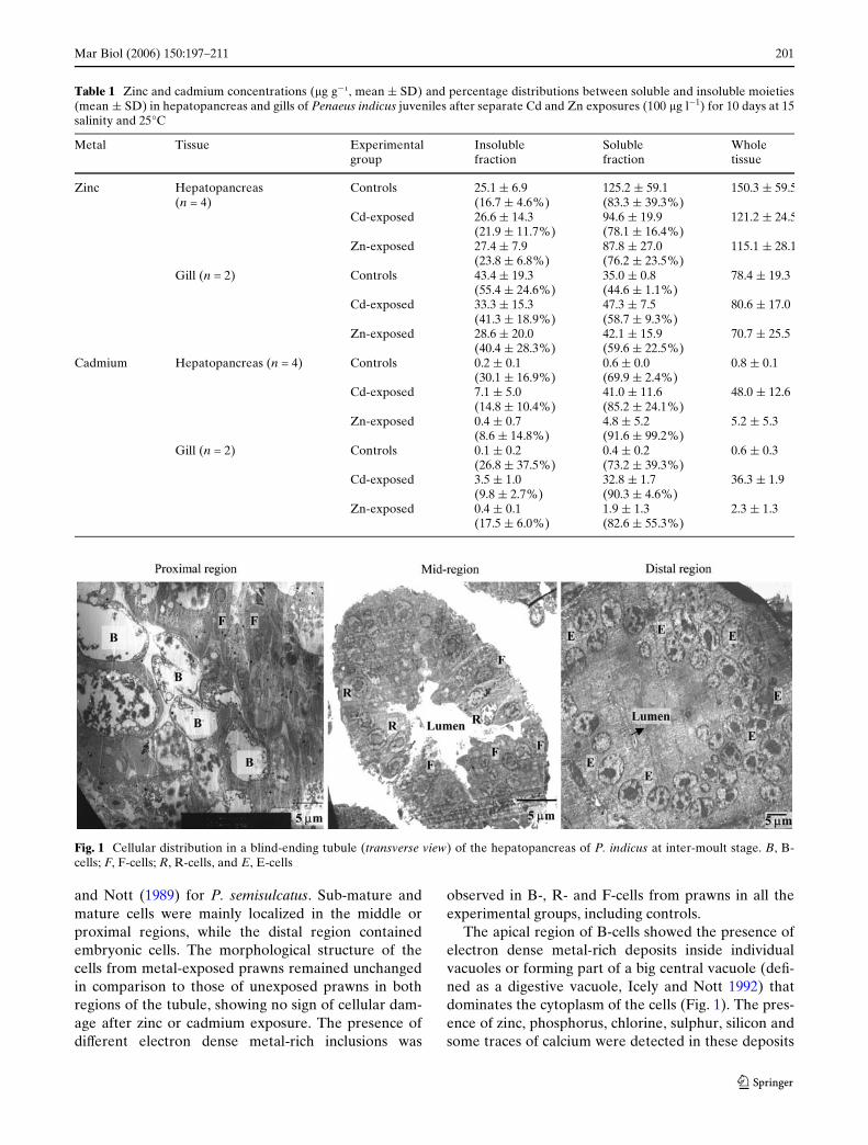
The four hepatopancreas cell types (Embryonic, E-cells; Fibrillar, F-cells; Blister-like, B-cells; and Resorp-tive, R-cells) reported by Gibson and Barker (1979)were recognised in P. indicus in this study. The Wfth celltype (Midget or M-cell) described by Al-Mohanna andNott (1987) could not be distinguished.
The distribution of the cells along a blind-endingtubule of the hepatopancreas of P. indicus (Fig. 1)clearly follows the previous description of Al-Mohanna
123
Mar Biol (2006) 150:197–211 201
and Nott (1989) for P. semisulcatus. Sub-mature andmature cells were mainly localized in the middle orproximal regions, while the distal region containedembryonic cells. The morphological structure of thecells from metal-exposed prawns remained unchangedin comparison to those of unexposed prawns in bothregions of the tubule, showing no sign of cellular dam-age after zinc or cadmium exposure. The presence ofdiVerent electron dense metal-rich inclusions was
observed in B-, R- and F-cells from prawns in all theexperimental groups, including controls.
The apical region of B-cells showed the presence ofelectron dense metal-rich deposits inside individualvacuoles or forming part of a big central vacuole (deW-ned as a digestive vacuole, Icely and Nott 1992) thatdominates the cytoplasm of the cells (Fig. 1). The pres-ence of zinc, phosphorus, chlorine, sulphur, silicon andsome traces of calcium were detected in these deposits
Table 1 Zinc and cadmium concentrations (�g g¡1, mean § SD) and percentage distributions between soluble and insoluble moieties(mean § SD) in hepatopancreas and gills of Penaeus indicus juveniles after separate Cd and Zn exposures (100 �g l¡1) for 10 days at 15salinity and 25°C
Metal Tissue Experimental group
Insoluble fraction
Soluble fraction
Whole tissue
Zinc Hepatopancreas (n = 4)
Controls 25.1 § 6.9 (16.7 § 4.6%)
125.2 § 59.1(83.3 § 39.3%)
150.3 § 59.5
Cd-exposed 26.6 § 14.3(21.9 § 11.7%)
94.6 § 19.9(78.1 § 16.4%)
121.2 § 24.5
Zn-exposed 27.4 § 7.9(23.8 § 6.8%)
87.8 § 27.0(76.2 § 23.5%)
115.1 § 28.1
Gill (n = 2) Controls 43.4 § 19.3(55.4 § 24.6%)
35.0 § 0.8(44.6 § 1.1%)
78.4 § 19.3
Cd-exposed 33.3 § 15.3(41.3 § 18.9%)
47.3 § 7.5(58.7 § 9.3%)
80.6 § 17.0
Zn-exposed 28.6 § 20.0(40.4 § 28.3%)
42.1 § 15.9(59.6 § 22.5%)
70.7 § 25.5
Cadmium Hepatopancreas (n = 4) Controls 0.2 § 0.1(30.1 § 16.9%)
0.6 § 0.0(69.9 § 2.4%)
0.8 § 0.1
Cd-exposed 7.1 § 5.0(14.8 § 10.4%)
41.0 § 11.6(85.2 § 24.1%)
48.0 § 12.6
Zn-exposed 0.4 § 0.7(8.6 § 14.8%)
4.8 § 5.2(91.6 § 99.2%)
5.2 § 5.3
Gill (n = 2) Controls 0.1 § 0.2(26.8 § 37.5%)
0.4 § 0.2(73.2 § 39.3%)
0.6 § 0.3
Cd-exposed 3.5 § 1.0(9.8 § 2.7%)
32.8 § 1.7(90.3 § 4.6%)
36.3 § 1.9
Zn-exposed 0.4 § 0.1(17.5 § 6.0%)
1.9 § 1.3(82.6 § 55.3%)
2.3 § 1.3
Fig. 1 Cellular distribution in a blind-ending tubule (transverse view) of the hepatopancreas of P. indicus at inter-moult stage. B, B-cells; F, F-cells; R, R-cells, and E, E-cells
123
202 Mar Biol (2006) 150:197–211
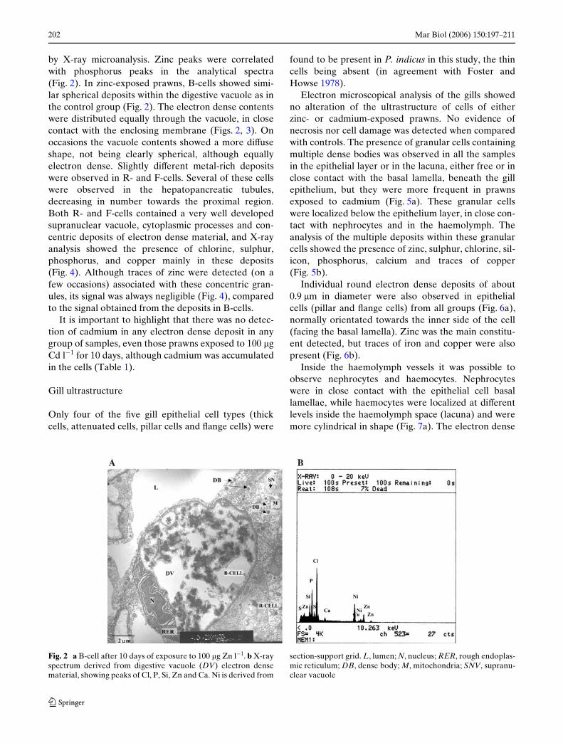
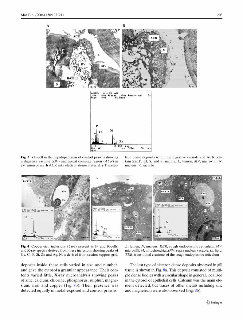
by X-ray microanalysis. Zinc peaks were correlatedwith phosphorus peaks in the analytical spectra(Fig. 2). In zinc-exposed prawns, B-cells showed simi-lar spherical deposits within the digestive vacuole as inthe control group (Fig. 2). The electron dense contentswere distributed equally through the vacuole, in closecontact with the enclosing membrane (Figs. 2, 3). Onoccasions the vacuole contents showed a more diVuseshape, not being clearly spherical, although equallyelectron dense. Slightly diVerent metal-rich depositswere observed in R- and F-cells. Several of these cellswere observed in the hepatopancreatic tubules,decreasing in number towards the proximal region.Both R- and F-cells contained a very well developedsupranuclear vacuole, cytoplasmic processes and con-centric deposits of electron dense material, and X-rayanalysis showed the presence of chlorine, sulphur,phosphorus, and copper mainly in these deposits(Fig. 4). Although traces of zinc were detected (on afew occasions) associated with these concentric gran-ules, its signal was always negligible (Fig. 4), comparedto the signal obtained from the deposits in B-cells.
It is important to highlight that there was no detec-tion of cadmium in any electron dense deposit in anygroup of samples, even those prawns exposed to 100 �gCd l¡1 for 10 days, although cadmium was accumulatedin the cells (Table 1).
Gill ultrastructure
Only four of the Wve gill epithelial cell types (thickcells, attenuated cells, pillar cells and Xange cells) were
found to be present in P. indicus in this study, the thincells being absent (in agreement with Foster andHowse 1978).
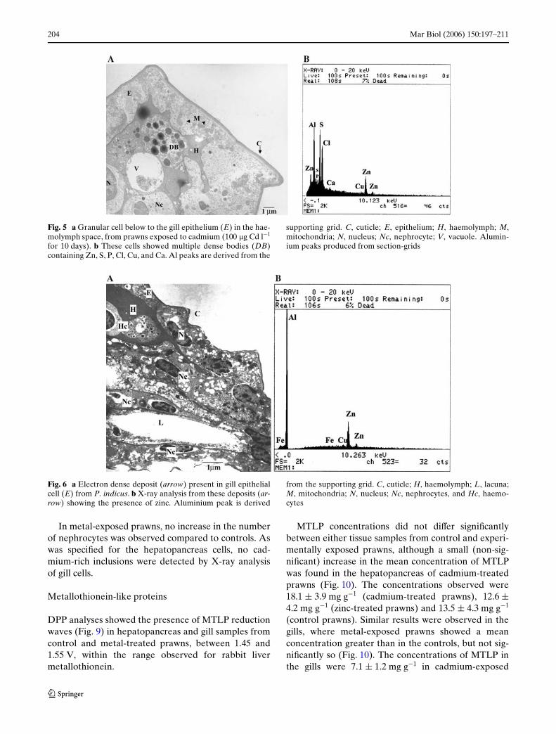
Electron microscopical analysis of the gills showedno alteration of the ultrastructure of cells of eitherzinc- or cadmium-exposed prawns. No evidence ofnecrosis nor cell damage was detected when comparedwith controls. The presence of granular cells containingmultiple dense bodies was observed in all the samplesin the epithelial layer or in the lacuna, either free or inclose contact with the basal lamella, beneath the gillepithelium, but they were more frequent in prawnsexposed to cadmium (Fig. 5a). These granular cellswere localized below the epithelium layer, in close con-tact with nephrocytes and in the haemolymph. Theanalysis of the multiple deposits within these granularcells showed the presence of zinc, sulphur, chlorine, sil-icon, phosphorus, calcium and traces of copper(Fig. 5b).
Individual round electron dense deposits of about0.9 �m in diameter were also observed in epithelialcells (pillar and Xange cells) from all groups (Fig. 6a),normally orientated towards the inner side of the cell(facing the basal lamella). Zinc was the main constitu-ent detected, but traces of iron and copper were alsopresent (Fig. 6b).
Inside the haemolymph vessels it was possible toobserve nephrocytes and haemocytes. Nephrocyteswere in close contact with the epithelial cell basallamellae, while haemocytes were localized at diVerentlevels inside the haemolymph space (lacuna) and weremore cylindrical in shape (Fig. 7a). The electron dense
Fig. 2 a B-cell after 10 days of exposure to 100 �g Zn l¡1. b X-rayspectrum derived from digestive vacuole (DV) electron densematerial, showing peaks of Cl, P, Si, Zn and Ca. Ni is derived from
section-support grid. L, lumen; N, nucleus; RER, rough endoplas-mic reticulum; DB, dense body; M, mitochondria; SNV, supranu-clear vacuole
123
Mar Biol (2006) 150:197–211 203
deposits inside these cells varied in size and number,and gave the cytosol a granular appearance. Their con-tents varied little, X-ray microanalysis showing peaksof zinc, calcium, chlorine, phosphorus, sulphur, magne-sium, iron and copper (Fig. 7b). Their presence wasdetected equally in metal-exposed and control prawns.
The last type of electron dense deposits observed in gilltissue is shown in Fig. 8a. This deposit consisted of multi-ple dense bodies with a circular shape in general, localizedin the cytosol of epithelial cells. Calcium was the main ele-ment detected, but traces of other metals including zincand magnesium were also observed (Fig. 8b).
Fig. 3 a B-cell in the hepatopancreas of control prawns showinga digestive vacuole (DV) and apical complex region (ACR) inextrusion phase. b ACR with electron dense material. c The elec-
tron dense deposits within the digestive vacuole and ACR con-tain Zn, P, Cl, S, and Si mainly. L, lumen; MV, microvilli; N,nucleus; V, vacuole
Fig. 4 Copper-rich inclusions (Cu-I) present in F- and R-cells,and X-ray spectra derived from these inclusions showing peaks ofCu, Cl, P, Si, Zn and Ag. Ni is derived from section-support grid.
L, lumen; N, nucleus; RER, rough endoplasmic reticulum; MV,microvilli; M, mitochondria; SNV, supra nuclear vacuole; Li, lipid;TER, transitional elements of the rough endoplasmic reticulum
123
204 Mar Biol (2006) 150:197–211
In metal-exposed prawns, no increase in the numberof nephrocytes was observed compared to controls. Aswas speciWed for the hepatopancreas cells, no cad-mium-rich inclusions were detected by X-ray analysisof gill cells.
Metallothionein-like proteins
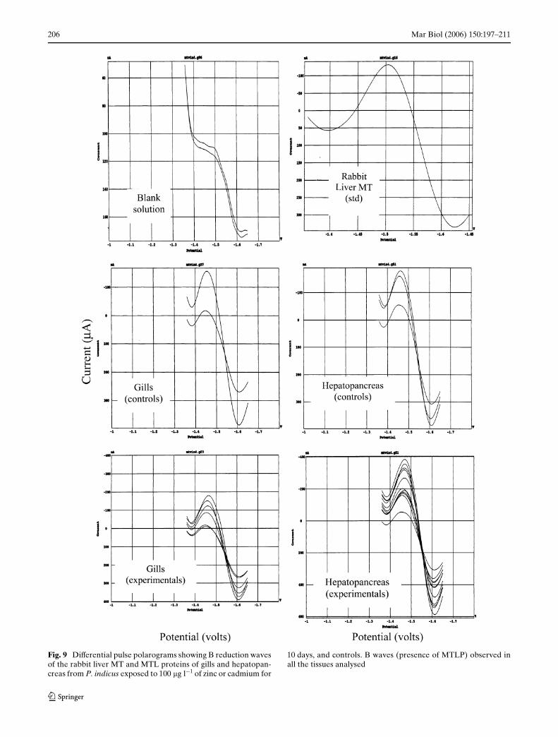
DPP analyses showed the presence of MTLP reductionwaves (Fig. 9) in hepatopancreas and gill samples fromcontrol and metal-treated prawns, between 1.45 and1.55 V, within the range observed for rabbit livermetallothionein.
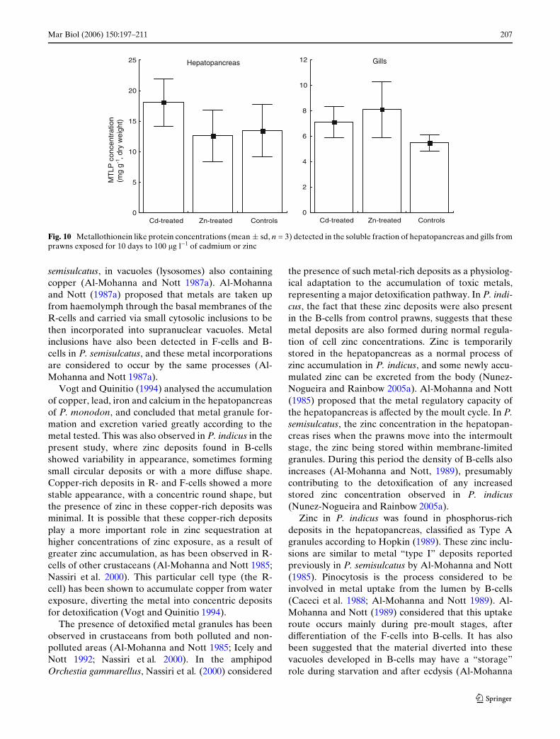
MTLP concentrations did not diVer signiWcantlybetween either tissue samples from control and experi-mentally exposed prawns, although a small (non-sig-niWcant) increase in the mean concentration of MTLPwas found in the hepatopancreas of cadmium-treatedprawns (Fig. 10). The concentrations observed were18.1 § 3.9 mg g¡1 (cadmium-treated prawns), 12.6 §4.2 mg g¡1 (zinc-treated prawns) and 13.5 § 4.3 mg g¡1
(control prawns). Similar results were observed in thegills, where metal-exposed prawns showed a meanconcentration greater than in the controls, but not sig-niWcantly so (Fig. 10). The concentrations of MTLP inthe gills were 7.1 § 1.2 mg g¡1 in cadmium-exposed
Fig. 5 a Granular cell below to the gill epithelium (E) in the hae-molymph space, from prawns exposed to cadmium (100 �g Cd l¡1
for 10 days). b These cells showed multiple dense bodies (DB)containing Zn, S, P, Cl, Cu, and Ca. Al peaks are derived from the
supporting grid. C, cuticle; E, epithelium; H, haemolymph; M,mitochondria; N, nucleus; Nc, nephrocyte; V, vacuole. Alumin-ium peaks produced from section-grids
Fig. 6 a Electron dense deposit (arrow) present in gill epithelialcell (E) from P. indicus. b X-ray analysis from these deposits (ar-row) showing the presence of zinc. Aluminium peak is derived
from the supporting grid. C, cuticle; H, haemolymph; L, lacuna;M, mitochondria; N, nucleus; Nc, nephrocytes, and Hc, haemo-cytes
123

Mar Biol (2006) 150:197–211 205
prawns, 8.1 § 2.2 mg g¡1 in zinc-exposed prawns and5.5 § 0.7 mg g¡1 in controls.
Discussion
Subcellular insoluble fraction
The formation of metal-rich insoluble deposits or gran-ules in the hepatopancreas and gills of P. indicus variedbetween tissues and among cells within each tissue. Nocadmium-rich deposits were identiWed but zinc-rich
deposits were present in B-, R- and F-cells of the hepa-topancreas, and in epithelial cells, haemocytes andnephrocytes in the gills.
In the particular case of the hepatopancreas, the B-cells contained the main zinc deposits, storing thisessential metal in lysosomal residual bodies, whichcongregated in a big supranuclear vacuole which iseventually extruded into the lumen following the nor-mal turnover process of the cells (Al-Mohanna andNott 1985; Andersen and Baatrup 1988). Similarreports of zinc deposits in hepatopancreatic cells(including B-cells) have been made in the case of P.
Fig. 7 a Electron dense deposits present in nephrocytes (Nc) andhaemocytes (Hc) cells from gills of P. indicus. b X-ray analysisfrom dense round deposits in nephrocytes (arrows) contained Zn,
Cl, and P mainly. C, cuticle; H, haemolymph; L, lacuna; MDB,multiple dense bodies; N, nucleus; V, vacuole
Fig. 8 a Multiple dense bodies (MDB, arrow) in epithelial cellsfrom gills of P. indicus, with Ca, P and Zn peaks after X-ray
microanalysis. Aluminium peak from uncoated grid (b). Nc,nephrocytes
123
206 Mar Biol (2006) 150:197–211
Fig. 9 DiVerential pulse polarograms showing B reduction wavesof the rabbit liver MT and MTL proteins of gills and hepatopan-creas from P. indicus exposed to 100 �g l¡1 of zinc or cadmium for
10 days, and controls. B waves (presence of MTLP) observed inall the tissues analysed
123
Mar Biol (2006) 150:197–211 207
semisulcatus, in vacuoles (lysosomes) also containingcopper (Al-Mohanna and Nott 1987a). Al-Mohannaand Nott (1987a) proposed that metals are taken upfrom haemolymph through the basal membranes of theR-cells and carried via small cytosolic inclusions to bethen incorporated into supranuclear vacuoles. Metalinclusions have also been detected in F-cells and B-cells in P. semisulcatus, and these metal incorporationsare considered to occur by the same processes (Al-Mohanna and Nott 1987a).
Vogt and Quinitio (1994) analysed the accumulationof copper, lead, iron and calcium in the hepatopancreasof P. monodon, and concluded that metal granule for-mation and excretion varied greatly according to themetal tested. This was also observed in P. indicus in thepresent study, where zinc deposits found in B-cellsshowed variability in appearance, sometimes formingsmall circular deposits or with a more diVuse shape.Copper-rich deposits in R- and F-cells showed a morestable appearance, with a concentric round shape, butthe presence of zinc in these copper-rich deposits wasminimal. It is possible that these copper-rich depositsplay a more important role in zinc sequestration athigher concentrations of zinc exposure, as a result ofgreater zinc accumulation, as has been observed in R-cells of other crustaceans (Al-Mohanna and Nott 1985;Nassiri et al. 2000). This particular cell type (the R-cell) has been shown to accumulate copper from waterexposure, diverting the metal into concentric depositsfor detoxiWcation (Vogt and Quinitio 1994).
The presence of detoxiWed metal granules has beenobserved in crustaceans from both polluted and non-polluted areas (Al-Mohanna and Nott 1985; Icely andNott 1992; Nassiri et al. 2000). In the amphipodOrchestia gammarellus, Nassiri et al. (2000) considered
the presence of such metal-rich deposits as a physiolog-ical adaptation to the accumulation of toxic metals,representing a major detoxiWcation pathway. In P. indi-cus, the fact that these zinc deposits were also presentin the B-cells from control prawns, suggests that thesemetal deposits are also formed during normal regula-tion of cell zinc concentrations. Zinc is temporarilystored in the hepatopancreas as a normal process ofzinc accumulation in P. indicus, and some newly accu-mulated zinc can be excreted from the body (Nunez-Nogueira and Rainbow 2005a). Al-Mohanna and Nott(1985) proposed that the metal regulatory capacity ofthe hepatopancreas is aVected by the moult cycle. In P.semisulcatus, the zinc concentration in the hepatopan-creas rises when the prawns move into the intermoultstage, the zinc being stored within membrane-limitedgranules. During this period the density of B-cells alsoincreases (Al-Mohanna and Nott, 1989), presumablycontributing to the detoxiWcation of any increasedstored zinc concentration observed in P. indicus(Nunez-Nogueira and Rainbow 2005a).
Zinc in P. indicus was found in phosphorus-richdeposits in the hepatopancreas, classiWed as Type Agranules according to Hopkin (1989). These zinc inclu-sions are similar to metal “type I” deposits reportedpreviously in P. semisulcatus by Al-Mohanna and Nott(1985). Pinocytosis is the process considered to beinvolved in metal uptake from the lumen by B-cells(Caceci et al. 1988; Al-Mohanna and Nott 1989). Al-Mohanna and Nott (1989) considered that this uptakeroute occurs mainly during pre-moult stages, afterdiVerentiation of the F-cells into B-cells. It has alsobeen suggested that the material diverted into thesevacuoles developed in B-cells may have a “storage”role during starvation and after ecdysis (Al-Mohanna
Fig. 10 Metallothionein like protein concentrations (mean § sd, n = 3) detected in the soluble fraction of hepatopancreas and gills fromprawns exposed for 10 days to 100 �g l¡1 of cadmium or zinc
0
5
10
15
20
25
Cd-treated Zn-treated Controls0
2
4
6
8
10
12
Cd-treated Zn-treated Controls
GillsHepatopancreas
MT
LP c
once
ntra
tion
(mg
g-1, d
ry w
eigh
t)
123
208 Mar Biol (2006) 150:197–211
and Nott 1989), but it is diYcult to conclude this in P.indicus from microscopic observations alone.
Transport of cell contents into, and from, the hae-molymph has been attributed mainly to R-cells, duringintermoult and premoult stages (Al-Mohanna and Nott1987b). Metal-phosphate deposits found in R-cells ofP. semisulcatus are considered to be resorbed duringand immediately after ecdysis, their calcium contentbeing required for hardening the exoskeleton (Al-Mohanna and Nott 1987b). The absence of signiWcantzinc deposits in R-cells of P. indicus suggests that B-cells are the predominant cell type involved in zincsequestration at sub-lethal concentrations of zinc expo-sure under the experimental conditions employed,most probably as zinc phosphate.
Penaeus indicus when exposed to 10 �g labelled zincl¡1 at 15 salinity and 25°C accumulated labelled Zn inthe hepatopancreas at a rate of 4.9 �g Zn g¡1 day¡1
(Nunez-Nogueira and Rainbow 2005a). During a sub-sequent 10 day depuration phase, the labelled zinc con-centration of the hepatopancreas decreased at4.7 �g g¡1 day¡1. On the other hand, the total zinc con-centration of the hepatopancreas of P. indicus did notchange, suggesting equal inXux and eZux of metal intoand out of the hepatopancreas. Metal inclusions in thehepatopancreas of Penaeus prawns have been reportedto be expelled into the lumen together with epithelialcells which undergo lysis once in the lumen, and thenall the debris is excreted through the faeces (Vogt andQuinitio 1994). This seems to explain, at least partially,the zinc loss observed in P. indicus (Nunez-Nogueiraand Rainbow 2005a).
This form of discharge is apparently related not onlyto the moult cycle as mentioned above, but also to thehepatopancreas cell cycle and feeding cycle (Hopkinand Nott 1980; Al-Mohanna and Nott 1989). Total ren-ovation of the hepatopancreatic epithelium requires afew days (5 days in adult decapods; Davis and Burnett1964), being faster in juveniles, and probably promotedin well fed organisms (Vogt and Quinitio 1994).According to Al-Mohanna and Nott (1989), this cellextrusion into the lumen occurs during the beginningand Wnal stages of the pre-moult period for F- and B-cells and at the end of post-moult for R-cells. The factthat accumulation and loss rates of Zn in the hepato-pancreas were similar (Nunez-Nogueira and Rainbow2005a) explains the observation that the total zinc con-centration of the hepatopancreas of P. indicus showedno signiWcant change in the experiments conductedhere with no net zinc accumulation in the hepatopan-creas (Table 1). If most of the senescent B-cells areexcreted before moulting, this could be the explanationfor part of the loss of labelled zinc of P. indicus during
the depuration phase observed previously (Nunez-Nogueira and Rainbow 2005a). Under the same exper-imental conditions as employed in the present study,the prawns showed a mean period of 10 days betweenmoults (Nunez-Nogueira and Rainbow 2005a), imply-ing that cellular discharges could have contributed dur-ing the depuration phase because most of the prawns inthat previous study (11 out of 13) were in the late pre-moult stage (between 6 and 18 days after the lastmoult) when dissected.
Gills also showed the presence of zinc deposits inepithelial cells, nephrocytes and haemocytes. Thesedeposits varied in appearance and content. Haemo-cytes found in the gill lacunae showed a granular cyto-plasm in all the experimental groups. The sphericalgranules showed considerable zinc and sulphur peaksafter X-ray microanalysis. Haemocytes in gills of deca-pods have been proved to be involved in metal seques-tration (e.g. mercury) after uptake from solution(Andersen and Baatrup 1988). The epithelial layer isconsidered to be the Wrst barrier against toxic metalexposure, while cells in the haemolymph are the site ofmain phagocytic activity and are capable of fast seques-tration of particulates within the gills (Martin and Hose1992). Sequestration of metals by these granulocytesprobably occurs during their passage through the openand closed vascular system of the gills (Foster andHowse 1978). The presence of single electron-densedeposits containing zinc facing the haemolymph in epi-thelial cells (Fig. 6) suggests a temporary storage of themetal in the epithelial layer before transfer to theblood, although zinc excretion through the gills hasbeen considered as a possible adaptation or reaction tometal pollution (Gilles and Pequeux 1983).
Nephrocytes within the gills also showed the pres-ence of zinc deposits. This cell type contains a vacuo-lated cytoplasm, with light granular contents. Thesegranules seem to be accumulated gradually accordingto the moult stage, being released into the blood duringthe next moult (Taylor and Taylor 1992). Nephrocytes,although not forming an epithelium, can appear ingroups of several cells in the septum or lying free sur-rounded by the haemolymph (Taylor and Taylor 1992).
In the case of cadmium, no cadmium-rich depositswere observed in any sample, either of hepatopancreasor gills. Nevertheless, conWrmation of cadmium accu-mulation in the tissue was obtained by AAS (Table 1),indicating that this non-essential metal remains in thecytosol (soluble fraction of the tissue) in a diVerentform. Compartmentalization analysis showed that 86%of cadmium was present in the soluble fraction of thehepatopancreas, indicating that cadmium is being han-dled and detoxiWed in a diVerent way from zinc, most
123
Mar Biol (2006) 150:197–211 209
probably by soluble metal-chelating molecules. One ofthe strategies for soluble trace metal detoxiWcation inaquatic invertebrates involves metallothioneins (orstrictly MTLP) (Roesijadi 1981, 1993; Engel and Roe-sijadi 1987; Carpene 1993; Viarengo 1999).
Subcellular soluble fraction
Metallothioneins (MTs) or MTLP have been found inthe hepatopancreas and gills of decapod crustaceans(Rainbow and Scott 1979; Wong and Rainbow 1986;Engel and Brouwer 1993; del Ramo et al. 1995; Canliet al. 1997; Legras et al. 2000; Mouneyrac et al. 2001;Pourang et al. 2004), including Penaeus species (Mok-nes et al. 1995). In the crab Carcinus maenas, Rainbowand Scott (1979) observed that cadmium-binding pro-teins can be present in the hepatopancreas of Weld-col-lected specimens as well as in crabs exposed to highconcentrations of cadmium in solution. Similar resultshave been observed in other decapods including theblue crab Callinectes sapidus (Engel and Brouwer1993), the crayWsh Procambarus clarkii (Del Ramoet al. 1995), and the Norway lobster Nephrops norvegi-cus (Canli et al. 1997). Moksnes et al. (1995) observedthat metallothionein concentrations in the hepatopan-creas of P. vannamei increased by more than 100% in adose-dependent response when the prawns wereexposed to an abnormally high cadmium concentration(1.5 mg Cd l¡1) for 9 days.
The kinetic study of newly taken up cadmium in thehepatopancreas and gills of P. indicus showed that thehepatopancreas accumulated labelled cadmium at arate of 8 ng day¡1, when exposed to 10 �g labelled Cdl¡1 (at 25°C and 15 salinity), while the gills showed anuptake rate of 6 ng day¡1, without signiWcant loss duringthe depuration phase (Nunez-Nogueira and Rainbow,2005b), showing that cadmium is stored in these tissues.During this study, 85% of total cadmium burden islocalized in the soluble fraction of the hepatopancreasafter 100 �g l¡1cadmium exposure for 10 days, while90% was found in the soluble fraction of the gills.
MTLP were found in hepatopancreatic and gill tis-sue from P. indicus (an average of 18 mg g¡1 in hepato-pancreas and 7.1 mg g¡1 in gills, respectively), with thepotential to play a role in cadmium sequestration anddetoxiWcation in this soluble cellular. The conclusionthat Cd is predominantly detoxiWed by MTLP in P.indicus is also supported by the absence of cadmium-rich insoluble subcellular inclusions.
MTs have been found in high concentrations(compared to other organs) in the hepatopancreasof decapod crustaceans (Chavez-Crooker et al.2003). Chavez-Crooker et al. (2003) considered that
hepatopancreatic E-cells in the lobster Homarus amer-icanus play a signiWcant role in heavy metal dietaryuptake and sequestration, based on their high metallo-thionein (MT) concentration with MT translocationduring cell division. These authors also suggest thatMTs may be involved only in metal sequestration inhepatopancreatic R- F- and B-cells.
The presence of MTLP in gill tissue suggest a rolefor the gills in detoxiWcation mechanisms and not onlyin transitory metal uptake and loss. Nimmo et al.(1977) considered that gills in P. duorarum could be asite for cadmium excretion, collecting the metal fromcirculating haemocytes to be accumulated within thegills for later elimination by the sloughing oV ofaVected portions of the gills. An increase in the num-ber of nephrocytes in gill Wlaments after cadmiumexposure has also been observed in P. japonicusexposed to 200 �g Cd l¡1 for 15 days (Soegianto et al.1999b), suggesting that this type of response occurs atacute metal exposure. It is highly probable that mostpart of the metal accumulated in the gill of P. indicus isrelated to the presence of nephrocytes, with a smallerpart stored temporarily in the cytoplasm of the gill epi-thelial cells, bound to MTLP (Chavez-Crooker et al.2003).
It is concluded that in P. indicus binding to MTLP isthe detoxiWcation mechanism for cadmium, while thedetoxiWcation of zinc involves both binding to MTLPand incorporation into insoluble metal-rich subcellularinclusions. Zinc-rich inclusions are developed withinB-, R- and F-cells in the hepatopancreas. Traces of zincare mainly localized within the digestive vacuole in B-cells associated with phosphorus, while R- and F-cellscontain zinc associated with sulphur and phosphate insupranuclear vacuoles. These latter deposits were lesscommon compared to those in B-cells. Subcellular zincdeposits in gills were detected in granular and epithe-lial cells, and within capillary vessels, particularly innephrocytes and haemocytes. Cadmium, on the otherhand, is only detected in the soluble fraction of thecells in both tissues and is not stored in metal-rich cel-lular inclusions. MTLP in the hepatopancreas and gillsare also involved in the sequestration and storage ofzinc, perhaps transporting zinc for deposition withininsoluble deposits.
Acknowledgments This work was supported by funding to GN-N from CONACyT and SEP-Mexico, and by funding to Dr. C.Amiard-Triquet (Service d’Ecotoxicologie, ISOMer, Nantes,France). It is a pleasure to acknowledge the considerable techni-cal support of Keith Pell (Queen Mary College, University ofLondon), and Dr. Alex Ball (The Natural History Museum, Lon-don), and for training and advice on the X-ray microanalysis andTEM analysis, respectively. We are also grateful for the technicalsupport provided by CEREA-UCO (Angers, France).
123
210 Mar Biol (2006) 150:197–211
References
Al-Mohanna SY, Nott JA (1985) The accumulation of metals inthe hepatopancreas of the shrimp Penaeus semisulcatus deHaan (Crustacea: Decapoda) during the moult cycle. In:Halwagy R, Clayton D, Behbehani M (eds) Marine environ-ment and pollution. University of Kuwait, Kuwait pp 195–207
Al-Mohanna SY, Nott JA (1987a) R-cells and the digestive cyclein Penaeus semisulcatus (Crustacea: Decapoda). Mar Biol95:129–137
Al-Mohanna SY, Nott JA (1987b) M-’Midget’ cells and moult cy-cle in Penaeus semisulcatus (Crustacea: Decapoda). J MarBiol Assoc UK 67:803–813
Al-Mohanna SY, Nott JA (1989) Functional cytology of the hepa-topancreas of Penaeus semisulcatus (Crustacea: Decapoda)during the moult cycle. Mar Biol 101:535–544
Amiard JC, Pineau A, Boiteau H, Metayer C, Amiard-Triquet C(1987) Application of atomic absorption spectrophotometryusing Zeeman eVect to the determination of eight trace ele-ments (Ag, Cd, Cr, Cu, Mn, Ni, Pb, and Se) in biologicalmaterials. Water Res 21:693–697
Andersen JT, Baatrup E (1988) Ultrastructural localization ofmercury accumulation in the gills, hepatopancreas, midgut,and antennal glands of the brown shrimp, Crangon crangon.Aquat Toxicol 13:309–324
Arruda-Freire C, Campbell-McNamara J (1995) Fine structure ofthe gills of the fresh-water shrimp Macrobrachium olfersii(Decapoda): eVect of acclimation to high salinity mediumand evidence for involvement of the lamellar septum in ionuptake. J Crust Biol 15:103–116
Bryan GW (1976) Some aspects of heavy metal tolerance inaquatic organisms. In: Lockwood APM (eds) EVects of pol-lutants on aquatic organisms. Cambridge University Press,London pp 7–34
Bryan GW, Langston WJ, Hummerstone LG, Burt GR (1985) Aguide to the assessment of heavy-metal contamination inestuaries using biological indicators. Occ Publ Mar Biol As-soc UK 4:1–92
Caceci T, Neck KF, Lewis DH, Sis RF (1988) Ultrastructure of thehepatopancreas of the PaciWc white shrimp, Penaeus vanna-mei (Crustacea: Decapoda). J Mar Biol Assoc UK 68:323–337
Canli M, Stagg RM, Rodger G (1997) The induction of metallo-thionein in tissues of the Norway lobster Nephrops norvergi-cus following exposure to cadmium, copper and zinc: therelationship between metallothionein and the metal. Envi-ron Pollut 96(3):343–350
Chavez-Crooker P, Pozo P, Castro H, Dice MS, Boutet I, TanguyA, Moraga D, Ahearn GA, (2003) Cellular localization ofcalcium, heavy metals, and metallothionein in lobster (Hom-arus americanus) hepatopancreas. Comp Biochem Physiol C136:213–224
Campbell MJ, Radecki Z, Trinkl A, Burns KI (2000) Report onthe intercomparison runs for the determination of trace andminor elements in cabbage material. Rep. IAEA/AL/123,IAEA-359, Vienna
Coquery M, Horvat M (1996) The analytical performance studyfor the MED POL area: determination of trace elements inmarine sediments SD-MEDPOL-1/TM and Wsh homogenateMA-MEDPOL-1/TM. Report IAEA, Monaco
Carpene E (1993) Metallothionein in marine molluscs. In: Dallin-ger R, Rainbow PS (eds) Ecotoxicology of metals inverte-brates. Lewis Publishers, Boca Raton pp 55–72
Chinni S, Yallapragada PR (2000) Toxicity of copper, cadmium,zinc and lead to Penaeus indicus postlarvae: eVects of indi-vidual metals. J Environ Biol 21:255–258
Correa-Junior JD, Allodi S, Amado-Filho GM, Farina M (2000)Zinc accumulation in phosphate granules of Ucides cordatushepatopancreas. Braz J Med Biol Res 33:217–221
Dall W, Moriarty DJ (1983) Functional aspects of nutrition anddigestion. In: Mantel LH (eds) The biology of Crustacea,internal anatomy and physiological regulation. AcademicInc. NewYork pp 215–261
Davis LE, Burnett AL (1964) A study of growth and cell diVerenti-ation in the hepatopancreas of the cryWsh. Dev Biol 10:122–153
Del Ramo J, Torreblanca A, Martinez MAP, Diaz-Mayans J(1995) QuantiWcation of cadmium-induced metallothioneinin crustaceans by the silver-saturation method. Mar EnvironRes 39:121–125
Engel DW, Brouwer M (1993) Crustaceans as models for metalmetabolism: I. EVects of the molt cycle on blue crab metalmetabolism and metallothionein. Mar Environ Res 35:1–5
Engel DW, Roesijadi G (1987) Metallothioneins: a monitoringtool. In: Vernberg FJ (eds) Pollution physiology of estuarineorganisms. University of South Carolina Press, USA, pp421–438
Foster CA, Howse HD (1978) A morphological study on gills ofthe brown shrimp, Penaeus aztecus. Tissue Cell 10:77–92
Gibson R, Barker PL (1979) The decapod hepatopancreas. Oce-anogr Mar Biol Ann Rev 17:285–346
Gilles R, Pequeux A (1983) Interactions of chemical and osmoticregulation with the environment. In: Vernberg FJ, VernbergWB (eds) The biology of Crustacea. Environmental adapta-tions, vol. 8. Academic, New York, pp 109–177
Grey DL, Dall W, Baker A (1983) A guide to the Australian Pen-aeid prawns. Northern Territory Government PrintingOYce, Darwin, Australia. 140 pp
Hirsch GC, Jacobs W (1930) Der Arbeitsrhythmus der Mitteld-armdruse von Astacus leptodactyus. II. Teil: Wachstum alsprimarer Faktor des Rhythmus eines polyphasischen organ-igen Sekretionssystems. Z Vergl Physiol 12:524–557
Holthuis LB (1980) FAO Species Catalogue. Vol. 1. Shrimps andprawns of the world. An annotated catalogue of species ofinterest to Wsheries. Food and Agriculture Organization ofthe United Nations, Rome, 271 pp
Hopkin SP (1989) Ecophysiology of metals in terrestrial inverte-brates. Elsevier Applied Science, London, 366 pp
Hopkin SP, Nott JA (1980) Studies on the digestive cycle of theshore crab Carcinus meanes (L.) with special reference to theB-cells in the hepatopancreas. J Mar Biol Assoc UK 60:891–907
Icely JD, Nott JA (1992) Digestion and absorption: digestive sys-tem and associated organs. In: Harrison FW, Humes AG(eds) Microscopic anatomy of invertebrates, Decapod Crus-tacea, vol. 10. Wiley-Liss Inc, New York, pp 147–201
Jacobs W (1928) Untersuchungen uber die Cytologie der Sekret-bildung in der Mitteldarmdruse von Aztecus leptodactylus. ZZellforsch Mikrosk Anat 8:1–62
Johnston W, Barber AA (1969) Reconstitution of functionalhemocyanin from apohemocyanin: the hepatopancreas ascopper donor. Comp Biochem Physiol 28:1259–1273
Joseph KO, Srivastava JP, Kadir PMA (1992) Acute toxicity ofWve heavy metals to the prawn, Penaeus indicus H. Milne Ed-wards in brackishwater medium. J Inland Fish Soc India24:82–84
Law RJ, Waldock MJ, Allchin CR, Laslett RE, Bailey KJ (1994)Contaminants in seawater around England and Wales: re-sults from monitoring surveys, 1990–1992. Mar Pollut Bull28:668–675
Legras S, Mouneyrac C, Amiard JC, Amiard-Trichet C, RainbowPS (2000) Changes in metallothionein concentrations in re-sponse to variation in natural factors (salinity, sex, weight)
123
Mar Biol (2006) 150:197–211 211
and metal contamination in crabs from a rich-metal estuary.J Exp Mar Biol Ecol 246:259–279
Le Reste L (1978) Biologie d’une population de crevettes Pena-eus indicus H. Milne Edwards sur la cote nord-ouest de Mad-agascar. O.R.S.T.O.M., Paris. 291 pp
Marigomez I, Soto M, Carajaville MP, Angulo E, Giamberini L(2002) Cellular and subcellular distribution of metals in mol-luscs. Microsc Res Technol 56:358–392
Martin GG, Hose E (1992) Vascular elements and blood (Hemo-lymph). In: Harrison FW, Humes AG (eds) Microscopicanatomy of invertebrates, Decapod Crustacea, vol. 10.Wiley-Liss Inc, New York, pp 117–149
Mason AZ, Jenkins KD (1995) Metal detoxiWcation in aquaticorganisms. In: Tessier A, Turner RA (eds) Metal speciationand bioavailability in aquatic systems, vol. 3. Wiley, Chiches-ter, pp 479–578
McClurg TP (1984) EVects of Xuoride, cadmium and mercury onthe estuarine prawn, Penaeus indicus. Water SA 10:40–45
Moksnes PO, Lindahl U, Haux C (1995) Metallothionein as a bi-oindicator of heavy metal exposure in the tropical shrimp,Penaeus vannamei: a study of dose-dependant induction.Mar Environ Res 39:143–146
Mouneyrac C, Amiard-Trichet C, Amiard JC, Rainbow PS (2001)Comparison of metallothionein concentrations and tissuedistribution of trace metals in crabs (Pachygrapsus mamora-tus) from a metal-rich estuary, in and out the reproductiveseason. Comp Biochem Physiol C 129:193–209
Nassiri Y, Rainbow PS, Amiard-Triquet C, Rainglet F, Smith BD(2000) Trace-metal detoxiWcation in the ventral caeca of Or-chestia gammarellus (Crustacea: Amphipoda). Mar Biol136:477–484
Nimmo DWR, Lightner DV, Bahner LH (1977) EVects of cad-mium on the shrimps, Penaeus duorarum, Palaemonetes pu-gio and Palaemonetes vulgaris. In: Vernberg FJ (Ed)Physiological responses of marine biota to pollutants. Aca-demic, New York, pp 131–183
Nunez-Nogueira G, Rainbow PS (2005a) Kinetics of zinc uptakefrom solution, accumulation and excretion by the decapodcrustacean Penaeus indicus. Mar Biol 147:93–103
Nunez-Nogueira G, Rainbow PS (2005b) Cadmium uptake andaccumulation by the decapod crustacean Penaeus indicus.Mar Environ Res 60:339–354
Nunez-Nogueira G, Smith BD, Rainbow PS (2006) AssimilationeYciency of zinc and cadmium in the decapod crustaceanPenaeus indicus. J Exp Mar Biol Ecol 332:75–83
Olafson RW, Olsson PE (1991) Electrochemical detection ofmetallothionein. Method Enzymol 205:205–213
Papathanassiou E, King PE (1984) EVects of starvation on theWne structure of the hepatopancreas in the common prawn
Palaemon Serratus (Pennant). Comp Biochem Physiol77A:243–249
Pérez-Farfante I, Kensley B (1997) Penaeoid and sergestoidshrimps and prawns of the world. Mémoires du Museum Na-tional d’Histoire Naturelle, Paris 175, pp 1–233
Pourang N, Dennis JH, Ghourchian H (2004) Tissue distributionand redistribution of trace elements in shrimp species withemphasis on the roles of metallothionein. Ecotox 13:519–533
Rainbow PS (1997) Ecophysiology of trace metal uptake in crus-taceans. Estuar Coast Shelf Sci 44:169–175
Rainbow PS (1998) Phylogeny of trace metal accumulation incrustaceans. In: Langston WJ, Bebianno M (eds) Metalmetabolism in aquatic environments. Chapman and Hall,London pp 285–319
Rainbow PS, Scott AG (1979) Two heavy metal-binding proteinsin the midgut gland of the crab Carcinus maenas. Mar Biol55:143–150
Roesijadi G (1981) The signiWcance of low molecular weight,metallothionein-like proteins in marine invertebrates: cur-rent status. Mar Environ Res 4:167–179
Roesijadi G (1993). Metallothioneins in metal regulation and tox-icity in aquatic animals. Aquat Toxicol 22:81–114
Soegianto A, Charmantier-Daures M, Trilles JP, Charmantier G(1999a) Impact of copper on the structure of gills and epipo-dites of the shrimp Penaeus japonicus (Decapoda). J CrustBiol 19:209–223
Soegianto A, Charmantier-Duares M, Trilles JP, Charmantier G(1999b) Impact of cadmium on the structure of gills andepipodites of the shrimp Penaeus japonicus (Crustacea:Decapoda). Aquat Living Resour 12:57–70
Taylor HM, Taylor EW (1992) Gills and lungs: exchange of gasesand ions. In: Hamson FW, Humes AG (eds) Microscopicanatomy of invertebrates, Decapod Crustacea, vol. 10.Wiley-Liss Inc, New York, pp 203–293
Thompson JAJ, Cosson RP (1984) An improved electrochemicalmethod for the quantiWcation of metallothioneins in marineorganisms. Mar Environ Res 11:137–152
Viarengo A, Burlando B, Dondero F, Marro A, Fabbri R (1999)Metallothionein as a tool in biomonitoring programmes.Biomarkers 4:455–466
Vogt G, Quinitio ET (1994) Accumulation and excretion of metalgranules in the prawn, Penaeus monodon, exposed to water-borne copper, lead, iron and calcium. Aquat Toxicol 28:223–241
Williams B (2000) Biostatistics. Chapman and Hall/CRC. BocaRaton, 201pp
Wong VWT, Rainbow PS (1986) Two metallothioneins in theshore crab Carcinus maenas. Comp Biochem Physiol A83(1):149–156
123


