Embed Size (px)
Citation preview
A Study Guide For
RADIATION BIOPHYSICS
NUC-412-GS
Course Author:
Thomas N. Massey, Ph.D.Ohio University
Athens, Ohio
Thomas Edison State CollegeDistance & Independent Adult Learning (DIAL)
COPYRIGHT © 1996 THOMAS EDISON STATE COLLEGEALL RIGHTS RESERVED
This course was produced for Distance & Independent Adult Learning at ThomasEdison State College by Ohio University Independent Study.
Editor: Elizabeth Houdek
Graphic Design and Layout: Libby Dudding
First printing November 1994Revised October 1996Revised September 1999, May 2000, July 2001, January 2005
The Study Guide Author
I, Thomas N. Massey, am pleased to be writing a guided study course onRadiation Biophysics. My earliest scientific research was on the radiationchemistry of an organic heterocycle. I have since been involved in research inradiochemistry and nuclear physics. The majority of my grounding inradiation safety was at San Jose State University's Nuclear Science Facility. This facility was the only teaching facility at an undergraduate level in theentire United States during my three years there. After graduating, I workedat Lawrence Livermore National Laboratory and shared office space with theHealth Physics Group for several years. I have been fortunate to be able toobserve the radiation safety practices at a large number of acceleratorfacilities both in the United States and abroad.
I wish to dedicate this work to my wife, Kathy. Her help in organizing thematerial and preparing this Study Guide was invaluable. The timely revisionof the Study Guide was made possible by the support and encouragement ofmy wife.
Radiation biophysics
Table of Contents
The Study Guide Author
Introduction to This Study Guide . . . . . . . . . . . . . . . . . . . . . . . . . . 1
Lessons
1 An Introduction to Radiation . . . . . . . . . . . . . . . . . . . . . . . . . . . . . . . . . . . . . . . . . . . 5
2 Interaction of Radiation with Matter . . . . . . . . . . . . . . . . . . . . . . . . . . . . . . . . . . . . 19
3 Current Issues in Radiation Biophysics . . . . . . . . . . . . . . . . . . . . . . . . . . . . . . . . . . 33
4 Radiation Chemistry . . . . . . . . . . . . . . . . . . . . . . . . . . . . . . . . . . . . . . . . . . . . . . . . 37
5 Target Theory Model . . . . . . . . . . . . . . . . . . . . . . . . . . . . . . . . . . . . . . . . . . . . . . . . 53
6 Gene Mutations . . . . . . . . . . . . . . . . . . . . . . . . . . . . . . . . . . . . . . . . . . . . . . . . . . . . 69
Midterm Examination Information. . . . . . . . . . . . . . . . . . . . . . . . . . . . . . . . . . . . . . 81
7 Survival Curves . . . . . . . . . . . . . . . . . . . . . . . . . . . . . . . . . . . . . . . . . . . . . . . . . . . . 83
8 Effects on Tissues . . . . . . . . . . . . . . . . . . . . . . . . . . . . . . . . . . . . . . . . . . . . . . . . . 103
9 Organs and Organisms . . . . . . . . . . . . . . . . . . . . . . . . . . . . . . . . . . . . . . . . . . . . . . 125
10 Risk Estimation; Background Radiation: Natural and Artificial . . . . . . . . . . . . . 147
11 Metabolism and Biological Effects of Radionuclide Uptake . . . . . . . . . . . . . . . . 175
12 Course Paper . . . . . . . . . . . . . . . . . . . . . . . . . . . . . . . . . . . . . . . . . . . . . . . . . . . . . 187
Final Examination Information . . . . . . . . . . . . . . . . . . . . . . . . . . . . . . . . . . . . . . . . 189
1
NUC-412-GSIntroduction To This Study Guide
This Study Guide contains the general instructions and lessons for RadiationBiophysics. General instructions for Guided Study (GS) are contained in yourCourse Manual from Thomas Edison State College. Further information onthis course is given below and in the Course Syllabus within the CourseManual.
COURSE OBJECTIVES
This course has been designed to provide current information on theinteraction of radiation with biological systems. Although this course isdirected primarily toward radiation safety professionals, it is general enoughto be of interest to the user of radiation sources and the informed generalpublic.
COURSE OVERVIEW
The prerequisites for the NUC-412-GS are introductory level courses inRadiation Science: Biology and Chemistry, and Radiation Science: Physics, or their equivalents. Corequisites are one year each of physics, calculus,biology, chemistry, and the course Radiation Interactions NUC-413-GS. Therequirement for a physics, chemistry and biology background is a minimumfor comprehending all pertinent portions of this course. Physics describes theradiation and its interaction with matter. Chemistry formulates the primaryand secondary chemical reactions induced. Biology organizes the topics ofthe larger-scale effects of the radiation. This course will cover these topics inmuch greater detail.
NUC-412-GS
2
REQUIRED TEXTBOOKS
ISBN 0-323-02555-2 Stewart C. Bushong, Radiologic Science forTechnologists: Physics, Biology, and Protection, 8th ed., Elsevier Mosby,2004.
ISBN 0-7817-2649-2 Eric J. Hall, Radiobiology for the Radiologist, 5th ed.,Lippincott Williams & Wilkins, 2000.
The Bushong text has been chosen for its clear explanation of the topics inradiation biology, radiation, and interaction of radiation with matter, althoughonly one-quarter of the book is devoted to health physics issues. Bushong hasa very straightforward and understandable approach to the topics. The Halltext was chosen because of the broad range of material on radiobiology that itcovers.
Supplemental Text
ISBN 0-471-97590-7 A.H.W. Nias, An Introduction to Radiobiology, 2nd ed.,Wiley, 1998.
This text is not required for the course, but it does have clear, undergraduate-level explanations of the topics in radiation biology. Reading assignmentsfrom this text are provided in the course lessons for your information.
Information on the Internet
Much new and important information on topics in radiobiology is availablefrom various places on the World Wide Web. A good place to start is a set oflinks at http://www.phy.ohiou.edu/~massey/radiation.html. Yourinstructor may have other suggestions, or you may want to use one of thesearch engines to look for a topic in your reading. A Web search may be agood source of material for your course paper (see Lessons 3 and 12).
Introduction To This Study Guide
3
THE WRITING ASSIGNMENTS
The writing assignments will consist of vocabulary, short-essay, and problemsrequiring deductive reasoning. The vocabulary work will be weighted as aquarter of the entire assignment. This weight on vocabulary is to encourageyour interdisciplinary communication skills. During the course of future workyou will need to communicate effectively with people who are relativelyunfamiliar with your discipline. Improving your vocabulary will enhanceyour ability to communicate with these individuals. Further, any reading onthe subject requires that you understand the exact meaning of the words usedto properly interpret the information in journals and professional meetings.
Problems based on deductive reasoning and short-essay questions will makeup the balance of the writing assignments. The percentage of each lessondevoted to deductive reasoning will vary from lesson to lesson depending onthe topic.
THE EXAMINATIONS
The course has two examinations, a midterm and a final. The midterm consistsof a series of questions that require short answers. The final contains a similarseries of questions and also has a series of multiple choice questions. Bothexams are designed to cover the important points in each lesson they cover. Questions based on interrelationships of various lessons will be asstraightforward as possible. The examinations are designed to test yourunderstanding of the material rather than your ability to take tests.
Each exam will be two (2) hours long. The midterm examination coversLessons 1–6. The final examination covers all of the material in the course.
NUC-412-GS
4
THE COURSE PAPER
The paper is intended to acquaint you with the literature in the field ofRadiation Biophysics. The paper is to be on a current topic in the field. Abasic background on current topics in Radiation Biophysics is given in Lesson3; however, you will choose the subject of the paper.
The topic should be chosen and sent to the instructor for approval andsuggestions as part of Written Assignment 2, which is described in the CourseSyllabus in the Course Manual.You will submit the paper as the assignmentfor Lesson 12, just prior to taking the final examination.The paper should be5–10 pages in length, plus a bibliography of at least 5 sources.The paper willbe counted as 10% of the total grade.
YOUR GRADE FOR THIS COURSE
The course work will consist of each individual lesson's writing assignment,the paper, a midterm exam, and a final exam. The percentage for eachelement is given below:
ITEM NUMBER OF ITEMS PERCENT OF FINAL GRADE
Lessons 11 20%Paper 1 10%Midterm Exam 1 35%Final Exam 1 35%
100%
5
˜ Preview ˜
READING ASSIGNMENT
Hall: Chapters 1 and 15
Bushong: Chapters 1 (all), 4 (pp.39-47), and 5 (all)
Nias: Chapters 1 (all), 4 (pp. 47-59), and 9 (pp. 141-151)
LESSON OBJECTIVES
Upon completion of this unit youwill review:
˜ the definition of ionizingversus non-ionizing radiation
˜ the basic definition of unitsused in radiation biophysics
˜ the properties ofelectromagnetic radiation
˜ the basics of radioactive decayand radiation
˜ the decay of the positron
(continued)
LESSON 1An Introduction to Radiation
DISCUSSION
Introduction
The initial part of radiation biophysics is understanding theinteraction of radiation with matter. In this lesson we willreview the basic units, general definitions, and concepts ofradiation. Each of the concepts discussed here is importantfor understanding the interaction of radiation withbiological systems.
A Brief History of Radiation Biology
X-rays were discovered by Wilhelm Roentgen in 1895. Thehistory of radiobiology began in 1896 with the observationof hair loss following irradiation. The crude dosimetry of“skin erythema” was used until 1928 when the roentgen (R)unit was adopted. In 1896 and 1898, Antoine HenryBecquerel and Marie Curie discovered the radioactiveproperties of uranium and radium. In 1927, Müllerdemonstrated dose-response relationships with the mutationrate in the Drosophila fruit fly. In 1956, Puck and Marcusdemonstrated the exponential radiation dose-responsecurve. In 1906, the “law” of Bergonié and Tribondeaudemonstrated that actively-dividing cell populations aremore radiosensitive than non-dividing tissue. Lastly, in1959, Elkind and Sutton showed thatcellular repair of radiation damage is possible.
An Introduction to Radiation
6
˜ the conversion of mass intoenergy
˜ the concept of half-life
˜ the concept of the Bohr’smodel of the hydrogen atom
˜ the deBroglie wavelength forparticles
˜ the Compton wavelength forparticles
Basic Units
There are two basic sets of units used in the scientificliterature, the MKS and the CGS system. The CGSsystem is composed of the units of centimeter forlength, grams for mass and seconds for time. TheMKS system has meters for length, kilograms formass and seconds for time. The MKS units usecoulombs for charge while the CGS units useelectrostatic units (esu).
The three most important units in radiation biologyand health physics are activity, absorbed dose, anddose equivalent. The earliest units applied in
radiobiology were the “skin erythema” dose and the pastille unit. A later unit tothe measurement of exposure was the roentgen (R) defined as 1 esu/cm3 of dryair, with units of charge per unit mass (CGS units). The roentgen unit is a unitof exposure; therefore, it does not indicate the amount of the dose actuallyabsorbed by the material.
Activity is the measure of the number of decays per second and is expressed incuries. The curie is 3.7 x 10¹⁰ disintegrations per second. A curie is roughly therate of decay of one radioactive material relative to the rate of decay in radium(1 curie per gram). The SI unit for measuring the amount of a radioactiveisotope, which decays in radioactivity with a half-life, is a becquerel (Bq),replacing the older unit of curie.
Absorbed dose is the energy absorbed per unit mass of tissue in the body. Theconcept of dose was developed where a rad is defined as 100 erg/g in the CGSsystem or a gray, defined as 1 J/kg in the MKS system. From the definition it iseasy to see that 1 gray = 100 rad. One centigray (cG) is exactly equal to onerad. The rad (radiation absorbed unit) is the absorption of 10!2 joules ofradiation per kilogram of material. [A tissue exposed to a dose of 1 roentgen ofx-rays will receive an absorbed dose of 0.95 rad; therefore, the R and rad areconsidered equivalent in most cases when gamma or x-rays are used.]
Lesson 1
7
The dose equivalent is the absorbed dose multiplied by a biologicaleffectiveness factor. The dose equivalent is defined by the formula:
where H is the dose equivalent, Q is the quality factor for the radiation, and N isthe dose distribution factor. The units of H are in rem if the dose is in rad, andin sieverts (Sv) if dose is in gray. The quality factor Q is often taken to be theequivalent of the relative biological effectiveness (RBE) which is defined by:
where DR is the standard radiation (usually gamma-rays or x-rays) and D Xisdose needed from the radiation X to achieve the same biological result.
Two types of dose may be calculated—whole body dose and organ dose. Wholebody dose is calculated by the following equation:
Whole Body Dose = Energy Absorbed/ Mass of Body
Organ dose is calculated by the following equation:
Organ Dose = Energy Absorbed/ Mass of Organ
This organ dose is important when describing exposure to ingestedradionuclides and when exposure is in a non-uniform radiation field.
Several related terms are used to describe the behavior of radiation. Theparticle fluence is defined as the number of particles passing through a givenarea, in units of particles per unit area. The particle fluence density is numberof particles passing through an area in a given time period or, equivalently, therate of change of the particle fluence. Since each particle can carry energy, theenergy fluence can be defined as the amount of energy passing through a givenarea. The energy fluence is also equal to the average particle energy times the
An Introduction to Radiation
8
particle fluence. The energy flux density can be defined as the amount ofenergy passing through a defined area per unit time or, equivalently, the rate ofchange of the energy fluence.
The concept of kerma is extremely important in determining the damage done tomaterials from tissues to semiconductors. Kerma is defined as the sum of all ofthe kinetic energy of all primary and secondary charged particles in a definedvolume element of a given material. The charged particle equilibrium is thecondition where the number of charges entering a volume is equal to and thesame sign as those leaving the volume. When this condition is met, kerma isequal to the dose.
Kerma is the preferred method of quantifying energy deposition in the medicalphysics community. The large kerma factor in the reaction 10B(n,")7Li is whythis reaction is being explored as a treatment for inoperable cancer. A largeprogram is underway at Lawrence Livermore National Laboratory to use allavailable nuclear data to model interactions of radiation with models of anindividual patient and calculate the kerma factors for planning radiation therapy.
Linear energy transfer (LET) is a measure of the energy loss along the length ofa particle's trajectory. The LET depends on both the type of particle and itsenergy. A high value for LET means more energy deposited in a given particle. Particles with high LET often also have high RBE and quality factors. This isdiscussed in greater detail in Lesson 7.
General Definitions
In review, radiation is defined as the energy emitted that can be transmittedthrough a vacuum as well as matter. The following are the two main types ofradiation:
1. Ionizing radiation, for example, X-rays, gamma-rays, " particles
2. Non-ionizing radiation, for example, visible light, microwaves and radiowaves.
Lesson 1
9
Ionizing radiation is capable of removing an electron from the atom with whichit interacts (ionization). Non-ionizing radiation frees particles throughinteraction with the medium. For instance, microwaves interact with a mediumto heat up the material.
Electromagnetic Radiation
Electromagnetic radiation is a special form of radiation. This radiation type istypified by the behavior of light. It has properties like both a wave and aparticle. The wave-like behavior is best shown in diffraction where lightinterferes with itself. The particle-like properties of light are clearly shownwhen the photoelectric effect is studied. In the photoelectric effect, photons of agiven frequency impinge on a metal, emitting monoenergetic electronsregardless of the intensity of the light. Thus, the photon acts a particle with onephoton knocking out one electron. Two commonly used formulas withelectromagnetic radiation should be recalled:
where E is the energy of the photon, h is Planck's constant, v is the frequency ofthe photon, lambda, 8, is the wavelength, and C = speed of light. For example,sunlight has its greatest intensity at a wavelength of 4.83 x 10⁻⁷ m. Thefrequency of this light can be calculated:
Because of their very high frequency and very short wavelength, X-rays andgamma rays ionize any molecule in their path and can penetrate the whole body. All electromagnetic radiation travels in a straight line and the intensity of theradiation falls off with distance travelled, as described by the inverse squarelaw.
An Introduction to Radiation
10
Radioactive Decay
There are three common modes of radioactive decay that can result in ionizingradiation. These basic decay modes are:
1. alpha decay—the emission of a helium nucleus.
2. beta decay (both $- and $+)—the emission of an electron and anantineutrino or the emission of an anti-electron (positron) and a neutrino,respectively. (The fate of the positron is discussed below.)
3. electron capture—the capture of an orbital electron by the nucleus withthe emission of a neutrino.
The origin of the names for these decays are discussed on pages 44-55 of theBushong text, if you are interested.
Gamma and X-ray Emission
Electromagnetic radiation is often emitted following radioactive decay. Youwill recall that electromagnetic radiation is named according to its origin, in thetransitions of the orbital electrons of the atom for X-rays, and in transitionwithin the nucleus for gamma-rays. Gamma-rays are commonly emitted duringthe deexcitation of the daughter nucleus following any of the nuclear decaymodes. The nuclear decays can often interact with the atomic electrons. Whenthis occurs, X-rays will result. The interaction of photons with matter isdiscussed in the next lesson.
Energy from Mass
All radioactive decay occurs due to the final products having less mass (andthus energy) than the initial nucleus. This difference of mass can be convertedto energy using Einstein's formula:
E = mC2
Lesson 1
11
where E is the energy, m is the mass and C is the speed of light. The energyavailable for a decay is always the maximum possible energy that can bedeposited per decay. For the alpha decay of 255U to 231Th, the mass difference is0.00499 amu.
Each individual decay gives off very little energy as compared to macroscopicsources of energy such as a stove flame, an electric heater, or your own bodyheat. We will review in the next lesson how radiation interacts with matter todeposit energy.
Positron Decay
A special point with $+ or positron decay is that it happens when an electron andits anti-particle, a positron, are created in the region of the nucleus. Theelectron is absorbed by the nucleus and the positron is emitted. Thus, for anucleus to decay by this mode, the nucleus must have twice the rest mass of anelectron available (i.e., 1.022 MeV). The positron is the anti-particle to theelectron and will eventually annihilate with an electron in the material, yieldingtwo photons of 0.511 MeV energy emitted back-to-back. This is known asannihilation radiation. (Positron emitters are becoming increasingly importantto medical imaging, as the two photons emitted by annihilation of the positroncan be used to precisely determine the position of the positron at the time ofannihilation in positron emission tomography or PET. PET scans are currentlyused for imaging the heart and brain in real time.)
An Introduction to Radiation
12
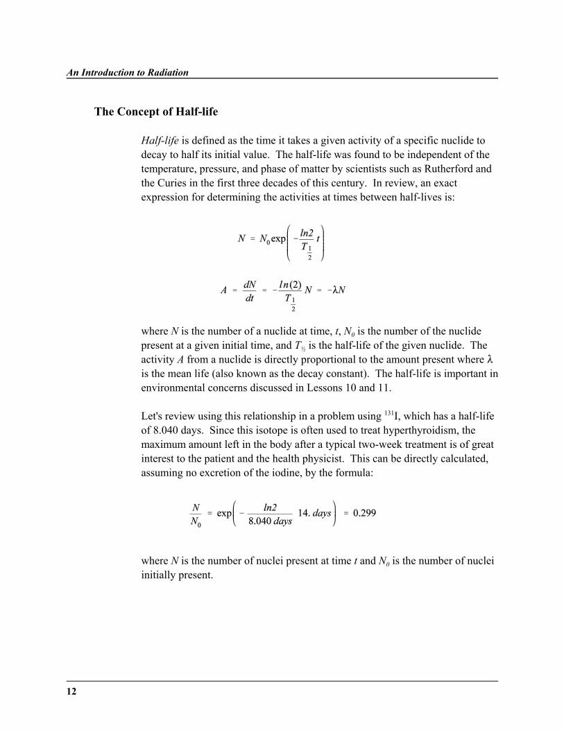
The Concept of Half-life
Half-life is defined as the time it takes a given activity of a specific nuclide todecay to half its initial value. The half-life was found to be independent of thetemperature, pressure, and phase of matter by scientists such as Rutherford andthe Curies in the first three decades of this century. In review, an exactexpression for determining the activities at times between half-lives is:
where N is the number of a nuclide at time, t, N0 is the number of the nuclidepresent at a given initial time, and T½ is the half-life of the given nuclide. Theactivity A from a nuclide is directly proportional to the amount present where 8is the mean life (also known as the decay constant). The half-life is important inenvironmental concerns discussed in Lessons 10 and 11.
Let's review using this relationship in a problem using 131I, which has a half-lifeof 8.040 days. Since this isotope is often used to treat hyperthyroidism, themaximum amount left in the body after a typical two-week treatment is of greatinterest to the patient and the health physicist. This can be directly calculated,assuming no excretion of the iodine, by the formula:
where N is the number of nuclei present at time t and N0 is the number of nucleiinitially present.
Lesson 1
13
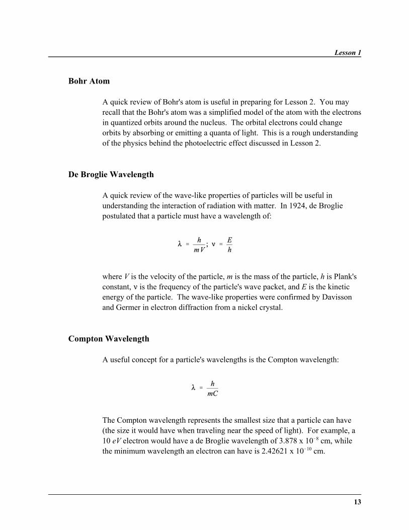
Bohr Atom
A quick review of Bohr's atom is useful in preparing for Lesson 2. You mayrecall that the Bohr's atom was a simplified model of the atom with the electronsin quantized orbits around the nucleus. The orbital electrons could changeorbits by absorbing or emitting a quanta of light. This is a rough understandingof the physics behind the photoelectric effect discussed in Lesson 2.
De Broglie Wavelength
A quick review of the wave-like properties of particles will be useful inunderstanding the interaction of radiation with matter. In 1924, de Brogliepostulated that a particle must have a wavelength of:
where V is the velocity of the particle, m is the mass of the particle, h is Plank'sconstant, < is the frequency of the particle's wave packet, and E is the kineticenergy of the particle. The wave-like properties were confirmed by Davissonand Germer in electron diffraction from a nickel crystal.
Compton Wavelength
A useful concept for a particle's wavelengths is the Compton wavelength:
The Compton wavelength represents the smallest size that a particle can have(the size it would have when traveling near the speed of light). For example, a10 eV electron would have a de Broglie wavelength of 3.878 x 10!8 cm, whilethe minimum wavelength an electron can have is 2.42621 x 10!10 cm.
An Introduction to Radiation
14
While the minimum wavelength of the electron is small, the size of the nucleusranges from 1.2 ! 8x10!13 cm. The wave function of an electron is much largerthan the nucleus at any energy. The positrons needed for $+ are created at thetime of decay by the creation of a positron and an electron from a photon nearthe nucleus. The electron is absorbed by the nucleus. The electrons for electroncapture are from the small amount of an electron's wave function found at thenucleus from the innermost electrons with wave functions at the origin (e.g., 1s,2s, 3s 4s, . . . ). Note: the interaction is largest when the two wave functionsinvolved in the interaction have roughly the same wavelength.
Lesson 1
15
WRITING ASSIGNMENT - Lesson 1Complete the following assignment and submit it according to the directions in yourThomas Edison course information booklet.
Vocabulary
Please write the term and its definition on your paper. Your definition shouldbe in your own words and limited to one or two sentences.
newtonjoulewattunits of length (centimeters and meters)frequencyhertzcoulombpotential differencevoltdose equivalent (sievert and rem)relative biological effectiveness (RBE)linear energy transfer (LET)particle flux densityenergy flux densityionizing radiation
ground statebecquerelhalf-lifeatomic numberneutron numberradionuclideisotonede Broglie wavelengthBohr atomgamma-rayunits of mass (grams and kilograms)ergsecondelectric current (ampere)exposure (roentgen)dose (rad and gray)capacityparticle fluenceenergy fluence
rest masscuriedecay constantmean-lifeatomic mass numbernuclideisobarauger electronsCompton wavelengthX-rayalpha particlevisible lightradio wavesdaughter nucleusparent nucleusmicrowaveexcretionskin erythema dosespecific activitywhole body doseorgan dose
Problems And questions
These problems and questions require numerical or short-essay answers. Pleaseshow your work and circle your final numerical answers. Limit your short-essayanswers to one page or less.
1. Sunlight has its maximum intensity at a wavelength of 4.83 x 10⁻⁷m; whatenergy does this correspond to in eV?
An Introduction to Radiation
16
2. In Figure 5-6 in the Bushong text, the electromagnetic spectrum is givenin terms of frequency and energy. Choose any three of theelectromagnetic radiations shown and calculate the wavelength of aphoton for that radiation (e.g., X-rays, gamma-rays, ultraviolet, visible,microwaves, etc.).
3. What is the wavelength and frequency of a photon emitted by a transitionof an electron from a n=2 orbit to a n=1 orbit?
4. What is the wavelength and frequency of a photon emitted by transition ofan electron from a n=4 orbit to a n=1 orbit?
5. Using standard symbols (i.e., AZ, e-, e+, etc.) define alpha decay, $+ decay,$- decay, and electron capture showing all particles emitted.
6. How much energy in MeV is given off in the following decays:
(a)
where the mass of 3H is 3.016049265 amu, e- is 5.4865 x 10 !4 amu, and3He is 3.016029308.
(b)
where the mass of 148Gd is 147.918112 amu, 144Sm is 143.911997 amu,and the mass of an alpha particle is 4.002603 amu.
(c)
where the mass of 26Al is 25.986892 amu, 26Mg is 25.982593, and theelectron mass is 5.4865 x 10!4 amu.
Lesson 1
17
7. You received a shipment 20 days ago of 131I for treatment ofhyperthyroidism. What fraction of the original shipment would you stillhave with a half-life of 8.040 days for 131I?
8. You are using a 60Co source labeled as having the activity of3.7 x 1010 counts/second in 1945. What is this source's current activity? (The half-life of 60Co is 5.271 years.)
9. What are the three most important types of units in radiobiology? Explain the meaning of each.
19
˜ Preview ˜READING ASSIGNMENT
Hall: Chapters 1 (all) and 7 (pp.112-114)
Bushong: Chapter 12
Nias: Chapters 4 (all) and 9 (pp.140-151)
LESSON OBJECTIVES
˜ This lesson will cover theinteractions of photons withmatter by the processes of:
1. Raleigh scattering2. Compton scattering3. Photoelectric effect4. Pair production
˜ The energy loss of theenergetic electrons producedby the last-mentioned threeprocesses occurs in threemain ways:
1. multiple-collision energytransfer
2. photoelectric process
(continued)
LESSON 2Interaction of Radiation with Matter
DISCUSSION
Introduction
This lesson will focus on the transfer of energy tomatter, specifically, the interaction of high-energyphotons with tissue. X-ray production will also bediscussed.
To complete the review of the interaction of radiationwith matter, the interaction of electrons, alphaparticles, and neutrons will be briefly surveyed.
Absorbed Energy
When radiation passes through tissue or other matter,energy is lost from the system. Some of the energyimparted is lost, whereas some is absorbed. This isshown in the following formula:
where ),AB is the energy absorbed, ), TR is theenergy transferred and ),L is the energy lost. Anexample of energy lost to a system is during Comptonscattering (discussed below), where the incomingphoton interacts with an electron and the scatteredphoton often escapes the material.
Interaction of Radiation with Matter
20
3. bremsstrahlung
4. direct collisions
˜ The idea of dose is reviewedand the basic definitionsrelated to this are included inthe written assignment.
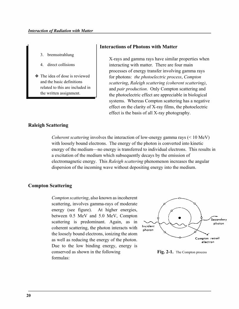
Interactions of Photons with Matter
X-rays and gamma rays have similar properties wheninteracting with matter. There are four mainprocesses of energy transfer involving gamma raysfor photons: the photoelectric process, Comptonscattering, Raleigh scattering (coherent scattering),and pair production. Only Compton scattering andthe photoelectric effect are appreciable in biologicalsystems. Whereas Compton scattering has a negativeeffect on the clarity of X-ray films, the photoelectriceffect is the basis of all X-ray photography.
Raleigh Scattering
Coherent scattering involves the interaction of low-energy gamma rays (< 10 MeV)with loosely bound electrons. The energy of the photon is converted into kineticenergy of the medium—no energy is transferred to individual electrons. This results ina excitation of the medium which subsequently decays by the emission ofelectromagnetic energy. This Raleigh scattering phenomenon increases the angulardispersion of the incoming wave without depositing energy into the medium.
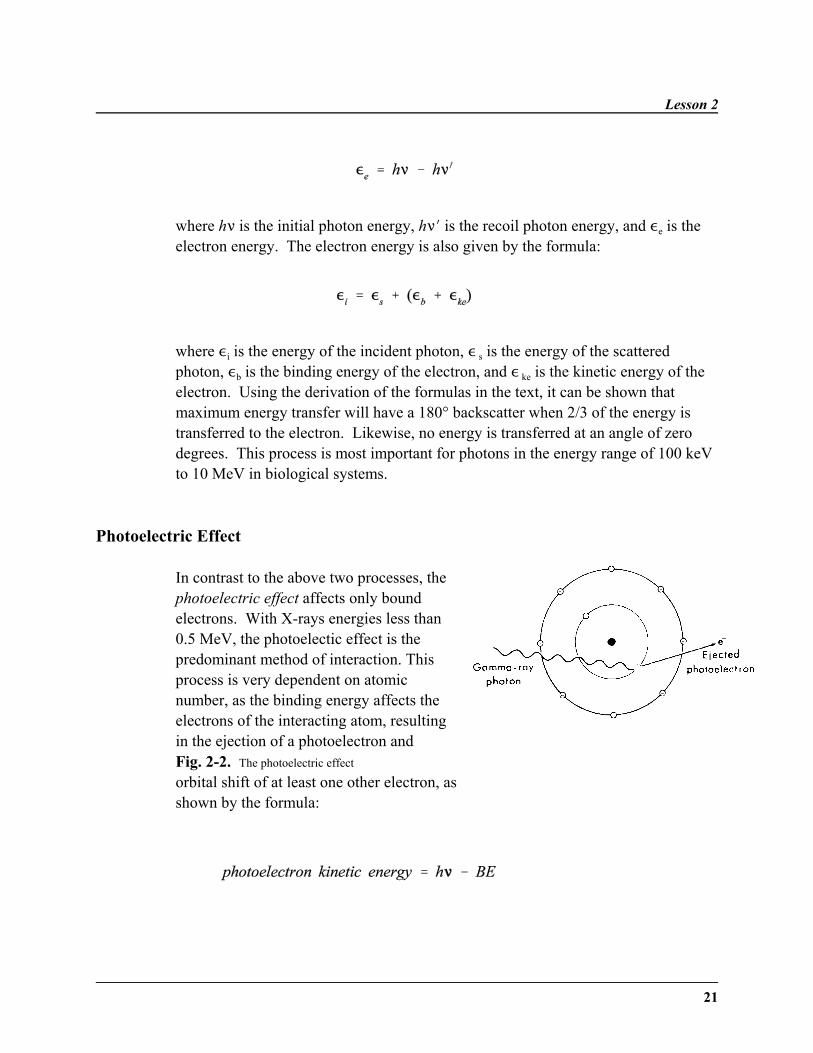
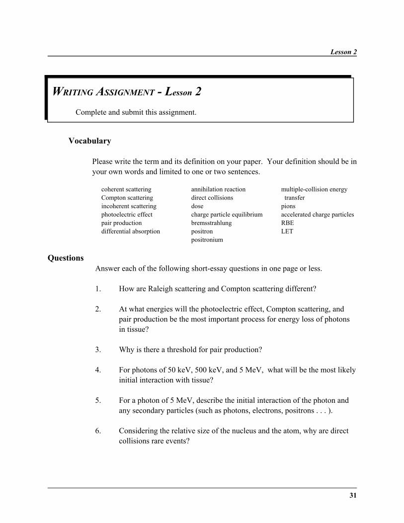
Compton Scattering
Compton scattering, also known as incoherentscattering, involves gamma-rays of moderateenergy (see figure). At higher energies,between 0.5 MeV and 5.0 MeV, Comptonscattering is predominant. Again, as incoherent scattering, the photon interacts withthe loosely bound electrons, ionizing the atomas well as reducing the energy of the photon.Due to the low binding energy, energy isconserved as shown in the following Fig. 2-1. The Compton process formulas:
Lesson 2
21
where h< is the initial photon energy, h<N is the recoil photon energy, and ,e is theelectron energy. The electron energy is also given by the formula:
where ,i is the energy of the incident photon, , s is the energy of the scatteredphoton, ,b is the binding energy of the electron, and , ke is the kinetic energy of theelectron. Using the derivation of the formulas in the text, it can be shown thatmaximum energy transfer will have a 180° backscatter when 2/3 of the energy istransferred to the electron. Likewise, no energy is transferred at an angle of zerodegrees. This process is most important for photons in the energy range of 100 keVto 10 MeV in biological systems.
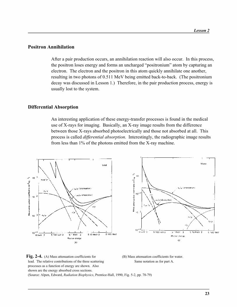
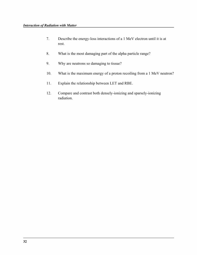
Photoelectric Effect
In contrast to the above two processes, thephotoelectric effect affects only boundelectrons. With X-rays energies less than0.5 MeV, the photoelectic effect is thepredominant method of interaction. Thisprocess is very dependent on atomicnumber, as the binding energy affects theelectrons of the interacting atom, resultingin the ejection of a photoelectron andFig. 2-2. The photoelectric effect
orbital shift of at least one other electron, asshown by the formula:

Interaction of Radiation with Matter
22
Fig. 2-3. Pair production
where h< is the photon energy and BE is the binding energy of the electron. Thisprocess is likely to occur if the energy of the incident photon is just greater than thebinding energy of the electron it interacts with. In fact, the probability of interactionis inversely proportional to the third power of the photon energy. It is alsoproportional to the third power of the atomic number of the absorbing material. Thephotoelectric cross-section relation to energy and atomic number of the absorbingmaterial is:
where E( is the photon energy and Z is the atomic number of the absorber.
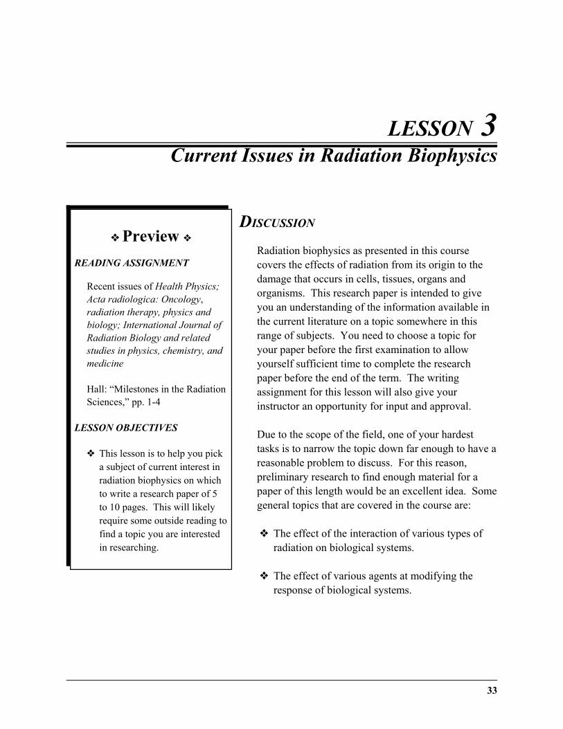
Pair Production
In the final energy-transfer process,pair production, an energetic photonapproaches the nucleus of an atom andcreates a new positron (a positivelycharged anti-particle of an electron) andan electron. At still higher energies, inexcess of 1.02 MeV, x-ray photons areinvolved in the process of pairproduction. Since two energyequivalents of the mass of the electronare required (2 x 0.511 MeV), this process only involves photons with at least 1.022MeV of energy. The kinetic energy distribution can be calculated with the aid of thefollowing formula:
where ,positron is the kinetic energy of the positron, and , electron in the kinetic energyof the electron.
Lesson 2
23
Positron Annihilation
After a pair production occurs, an annihilation reaction will also occur. In this process,the positron loses energy and forms an uncharged “positronium” atom by capturing anelectron. The electron and the positron in this atom quickly annihilate one another,resulting in two photons of 0.511 MeV being emitted back-to-back. (The positroniumdecay was discussed in Lesson 1.) Therefore, in the pair production process, energy isusually lost to the system.
Differential Absorption
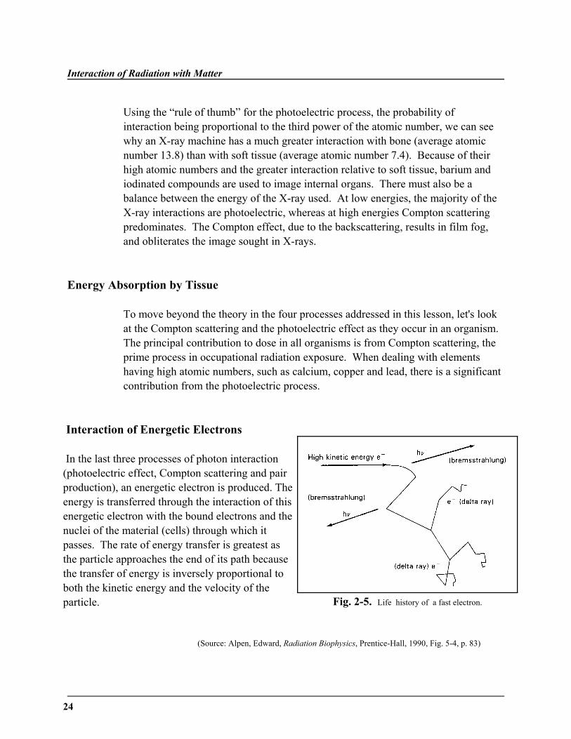
An interesting application of these energy-transfer processes is found in the medicaluse of X-rays for imaging. Basically, an X-ray image results from the differencebetween those X-rays absorbed photoelectrically and those not absorbed at all. Thisprocess is called differential absorption. Interestingly, the radiographic image resultsfrom less than 1% of the photons emitted from the X-ray machine.
Fig. 2-4. (A) Mass attenuation coefficients for (B) Mass attenuation coefficients for water.lead. The relative contributions of the three scattering Same notation as for part A.processes as a function of energy are shown. Alsoshown are the energy absorbed cross sections.(Source: Alpen, Edward, Radiation Biophysics, Prentice-Hall, 1990, Fig. 5-2, pp. 78-79)
Interaction of Radiation with Matter
24
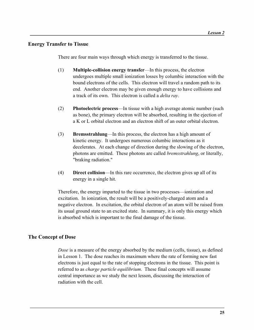
Fig. 2-5. Life history of a fast electron.
Using the “rule of thumb” for the photoelectric process, the probability ofinteraction being proportional to the third power of the atomic number, we can seewhy an X-ray machine has a much greater interaction with bone (average atomicnumber 13.8) than with soft tissue (average atomic number 7.4). Because of theirhigh atomic numbers and the greater interaction relative to soft tissue, barium andiodinated compounds are used to image internal organs. There must also be abalance between the energy of the X-ray used. At low energies, the majority of theX-ray interactions are photoelectric, whereas at high energies Compton scatteringpredominates. The Compton effect, due to the backscattering, results in film fog,and obliterates the image sought in X-rays.
Energy Absorption by Tissue
To move beyond the theory in the four processes addressed in this lesson, let's lookat the Compton scattering and the photoelectric effect as they occur in an organism. The principal contribution to dose in all organisms is from Compton scattering, theprime process in occupational radiation exposure. When dealing with elementshaving high atomic numbers, such as calcium, copper and lead, there is a significantcontribution from the photoelectric process.
Interaction of Energetic Electrons
In the last three processes of photon interaction(photoelectric effect, Compton scattering and pairproduction), an energetic electron is produced. Theenergy is transferred through the interaction of thisenergetic electron with the bound electrons and thenuclei of the material (cells) through which itpasses. The rate of energy transfer is greatest asthe particle approaches the end of its path becausethe transfer of energy is inversely proportional toboth the kinetic energy and the velocity of theparticle.
(Source: Alpen, Edward, Radiation Biophysics, Prentice-Hall, 1990, Fig. 5-4, p. 83)
Lesson 2
25
Energy Transfer to Tissue
There are four main ways through which energy is transferred to the tissue.
(1) Multiple-collision energy transfer—In this process, the electronundergoes multiple small ionization losses by columbic interaction with thebound electrons of the cells. This electron will travel a random path to itsend. Another electron may be given enough energy to have collisions anda track of its own. This electron is called a delta ray.
(2) Photoelectric process—In tissue with a high average atomic number (suchas bone), the primary electron will be absorbed, resulting in the ejection ofa K or L orbital electron and an electron shift of an outer orbital electron.
(3) Bremsstrahlung—In this process, the electron has a high amount ofkinetic energy. It undergoes numerous columbic interactions as itdecelerates. At each change of direction during the slowing of the electron,photons are emitted. These photons are called bremsstrahlung, or literally,"braking radiation."
(4) Direct collision—In this rare occurrence, the electron gives up all of itsenergy in a single hit.
Therefore, the energy imparted to the tissue in two processes—ionization andexcitation. In ionization, the result will be a positively-charged atom and anegative electron. In excitation, the orbital electron of an atom will be raised fromits usual ground state to an excited state. In summary, it is only this energy whichis absorbed which is important to the final damage of the tissue.
The Concept of Dose
Dose is a measure of the energy absorbed by the medium (cells, tissue), as definedin Lesson 1. The dose reaches its maximum where the rate of forming new fastelectrons is just equal to the rate of stopping electrons in the tissue. This point isreferred to as charge particle equilibrium. These final concepts will assumecentral importance as we study the next lesson, discussing the interaction ofradiation with the cell.
Interaction of Radiation with Matter
26
Interaction of " and $ Particles with Matter
The interaction of $ particles (electrons from the nucleus) and alphaparticles with matter results primarily in fast electrons which will interactas described above. These interactions are primarily important for internalexposure for alpha decay and unshielded radiation sources for $ decay.
One additional feature for $ decay is the possibility of producingCherenkov radiation. When a charged particle travels faster than the speedof light in the medium, light is produced in a forward cone. This isresponsible for the blue light which is observed in pool-type reactors orvery radioactive beta sources stored in water. This effect is alsoresponsible for the flashes of light due to cosmic rays seen by astronants.
The interaction of alpha particles with matter is based primarily on thecoulomb interaction. The main components of this interaction are:
(a) elastic scattering from electrons
(b) inelastic scattering from electrons
(c) elastic scattering from nuclei
(d) inelastic scattering from nuclei
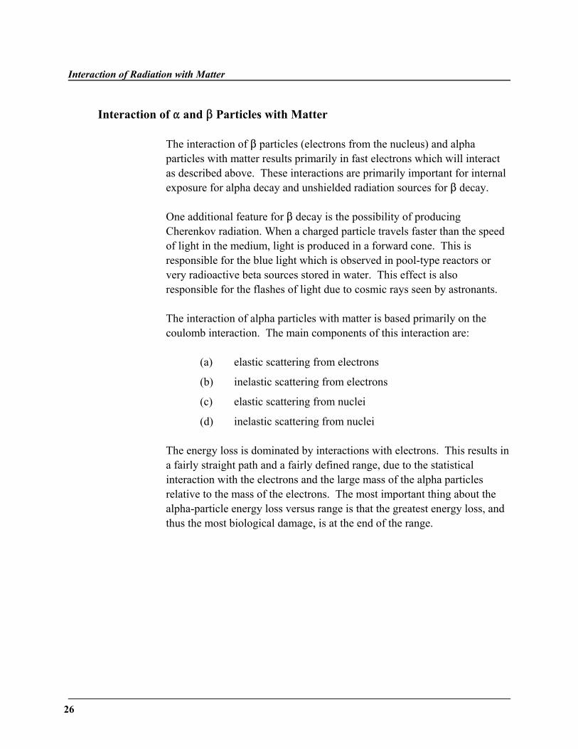
The energy loss is dominated by interactions with electrons. This results ina fairly straight path and a fairly defined range, due to the statisticalinteraction with the electrons and the large mass of the alpha particlesrelative to the mass of the electrons. The most important thing about thealpha-particle energy loss versus range is that the greatest energy loss, andthus the most biological damage, is at the end of the range.
Lesson 2
27
Fig. 2-6. Bragg curve for 214Po alpha particles (E = 7.69 MeV) showing specific ionization asfunction of distance traveled from the source in air.
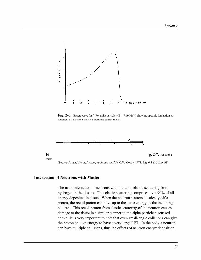
Fi g. 2-7. An alphatrack.
(Source: Arona, Victor, Ionizing radiation and life, C.V. Mosby, 1971, Fig. 6-1 & 6-2, p. 91)
Interaction of Neutrons with Matter
The main interaction of neutrons with matter is elastic scattering fromhydrogen in the tissues. This elastic scattering comprises over 90% of allenergy deposited in tissue. When the neutron scatters elastically off aproton, the recoil proton can have up to the same energy as the incomingneutron. This recoil proton from elastic scattering of the neutron causesdamage to the tissue in a similar manner to the alpha particle discussedabove. It is very important to note that even small-angle collisions can givethe proton enough energy to have a very large LET. In the body a neutroncan have multiple collisions, thus the effects of neutron energy deposition

Interaction of Radiation with Matter
28
can be multiplied under some extreme situations.
Lesson 2
29
Of secondary importance to the dose from neutrons are neutron-inducedcharged-particle reactions. Below 1.0 MeV, the neutron capture reaction ofthe type 12C(n,()13C can be a strong contributor to the absorbed dose. Neutron-induced reactions of the type 12C(n,")9Be also contribute to theabsorbed dose. Because of the high LET of low-energy alphas, the reaction10B(n,")7Li is currently being used with monoclonal antibodies attached toboron compounds to deliver a dose directly to the site of inoperable cancer.
Linear Energy Transfer
The importance of linear energy transfer is that as the density of ionizationincreases, there is an increasing probability that radiation energy will bedeposited directly in a biological molecule so that damage will occur; thehigher the linear energy transfer, the more the biological damage. As linearenergy transfer increases, the relative biological effectiveness increases. Alpha particles are more effective than neutrons, which are more effectivethan x-rays. With densely-ionizing radiation, the oxygen effect is muchless, as is the capacity of the cells to shed sublethal and potentially lethalradiation damage. There is also less variation in the radiosensitivity ofcells in different phases of the cell cycle. The “overkill effect” occurs withlinear energy transfer above keV/:m, due to the fall in relative biologicaleffectiveness. This will be discussed in Lesson 7 in relation to thebiological implications.
Summary of the Interaction of Different Types of Radiationwith Matter
1. Alpha particlesThe alpha particles are positively-charged since they consistof two protons and two neutrons. As they are massive, theymove slowly through tissue and penetrate only a shortdistance. Alpha particles are very densely-ionizing, with thegreatest density of ionization occurring at the end of theshort and straight alpha particle track. They come to rest ashelium atoms in the tissue.
Interaction of Radiation with Matter
30
2. NeutronsThe neutrons have no electric charge, so no ionization isproduced directly. Interaction can only result from directcollisions with atomic nuclei. Slow neutrons enter atomicnuclei and are "captured," a process which makes hydrogenand nitrogen radioactive. Fast neutrons interact mainly byelastic collisions with nuclei. Elastic scattering occurs withnuclei of oxygen, carbon, and nitrogen. Inelastic scatteringoccurs with heavier atomic nuclei in tissue. The absorptionof neutrons is proportional to the concentration of hydrogenin tissues.
3. Pions (Negative Pi- mesons)These are subatomic particles with a mass 276 times that ofan electron but with the same negative charge. Thedeposition of energy mainly occurs when they slow and arecaptured by nuclei present in tissues.
4. Accelerated charge particlesAtomic nuclei of carbon, helium, neon, argon, and otherelements have physical and biological properties similar tonegative pions. The dose delivered at "the depth" is moreeffective because they are more densely ionizing thanorthodox radiation.
5. Beta particlesBeta particles, which are electrons, are also used in cancertreatments. Because they are negatively charged and have asmall mass, electrons are deflected easily from their track,producing an erratic path. The greatest density of ionizationoccurs at the end of this track.
6. Gamma raysInteract with the electrons within a material via the coulombinteraction. The elastic and inelastic scattering of thephoton by the electrons in a material result in high-energyelectrons. These energetic electrons then transfer energy tothe surrounding material. At high enough energies, aphoton can produce a positron-electron pair.
Lesson 2
31
WRITING ASSIGNMENT - Lesson 2Complete and submit this assignment.
Vocabulary
Please write the term and its definition on your paper. Your definition should be inyour own words and limited to one or two sentences.
coherent scatteringCompton scatteringincoherent scatteringphotoelectric effectpair productiondifferential absorption
annihilation reactiondirect collisionsdosecharge particle equilibriumbremsstrahlungpositronpositronium
multiple-collision energy transferpionsaccelerated charge particlesRBELET
QuestionsAnswer each of the following short-essay questions in one page or less.
1. How are Raleigh scattering and Compton scattering different?
2. At what energies will the photoelectric effect, Compton scattering, andpair production be the most important process for energy loss of photonsin tissue?
3. Why is there a threshold for pair production?
4. For photons of 50 keV, 500 keV, and 5 MeV, what will be the most likelyinitial interaction with tissue?
5. For a photon of 5 MeV, describe the initial interaction of the photon andany secondary particles (such as photons, electrons, positrons . . . ).
6. Considering the relative size of the nucleus and the atom, why are directcollisions rare events?
Interaction of Radiation with Matter
32
7. Describe the energy-loss interactions of a 1 MeV electron until it is at rest.
8. What is the most damaging part of the alpha particle range?
9. Why are neutrons so damaging to tissue?
10. What is the maximum energy of a proton recoiling from a 1 MeV neutron?
11. Explain the relationship between LET and RBE.
12. Compare and contrast both densely-ionizing and sparsely-ionizingradiation.
33
˜ Preview ˜READING ASSIGNMENT
Recent issues of Health Physics;Acta radiologica: Oncology,radiation therapy, physics andbiology; International Journal ofRadiation Biology and relatedstudies in physics, chemistry, andmedicine
Hall: “Milestones in the RadiationSciences,” pp. 1-4
LESSON OBJECTIVES
˜ This lesson is to help you picka subject of current interest inradiation biophysics on whichto write a research paper of 5to 10 pages. This will likelyrequire some outside reading tofind a topic you are interestedin researching.
LESSON 3Current Issues in Radiation Biophysics
DISCUSSION
Radiation biophysics as presented in this coursecovers the effects of radiation from its origin to thedamage that occurs in cells, tissues, organs andorganisms. This research paper is intended to giveyou an understanding of the information available inthe current literature on a topic somewhere in thisrange of subjects. You need to choose a topic foryour paper before the first examination to allowyourself sufficient time to complete the researchpaper before the end of the term. The writingassignment for this lesson will also give yourinstructor an opportunity for input and approval.
Due to the scope of the field, one of your hardesttasks is to narrow the topic down far enough to have areasonable problem to discuss. For this reason,preliminary research to find enough material for apaper of this length would be an excellent idea. Somegeneral topics that are covered in the course are:
˜ The effect of the interaction of various types ofradiation on biological systems.
˜ The effect of various agents at modifying theresponse of biological systems.
Current Issues in Radiation Biophysics
34
˜ The response of cells, tissues, organs and organisms to radiation.
˜ Genetic damage caused by radiation.
˜ Background radiation from man-made and natural sources.
Of general interest are several human disasters, accidents, and intentionalexposure:
˜ Exposure of early radiologists
˜ Exposure of overseas flight crews
˜ Exposures from radiation therapy and medical procedures
˜ The atomic bombs at Hiroshima and Nagasaki
˜ Three Mile Island maximum exposures
˜ Chernobyl accident and cleanup
˜ The natural reactor found in Africa
Some topics of great interest to people in radiation safety are:
˜ The human biological response at low doses of radiation is very importantfor setting the safety standards:
(a) Is the shape linear or quadratic?(b) Is there a threshold dose (or is there a natural incidence for this)?
˜ The response of biological systems to radioactive labeled compounds.
˜ How can the scope of the radon problem be determined on a human scale?
˜ What is the result of occupational exposures to radiation of flight crews,astronauts, radium dial painters, tritium dial painters, etc.?
Lesson 3
35
WRITING ASSIGNMENT - Lesson 3Please write a one- or two-sentence description of the topic and a tentative outline of thepaper. Include a list of references found to date. The final paper should be written tocorrespond to the style of a journal of your choice or whatever style your mentor requires. The bibliography should include at least five different sources, three of which should be fromone or more journals.
˜ What are the results of radiation experiments conducted in secret afterWorld War II?
˜ What is really known about the Silkwood incident?
˜ What is the industrial safety in the nuclear industry?
˜ What are the ethics of public exposure to radiation?
To begin your search, you may wish to visit the Web site:
http://www.phy.ohiou.edu/~massey/radiation.html
This Web site has links to some of the good starting points to information on biophysicsavailable on the World Wide Web.
You will find a full list of references to amplify and expand on the discussion in the text at theend of each chapter in the primary text and the supplemental references by Nias. Thesereferences can serve as starting points of your literature search in two ways. The common way isto look up the reference. However, with the Science Citation Index you can find all recent worksthat cite a particular paper. In addition, more recent works by the same author may also be foundby a search on that particular author. Using the Science Citation Index is one way in which allpapers on a given subject may be found quickly.
37
˜ Preview ˜READING ASSIGNMENT
Hall: Chapters 1 (all), 2 (all), and9 (pp. 136-137)
Bushong: Chapter 35
Nias: Chapters 4 (pp. 55-62), 5 (all), and 6 (all)
LESSON OBJECTIVES
After this lesson, you should beable to:
˜ Define spur, blob, and track inthe process of energy transfer.
˜ List the products of waterradiolysis.
˜ Describe the three stages ofwater radiolysis.
˜ Explain the difference betweendirect and indirect energytransfer.
(continued)
LESSON 4Radiation Chemistry
DISCUSSION
Radiolysis of Water: Energy Transfer
This lesson deals with the effects of radiation on thecell, via the process of water radiolysis. Energytransfer will be studied, as well as direct and indirecttransfer of this energy. While briefly mentioning theeffects of radiation on other cell organelles andcellular macromolecules, the effect of radiation on thecell's DNA will be the main focus. The role ofradiation on cell aging will be explored. The cellularbasis of cancer, in regards to growth kinetics, will beaddressed. Lastly, cellular death and mitotic deathwill be differentiated, and the formation of giant cellswill be discussed.
From the previous lesson, one might assume that theprocess of energy transfer from an electron slowing-down is a continuous process. This does not occur,however; the energy is deposited in discrete eventsclassified as:
1. spur: 6–100 eV
2. blob: 100–500 ev
3. short track: 500–50,000 ev
Radiation Chemistry
38
˜ Explain the relationshipbetween molecular weight andtarget size.
˜ Describe the effects ofradiation on various cellularorganelles.
˜ Describe the effects ofradiation on the cellularmacromolecules.
˜ Describe the five types ofDNA damage.
˜ Explain the difference betweencellular and mitotic death.
˜ Describe the formation of agiant cell.
˜ Describe how DNA damagecan result in cancer.
˜ Explain the role of radiation inthe three models of cellularaging.
During these energy transfers, three events canoccur—ionization, excitation, or superexcitation. Insuperexcitation, the energy available is greater thanthat needed for ionization. All three of these eventsresult in unstable molecules.
As we learned in the last lesson, delta rays (secondaryelectrons) are also formed, which then form their ownspurs, blobs, and short tracks. A 1 Mev electrondeposits 65% of its energy in spurs, 15% in blobs, and20% in short tracks. However, each of theseinteraction sites are separated to the degree that verylittle interaction occurs between them.
Radiolysis Products
Cells are predominantly water. Seventy-five percentof cell volume is accounted for by water. Watermakes up the cytoplasm of the cell. These excitationsand ionizations occur in the water of the cell, formingthe following products: H2O+, H+, OH!, and H3O+. Some of these reactions are shown here:
The primary products of radiolysis in the first 10-11 seconds are , andeaq. The hydrated electron, eaq, is much more stable than a free electron. Theseproducts, while still close together, may recombine and convert their energy tothermal energy. However, after 10!11s, diffusion separates the products, makingrecombination impossible.
Lesson 4
39
After primary radiolysis and recombination, the third stage of the radiolysis of wateroccurs—chemical stage. These reactions, which are very complex, result in theproduction of various chemical ions and molecules, such as , , ,
, etc. The important result of “ionizing” radiation is the production of free
radicals, which disrupt the normal molecular structures and damage the biologicaltarget.
For ordinary radiation, the highest yields are the free radicals eaq and ; however,the yield of eaq may be reduced by scavenger molecules. Densely-ionizing radiationhas an increased yield of H2O2, which accounts for the lower oxygen effect withdensely-ionizing radiation, a concept which will be further explored in Lesson 7.
Time Scale
The above-mentioned reactions occur within a very brief time frame, which makesthem difficult to study. Therefore, various techniques are used to study theprocesses involved in irradiation of a cell. The physical processes of ionization andexcitation occur in 10!18 seconds. The pulse radiolysis technique is used to detectreactions with free radical ions, a process lasting 10!12 seconds. The rapid mixtechnique is used to study the decay of radicals in target molecules, reactionsoccurring in 10!6 seconds. Low dose rate and mutant cells are used to observe theenzymatic processes that remove toxic radiation products and repair damagedmolecules. The Puck and Marcus process, which can be used for a period of severalweeks, studies the altered cell molecules, which may lead to cell death and the lossof proliferative capacity.
Direct & Indirect Action
From a biological point of view, it makes little difference whether a molecule isdamaged directly or indirectly. At the point reached by the previous reactionequations, only the water present in the cytoplasm of the cell has been affected,resulting in the production of various hydrated species. In order to permanentlydamage the cell, the biological molecules, such as DNA or proteins, of the cell mustbe affected. This can occur in two ways—direct action and indirect action. Thedirect effect of ionizing radiation is represented by the following two equations:
Radiation Chemistry
40
The indirect effect also operates, producing the free radicals OH and H, whichcombine with organic molecules in the following fashion:
In indirect action, a scavenger molecule, such as Fe2+, will interact with theradicals in order to bring the hydrolysis to an end. The scavenger will thendiffuse and interact with a biological molecule, such as DNA. The hydroxylradical, , is the predominant molecular species in the diffusion-limitedreactions. An example of the use of these scavengers is the quantification of theamount of radiation dose using the Fricke dosimeter. The oxidation of theferrous ion ( Fe2+) to the ferric ion (Fe3+) in a radiation field is detected bysimple photometric methods. This dosimetric method is a standard method formeasurement of doses in 1000-of-rad range.
It is very important to note that the indirect effects occur more frequently withsparsely-ionizing radiation; whereas, the direct effect operates more withdensely-ionizing radiation.
To repeat an important point, in direct action, the high-energy electron's energywill be deposited directly on the biological molecule (DNA). Due to theabsence of intervention of radiolytic species, a dose-response relationship canbe derived:
wherego = undamaged moleculesg = molecules remaining after dosek = inactivation constantD = dose
Lesson 4
41
Mol
ecul
ar w
eigh
t (ra
diat
ion
targ
et si
ze),
dalto
ns
Molecular weight(physical chemical measurement), daltons
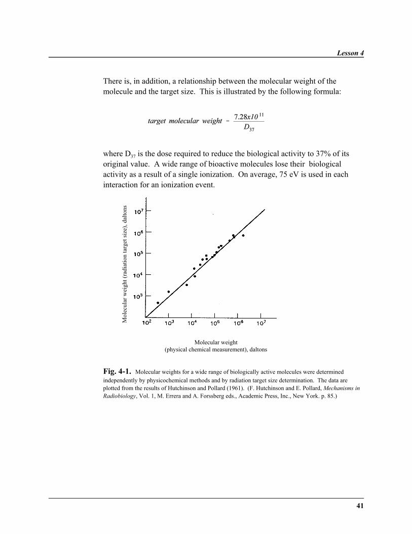
There is, in addition, a relationship between the molecular weight of themolecule and the target size. This is illustrated by the following formula:
where D37 is the dose required to reduce the biological activity to 37% of itsoriginal value. A wide range of bioactive molecules lose their biologicalactivity as a result of a single ionization. On average, 75 eV is used in eachinteraction for an ionization event.
Fig. 4-1. Molecular weights for a wide range of biologically active molecules were determinedindependently by physicochemical methods and by radiation target size determination. The data areplotted from the results of Hutchinson and Pollard (1961). (F. Hutchinson and E. Pollard, Mechanisms inRadiobiology, Vol. 1, M. Errera and A. Forssberg eds., Academic Press, Inc., New York. p. 85.)
Radiation Chemistry
42
Radiation and DNA: DNA Structure
As a review, cells group in patterns that are recognizable as tissues. The tissuesform organs. The entire organism consists of an orderly arrangement of suchorgans. This paragraph offers a brief review of tissue types which will make theinformation on the sensitivity to radiation and causation of cancer easier tounderstand.
Mammalian cells are classified into five types of tissue:
1. Epithelial tissues consist of cells that grow into sheets that cover organsand line cavities. Such cells may be squamous, cuboidal, or columnar.
2. Connective tissue cells form the structural units of the body, such as bone,cartilage, and tendon.
3. Muscular tissues have the property of contracting to produce movement ortension.
4. Blood cells consist of red cells, which are specialized for the transport ofoxygen, white cells, which combat infection, megakaryocytes, whichproduce platelets, and lymphocytes, which aid in the immune response.
5. Nervous tissue, which includes the brain, the spinal cord and all of thenerves in the body.
This section will focus on the effects of radiation to the cell, by looking at theeffects of radiation on cell physiology. The cell contains many organelles or“little organs” which may be affected by radiation. For example, the ribosomesare small, spherical bodies that are the site of protein synthesis. They consist ofribosomal RNA and protein. Another organelle, the Golgi apparatus, stores andtransports secretory products and aids in formation of the cell wall. Lysosomesare sacs containing enzymes responsible for the digestion of material in foodvacuoles. They also aid in the breakdown of damaged cell structures. Lastly,the centrioles play a role in cell division. Interaction between the organelles isessential for the cell to remain viable.
Lesson 4
43
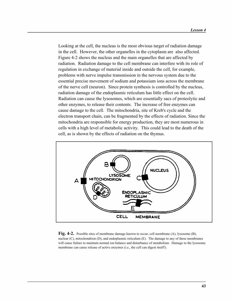
Looking at the cell, the nucleus is the most obvious target of radiation damagein the cell. However, the other organelles in the cytoplasm are also affected. Figure 4-2 shows the nucleus and the main organelles that are affected byradiation. Radiation damage to the cell membrane can interfere with its role ofregulation in exchange of material inside and outside the cell, for example,problems with nerve impulse transmission in the nervous system due to theessential precise movement of sodium and potassium ions across the membraneof the nerve cell (neuron). Since protein synthesis is controlled by the nucleus,radiation damage of the endoplasmic reticulum has little effect on the cell. Radiation can cause the lysosomes, which are essentially sacs of proteolytic andother enzymes, to release their contents. The increase of free enzymes cancause damage to the cell. The mitochondria, site of Kreb's cycle and theelectron transport chain, can be fragmented by the effects of radiation. Since themitochondria are responsible for energy production, they are most numerous incells with a high level of metabolic activity. This could lead to the death of thecell, as is shown by the effects of radiation on the thymus.
Fig. 4-2. Possible sites of membrane damage known to occur, cell membrane (A), lysosome (B),nuclear (C), mitochondrion (D), and endoplasmic reticulum (E). The damage to any of these membraneswill cause failure to maintain normal ion balance and disturbance of metabolism. Damage to the lysosomemembrane can cause release of active enzymes (i.e., the cell can digest itself!).
Radiation Chemistry
44
Irradiation not only affects the organelles but the macromolecules whichcompose the cellular structures. Irradiation has the following specific effects onmacromolecules in the cell:
1. Carbohydrates undergo degradation.
2. Lipids experience oxidation in a chain reaction.
3. Proteins suffer breakage of hydrogen bonds responsible for thesecondary and tertiary structure. Changes in the secondary andtertiary structure can cause loss of function in a protein or anenzyme.
4. Nucleic acids have change/loss of base, single strand breaks, anddouble strand breaks.
It is important to note that the macromolecules may have differentradiosensitivities in different areas or organelles of the cell. Radiation damageto the cell is followed by the expression of molecular damage in the followingsequence : DNA6RNA6proteins6lipids and other macromolecules.
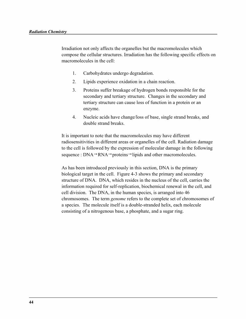
As has been introduced previously in this section, DNA is the primarybiological target in the cell. Figure 4-3 shows the primary and secondarystructure of DNA. DNA, which resides in the nucleus of the cell, carries theinformation required for self-replication, biochemical renewal in the cell, andcell division. The DNA, in the human species, is arranged into 46chromosomes. The term genome refers to the complete set of chromosomes ofa species. The molecule itself is a double-stranded helix, each moleculeconsisting of a nitrogenous base, a phosphate, and a sugar ring.
Lesson 4
45
Ribose-phosphate ———————> backbone
Fig. 4-3. DNA Replication
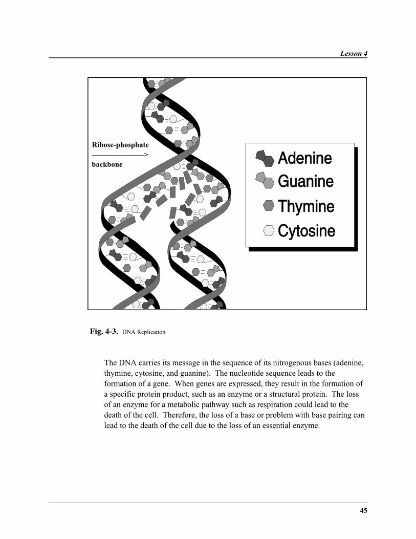
The DNA carries its message in the sequence of its nitrogenous bases (adenine,thymine, cytosine, and guanine). The nucleotide sequence leads to theformation of a gene. When genes are expressed, they result in the formation ofa specific protein product, such as an enzyme or a structural protein. The lossof an enzyme for a metabolic pathway such as respiration could lead to thedeath of the cell. Therefore, the loss of a base or problem with base pairing canlead to the death of the cell due to the loss of an essential enzyme.
Radiation Chemistry
46
Adenine Cytosine
Guanine Thymine
Fig. 4-4. DNA Bases
Lesson 4
47
DNA Damage
There are five main ways in which damage can occur to DNA:
1. Altered functional group of purine/pyrimidine
2. Loss of purine/pyrimidine
3. Free-radical transfer, causing loss of base and chain breakage
4. Single-strand break
5. Double-strand break
Of the mechanisms listed above, attacks on the bases (purines and pyrimidines)are not as serious. There are many enzymatic repair systems in place for thissort of damage. Attacks on the sugar-phosphate backbone, that is single-strandand double-strand breaks, are of the greatest importance.
Most forms of DNA damage, base damage and single-strand breaks, only rarelycause loss of function in the cell. Many repair mechanisms, which arediscussed in Lesson 6, exist to repair such damages.
Double-strand breaks, on the other hand, are much more serious. With thebreak, there is no template for damage repair. Repair may be faulty, resulting ingenetic mutation or loss of reproductive capacity. As discussed earlier in thelesson, double-strand breaks can occur by direct action through interaction withhigh energy electrons, or by indirect action, through interaction with theproducts of water radiolysis.
Cancer
DNA is damaged by irradiation. Such damage can result in a lack of control ofdivision of the cell. Such a condition is cancer. Cancer is a disorder of cellgrowth. The more common cancers are made up of epithelial cell types and arecalled carcinomas. Connective tissue cells may also become
Radiation Chemistry
48
malignant and are called sarcomas. There is no difference in intrinsicradiosensitivity between normal and malignant cells of the same tissue type. Tumor cell populations may show decreased radiosensitivity, however, when afraction of cells are lacking in oxygen.
Tumors are of two types: benign and malignant. Benign tumors have a moreorganized structure, remain localized, and retain the characteristics of originaltissue cells. Malignant tumors grow rapidly and invasively, destroyingneighboring tissues.
Total cell number increases in only two instances: during childhood and intumors. Tumors increase in size due to an increasing number of cells; however,tumors may also increase in size due to proliferation of connective tissue orvascular elements, increased blood supply, hemorrhage, or cyst formation.
Cell Death
Cancer cells may appear immortal due to their ability to divide indefinitely.This observation leads to the concept of cell death and mitotic death. Absolutesensitivities of living cells to the lethal effects of radiation vary widely, but, ingeneral, mammalian cells are particularly sensitive. Except for cells in thelymphocyte series and the oocyte, which may show death during interphase,mammalian cells respond to moderate radiation doses by mitotic death. Duringmitotic death, the cells do not die immediately after irradiation but begin to diewhen they come into mitosis. Even though they have not lost functionalintegrity, they are doomed to perish due to the lack of reproductive capacity.These cells are "sterilized."
Interphase death, which is an immediate death, is uncommon with ionizingradiation. In interphase death, the cells die before they reach the next mitosis.Mitotic death, which is death at the time of mitosis, occurs at about 1.5 Gy.Some cells sterilized by radiation will enlarge and become polyploid, forming"giant cells." This occurs because the process of mitotic division has beeninhibited, but all other metabolic functions of the cell have continued normally.This cell can not grow in number, only in size due to synthesis ofmacromolecules. In terms of reproduction, this cell is dead, even though all
Lesson 4
49
its other functions are intact. Giant cells only form in cell populations thatundergo mitosis normally. Other cells damaged by radiation will proliferateinto colonies of ten before mitosis ceases.
Regardless of the manner of death, pyknosis occurs in the dying cell. In thisprocess, the DNA condenses to form a compact mass, which is followed bykaryolysis. All that remains of the cell is a non-nucleated mass of cytoplasm.
Radiation and Cell Aging
Radiation is known to cause life-shortening in both humans and animals. Theconcept of radiation life-shortening can be understood by looking at the level ofthe cell. There are three hypotheses of aging of the cell: wear-and-tearhypothesis, accumulation of toxins, and somatic mutations. The wear-and-tearhypothesis suggests that a cell will accumulate injuries until it eventuallyreaches a point where cell death results. Free radicals, such as those formed byradiation-induced water hydrolysis, can greatly increase the injuries to the cell. The accumulation-of-waste hypothesis, which is due in part to cross-linking ofcollagen, is not believed to be affected by radiation. According to the somatic-mutation hypothesis, mutations to the DNA causes its information to becomegarbled and faulty. This causes problems in protein synthesis, leading to flawsin cell structure and function. In summary, radiation is known to damageDNA—it possibly leads to life-shortening by information loss in somaticmutations, wear-and-tear caused cell injuries, and from accumulation of toxins(that the cell can no longer metabolize).
Therefore, this lesson has started to explore the damage radiation can do to thecell, leading to problems on the organism level. Table 4-1 is an excellentsummary of the effects of radiation on each biological level.
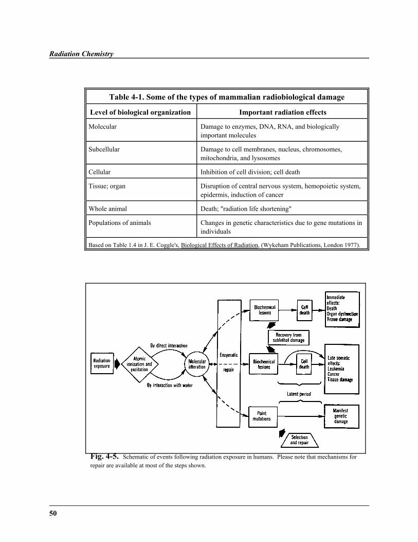
Radiation Chemistry
50
Table 4-1. Some of the types of mammalian radiobiological damage
Level of biological organization Important radiation effects
Molecular Damage to enzymes, DNA, RNA, and biologicallyimportant molecules
Subcellular Damage to cell membranes, nucleus, chromosomes,mitochondria, and lysosomes
Cellular Inhibition of cell division; cell death
Tissue; organ Disruption of central nervous system, hemopoietic system,epidermis, induction of cancer
Whole animal Death; "radiation life shortening"
Populations of animals Changes in genetic characteristics due to gene mutations inindividuals
Based on Table 1.4 in J. E. Coggle's, Biological Effects of Radiation, (Wykeham Publications, London 1977).
Fig. 4-5. Schematic of events following radiation exposure in humans. Please note that mechanisms forrepair are available at most of the steps shown.
Lesson 4
51
WRITING ASSIGNMENT - Lesson 4Complete and submit this following assignments.
This lesson has also shown how radiation, through the atomic ionizations andexcitations in the water radiolytic process, can affect the cell. These chemicalspecies ( , , and others) can lead to cell death through biochemical
lesions of the DNA or direct genetic mutation. Figure 4-5 is a summaryoverview of our study of the effects of radiation on the cells (Lessons 5–7),tissues (Lesson 8), organs (Lesson 9), and organisms (Lesson 9). Cellular deathand mitotic death were introduced, as well as the formation of giant cells. Abrief overview of cancer was also given in this lesson.
Vocabulary
Please write the term and its definition on your paper. Your definition shouldbe in your own words and limited to one or two sentences.
spurblobshort trackdelta rayscavenger moleculeradiation life-shorteningsingle strand break
double strand breakrecombinationrestitutionpulse radiolysisPuck and Marcus processgiant cellinterphase death
mitotic deathsuperexcitationhydrated electronFricke dosimetercontinuous slowing-down approximation
Questions
Answer each of the short-essay questions below in one page or less.
1. Explain the three hypotheses of cell life-shortening. Which hypothesesare affected by radiation?
Radiation Chemistry
52
2. What are the three stages of water radiolysis? What happens in eachstage?
3. What is the difference between direct and indirect energy transfer?
4. What is radiation's effect on the following:
(a) nucleus(b) endoplasmic reticulum(c) cell membrane(d) mitochondria
5. How could the loss of a single nitrogenous base lead to the death of a cell?
6. What are the chemical reactions for restitution and damage fixation?
7. Explain the role of the dissociation reaction, addition reaction, andhydrogen atom extraction in water radiolysis.
8. What is cancer? Explain the significance of the lack of growth kinetics.
9. What is the effect of irradiation on the four macromolecules?
10. How is a “giant cell” formed?
5353
˜ Preview ˜READING ASSIGNMENT
Hall: Chapters 2, 3, and 4
Bushong: Chapter 35
Nias: Chapter 8
LESSON OBJECTIVES
This lesson should help you to:
˜ Explain the importance ofclonogenic survival.
˜ Discuss the assumptions in thetarget theory model.
˜ Explain the difference betweenexponential and sigmoidalsurvival curves.
(continued)
LESSON 5 Target Theory Model
DISCUSSION
Cell Survival Curve
This lesson will study the effects of radiation on thecell. Later lessons will look at the effect of radiationon the tissues, organs, and organisms. Two types ofsurvival curves will be discussed—exponential andsigmoidal survival curves. Two aspects of targettheory will be studied—single-hit and multiple-hittargets. Lastly, the effects of radiation on DNA willbe explored, specifically the action of radiation on thecyclins. Various types of DNA damage will bedetailed.
To begin to understand single- and multiple-hit targettheory, we need to understand some of the basics ofthe cell survival curve. The cell survival curvedescribes the relationship between the fractionalsurvival of a population of irradiated cells and thedose of radiation to which the cells have beenexposed. This survival is specifically clonogenicsurvival. A survivor that has retained itsreproductive integrity and is able to proliferateindefinitely to reproduce a large clone or colony issaid to be clonogenic. Or in other words, clonogenicsurvival is the ability of a progenitor (clone) toproduce at least 50 clones. This is closely related tothe mitotic death discussed in Lesson 4, as each cellmust pass through mitosis six times to produce atleast 50 clones.
Target Theory Model
54
˜ Discuss the difference in cellsused in single and multi-hittarget models.
˜ Define D37 as used in the single-hit model.
˜ Explain the importance ofthreshold phenomenon.
˜ Explain why dicentrics are usedfor radiation dosimetry.
˜ Discuss briefly the linearquadratic model of cell damage.
˜ Name two types ofchromosomal breaks and wherethey occur in the cell cycle.
˜ Discuss response of thechromosomes to different typesof breaks.
˜ Discuss the importance of thep53 protein.
˜ Describe how radiation is able toaffect the cell cycle.
˜ Define balanced translocationand dicentric fragment.
˜ Briefly discuss alternative cellsurvival theories.
Clones are offspring that are identical to the parent.This survival does not refer to true survival—it isvery difficult to affect respiration and metabolismwith incoming radiation. Rather, it refers toreproductive or mitotic death. In general, a dose of10,000 rads (100 grays) is necessary to destroy cellfunction in nonproliferating systems, in contrast to200 rads for proliferating populations. Non-dividingpopulations, therefore, are much more difficult todestroy.
Cell Survival Curves
A cell survival curve describes the relationshipbetween the radiation dose and the proportion of cellsthat survive. Survival curves consist of a dose plottedon the linear scale and the surviving fraction on thelogarithmic scale. At low doses for sparsely-ionizingradiations, the survival curve starts out straight on thelog linear plot with a finite initial slope. At higherdoses, the curve bends. At very high doses, thesurvival curve tends to straighten again, returning toan exponential function of dose. On the other hand,for densely-ionizing radiation, the cell survival curveis a straight line from the origin, indicating anexponential function of dose.
Target Theory
Lea produced one of the earliest models for cellkilling: target theory. He made the followingassumptions.
1. The killing of the cell is a multistep process.
Lesson 5
5555
2. The first step in the process is the absorption of a critical amount ofenergy.
3. Molecular lesions will result from the ionization and excitation caused bythe energy.
4. The expression of the lesions causes the loss of the ability of the cell toreproduce.
Most interestingly, when he formulated these assumptions, Lea was not awareof the effects of radiation on DNA; however, his model fits well with today'sunderstanding of the direct and indirect effects of radiation on DNA, covered inthe previous lesson.
In the target model, the survival curve is described in terms of the initial slope,D1, due to single event killing, a final slope, D 0, due to multiple event killing,and n or Dq to represent the width of the shoulder (termed the quasithreshold).
There are two main types of survival curves—the exponential survival curveand the sigmoidal (shouldered) survival curve. The exponential survival curve,whose graph is commonly shown as a straight line in the plot of the logarithmof the surviving fraction versus the applied dose, shows that the loss ofclonogenic potential is related to dose in a strictly exponential fashion. Thiscurve is applied to yeast, bacteria, and mammalian sperm, for example.
Whereas the exponential survival implies that simple, single events result inclonogenic death, the sigmoidal curve adheres to Lea's belief that there must bean accumulation of DNA damage before the cell experiences a clonogeneticdeath. The logarithmic graph shows a shoulder and a straight portion, theshoulder demonstrating the need for a minimum dose before clonogenetic death(see Figure 3-3 on page 36 of Hall).
It is interesting to note that when the radiation dose is delivered in a series ofequal fractions, separated by time for repair, the effective survival curve is astraight line from the origin through a point, that is, it is an exponentialfunction. This concept will be further discussed in Lesson 7 in the Dose Ratesection.
Target Theory Model
56
According to target theory, for a cell to die following radiation exposure, itstarget molecule must be inactivated. The target is the area occupied by thetarget molecule or by a sensitive site on the target molecule. The target isprobably DNA. A hit occurs when there is interaction between radiation andthe target. As shown in previous lessons, the hits occur through both direct andindirect effects.
Target Theory: Single- and Multi-hit
There are two models in target theory: single hit and multi-hit. The single-hittheory applies to bacteria and viruses. Bacteria are prokaryotic. Prokaryoticcells lack a true nucleus. The DNA exists as a single, circular, double-strandedmolecule. The multi-target, single-hit theory is applicable to human cells, orany complex cells such as plant or animal. These complex cells are eukaryotic. Eukaryotic cells have a true nucleus, one that is membrane-bound. The genomeis divided into discrete units called chromosomes.
In the single-hit model, a single sensitive site is destroyed by an inactivatingionization, resulting in the death of the cell. Since the DNA exists as a singlestrand, it is easier to destroy its expression. When the radiation reaches a levelsufficient to kill 63% of cells (37% survival), it is called D37. The D37 is ameasure of the radiosensitivity of the cell. A low D37 represents a highlyradiosensitive cell. A high D37 represents a radioresistant cell. The equation fora single-hit is given below:
where S is the surviving fraction, N is the number of cells following dose D, andN0 is the initial number of cells.
In the multi-target, single-hit model, a threshold phenomenon occurs— multipletargets must be destroyed to kill the cell. At lower doses, more cells willsurvive because not all the sensitive sites (possibly genes on the chromosomes)are destroyed. As in the single-hit model, fewer cells survive when subjected toan increasing radiation dose. At very high dose levels, the dose-response
Lesson 5
5757
becomes the same as single-hit because all additional sites have been destroyed. The equation for the survival fraction, S, of a cell colony in this model is:
where D0 is the dose required to reduce the survival to 37% from a single target,and n is the number of targets in the cell.
Chromosomal Damage
Whereas the target theory is the most widely-known theory of cell survival,there are several other theories mentioned in the text. For example, themolecular theory of radiation action, or linear-quadratic model (L-Q), assumesthat the action of radiation is the rupturing of the bonds of DNA, producinglesions. These lesions will be completely or partially expressed as a result ofrepair processes. This linear quadratic model, which is commonly used inclinical radiotherapy, also deals with dual radiation action, meaning it assumesthere are two components of cell killing by radiation, one that is proportional todose and one that is proportional to the square of dose. Therefore, this modelhas a continuously bending survival curve, which has the expected initial slopebut which never becomes exponential, even at high doses. Unfortunately, theactual cell survival curve does not really support this model since most actualsurvival curves do not fit the linear quadratic equation.
The sublesions produce by radiation may interact to form a lesion. The repair-misrepair model (RMR) theorizes that the irradiation stage is followed by aperiod of cell repair or damage enhancement. The cell passes through variousphases, which may be followed by permanent alteration of the genetic material.The lethal-potentially lethal (LPL) model proposes that B lesions mayconverted to an undamaged A state or lethal C state by various processes.
Target Theory Model
58
Radiation and the Cell Cycle
Before further discussing damage to chromosomes, a short review of mitosisand meiosis might be useful. Mitosis produces two identical daughter cells. Meiosis leads to the production of one ovum (in oogenesis) or four sperm (inspermatogenesis). The products of meiosis are different due to crossing-over inprophase I. Meiosis is basically two divisions, resulting in the production ofhaploid cells. Study the Figure 5-1 to review mitosis.
Studies of chromosomal damage are primarily done using metaphasechromosomes of lymphocytes of mammals. New methods are allowing thestudy of chromosomes during interphase. These studies separate the damage tothe chromosome according to which phase the chromosome was in the cellcycle. Study of the cell cycle is also due to autoradiography. Study of theeffects of radiation on cell cycles is improved through the use of synchronouslydividing cell cultures. Cells may be synchronized by physical methods, such asmitotic selection, volume selection, or by chemical methods, through the use ofphase-specific drugs such as vincristine and cytosine arabinoside. Whereas thechemical methods can be used “in vivo,” the physical methods cannot be.
Fig. 5-1. Chromosome replication and the mammalian cell cycle showing mitosis for somatic cells.
The growth kinetic state of a cell population depends upon how many cells arepassing through their proliferative cycle and how long that cycle is. The cellcycle is defined as the interval between the midpoint of mitosis in a cell and themidpoint of the subsequent mitosis in both daughter cells. The type
Lesson 5
5959
of damage to the chromosome is dependent upon the location of the cell in thecell cycle.
Looking at the cell cycle, there are four phases in the life of the cell—Gap I(G1), Synthesis(S), Gap II(G 2), and Mitosis (M) (see Fig. 5-1). Progression through the cell cycle is based on the sequential transcription andtranslation of the cell cycle genes. G1 may be extended into G 0; however,G1 cells are already committed to DNA synthesis and division, whereas G 0
cells need a stimulus to enter the cell cycle.
During the S phase, DNA is replicated through the production of sisterchromatids. G1 and G 2 are both lacking in this DNA synthesis. G 1(Gap) is thephase in which the cell “decides” whether to divide. Some cells arepermanently arrested in this state. G2, which is shorter than G 1, is the phasedirectly before mitosis. Cell division occurs in the mitosis phase.
The length of the cell cycle varies due to variations in the length of G1 for thecells. M, S, and G2 vary comparatively little between different cells in differentcircumstances.
The following principles usually apply to the cell cycle in irradiated cells.
1. Cells are most sensitive at or close to mitosis.2. Resistance is greatest in the latter part of the S phase.3. If G1 has appreciable length, a resistant period is evident early in the
G1 phase, followed by a sensitive period towards the end of G 1
4. G2 is usually sensitive, perhaps as sensitive as the M phase.5. The radiosensitivity of a heterogeneous population of cells is dominated
by the most resistant component.
Cell cycle sensitivity may be due to changes in the formation of the DNAcontent during the S and M phases, as well as variations in the level of thesulfhydryl compounds in the cell, which serve as natural radioprotectors.However, cell cycle sensitivity may also be due to the presence or absence of aclass of proteins called cyclins.
Target Theory Model
60
Cell Cycle Controls
According to recent research, starting with the G1 phase, and then at specifictimes in the cell cycle, the concentration of cyclins increase in the nucleus.Cyclins are a family of closely-related proteins, consisting of eight types (typesA-H). Cyclins form cyclin-Cdk complexes, which is the main control elementthat regulates the progression and activity of each phase in the cell cycle. Thedecision to end cell cycle progression occurs at the cell cycle checkpoints.
Each checkpoint monitors different aspects of DNA replication andchromosome segregation. Each regulates a different phase of the cell cycle.Ionizing radiation triggers the checkpoints in G1, S, and G 2; however, theG1 checkpoint can permanently stop the cell cycle.
Three cellular responses can occur at the G1 checkpoint:
1. Apoptosis - Apoptosis is a metabolically-activated form of cell death,which serves to eliminate cells which have sustained an unusually highlevel of DNA damage.
2. Cell senescence - Cell senescence acts as an internal constraint on the life-span of a cell line. It is a quiescent state that is triggered after a cell hasundergone a finite number of divisions.
3. G1 arrest - In this response the cell is permanently stopped in the G1
phase.
Each of these three responses involves the critical protein p53, a transactivatingprotein which initiates the transcription of other genes. A mutation in p53 canlead to carcinogenesis, evidenced by the fact that 50% of human tumors havecells with mutated p53. Such a mutated cell can become the "seed" of cancerwithin an otherwise healthy body.
Lesson 5
6161
Division Delay
Irradiation of the cell can result in two consequences: division delay and DNAdamage, which will be discussed in the next section. Irradiation can produce adivision delay which is expressed as a delay in entering mitosis. The maximaldelay is achieved when cells are irradiated in the G2 phase, due to a“checkpoint” which is known as the “G2 block.” The G 2 block is due to a dose-dependent inhibition of the synthesis of proteins for mitosis, such as centrioleproteins. The delay of S phase cells is due to depression of DNA synthesis andprolongation of the G2 period. The delay of cells irradiated in the G1 phase ismerely expressed as a prolongation of both S and G 2 phase. Recovery from theG2 block is characterized by the reappearance of mitotic cells. The time neededfor this to occur is a measure of radiation-induced division delay.
Using the cell cycle, we will look at the three types of chromosomaldamage—chromosome, chromatid, and subchromatid. Cells that are in G2, thephase after synthesis occurs, will have damage of the chromatid type. Thesechromatid aberrations may follow irradiation later in the cell cycle when eachchromosome will have divided into two chromatids held together by acentromere, so that the damage may only be evident in one of two chromatids ofthe pair. Such a break occurs in a single chromatid arm and leaves the oppositearm of the same chromosome undamaged. One example of a lethal chromatidaberration is the anaphase bridge.
If the irradiation is delievered early in the cell cycle, before DNA synthesis hasbegun, then chromosomes will not yet have duplicated and damage will occur towhole chromosomes. This is visible at the next mitosis as chromosomeaberrations. The radiation-induced break will be in a single strand of chromatin.This strand of chromatin will replicate the break in an identical strand laid downduring synthesis. Therefore, cells that were in G1, the phase before synthesis,will have damage of the chromosome type, since these changes will bereplicated when the cell enters the synthesis phase.
Target Theory Model
62
The most frequent aberrations are exchanges and deletions. The simple deletionis the result of a single break in the chromosome or chromatid and the exchangeis a new arrangement following the joining of breakage ends that result fromseparate breaks.
There is always a strong driving force for the DNA to rejoin. Most of thedamage due to single-hit breaks is restored to the original state. Occasionally,the fragments formed in a single hit will rejoin incorrectly. An acentricfragment forms when the large fragment joins end-to-end to form a circularchromosome, leaving a small fragment without a centromere (see Figure 5-2).
In multiple breaks, the terminal deletion is a result of a single break across thechromosome; the remainder of the interchange involves a double break andabnormal rejoining. The centric ring is the most common aberration.
With two hits, dicentric products are formed in G1, when adjacent chromosomeseach suffer one hit and recombine. Dicentric products result whentranslocation (movement of chromosome separated in meiosis) is unequal. Theproduct may have two centromeres (dicentric) which leads to the formation ofbridges which are fatal to the cell. The dicentric product will remain in thecenter of the cell during division, not allowing complete separation of thedaughter cells. Ring chromosomes are also produced.
In G2, at the chromatid level of damage, fragments may recombine, omittingsmall fragments—interstitial deletion. If reordering of the fragments occur,without any loss of material, the process is referred to as inversion. Examplesof two-hit damage are shown in Figure 5-3. An example of reciprocaltranslocation is shown in Figure 5-4. The dicentrics are used as the basis forbiological dosimetry, because the dicentric is the most consistent index ofradiation damage. It represents 60% of all observed unstable aberrationsfollowing acute irradiation.
At the chromatid level, such unstable aberrations are likely to be lethal andwithin a few cell cycles would be selectively eliminated from the naturalproliferating population of cells. Stable aberrations are more serious becausethey can pass through successive cycles. Non-lethal chromosome aberrationsinclude symmetrical translocation and a small interstitial deletion.
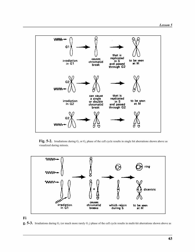
Lesson 5
6363
Fig. 5-2. Irradiations during G1 or G2 phase of the cell cycle results in single hit aberrations shown above asvisualized during mitosis.
Fig. 5-3. Irradiations during G1 (or much more rarely G 2) phase of the cell cycle results in multi-hit aberrations shown above as

Target Theory Model
64
visualized during mitosis.

Lesson 5
6565
Fig. 5-4. Radiation-induced reciprocal translocation.
Damage due to multiple hits can be extreme. At the chromosome level (in G1),breaks can form a dicentric product or undergo an isocentric break. Anisocentric break occurs at the same location in both chromatids.
G1: chromosome damage ———> translocation, dicentric chromosomeG2: chromatid damage ———> interstitial deletion, minute, inversion
Chromosome Aberration Dosimetry
An application of the knowledge of radiation damage to chromosomes is its usein chromosome aberration dosimetry. In the event of radiation exposure ablood sample can be examined by a lab specializing in this work and a ratherprecise estimate of the dose can be obtained in the range of 35 to 500 Rem. This technique requires the services of a highly skilled technician for up to twoweeks. Calibration curves enable accidental radiation exposure to be estimatedin terms of such chromosome aberrations. The number of cells showingaberration decrease with time, but the chromosome aberrations are stillobservable after 25 years.
This lesson has explored the effects of radiation on the chromosomes of the cell. Lesson 6 will explain further the effect of radiation on the cell's DNA. Geneticmutations are a the result of chromosome interaction with radiation. Repairmechanisms for this genetic damage will also be explored in Lesson 6.
Target Theory Model
66
The response of the cell to radiation is expressed as a survival curve. The targettheory model (single and multi-hit) explains how radiation may inactivate thecell. Radiation can either delay the cell cycle or produce chromatid orchromosome damage, depending upon the cell's position in the cell cycle.
Summary
˜ The interaction of radiation with prokaryotic cells and viruses is describedby single hit theory.
˜ Multi-target, single hit theory explains the interaction of radiation witheukaryotic organisms.
˜ Threshold phenomenon refers to the accumulation of damage in the DNAbefore cell death occurs.
˜ There are three primary changes to the chromosome due to irradiation—subchromatid, chromatid, and chromosome level damage.
˜ The presence of dicentrics in the bloodstream is used for radiationdosimetry.
˜ The cyclins, which are responsible for checkpoints in the cell cycle, maybe inactivated by radiation.
˜ Division delay is an example of a potential effect of radiation on the cell.
˜ The p53 protein is commonly mutated by radiation in many cancers.
Lesson 5
6767
WRITING ASSIGNMENT - Lesson 5Complete and submit the following assignment.
Vocabulary
Please write the term and its definition on your paper. Your definition shouldbe in your own words and limited to one or two sentences.
cell survival curveclonogenic survivalreproductive deathtarget theoryprokaryoticeukaryoticD37
threshold phenomenon
chromatidinversionbalanced translocationdicentric producthit probabilitymean lethal doselinear-quadratic modeldual radiation action
theoryrepair-misrepair modelaptosiscell senescencecyclinscell cycleG2 blockanaphase bridge
questions
Write short-essay (1 page or less) answers for the following.
1. Why aren't all cells which are alive included in the cell survival curve?
2. What are the four important assumptions made by Lea in the targetmodel?
3. What is the difference between the exponential and sigmoidal cell survivalcurves?
4. In what type of cells does single hit occur? Multiple hits?
5. Why does the threshold phenomenon occur in the multi-target, single-hitmodel?
Target Theory Model
68
6. When do the three types of chromosomal breaks occur?
7. Which is the most severe break? Why?
8. What are the differences among the linear-quadratic theory, target theory,and dual radiation models?
9. What are the weaknesses in the target theory? Linear-quadratic theory?
10. How does the survival curve differ for densely-ionizing and sparsely-ionizing radiation?
11. Describe briefly how the following survival curves would appear:(a) linear quadratic survival curve(b) multi-target survival curve(c) fractionation of radiation dose
12. What is the importance of the p53 gene? What may happen with radiationexposure?
13. How does resistance to radiation vary throughout the phases of the cellcycle?
14. What is a dicentric? Why is it important in radiobiology?
15. How do chromatid breaks occur? Give an example of such damage.
69
˜ Preview ˜READING ASSIGNMENT
Hall: Chapter 11
Bushong: Chapter 35 (pp. 503-505)
Nias: Chapter 18 (pp. 320-324)
LESSON OBJECTIVES
By the end of this lesson youshould be able to:
˜ Name and explain the threetypes of genetic mutations.
˜ Name several commonexamples of each mutationtype.
˜ Discuss the findings of Müllerand the Russells.
(continued)
LESSON 6Gene Mutations
DISCUSSION
This lesson will focus on genetic mutations due toradiation. Lesson Five addressed damage tochromosomes. This lesson will discuss the result ofthe irradiation of the chromosomes—geneticmutation, focusing on the work of Müller and theRussells. Genes are the functional units of thechromosome, and their expression leads to theproduction of a specific protein. Repair mechanismsand disorders of these mechanisms will also bestudied.
Gene Mutation Classification
A gene is a finite segment of DNA specified by anexact sequence of bases. The number of genes in thehuman is 0.5 x 10⁵ per haploid set of chromosomes.Chromosome mutations are the result of either anincrease or decrease in the number of genes in thenucleus. Point mutations, which result from smallalterations in DNA, occur in proportion to dose andare independent of dose rate.
On the whole, genetic mutations are a burden to thepopulation, not a benefit. Radiation-inducedmutations add to the spontaneous mutations,increasing the genetic burden. There is no question
Gene Mutations
70
˜ Explain the importance of thework of Müller and theRussells.
˜ Define the concept of doublingdose and genetically significantdose.
˜ Discuss normal mutation ratesin the human population.
˜ Explain the problemsassociated with the lack of arepair mechanism.
˜ Explain how radiation canaffect the mutation rate.
˜ Discuss the effects of theamount of DNA and dose onmutation rate.
that radiation is deleterious to the species as a whole.Most importantly, radiation does not result in geneticeffects that are new or unique, but increases thefrequency of some mutations that already occurspontaneously or naturally.
To review briefly, the human genome (complete set ofchromosomes) consists of 23 pairs ofchromosomes—a total of 46 chromosomes. Ahomologous chromosome pair consists of twochromosomes which are "alike," meaning they havethe same type of genetic information. Twenty-two ofthe pairs are somatic chromosomes. The twenty-thirdpair consists of the sex chromosomes. The femalehas two X chromosomes, whereas the male has onlyone X chromosome. The other chromosome in themale is a Y chromosome, which has very little geneticinformation on it.
Gene mutations may be classified as dominant,recessive, or sex-linked. A genetic mutation is achange in a DNA base leading to the production of analtered protein or no protein product at all.
A dominant mutation is one that appears in the immediate offspring if thealtered gene is found in at least one of the parents. This dominant mutationneed be in the gene of only one of the chromosomes of the homologous pair tobe expressed. There are 700 such dominant gene diseases includingpolydactyly, achondroplasia, Huntington's chorea, and retinoblastoma.
A recessive mutation will appear only in offspring that have received the samemutation from both parents. For a recessive mutation to be observed, unlesssex-linked, both chromosomes of the homologous pair must carry the alteredgene. This means that the gene must be inherited from both parents;consequently, many generations may pass before it is expressed. There arenearly 500 recessive diseases, including sickle cell anemia, cystic fibrosis, andTay-Sachs disease.
Sex-linked mutations occur in the sex chromosomes—female (XX) and male
Lesson 6
71
(XY). Since the male has a dissimilar pair of sex chromosomes that do notcontain complementary genes for all the traits, the male will express “recessive”mutations. This type of mutation is called sex-linked. There is more than 80such sex-linked conditions, including hemophilia, color-blindness, andDuchenne muscular dystrophy.
Mutations occur in both germ cells and somatic cells. Mutations occurring inthe sex cells are more disastrous than those occurring in the somatic cells (bodycells) because the mutation can be passed to the offspring. Mutations of asomatic cell will merely lead to the death of that one cell. For example,mutation of an epidermal cell (skin) may result in death of that cell or cancer. However, the organism will continue to survive if only this one cell is affected. Its offspring will not be affected.
As a rule of thumb, the genetic effects will be most pronounced in the firstgeneration after irradiation; however, the total number of deleterious effects ofgenetic mutations following irradiation will be much greater in all subsequentgenerations.
Radiation can affect future offspring in another way besides mutation—chromosome distribution. Errors in chromosome distribution can result in cellscontaining too many or too few chromosomes. For example, Down's syndromeresults from an extra chromosome 23. Most of the time an incorrectchromosome number leads to embryonic death, accounting for 40% ofspontaneous abortions and 6% of stillbirths. Radiation is more effective atbreaking chromosomes than in causing errors in chromosome distribution. Such a chromosome imbalance, if it does not cause the death of the embryo,leads to physical abnormalities, usually accompanied by mental deficiency.
Experimental Studies
The early research on the genetic effects of radiation was done by H. J. Müller,who won a Nobel Prize for his studies. He studied the Drosophila fruit fly byobserving mutations on the X chromosome. The results showed a linearnon-threshold response of the mutation rate to radiation dose. This type ofresponse is also known as stochastic response. (This type of response has beenused by the National Council on Radiation Protection to model the humanresponse to low doses of radiation.) Müller also concluded that the observedmutations were of the same quality as those which occur spontaneously. Theresponse observed was independent of dose rate, indicating that the damage is
Gene Mutations
72
due to a single hit on a gene.
After World War II, L.B. and W.L. Russell began a large mouse colony whichwas exposed to radiation dose rates from 0.001 to 0.90 rad per minute and totaldoses of 1000 rad (10 Gy). They observed specific expressions of geneticinformation, i.e., the coat color and eye color. These studies have continuedwith over one million mice, giving the project the common name of theMegamouse Project.
A major conclusion of this study is that there is a dose-rate effect for mammals. This study observed that the mutations were more frequent for mice exposed ata high dose rate than in mice which received the same dose but at a lower rate. This is termed a dose-rate effect, as the mutation rate depends upon the rate atwhich the dose is given, not on the total dose.
The study implies a repair mechanism for radiation damage in mammals. It isimportant to note that the mutation rate is about 15 times greater than that foundin fruit flies. In the target model, this would imply that the actual size of thegenome on the chromosomes is about 15 times larger in mammals than in fruitflies.
As has been shown in numerous studies in addition to Russells', there is arelationship between radiation-induced damage and the size of the nucleus,chromosomes, or DNA content. As the DNA content increases betweenspecies, the mutation rate per gene multiplies by almost the same factor. Thiscorrelation may result from an increased amount of DNA associated withregulating the gene activity as the DNA content increases. This relation isshown in Figure 6-1.
The Megamouse Project led to many other conclusions in addition to that ofdose rate. There are two ways to describe the estimated genetic risk ofradiation. The first method is to compare radiation-induced mutations withthose that occur spontaneously. The results are expressed in terms of doubling-dose. This is the relative mutation risk. The second way of describing the director absolute mutation risk quotes the incidence of disorders resulting frommutations in the first generation.
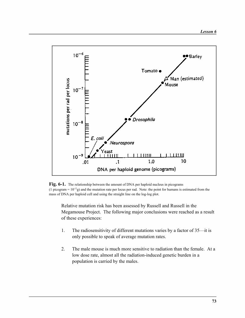
Lesson 6
73
Fig. 6-1. The relationship between the amount of DNA per haploid nucleus in picograms (1 picogram = 10-12g) and the mutation rate per locus per rad. Note: the point for humans is estimated from themass of DNA per haploid cell and using the straight line on the log-log plot.
Relative mutation risk has been assessed by Russell and Russell in theMegamouse Project. The following major conclusions were reached as a resultof these experiences:
1. The radiosensitivity of different mutations varies by a factor of 35—it isonly possible to speak of average mutation rates.
2. The male mouse is much more sensitive to radiation than the female. At alow dose rate, almost all the radiation-induced genetic burden in apopulation is carried by the males.
Gene Mutations
74
3. The genetic consequences of a given dose can be greatly reduced if a timeinterval is allowed between irradiation and conception. Consequently, ifpersons are exposed to a significant dose of radiation, it is recommendedthat six months be allowed to elapse between the exposure to the radiationand planned conception to minimize the genetic consequences.
The Concept of a Doubling Dose
The concept of a doubling dose is very useful in describing genetic damage. The doubling dose is the amount of radiation needed to produce twice the rateof genetic mutations that would have been observed without radiation. Forhumans the doubling dose ranges from 50 to 250 rads (0.5 and 2.5 Gy). A moreexact estimate of the doubling dose for humans is 1 Gy (100 rads) based onlow-dose-rate exposure. Drosophila fruit flies have doubling dose 15 timesgreater than this.
The figure for doubling dose for humans is a calculated quantity, based on themeasured mutation rate per locus on the mouse, adjusted for the estimated lociin humans.
The lowest possible doubling dose is 3 rems, the average amount of radiationreceived from natural sources during the typical reproductive lifetime of 30years. Estimates of doubling dose from Hiroshima and Nagasaki is 1.56 Sv(156 rems).
The following guiding principles apply to doubling dose:
1. Most mutations, whether spontaneous or induced by radiation, areharmful.
2. Any dose of radiation, however small, entails some genetic risk.
3. The number of mutations produced is proportional to the dose.
4. Risk estimates based on experiments with the mouse are very similar forhuman values.
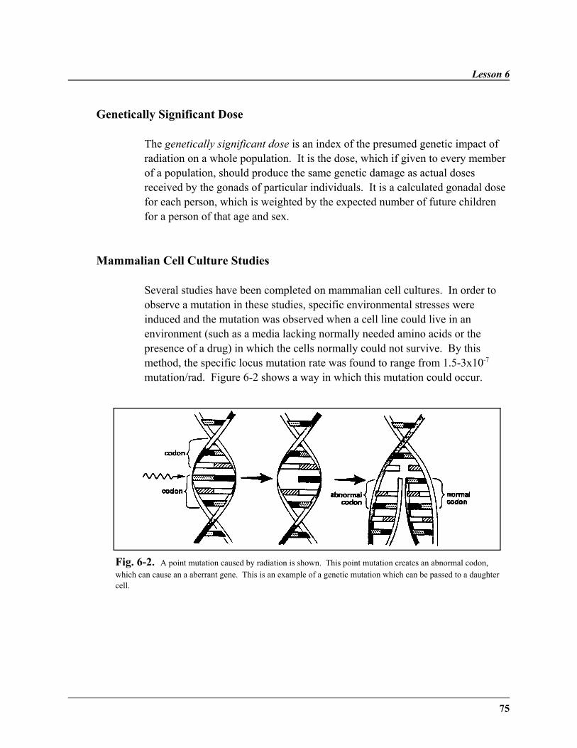
Lesson 6
75
Genetically Significant Dose
The genetically significant dose is an index of the presumed genetic impact ofradiation on a whole population. It is the dose, which if given to every memberof a population, should produce the same genetic damage as actual dosesreceived by the gonads of particular individuals. It is a calculated gonadal dosefor each person, which is weighted by the expected number of future childrenfor a person of that age and sex.
Mammalian Cell Culture Studies
Several studies have been completed on mammalian cell cultures. In order toobserve a mutation in these studies, specific environmental stresses wereinduced and the mutation was observed when a cell line could live in anenvironment (such as a media lacking normally needed amino acids or thepresence of a drug) in which the cells normally could not survive. By thismethod, the specific locus mutation rate was found to range from 1.5-3x10⁻⁷
mutation/rad. Figure 6-2 shows a way in which this mutation could occur.
Fig. 6-2. A point mutation caused by radiation is shown. This point mutation creates an abnormal codon,which can cause an a aberrant gene. This is an example of a genetic mutation which can be passed to a daughtercell.
Gene Mutations
76
To give you some perspective on the mutations induced by radiation, a shortoverview of natural mutations and DNA repair is needed. Human mutationrates have been observed from 0.5 to 10 per 100,000 for Huntington's Chorea toneurofibromatosis, respectively. Huntington's Chorea is a dominant diseasewhich does not appear until middle-age in humans, after they have alreadypassed the gene to offspring. Neurofibromatosis is commonly known asElephant Man's Disease. Similar mutation rates have been observed in suchdiverse organisms as bacteria and hamsters.
The process of DNA repair may involve enzyme recognition of a distortedsection of a DNA strand, excision of the defective sequence, and replacement ofthis section by a new sequence of nucleotides. This new sequence ofnucleotides is formed as a complement to the undamaged DNA strand. Glycolase enzymes are highly specific in removing damaged bases. Endonuclease enzymes nick off sites that are lacking a base or have a damagedbase. Polymerases act to refill a new sequence of nucleotides. Ligases will joinadjacent nucleotides.
The DNA aberrations which are not directly repaired by excision andreplacement may be repaired in a process of "multiplicity reactivation." Figure6-3 shows this process occurring to form a thymine dimer, which is a commonproduct of UV irradiation.
When these repair mechanisms are not functioning, normal backgroundradiation can cause massive damage to the organism. Xeroderma pigmentosumis a classic example of an inability to repair DNA damage from UV radiation. Humans with the disorder suffer from numerous skin malignancies, resulting ineventual death. Cockayne Syndrome results when ultraviolet radiation inhibitsDNA replication. Humans with this disorder suffer from dwarfism,microcephaly, and premature aging.
As a further example of the importance of DNA repair, consider individualswith ataxia telangrectasia, a disorder of DNA repair. Heterozygotes for thisdisorder are very sensitive to radiation-induced cancer. They are at an increasedrisk for all types of cancer, with breast cancer in females being prominent.While comprising only 1% of the population, these AT heterozygotes accountfor 20% of breast cancer occurrence, especially in younger women.
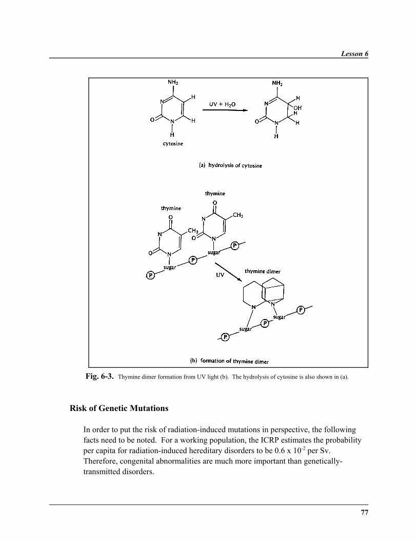
Lesson 6
77
Fig. 6-3. Thymine dimer formation from UV light (b). The hydrolysis of cytosine is also shown in (a).
Risk of Genetic Mutations
In order to put the risk of radiation-induced mutations in perspective, the followingfacts need to be noted. For a working population, the ICRP estimates the probabilityper capita for radiation-induced hereditary disorders to be 0.6 x 10⁻² per Sv. Therefore, congenital abnormalities are much more important than genetically-transmitted disorders.
Gene Mutations
78
It has been estimated that the average American receives less than five radsfrom artificial radiation sources over a 30-year period. Furthermore, no morethan one to six percent of spontaneous mutations in humans may be ascribed tobackground radiation. The low rate of mutation in the modern environment canbe attributed in part to the mechanism for DNA repair. If a single location on aDNA strand is damaged by ultraviolet light, chemicals, or radiation, repair canoccur. As a rule, when an acute exposure with a large dose is absorbed, a largeproportion of genetic mutations can be avoided if conception is deferred for atleast six months.
Summary
This lesson has discussed the irradiation effects on genes. Repair mechanisms,dominant in more complex organisms, are important "fixers" of irradiationdamage. Lesson 7 will adress factors, such as oxygen and temperature, whichcan further enhance or lessen the effects of irradiation on the chromosomes.
˜ There are three types of genetic mutations: dominant, recessive, and sex-linked mutations.
˜ The size of the genome of an organism can affect dose-rate effect.
˜ Doubling dose is also related to the size and repair mechanisms of anorganism.
˜ Repair mechanisms serve to "fix" the genetic mutations caused bybackground radiation.
˜ Radiation can induce both genetic mutations and problems withchromosome distribution.
˜ Genetically-significant dose helps assess the risk of genetic mutations in apopulation.
˜ The risk of genetic mutation due to radiation sources is not great.
Lesson 6
79
WRITING ASSIGNMENT - Lesson 6Complete and submit the following assignment.
Vocabulary
Please write the term and its definition on your paper. Your definition shouldbe in your own words and limited to one or two sentences.
dominant mutationrecessive mutationpoint mutationsex-linked mutationstochastic responseframe-shift mutation
doubling dosexeroderma pigmentosumgene mutationgenegenetically significant dose
gonadal doserelative mutation riskdirect mutation riskgenetic detrimentgenetic death
questions
All of the questions below ask for a short-essay response. Please limit yourresponse to one page or less.
1. What are the three types of mutations? Explain the differences betweenthem.
2. Why are germ-cell mutations more damaging than somatic cell mutations?
3. What was the major finding of the Russell and Russell study?
4. What is the importance of doubling dose?
5. What limits the appearance of mutations in the human population?
6. Explain the process of DNA repair.
Gene Mutations
80
7. Using Figure 6-1, one can observe that the so-called “simple plants” suchas barley and tomatoes have a lower mutation rate than man. Using yourknowledge of genetics, why is this so? (Hint: How are the extremelylarge strawberries different genetically from ordinary strawberries?)
8. Explain why the following statement is true: “Radiation does not result inmutations that are new or unique.”
9. What is the impact of chromosome-distribution problems caused byradiation?
10. What were the findings of the Megamouse Project conducted by Russell &Russell?
11. What is the link between radiation, mutation, and breast cancer?
81
Midterm Examination Information
The midterm examination will cover Lessons 1-6. The exam consists of aseries of questions each requiring a short answer.
83
˜ Preview ˜READING ASSIGNMENT
Hall: Chapters 6 (all), 7 (all), 9(all), 22 (pp. 397-411), and 25 (all)
Bushong: Chapters 34 (all),35 (all), and 36 (pp. 516-520)
Nias: Chapters 4 (pp. 60-61), 7 (all), 8 (all), 16 (all), and 17 (all)
LESSON OBJECTIVES
By the end of this lesson, youshould be able to:
˜ Define terms used inunderstanding survivalcurves: mean lethal dose(Dm), threshold dose, Dq, andDo.
(continued)
LESSON 7Survival Curves
DISCUSSION
This lesson will focus on modifications to theradiation response of the cell, particularly the effectsof oxygen and energy transfer on the response of thecell to radiation. Survival curves will be explained ingreater detail. Lastly, the effects of sex, age, water,chemicals (radiosensitizers and radioprotectors) onirradiation will be discussed. Dose rate will beaddressed, in context with radiotherapy.
Dose-Response Curves
Several clonogenic assays can determine dose-response relationships for cells of normal tissues. Theassays for skin colony, testes, kidney tubule, andjejunum crypts can determine the reproductiveintegrity of individual cells. Bone marrow cells,thyroid cells, and mammary cells all involvetransplantation into another site. Skin reactions inpigs, lung response in mice, and spinal cord damagecan be used to determine functional endpoints fordose-response curves.
Survival Curves
84
˜ Define and explain the fourtypes of radiation damage.
˜ Describe the characteristics ofthe survival curve.
˜ Explain the effects of oxygen onradiation response.
˜ Describe basic concepts ofradiotherapy in regards tooxygen, chemicals, and doserate.
˜ Explain the usage of the termsOER and RBE.
˜ Discuss the modification of theradiation response due to water,temperature, age, and sex.
˜ Describe the effects ofchemicals on the radiationresponse.
˜ Explain the body's response tofractionation and protraction.
˜ Explain the relationshipbetween LET and sublethaldamage.
˜ Differentiate between sublethaland potentially lethal damageat the cellular level.
˜ Discuss the importance offractionation and protractionon the response of anorganism.
In general, the response of a tissue or organ toradiation depends on two factors: (1) inherentsensitivity of the individual cells, and (2) kinetics ofthe population as a whole of which the cells are apart.
In highly-differentiated tissues that have specializedfunctions, the cell survival curves are irrelevant dueto the lack of mitotic future of the cells. The radiationneeded to destroy the function of the cell is muchgreater than that to stop mitosis of the cell.
Survival Curves
The survival curves of mammalian cells can be usedto obtain direct information on their response toradiation.
When the logarithm of a typical cell survival isplotted versus dose on a linear scale, several featuresbecome evident. At larger doses of radiation, thegraph becomes a straight line on this scale. At lowdoses, a shoulder on the curve is found, which isquantified by the threshold dose. The width ofshoulder of the survival curve indicates the degree ofsublethal damage, which is more readily repaired inthe S phase.
As for specific values, the mean lethal dose, Dm, isdefined as that amount of radiation required to reducethe survival by 37%. It is equal to D37 in the linearportion of the curve. A large Dm indicates aradioresistant line of cells, whereas a small Dm ischaracteristic of cells with a high radiosensitivity.The D0 is the slope of the exponential portion of thecurve, which is the nonlethal dose. The thresholddose, Dq, is a measure of how much damage occursbefore it is
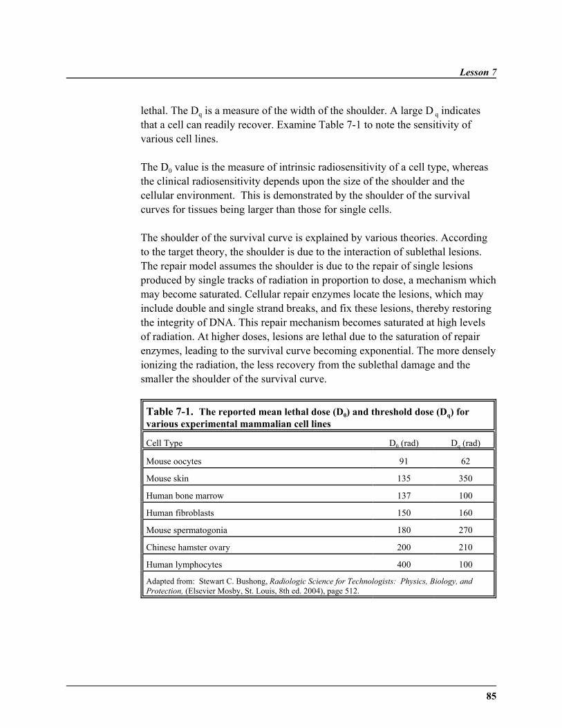
Lesson 7
85
lethal. The Dq is a measure of the width of the shoulder. A large D q indicatesthat a cell can readily recover. Examine Table 7-1 to note the sensitivity ofvarious cell lines.
The D0 value is the measure of intrinsic radiosensitivity of a cell type, whereasthe clinical radiosensitivity depends upon the size of the shoulder and thecellular environment. This is demonstrated by the shoulder of the survivalcurves for tissues being larger than those for single cells.
The shoulder of the survival curve is explained by various theories. Accordingto the target theory, the shoulder is due to the interaction of sublethal lesions.The repair model assumes the shoulder is due to the repair of single lesionsproduced by single tracks of radiation in proportion to dose, a mechanism whichmay become saturated. Cellular repair enzymes locate the lesions, which mayinclude double and single strand breaks, and fix these lesions, thereby restoringthe integrity of DNA. This repair mechanism becomes saturated at high levelsof radiation. At higher doses, lesions are lethal due to the saturation of repairenzymes, leading to the survival curve becoming exponential. The more denselyionizing the radiation, the less recovery from the sublethal damage and thesmaller the shoulder of the survival curve.
Table 7-1. The reported mean lethal dose (D0) and threshold dose (Dq) forvarious experimental mammalian cell lines
Cell Type D0 (rad) Dq (rad)
Mouse oocytes 91 62
Mouse skin 135 350
Human bone marrow 137 100
Human fibroblasts 150 160
Mouse spermatogonia 180 270
Chinese hamster ovary 200 210
Human lymphocytes 400 100
Adapted from: Stewart C. Bushong, Radiologic Science for Technologists: Physics, Biology, andProtection, (Elsevier Mosby, St. Louis, 8th ed. 2004), page 512.
Survival Curves
86
The existence of a threshold in cell-survival curves implies that some damagemust accumulate before it is fatal to the cell. The larger the value of Dq, themore damage that must accumulate. This damage to cells prior to cell death iscalled sublethal damage. In radiation therapy it is very important to note thatwhen a dose is split into two parts separated by enough time, a threshold isobserved for each part of the dose. Thus by properly spacing treatment, it ispossible to reduce the damage to healthy cells during radiation treatment. Thisconcept of dose fractionation will be further explored later in this lesson.
Types of Radiation Damage
There are four main types of radiation damage to a cell:
1. Non-lethal damage involves a lesion which does not prevent proliferation,but it affects the rate of such proliferation.
2. Lethal damage is damage which is irreparable, irreversible, and leadsirrevocably to cell death.
3. Sublethal damage is damage which is repaired in hours unless additionalsublethal damage is added, with which it interacts to form lethal damage. Basically, sublethal damage, which is chiefly evident at low dose levels, isshed after the passage of a relatively short period of time. It has beenshown that the repair of sublethal damage reflects the repair and therejoining interval of double strand breaks before they can interact to formlethal lesions.
4. Potentially lethal damage is the damage that can be modified bypostirradiation environmental conditions. This type of damage is manifestin those cell populations which are proliferating and are well nourished. This type of damage is only lethal if cells are stimulated to divide beforerepair occurs. Therefore, if mitosis can be delayed by suboptimal growthconditions, the DNA damage can be repaired. Potentially lethal damagelimits the effectiveness of radiotherapy on tumors. There is no potentiallylethal damage repair following exposure to high linear energy transferradiations.
Lesson 7
87
Four Rs of Radiobiology
In radiotherapy, these types of cellular damage are regulated by the four Rs ofradiotherapy. The four Rs of radiobiology—repair, reassortment, repopulation,and reoxygenation—are affected by the type of radiation dose.
1. There is a prompt repair of sublethal repair damage.
2. There is a progression of cells through the cell cycle during the intervalbetween the split doses, called reassortment.
3. There is an increase of surviving fraction due to cell division, orrepopulation. When the time interval between two dose fractions exceedsthe cell cycle, there will be an increase in the number of cells survivingdue to cell proliferation.
4. The fractionation of the dose allows greater affect through reoxygenationof the tumour, which increases the radiosensitivity.
Cell Cycle Effects
As addressed in Lesson 5, during the cell cycle the radiation sensitivity of thecells can change dramatically. Studies using cells which have their cell cyclessynchronized have shown the results found in Figure 7-1 below. To reviewLesson 5, the mitosis phase, where the cell divides, is always the most sensitive,while late in the S phase, where DNA synthesis occurs, is the least sensitive. This knowledge is useful in the planing of cancer treatments.
The Effect of Oxygen and LET on Cell Survival
The survival of a cell can be affected by factors other than the radiation dose. The most biologically variable factor that modifies the radiation response is theamount of oxygen in the system. The best clinical radiation sensitizer is oxygendissolved in the tissues at physiological concentration. In general, anycompound that is electron-affinic, such as nitroimidazoles, will tend to be
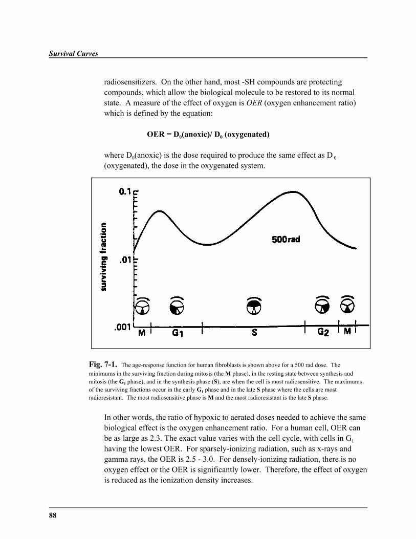
Survival Curves
88
radiosensitizers. On the other hand, most -SH compounds are protectingcompounds, which allow the biological molecule to be restored to its normalstate. A measure of the effect of oxygen is OER (oxygen enhancement ratio)which is defined by the equation:
OER = D0(anoxic)/ D0 (oxygenated)
where D0(anoxic) is the dose required to produce the same effect as D 0
(oxygenated), the dose in the oxygenated system.
Fig. 7-1. The age-response function for human fibroblasts is shown above for a 500 rad dose. Theminimums in the surviving fraction during mitosis (the M phase), in the resting state between synthesis andmitosis (the G1 phase), and in the synthesis phase (S), are when the cell is most radiosensitive. The maximumsof the surviving fractions occur in the early G1 phase and in the late S phase where the cells are mostradioresistant. The most radiosensitive phase is M and the most radioresistant is the late S phase.
In other words, the ratio of hypoxic to aerated doses needed to achieve the samebiological effect is the oxygen enhancement ratio. For a human cell, OER canbe as large as 2.3. The exact value varies with the cell cycle, with cells in G1
having the lowest OER. For sparsely-ionizing radiation, such as x-rays andgamma rays, the OER is 2.5 - 3.0. For densely-ionizing radiation, there is nooxygen effect or the OER is significantly lower. Therefore, the effect of oxygenis reduced as the ionization density increases.
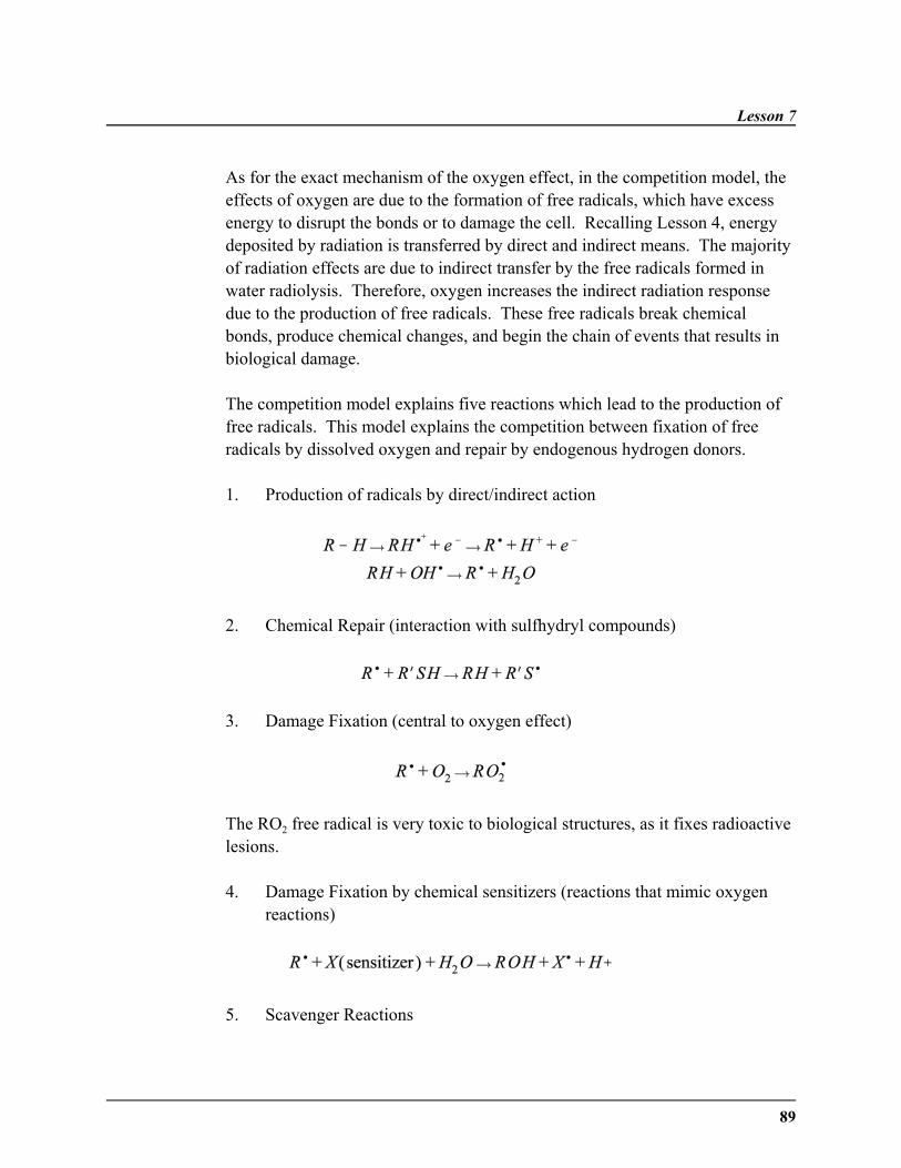
Lesson 7
89
As for the exact mechanism of the oxygen effect, in the competition model, theeffects of oxygen are due to the formation of free radicals, which have excessenergy to disrupt the bonds or to damage the cell. Recalling Lesson 4, energydeposited by radiation is transferred by direct and indirect means. The majorityof radiation effects are due to indirect transfer by the free radicals formed inwater radiolysis. Therefore, oxygen increases the indirect radiation responsedue to the production of free radicals. These free radicals break chemicalbonds, produce chemical changes, and begin the chain of events that results inbiological damage.
The competition model explains five reactions which lead to the production offree radicals. This model explains the competition between fixation of freeradicals by dissolved oxygen and repair by endogenous hydrogen donors.
1. Production of radicals by direct/indirect action
2. Chemical Repair (interaction with sulfhydryl compounds)
3. Damage Fixation (central to oxygen effect)
The RO2 free radical is very toxic to biological structures, as it fixes radioactivelesions.
4. Damage Fixation by chemical sensitizers (reactions that mimic oxygenreactions)
5. Scavenger Reactions
Survival Curves
90
The superoxide ion (O2CG), which is formed by water radiolysis, is believed to be
an intermediate in the oxygen effect. The superoxide dismutase enzyme acts onthis radical to form oxygen and hydrogen peroxide, thereby protecting the cellfrom damage by the free radical. Cells with reduced superoxide dismutaseenzymes are much more vulnerable to the effects of oxygen. In summary,oxygen increases the effect of radiation by the action of various oxygenated freeradicals, and this increase is quantified by the OER formula.
It is important to remember that as the oxygen concentration increases, thematerial becomes progressively more sensitive to radiation until in the presenceof 100% oxygen, it is about 3 times as sensitive as under complete anoxia.
Oxygen Effect in Tumors
This section will illustrate the practical importance of the oxygen effect inradiotherapy. The presence of a relatively small proportion of hypoxic cells intumors can limit the success of radiotherapy. Tumors consist of two populationsof cells—one well-oxygenated fraction and the hypoxic fraction, which mayaccount for 10-15% of a human tumor. As the tumor is irradiated, the radiationwill kill the more radiosensitive outer cells but not the more radioresistant,oxygen-deprived inner cells. As a result, there will be an increase in the supplyof nutrients to these inner cells, which will again start proliferating if they haveretained their reproductive potential. Therefore, though these cells are only asmall fraction of the tumor, they play an important role in theradioresponsiveness of a tumor. Administration of hyperbaric oxygen is used toensure the radiosensitivity of the tumor. Hyperbaric oxygen administrationincreases the success rates of radiotherapy for head and neck tumors.
Survival curves are biphasic for tumors, with a steep initial slope for the well-oxygenated proportion of the tumor cell population and a shallower final curveover the higher doses where the radioresistant hypoxic cells predominate.Overall, the most sensitive tumors have much less than 1% hypoxic cells,whereas resistant tumors have more than 10% hypoxic cells.
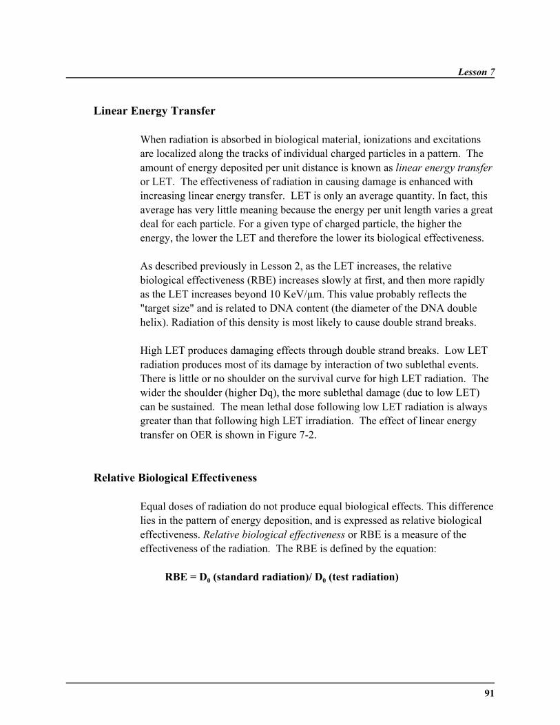
Lesson 7
91
Linear Energy Transfer
When radiation is absorbed in biological material, ionizations and excitationsare localized along the tracks of individual charged particles in a pattern. Theamount of energy deposited per unit distance is known as linear energy transferor LET. The effectiveness of radiation in causing damage is enhanced withincreasing linear energy transfer. LET is only an average quantity. In fact, thisaverage has very little meaning because the energy per unit length varies a greatdeal for each particle. For a given type of charged particle, the higher theenergy, the lower the LET and therefore the lower its biological effectiveness.
As described previously in Lesson 2, as the LET increases, the relativebiological effectiveness (RBE) increases slowly at first, and then more rapidlyas the LET increases beyond 10 KeV/µm. This value probably reflects the"target size" and is related to DNA content (the diameter of the DNA doublehelix). Radiation of this density is most likely to cause double strand breaks.
High LET produces damaging effects through double strand breaks. Low LETradiation produces most of its damage by interaction of two sublethal events.There is little or no shoulder on the survival curve for high LET radiation. Thewider the shoulder (higher Dq), the more sublethal damage (due to low LET)can be sustained. The mean lethal dose following low LET radiation is alwaysgreater than that following high LET irradiation. The effect of linear energytransfer on OER is shown in Figure 7-2.
Relative Biological Effectiveness
Equal doses of radiation do not produce equal biological effects. This differencelies in the pattern of energy deposition, and is expressed as relative biologicaleffectiveness. Relative biological effectiveness or RBE is a measure of theeffectiveness of the radiation. The RBE is defined by the equation:
RBE = D0 (standard radiation)/ D0 (test radiation)
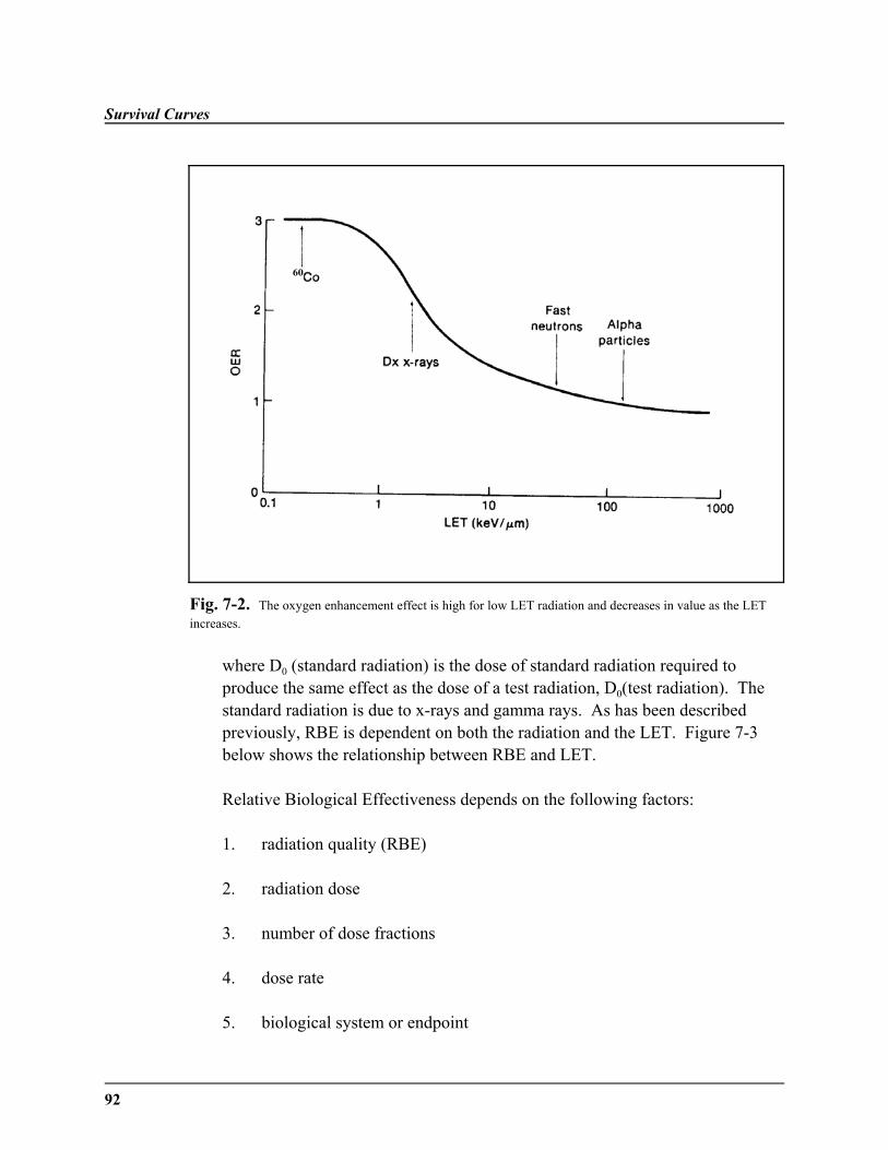
Survival Curves
92
Fig. 7-2. The oxygen enhancement effect is high for low LET radiation and decreases in value as the LETincreases.
where D0 (standard radiation) is the dose of standard radiation required toproduce the same effect as the dose of a test radiation, D0(test radiation). Thestandard radiation is due to x-rays and gamma rays. As has been describedpreviously, RBE is dependent on both the radiation and the LET. Figure 7-3below shows the relationship between RBE and LET.
Relative Biological Effectiveness depends on the following factors:
1. radiation quality (RBE)
2. radiation dose
3. number of dose fractions
4. dose rate
5. biological system or endpoint
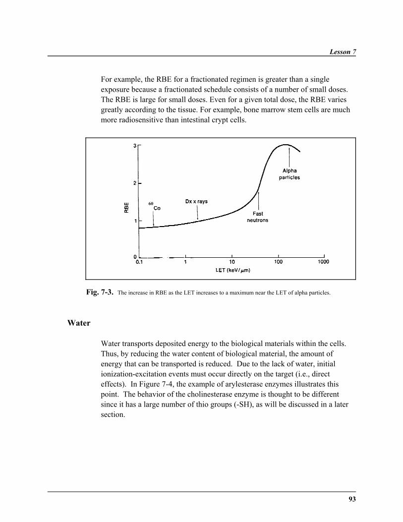
Lesson 7
93
For example, the RBE for a fractionated regimen is greater than a singleexposure because a fractionated schedule consists of a number of small doses.The RBE is large for small doses. Even for a given total dose, the RBE variesgreatly according to the tissue. For example, bone marrow stem cells are muchmore radiosensitive than intestinal crypt cells.
Fig. 7-3. The increase in RBE as the LET increases to a maximum near the LET of alpha particles.
Water
Water transports deposited energy to the biological materials within the cells. Thus, by reducing the water content of biological material, the amount ofenergy that can be transported is reduced. Due to the lack of water, initialionization-excitation events must occur directly on the target (i.e., directeffects). In Figure 7-4, the example of arylesterase enzymes illustrates thispoint. The behavior of the cholinesterase enzyme is thought to be differentsince it has a large number of thio groups (-SH), as will be discussed in a latersection.
Survival Curves
94
Fig. 7-4. Relative radiosensitivity of two enzymes when they are irradiated under different conditionsof hydration. (Augustinsson, Jonsson,and Sparrman, Acta Chem. Scand. 15, 1961)
Age
The age of a biological structure is also an important factor in theradiosensitivity of an organism. For humans, the radiosensitivity is greatest inutero when the cells are rapidly dividing. This is due to the increasedsensitivity of the cell while in mitosis. A second period of increased sensitivityis in the aged. This is due to a decreased rate of division, and thereby lowreplacement of cells, in an aged organism. Once a cell is destroyed, the losswill not be replaced—function will suffer.
Sex
Surprisingly, sex is indeed important in radiosensitivity. For humans, womenare five to ten percent more radiation resistant than males. Both human sexeshave 46 chromosomes, 44 of which are autosomes. The remaining two are sexchromosomes. Women have two X chromosomes, whereas males have an X anda Y chromosome. The Y chromosome has very little genetic information. Recessive traits on the X chromosome will be expressed in the male due to hislack of similar genes in his sex chromosomes. Therefore, women have twicethe genetic material in their sex chromosomes (XX vs. XY). The additionalDNA could aid in modifying the effects of the radiation.
Lesson 7
95
Chemicals (Radiosensitizers and Radioprotectors)
Chemicals can have either a positive or negative influence on radiosensitivity,being either radiosensitizers or radioprotectors. Most of the trueradioprotectors are sulfhydryl compounds. Sulfhydryl compounds protect byscavenging free radicals, aiding in the chemical restitution of the targetmolecule. Thiols (R-SH) are one example of chemicals that decreaseradiosensitivity. Other examples of chemicals which protect are cysteine andcysteamine, both of which contain sulfhydryl groups. Seven hundred of thesechemicals have been tested to improve survival by up to a factor of two. Unfortunately, all of the chemicals tested to date have strong side effects andare often more damaging than the radiation itself, mainly due to the highconcentration required to have any protective effect. They cause nausea andvomiting at levels needed for radioprotection.
More effective radioprotectors have been developed, including WR-638(Cytaphos), WR-2721 (amifostine), and WR-1607 (d-Con). Amifostine offersprotection to the hematopoietic system, gut, and salivary glands. The dose-limiting toxicity is hypotension, sneezing, and somnolence. An important termin conjunction with radioprotection is dose reduction factor, which is the ratioof radiation doses required to produce the same biological effect in the absenceand presence of radioprotectors.
Radiosensitizers are chemical or pharmacologic agents that increase the lethaleffects of radiation when administered in conjunction with it. Radiosensitizingchemicals increase the transport of energy or increase the damage to theorganism. Currently, they offer no gain in radiotherapy because they do notshow a differential effect between tumors and normal tissues.
Only two types of sensitizers have a practical use—halogenated pyrimidines andhypoxic cell sensitizers. Halogenated pyrimidines have a differential effect due totumor cells having a more rapid cell cycle. They weaken DNA chains by beingincorporated in place of thymidine. Use of 5-iododeoxyuridine and5-bromodeoxyuridine, as an adjunct to radiotherapy, began in the 1970s.
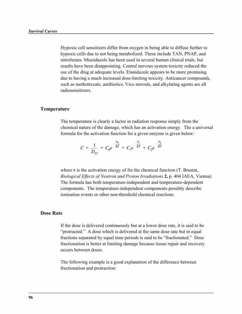
Survival Curves
96
Hypoxic cell sensitizers differ from oxygen in being able to diffuse further tohypoxic cells due to not being metabolized. These include TAN, PNAP, andnitrofurans. Misoidazole has been used in several human clinical trials, butresults have been disappointing. Central nervous system toxicity reduced theuse of the drug at adequate levels. Etanidazole appears to be more promisingdue to having a much increased dose-limiting toxicity. Anticancer compounds,such as methotrexate, antibiotics, Vico steroids, and alkylating agents are allradiosensitizers.
Temperature
The temperature is clearly a factor in radiation response simply from thechemical nature of the damage, which has an activation energy. The a universalformula for the activation function for a given enzyme is given below:
where g is the activation energy of for the chemical function (T. Brustat,Biological Effects of Neutron and Proton Irradiations 2, p. 404 IAEA, Vienna). The formula has both temperature-independent and temperature-dependentcomponents. The temperature-independent components possibly describeionization events or other non-threshold chemical reactions.
Dose Rate
If the dose is delivered continuously but at a lower dose rate, it is said to be“protracted.” A dose which is delivered at the same dose rate but in equalfractions separated by equal time periods is said to be “fractionated.” Dosefractionation is better at limiting damage because tissue repair and recoveryoccurs between doses.
The following example is a good explanation of the difference betweenfractionation and protraction:
Lesson 7
97
Protraction: A mouse is given 600 rads at 200 rads/hour for 3 minutescontinuously.
Fractionation: A mouse is given 600 rads at 200 rads/min in 12 fractionsof 50 rads, separated by 24 hours.
Dose Rate—Protraction
As the dose rate is lowered and the exposure time extended, the biological effect ofa given dose is greatly reduced. Since a continuously low dose rate of radiationmay be considered to consist of infinitely small fractions, the survival curve willhave no shoulder and be shallower than for single acute exposure.
Some cell lines repair sublethal damage rapidly, some more slowly. This isreflected in the survival curves at low dose rates. For example, testis tissue is moresensitive than small intestine tissue. A point is reached at which all sublethaldamage is repaired, resulting in a limiting slope.
There are several factors which determine the response of renewal tissues tocontinual radiation.
1. Cellular sensitivity of the stem cells involved.
2. Duration of the cell cycle (i.e., more damage to cells with a long cell cycle).
3. Ability of some tissues to adapt to the new trauma of continual radiation.
When protraction of radiation dose occurs, there is an increase in recovery fromsublethal damage in the surviving cells, leading to more proliferation during theirradiation. As a result of this protracted dose, the radiation may appear to havelittle or no effect at all. If the dose rate is greatly lowered, there will be time forproliferation of undamaged cells.
At intermediate dose rates, a cell population may only fall to a survival levelthat is “steady state,” due to the balance of continuous radiation damage and therecovery of damaged cells. As a rule, a given dose rate of continuousirradiation will be more effective in cells with a longer cell cycle.
Survival Curves
98
In action, protraction, a method whereby the overall time within the normalradiotherapy range is prolonged, has a sparing effect on the late reactions but alarge sparing effect on early reactions. Early-responding tissues are skin,mucosa, and intestinal epithelium; late-responding tissues include the spinalcord.
Dose Rate—Fractionation
The goal of fractionation in radiotherapy is to kill all tumor cells withoutproducing serious damage to the normal surrounding tissues. This is bestachieved by giving the total dose of radiation in a specific number of fractionsover a time period, generally five treatments a week for six weeks. Repair,reassortment, repopulation, and reoxygenation, the four Rs of radiotherapy,determine the effectiveness of the fractionation. For example, dividing a doseinto fractions spares normal tissues because of repair of sublethal damagebetween dose fractions and repopulation of cells if the overall time issufficiently long. Fractions also increase damage to a tumor because ofreoxygenation and reassortment of cells into radiosensitive phases of the cycle.
With very large tumors, the incidence of late-tissue effects may outweigh thebeneficial effects of tumor volume reduction. The fraction size is the dominantfactor in determining the late effects while overall treatment time has littleinfluence. By contrast, fraction size and overall treatment time both determinethe response of acutely-responding tissues. Therefore, the late effects on lung,kidney, and spinal cord determine the tolerance dosage. Fractionation of theradiation dose produces better tumor control for a given level of normal tissuetoxicity than a single large dose.
According to the nominal standard dose system (NSD), the total dose for thetolerance of connective tissue is related to the number of fractions (N) and theoverall treatment time (T) by the following formula (Nias, page 289):
Total dose = (NSD) T0.11 N0.24
Fractionation can also involve multiple fractions per day—hyperfractionationand accelerated treatment. In hyperfractionation, the treatment time remains thesame but the number of fractions are doubled. The intent is to further

Lesson 7
99
reduce late effects and achieve better tumor control. Accelerated treatmenthalves the treatment time by giving two fractions a day. This process reducesthe repopulation in rapidly proliferating tumors. The use of either of these twoprocesses depends on clonogenic cell number doubling time.
The dose rate is often very important in radiation effects on the cell ororganism. For some cells, a threshold dose is required before any effect can beseen. By splitting a dose in several parts below this threshold level, a bettersurvival rate occurs than when the dose is applied at once. Protraction andfractionation are two methods to reduce the effects of the dose delivered.
Summary
This lesson has focused on possible modifications in the radiation dose receivedby an organism. The survival curves, which illustrate the survival of apopulation of cells, can show the effects of factors such as sex, age, and doserate. In Lesson 8, the focus will shift from cells to tissues and organs. Thebasics of irradiation at the cellular level will also apply to the tissues andorgans.
˜ Graphing a survival curve gives a great deal of pertinent information suchas threshold dose and mean lethal dose.
˜ Oxygen has an important influence on the effects of radiation due todestruction of the cell by a free radical.
˜ The effects of radiation are influenced by cell cycle position, water, linearenergy transfer, age, sex, chemicals, and temperature.
˜ The effects of dose rate on radiation is important for therapeutic radiationuse.
˜ Radiosensitizers and radioprotectors are limited in their use due totoxicity.
Survival Curves
100
WRITING ASSIGNMENT - Lesson 7Complete and submit the following assignment.
Vocabulary
Please write the term and its definition on your paper. Your definition shouldbe in your own words and limited to one or two sentences.
threshold dosemean lethal dosesublethal damageOERRBEthiolsprotraction
fractionationdose modifying effectcompetition modelsuperoxideplating efficiencyfeeder cellsconditioning dose
challengepotentially lethal damageradiosensitizersmethotrexateradioprotectorsamifostinedose reduction factor
Questions
Anwer each of the short-answer essay questions below in one page or less.
1. What is the effect of temperature on radiation in the following temperatureintervals?
a. T < 100 Kb. 100 < T < 170 Kc. 170 < T < 420 K
2. Why is superoxide or any free radical important in the oxygen effect?
3. What is the main use of split-dose technique?
Lesson 7
101
4. Using the mouse example from this lesson, supply another set of figures toexplain fractionation and protraction.
5. How is plating efficiency important in understanding the worth of survivalcurves?
6. What is the significance of the shoulder in the survival curve?
7. In Figure 7-5 on the next page, label the graph of the survival curve ofmammalian cells with the following terms: extrapolation number,quasithreshold, slope, D0.
8. Explain the relationship between high LET/low LET and sublethaldamage.
9. According to the cell cycle, which cell population is most radiosensitive?least radiosensitive?
10. Compare and contrast the four types of cellular damage due to radiation.
11. What are the "four Rs" of radiobiology? Explain each briefly.
12. Explain the oxygen effect seen in tumors during radiotherapy.
13. What are the five factors determining relative biological effectiveness(RBE)?
14. What are the problems which arise in the use of radioprotectors andradiosensitizers?
15. What is meant by hyperfractionation and accelerated treatment? Why arethese methods useful?
16. What are the three factors which determine the response of renewal tissuesto continual radiation?
17. What is the role of fractionated and protracted radiotherapy in thetreatment of tumors with regard to late/early effects?

Fig. 7-5
103
˜ Preview ˜READING ASSIGNMENT
Hall: Chapters 12, 13, 19, and 21
Bushong: Chapters 33 (all), 34(pp. 498-500), and 36 (pp. 520-530)
Nias: Chapters 8 and 13
LESSON OBJECTIVES
By the end of this lesson, youshould be able to:
˜ Define and explain the Law ofBergonié and Tribondeau.
˜ Explain the significance ofCasarett's classification oftissue.
(continued)
LESSON 8Effects on Tissues
DISCUSSION
Overview
Lessons 5 and 6 looked at the effects of radiation on thecells. This lesson focuses on tissues, collections of cellsof similar structure and function. Tissues combine toform organs, an overall integrated organization of tissues. Radiation affects the whole body through the hierarchy ofthe cell - tissue - organ - system organization.
The effects of radiation on a tissue system are dependenton the rate of division or growth kinetics of that tissue. For example, radiation has a much greater effect on arapidly dividing system (blood-forming organs) than on aslowly dividing system (central nervous system). Thislesson will focus on the nonstochastic (dose-threshold)effects on various tissues and organs, as well as neoplastictissue (embryo and fetus).
The Law of Bergonié and Tribondeau
As a historical note, the Law of Bergonié and Tribondeauis of great interest in the development of radiobiology. In1906, the French scientists Bergonié and Tribondeaumade a series of observations of the radiosensitivity ofcells of rat testis. In the rat, cell populations with thehigher rate of cell division show the earlier response toradiation.
Effects on Tissues
104
˜ List the tissues which arehighly sensitive, moderatelysensitive, and slightly sensitiveto radiation.
˜ Explain the relationshipbetween radioresistance,growth kinetics, andradioresponsiveness.
˜ Discuss the effects of radiationon digestive organs, liver,kidney, thoracic organs, andlens of the eye.
˜ Discuss the effects of radiationon hematopoietic system,digestive system, germ cells,epidermis, eye, and centralnervous system.
˜ Describe the effects ofradiation on an embryo.
˜ Explain the increasedsensitivity of fetal andembryonic tissue.
˜ Explain the findings ofHiroshima and Nagasaki, inregards to radiation's effect onembryo development.
˜ Define and explain the termsstochastic and non-stochastic.
(continued)
From these observations they formulated a law aboutthe radiation sensitivity of cells and tissues. Theradiosensitivity laws can be stated as:
1. Stem cells are radiosensitive. The more maturethe cell, the more radiation resistant.
2. The older the tissue or organ, the more radiationresistant it is.
3. The lower the metabolic activity, the higher theradioresistance of the tissue.
4. As the proliferation rate of cells increase and thegrowth rate of tissue increases, theradiosensitivity increases.
This law has been proved in several different tests,but it does have several important exceptions such asthe radiation sensitivity of lymphocytes.
The lymphocyte is extremely radiosensitive, whilealso being non-dividing and mature.The smalllymphocyte breaks the rules in that it does not usuallydivide, dies an interphase death, and at the same time,is one of the most sensitive mammalian cells.
Casarett's Classificatifon
The classification of cells by Casarett divides cellsinto four divisions by radiosensitivity, which isaffected by the growth kinetics of the cell. Casarett'sclassification of mammalian cell sensitivity dividesthe cells into four major categories, numbered I to IV,as shown below:
Lesson 8
105
˜ Explain why sterility is oneof the classic effects ofradiation.
˜ Describe the late effects ofradiation on the esophagus,small intestine, largeintestine, and stomach.
˜ Describe the concept of earlyand late effects.
˜ Describe the nonstochasticlate effects in humans.
I. Vegetative Intermitotic - cells which divideregularly and have no differentiation
II. Differentiating Intermitotic - cells which divideregularly, some of which are differentiated.
III. Reverting Postmitotic - cells which do notdivide regularly.
IV. Fixed Postmitotic - cells which do not divideand are highly differentiated.
(Adapted from Table 8.1, page 168 of Casarett, Radiation Biology,Prentice-Hall, 1968)
Specific cell types are divided into the four categorieson page 343 of Hall's text. Because this chart is in thetext, a slightly different version is shown in Table 8-1.
Radiosensitive Tissue
The sensitivity of a cell to radiation is determined by its state of maturity and itsfunctional role. Immature cells, such as stem cells, are much more sensitivethan mature cells due to their high rate of division. The following list showssensitivity of various cell types in a summary form.
High Lymphocytes (white blood cells)Spermatogonia (sperm-producing cells)Erythroblasts (immature red blood cells)Intestinal crypt cells (gland cells between villi)
Mid Spermatids (non-dividing immature sperm)Osteoblasts (immature bone cells)
Low Muscle cellsNerve cells
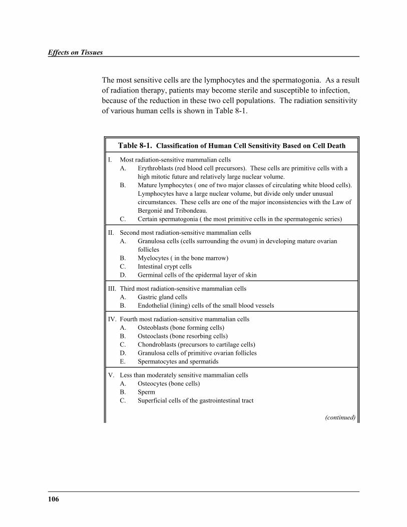
Effects on Tissues
106
The most sensitive cells are the lymphocytes and the spermatogonia. As a resultof radiation therapy, patients may become sterile and susceptible to infection,because of the reduction in these two cell populations. The radiation sensitivityof various human cells is shown in Table 8-1.
Table 8-1. Classification of Human Cell Sensitivity Based on Cell Death
I. Most radiation-sensitive mammalian cellsA. Erythroblasts (red blood cell precursors). These cells are primitive cells with a
high mitotic future and relatively large nuclear volume.B. Mature lymphocytes ( one of two major classes of circulating white blood cells).
Lymphocytes have a large nuclear volume, but divide only under unusualcircumstances. These cells are one of the major inconsistencies with the Law ofBergonié and Tribondeau.
C. Certain spermatogonia ( the most primitive cells in the spermatogenic series)
II. Second most radiation-sensitive mammalian cellsA. Granulosa cells (cells surrounding the ovum) in developing mature ovarian
folliclesB. Myelocytes ( in the bone marrow)C. Intestinal crypt cellsD. Germinal cells of the epidermal layer of skin
III. Third most radiation-sensitive mammalian cellsA. Gastric gland cellsB. Endothelial (lining) cells of the small blood vessels
IV. Fourth most radiation-sensitive mammalian cellsA. Osteoblasts (bone forming cells)B. Osteoclasts (bone resorbing cells)C. Chondroblasts (precursors to cartilage cells)D. Granulosa cells of primitive ovarian folliclesE. Spermatocytes and spermatids
V. Less than moderately sensitive mammalian cellsA. Osteocytes (bone cells)B. Sperm
C. Superficial cells of the gastrointestinal tract
(continued)
Lesson 8
107
VI. Fairly large dose of radiation necessary to alter these mammalian cellsA. Parencymal and duct cells of glandsB. Fibroblasts (these form the intercellular fibrous matrix)C. Endothelial cells of the large blood vesselsD. Erythrocytes (red blood cells)
VII. Fairly radiation-resistant mammalian cellsA. Fibrocytes (connective tissue cells)B. Reticular cells (fixed hematopoetic stem cells)C. Chondrocytes (cartilage cells)D. Phagocytes (scavengers)
VIII. Least sensitive to radiation of all mammallian cellsA. Muscle CellsB. Nerve Cells (it may be that the nerve cells are sensitive, but it takes a long time to
take effect)
Adapted from R. P. Yaffe and G. L. Helgeson, Practical Health Physics, California Book Company,Berkley 1971.
Growth Kinetics and Radiosensitivity
The faster the growth kinetics of a cell population, the earlier the radiationresponse; however, this radioresponsiveness does not depend uponradiosensitivity of the cells. For example, liver tissue appears to beradioresistant; however, its radio-responsiveness appears slower due to itsslower division time.
Tissues that show a response soon after radiotherapy do so because of the fasterkinetics of their cell population. There may also be a latent period before theappearance of radiation damage in the tissue. The larger the radiation dose andthe larger the volume inactivated, the higher the likelihood of an "avalanche" ofcell death which will reduce the cell number below the minimum needed fortissue function, resulting in an earlier appearance of radiation effects. Whereasloss of function is mainly due to loss of parenchymal cells, it may also be due toblood vessel damage and fibrosis. Therefore, radioresponsiveness depends upona combination of the particular vascular architecture of the tissue and theintricate interaction of cell death, recovery, and repopulation.
Effects on Tissues
108
It is important to note that overall, radiation has an aging effect on the tissuesand organs, in that the organ syndrome occurs in the aged, but radiationincreases the incidence and lowers the age when it occurs. This is especiallyevident in the effects of radiation on the kidney and the lens of the eye, whichwill be described later in the lesson.
Radiosensitivity of Organs
The radiosensitivity of an organ depends upon the radiosensitivity of the tissueswhich compose it. The general pattern of radiation damage to organs is thefollowing:
1. inhibition of cell division in mitotically active cells
2. chromosome aberrations
3. hemorrhage and edema
4. removal of debris by phagocytic cells
5. cell regeneration
Hematopoietic Tissue
The hematopoietic system is basically one of the most radiosensitivecell-renewal populations of the organism. The bone marrow varies in structureand cellular composition with age and with anatomical location. It has one ofthe lowest dose-limiting levels. The hematopoietic system consists of bonemarrow (myelopoietic), circulating blood (erythropoietic), and lymphoid tissue(thrombopoietic).
The system produces most of the circulating blood cells. The principal effect ofradiation on this system is to depress the number of blood cells in the peripheralcirculation.
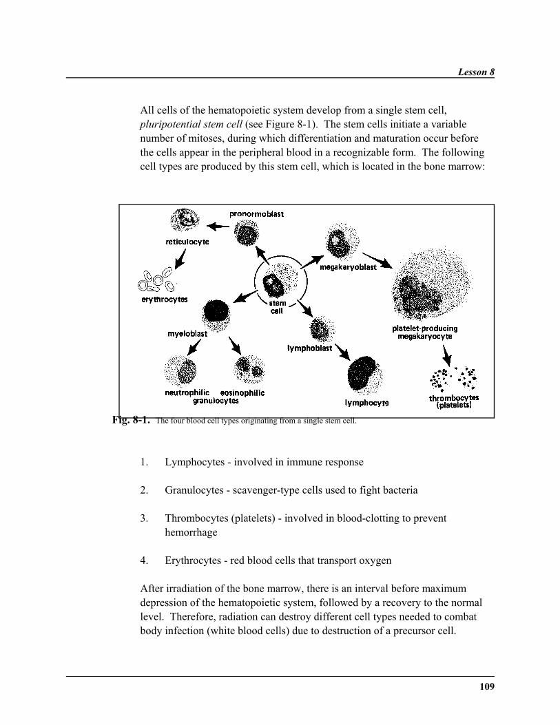
Lesson 8
109
All cells of the hematopoietic system develop from a single stem cell,pluripotential stem cell (see Figure 8-1). The stem cells initiate a variablenumber of mitoses, during which differentiation and maturation occur beforethe cells appear in the peripheral blood in a recognizable form. The followingcell types are produced by this stem cell, which is located in the bone marrow:
Fig. 8-1. The four blood cell types originating from a single stem cell.
1. Lymphocytes - involved in immune response
2. Granulocytes - scavenger-type cells used to fight bacteria
3. Thrombocytes (platelets) - involved in blood-clotting to preventhemorrhage
4. Erythrocytes - red blood cells that transport oxygen
After irradiation of the bone marrow, there is an interval before maximumdepression of the hematopoietic system, followed by a recovery to the normallevel. Therefore, radiation can destroy different cell types needed to combatbody infection (white blood cells) due to destruction of a precursor cell.

Effects on Tissues
110
Anemia, which results from the loss of production of red blood cells, will notappear before the loss of the circulating red blood cells. The timing fordestruction of various cell types depends upon the kinetics of each cell type. Forexample, since red blood cells have relative long life (120 days in man), theeffects of anemia are seen later than the effects on the immune system. As isalso expected, mature cells are less radiosensitive than undifferentiatedprecursors. For example, activated T and B cells are less sensitive than are theirprecursors.
Thoracic Organs
Due to the rapid cell renewal system in the esophagus, radiation effects appearin the esophagus before the lungs. Radiation pneumonitis results from damageto alveolar cells (air sac cells) and capillary cells. Starvation cell death may alsooccur, which results in early esophageal damage and later lung fibrosis.
The heart, large artery, and large veins are less affected by radiation than thecapillaries, which are affected by occlusions. In fact, capillary tissue is the mostimportant limiting tissue in irradiation of a patient.
Thyroid
The normal thyroid is more radioresistant than the hyperactive thyroid. Highdoses of radiation can permanently damage normal thyroid tissue, leading tohypothyroidism.
Kidney
Due to slow cell turnover, it takes several months for radiation injury to occur inthe kidney. Acute radiation nephritis may cause up to 30% mortality; however,even those who recover will have chronic radiation nephritis.
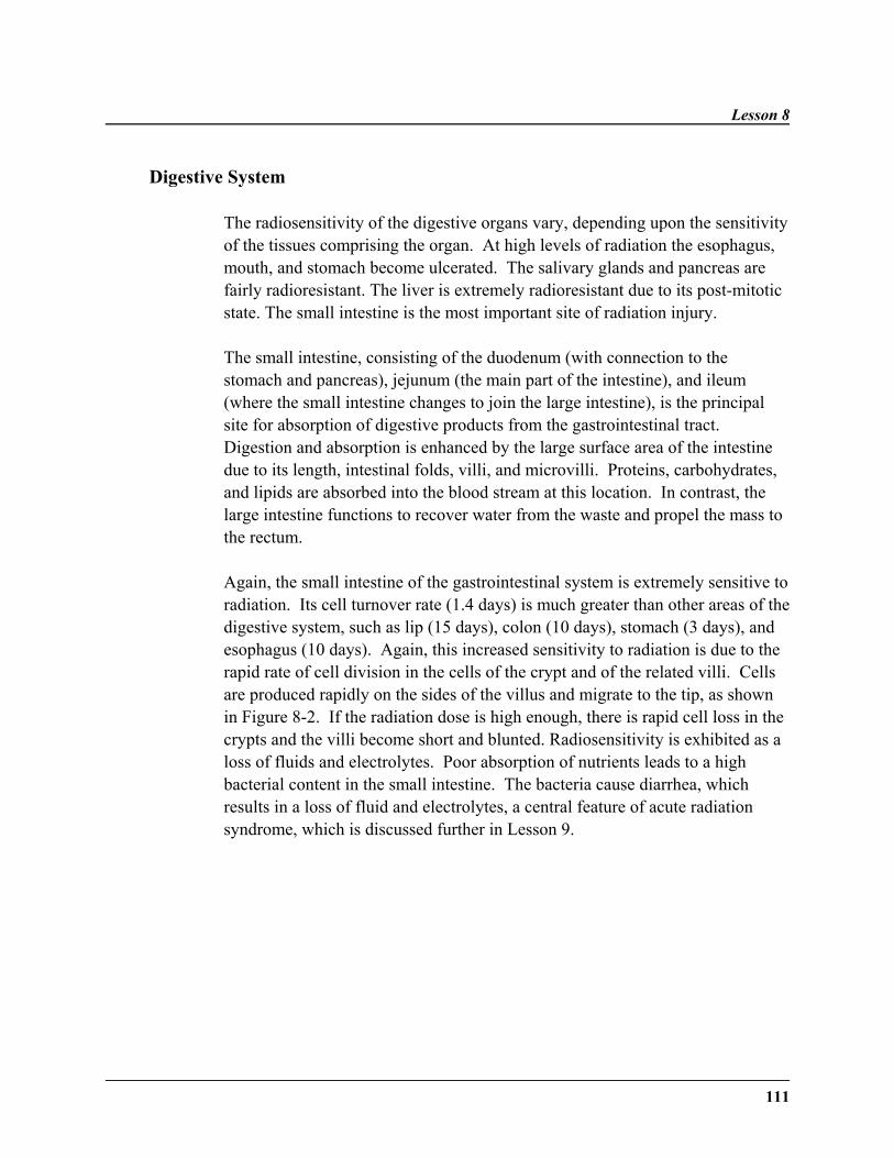
Lesson 8
111
Digestive System
The radiosensitivity of the digestive organs vary, depending upon the sensitivityof the tissues comprising the organ. At high levels of radiation the esophagus,mouth, and stomach become ulcerated. The salivary glands and pancreas arefairly radioresistant. The liver is extremely radioresistant due to its post-mitoticstate. The small intestine is the most important site of radiation injury.
The small intestine, consisting of the duodenum (with connection to thestomach and pancreas), jejunum (the main part of the intestine), and ileum(where the small intestine changes to join the large intestine), is the principalsite for absorption of digestive products from the gastrointestinal tract. Digestion and absorption is enhanced by the large surface area of the intestinedue to its length, intestinal folds, villi, and microvilli. Proteins, carbohydrates,and lipids are absorbed into the blood stream at this location. In contrast, thelarge intestine functions to recover water from the waste and propel the mass tothe rectum.
Again, the small intestine of the gastrointestinal system is extremely sensitive toradiation. Its cell turnover rate (1.4 days) is much greater than other areas of thedigestive system, such as lip (15 days), colon (10 days), stomach (3 days), andesophagus (10 days). Again, this increased sensitivity to radiation is due to therapid rate of cell division in the cells of the crypt and of the related villi. Cellsare produced rapidly on the sides of the villus and migrate to the tip, as shownin Figure 8-2. If the radiation dose is high enough, there is rapid cell loss in thecrypts and the villi become short and blunted. Radiosensitivity is exhibited as aloss of fluids and electrolytes. Poor absorption of nutrients leads to a highbacterial content in the small intestine. The bacteria cause diarrhea, whichresults in a loss of fluid and electrolytes, a central feature of acute radiationsyndrome, which is discussed further in Lesson 9.
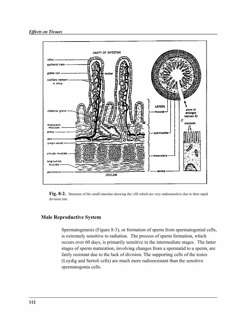
Effects on Tissues
112
Fig. 8-2. Structure of the small intestine showing the villi which are very radiosensitive due to their rapiddivision rate.
Male Reproductive System
Spermatogenesis (Figure 8-3), or formation of sperm from spermatogonial cells,is extremely sensitive to radiation. The process of sperm formation, whichoccurs over 60 days, is primarily sensitive in the intermediate stages. The latterstages of sperm maturation, involving changes from a spermatid to a sperm, arefairly resistant due to the lack of division. The supporting cells of the testes(Leydig and Sertoli cells) are much more radioresistant than the sensitivespermatogonia cells.
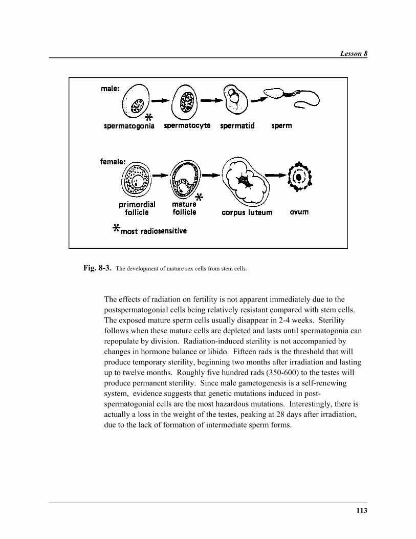
Lesson 8
113
Fig. 8-3. The development of mature sex cells from stem cells.
The effects of radiation on fertility is not apparent immediately due to thepostspermatogonial cells being relatively resistant compared with stem cells.The exposed mature sperm cells usually disappear in 2-4 weeks. Sterilityfollows when these mature cells are depleted and lasts until spermatogonia canrepopulate by division. Radiation-induced sterility is not accompanied bychanges in hormone balance or libido. Fifteen rads is the threshold that willproduce temporary sterility, beginning two months after irradiation and lastingup to twelve months. Roughly five hundred rads (350-600) to the testes willproduce permanent sterility. Since male gametogenesis is a self-renewingsystem, evidence suggests that genetic mutations induced in post-spermatogonial cells are the most hazardous mutations. Interestingly, there isactually a loss in the weight of the testes, peaking at 28 days after irradiation,due to the lack of formation of intermediate sperm forms.
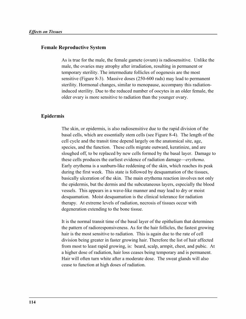
Effects on Tissues
114
Female Reproductive System
As is true for the male, the female gamete (ovum) is radiosensitive. Unlike themale, the ovaries may atrophy after irradiation, resulting in permanent ortemporary sterility. The intermediate follicles of oogenesis are the mostsensitive (Figure 8-3). Massive doses (250-600 rads) may lead to permanentsterility. Hormonal changes, similar to menopause, accompany this radiation-induced sterility. Due to the reduced number of oocytes in an older female, theolder ovary is more sensitive to radiation than the younger ovary.
Epidermis
The skin, or epidermis, is also radiosensitive due to the rapid division of thebasal cells, which are essentially stem cells (see Figure 8-4). The length of thecell cycle and the transit time depend largely on the anatomical site, age,species, and the function. These cells migrate outward, keratinize, and aresloughed off, to be replaced by new cells formed by the basal layer. Damage tothese cells produces the earliest evidence of radiation damage—erythema. Early erythema is a sunburn-like reddening of the skin, which reaches its peakduring the first week. This state is followed by desquamation of the tissues,basically ulceration of the skin. The main erythema reaction involves not onlythe epidermis, but the dermis and the subcutaneous layers, especially the bloodvessels. This appears in a wave-like manner and may lead to dry or moistdesquamation. Moist desquamation is the clinical tolerance for radiationtherapy. At extreme levels of radiation, necrosis of tissues occur withdegeneration extending to the bone tissue.
It is the normal transit time of the basal layer of the epithelium that determinesthe pattern of radioresponsiveness. As for the hair follicles, the fastest growinghair is the most sensitive to radiation. This is again due to the rate of celldivision being greater in faster growing hair. Therefore the list of hair affectedfrom most to least rapid growing, is: beard, scalp, armpit, chest, and pubic. Ata higher dose of radiation, hair loss ceases being temporary and is permanent. Hair will often turn white after a moderate dose. The sweat glands will alsocease to function at high doses of radiation.
Lesson 8
115
Fig. 8-4. A cross-section view of the skin. Note the basal cells and hair follicles which are most radiosensitive.
Eye - Cataractogenesis
Of all the eye components, only the lens is radiosensitive. This is due to theturnover of the lens cells. The sensitivity of the lens is species-dependent andage-dependent. Neutrons are particularly effective in forming cataracts. Cataracts result from the opacification of these cells. The average latency periodfor cataracts is 2-3 years.
Effects on Tissues
116
Central Nervous System
This system was once thought extremely radioresistant because of the low rateof cell division of the neurons; however, this false belief was due to the latemanifestation of radiation damage. The spinal cord is much more sensitive thanthe brain. Upon irradiation, the brain suffers from brain edema.
The earlier mode of injury affects white matter through damage to thesupporting glia cells. The later mode of injury is vascular in nature. When thebrain is exposed to high doses of radiation in radiotherapy, there is a risk of latebrain necrosis. Chronic radiation encephalopathy may occur 3-4 months afterradiotherapy. A single very high dose may lead to acutemeningoencephalopathy.
Fetal and Embryonic Tissue
Unlike normal tissues which have a resting or G0 cell population which canreplace damaged cells, the neoplastic tissue of an embryo or fetus is composedof rapidly dividing cells that are extremely radiosensitive. The three differentperiods of intrauterine development—preimplantation, organogenesis, and fetaldevelopment and growth—are each affected differently by radiation.
The classic effects of irradiation during pregnancy are the following:
1. Lethal effects, induced by radiation before or immediately after embryoimplantation or at high doses during the other stages, expressed as prenatalor neonatal death.
2. Malformations during major organogenesis, when the main bodystructures are formed.
3. Growth disturbances, induced at all stages of development but especiallyin the latter part of pregnancy.
4. Carcinogenic effects of radiation are high in utero.
Lesson 8
117
The principal factors of importance in the effects of irradiation in utero are thedose, stage of gestation, and the dose rate.
During preimplantation (0-9 days in man), there are several cell divisionsbefore the fertilized ovum reaches the uterus. The loss of several cells due toirradiation causes death of the organism. Losses at this stage are not detectablein humans. This is the most sensitive stage to lethal effects of radiation. Growthretardation is not observed at this stage, because this is an "all or nothing" effectof radiation due to the small number of and the general nature of the cells.
Organogenesis, running from 10 days to six weeks after fertilization in humans,is the stage in which organs and systems are formed. Radiation administeredearly in this period would affect the nervous system early and the skeletalsystem later in the stage, due to the production of a specific defect during theperiod of organogenesis. Death or malformation can occur from irradiation. This is the most disastrous period for irradiation. The loss of a system, such asgastrointestinal, could be fatal for the organism. If death occurs as a result ofirradiation in this sage, it is likely to be neonatal death, occurring at or about thetime of birth. Embryos also exhibit the greatest intrauterine growth retardation.This is a weight reduction which is due to cell depletion. Therefore, there is anassociation between growth retardation and teratogenesis.
The fetal stage in humans runs from six weeks to term. Higher doses ofradiation are needed to cause death in the fetal stage—smaller head size is acommon result in this stage. Most organs are formed and growth is occurring inthis period. Further information about these effects is given in Table 8-2 andFigure 8-5.
Irradiation in this stage can cause effects on the hematopoietic system, liver,kidney, and developing gonads. Much higher doses of radiation are required tocause lethality in this period. Although irradiation during organogenesis showsthe most temporary growth retardation, irradiation during the fetal stage exhibitsthe largest degree of permanent growth retardation.
According to data from both medical exposure and Hiroshima/Nagasaki, themost common effects of in utero irradiation are microcephaly (sometimescombined with mental retardation), other central nervous system defects, and
Effects on Tissues
118
growth retardation. The highest risk of mental retardation occurs at 8-15 weekswhen the relevant tissue, the brain cortex, is being formed. Cells killed beforeeight weeks of gestation can cause small head size without mental retardation.The malformation of body structures, which is expected due to irradiationduring organogenesis, is rare in the Japanese survivors.
A great deal of data concerning radiation effects have come from X-rayexposure in expectant mothers. Exposure to medical radiation has led to thefollowing conclusions:
1. Large doses of radiation (250 rads) at 2-3 weeks of gestation do notproduce severe abnormalities in most children although many embryos areaborted.
2. Irradiation at 4-11 weeks of gestation leads to severe abnormalities ofmajor organs in most children.
3. Irradiation at 11-16 weeks produces stunted growth, microcephaly, mentalretardation, eye, skeletal, and genital organ abnormalities.
4. Irradiation at thirty weeks leads to functional disabilities.
5. Susceptibility to carcinogenic effects of radiation is high during the inutero period. Diagnostic exposure increases the natural cancer incidenceby a factor of 1.5 to 2.
Overall, the maximum permissable dose to the fetus during gestation is5 mSv, with a monthly limit of 0.5 mSv. A dose of 10 rads to the embryo duringthe sensitive period of gestation (10 days - 26 weeks) is regarded as the cut-offpoint, above which a therapeutic abortion may be considered.
The “10-day” rule refers to the fact that X-ray examinations of the lowerabdomen in women should only be carried out in the first ten days following thefirst day of the menstrual cycle. Since 1984, it is believed the risk of radiationinjury could be greater in the seven weeks before the mother becomes pregnantthan in the first two weeks of pregnancy.
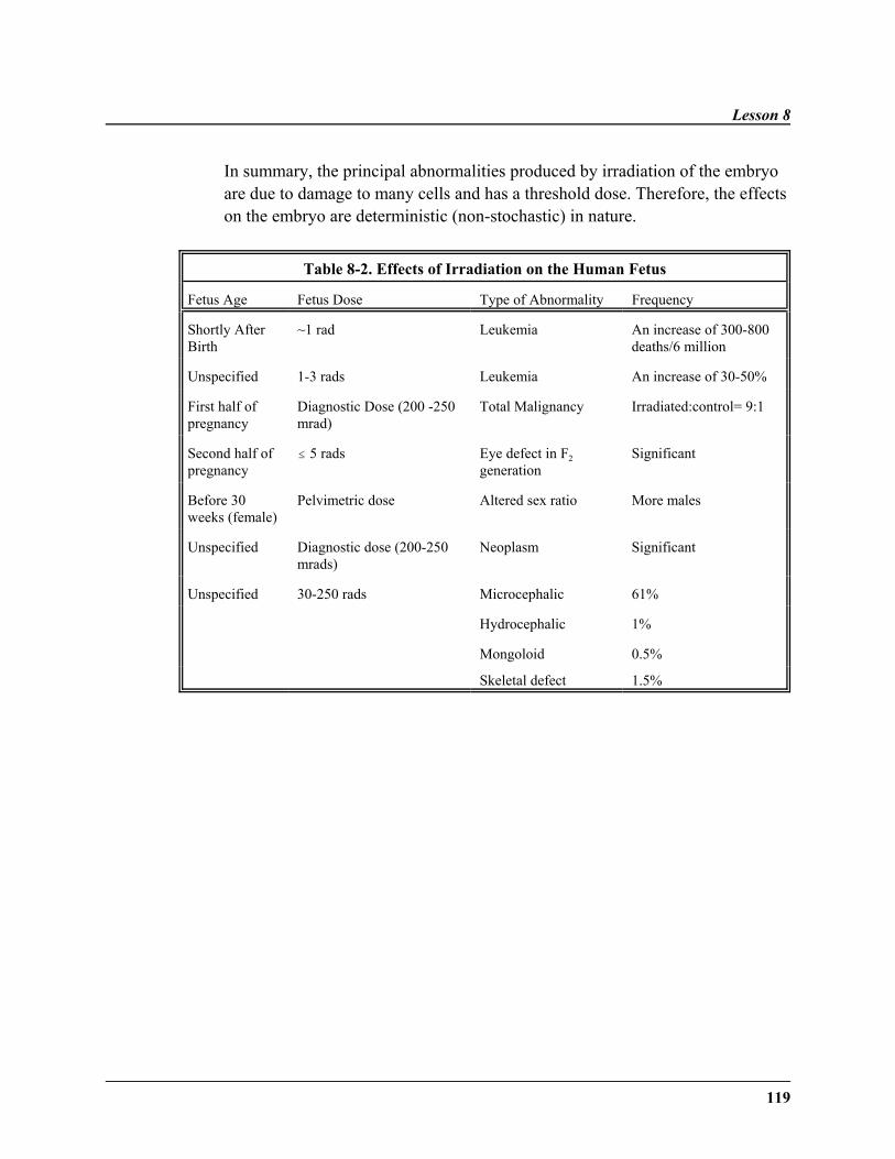
Lesson 8
119
In summary, the principal abnormalities produced by irradiation of the embryoare due to damage to many cells and has a threshold dose. Therefore, the effectson the embryo are deterministic (non-stochastic) in nature.
Table 8-2. Effects of Irradiation on the Human Fetus
Fetus Age Fetus Dose Type of Abnormality Frequency
Shortly AfterBirth
~1 rad Leukemia An increase of 300-800deaths/6 million
Unspecified 1-3 rads Leukemia An increase of 30-50%
First half ofpregnancy
Diagnostic Dose (200 -250mrad)
Total Malignancy Irradiated:control= 9:1
Second half ofpregnancy
# 5 rads Eye defect in F2generation
Significant
Before 30weeks (female)
Pelvimetric dose Altered sex ratio More males
Unspecified Diagnostic dose (200-250mrads)
Neoplasm Significant
Unspecified 30-250 rads Microcephalic 61%
Hydrocephalic 1%
Mongoloid 0.5%
Skeletal defect 1.5%
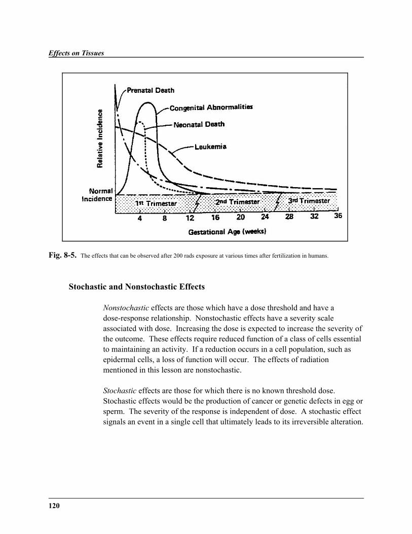
Effects on Tissues
120
Fig. 8-5. The effects that can be observed after 200 rads exposure at various times after fertilization in humans.
Stochastic and Nonstochastic Effects
Nonstochastic effects are those which have a dose threshold and have adose-response relationship. Nonstochastic effects have a severity scaleassociated with dose. Increasing the dose is expected to increase the severity ofthe outcome. These effects require reduced function of a class of cells essentialto maintaining an activity. If a reduction occurs in a cell population, such asepidermal cells, a loss of function will occur. The effects of radiationmentioned in this lesson are nonstochastic.
Stochastic effects are those for which there is no known threshold dose. Stochastic effects would be the production of cancer or genetic defects in egg orsperm. The severity of the response is independent of dose. A stochastic effectsignals an event in a single cell that ultimately leads to its irreversible alteration.
Lesson 8
121
Radiologists look at the effects of radiation as early or late effects. Protractionand fractionation, both discussed in Lesson 7, can affect the appearance of earlyand late effects. The early effects of radiation are based on the proliferation ofcells (division), as evidenced by the effect on testes and intestines.
The esophagus, small intestine, large intestine, and stomach all suffer the samelate nonstochastic effects. Irradiation of the organs leads to stricture of theorgan (stenosis). Stenosis can then lead to blockage of the organ. Fiberatrophyof the tissues also occurs. These late effects are due not to interference withdivision but to changes in the microvasculature of the cell or cellular DNA.
The liver is radioresistant due to lack of cell division in the mature cells. However, radiation-induced hepatitis can occur. Irradiation of the kidneysresults in arteriolar nephrosclerosis. Nonstochastic late effects on the lungsincludes radiation-induced pneumonitis. Whereas little is known about theeffect on the brain, irradiation of the spinal cord can lead to paralysis. Irradiation of the lens of the eye results in cataracts.
As for stochastic effects, the primary early effect is acute radiation syndrome. The primary importance of late stochastic effects lies in radiation-inducedmalignancy and genetic effects. Both of these effects, early and late, will bestudied in the next lesson.
Summary
˜ The effects of radiation on a tissue are due primarily to the rate of divisionof the cells making up the tissue.
˜ The effects of radiation on the sperm and ovaries are especially importantbecause of the possibility of effects on future generations due to geneticmutations.
˜ Radiation has an aging effect on kidneys and eyes.
˜ The effect of radiation on the skin can be used to estimate the effects ofradiation on the body.
Effects on Tissues
122
WRITING ASSIGNMENT - Lesson 8Complete and submit the following assignment.
˜ Embryonic tissue is extremely radiosensitive due to the increased rate ofcell division.
˜ Irradiation of the embryo can lead to death, organ malformation, or growthdeficits.
˜ Stochastic and nonstochastic effects differ in dose threshold and dose-response relationship.
Vocabulary
Please write the term and its definition on your paper. Your definition shouldbe in your own words and limited to one or two sentences.
stochastic effectsnonstochastic effectscell cyclespermatogenesisorganogensiserythemawet desquamation
stenosistissueorgangrowth fractionHewitt assayspleen colony assaygastrointestinal crypt assay
pre-implantation perioddry desquamationarteriolar nephrosclerosisnominal standard dosefiberatrophyavalancheten-day rulemicrocephaly
Questions
All of the questions below are short-essay. Please try to confine your responseto each question to one page or less.
1. What single principle determines the early effects of radiation?
2. What type of cells are most radiosensitive? least sensitive? Whatdetermines the radiosensitivity of the cell?
Lesson 8
123
3. What is important about erythema and wet desquamation?
4. How are the stomach and intestines affected by radiation?
5. What stages of spermatogenesis are most sensitive? Why?
6. Why does temporary sterility in men take two months after irradiation todevelop?
7. Explain what radiation does to each of the three stages of embryonic andfetal development.
8. Why does anemia develop after irradiation?
9. Explain the importance of the Hewitt assay and the spleen-colony assay.
10. Explain the role of radioresponsiveness of tissue in radiotherapy.
11. What is the effect of radiation on the hematopoietic system?
12. What is the effect of radiation on the nervous system?
13. Explain the statement “Radiation has an aging effect on the body.” Citetwo examples of this “aging effect.”
14. What are the three classic effects of radiation on the developing embryo?
15. What were the principal effects on embryos irradiated during the atomicbombings of Hiroshima and Nagasaki?
125
˜ Preview ˜READING ASSIGNMENT
Hall: Chapters 8 (pp. 129-135),10 (all), 17 (pp. 288-296 and 306-308), and 19 (pp. 339-355)
Bushong: Chapters 36 (pp. 517-520) and 37 (all)
Nias: Chapters 14 and 15
LESSON OBJECTIVES
By the end of this lesson, youshould be able to:
˜ Define the terms LD50/30 anddoubling dose.
˜ Name and explain the threestages of acute radiationsyndrome.
˜ Explain the general prognosticsigns for acute radiationexposure.
(continued)
LESSON 9Organs and Organisms
DISCUSSION
Overview
This lesson will primarily focus on the stochasticeffect of radiation on a specific organ and the overalleffect on the organism. Early and late effects(malignancy and genetic problems) will be explored. The effects of radiation on the organs and theorganism will be addressed using examples frommedicine and various occupations. Carcinogenesis isa late stochastic effect, as compared to the latenonstochastic effects on tissues described in Lesson 8. Various theories of carcinogenesis will be examinedin the context of ionizing radiation. The effects to bestudied are non-threshold, non-dose-responserelationships.
Early Stochastic Effects
The most devastating human response to radiation isdeath, which occurs at a minimum dose of 100 radsentered over the whole body. Acute radiationlethality is an early effect of radiation not commonly encountered in diagnostic radiology. Acute radiationlethality is measured by the LD50/30 in humans andanimals. The LD50/30 is the dose of radiation to theentire body which will result in death within 30 daysto 50% of the subjects irradiated.
Organs and Organisms
126
˜ Discuss the three-stage processof clonal theory.
˜ Explain Birenbaum's initiation-promotion hypothesis.
˜ Describe specific effects ofoccupational radiationexposure.
˜ Discuss specific effects ofmedical radiation exposure.
˜ Discuss specific effects ofnuclear weapon exposure.
˜ Explain the late radiation effectof genetic mutation.
˜ Describe the basis of thesymptoms for acute radiationsyndrome.
˜ Discuss the study of Furth &Furth on carcinogenesis.
˜ Describe oncogenes and theirrole in cancer.
˜ Describe the effects of ionizingradiation on cancer.
The LD50 is the dose of any agent or material thatcauses a mortality of 50% in the experimental groupswithin a specified period of time. The LD50 is aconvenient way to smooth out the problems ofheterogeneous populations, by reducing the skewingeffects due to extreme subjects. The peak incidenceof death for humans occurs from days 30-60.Therefore, LD50/60 is more commonly used forhumans, rather than the LD50/30 which is more oftenused with animals. However, the LD50/30 is the mostconvenient form of the expression.
The best estimate for LD50/60 for humans is 3.25 Gy or325 rem for young, healthy adults. This reflects theobservation that larger species are more susceptible tohemopoietic damage than are smaller species. As thewhole-body irradiation dose increases, the averagetime between exposure and death decreases. Thistime is known as the mean survival time. In dosesabove 500 rem, death from hemopoietic syndrome 3-4 weeks later is common. Survival may be possibleif infection, bleeding, or physical trauma are avoidedduring the blood cell nadir. Bone marrow transplantshave been used to try to “rescue” patients who havereceived a dose between 800-1000 rads.
Acute Radiation Syndrome
The early stochastic effects of radiation are found inacute radiation syndrome. Early radiation lethality isdefined as death occurring within a few weeks thatcan be attributed to a specific high-intensity exposureto radiation. Thirty people died of acute radiationexposure at Chernobyl in April 1986. In fact, much
of the recent information on dose levels were obtained by the Chernobylaccident in 1986. The syndrome consists of three stages—prodromal stage,latent period, and manifest illness stage.
Lesson 9
127
The nature of radiation lethality, the timing of death, and whether death occursat all depends upon various cell populations, specifically their depletion andtheir ability to recover to a normal level. It is the balance between the gradualremoval of the aging mature cells and the arrival of the new mature cells thatwill determine whether the tissue remains intact enough for continued integrityof the tissue and the life of the affected human.
The prodromal stage, occurring from a few hours after the irradiation to day oneor two, consists of acute symptoms, which last for a limited period of time. This period can extend for four weeks. The various symptoms of this syndromevary with respect to the time of onset, maximum severity, and duration,depending on the size of dose. Often a severe prodromal response indicates apoor clinical prognosis. Symptoms include both gastrointestinal andneuromuscular symptoms.
During the latent period, the patient will be free of visible effect; however, thelethal effects of the manifest illness stage are developing even though thehuman appears symptom free.
During the manifest illness stage, the clinical signs and symptoms occur. Thereis a decrease in red blood cells, white blood cells, and platelets. This is due tothe radiation's effect on the precursor blood cells. The gastrointestinalsymptoms are nausea, vomiting, and diarrhea. The diarrhea and dehydration aredue in part to the destruction of the villus cells. Neuromuscular symptomsinclude apathy, sweating, fever, and headache.
These symptoms demonstrate how problems with the division of cells in thetissue can lead to the illness of the organism. It is due to the different cellularresponses that gastrointestinal syndrome occurs before the hemopoieticsyndrome. Since hemopoietic stem cells are more radiosensitive than gut cells,survivors of the gastrointestinal syndrome may still die from the hemopoieticsyndrome.
Organs and Organisms
128
Syndromes of Acute Radiation Exposure
There are three dose-related syndromes in acute radiation sickness: hematologic syndrome, gastrointestinal syndrome, and central nervous systemsyndrome.
The hematopoietic syndrome appears at a radiation level of 300-900 rads. Thesymptoms of this syndrome occur later because mature circulating cells die offand are not then re-supplied by the destroyed precursor population.
The first phase of the hemopoietic phase is due to the lesions of the blood-forming organs, leading to hemorrhage and anemia. The bone marrow, spleen,and lymph nodes atrophy. During 5-6 weeks, infection is important due todestruction of lymphocytes and granulocytes, lack of antibody production, andulcerations permitting bacteria entry. If the patient survives, convalescencebegins after two months.
The gastrointestinal syndrome arises at the higher dose of 500-1200 rads. Thissyndrome may result in death in 5-8 days after irradiation. This syndromepredominates at low whole-body doses, in particular doses to the abdomen. Thesymptoms include nausea, vomiting, prolonged diarrhea, dehydration, weightloss, and complete exhaustion. Death is due to destruction of the functioningcells of the villi in the small intestine. Loss of the intestinal barrier leads to abacterial invasion of the bloodstream and the peritoneal cavity, release ofproteolytic enzymes, and the loss of water and electrolytes. Symptoms ofgastrointestinal origin may disappear after 2-3 days and recur by the fifth daydue to loss of the intestinal cells. At this time, peristalsis will end and severeparalytic ileus may occur. Circulation failure, coma, and death may follow.However, some victims may survive this phase due to fluid replacement therapyand antibiotic treatment.
The slowly-dividing cells of the central nervous system are not affected untilthe immense dose of 10 K-100 K rads. The cerebrovascular syndrome results indeath 24-48 hours after exposure. The modes of death in the gastrointestinal andhemopoietic syndromes are due to the depletion of stem cells of a critical self-renewing tissue. The exact cause of death in the cerebrovascular syndrome isunclear. Some believe neuronal damage secondary to vascular damage, edema,and increased intracranial pressure may cause the symptoms.
Lesson 9
129
At doses which bring on this syndrome, all three syndromes will occur givenenough time; however, the cerebrovascular damage brings death very quicklybefore the other syndromes are observed. This symptoms again include the lossof movement, respiratory distress, disorientation, seizures, and coma. Refer toTable 9-1 and Figure 9-1 for a summary of information on the three syndromes.
Prognosis
In general, prodromal nausea and vomiting are more prolonged at high doses.Early CNS symptoms indicate a very high and lethal dose. The longer anindividual survives after the first 2-3 weeks, the better the prognosis. Thoseshowing the CNS syndrome will die. Those with continual nausea, vomiting,and diarrhea will probably die. Those with brief nausea and vomiting willprobably survive.
The very young and the very old are more radiosensitive than the middle-agedand young adults. Females are also more radioresistant than males. The threesyndromes of acute radiation syndrome require different levels of radiation fortheir emergence.
The lymphocyte count is valuable as a criterion for judging radiation injury. Thetotal white cell count is of particular value for following the patient throughoutthe course of the syndrome.
When only the lower body is irradiated, a higher dose is tolerated and thegastrointestinal tract is the primary target. When only the upper body isirradiated, an even higher dose is tolerated, and the bone marrow syndrome ismore common. Survival is more likely if the radiation dose is protracted and ifonly part of the body is exposed. When a very large volume of tissue isirradiated, damage will be evident after a relatively low dose.
(continued)
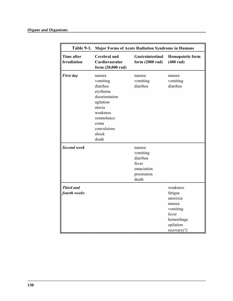
Organs and Organisms
130
Table 9-1. Major Forms of Acute Radiation Syndrome in Humans
Time afterIrradiation
Cerebral andCardiovascularform (20,000 rad)
Gastrointestinalform (2000 rad)
Hemopoietic form(400 rad)
First day nauseavomitingdiarrheaerythemadisorientationagitationataxiaweaknesssomnolencecomaconvulsionsshockdeath
nauseavomitingdiarrhea
nauseavomitingdiarrhea
Second week nauseavomitingdiarrheafeveremaciationprostration death
Third andfourth weeks
weaknessfatigueanorexianauseavomitingfeverhemorrhageepilationrecovery(?)
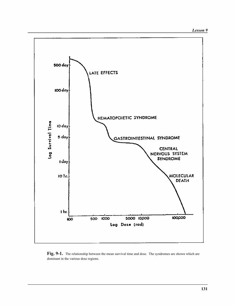
Lesson 9
131
Fig. 9-1. The relationship between the mean survival time and dose. The syndromes are shown which aredominant in the various dose regions.
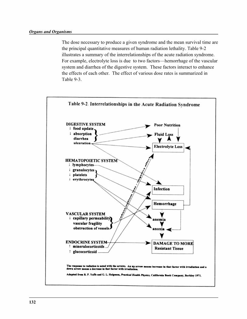
Organs and Organisms
132
The dose necessary to produce a given syndrome and the mean survival time arethe principal quantitative measures of human radiation lethality. Table 9-2illustrates a summary of the interrelationships of the acute radiation syndrome. For example, electrolyte loss is due to two factors—hemorrhage of the vascularsystem and diarrhea of the digestive system. These factors interact to enhancethe effects of each other. The effect of various dose rates is summarized inTable 9-3.
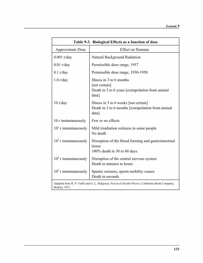
Lesson 9
133
Table 9-3. Biological Effects as a function of dose
Approximate Dose Effect on Humans
0.001 r/day Natural Background Radiation
0.01 r/day Permissible dose range, 1957
0.1 r/day Permissible dose range, 1930-1950
1.0 r/day Illness in 3 to 6 months [not certain]Death in 3 to 6 years [extrapolation from animaldata]
10 r/day Illness in 3 to 6 weeks [not certain]Death in 3 to 6 months [extrapolation from animaldata]
10 r instantaneously Few or no effects
102 r instantaneously Mild irradiation sickness in some people No death
103 r instantaneously Disruption of the blood forming and gastrointestinaltissue100% death in 30 to 60 days.
104 r instantaneously Disruption of the central nervous systemDeath in minutes to hours
105 r instantaneously Spastic seizures, sperm mobility ceasesDeath in seconds
Adapted from R. P. Yaffe and G. L. Helgeson, Practical Health Physics, California Book Company,Berkley 1971.
Organs and Organisms
134
Late Stochastic Effects - Carcinogenesis
Cancer is a gross distortion of cell behavior caused by numerous gene mutationsand numerous abnormalities in the production and functioning of proteins. Thecellular basis for cancer is discussed in Lesson 3. Cancer is a class of diseases,all pertaining to unlimited cell growth that is potentially fatal. Cancer initiatesfrom a single cell that has been transformed due to a particular change in itsDNA. Ninety percent of human cancers are carcinomas.
A neoplastic cell is hyperresponsive to growth factors, underresponsive togrowth inhibitors, and has an increase in metabolic transport capabilities. Acancer cell tends to have an irregular shape, an abnormally appearing nucleus, ismore mobile, is invasive, and shows genomic instability. In a benign tumor, theneoplasm remains as a well-defined cluster and does not spread to neighboringcells. A malignant tumor is capable of invading surrounding tissues, due todisruption of intracellular adhesion, which enables invasive tumor cells to insertthemselves between cells of surrounding tissue and to migrate. As it spreadsthroughout the body, the tumor can invade and destroy tissue until the organismis so compromised that death results.
Carcinogenesis is the primary late effect of radiation. Unlike early effects whichare due to the radiation's effect on division, the late effects are possibly due tochanges within the cell's DNA or microvasculature. Radiation carcinogenesishas mainly been studied in experimental animals, such as the landmark study byFurth & Furth (1936) on mice. However, carcinogenesis in variousoccupational groups, which will be described later in the lesson, do demonstratethe stochastic nature of cancer. These and other studies have led to adose-response curve (Hall: Figure 10-5, page 152). The graph shows thatincidence vs. dose curve rises sharply for low doses. There is a maximum valueof incidence above which incidences decrease. At this saturation point, thenumber of cells remaining alive to produce a tumor are greatly reduced.
As for general information about radiation-induced carcinogenesis, the timeinterval between irradiation and the appearance of a malignancy is the latentperiod. Leukemia has the shortest latent period. Tumors may not appear for 20-50 years, due to the effects of age of expression.
Lesson 9
135
In general, those exposed at an early age are more susceptible to radiation-induced carcinogenesis than those exposed at older ages. For example, breastcancer is more common in young females exposed to radiation. There areexceptions to the rule. For example, susceptibility to radiation-inducedleukemia is constant throughout life; whereas, susceptibility to respiratorycancer increases in middle age.
The following three sections will examine the prominent theories dealing withthe development of cancer in an organism—oncogenes, cooperating genes, andsuppressor genes.
Clonal Theory/ Oncogenes
Although a virus causes cancer by inserting an oncogene from its own genomeinto that of the cell, the mechanism of transformation by radiation is to causechanges in a normal proto-oncogene native to that cell. These changes cause theproto-oncogene to be activated. The proto-oncogenes are present in every celland may serve to regulate cell growth and differentiation. They act in adominant manner. Cells containing this activated oncogene becometransformed, evidenced by a lack of contact inhibition.
The radiation causes activation of the proto-oncogenes in three ways:
• point mutation,
• rearrangement or translocation of a chromosome which places anoncogene near a promoter sequence,
• gene amplification.
The studies support a three-stage process in the development of cancer due tochanges in the proto-oncogene:
1. Transformation of cell with lack of response to growth controlmechanisms,

Organs and Organisms
136
2. Invasion of transformed cells to surrounding tissues,
3. Migration of cells to other body locations to establish a tumor(metastasis).
This process is known as the clonal theory, which assumes that a singleradiation interaction alters the DNA of a single cell, thereby producing cancer inthe cell.
Studies on animals and observations of human patients have shown that perhapsthe clonal theory is too simplified. Levels of hormones are extremelyinfluential in cancers of endocrine glands (ovaries, pituitary, etc.). Murineleukemias, leukemia in rats and mice, are dependent on viruses. Immunocompetence is an important modifier in the survival of the cancerouscell.
Initiation - Promotion Hypothesis/Cooperating Genes
There is typically a latency, or waiting period, between the radiation event andthe cancer, depending on the type of malignancy. This can be explained byBerenblum's initiation-promotion hypothesis. The first step—initiation—is thestep in which the cell is altered or activated. During the second step—promotion—the exposure of the cell to various agents leads to the expression ofthe cancer.
Since the two steps of initiation and promotion are involved in carcinogenesis,two cooperating oncogenes are needed for expression of the malignantphenotype. For example, the combination of myc and ras might be required forcancer to develop. Myc oncogenes confer immortality, whereas ras oncogenesconfer loss of contact inhibition.
Suppressor Genes Theory
Normal cells contain a gene to suppress the neoplastic potential of tumor cells.This suppressor gene may be located on chromosome 11. Mutation of thesuppressor gene p53 may result in cancers of the lung, esophagus, breast, liver,
Lesson 9
137
and brain. Loss of the protein p105Rb may lead to retinoblastoma. Somatichomozygosity may also result in an increased incidence of retinoblastoma,colon cancer, etc. Most suppressor genes are recessive.
Other Theories of Carcinogenesis
There are two other theories of carcinogenesis which deserve note. Thesomatic-mutation hypothesis deals with the damage of the cell due to radiation. However, the viral hypothesis states the possibility of a virus weakening thecell, thereby leading to vulnerability for cancer development. Another similartheory views a virus as the direct cause of the cancer.
Effect of Ionizing Radiation on Cancer
This section will discuss the actual effects of ionizing radiation on tumorsuppressor genes and oncogenes. Ionizing radiation is an activator ofoncogenes and an inactivator of tumor-suppressor genes. Oncogene activationpromotes cellular proliferation, and the checks on this proliferation are removedthrough the inactivation of tumor suppressor genes. Specific oncogenes andtumor suppressor genes have been correlated with specific cancers; therefore,genetic mutations do lead to cancer. More than seventy oncogenes and a dozentumor-suppressor genes have been identified.
The oncogenes are activated by the mutation or amplification of normal proto-oncogenes, which are genes that are part of regulatory pathways that exertinfluence through phosphorylation of target proteins, formation of protein-protein complexes, or regulation of transcription of target genes. Oncogenes aredominant, positive regulators that stimulate cell growth. Oncogenes may beactivated through a variety of cytogenic events, such as large chromosomedeletions, inversions, and translocation. For example, the ras oncogene ismutated in about 30% of all human cancers, including bladder and colon cancer.Human B-cell lymphomas are due to translocation of bcl-2 proto-oncogenes in85% of leukemia cases. The cyclin-D1 oncogene is an amplified geneassociated with parathyroid cancer. The ret oncogene is associated withpapillary adenocarcinomas of the thyroid, the major type of thyroid cancerfound among atomic-bomb survivors, including children affected by theChernobyl accident.

Organs and Organisms
138
The bcr/abl oncogene is strongly linked with a major form of radiation-inducedB-cell leukemia called chronic myelogenous leukemia.
The normal role of tumor suppressor genes, which act in a complementaryfashion with proto-oncogenes, is to inhibit cell growth, with their proteinproducts acting as stops on the cell cycle. Tumor suppressor genes may beinactivated through the induction of point mutations, chromosomerearrangements, or the loss of part or all of a chromosome. The p53 gene,involved in the G1 checkpoint, is mutated in over 50% of all human cancers.Mutations of the core domain of the DNA binding region of p53 are correlatedwith human cancer. A mutation of rb can lead to retinoblastoma, a rarechildhood cancer of the retina.
Therefore, the ionizing radiation serves to inactivate tumor suppressor genesand to activate proto-oncogenes through the following means. Most singlestrand breaks are repaired without the loss of information and only a slight riskof genetic mutation. Base alterations and basic sites can cause single basechanges known as point mutations, which are easily repaired. Although single-strand breaks, basic sites, and base alterations are induced by both ionizingradiation and normal metabolism, the double strand break is causedpreferentially by ionizing radiation. The risk of double strand breaks is low; forexample, background radiation levels would produce only one double strandbreak per 10 cells per year. Double strand breaks are difficult to repair and mayresult in the loss of genetic information due to the lack of a complementaryDNA strand; however, a process called homologous recombination may act torestore the double strand break.
Occupational Exposure
Organ-specific radiation cancers can be found clustered in various occupations,medical exposures, and environmental hazards. As is shown by Hall in Figures10-5 to 10-8 on pages 152-155, the sensitivity for radiogenic cancers variesgreatly with the organs affected. The two organs now believed to be the mostsensitive for cancer are the female breast and the lung.
Lesson 9
139
Radiology
Radiologists of today do not have a shortened life span; however, they were atmuch greater risk from the 1930s to 1950s before modern procedures existed.Until about 1950, radiologists in the US were observed to have excess cancermortality, especially leukemia, lymphoma, and multiple myelomas, whencompared to practitioners in other medical specialities. According to a survey in1956 of 80,000 obituaries of doctors, the mean age of death of radiologists was60.5 years, as compared to a mean age of 65.7 for doctors not exposed to dailyradiation.
Uranium Miners
One of the decay products of uranium is radon (Rn), which is a gas emanated bythe rocks to the air. Exposure to radon in the mines leads to lung cancer. Radon was inhaled by the miners, depositing atoms of radioactive material intheir lungs. Studies involving the pitchblende miners in Saxony indicate thatalpha radiation produces the tumors in the lungs. The average exposure of theminers is 15-20 years; in addition, it is difficult to separate the effects of radonfrom the effects of cigarette smoking. One study of uranium miners around theworld showed an 80% increase in lung cancer deaths over what was seen inunexposed miners. Radon will be further discussed in Lesson 11.
Radium Dial Painters
In the 1920s and the 1930s, watch dials were painted with luminous paintscontaining radium, which were ingested by the watch workers. This led to bonecancer due to the intense alpha radiation. The radium acted chemically ascalcium and was deposited in the bone. This group of workers had bonesarcomas and carcinomas of the epithelial cells of the sinuses. It was thedangers faced by the radium-dial painters which revealed the danger ofoccupational exposure to radiation. In one study of this population, of 154subjects who received skeletal doses of greater than 20,000 rem, 62 subjectsdeveloped skeletal tumors.
Organs and Organisms
140
Medical Exposure
Radium Injections
Excess cancer was also due to injection of radium, which was widely taken inGermany (1944-1951) for its alleged curative power. The radium was laterdeposited in bone, leading to an incidence of bone sarcoma 280 times that of anunexposed population.
Ankylosing Spondylitis
Ankylosing spondylitis is a disease of the spine. This arthritis-like condition, inwhich patients walk hunched over, was treated by radiation in the 1940s and1950s in Great Britain. There is a higher risk of leukemia in these patients.These patients also had increased malignancy due to non-Hodgkin's lymphomaand cancers of the esophagus, lung, bone, breast, and brain.
Women with Tuberculosis
Women, in both Nova Scotia and New England, who received fluoroscopictherapy for tuberculosis have a higher rate of breast cancer. Patients receivingradiotherapy for postpartum mastitis also showed an excess incidence of breastcancer.
Children with Enlarged Thymus
Thymic enlargement occurred in two populations of children in the 1940s and1950s and was treated with radiation in Ann Arbor, Michigan, and Rochester,New York. Twenty years after irradiation of the thymus, an increase occurredin the rate of thyroid cancer. Both malignant and benign thyroid tumors havebeen observed. Thyroid cancer has also been observed in those with tineacapitis (ringworm of the scalp), a condition once treated with X-rays.
Lesson 9
141
In Utero Diagnosis
The embryo becomes less sensitive to radiation effects as development moveson but is most sensitive during the first trimester (organogenesis). Radiationcauses an increase in childhood malignancy, mental retardation, skeletal andorgan abnormalities, and central nervous system problems, as can be seen inTable 8-2 (previous lesson). Studies indicate a strong link between excessincidence of leukemia and children irradiated in utero.
Nuclear Weapon Detonation
Hiroshima and Nagasaki
Hiroshima and Nagasaki survivors have a statistically significant increase inleukemia, breast, thyroid, and skin cancer. There was a great increase inincidence of leukemia in the residents of these cities after the dropping of thebomb, having its peak incidence five years afterward. Of nearly 300,000people, 100,000 were killed from the blast and its early effects. Of thesurvivers, 100,000 were unaffected, whereas the remainder suffered from lateeffects (leukemia, breast cancer). There was a fifteen percent increase ingeneral mortality of those people who had been within 1200 meters of thehypocenter of the atomic bomb (see Figures 9-2 and 9-3).
The best study on radiation induction of cancer in humans is the Life-SpanStudy of Survivors of the atomic bomb attacks on Hiroshima and Nagasaki,which followed 80,000 individuals over the period 1958-1987. These subjectsrepresent all ages, both sexes, and a wide range of doses. Of this population,only 0.35% (500 people) died from radiation-induced solid cancers. Likewise,only 0.087% (75 people) have developed radiation-induced leukemia.Statistically significant non-carcinogenic prenatal effects have been observed,including severe mental retardation, small head size, and low intelligencescores. The IQ downward shift in embryos, during 8-15 weeks after conception,is estimated to be approximately 30 IQ points per 100 rem.
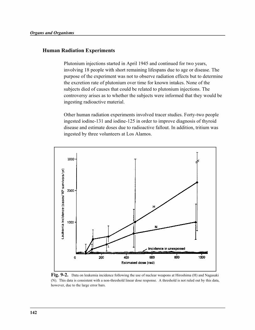
Organs and Organisms
142
Human Radiation Experiments
Plutonium injections started in April 1945 and continued for two years,involving 18 people with short remaining lifespans due to age or disease. Thepurpose of the experiment was not to observe radiation effects but to determinethe excretion rate of plutonium over time for known intakes. None of thesubjects died of causes that could be related to plutonium injections. Thecontroversy arises as to whether the subjects were informed that they would beingesting radioactive material.
Other human radiation experiments involved tracer studies. Forty-two peopleingested iodine-131 and iodine-125 in order to improve diagnosis of thyroiddisease and estimate doses due to radioactive fallout. In addition, tritium wasingested by three volunteers at Los Alamos.
Fig. 9-2. Data on leukemia incidence following the use of nuclear weapons at Hiroshima (H) and Nagasaki(N). This data is consistent with a non-threshold linear dose response. A threshold is not ruled out by this data,however, due to the large error bars.
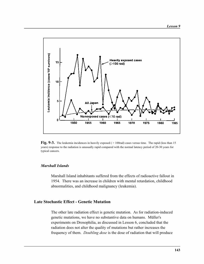
Lesson 9
143
Fig. 9-3. The leukemia incidences in heavily exposed ( > 100rad) cases versus time. The rapid (less than 15years) response to the radiation is unusually rapid compared with the normal latency period of 20-30 years fortypical cancers.
Marshall Islands
Marshall Island inhabitants suffered from the effects of radioactive fallout in1954. There was an increase in children with mental retardation, childhoodabnormalities, and childhood malignancy (leukemia).
Late Stochastic Effect - Genetic Mutation
The other late radiation effect is genetic mutation. As for radiation-inducedgenetic mutations, we have no substantive data on humans. Müller'sexperiments on Drosophilia, as discussed in Lesson 6, concluded that theradiation does not alter the quality of mutations but rather increases thefrequency of them. Doubling dose is the dose of radiation that will produce
Organs and Organisms
144
twice the frequency of genetic mutations as would have been observed withoutthe radiation. Data from mice leads to the conclusion that mammals have somecapacity to repair genetic damage. Most radiation-induced mutations arerecessive, meaning they must be present in both male and female to produce thetrait as discussed in Lesson 6.
Summary
In summary, this lesson has looked at the stochastic effects of radiation. Acuteradiation exposure involves the appearance of three syndromes—cardiovascular, gastrointestinal, and cerebrovascular. The late stochastic effectof cancer may be mediated by oncogenes, tumor suppressor genes, cooperatinggenes, and viruses, all of which are affected by ionizing radiation. Occupationalexposure and the events of Hiroshima and Nagasaki deal with carcinogenesis invarious organs. Another prominent late stochastic effect is genetic mutation.
˜ Acute radiation sickness is an early stochastic effect.
˜ Carcinogenesis and genetic mutations are a late stochastic effect.
˜ Stochastic effects are non-threshold, dose-response relationships.
˜ Occupational exposure has led to many late stochastic effects in people,such as lung cancer, bone cancer, and leukemia.
˜ The three main syndromes of acute radiation exposure arecerebrovascular, gastrointestinal, and cardiovascular.
˜ According to the oncogene theory, the ras oncogene is mutated in 30% ofall human cancers.
˜ According to the tumor suppressor theory, the p53 gene is mutated in 50%of all cancers.
˜ The events of Hiroshima and Nagasaki are among the primary sources ofinformation for radiation exposure.
Lesson 9
145
WRITING ASSIGNMENT - Lesson 9Complete and submit the following assignment.
Vocabulary
Please write the term and its definition on your paper. Your definition shouldbe in your own words and limited to one or two sentences.
LD50/30
acute radiation syndromecarcinogenesisprodromal stagelatent stageclonal theory
metastasisinitiation-promotionankylosing spondylitisrecessive traitsdoubling dosemurine leukemiaproto-oncogene
oncogenetumor-suppressor genep53 generas oncogenecancerbenign tumorLife-Span Study
Questions
Answer each of the following short-answer essay questions in one page or less.
1. What is the course of illness for acute radiation syndrome?
2. What is the basis of acute radiation syndrome?
3. What is the basis for late stochastic effects? What are these effects?
4. What are the three stages in the clonal theory?
5. What are some problems with the clonal theory?

Organs and Organisms
146
6. What organs are affected by radiation cancers in the followingpopulations?
a. radium dial paintersb. people with ankylosing spondylitisc. uranium mine workers
7. What were the effects of radiation exposure on the inhabitants ofHiroshima, Nagasaki, and the Marshall Islands?
8. What is the importance of nominal standard dose in the field of radiology?
9. What is the main difference between mutations due to radiation and thosedue to natural effects?
10. Why is a victim not cured if he has survived for ten days after radiationexposure?
11. What are some general guidelines for the prognosis of those experiencingextreme radiation exposure?
12. What is the danger in the latent period?
13. Why are infectious organisms so destructive in the hematopoieticsyndrome?
14. How can ionizing radiation lead to an increase in cancer, according to thepopular cancer theories?
15. In what ways has radiation resulting from medical exposure contributed tocancer deaths?
16. What is the controversy surrounding the plutonium experiments andnuclear fallout?
17. What is the danger of nuclear fallout?
147
˜ Preview ˜READING ASSIGNMENT
Hall: Chapters 14 and 15
Bushong: Chapters 36 (pp. 517-520) and 37 (all)
Nias: Chapters 18, 19, and 20
LESSON OBJECTIVES
By the end of this lesson, youshould be able to:
˜ List the relative contribution ofradiation sources to naturalbackground radiation.
˜ Define and contrast relativerisk, excess risk and absoluterisk.
(continued)
LESSON 10Risk Estimation; background Radiation:
NATURAL AND ARTIFICIAL
DISCUSSION
Introduction
Work in radiobiology has focused on the dose responseof biological systems to radiation. Guidelines forexposure limits have been formulated from variouscommittees, using various risk models. These efforts areintended to improve therapeutic radiology proceduresand to determine the response to low-level radiation. This lesson will focus on estimating risk and on thesources of background radiation, both artificial andnatural. Various sources of background radiation, suchas cosmic rays and even ceramic cookware, will beintroduced. You will also have an opportunity todetermine your personal background radiation level.
An Organism's Response to Radiation
The response of an organism to radiation (the doseresponse) can have several shapes. The doseresponse can be linear or nonlinear and threshold ornon-threshold. To observe effects due to lowdosages, very large numbers of animals need to beirradiated. This is necessary to obtain reasonablestatistics on these rare events. Usually, the estimate
Risk Estimation; Background Radiation: Natural and Artificial
148
˜ Discuss the three sources ofnatural background radiation.
˜ Explain the concept ofexposure limits.
˜ Explain the importance ofthe radon problem.
˜ Discuss the three sources ofartificial radiation.
˜ Discuss the complexities ofradiation used in medicalexposure.
˜ Explain the importance ofdoubling dose.
˜ Explain the role ofgeography in externalterrestrial radiation.
of radiation effects at low dose is based on studies oflarge numbers of animals at higher doses andextrapolated to the low dose. For this extrapolation, alinear, non-threshold mode is used to estimate theresponse at lower doses. Recent work by thecommittee on Biological Effects of IonizingRadiation (BEIR) has found that after reviewing theliterature extensively, a quadratic non-thresholdbehavior described the data better than a linear, non-threshold response at low energies. However, sincethe linear, non-threshold response is a conservativeestimate of the response, this has been kept as thestandard for estimation of risk at low doses.
Risk Models
There are three models which deal with risk due toradiation exposure—absolute risk, relative risk, andtime-dependent relative risk models.
In the absolute risk model, radiation induces a group of cancers above and beyond thenatural incidence
and is unrelated to it. In the relative risk model, the effect of radiation is toincrease the natural incidence of cancer at all ages subsequent to exposure bya given factor. This model predicts a large excess number of cancersappearing late in life after irradiation. In the time-dependent relative-riskmodel, it is assumed that the excess incidence of cancer is a function of dose,(dose)2, age at exposure, and time since exposure. This model has been usefulin understanding epidemiology data from such events as the Hiroshima andNagasaki atomic bomb exposures and the exposures from the Chernobyldisaster.
The various models are expressed in formulas that allow for estimation ofrisk. For example, relative risk is defined as the ratio of observed cases to theexpected cases:
Lesson 10
149
The relative risk is computed by comparing the number of persons in theexposed population showing a late effect with the number who developed thesame late effect in an unexposed population. A relative risk of 1 indicates norisk at all. A relative risk of 1.75 would indicate the frequency of the lateresponse is 75% higher in the irradiated population. A relative risk of lessthan 1 indicates a protective benefit.
EXAMPLE 1:
In a study of radiation-induced leukemia following diagnostic levels ofradiation, 237 cases were observed in 100,000 people. The normal incidenceof leukemia in the United States is 150 cases per 100,000. What is the relativerisk of the radiation-induced leukemia?
Answer: Relative Risk = Observed/Expected= 237/150=1.58 (i.e., 58% higher in irradiated population)
Excess risk can also be calculated. Excess risk is the difference between theobserved cases and the expected cases:
The excess cases found are assumed to be radiation induced.
EXAMPLE 2:
Forty cases of skin cancer were observed in a population of 1000 radiologists.The incidence in the general population is 0.5/100,000. How many excessskin cancers were produced in this population?
Answer: Excess Cases = Observed - Expected= 40 - .005= 40 (All 40 represent risk)
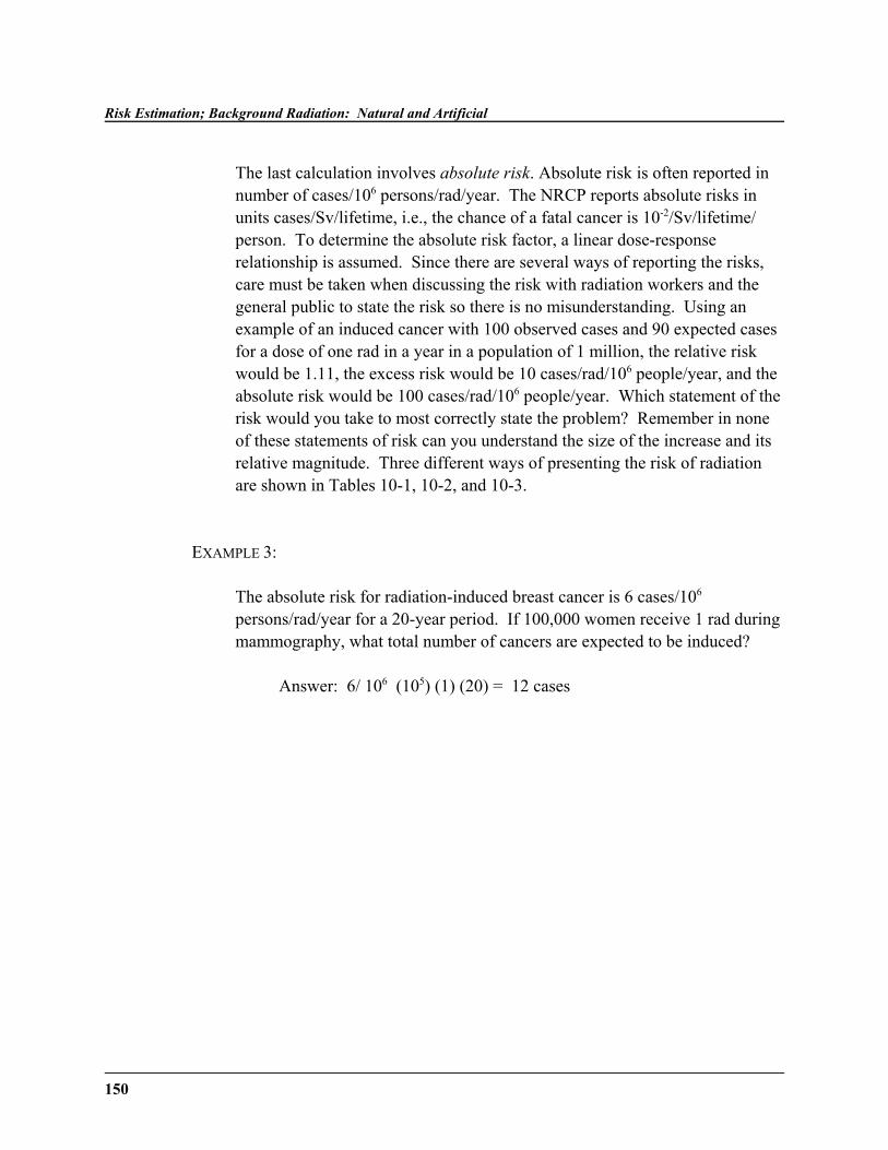
Risk Estimation; Background Radiation: Natural and Artificial
150
The last calculation involves absolute risk. Absolute risk is often reported innumber of cases/106 persons/rad/year. The NRCP reports absolute risks inunits cases/Sv/lifetime, i.e., the chance of a fatal cancer is 10-2/Sv/lifetime/person. To determine the absolute risk factor, a linear dose-responserelationship is assumed. Since there are several ways of reporting the risks,care must be taken when discussing the risk with radiation workers and thegeneral public to state the risk so there is no misunderstanding. Using anexample of an induced cancer with 100 observed cases and 90 expected casesfor a dose of one rad in a year in a population of 1 million, the relative riskwould be 1.11, the excess risk would be 10 cases/rad/106 people/year, and theabsolute risk would be 100 cases/rad/106 people/year. Which statement of therisk would you take to most correctly state the problem? Remember in noneof these statements of risk can you understand the size of the increase and itsrelative magnitude. Three different ways of presenting the risk of radiationare shown in Tables 10-1, 10-2, and 10-3.
EXAMPLE 3:
The absolute risk for radiation-induced breast cancer is 6 cases/106
persons/rad/year for a 20-year period. If 100,000 women receive 1 rad duringmammography, what total number of cancers are expected to be induced?
Answer: 6/ 106 (105) (1) (20) = 12 cases
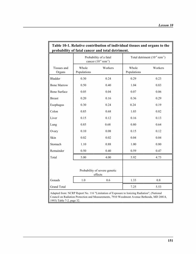
Lesson 10
151
Table 10-1. Relative contribution of individual tissues and organs to theprobability of fatal cancer and total detriment.
Probability of a fatalcancer (10-4 rem-1)
Total detriment (10-4 rem-1)
Tissues andOrgans
WholePopulations
Workers WholePopulations
Workers
Bladder 0.30 0.24 0.29 0.23
Bone Marrow 0.50 0.40 1.04 0.83
Bone Surface 0.05 0.04 0.07 0.06
Breast 0.20 0.16 0.36 0.29
Esophagus 0.30 0.24 0.24 0.19
Colon 0.85 0.68 1.03 0.82
Liver 0.15 0.12 0.16 0.13
Lung 0.85 0.68 0.80 0.64
Ovary 0.10 0.08 0.15 0.12
Skin 0.02 0.02 0.04 0.04
Stomach 1.10 0.88 1.00 0.80
Remainder 0.50 0.40 0.59 0.47
Total 5.00 4.00 5.92 4.73
Probability of severe geneticeffects
Gonads 1.0 0.6 1.33 0.8
Grand Total 7.25 5.53
Adapted from: NCRP Report No. 116 "Limitation of Exposure to Ionizing Radiation", (NationalCouncil on Radiation Protection and Measurements, 7910 Woodmont Avenue Bethesda, MD 20814,1993) Table 7-2, page 32.
Risk Estimation; Background Radiation: Natural and Artificial
152
Table 10-2. Risk factor consequence in life shortening
Risk Factors of disease, occupation,or other conditions.
Expected days of life lost
Being male rather than female 2800
Heart disease 2100
Being single 2000
One pack of cigarettes a day 1600
Working as a coal miner 1100
Cancer 980
30 pounds overweight 900
Stroke 520
All Accidents 435
Service in Vietnam 400
Motor vehicle accidents 200
Average Occupational Accidents 74
Speed limit increase from 55 to 65 mph 40
Radiation Worker 12
Airplane Crashes 1
Adapted from S.C. Bushong, Radiologic Science for Technologists: Physics, Biology, and Protection,7th ed. ( Elsevier Mosby, St. Louis, 2004) Table 37-2, page 534.

Lesson 10
153
Table 10-3. Yearly Average Risk of Death from Various Causes.
Cause Your chance of dying this year
All causes(all ages) 1 in 100
20 cigarettes per day 1 in 280
Heart disease 1 in 300
Cancer 1 in 520
All Causes (25 year old) 1 in 700
Stroke 1 in 1200
Motor vehicle accident 1 in 4000
Drowning 1 in 30,000
Alcohol (light drinker) 1 in 50,000
Air Travel 1 in 100,000
Radiation, 100 mrad 1 in 100,000
Texas Gulf Coast Hurricane 1 in 4,500,000
Being a rodeo cowboy 1 in 6,200,000
Adapted from S.C. Bushong, Radiologic Science for Technologists: Physics, Biology, and Protection,7th ed. ( Elsevier Mosby, St. Louis, 2004) Table 37-6, page 543.
Exposure Limits
The risk models are used to aid the various regulatory committees in imposinglimits. The earliest limits on radiation were based on preventing the onset ofobvious effects such as skin ulcerations, which appeared after intenseexposures to radiation. Later limits were based on preventing delayed effects,such as cancer. In 1902, the first dose limit of 10 rad was recommended. In1924, a "tolerance dose rate," a dose that could be tolerated indefinitely, wasdetermined. Essentially, early limits were a judgment call based on theabsence of observed biological harm.
Risk Estimation; Background Radiation: Natural and Artificial
154
The ICRP (International Commission on Radiological Protection) andUNSCEAR (United Nations Committee on the Effects of Atomic Radiation),and NCRP ( National Council on Radiation Protection and Measurements)estimate the risk associated with exposure to low levels of radiation andrecommend dose limits for radiation workers and the general public. Theyhave estimated the low-dose/low dose-rate risk by extrapolation of risksobtained from high-dose/high dose-rate atomic bomb survivor data and otherradiation studies. These other studies include radiation accidents,occupational exposures, and medical exposures. There are several means ofobtaining extrapolations:
1. Linear-dose response, no threshold hypothesis (LNT)
This most common model implies that the risk is proportional to dose allthe way down to zero dose. It is calculated by extrapolating the straightline drawn through the high-dose data all the way down to zero. Thismodel is equivalent to a stochastic model of radiation effects. Asmentioned earlier, the LNT is the standard for estimation of risk at lowdoses.
2. Threshold
This model assumes there is some value of dose below which there is noeffect.
3. Sublinear
This model assumes that the effect per unit dose at low doses is less thanat high doses.
4. Superlinear
This model assumes that the effects per unit dose at low dose is greaterthan at high doses.
Lesson 10
155
5. Adaptative Response (Radiation Hormesis)
This model assumes that very low doses have a protective effect on theorganism.
Although all of these models have valid points, the results of most animal andcellular experiments favor the LNT or sublinear hypothesis.
The three organizations use the LNT hypothesis as a conservative basis forestimating risk. The risk factor for doses less than 20 rem or 0.6 rem/hr is setequal to one-half the risk factor for high doses (1.2 x 10!3/rem). Therefore, therisk factor for radiation-induced cancer mortality is 6 x 10!4 per rem for thegeneral population. The risk factors for low dose or low dose-rate radiationexposure is 5 x 10⁻⁴ per rem for the general public and 4 x 10!4 per rem forworkers, both values applied to exposures in excess of natural backgroundlevels.
The annual dose limits recommended by the NCRP are, for occupationalexposures, 5 rem for stochastic effects, 15 rem for the lens of the eye, and 50rem for all other organs. It is recommended that a worker's lifetime effectivedose not exceed one rem multiplied by the worker's age in years.
The most objective method of comparing the risks of radiation with those ofother hazards is to study mortality statistics from equivalent groups in thepopulation. It is recommended that the general population should not receive anannual dose to the whole body greater than 5mSv over a period of thirty years.Basically, for those exposed to radiation as their occupation, the dose is tentimes higher than that for the general population. As a rule, the dose can usuallybe kept within one tenth of the recommended limit. The limits are higher forindividual organs than for the whole body, due to "weighting factors" of theorgan exposed.
The ICRP recommends that the public should not receive an additional dose of1 mSv/year above the background radiation; however, the natural backgroundradiation already amounts to twice this level (2.2mSv/ year). Overall, the mostbasic principle of radiation protection is to keep radiation sources as low asreasonably achievable (ALARA).
Risk Estimation; Background Radiation: Natural and Artificial
156
Latent Period
The calculation of risk is complicated by the fact that most radiation effects donot show themslves immediately. There is always a long latent period betweenthe time of irradiation and the clinical appearance of cancer. The latent period isshorter for leukemia than other cancers, with a minimum period of 4-5 yearsand a mean of eight years. This compares with a minimum of ten years and amean of 15 years for the appearance of other cancers. It is important toremember that the risk of cancer/leukemia from radiation depends not only onthe total dose but on dose rate. High LET radiations are more effective inproducing transformation of cells than low LET radiation.
Doubling Dose
A useful concept in radiobiology is the doubling dose (also discussed in Lesson6) in relation to genetic mutations. The doubling dose is defined as the dose ofradiation required to double the natural incidence of a genetic effect. This isvery useful as it gives an automatic perspective on the process. The U.N.Scientific Committee on the Effects of Atomic Radiation, UNSCEAR, revieweda study of 750,000 live births and concluded that the spontaneous mutation rateis 10.5%. Further, most of the genetic effects are believed by the BiologicalEffects of Ionizing Radiation committee (BEIR) to have a doubling dose of 50to 250 rem. There has not been any observance of statistically significanthereditary health effects of ionizing radiation in humans.
Species Risk
The lesson has focused on the risk for humans. However, the risks andeffectiveness of radiation also varies with other species.
Species which reproduce asexually are more radiation resistant than aresexually reproducing species. Whereas sexual reproduction requires at leasttwo survivors, asexual reproduction can proceed with only one individual.

Lesson 10
157
Short-lived species with a short gestation period are more susceptible than long-lived species, due to rapid cell division in the former. Embryonic, young, andvery old are more radiosensitive. Warm-blooded animals are moreradiosensitive than cold-blooded organisms.
Overall, large herbivorous mammals are the most radiosensitive due to theircharacteristics of being warm-blooded, sexual, and exposed most of the time. The following organisms are ranked in radiosensitivity from greatest toleast—large mammals, smaller mammals, birds, plants, one-celled animals andplants, bacteria, and viruses. Man is ranked among the large mammals inradiosensitivity.
Background Radiation: Natural and Artificial
An average American receives 360 millirems of ionizing radiation every year,300 millirems per year (78%) from natural sources. The dose to which a humanis exposed each year comes from three main sources: natural sources, enhancednatural sources, and man-made sources. 18% percent of radiation comes fromartificial sources, the majority of which is due to medical procedures, while theremaining 4% comes from natural background radiation.
Enhanced natural sources are sources in which human activity has increased thelevel of individual exposure. They are natural in origin and include suchactivities as air travel at high altitudes and phosphate mining.
Man-made exposure is due to X-rays, radiopharmaceuticals in medicine,accidents in nuclear power plants, and atmospheric testing of nuclear weapons.Of these sources, the most significant exposure is medical. The exposure due tofall-out from 1959-1962 never reached more than a small fraction of that frommedical diagnosis. The overall effective dose from medical radiation is less thanhalf that from natural background radiation. These sources will be explored indepth later in the lesson. A personal radiation dose chart is included for you onpages 163-164 so that you can estimate your individual dose from backgroundradiation.
Risk Estimation; Background Radiation: Natural and Artificial
158
Natural Sources
The sources of background radiation are summarized in Table 10-4. Naturalradiation comes from the following sources:
1. cosmic radiation
2. external terrestrial radiation
3. internal terrestrial radiation.
These natural sources affect all of the world's population all the time. Thenatural background annual, world-wide, average whole-body effective dose is238 mrem, a value which varies widely among locations. In contrast, the doseassociated with internal radiation (due mainly to potassium-40) varies much lessfrom person to person. Epidemiological studies have generally failed to find astatistically significant correlation between cancer mortality and levels ofbackground radiation. The hypothetical risk associated with the dose fromnatural background radiation is merely a small fraction of risks faced by allhumans each day, such as car wrecks.
We will now look at the three sources of natural radiation more closely.
Cosmic radiation is due to an ionizing component and neutrons. The primarycosmic rays have the same composition as the atomic abundance in the solarsystem. The energy of the primary cosmic rays can exceed 10 TeV (10,000,000MeV). The primary cosmic ray interacts with the atoms of the upper atmosphereand produces electromagnetic and particulate radiation on earth. The dose rateon the ground varies with altitude, latitude, and longitude. The cosmic rayintensity is least in the equatorial regions and rises towards the poles. Theionizing component consists of protons (75-90%), helium nuclei (10-18%), andions with an atomic number greater than 3 (1-7%).
In addition to the cosmic ray background, there are also high energy X-rays andgamma-rays present (see Figure 10-1). However, the dose from theelectromagnetic radiation in much less than from the particulate radiation.
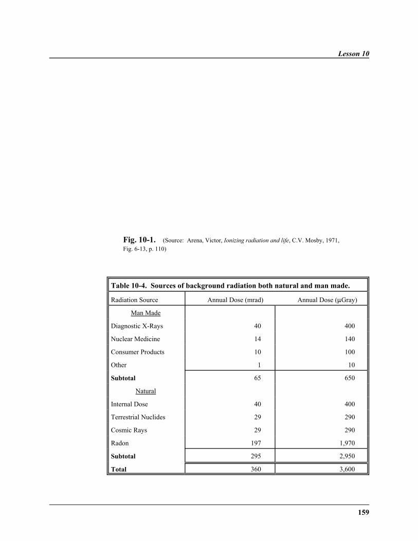
Lesson 10
159
Fig. 10-1. (Source: Arena, Victor, Ionizing radiation and life, C.V. Mosby, 1971,Fig. 6-13, p. 110)
Table 10-4. Sources of background radiation both natural and man made.
Radiation Source Annual Dose (mrad) Annual Dose (:Gray)
Man Made
Diagnostic X-Rays 40 400
Nuclear Medicine 14 140
Consumer Products 10 100
Other 1 10
Subtotal 65 650
Natural
Internal Dose 40 400
Terrestrial Nuclides 29 290
Cosmic Rays 29 290
Radon 197 1,970
Subtotal 295 2,950
Total 360 3,600
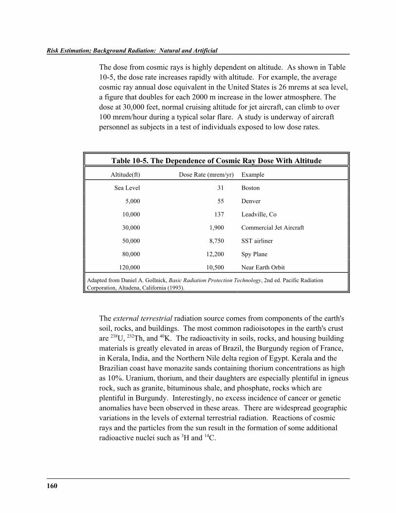
Risk Estimation; Background Radiation: Natural and Artificial
160
The dose from cosmic rays is highly dependent on altitude. As shown in Table10-5, the dose rate increases rapidly with altitude. For example, the averagecosmic ray annual dose equivalent in the United States is 26 mrems at sea level,a figure that doubles for each 2000 m increase in the lower atmosphere. Thedose at 30,000 feet, normal cruising altitude for jet aircraft, can climb to over100 mrem/hour during a typical solar flare. A study is underway of aircraftpersonnel as subjects in a test of individuals exposed to low dose rates.
Table 10-5. The Dependence of Cosmic Ray Dose With Altitude
Altitude(ft) Dose Rate (mrem/yr) Example
Sea Level 31 Boston
5,000 55 Denver
10,000 137 Leadville, Co
30,000 1,900 Commercial Jet Aircraft
50,000 8,750 SST airliner
80,000 12,200 Spy Plane
120,000 10,500 Near Earth Orbit
Adapted from Daniel A. Gollnick, Basic Radiation Protection Technology, 2nd ed. Pacific RadiationCorporation, Altadena, California (1993).
The external terrestrial radiation source comes from components of the earth'ssoil, rocks, and buildings. The most common radioisotopes in the earth's crustare 238U, 232Th, and 40K. The radioactivity in soils, rocks, and housing buildingmaterials is greatly elevated in areas of Brazil, the Burgundy region of France,in Kerala, India, and the Northern Nile delta region of Egypt. Kerala and theBrazilian coast have monazite sands containing thorium concentrations as highas 10%. Uranium, thorium, and their daughters are especially plentiful in igneusrock, such as granite, bituminous shale, and phosphate, rocks which areplentiful in Burgundy. Interestingly, no excess incidence of cancer or geneticanomalies have been observed in these areas. There are widespread geographicvariations in the levels of external terrestrial radiation. Reactions of cosmicrays and the particles from the sun result in the formation of some additionalradioactive nuclei such as 3H and 14C.
Lesson 10
161
Radon
As mentioned previously, radon is the major component in external terrestrialradiation. Recent surveys of homes in the United States have revealed a veryhigh incidence of radioactive radon gas and its daughter products. The largestcontributor to effective dose is radon for non-smokers in the United States.Radon is a chemically inert gas which is formed in several of the natural decaychains which start with thorium or uranium nuclei. Due to the chemicalproperties of radon, it is easily transported to the surface through small cracks inthe rocks from the area where it is produced. During the last two decades, therehas been a shift to improved sealing of houses, which has understandablyexacerbated the problem. The average concentration in the U.S. is 376 mBq/Lin the living area and much more in the basement. The current estimate of anaverage annual dose to the lungs from radon is 197 mrem.
Many variables influence radon levels, including the time of day, the season,geology of the soil, home construction, barometric pressure, humidity, moisturein the soil, and the rate of ventilation in the home. There are two common homeradon measurement devices—charcoal canisters and alpha track monitors.
Water is a major source of radon. Water obtained from surface sources, such aslakes and reservoirs, is low in radon; however, water from wells has highconcentrations of radon gas.
As explained in the previous lesson, radon contaminates many undergroundmines, leading to an increased risk of lung cancer. Radon gas leads toirradiation of the surface of the lungs when the gas and its products are inhaled.The radon decays to polonium, which emits alpha particles, which causes “hotspots” of radiation damage. Six to twelve percent of cases of lung cancer aredue to radon, amounting to 5,000-10,000 cases per year. The main evidencethat exposure to radon increases mortality from lung cancer comes from minersworking underground where there are high concentrations of radon decayproducts. Since lung cancer due to radon is a stochastic effect, the bestprecaution is to provide adequate ventilation in houses.
Risk Estimation; Background Radiation: Natural and Artificial
162
Interestingly, radon data shows a negative correlation between radon and lungcancer up to concentrations of at least 7 picocuries per liter, implying that radonexposure has a hormetic effect at this level.
Internal Terrestrial Radiation
The internal terrestrial exposure is due to the ingestion of small traces ofradioactive materials present in food supplies or inhaled as airborne particles.Radioactive thorium, radium, and lead can be detected in most persons;however, only 40K makes an appreciable contribution. Other radioisotopesinvolved in internal terrestrial exposure include 14C and 226Ra.
The predominant radioactive element in normal foods and human tissues is 40K. It is concentrated particularly in muscle. We also ingest uranium, radium, andthorium in food we eat. 226Ra is chemically similar to calcium and barium, so itpasses easily into the food chain. It is present in all foods, especially cereals andbrazil nuts, which concentrate radium. The excess radium concentrates in theskeleton. On a facetious note, some scientists believe it is best to sleep in twinbeds so as not to receive additional radiation from each other's body.

Lesson 10
163
Personal Radiation Dose Chart
(Adapted from Personal Radiation Dose, American Nuclear Society, 1990with further data from NCRP Report No. 93 and NCRP Report No.95
From Roger Eckhardt, “Ionizing Radiation-It's Everywhere!” Radiation Protection and the Human Radiation Experiments,
Los Alamos Science 23 48 (1995).
Estimate your annual effective dose in millirem by adding the numbers in the right hand column,including the numbers you choose from each category with a blank space.
Where you live:Cosmic Radiation at sea levelFor your elevation in feet:500-1000 ft: 21000-2000 ft: 52000-3000 ft: 93000-4000 ft: 154000-5000 ft: 21
5000-6000 ft: 296000-7000 ft: 407000-8000 ft: 538000-9000 ft: 709000-10000 ft: 107
Terrestrial:Live in a state bordering the Gulf or Atlanticfrom Texas east and north: 23
Live in Colorado Plateau or Rocky MountainState: 29
Live anywhere else in the United States: 46
Internal:What you eat and drinkRadon: Insert a value for your average radonlevel (in picocuries per liter x 100) or use theU.S. average of 200 mrem
(continued)
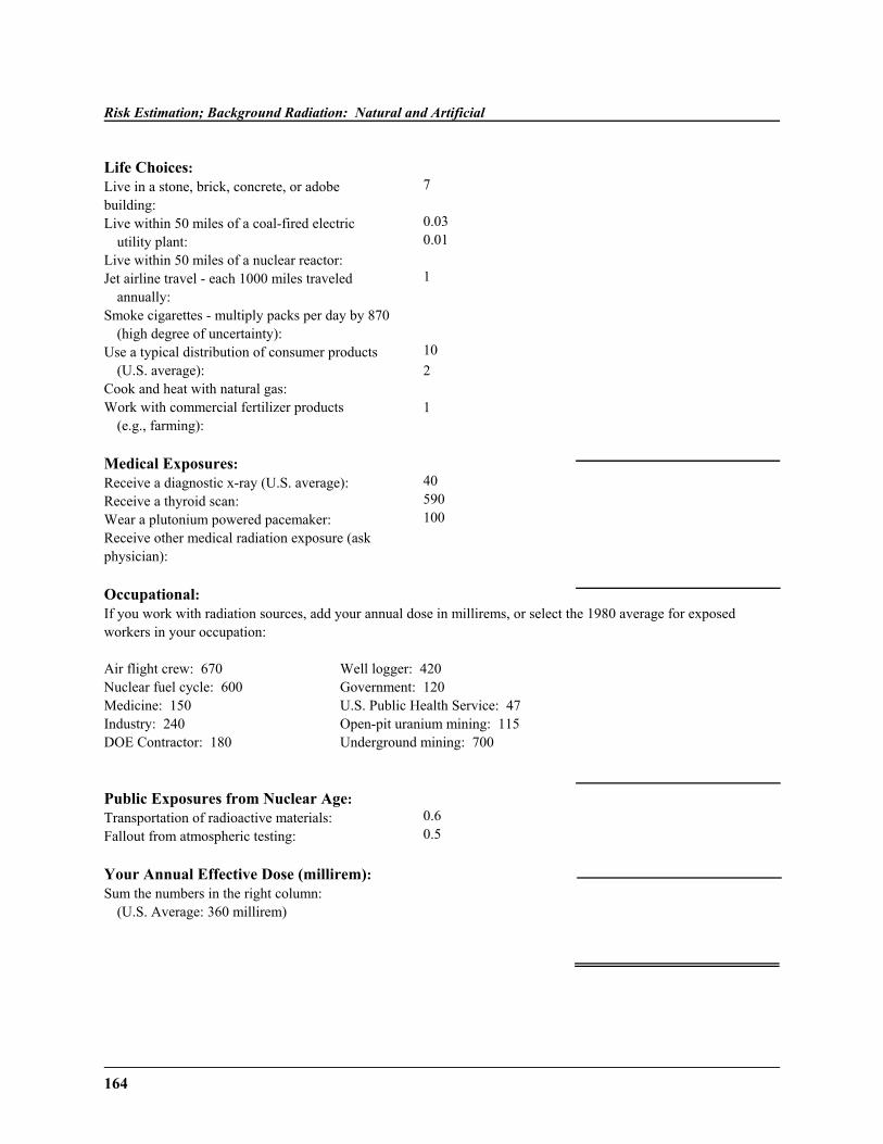
Risk Estimation; Background Radiation: Natural and Artificial
164
Life Choices:Live in a stone, brick, concrete, or adobe building:Live within 50 miles of a coal-fired electric
utility plant:Live within 50 miles of a nuclear reactor:Jet airline travel - each 1000 miles traveled
annually:Smoke cigarettes - multiply packs per day by 870
(high degree of uncertainty):Use a typical distribution of consumer products
(U.S. average):Cook and heat with natural gas:Work with commercial fertilizer products
(e.g., farming):
7
0.030.01
1
102
1
Medical Exposures:Receive a diagnostic x-ray (U.S. average):Receive a thyroid scan:Wear a plutonium powered pacemaker:Receive other medical radiation exposure (askphysician):
40590100
Occupational:If you work with radiation sources, add your annual dose in millirems, or select the 1980 average for exposedworkers in your occupation:
Air flight crew: 670Nuclear fuel cycle: 600Medicine: 150Industry: 240DOE Contractor: 180
Well logger: 420Government: 120U.S. Public Health Service: 47Open-pit uranium mining: 115Underground mining: 700
Public Exposures from Nuclear Age:Transportation of radioactive materials:Fallout from atmospheric testing:
0.60.5
Your Annual Effective Dose (millirem):Sum the numbers in the right column:
(U.S. Average: 360 millirem)
Lesson 10
165
Consumer Products
Many consumer products are radiation sources, such as radioluminoustimepieces (containing radium) and smoke detectors (containing 241Am, an alphasource). The main exposure from smoke detectors is to the workers assemblingthe detector. Static eliminators, which are used to clean negatives and CDs,contain 210Po.
Ceramic dinnnerware is a source of ionizing radiation, due to the glazescontaining uranium oxides and sodium uranite. Dental products, such ascrowns, and opthalmic glasses, especially rose-tinted glasses, containradioactive material.
Tobacco products have an above-average concentration of lead 210Pb and 210Po.Smoking of tobacco leads to large exposures of radiation in the lungs, leading tothe belief that radiation may be one of the major causes of lung cancer forsmokers.
In addition, a masonry home, such as brick, stone, adobe, or concrete, also leadsto an increase in radiation exposure.
Artificial Sources of Radiation
The artificial sources of radiation are due to the following:
1. fallout from nuclear testing
2. medical treatment
3. reactors.
It is important to note that these artificial sources only affect a small fraction ofthe population at any one time.
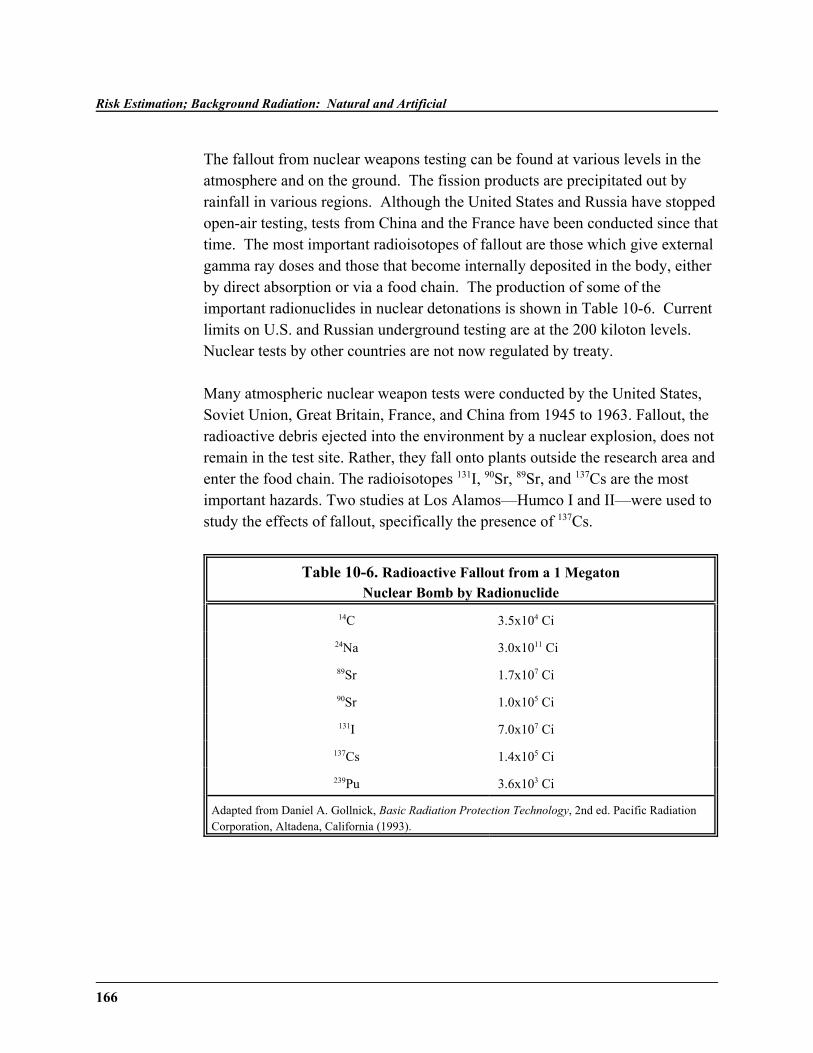
Risk Estimation; Background Radiation: Natural and Artificial
166
The fallout from nuclear weapons testing can be found at various levels in theatmosphere and on the ground. The fission products are precipitated out byrainfall in various regions. Although the United States and Russia have stoppedopen-air testing, tests from China and the France have been conducted since thattime. The most important radioisotopes of fallout are those which give externalgamma ray doses and those that become internally deposited in the body, eitherby direct absorption or via a food chain. The production of some of theimportant radionuclides in nuclear detonations is shown in Table 10-6. Currentlimits on U.S. and Russian underground testing are at the 200 kiloton levels. Nuclear tests by other countries are not now regulated by treaty.
Many atmospheric nuclear weapon tests were conducted by the United States,Soviet Union, Great Britain, France, and China from 1945 to 1963. Fallout, theradioactive debris ejected into the environment by a nuclear explosion, does notremain in the test site. Rather, they fall onto plants outside the research area andenter the food chain. The radioisotopes 131I, 90Sr, 89Sr, and 137Cs are the mostimportant hazards. Two studies at Los Alamos—Humco I and II—were used tostudy the effects of fallout, specifically the presence of 137Cs.
Table 10-6. Radioactive Fallout from a 1 MegatonNuclear Bomb by Radionuclide
14C 3.5x10⁴ Ci
24Na 3.0x10¹¹ Ci
89Sr 1.7x10⁷ Ci
90Sr 1.0x10⁵ Ci
131I 7.0x10⁷ Ci
137Cs 1.4x10⁵ Ci
239Pu 3.6x10³ Ci
Adapted from Daniel A. Gollnick, Basic Radiation Protection Technology, 2nd ed. Pacific RadiationCorporation, Altadena, California (1993).
Lesson 10
167
Chernobyl has been the most recent large source of fallout. The Chernobylaccident occured in Russia in 1986. Thirty-one people died from the accident,and 135,000 people in the region were permanently evacuated. These evacuatedpeople had an average dose of 12 rem, a dose 1500 times greater than that ofpeople near Three Mile Island. Pripjat is a “radiation ghost town.” Three millionacres of agricultural land has been lost due to contamination.
The artificial background dose due to reactors is still quite controversial. Themore accepted estimate is that the dose from the nuclear reactors is less thanone percent of the total background radiation. This is difficult to estimate, dueto uncertainties about the chance of a catastrophic accident which releasesradiation. For instance, the Chernobyl reactor disaster resulted in fallout whichincreased background radiation not only for the local population, but also for alarge part of western Europe.
Occupational Dose
Those who choose to work with ionizing radiation may be exposed to higherdoses than the general public. Most workers in nuclear power stations andmedical radiation are in a low dose category. The higher dose group includesuranium miners, crews of jetcraft, nuclear reactor workers, and nuclear fuelreprocessing workers.
Medical Use
Medical exposure is one of the most significant external man-made sources ofradiation. Medical exposure is primarily due to diagnostic X-ray of the chest,head or other body parts. The therapeutic X-ray delivers much larger doses ofradiation. On average, the dose from diagnostic x-rays is estimated at 23mrem/year (0.23 mSv/year). Nuclear medical diagnostic procedures areestimated to average 14 mrem/yr (140 :Sv/yr). Therapeutic radiation, primarilyto control cancer, can reach up to 6000 rads per patient.
The medical use of radiation involves offsetting a risk against a benefit. Thedetriment in regards to the annual collective dose of persons is the following:
Risk Estimation; Background Radiation: Natural and Artificial
168
4%/Sv fatal cancer0.8%/Sv of nonfatal cancer0.8%/Sv heritable effects (mutations)
This risk must be weighed against the benefit that millions receive fromimproved diagnosis. A dose of one sievert to the population would cause a 5%lifetime risk of fatal cancer.
The two tissues that are dose-limiting for medical radiation procedures are thebone marrow, due to leukaemogenesis, and the gonads, because of geneticeffects. In examinations of the lower trunk, the gonads receive a high dose,unless they are shielded with protective devices.
To summarize, there are three potential hazards resulting from medicalradiation:
1. heritable effects in future generations2. risk of cancer or leukemia in patients themselve3. anomalies or malignancies in irradiated embryos
The dose for medical exposure is not a contributor to the annual dose for anindividual.
As shown by the chart (Bushong, p. 5, Figure 1-3; Hall, p. 203, Figure 14-5;Nias, p. 7, Figure 1-4), the dose from natural sources accounts for 82% of theaverage dose. Radon, by itself, accounts for 55%, being the largest singleradiation source. The cosmic and terrestrial sources account for 11% of thetotal. Internal sources, such as 40K, account for 11%. Manmade sources accountfor 18% of the dose, the largest being 11% from medical X-rays. Consumerproducts account for 4% of the dose, whereas nuclear fallout, nuclear powerplants, and occupational exposure contribute less than 1% of the total dose. These values for the estimated background will vary depending upon the sourceof the dose estimates and the assumptions used in making them. A fewreasonable alternate choices for the personal radiation dose chart will give youan understanding of the variability of the background radiation.
Lesson 10
169
Summary
In summary, Lesson 10 has dealt with the risk models and exposure limits forradiation. It has examined the various sources of background radiation, bothnatural and artificial sources. Sources examined in depth included radon,medical exposure, and fallout. You have had also has an opportunity to put theinformation in perspective by completing the personal radiation dose chart.
˜ Dose response curves can differ for organisms of different species.
˜ Relative risk, excess risk, and absolute risk are all measures for expressingthe risk of an event.
˜ Natural background radiation is due to cosmic rays, external terrestrialradiation, and internal terrestrial radiation.
˜ Artificial sources of radiation are reactors, fallout, and medical treatment.
˜ Using the LNT model of risk, the regulatory committees have calculatedexposure limits.
˜ Radon, which is the primary source of external terrestrial radiation, hasbeen implicated in lung cancer.
˜ Potassium-40 is the main source of internal terrestrial radiation.
˜ The use of radiation in medicine is relatively safe; however, the risks andbenefits must be assessed.
Risk Estimation; Background Radiation: Natural and Artificial
170
WRITING ASSIGNMENT - Lesson 10Complete and submit the following assignment.
Vocabulary
Please write the term and its definition on your paper. Your definition should be inyour own words and limited to one or two sentences.
dose responseBEIRrelative riskexcess riskabsolute riskradon
external terrestrial radiationcosmic raysinternal terrestrial radiationthreshold doseradioactive isotopespotassium-40
latent periodALARAradiation hormesislifetime effective doseenhanced natural sourcestime-dependent relative-risk
model
Problems and Questions
These problems and questions require numerical or short-essay answers. Showyour work and circle your final numerical answers. Limit your short-essayanswers to one page or less.
1. Why are experiments looking for radiation effects normally done at highradiation dose rates rather than at low dose rates?
2. Given: the risk of dying today is 1 in 10,000, the risk of being hit andkilled today if you ride a bicycle is 1 in 5,000, and the risk of dying todayif you wear a safety belt and drive defensively is 1 in 20,000.
What is: the absolute risk of dying today, dying today if you ride a bike,and dying today if you wear a seat belt and drive defensively? What is therelative risk for the same cases? What is the excess risk for the samecases?
Lesson 10
171
3. Using Table 10-3, what is the relative risk of dying in a year assuming thatyou:
a. are 25 years oldb. drink (light drinker)c. will have a Gulf Coast hurricane visit you versus 100 mr of
radiation?
4. How much background radiation do we get from cosmic rays?
5. How much background radiation do we get from medical uses?
6. What are some important isotopes in natural internal radiation?
7. Why is radon present in homes? It wasn't there when they were first built. Where does it come from?
8. Given the dose of 23 mrem/year from medical exposure and using Table10-1, what is the probability of fatal cancers for whole populations forbreast and colon cancer?
9. Given the dose of 6,000 rads from therapeutic radiation, what is theprobability of fatal cancers for stomach, bone marrow, and bone surface?
10. What is the largest source of artificial radiation?
11. Complete the personal dose chart on pages 173-174.
12. What are some of the radiation sources you may be in contact with in yourhome?
13. What is the role of geography in background exposure? How dangerous isbackground exposure?
14. What is the importance of exposure limits? Who decides what the limitsare?
Risk Estimation; Background Radiation: Natural and Artificial
172
15. What are the two dose-limiting tissues in medical exposure to radiation?What are the three potential hazards in medical radiation?
16. Why do uranium miners who smoke have one of the highest cancer ratesof any occupational group?
17. Place the following radiation sources in rank order (largest to smallest): fallout, cosmic sources, radon, nuclear power plants, internal sources,medical exposure.
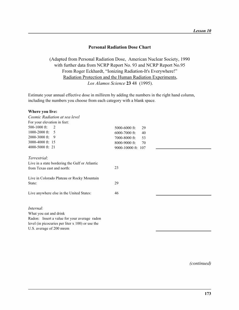
Lesson 10
173
Personal Radiation Dose Chart
(Adapted from Personal Radiation Dose, American Nuclear Society, 1990with further data from NCRP Report No. 93 and NCRP Report No.95
From Roger Eckhardt, “Ionizing Radiation-It's Everywhere!” Radiation Protection and the Human Radiation Experiments,
Los Alamos Science 23 48 (1995).
Estimate your annual effective dose in millirem by adding the numbers in the right hand column,including the numbers you choose from each category with a blank space.
Where you live:Cosmic Radiation at sea levelFor your elevation in feet:500-1000 ft: 21000-2000 ft: 52000-3000 ft: 93000-4000 ft: 154000-5000 ft: 21
5000-6000 ft: 296000-7000 ft: 407000-8000 ft: 538000-9000 ft: 709000-10000 ft: 107
Terrestrial:Live in a state bordering the Gulf or Atlanticfrom Texas east and north: 23
Live in Colorado Plateau or Rocky MountainState: 29
Live anywhere else in the United States: 46
Internal:What you eat and drink Radon: Insert a value for your average radonlevel (in picocuries per liter x 100) or use theU.S. average of 200 mrem
(continued)
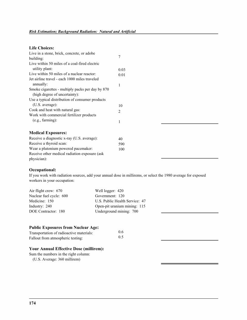
Risk Estimation; Background Radiation: Natural and Artificial
174
Life Choices:Live in a stone, brick, concrete, or adobe building:Live within 50 miles of a coal-fired electric
utility plant:Live within 50 miles of a nuclear reactor:Jet airline travel - each 1000 miles traveled
annually:Smoke cigarettes - multiply packs per day by 870
(high degree of uncertainty):Use a typical distribution of consumer products
(U.S. average):Cook and heat with natural gas:Work with commercial fertilizer products
(e.g., farming):
7
0.030.01
1
102
1
Medical Exposures:Receive a diagnostic x-ray (U.S. average):Receive a thyroid scan:Wear a plutonium powered pacemaker:Receive other medical radiation exposure (askphysician):
40590100
Occupational:If you work with radiation sources, add your annual dose in millirems, or select the 1980 average for exposedworkers in your occupation:
Air flight crew: 670Nuclear fuel cycle: 600Medicine: 150Industry: 240DOE Contractor: 180
Well logger: 420Government: 120U.S. Public Health Service: 47Open-pit uranium mining: 115Underground mining: 700
Public Exposures from Nuclear Age:Transportation of radioactive materials:Fallout from atmospheric testing:
0.60.5
Your Annual Effective Dose (millirem):Sum the numbers in the right column:
(U.S. Average: 360 millirem)
175
˜ Preview ˜READING ASSIGNMENT
Hall: Chapter 14 (pp. 218-224)
Nias: Chapter 18 (pp. 314-320)
LESSON OBJECTIVES
By the end of this lesson, youshould be able to:
˜ Name and explain the threeways in which radionuclidesmay enter the body.
˜ Define biological half-life andexplain the need for thisconcept.
˜ Define discrimination ratio.
˜ Describe importance of tritiumas a radionuclide.
(continued)
LESSON 11Metabolism and Biological Effects of Radionuclide
Uptake
DISCUSSION
Radionuclide Entry into the Body
This lesson will deal with the metabolism ofradionuclides. Radioactive isotopes are unstableatoms that disentegrate randomly with the emission ofionizing radiation, including alpha and beta particles,gamma rays, or a combination of these. It willintroduce the concepts of discrimination ratio,biological half-life, and radiotoxicity. Committeddose and the committed dose formula will bediscussed. Important radionuclides and theirproperties will be discussed. The history and basicconcepts of nuclear medicine, in relation to theproperties of radioisotopes, will be examined.
Concepts of Nuclear Medicine
The first person to suggest the use of radioisotopes inbiology and medicine was the chemist Hevesy.Nuclear medicine was a late starter compared withradiation therapy and X-ray diagnosis. Ten to twelvemillion doses are administered each year.
In nuclear medicine, the radiation is deliveredinternally from radioactive isotopes administered tothe patient by injection or by mouth. Some isotopes
Metabolism and Biological Effects of Radionuclide
176
˜ Define effective doseequivalent.
˜ List and explain the threecritical doses in nuclearmedicine.
˜ Describe the effects ofradionuclides of noble gas,alkali metals, and radium on thehuman body.
˜ Explain the significance ofradioactive iodine and itsmetabolism.
˜ Describe the sources of bothmanmade and natural radiation.
˜ Describe the factors whichdetermine radiotoxicity.
˜ Rank the radionuclides in orderof radiotoxicity.
˜ Explain the importance ofcommitted dose.
˜ Work problems with thecommitted dose formula.
are used for their imaging properties, being generallydistributed throughout the body. Others are chosenfor the property of being localized in a particularorgan.
Nuclear medicine using radionuclides is based on theaccumulation of the isotope in the organ of interest (the target organ). However, the dose to the criticalorgan will limit the amount of radioisotope that maybe administered.
It is important to remember that the pathway of theradionuclides is determined by the parent element. Itis not affected by radioactive decay or energy release. In addition, if the element involved is not normallymetabolized, its fate will be determined by themetabolic activity of its nearest neighbor in theperiodic chart.
The factors affecting the absorption of a radioisotopeare the following:
1. Distribution of the radionuclide within the bodyand its uptake in certain critical organs.
2. Nonhomogenous distribution of the nuclideeven within the critical organ.
3. Biological half-life of the nuclide, which mayvary with the patient's age and may be modifiedby disease or pathologic conditions. Forexample, variation in the excretion due todisease can influence the radiation dose.
The effective dose equivalent recognizes the varying sensitivity of differentbody tissues. Remember, the dose equivalent is calculated by multiplying theabsorbed dose by the appropriate quality factor.
Lesson 11
177
Three doses are critical:
1. The total body dose, which will determine the risk of leukemia.
2. The dose to the critical organ, due to susceptibility to radiation-induced cancer.
3. The gonadal dose, since this is a measure of genetic hazard.
The consequences of a dose are reduced if the radiation is spread over a periodof time compared with a single acute exposure.
Radionuclides have three ways in which they enter the body:
1. Inhalation2. Ingestion3. Injection
In the process of ingestion, radionuclides may enter the oral cavity directly. However, a significant alternative means of entry is via the mucus formed,containing particulate matter taken in by the tracheo-bronchial tree. Afteringestion, the material follows the normal path of the gastrointestinal tract. Theradionuclides will be absorbed into the bloodstream (or lymphatic system) orexpelled from the body.
In inhalation, there are three distinct regions which retain different-sizedparticles. The nasopharynx retains large particles, whereas the deep lung isreached by only the smallest of particles. The tracheo-bronchial tree retainsparticles of intermediate size. As in the gastrointestinal tract, the substances areeventually absorbed into the bloodstream of the organism.
Injection of radionuclides into the body can result from medical procedure oraccident. Radionuclides are routinely injected for both diagnostic tests andradiation therapy. During work with radionuclides, injection is possible throughtrauma to the skin tissue.
Metabolism and Biological Effects of Radionuclide
178
Discrimination Ratio
To repeat an important point, the nuclides which are isotopes of naturalelements in the body are metabolized the same as the natural element. Forexample, I, C, K, Na, Ca, and H are all metabolized as the natural,nonradioactive form of the element. For example, the radium ingested by thewatch workers, which was described in Lesson 9, was metabolized like itsnearest periodic table neighbor—calcium. Therefore, radium was deposited inthe bone, as calcium would be in normal metabolism. However, the foreignnuclide is at a disadvantage when compared to the normal element relative tothe number of atoms of the same element normally present in your body. Thediscrimination ratio describes the competition between a given radionuclide andits normally occurring stable isotopes.
Biological Half-Life
It is difficult to determine how much of a dose has been administered to a tissuefor many reasons. There is a non-uniform distribution of the isotope in bodyorgans. Once in the body, the isotope is undergoing radioactive decay. Thereare variable and organ-dependent rates of activity equilibrium for the isotope. There are two situations in which knowing the dose administered isimportant—use of radionuclides in medical diagnosis and administration ofexposure to radionuclides in the environment. In these two cases, urine andfeces assay are sometimes needed to determine the amount of a radionuclidepresent. From the assay, a model of the human metabolism of the elementassayed can be used to estimate the internal dose.
In order to calculate the doses administered to the body, one needs to use theconcept of biological half-life. As the formula states:
where Teff = effective half-lifeTp = physical half-lifeTb = biological half-life
Lesson 11
179
An example of a biological half-life is the excretion of tritium from humans. When irradiated water is ingested, the amount of tritium present in the urinedecreased with a biological half-life of approximately 10 days when wateruptake was normal (2.7 liter-day). Further work showed that this half-life isreduced to 2.4 days when water intake was increased to 12.8 l/day. Thus, thebiological half-life can be modified by external factors.
To reiterate an imporatant basic point, the half-life is a unique feature of eachisotope, as is the type of radiation emitted and the main energy of that radiation.Uranium-238 has the longest half-life and the lowest energy of gammaradiation. Long half-lives present problems to the nuclear power industry,which has to store or process these nuclear waste products without hazard to thepublic.
Radionuclides in Nuclear Medicine
Many radioisotopes have been used in various capacities in nuclear medicinetechniques, including the following: Sodium-24 has been used to measure thecirculation time of blood; Iodine-131 has been used to measure thyroid function,leading to a possible diagnosis of hypothyroidism or hyperthyroidism; Iodine-131 can also be used to diagnose both internal bleeding and fat malabsorption;Iron-59 is used to study the formation rate of red blood cells in people withdisease (such as anemia) and in healthy people; Chromium-51 is useful indetermining the length of red blood cell survival; Potassium-40, whichconcentrates in the muscle, can be used to measure the body's muscle mass.
Important Radionuclides
The lesson will now focus on several radionuclides of biological importance,many of which have caused malignancies in the previously-mentionedoccupational groups.
Metabolism and Biological Effects of Radionuclide
180
1. Tritium ( 3H):
Tritium primarily is present as a result of nuclear weapon testing in theenvironment. Exposure most likely occurs by ingestion or inhalation ofwater containing tritium. This water then goes into the human soft tissuemass, possibly being integrated into cells. The biological hazards oftritium in water are low.
2. Noble Gases - Krypton and Radon
These noble gases lack chemical reactivity, limiting any hazards as aresult. They are soluble in body fat. Krypton is produced by the nuclearpower industry. Radon is widely distributed in the environment, and aswas shown in the previous lesson, can cause lung cancer. This is afrequent occurrence for uranium mine workers.
3. Alkali Metals
Potassium, K, is present in all organisms; 40K contributes to thebackground dose for humans. Cesium behaves as potassium, beingabsorbed from the gastrointestinal tract to muscle. It is present in themuscle of meat animals, which may be ingested by humans.
4. Alkaline Earths
All these elements act as calcium and are concentrated in bone. Strontium,found in fission products, appears in food cycles where calcium-richproducts such as milk and milk products are eaten.
5. Radium
All the decay products of radium are found in the earth's crust (U, Ra, Pb). Ingestion of radium leads to sarcomas of bone, as shown by the experienceof the radium dial painters. Treatments with radium in the past led tobony tumors.
Lesson 11
181
6. Halogens - Iodine
Iodine radionuclides are produced by the fission of uranium or plutonium. In nuclear accidents, it is released as a gas and moves rapidly through thefood chain as iodine fallout on forage for cows, where it then concentratedin milk, and the milk is then ingested by humans. Iodine has a half-life ofonly 8 days, so its hazard due to release is short-lived. In the body, iodineis concentrated in the thyroid. To prevent malignancy, a potassium iodidesalt is given to compete with radioactive iodine for incorporation in thethyroid. Radioactive iodine-131 is widely used in the treatment ofhyperthyroidism, the major benign disease for which radiation is thetreatment of choice.
7. Uranium
Uranium, which occurs naturally in the earth's crust, is a nephrotoxicagent, producing severe kidney damage.
8. Plutonium
Plutonium is a man-made element first synthesized by the ManhattanProject. It has been ingested by humans in the past in controversialexperiments. Plutonium enters the body by inhalation of aerosolscontaining the element. It settles in the deep lung or may be deposited inbone. No plutonium-related disease has yet been found in humans.
Radiotoxicity
The radionuclides described above each have different effects on the body, thatis, they have different radiotoxicities. The radiotoxicity of the radionuclidedepends on the following four factors:
1. energy and type of radiation emitted by isotopes
2. physical half-life of isotopes

Metabolism and Biological Effects of Radionuclide
182
3. biological half-life of isotopes
4. whether localized concentration of isotopes occurs in critical organs of thebody.
Because internally deposited radioactive isotopes may be accumulatedselectively in critical organs, the actual radiation dose will depend upon thisfactor, as well as the half-life, energy, and type of radiation emitted.
Using the four factors, some of the radionuclides mentioned in this lesson canbe placed in the following three levels of toxicity:
Very Toxic: Ca, Sr, IModerately Toxic: P, S, Fe, Co, CsSlightly Toxic: H, C, Na, K
Calcium and strontium radionuclides are very toxic because they concentrate inbone and have a low turnover rate, i.e., long biological half-life. Likewise, theradioisotope of iodine concentrates in the thyroid. The radionuclides ofhydrogen and sodium are among the least toxic because of their high turnoverrate and elimination from the body.
These factors can be changed drastically when a radionuclide is used as a labelin a biologically active compound. For instance, if 14C-labeled purine isingested, it will be incorporated in the DNA and RNA. It will have a muchlonger biological half-life than free 14C, and, when the labeled 14C does finallydecay, the damage to the organism will be much greater.
Lastly, a useful concept for the reporting of internal contamination is thecommitted dose. The committed dose is defined as:
where D(t) is the dose rate for the radionuclide at a given time t, T0 is the initialdate of exposure, and )t is the time from the initial exposure until the lifeexpectancy of the exposed individual. For current regulations, the committeddose is calculated from the time of exposure to the expected end of life.
Lesson 11
183
An example of this is an exposure to tritium, which has an initial internal doseof 2.0 rem/day. The biological half-life for tritium is 7 days. The committeddose is then
The integral is then easily solved for the )t of the remaining life expectancy ofthe exposed individual.
Summary
In summary, radionuclides are an important component of nuclear medicine.The properties of the specific radionuclide, such as half-life and distributionproperties, determine the use of the radionuclide in therapy. Radiotoxicity of theelement is a critical factor in the use of the radionuclide. Critical organ dose,total body dose, and gonadal dose are values used to limit adverse effects to thepatient.
˜ Radionuclides have three methods to enter the body—inhalation,ingestion, and injection.
˜ Radionuclides differ in their metabolism from their stable counterparts.
˜ Radionuclides have different radiotoxicities, depending on the biologicaland physical half-life, location of concentration, and type of radiationemitted.
˜ Many of the radionuclides have disastrous effects on an organism, such ascancer and kidney damage.
˜ Total body dose, critical organ dose, and gonadal dose are all criticalvalues in nuclear medicine.
˜ Nuclear medicine is a new field in comparison to the older field ofradiation therapy.
Metabolism and Biological Effects of Radionuclide
184
WRITING ASSIGNMENT - Lesson 11Complete and submit the following assignment.
Vocabulary
Please write the term and its definition on your paper. Your definition should be inyour own words and limited to one or two sentences.
radionuclidediscrimination ratiobiological half-lifetritium
radiumplutoniumuranium seriesphysical half-life
committed doseeffective dose equivalentgonadal dosecritical organ
Questions
All of the questions below are short essay. Please try to keep your answers toone page or less.
1. What are the three ways in which radionuclides can enter the body?
2. Why do scientists use the concept of biological half-life?
3. What determines how specific radionuclides are metabolized?
4. Why is it dangerous to humans if a cow eats a radionuclide of an alkalimetal?
5. Why are noble gas radionuclides not quite as reactive as otherradionuclides?
6. Why does one ingest potassium iodide when exposed to radioactive iodineisotopes?
Lesson 11
185
7. What are the usual uses for the following radionuclides: krypton, sodium,barium, strontium, xenon, and tritium?
8. What is the largest source of natural radiation? How does one handle andlessen the effects from this source?
9. What are some of the consumer products which lead to man-madebackground radiation? How can one lessen their effects?
10. Why are strontium and iodine much more damaging to the body thantritium?
11. What are the three critical values in nuclear medicine?
12. How does the concept of internal terrestrial exposure relate to radionuclideconcepts?

187
LESSON 12Course Paper
At this point, you should have written your course paper, following the formatsuggested by your mentor in response to your assignment in Lesson 3. Remember that the grade for this paper counts 10 percent of your coursegrade. Present your work in a neat, well-organized fashion and double-checkyour spelling and grammar. Make and keep a copy of your paper before youmail it, in case of a postal service problem.
189
Final Examination Information
The final examination will cover all the course material. The format willinclude both multiple choice questions and a series of questions eachrequiring a short answer.