Embed Size (px)
Citation preview

Cell, Vol. 114, 135–146, July 11, 2003, Copyright 2003 by Cell Press
Structure of the Rho Transcription Terminator:Mechanism of mRNA Recognitionand Helicase Loading
this process by a number of factors, such as NusG,which serve as antiterminators to ameliorate the termi-nation properties of the enzyme (Sullivan and Gottes-man, 1992).
Tethering of Rho to a rut site is independent of ATP
Emmanuel Skordalakes and James M. Berger*Department of Molecular and Cell BiologyUniversity of California, Berkeley239 Hildebrand Hall, #3206Berkeley, California 94720
binding and/or hydrolysis (Galluppi and Richardson,1980; McSwiggen et al., 1988) and is mediated by anN-terminal domain in each protomer of the Rho hexamerSummary(Martinez et al., 1996a, 1996b; Allison et al., 1998; Bri-ercheck et al., 1998). Previous high-resolution NMR andIn bacteria, one of the major transcriptional termina-X-ray crystal structures of the N-terminal mRNA bindingtion mechanisms requires a RNA/DNA helicase knowndomain of Rho have provided a structural view of thisas the Rho factor. We have determined two structuresfold and its interactions with target substrates (Allisonof Rho complexed with nucleic acid recognition siteet al., 1998; Briercheck et al., 1998; Bogden et al., 1999).mimics in both free and nucleotide bound states toThe domain consists of a three-helix bundle that rests3.0 A resolution. Both structures show that Rho formson top of a five-stranded � barrel. The barrel belongsa hexameric ring in which two RNA binding sites—ato the oligonucleotide/oligossacharide binding (OB) pro-primary one responsible for target mRNA recognitiontein superfamily and is primarily responsible for Rho’sand a secondary one required for mRNA translocationpreferential interaction with cytosine-rich nucleic acidsand unwinding—point toward the center of the ring.(McSwiggen et al., 1988; Geiselmann et al., 1992; Rich-Rather than forming a closed ring, the Rho hexamerardson and Richardson, 1992; Modrak and Richardson,is split open, resembling a “lock washer” in its global1994; Platt, 1994). Curiously, the domain does not spe-architecture. The distance between subunits at thecifically recognize the 2�-hydroxyl of RNA and can asso-opening is sufficiently wide (12 A) to accommodateciate with ssDNA in vitro, a property that has been usedsingle-stranded RNA. This open configuration mostto great effect for dissecting Rho function (Oda andlikely resembles a state poised to load onto mRNATakanami, 1972; Richardson, 1982; Brennan et al., 1987).and suggests how related ring-shaped enzymes may
The C-terminal region of Rho contains signature se-be breached to bind nucleic acids.quence motifs that are hallmarks of Walker-type ATPbinding proteins. The closest homolog to Rho is the F1IntroductionATP synthase, an �3�3 heterohexameric complex thatassembles into rings and generates ATP from a proton-Appropriate termination of transcription is essential formotive force (Boyer, 1997). Consistent with this relation-proper regulation of gene expression. In bacteria, mRNAship, EM studies have shown that Rho also forms hex-transcription termination is carried out by two distinctameric rings, although in the absence of RNA, thesemechanisms (Richardson and Greenblatt, 1996). In in-rings exist in both closed and “notched” open formstrinsic termination, an mRNA stem loop structure is(Oda and Takanami, 1972; Gogol et al., 1991; Yu et al.,thought to induce RNA polymerase to pause, promoting2000). Notched rings have been interpreted either to bethe release of both the polymerase and the RNA tran-missing a subunit or to be a natural functional state ofscript from the template DNA. This mechanism accountsthe protein for loading RNA into the hole of the hexamerfor approximately half of the termination sites in Esche-(Yu et al., 2000). Open Rho rings can be converted into
richia coli, and resembles the transcription terminationthe closed form by the binding of extended single-
mechanism used by eukaryotic pol III. In contrast, thestranded nucleic acids, which have been proposed to
second mechanism of transcription termination is pro- wrap around the perimeter of the hexamer (Galluppitein mediated and relies on an enzyme found throughout and Richardson, 1980; McSwiggen et al., 1988; Gan andbacteria known as the Rho factor (Brown et al., 1981; Richardson, 1999; Yu et al., 2000).Opperman and Richardson, 1994). As Rho loads onto nascent mRNAs, the single-
Since its discovery in 1969 by Roberts (Roberts, 1969), stranded substrate is thought to bind in the hole of theRho has become a paradigm for understanding how hexameric ring, where it interacts with a secondary RNAexogenous proteins can regulate the termination of tran- binding site in the C-terminal domains (Burgess andscription by RNA polymerase. In vivo, Rho loads onto Richardson, 2001a; Burgess and Richardson, 2001b).mRNAs at a cytosine-rich region of 40 or more bases Two regions, the Q and R loops, have been implicatedknown as a Rho utilization (rut) site (Morgan et al., 1985; in RNA associations at the secondary site (Miwa et al.,Alifano et al., 1991). Once bound, Rho acts as a hexa- 1995; Burgess and Richardson, 2001a; Wei and Richard-meric, 5�→3� ATP-dependent helicase to translocate to son, 2001b), and RNA binding in the hole of the hexa-the site of transcription and disengage the polymerase meric ring can stimulate ring closure when the second-(Oda and Takanami, 1972; Lowery-Goldhammer and ary sites become affixed to the message (Gogol et al.,Richardson, 1974; Morgan et al., 1983; Brennan et al., 1991; Yu et al., 2000). After loading appropriately, the1987; Richardson, 2002). Rho can be regulated during C-terminal domains are thought to engage in cycles of
ATP binding and hydrolysis, translocating Rho along themRNA until it reaches RNA polymerase and unwinds*Correspondence: [email protected]

Cell136
the RNA/DNA heteroduplex at the site of transcription ssDNA and another bound to ssRNA and AMPPNP. ThessDNA-Rho complex was phased to intermediate reso-(Lowery-Goldhammer and Richardson, 1974; Brennanlution (6.0 A) by single isomorphous replacement fromet al., 1987, 1990; Walstrom et al., 1997; Burgess anda tantalum bromide derivative, followed by high-resolu-Richardson, 2001a; Kim and Patel, 2001; Stitt, 2001).tion phasing using selenomethionine-based MAD. TheTwo models have been advanced to describe the physi-ssRNA/AMPPNP assembly was solved by molecular re-cal nature of the translocation/unwinding reactionsplacement. Both structures were refined to 3.0 A resolu-(Richardson, 2002; Delagoutte and von Hippel, 2003). Intion (Tables 1 and 2).one, known as the tethered tracking mechanism, Rho
The crystalline asymmetric unit contains six Rho pro-is thought to remain attached to the rut site while translo-tomers assembled into a discrete hexameric particle.cating toward the 3� end of the mRNA (Steinmetz andEach subunit is peanut-shaped, with dimensions of 65 APlatt, 1994). In the other model, Rho is thought to detachin height, 50 A in depth, and 35 A in width (Figure 1A),from the rut site upon the onset of translocation (Geisel-and is composed of an N- and C-terminal domain joinedmann et al., 1993; von Hippel and Delagoutte, 2001).together by an extended 30 residue linker. AlthoughDespite extensive efforts, several questions regardingthere is clear electron density for five out of sixthe mechanism of Rho remain unanswered. For exam-N-terminal domains in the hexamer, the N-terminal do-ple, it is not known how the primary and secondary RNAmain of protomer B is partially disordered, and electronbinding sites are oriented with respect to each other,density can only be seen for its C-terminal half.and whether this arrangement can accommodate the
The Rho N-terminal domain consists of two subdo-simultaneous RNA binding events predicted by the teth-mains: a three-helix bundle followed by a five-strandedered tracking model. It has also remained unclear how� barrel (Figure 1A). The � barrel is an OB-type fold,an extended nucleic acid segment gains entry into thewhich is found in a wide variety of single-stranded nu-topologically closed-ring system of the Rho particle, acleic acid binding molecules that include proteins suchproblem that exists for a variety of hexameric helicases.as type II aminoacyl tRNA synthases and cold shockTo begin to address these questions, we determinedproteins (Cavarelli et al., 1993; Newkirk et al., 1994;the structure of the full-length Escherichia coli Rho pro-Schindelin et al., 1994). An indented face on one sidetein bound to an ssDNA recognition site mimic to 3.0 Aof this subdomain forms the primary RNA binding siteresolution. The same structure bound to ssRNA and theof Rho (Modrak and Richardson, 1994; Briercheck et al.,ATP analog AMPPNP (adenosine 5�-(�,�-imido)triphos-1998; Bogden et al., 1999). Structural comparisons ofphate) was also determined to 3.0 A resolution. Botheither the helical-bundle or the OB-fold subdomains be-structures show that Rho forms hexameric rings in whichtween different protomers and isolated Rho N-terminalthe primary RNA binding sites of the N-terminal domainsdomains solved previously by NMR and X-ray crystallog-face toward the interior of the particle, positioning theraphy show that the individual structures of these seg-3� end of nucleic acid exiting from the rut-recognitionments do not vary greatly (C� RMSD 0.73A) (Allison etsurface directly toward the hole of the hexamer. Nucleo-al., 1998; Briercheck et al., 1998; Bogden et al., 1999).tide binds in a crevice between the C-terminal domainsHowever, when comparing both subdomains in unison,of neighboring protomers and is liganded by residuesthe overall RMSDs for the entire N-terminal domain be-of the Walker-A and -B motifs. Secondary RNA bindingcome higher (C� RMSD 1.1A) due to small variations inmotifs, which comprise the acceptor sites for translocat-the relative orientations between the helical bundle anding along RNA, line the perimeter of the interior hole:the OB fold. In the Rho hexamer, these differences aresix Q loops together form a narrow constriction in themost prominent for protomers A and F.center of the ring, while each R loop lies above the
Each of the six C-terminal domains of Rho consists ofP loop of the Walker-A motif. Strikingly, the Rho ring isseven parallel � strands (�6–�13) sandwiched betweensplit open in both the AMPPNP bound and unboundseveral � helices (Figure 1A). There is clear electron
states, adopting an architecture akin to that of a lockdensity for residues 155–417 in all six C-terminal do-
washer. Taken together, these structural features illumi-mains, although the last 15 residues exhibit higher than
nate the mechanism by which Rho associates with rut average temperature factors than the rest of the model.sites and loads the 3� end of the mRNA into the interior The average C� RMSD between each of the six mono-of the ring prior to the start of translocation. mers in the hexamer is 0.58 A. As expected from se-
quence analysis, the fold of the C-terminal domain be-Results and Discussion longs to the RecA superfamily of ATP binding proteins.
A structural survey of such folds in the database showsThe Rho Monomer Structure that this region of Rho is most similar to the mitochon-The full-length Rho protein used for these studies is drial F1 ATPase (C� RMSD 2.1 A) (Abrahams et al., 1994;hexameric in solution, as judged by both gel filtration Bird et al., 1998b).and dynamic light scattering (data not shown). Although There are several key signature sequence motifs lo-preliminary crystals of the purified protein grew readily, cated in the Rho C-terminal domain. One is the P loop,extensive optimization of initial crystallization condi- which is part of the Walker-A motif found in all RecA-tions was required to obtain crystals that diffracted to family ATPases and is required for ATP binding anda useful resolution. The key elements in this process hydrolysis. Each P loop is formed by the �6/�7 connec-proved to be cocrystallization of the protein with mild tor segment and is juxtaposed with an adjacent pro-detergents and the use of an appropriate length and tomer. A second important motif in Rho’s C-terminalsequence of nucleic acid substrate. In all, two different domain is the R loop, which connects the secondary
structural elements �11/�12 and is located just belowRho structures were obtained: one in complex with

Rho Termination Factor Structure137
Table 1. Data Collection and Phasing Statistics
Rho-DNA Rho-RNAComplex Complex Ta6Br12
2� Se-�1 Se-�2
Data set (� A) 1.245 1.0 0.9794Resolution (A) 50–3.0 50–3.0 50–4.6 50–3.6 50–3.6Completeness (%) 97.9 (97.8) 96.1 (95.0) 95.7 (99.1) 94.3 (94.8) 96.2 (96.7)Redundancy 2.2 (2.2) 2.2 (2.2) 2.5 (2.5) 2.3 (2.3) 2.3 (2.3)Rsym (%)a 4.6 (27.2) 5.0 (33.2) 6.9 (30.4) 6.3 (24.5) 6.7 (29.4)I/� 15.5 (3.5) 15.3 (2.5) 10.0 (3.0) 12.9 (5.0) 11 (4.5)
Phasing Analysis Ta6Br122� Se
Resolution (A) 20–6.0 20–3.6Rcullis (%,acent/cent, iso)b 0.78/0.68 0.43/0.40Rcullis (%, anom)b 0.80 0.78Number of sites 6 83Mean figure of merit (FOM) 0.432 0.308
Values in parentheses are for the highest-resolution bin.a Rsym � j|Ij – �I�|/�I�, where Ij is the recorded intensity of the reflection j and �I� is the mean recorded intensity over multiple recordings.b RCullis � ||FPH FP| � FH|/|FPH FP|, where FP and FPH are the structure amplitudes of the parent and the heavy-atom derivative and FH isthe calculated heavy-atom structure factor.
the P loop. A third motif, the Q loop, is formed by an full-length, ring-shaped ATPases observed crystallo-graphically to date have been imaged with their rings8 residue segment that projects toward the center of
the hexamer. Together, the Q and R loops are thought closed. Open configurations of oligomeric ATPaseshave been observed previously for only a few proteins,to form Rho’s secondary RNA binding site (Burgess and
Richardson, 2001a; Wei and Richardson, 2001a, 2001b; including the recombination protein RecA and a trun-cated form of the bacteriophage T7 gp4 primase/heli-Xu et al., 2002).case (Story et al., 1992; Sawaya et al., 1999). However,the particles in these structures do not exist as discreteThe Rho Assembly
The six subunits of Rho pack laterally into a hexameric hexamers and instead assemble into continuous fila-ments with 61 symmetry. EM analyses have shown thatring (Figures 2A and 2B). Subunit-subunit interactions
occur between both the N- and C-terminal domains of Rho hexamers are able to individually form both openand closed rings (Oda and Takanami, 1972; Gogol eteach protomer. Contacts between adjacent N-terminal
domains are generated between the �2/�3 connector al., 1991; Yu et al., 2000), although the purpose, function,and oligomeric organization of the open conformationloop in the helical bundle of one protomer and the ex-
tended linker that joins the N- and C-terminal domains have remained somewhat enigmatic. Our ability to trapand image an open conformation reminiscent of thatof the adjacent subunit. In the C-terminal domain, �11
in one protomer packs against the �7/�8 and �8/�9 seen previously by EM now suggests that this conforma-tion is a stable and natural form accessed by the protein.junctions of the neighboring subunit. The connector loop
for secondary elements �11/�12 is also located at the The diameter of the Rho ring is 120 A, with a largeinterior hole that varies in width from 20–35 A. The widestC-terminal subunit-subunit interface and is positioned
adjacent to and above the P loop of the ATP binding point of the hole is defined by the perimeter of theN-terminal domains, while the most narrow constrictionsite.
Strikingly, the Rho ring is split open, giving rise to a is formed by the Q loop signature sequence motifs. Thelock-washer arrangement of the hexamer arises fromglobal structure reminiscent of a lock washer (Figures 2A
and 2B). This conformation is unusual, because nearly all an upward rotation of one protomer with respect to
Table 2. Model Refinement
Rho-DNA Complex Rho-RNA Complex
Resolution (A) 20–3.0 20–3.0Rfree (%)a 29.6 30.3Rwork (%)b 27.0 27.0RMSDbond (A) 0.014 0.010RMSDangle (�) 1.47 1.25Favored 85.6 84.6Additionally allowed 12.4 13.2Generously allowed 1.5 1.7Total atoms (protein) 18,881 18,881Total atoms (substrates) 190 386Total atoms (Water) 20 15
a Rfree is the R value calculated for a test set of reflections, comprising a randomly selected 5% of the data that is not used during refinement.b Rwork, free � ||Fobs| � |Fcalc||/|Fobs|.

Cell138
Figure 1. Structure and Topology of the Rho Protomer
(A) Structure of Rho protomer-D showing the relative orientations of the N- (cyan) and C- (red) terminal domains. Each N-terminal domainconsists of a helical bundle and an OB-fold subdomain. The C-terminal domain is connected to the N-terminal domain by an extended linker(yellow) and consists of a RecA-type ATPase binding fold followed by a small helical subdomain. The P loop (blue), the Q loop (magenta),and the R loop (green) are highlighted. Secondary structure elements are labeled.(B) Exploded views of the N- and C-terminal domain interface. Both domains are rotated by 90� opposite to each other. Residues involvedin interdomain contacts are shown as blue rods and labeled.
another by approximately 15� about an axis that lies in ating with mRNA in the absence of accessory factors(Burgess and Richardson, 2001a, 2001b), this structurala plane perpendicular to the pseudo 6-fold rotation axis
of symmetry (Figure 2C). This swivel generates a helical organization appears consistent with a particle statecompetent to load onto extended nucleic acid chains.rise of approximately �8.5 A along the rotational axis of
the particle. By propagating these displacements aboutthe ring of the hexamer, the midpoints of protomers A Rho and Primary Site RNA Interactions
Rho has two distinct nucleic acid binding sites. Theand F at either end of the ring are offset from each otherby 45 A, giving rise to a 12 A wide gap that breaches primary mRNA binding sites are formed by the
N-terminal domains, which have the ability to bind eitherthe ring and allows access to the interior of the particle(Figure 2). It is interesting to note that the width of this single-stranded DNA or RNA (Galluppi and Richardson,
1980; Chen et al., 1986; McSwiggen et al., 1988; Modrakgap is sufficient to allow for the entry of a single-stranded nucleic acid into the hole of the hexamer. and Richardson, 1994). This feature was used to grow
crystals of Rho in complex with target site mimics com-The intersubunit angular rise present in the Rho com-plex (15�) is within a range seen for the related ATPases posed of either nucleic acid. Earlier studies have shown
that each N-terminal domain binds a dinucleotide seg-RecA and T7 gp4. In RecA, however, the subunit-subunitrocking angle is more extreme and along the 61 helical ment in a network of contacts that explain the preference
of Rho for cytosine (Bogden et al., 1999). The first nucle-axis (18�), which leads to filamentous assembly of theprotein (Story et al., 1992). In contrast, for T7 gp4, two otide base packs into a hydrophobic enclosure that is
formed by the side chains of Tyr80, Glu108, and Tyr110,intersubunit rotation angles of 15� in the plane of thering are offset by a downward 30� swing every third and is too small to comfortably hold purine bases. For
the second nucleotide, the cytosine base stacks on thesubunit that leads to ring closure (Singleton et al., 2000).Modulation of subunit-subunit orientations in these pro- aromatic side chain of Phe64, while its O2, N3, and N4
groups interact with the side chains of the neighboringteins is thought to occur in response to the combinedaction of ATP turnover and nucleic acid associations, Arg66 and Asp78 (Bogden et al., 1999). No contacts
are seen to the 2� hydroxyl of the bound nucleic acid,which drive either strand exchange (RecA) or DNA trans-location and unwinding (T7 gp4). In Rho, it appears that explaining why Rho is able to bind both ssDNA and
ssRNA.this common hinging mechanism has been further co-opted to allow the protein to enter a ring-open conforma- In our full-length Rho complex, the primary RNA bind-
ing sites of the N-terminal domains lie equidistant fromtion without perturbing the oligomerization state of theprotein. Because the interior of Rho is capable of associ- each other about the periphery of the hexamer. There

Rho Termination Factor Structure139
Figure 2. The Rho Hexamer
(A) Front view of the particle. The six subunitsof Rho pack into an open hexameric ring.Each protomer is represented by a differentcolor. The orientation of protomer C (yellowsubunit) is similar to that shown in Figure 1A.(B) Topdown view of (A). Protomers are la-beled A–F.(C) Schematic of the relative rise and offsetof adjacent Rho subunits as they wind aboutthe pseudo-6-fold axis of the ring (verticalline). Ovals are colored according to the colorscheme in (A) and (B). The gap betweenmonomers A and F is 12 A, and the helicalpitch is 45 A.
is clear electron density for nucleic acid in the OB-fold density for nucleic acid outside of the primary bindingsite.binding cleft of five of the six N-terminal domains. The
one N-terminal domain that lacks density for the DNA Surprisingly, the arrangement between the N- andC-terminal regions of each protomer is such that theor RNA substrate belongs to protomer B; the helical
bundle of this domain is also disordered. As anticipated, primary RNA binding clefts face inward, toward the cen-tral hole of the ring (Figure 3A). This finding was unex-each binding site accommodates an oligo-(deoxy)
cytadylic acid dinucleotide as seen for the isolated Rho pected, because earlier data had suggested that theprimary mRNA binding cleft might reside on the outerN-terminal domain/RNA complex solved previously
(Bogden et al., 1999). There is no observable electron periphery of the ring (Yu et al., 2000). Several lines of
Cell140
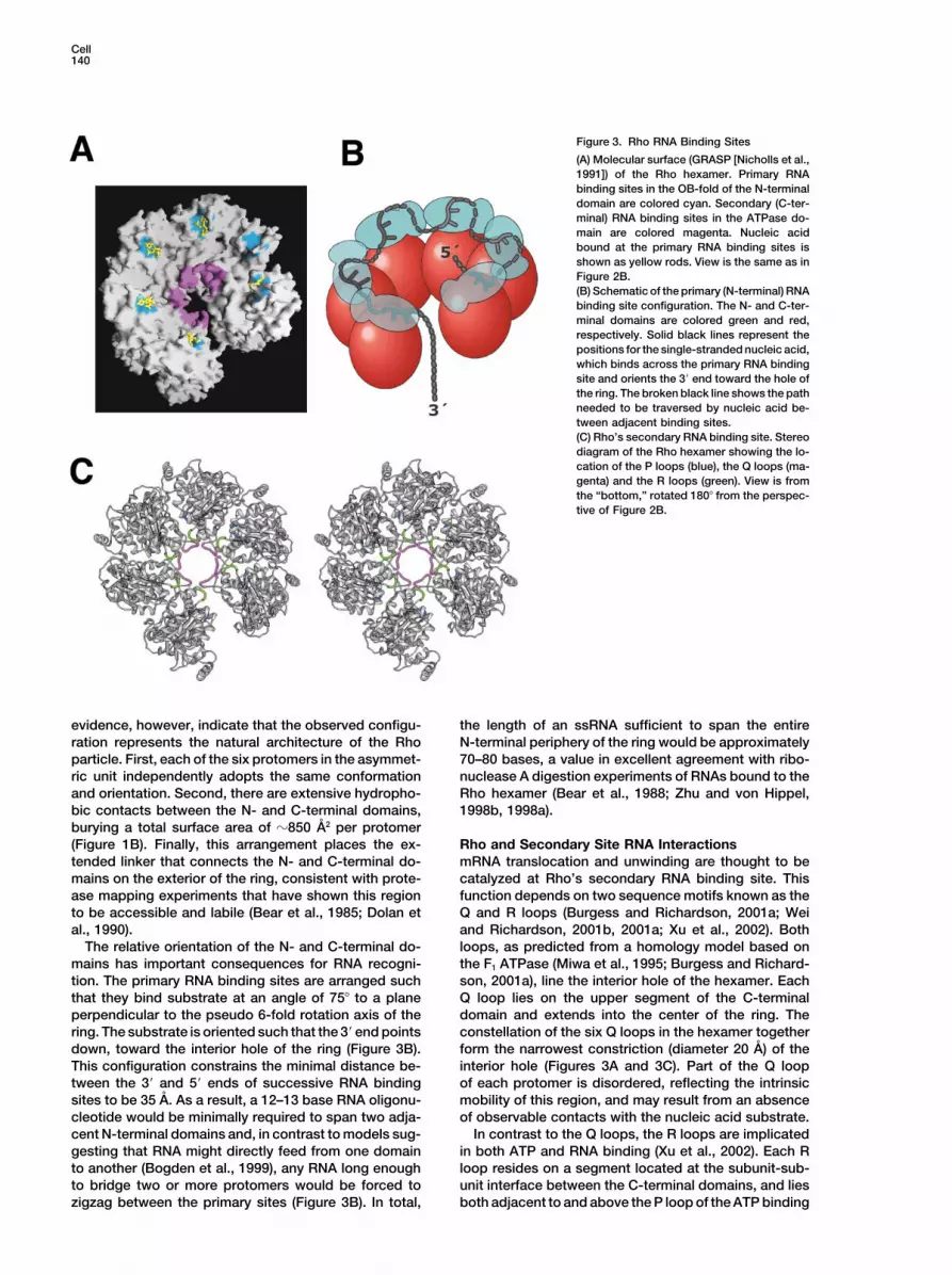
Figure 3. Rho RNA Binding Sites
(A) Molecular surface (GRASP [Nicholls et al.,1991]) of the Rho hexamer. Primary RNAbinding sites in the OB-fold of the N-terminaldomain are colored cyan. Secondary (C-ter-minal) RNA binding sites in the ATPase do-main are colored magenta. Nucleic acidbound at the primary RNA binding sites isshown as yellow rods. View is the same as inFigure 2B.(B) Schematic of the primary (N-terminal) RNAbinding site configuration. The N- and C-ter-minal domains are colored green and red,respectively. Solid black lines represent thepositions for the single-stranded nucleic acid,which binds across the primary RNA bindingsite and orients the 3� end toward the hole ofthe ring. The broken black line shows the pathneeded to be traversed by nucleic acid be-tween adjacent binding sites.(C) Rho’s secondary RNA binding site. Stereodiagram of the Rho hexamer showing the lo-cation of the P loops (blue), the Q loops (ma-genta) and the R loops (green). View is fromthe “bottom,” rotated 180� from the perspec-tive of Figure 2B.
evidence, however, indicate that the observed configu- the length of an ssRNA sufficient to span the entireN-terminal periphery of the ring would be approximatelyration represents the natural architecture of the Rho
particle. First, each of the six protomers in the asymmet- 70–80 bases, a value in excellent agreement with ribo-nuclease A digestion experiments of RNAs bound to theric unit independently adopts the same conformation
and orientation. Second, there are extensive hydropho- Rho hexamer (Bear et al., 1988; Zhu and von Hippel,1998b, 1998a).bic contacts between the N- and C-terminal domains,
burying a total surface area of �850 A2 per protomer(Figure 1B). Finally, this arrangement places the ex- Rho and Secondary Site RNA Interactions
mRNA translocation and unwinding are thought to betended linker that connects the N- and C-terminal do-mains on the exterior of the ring, consistent with prote- catalyzed at Rho’s secondary RNA binding site. This
function depends on two sequence motifs known as thease mapping experiments that have shown this regionto be accessible and labile (Bear et al., 1985; Dolan et Q and R loops (Burgess and Richardson, 2001a; Wei
and Richardson, 2001b, 2001a; Xu et al., 2002). Bothal., 1990).The relative orientation of the N- and C-terminal do- loops, as predicted from a homology model based on
the F1 ATPase (Miwa et al., 1995; Burgess and Richard-mains has important consequences for RNA recogni-tion. The primary RNA binding sites are arranged such son, 2001a), line the interior hole of the hexamer. Each
Q loop lies on the upper segment of the C-terminalthat they bind substrate at an angle of 75� to a planeperpendicular to the pseudo 6-fold rotation axis of the domain and extends into the center of the ring. The
constellation of the six Q loops in the hexamer togetherring. The substrate is oriented such that the 3� end pointsdown, toward the interior hole of the ring (Figure 3B). form the narrowest constriction (diameter 20 A) of the
interior hole (Figures 3A and 3C). Part of the Q loopThis configuration constrains the minimal distance be-tween the 3� and 5� ends of successive RNA binding of each protomer is disordered, reflecting the intrinsic
mobility of this region, and may result from an absencesites to be 35 A. As a result, a 12–13 base RNA oligonu-cleotide would be minimally required to span two adja- of observable contacts with the nucleic acid substrate.
In contrast to the Q loops, the R loops are implicatedcent N-terminal domains and, in contrast to models sug-gesting that RNA might directly feed from one domain in both ATP and RNA binding (Xu et al., 2002). Each R
loop resides on a segment located at the subunit-sub-to another (Bogden et al., 1999), any RNA long enoughto bridge two or more protomers would be forced to unit interface between the C-terminal domains, and lies
both adjacent to and above the P loop of the ATP bindingzigzag between the primary sites (Figure 3B). In total,

Rho Termination Factor Structure141
Figure 4. ATP Associations
(A) Stereo view of specific Rho and AMP-PNP interactions. Fo-Fc electron density contoured at 2� is shown for the bound nucleotide. Residuesimportant for nucleotide binding and hydrolysis are shown in ball and stick. Residues from two adjacent C-terminal domains form the activesite and are colored blue and green.(B) Rho nucleotide binding pocket conformation. The two adjacent C-terminal domains are colored red and gray (protomers D and E). Theactive site is partly closed and can bind nucleotide, but does not appear fully organized to carry out hydrolysis. The P loop (blue) is shownnext to the R loop motif (green).(C) F1 ATPase nucleotide binding pocket conformation (1bmf). Two ATPase domains are colored yellow and cyan, and bound nucleotide isshown as black rods. The active site cleft is fully closed and competent to carry out nucleotide hydrolysis.
pocket (Figures 3C and 4B). Part of each R loop also explaining why RNA appears to be readily captured fortranslocation following primary site occupancy (Kim andlines the interior hole of the Rho hexamer. The position
of the R loop and its contact with the P loop indicates Patel, 2001).that this motif could function as part of an allostericeffector switch that directly couples RNA binding in the Nucleotide Associations and Rho Function
The ATP binding pocket of Rho is located at the interfacehole of the hexamer to the ATP binding and hydrolysissite (Richardson and Conaway, 1980; Shigesada and between the C-terminal domains of adjacent protomers
(Figure 4B). Part of this region is formed by signatureWu, 1980; Richardson, 1982; Engel and Richardson,1984; Kim and Patel, 2001). Consistent with this hypoth- sequence motifs such as the adjacent Walker-A
(GXGXXGK(S/T) and Walker-B (D(D/E)XX) segments. Foresis, RNA binding to the secondary state coincides withclosure of the hexameric ring and stimulation of the native crystals grown in the presence of AMPPNP, clear
electron density for bound nucleotide is seen in all sixATPase activity (Gogol et al., 1991; Gan and Richard-son, 1999), presumably by introducing conformational ATP binding pockets of the hexameric ring (Figure 4A).
In contrast, weak electron density is observed for nucle-changes between subunits and residues around the ATPbinding site (Lowery-Goldhammer and Richardson, otide in the structure determined with the selenomethio-
nine protein, indicating that the occupancy at this site1974). It is interesting to note that the positioning of theprimary RNA binding sites places the 3� RNA end near is low. This difference appears due to the insertion of
the hydrophobic side chain SeMet186 of the seleno-the secondary sites from the outset of mRNA binding,

Cell142
Figure 5. Schematic Model for Rho Function
Numbers correspond to stages outlined in thetext. Asterisks represent catalytic sitesthought to be competent for ATP hydrolysis.
methionine protein into the adenine binding pocket of ing both an entry point into what would otherwise bethe topologically confined interior of the ring and sug-each of the six subunits. Nonetheless, superpositiongesting a means by which the protein may enter a nu-of the ATP bound and -free structures shows that thecleic acid “loading” configuration. This general organi-organization and conformation of the ATP binding siteszation provides an elegant means to ensure that targetare the same.mRNAs are efficiently captured by the protein, therebyCocrystallization (as opposed to soaking) of Rho withactivating the particle and initiating the process of tran-AMPPNP shows that nucleotide can associate with anscription termination.open-ring state of the protein. The interactions between
The significance of the Rho open-ring state may beRho and the adenosine base of bound AMPPNP areapplicable to other toroidal helicases and translocases.typical of those seen for other RecA-type proteins, inA long-standing puzzle has been to understand howwhich aliphatic or aromatic side chains sandwich theextended nucleic acid segments gain entry to the interiorpurine moiety in a “hydrophobic pincer” (Figure 4A)of such protein rings, where unwinding and transloca-(Boyer, 1997; Bird et al., 1998a; Singleton et al., 2000;tion are thought to occur. Many helicases, such as DnaB,Xu et al., 2000). Other nucleotide associations are notpapilloma virus E1, and the MCM complex, use special-canonical, however. For example, residues in theized loading proteins to accomplish this task (Lusky etWalker-A and -B regions do not make standard contactsal., 1993; Seo et al., 1993; Donovan et al., 1997; Tanakato the �- and �-phosphates of AMPPNP, nor is magne-et al., 1997; Weinreich et al., 1999; Barcena et al., 2001).sium evident in our structure (Figure 4A). These alteredOthers, such as Rho, appear to load onto target sub-features arise because the subunit-subunit orientationstrates without accessory factors. The ability of Rho toof the open ring splays apart the ATPase domains asspontaneously switch between open- and closed-ringcompared to other RecA-type proteins that have formedstructures provides an immediate solution to the loadingcompetent active sites (Figures 4B and 4C). Given thatproblem (Gogol et al., 1991; Yu et al., 2000; Richardson,the open-ring form of Rho imaged here appears consis-2002). By utilizing intersubunit hinging motions evidenttent with an mRNA loading state, as opposed to anin a number of RecA-type ATPases, our structure showsactively translocating helicase, such atypical nucleotidehow Rho can configure the relative orientation of neigh-associations are perhaps not surprising, although theseboring RecA-type folds to allow ring opening withoutdata indicate that ring opening can still occur when Rhofully exposing subunit interfaces that might otherwiseis bound to ATP, as would be expected when the proteinfoster filamentation. As a result, the Rho model may beoperates in vivo.applicable for understanding how other ring systemsare breached to bind nucleic acids, and may potentially
Rho Architecture and Mechanistic Implications represent a structural state accessed for some helicasesAs a collective, the structures observed here explain through loader protein activity.several important features of Rho function. First, Rho’s Another interesting finding is that the Rho hexamerprimary RNA binding sites line the perimeter of the hex- can remain open and, thus, competent to bind mRNAamer and face inward toward the center of the particle. at its secondary binding sites even when Rho’s ATP andSecond, the primary sites orient the 3� end of nucleic primary mRNA binding sites are occupied. This observa-acid target sequences into the interior hole, which is tion allows us to expand upon models explaining howformed by motifs known to be critical for coupling mRNA Rho recognizes rut sites and loads onto RNA in vivotranslocation and unwinding to ATP turnover. Third, the (Richardson, 2002; Delagoutte and von Hippel, 2003).
Extensive previous work, together with our structuralring of the hexamer is split by a gap �12 A wide, provid-

Rho Termination Factor Structure143
the native protein overexpressed in 2XYT media by induction at andata, suggest that Rho can oscillate between closed-OD600 of 0.3–0.5 with 1 mM IPTG at 37�C for 3 hr. Cells were lysedand open-ring states, either of which may associate withby sonication in 50 mM Tris-HCl (pH 7.5), 10% glycerol, 50 mM KCl,ATP and target mRNAs (Figure 5, stages 1 and 2). In the1 mM TCEP (Tris(2-carboxyethyl)phosphine hydrochloride) on ice.
open state, ATP would be loosely associated with Rho The protein was twice purified over a POROS-HS column (Perseptiveand only partially liganded as observed here. In contrast, Biosystems) then further purified with two successive Sephacryl
S-300 (Amersham) sizing columns in 10 mM Tris-HCl (pH 7.5), 50entry into the closed state would properly position andmM NaCl, and 1 mM TCEP. The protein at this stage was more thancoordinate nucleotide for hydrolysis. Natural conversion99% pure as judged by SDS-PAGE and Coomassie staining andbetween the two structural states would allow ATP hy-was monodisperse as judged by dynamic light scattering (DynaPro,drolysis to be observed in the absence of RNA (Stitt,model 99-CP, Protein Solutions). Protein was concentrated by ultra-
2001), but turnover would not be fully activated, both filtration (Centriprep-10, Amicon) to 15 mg/ml. Selenomethioninebecause of the lack of RNA effector signals through protein was overexpressed in minimal media using the protocol of
Van Duyne and coworkers (Van Duyne et al., 1993) and was purifiedthe Q and R loops and because a population of Rhosimilar to native Rho.molecules might be open at any given moment.
Loading and priming of Rho would begin through theCrystallization and Data Collectionassociation of a rut site with the primary RNA bindingInitial crystals of full-length Rho diffracted poorly. Cocrystals withsites in the N-terminal domains. The configuration ofDNA or RNA substrates resulted in crystals of different morphologythe N-terminal domains automatically orients the 3� endto those obtained in the absence of nucleic acid and proved instru-
of message toward the interior hole of the protein. Were mental in solving the Rho structure. A number of single-strandedthe ring to be open during rut site binding, mRNA could DNA and RNA substrates were tested in cocrystallization trials.
DNAs and RNAs varied in length (6–19 bases) and composition (onesimply thread its way through the gap between mono-or two binding sites), and were designed based on Rho’s specificitymers and into the interior of the hexamer to associatefor the unstructured, cytosine-rich mRNA regions present in rutwith the secondary RNA binding sites (Figure 5, stagesites. The best quality crystals in terms of size, stability, and diffrac-4). In contrast, if the ring were closed upon rut sitetion were those cocrystallized with a 15-mer DNA substrate (AACC
recognition (Figure 5, stage 3), mRNA entry would need CAAGAACCCAA) or with an 8-mer RNA substrate (CU)4.to wait until the ring spontaneously opens. It remains a Cocrystals of Rho in complex with the single-stranded DNA sub-
strate were grown by vapor diffusion at room temperature. Stockpossibility that mRNA binding to the primary, N-terminalprotein solutions were dialyzed in 10 mM Tris-HCl (pH 7.5) and 50domain binding sites might directly favor ring openingmM NaCl at 4�C prior to crystallization. One volume of protein solu-(Yu et al., 2000; Kim and Patel, 2001).tion was mixed with one volume of crystallization solution containingOnce bound in the interior, RNA would be sensed by100 mM Na•Cacodylate (pH 6.5), 100 mM NaCl, 5% PEG 8K, 40%
the Q and R loop regions, allosterically activating Rho glycerol, 0.6 mM n-Nonyl-�-D-thiomaltoside, and 2 mM TCEP; theand converting the protein into a stable, closed configu- well solution was diluted with an equivalent amount of H2O prior to
sealing the drop over the reservoir. Small crystals appeared over-ration (Figure 5, stage 5). Based on comparisons withnight and grew to average dimensions of 100 100 200 �M inclosed-ring RecA-type proteins, ring closure would pre-1 week. Crystals were introduced into cryoprotectant, containing asumably occur through conformational changes be-mix of 50 mM Tris-HCl (pH 7.5), 50 mM NaCl, 3% PEG 8K, 25%tween a subset of the ATPase domains, both formingglycerol, and 5% PEG 400, in a fast, single step, then flash frozen
competent set of active sites and compensating for the in liquid nitrogen. The crystals belong to the monoclinic space group15� subunit-subunit angular rise observed for the open- C2 with unit cell dimensions of a � 119.2 A, b � 205.8 A, c � 148.0 A,
and � � 95.3�. There is one hexamer per asymmetric unit.ring form. Ensuing cycles of ATP turnover would serve tomodulate the relative orientation of neighboring ATPasedomains, providing directed structural changes to pro- Structure Determination and Refinement
All data were collected at Beamline 8.3.1 at the Advanced Lightpel the protein 5�→3� along the message (Brennan etSource (ALS) using an ADSC Quantum-Q210 CCD detector. Diffrac-al., 1987; Singleton et al., 2000). As translocation begins,tion data were processed using DENZO and SCALEPACK (Otwinow-Rho could either dissociate from the rut site or couldski, 1997) (Table 1). Initial attempts to calculate phases using seleno-
remain bound to the target motif as predicted by the methionine MAD or SAD experiments were unsuccessful, so atethered tracking model (Figure 5, stages 6 and 7). Al- tantalum derivative was prepared by soaking a crystal overnight
with saturated solution of Ta6Br122�. Heavy atom positions werethough our structure cannot rule out either mechanism,
identified by SOLVE (Terwilliger and Berendzen, 1999), andit is interesting to note that the N-terminal domains openMLPHARE (CCP4, 1994) was used to calculate initial phases to 6.0 Aout from the center of the ring, creating a wide depres-based on isomorphous and anomalous differences for this derivativesion across the top of the particle. The size of this de-(Table 1). Selenium sites (83 out of 96) were located by calculating
pression appears ample enough to remain associated anomalous difference maps to 3.6 A resolution using the tantalumwith a RNA segment while single-stranded RNA is phases (Table 1). The selenium sites were refined initially with
MLPHARE and then with SHARP (de la Fortelle and Bricogne, 1997),spooled away from the protein during translocation. Fu-followed by DM solvent flattening (Cowtan, 1994) to yield startingture efforts focused toward assembling RNA bound,electron density maps.closed ring structures will be needed to help distinguish
Five out of the six N-terminal domains were located in the solvent-between these models and to better illuminate the physi-flattened density by the program FFFEAR (Cowtan K., 1998) using
cal coupling between ATP usage, translocation, and un- the X-ray model 2a8v (Bogden et al., 1999). The sixth domain waswinding. placed into the map manually and the fit optimized using RSR-RIGID
in O (Jones T.A., 1991). The N-terminal domains were used to derivethe six NCS (noncrystallographic symmetry) operators within theExperimental Proceduresasymmetric unit and the experimental electron density map wassubsequently improved using solvent flattening with NCS multido-Protein Expression and Purification
The open reading frame encoding the full-length Escherichia coli main averaging and phase extension in DM. The C-terminal domainof the mitochondrial F1 ATPase model (1bmf) was manually dockedRho protein was PCR amplified and cloned into pET24b. The plasmid
was transformed into the Escherichia coli strain BL21 (pLysS), and and rebuilt into this DM-averaged density using O.

Cell144
The model was refined with REFMAC5 (Murshudov G.N., 1997), Brennan, C.A., Steinmetz, E.J., Spear, P., and Platt, T. (1990). Speci-ficity and efficiency of rho-factor helicase activity depends on mag-using ARP (Lamzin and Wilson, 1993) for water building. In the final
cycles of refinement, TLS (translation libration and screw-rotation) nesium concentration and energy coupling to NTP hydrolysis. J.Biol. Chem. 265, 5440–5447.refinement of rigid groups (Winn et al., 2001) was carried out as
implemented in REFMAC5 (Table 2). The refined selenomethionine Briercheck, D.M., Wood, T.C., Allison, T.J., Richardson, J.P., andRho/DNA structure was used to solve the native Rho/RNA•AMPPNP Rule, G.S. (1998). The NMR structure of the RNA binding domain ofcomplex by molecular replacement using the program AMORE (Na- E. coli rho factor suggests possible RNA-protein interactions. Nat.vaza, 2001). The resultant solution was DM solvent flattened and Struct. Biol. 5, 393–399.multidomain averaged, revealing clear electron density in Fo-Fc maps
Brown, S., Brickman, E.R., and Beckwith, J. (1981). Blue ghosts: afor AMPPNP in all six ATP binding sites. The structure was thennew method for isolating amber mutants defective in essential genesrefined using REFMAC5. The Rho/DNA and the Rho/RNA structuresof Escherichia coli. J. Bacteriol. 146, 422–425.were refined to a final Rwork 27.0% and 27.0% and an Rfree 29.6%Burgess, B.R., and Richardson, J.P. (2001a). RNA passes throughand 30.3%, respectively. A total of 2472 out of 2502 residues arethe hole of the protein hexamer in the complex with the Escherichiaaccounted for in either of the two structures (Table 2). Geometriccoli Rho factor. J. Biol. Chem. 276, 4182–4189.analysis were carried out with Procheck (Laskowski et al., 1993)
and structural superpositions with LSQKAB (CCP4, 1994). Burgess, B.R., and Richardson, J.P. (2001b). Transcription factorRho does not require a free end to act as an RNA-DNA helicase on
Acknowledgments an RNA. J. Biol. Chem. 276, 17106–17110.
Cavarelli, J., Rees, B., Ruff, M., Thierry, J.C., and Moras, D. (1993).The authors are grateful to James Holton at Beamline 8.3.1 of the Yeast tRNA(Asp) recognition by its cognate class II aminoacyl-tRNAAdvanced Light source for assistance with data acquisition and to synthetase. Nature 362, 181–184.David King for mass spectroscopy analyses. We would also like to
CCP4 (Collaborative Computational Project 4) (1994). The CCP4thank James Keck, Deborah Fass, David Akey, Jan Erzberger, andsuite: programs for protein crystallography. Acta Crystallogr. D 50,Scott Gradia for critical reading the manuscript, as well as members760–763.of the Berger Lab for helpful discussions and insights. This workChen, C.Y., Galluppi, G.R., and Richardson, J.P. (1986). Transcrip-was supported by generous assistance from the G. Harold and Leilation termination at lambda tR1 is mediated by interaction of rho withY. Mathers Charitable Foundation.specific single-stranded domains near the 3� end of cro mRNA. Cell46, 1023–1028.Received: March 19, 2003
Revised: May 20, 2003 Cowtan, K. (1994). A CCP4 density modification package. JointAccepted: June 25, 2003 CCP4 ESF-EACBM. Newslett. Prot. Crystallogr. 31, 34–38.Published: July 10, 2003 Cowtan, K. (1998). Modified phased translation functions and their
application to molecular fragment location. Acta Crystallogr. D 54,References 750–756.
de la Fortelle, E., and Bricogne, G. (1997). Maximum-likelihood heavyAbrahams, J.P., Leslie, A.G., Lutter, R., and Walker, J.E. (1994).
atom parameter refinement for multiple isomorphous replacementStructure at 2.8 A resolution of F1-ATPase from bovine heart mito-
and multiwavelength anomalous diffraction methods. Methods En-chondria. Nature 370, 621–628.
zymol. 276, 472–494.Alifano, P., Rivellini, F., Limauro, D., Bruni, C.B., and Carlomagno,
Delagoutte, E., and von Hippel, P.H. (2003). Helicase mechanismsM.S. (1991). A consensus motif common to all Rho-dependent pro-and the coupling of helicases within macromolecular machines. Partkaryotic transcription terminators. Cell 64, 553–563.II: integration of helicases into cellular processes. Q. Rev. Biophys.
Allison, T.J., Wood, T.C., Briercheck, D.M., Rastinejad, F., Richard- 36, 1–69.son, J.P., and Rule, G.S. (1998). Crystal structure of the RNA-binding
Dolan, J.W., Marshall, N.F., and Richardson, J.P. (1990). Transcrip-domain from transcription termination factor rho. Nat. Struct. Biol.tion termination factor rho has three distinct structural domains. J.5, 352–356.Biol. Chem. 265, 5747–5754.
Barcena, M., Ruiz, T., Donate, L.E., Brown, S.E., Dixon, N.E., Rader-Donovan, S., Harwood, J., Drury, L.S., and Diffley, J.F. (1997).macher, M., and Carazo, J.M. (2001). The DnaB.DnaC complex: aCdc6p-dependent loading of Mcm proteins onto pre-replicativestructure based on dimers assembled around an occluded channel.chromatin in budding yeast. Proc. Natl. Acad. Sci. USA 94, 5611–EMBO J. 20, 1462–1468.5616.
Bear, D.G., Andrews, C.L., Singer, J.D., Morgan, W.D., Grant, R.A.,Engel, D., and Richardson, J.P. (1984). Conformational alterationsvon Hippel, P.H., and Platt, T. (1985). Escherichia coli transcriptionof transcription termination protein rho induced by ATP and by RNA.termination factor rho has a two-domain structure in its activatedNucleic Acids Res. 12, 7389–7400.form. Proc. Natl. Acad. Sci. USA 82, 1911–1915.Galluppi, G.R., and Richardson, J.P. (1980). ATP-induced changesBear, D.G., Hicks, P.S., Escudero, K.W., Andrews, C.L., McSwiggen,in the binding of RNA synthesis termination protein Rho to RNA. J.J.A., and von Hippel, P.H. (1988). Interactions of Escherichia coliMol. Biol. 138, 513–539.transcription termination factor rho with RNA. II. Electron micros-
copy and nuclease protection experiments. J. Mol. Biol. 199, Gan, E., and Richardson, J.P. (1999). ATP and other nucleotides623–635. stabilize the Rho-mRNA complex. Biochemistry 38, 16882–16888.
Bird, L.E., Brannigan, J.A., Subramanya, H.S., and Wigley, D.B. Geiselmann, J., Yager, T.D., and von Hippel, P.H. (1992). Functional(1998a). Characterisation of Bacillus stearothermophilus PcrA heli- interactions of ligand cofactors with Escherichia coli transcriptioncase: evidence against an active rolling mechanism. Nucleic Acids termination factor rho. II. Binding of RNA. Protein Sci. 1, 861–873.Res. 26, 2686–2693. Geiselmann, J., Wang, Y., Seifried, S.E., and von Hippel, P.H. (1993).Bird, L.E., Subramanya, H.S., and Wigley, D.B. (1998b). Helicases: A physical model for the translocation and helicase activities ofa unifying structural theme? Curr. Opin. Struct. Biol. 8, 14–18. Escherichia coli transcription termination protein Rho. Proc. Natl.
Acad. Sci. USA 90, 7754–7758.Bogden, C.E., Fass, D., Bergman, N., Nichols, M.D., and Berger,J.M. (1999). The structural basis for terminator recognition by the Gogol, E.P., Seifried, S.E., and von Hippel, P.H. (1991). StructureRho transcription termination factor. Mol. Cell 3, 487–493. and assembly of the Escherichia coli transcription termination factor
rho and its interaction with RNA. I. Cryoelectron microscopic stud-Boyer, P.D. (1997). The ATP synthase–a splendid molecular ma-ies. J. Mol. Biol. 221, 1127–1138.chine. Annu. Rev. Biochem. 66, 717–749.
Brennan, C.A., Dombroski, A.J., and Platt, T. (1987). Transcription Jones, T.A., Zou, J.Y., Cowan, S.W., and Kjeldgaard, M. (1991).Improved methods for building protein models in electron densitytermination factor rho is an RNA-DNA helicase. Cell 48, 945–952.
Rho Termination Factor Structure145
maps and the location of errors in these models. Acta Crystallogr. simultaneous interaction at two kinds of nucleic acid-binding sites.J. Biol. Chem. 257, 5760–5766.A 47, 110–119.
Richardson, J.P., and Conaway, R. (1980). Ribonucleic acid releaseKim, D.E., and Patel, S.S. (2001). The kinetic pathway of RNA bindingactivity of transcription termination protein rho is dependent on theto the Escherichia coli transcription termination factor Rho. J. Biol.hydrolysis of nucleoside triphosphates. Biochemistry 19, 4293–Chem. 276, 13902–13910.4299.Lamzin, V.S., and Wilson, K.S. (1993). Automated refinement of pro-Richardson, J.P., and Greenblatt, J. (1996). Escherichia coli andtein models. Acta Crystallogr. D 49, 129–147.salmonella. In Cellular and Molecular Biology, Second Edition, F.C.Laskowski, A.R., MacArthur, W.M., Moss, S.D., and Thornton, M.J.Neidhardt, R. Curtiss III, J.L. Ingraham, E.C.C. Lin, K.B. Low, B.(1993). PROCHECK: a program to check the stereochemical qualityMagasanik, W.S. Reznikoff, M. Riley, M. Schaechter, and H.E. Um-of protein structures. J. Appl. Cryst. 26, 283–291.barger, eds. (Washington, D.C.: American Society for Microbiology),
Lowery-Goldhammer, C., and Richardson, J.P. (1974). An RNA- pp. 822–848.dependent nucleoside triphosphate phosphohydrolase (ATPase)
Richardson, L.V., and Richardson, J.P. (1992). Cytosine nucleosideassociated with rho termination factor. Proc. Natl. Acad. Sci. USAinhibition of the ATPase of Escherichia coli termination factor rho:71, 2003–2007.evidence for a base specific interaction between rho and RNA. Nu-
Lusky, M., Hurwitz, J., and Seo, Y.S. (1993). Cooperative assembly cleic Acids Res. 20, 5383–5387.of the bovine papilloma virus E1 and E2 proteins on the replication
Roberts, J.W. (1969). Termination factor for RNA synthesis. Natureorigin requires an intact E2 binding site. J. Biol. Chem. 268, 15795–224, 1168–1174.15803.Sawaya, M.R., Guo, S., Tabor, S., Richardson, C.C., and Ellenberger,Martinez, A., Burns, C.M., and Richardson, J.P. (1996a). ResiduesT. (1999). Crystal structure of the helicase domain from the replica-in the RNP1-like sequence motif of Rho protein are involved in RNA-tive helicase-primase of bacteriophage T7. Cell 99, 167–177.binding affinity and discrimination. J. Mol. Biol. 257, 909–918.Schindelin, H., Jiang, W., Inouye, M., and Heinemann, U. (1994).Martinez, A., Opperman, T., and Richardson, J.P. (1996b). MutationalCrystal structure of CspA, the major cold shock protein of Esche-analysis and secondary structure model of the RNP1-like sequencerichia coli. Proc. Natl. Acad. Sci. USA 91, 5119–5123.motif of transcription termination factor Rho. J. Mol. Biol. 257,Seo, Y.S., Muller, F., Lusky, M., Gibbs, E., Kim, H.Y., Phillips, B.,895–908.and Hurwitz, J. (1993). Bovine papilloma virus (BPV)-encoded E2McSwiggen, J.A., Bear, D.G., and von Hippel, P.H. (1988). Interac-protein enhances binding of E1 protein to the BPV replication origin.tions of Escherichia coli transcription termination factor rho withProc. Natl. Acad. Sci. USA 90, 2865–2869.RNA. I. Binding stoichiometries and free energies. J. Mol. Biol. 199,Shigesada, K., and Wu, C.W. (1980). Studies of RNA release reaction609–622.catalyzed by E. coli transcription termination factor rho using iso-Miwa, Y., Horiguchi, T., and Shigesada, K. (1995). Structural andlated ternary transcription complexes. Nucleic Acids Res. 8, 3355–functional dissections of transcription termination factor rho by ran-3369.dom mutagenesis. J. Mol. Biol. 254, 815–837.Singleton, M.R., Sawaya, M.R., Ellenberger, T., and Wigley, D.B.Modrak, D., and Richardson, J.P. (1994). The RNA-binding domain(2000). Crystal structure of T7 gene 4 ring helicase indicates a mech-of transcription termination factor rho: isolation, characterization,anism for sequential hydrolysis of nucleotides. Cell 101, 589–600.and determination of sequence limits. Biochemistry 33, 8292–8299.Steinmetz, E.J., and Platt, T. (1994). Evidence supporting a tethered
Morgan, W.D., Bear, D.G., and von Hippel, P.H. (1983). Rho-depen-tracking model for helicase activity of Escherichia coli Rho factor.
dent termination of transcription. I. Identification and characteriza-Proc. Natl. Acad. Sci. USA 91, 1401–1405.
tion of termination sites for transcription from the bacteriophageStitt, B.L. (2001). Escherichia coli transcription termination factorlambda PR promoter. J. Biol. Chem. 258, 9553–9564.Rho binds and hydrolyzes ATP using a single class of three sites.
Morgan, W.D., Bear, D.G., Litchman, B.L., and von Hippel, P.H.Biochemistry 40, 2276–2281.
(1985). RNA sequence and secondary structure requirements forStory, R.M., Weber, I.T., and Steitz, T.A. (1992). The structure of therho-dependent transcription termination. Nucleic Acids Res. 13,E. coli recA protein monomer and polymer. Nature 355, 318–325.3739–3754.Sullivan, S.L., and Gottesman, M.E. (1992). Requirement for E. coliMurshudov G.N., Vagin, A.A., and Dodson, E.J. (1997). RefinementNusG protein in factor-dependent transcription termination. Cell 68,of macromolecular structures by the maximum-likelihood method.989–994.Acta Crystallogr. D 53, 240–255.Tanaka, T., Knapp, D., and Nasmyth, K. (1997). Loading of an McmNavaza, J. (2001). Implementation of molecular replacement inprotein onto DNA replication origins is regulated by Cdc6p andAMoRe. Acta Crystallogr. D Biol. Crystallogr. 57, 1367–1372.CDKs. Cell 90, 649–660.
Newkirk, K., Feng, W., Jiang, W., Tejero, R., Emerson, S.D., Inouye,Terwilliger, T.C., and Berendzen, J. (1999). Automated MAD andM., and Montelione, G.T. (1994). Solution NMR structure of the majorMIR structure solution. Acta Crystallogr. D 55, 849–861.cold shock protein (CspA) from Escherichia coli: identification of aVan Duyne, G.D., Standaert, R.F., Karplus, P.A., Schreiber, S.L.,binding epitope for DNA. Proc. Natl. Acad. Sci. USA 91, 5114–5118.and Clardy, J. (1993). Atomic structures of the human immunophilinNicholls, A., Sharp, K.A., and Honig, B. (1991). Protein folding andFKBP-12 complexes with FK506 and rapamycin. J. Mol. Biol. 229,association: insights from the interfacial and thermodynamic prop-105–124.erties of hydrocarbons. Proteins 11, 281–296.von Hippel, P.H., and Delagoutte, E. (2001). A general model forOda, T., and Takanami, M. (1972). Observations on the structure ofnucleic acid helicases and their “coupling” within macromolecularthe termination factor rho and its attachment to DNA. J. Mol. Biol.machines. Cell 104, 177–190.71, 799–802.Walstrom, K.M., Dozono, J.M., Robic, S., and von Hippel, P.H. (1997).Opperman, T., and Richardson, J.P. (1994). Phylogenetic analysisKinetics of the RNA-DNA helicase activity of Escherichia coli tran-of sequences from diverse bacteria with homology to the Esche-scription termination factor rho. 1. Characterization and analysis ofrichia coli rho gene. J. Bacteriol. 176, 5033–5043.the reaction. Biochemistry 36, 7980–7992.
Otwinowski, Z., and Minor, W. (1997). Processing of X-ray diffractionWei, R.R., and Richardson, J.P. (2001a). Identification of an RNA-data collected in oscillation mode. Methods Enzymol. 276, 307–326.binding Site in the ATP binding domain of Escherichia coli Rho by
Platt, T. (1994). Rho and RNA: models for recognition and response. H2O2/Fe-EDTA cleavage protection studies. J. Biol. Chem. 276,Mol. Microbiol. 11, 983–990. 28380–28387.Richardson, J. (2002). Rho-dependent termination and ATPases in Wei, R.R., and Richardson, J.P. (2001b). Mutational changes of con-transcript termination. Biochim. Biophys. Acta 1577, 251–260. served residues in the Q-loop region of transcription factor Rho greatly
reduce secondary site RNA-binding. J. Mol. Biol. 314, 1007–1015.Richardson, J.P. (1982). Activation of rho protein ATPase requires

Cell146
Weinreich, M., Liang, C., and Stillman, B. (1999). The Cdc6p nucleo-tide-binding motif is required for loading mcm proteins onto chroma-tin. Proc. Natl. Acad. Sci. USA 96, 441–446.
Winn, M.D., Isupov, M.N., and Murshudov, G.N. (2001). Use of TLSparameters to model anisotropic displacements in macromolecularrefinement. Acta Crystallogr. D 57, 122–133.
Xu, H., Frank, J., Niedenzu, T., and Saenger, W. (2000). DNA helicaseRepA: cooperative ATPase activity and binding of nucleotides. Bio-chemistry 39, 12225–12233.
Xu, Y., Kohn, H., and Widger, W.R. (2002). Mutations in the rhotranscription termination factor that affect RNA tracking. J. Biol.Chem. 277, 30023–30030.
Yu, X., Horiguchi, T., Shigesada, K., and Egelman, E.H. (2000). Three-dimensional reconstruction of transcription termination factor rho:orientation of the N-terminal domain and visualization of an RNA-binding site. J. Mol. Biol. 299, 1279–1287.
Zhu, A.Q., and von Hippel, P.H. (1998a). Rho-dependent terminationwithin the trp t’ terminator. I. Effects of rho loading and templatesequence. Biochemistry 37, 11202–11214.
Zhu, A.Q., and von Hippel, P.H. (1998b). Rho-dependent terminationwithin the trp t’ terminator. II. Effects of kinetic competition and rhoprocessivity. Biochemistry 37, 11215–11222.
Accession Numbers
Coordinates for both Rho complexes have been deposited in theRCSB PDB database and are available under the accession codes1PV4 and 1PVO.


