Embed Size (px)
Citation preview

RESEARCH ARTICLE
Striatal Infarction Elicits SecondaryExtrafocal MRI Changes in IpsilateralSubstantia NigraBenjaminWinter1,2, Peter Brunecker1, Jochen B. Fiebach1, Gerhard Jan Jungehulsing1,3,Golo Kronenberg1,2,4,5☯, Matthias Endres1,2,6,7,8☯*
1 Center for Stroke Research Berlin (CSB), Charité-Universitätsmedizin Berlin, Berlin, Charitéplatz 1,10117,Berlin, Germany, 2 Department of Neurology, Charité-Universitätsmedizin Berlin, Charitéplatz 1, 10117,Berlin, Germany, 3 Department of Neurology, Jüdisches Krankenhaus Berlin, Heinz-Galinski-Strasse 1,13347, Berlin, Germany, 4 Department of Psychiatry, Charité-Universitätsmedizin Berlin, Charitéplatz 1,10117, Berlin, Germany, 5 Max-Delbrück Center and Charité Medical Faculty, Experimental and ClinicalResearch Center, Lindenbergerweg 80, 13125, Berlin, Germany, 6 Excellence Cluster Neurocure, Charité-Universitätsmedizin Berlin, Charitéplatz 1, 10117, Berlin, Germany, 7 German Center for NeurodegenerativeDiseases (DZNE), Ludwig-Erhard-Allee, 53175, Bonn, Germany, 8 German Centre for CardiovascularResearch (DZHK), Oudenarder Straße 16, 13347, Berlin, Germany
☯ These authors contributed equally to this work.* [email protected]
AbstractFocal ischemia may induce pathological alterations in brain areas distant from the primary
lesion. In animal models, exofocal neuron death in the ipsilateral midbrain has been
described after occlusion of the middle cerebral artery (MCA). Using sequential magnetic
resonance imaging (T2- and diffusion-weighted) at 3 Tesla, we investigated acute ischemic
stroke patients on days 1, 2, 6, 8, and 10 after stroke onset. Sixteen consecutive patients
who had suffered a stroke involving the caudate nucleus and/or putamen of either hemi-
sphere were recruited into the study. Four additional patients with strokes sparing the cau-
date nucleus and putamen but encompassing at least one-third of the MCA territory served
as controls. Ischemic lesions involving striatal structures resulted in hyperintense lesions in
ipsilateral midbrain that emerged between days 6 and 10 after stroke and were not present
on the initial scans. In contrast, none of the control stroke patients developed secondary
midbrain lesions. Hyperintense lesions in the pyramidal tract or the brain stem caused by
degeneration of the corticospinal tract could be clearly distinguished from these secondary
midbrain gray matter lesions and were detectable from day 2 after ischemia. Co-registration
of high-resolution images with a digitized anatomic atlas revealed localization of secondary
lesions primarily in the substantia nigra pars compacta. Apparent diffusion coefficient
(ADC) values in the secondary lesions showed a delayed sharp decline through day 10.
Normalization of ADC values was observed at late measurements. Taken together, our
study demonstrates that striatal infarction elicits delayed degenerative changes in ipsilateral
substantia nigra pars compacta.
PLOS ONE | DOI:10.1371/journal.pone.0136483 September 1, 2015 1 / 11
a11111
OPEN ACCESS
Citation:Winter B, Brunecker P, Fiebach JB,Jungehulsing GJ, Kronenberg G, Endres M (2015)Striatal Infarction Elicits Secondary Extrafocal MRIChanges in Ipsilateral Substantia Nigra. PLoS ONE10(9): e0136483. doi:10.1371/journal.pone.0136483
Editor: Christoph Kleinschnitz, Julius-Maximilians-Universität Würzburg, GERMANY
Received: September 21, 2014
Accepted: December 24, 2014
Published: September 1, 2015
Copyright: © 2015 Winter et al. This is an openaccess article distributed under the terms of theCreative Commons Attribution License, which permitsunrestricted use, distribution, and reproduction in anymedium, provided the original author and source arecredited.
Data Availability Statement: Most data are availablewithin the paper and Supporting Information files.However, due to legal and ethical restrictions, theMRI images are not publicly available. Single imageswill be provided upon request from Dr. BenjaminWinter / [email protected].
Funding: ME receives funding from the DeutscheForschungsgesellschaft (Excellence clusterNeuroCure; SFB TR 43, KFO 247, KFO 213),Bundesministerium für Bildung und Forschung(Center for Stroke Research Berlin), European Union(European Stroke Network, WakeUp, Counterstroke),Volkswagen Foundation (Lichtenberg Program) and

IntroductionMidbrain changes associated with the loss of basal ganglia neurons have been described inexperimental models of brain ischemia in rats [1–6], mice [7, 8], gerbils [9], and cynomolgusmonkeys [10]. Magnetic resonance imaging (MRI) studies in experimental rodents demon-strate transient exofocal signal changes in ipsilateral midbrain. In particular, apparent diffusioncoefficient (ADC) values show a significant delayed decrease and T2 values show a significantdelayed increase in the ipsilateral midbrain after occlusion of the middle cerebral artery [8, 11,12]. On the histological level, these subacute MRI changes after experimental brain ischemiaare associated with alterations such as cellular swelling and changes in neuronal morphology[11, 13].
The anecdotal histopathological reports of human autopsy brain tissue published so farindicate nerve cell loss in the ipsilateral substantia nigra during the chronic phase after a largeinfarction of the basal ganglia [14, 15]. Correspondingly, several MRI investigations of patientswith a stroke in the striatum also revealed a T2-weighted hyperintensity appearing as a sub-acute exofocal alteration in the ipsilateral midbrain [16–19]. Here, we present a systematic pro-spective study of a series of 16 consecutive patients with striatal infarction admitted to ourstroke unit between January 2009 and May 2010. Four additional patients whose ischemiclesions spared the striatum served as controls. Sequential high-resolution anatomical MRI at 3Tesla was used to delineate the emergence and the precise neuroanatomic localization of sec-ondary MRI changes in the midbrain.
Methods
Patients and ControlsData presented here were collected within the framework of a larger project designated the"1000Plus study”, a prospective, single-center observational investigation [20, 21]. Study detailsare posted at www.clinicaltrials.gov (NCT00715533). Based on the initial MRI assessment (seebelow), 16 consecutive patients who had suffered an acute ischemic stroke involving the cau-date nucleus and/or putamen of either hemisphere were recruited into the study (‘striatalstroke patients’). Four additional patients who had suffered an acute ischemic stroke sparingcaudate nucleus and putamen but typically encompassing at least 1/3 of the middle cerebralartery (MCA) cortical territory served as controls (‘control stroke patients’). Exclusion criteriaincluded severe aphasia or inability to provide informed consent, hemorrhagic stroke, severebrain damage before admission, moderate to severe dementia (MMSE< 18) as well as contra-indications to MRI. The protocol had been approved by the local ethics committee and writteninformed consent was obtained prior to the study from each participant according to the Dec-laration of Helsinki.
Image AcquisitionAll MRI scans were acquired on a 3 T whole-body scanner (TIM Trio; Siemens AG, Erlangen,Germany) dedicated entirely to clinical research. The baseline scan was obtained within 24hours of stroke onset (day 1). Follow-up scans were performed on days 2, 6, 8, and 10. In somepatients, additional later scans were performed as shown in Table 1. On days 1 and 2, the MRIprotocol for this investigation included the following transversely-oriented sequences with aconsistent field-of-view of 230 mm: T2�-weighted imaging (2D-FLASH; TA = 73 s; TE = 20ms; TR = 620 ms; flip angle = 20°; matrix = 256 x 205; 25 slices with 5 mm thickness and 5.5mm spacing); diffusion-weighted imaging (DWI) (SE-EPI; TA = 131 s; TE = 93 ms; TR = 7600ms; matrix = 192 x 192, 50 slices with in-plane resolution of 1.2 mm, thickness of 2.5 mm and
Secondary Midbrain Lesion after Striatal Infarction
PLOS ONE | DOI:10.1371/journal.pone.0136483 September 1, 2015 2 / 11
the Corona Foundation. The funders had no role instudy design, data collection and analysis, decision topublish, or preparation of the manuscript.
Competing Interests: The authors have declaredthat no competing interests exist.
no interslice gap; 6 directions; b-values = 0 and 1000 s/mm2). On days 6, 8, and 10, the protocolincluded DWI as described above as well as coronal T2-weighted imaging (TSE; TA = 157 s;TE = 79 ms; TR = 5540 ms; matrix = 384 x 307; 30 slices with in-plane resolution of 0.6 mm,thickness of 3 mm and spacing of 3.3 mm).
Postprocessing and Data AnalysisThe following definitions apply: (a) ‘Primary ischemic lesion’means the early T2 hyperintensesignal in the MCA territory. (b) The term ‘secondary MRI lesion’ is used for delayed T2/DWIsignal changes that emerge in the ipsilateral midbrain after stroke. ROIs of these two lesiontypes were used independently in further analyses. As described in detail below, lesions wereidentified separately on T2 and DWI scans. However, because of higher spatial resolution andeasier-to-process coronal orientation, volumetry and lesion maps were generated from T2scans only. In general, DWI lesions matched T2 lesions well. However, because of differentscan techniques, we did not calculate the degree of overlap between DWI and T2 lesions.
For coronal T2 scans obtained on day 10 (or on the latest available scan before day 10),binary masks distinguishing between the hyperintense-appearing primary and secondarylesions, (coded as “1”), and non-infarcted areas (“0”) were generated for later usage as regionsof interests (ROI) and as the basis for subsequent volumetric measurements using the MRIcro
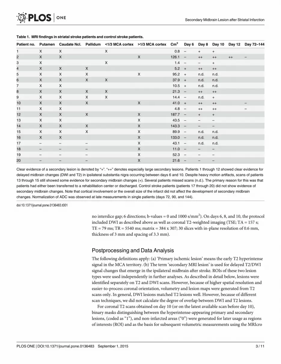
Table 1. MRI findings in striatal stroke patients and control stroke patients.
Patient no. Putamen Caudate Ncl. Pallidum <1/3 MCA cortex >1/3 MCA cortex Cm3 Day 6 Day 8 Day 10 Day 12 Day 72–144
1 X X X 0.8 – + +
2 X X X 126.1 – ++ ++ ++ –
3 X X 1.4 – – +
4 X X X 5.2 + ++ ++
5 X X X X 95.2 + n.d. n.d.
6 X X X X 37.9 + n.d. n.d.
7 X X 10.5 + n.d. n.d.
8 X X X X 21.3 – ++ ++
9 X X X X 14.4 – n.d. +
10 X X X X 41.0 + ++ ++ –
11 X X 4.8 – ++ ++ –
12 X X X X 187.7 – + +
13 X X X 43.5 – – –
14 X X X X 143.3 – – –
15 X X X X 89.9 – n.d. n.d.
16 X X X 133.0 – n.d. n.d.
17 – – – X 43.1 – n.d. n.d.
18 – – – X 11.0 – – –
19 – – – X 52.3 – – –
20 – – – X 21.6 – – –
Clear evidence of a secondary lesion is denoted by “+”. “++” denotes especially large secondary lesions. Patients 1 through 12 showed clear evidence for
delayed midbrain changes (DWI and T2) in ipsilateral substantia nigra occurring between days 6 and 10. Despite heavy motion artifacts, scans of patients
13 through 15 still showed some evidence for secondary midbrain changes (+). Several patients missed scans (n.d.). The primary reason for this was that
patients had either been transferred to a rehabilitation center or discharged. Control stroke patients (patients 17 through 20) did not show evidence of
secondary midbrain changes. Note that cortical involvement or the overall size of the infarct did not affect the development of secondary midbrain
changes. Normalization of ADC was observed at late measurements in single patients (days 72, 90, and 144).
doi:10.1371/journal.pone.0136483.t001
Secondary Midbrain Lesion after Striatal Infarction
PLOS ONE | DOI:10.1371/journal.pone.0136483 September 1, 2015 3 / 11

software (http://www.mricro.com). Lesion segmentation was performed manually by an expe-rienced investigator. To combine left- and right-sided infarcts into a single analysis, all imageswith right-hemispheric strokes were mirrored right to left.
Furthermore, coronal T2-weighted images were co-registered with coronal section outlinesof a digitized atlas of the human brain [22, 23] using an in-house developed software(“THAT”). In a first step, the interhemispheric midsagittal plane and the orthogonal planethrough the bicommissural line between the anterior and posterior commissures were deter-mined as reference planes. Then the images were repetitively transformed (translation, rota-tion, linear scaling) by the investigator with respect to structural landmarks (such as thalamus,caudate nucleus, corticospinal tract, putamen, wall of the third ventricle) [22]. Transformationsgenerated during the co-registration procedure were used to spatially adjust the ROIs to thestereotactic space of the digital atlas. The transformed image data as well as their correspondingROIs were subsequently re-sliced into two different iso-voxel spaces (1 x 1 x 1 mm; 0.25 x 0.25x 0.25 mm) using SPM8 (Statistical Parametric Mapping; Wellcome Department for CognitiveNeurology, University College London, UK) running on MATLAB 7.3 (The MathWorks, Inc.;Natick, MA, USA). Finally, the relative frequencies of the primary and secondary lesions werevoxel-wisely calculated for both iso-voxel spaces and the resulting maps were subsequentlycolor-coded. These cluster maps were overlaid on the coronal section outlines of the digitalanatomic atlas to determine the lesion topography more precisely.
For the determination of changes in ADC in the secondary midbrain lesion as a function oftime, the respective ROI was delineated in axial DWI images (trace-weighted) obtained on day10 or on the latest available scan between days 6 and 10, respectively, using MRIcro. Imagingdata of preceding scans were co-registered with this reference using SPM8 and the mean ADCvalues of the lesions were quantified for all available time points. For the determination of con-tralateral ADC values as an intra-subject reference value, ROIs were mirrored to the corre-sponding contralateral neuroanatomic structures.
To visualize the spatial distribution of the primary lesion, DWI data sets on day 10 or on thelatest available scan, respectively, and their corresponding ROIs were additionally normalizedto the brain template provided by the International Consortium for Brain Mapping (ICBM)using SPM8.
Statistical AnalysisValues are presented as means ± SEM. Ipsilateral and contralateral ADC measurements werecompared using paired t-tests.
ResultsSixteen consecutive striatal stroke patients and four control stroke patients were enrolled intothe study. Patients’ baseline characteristics are summarized in Table 2. The primary ischemic
Table 2. Baseline characteristics of study participants.
striatal stroke patients control stroke patients
No. 16 4
Age (mean ± S.E.M.) 70.4 ± 9.7 60.3 ± 26.3
Female gender, n (%) 8 (50) 3 (75)
NIHSS score, median (25th and 75th percentiles) 12 (5.5; 16.3) 5 (3; 9.8)
Right side of infarction, no. (%) 7 (44) 4 (100)
doi:10.1371/journal.pone.0136483.t002
Secondary Midbrain Lesion after Striatal Infarction
PLOS ONE | DOI:10.1371/journal.pone.0136483 September 1, 2015 4 / 11

lesion was evaluated on day 2. Follow-up scans were performed as indicated in Table 1 usingT2-weighted and DWI scans.
Fig 1 illustrates the emergence of secondary exofocal midbrain changes. Primary ischemicinfarcts (Fig 1A and 1B) involving striatal structures resulted in hyperintense ipsilateral midbrainlesions that emerged between days 6 and 10 after stroke (red arrows in Fig 1A and 1B). None ofthe 4 patients with MCA infarctions sparing striatal structures showed any evidence of such sec-ondary changes through day 10 (Table 1; patients 17–20). Note that exofocal midbrain changesin gray matter have to be distinguished from hyperintense lesions in the pyramidal tract withinthe brain stem white matter. The latter result fromWallerian degeneration of the corticospinaltract (blue arrows in Fig 1B) and become clearly detectable from day 2 of stroke onwards.
Next, we studied the anatomic distributions and relative frequencies of the primary ischemiclesions in relation to the emergence of secondary midbrain changes (Table 1; Figs 1C and 2A).Infarcts associated with delayed exofocal midbrain changes (red color-coded in Fig 1C) consistently
Fig 1. Subacute hyperintensity in ipsilateral midbrain at a delayed time point after striatal stroke. (A, B) MRI scans of two exemplary patients showingprimary ischemic lesion confined to striatum (A) or involving striatum (B) in axial diffusion-weighted (DWI, left) and coronal T2-weighted (T2) imaging (whitearrows; 2nd from left). On the right side, coronal views through the midbrain display the development of an ipsilateral hyperintense lesion occuring betweendays 6 to 10 after stroke (red arrows). Note that corticospinal degeneration (blue arrows in B) associated with cortical involvement is detectable before theemergence of these secondary exofocal changes in midbrain. (C) Frequency of the anatomic distributions of the primary ischemic lesions (12 striatal strokeand 4 control stroke patients). Lesions are overlayed on the ICBM human brain template. Infarcts associated with secondary midbrain changes are coded inred, infarcts not associated with midbrain changes are coded in blue. Only frequencies of at least 25% are displayed. (D) Localization of secondary exofocalmidbrain changes (n = 12 striatal stroke patients). For the purpose of this illustration, secondary lesions (day 10 or latest available scan before day 10) wereadjusted to and superimposed on coronal and transverse T1-weighted images of a single patient.
doi:10.1371/journal.pone.0136483.g001
Secondary Midbrain Lesion after Striatal Infarction
PLOS ONE | DOI:10.1371/journal.pone.0136483 September 1, 2015 5 / 11

involved striatal structures (caudate nucleus, putamen; Fig 2A). Similarly, we studied the frequen-cies of the precise anatomic localizations of secondary exofocal lesions (Figs 1D and 2B). Co-regis-tration with a digitized anatomic atlas [22, 23] showed that the core area of the secondary exofocalmidbrain lesions was consistently located in the pars compacta of the substantia nigra.
Finally, we measured ADC values of the ipsilateral secondary exofocal lesions over time. Ascompared to the corresponding midbrain area of the contralateral hemisphere, ADC values inthe ipsilateral lesion showed a delayed sharp decline through day 10 (Fig 3).
Fig 2. Ischemic lesions of striatum elicit secondary changes in ipsilateral substantia nigra. (A) Color coding of the relative frequency of “ischemic”voxels in the primary ischemic lesion of striatal stroke patients (n = 12) who subsequently developed secondary midbrain changes. Cd: Caudate nucleus(nmax = 11); Pu: putamen (nmax = 10). (B) Color coding of the relative frequency of voxels showing secondary changes in midbrain in these patients. SNC:substantia nigra, pars compacta (nmax = 9). SNR: substantia nigra, pars reticulata. Numbers ranging from -12.5 to +21.2 denote Talairach y-coordinates.
doi:10.1371/journal.pone.0136483.g002
Secondary Midbrain Lesion after Striatal Infarction
PLOS ONE | DOI:10.1371/journal.pone.0136483 September 1, 2015 6 / 11

DiscussionThis study has the following major findings: Ischemic lesions in the striatum lead to secondarychanges in ipsilateral midbrain gray matter. Co-registration of high-resolution images at 3 Twith a neuroanatomic atlas revealed that in human stroke patients these secondary midbrainchanges emerge particularly in the substantia nigra pars compacta. As compared to hyperin-tense lesions in the pyramidal tract, which are caused by Wallerian degeneration of the corti-cospinal tract, secondary midbrain hyperintensities were not present on the initial MRI scansbut appeared in a delayed fashion between days 6 and 10 after striatal stroke.
Cranial MRI provides an excellent anatomic definition of the location of the primary ische-mic lesion and can also be used in the subacute and chronic phases after stroke. It is thereforeimportant that clinicians be aware of the delayed MRI changes described here so as not to con-fuse them with a second stroke. As compared to an earlier study of striatal stroke patients at 0.5T, which reported the first occurrence of a T2 hyperintensity in the midbrain, on average, at
Fig 3. Decline in ADC values in ipsilateral midbrain after striatal stroke. ADC values in the exofocal midbrain lesion (blue squares) showed a significantdelayed decrease relative to the corresponding nonlesioned (red circles) contralateral midbrain area (paired t-tests; day 6: p<0.02 [n = 12], day 8: p<0.005[n = 9], day 10: p<0.0005 [n = 7]).
doi:10.1371/journal.pone.0136483.g003
Secondary Midbrain Lesion after Striatal Infarction
PLOS ONE | DOI:10.1371/journal.pone.0136483 September 1, 2015 7 / 11

more than 14 days after stroke onset [16], our study at higher field strengths revealed that sec-ondary changes in the substantia nigra are already underway during the first week after stroke.The combination of a T2 hyperintensity and decreased ADC values suggests both cytotoxicedema as well as vasogenic edema[24–26]. A similar pattern of MRI changes in midbrain asdescribed here for human stroke patients with striatal involvement has previously beenreported after occlusion of the middle cerebral artery in rats [12]. Conceivably, such secondarychanges may emerge as a novel target for neuroprotection with an extended time window [8,27]. Importantly, in an experimental study in mice subjected to transient occlusion of theMCA, the exofoxal T2 hyperintensity in the midbrain was associated with activation of micro-glia and clearly preceded overt neuron loss [8].
While experimental stroke studies in rats mainly point to involvement of the pars reticulataof the substantia nigra, our study, as well as an earlier MRI study in human patients [16],found that secondary changes were especially prominent in, albeit not limited to, the pars com-pacta (Fig 2B). Several explanations have been offered for this apparent discrepancy, includingdifferences in age, species, or the exact location of the primary ischemic lesion in the striatum[16]. The striatonigral pathway predominantly projects to the pars reticulata of the ipsilateralsubstantia nigra [28]. Destruction of this inhibitory GABAergic pathway after focal stroke mayresult in disinhibition, and ultimately, delayed transneuronal (i.e., transsynaptic) degeneration,primarily of neurons in the pars reticulata of the substantia nigra [2, 29]. By contrast, disrup-tion of the nigrostriatal pathway may result in retrograde degeneration of dopaminergic (i.e.tyrosine hydroxylase-expressing) neurons located primarily in the substantia nigra pars com-pacta [30]. It is likely that both of these mechanisms also come into play in human striatalstroke patients, depending on the precise neuroanatomic location of the primary striatal lesion.
What might be the clinical relevance of delayed exofocal neurodegeneration? In the currentstudy, no apparent clinical deterioration was observed with the onset of secondary midbrainMRI changes. However, using a middle cerebral artery occlusion model in the mouse, we previ-ously demonstrated that antidepressants prevent delayed neurodegeneration in the midbrainand thereby attenuate the depression-like behavioral syndrome that typically evolves in thesubacute stages of recovery [31]. We speculate that, in stroke survivors, secondary neuronalloss typically manifests in a delayed fashion in the form of subtle cognitive and neuropsychiat-ric deficits [32], which may negatively impact long-term functional outcomes. Conceivably,exofocal neuronal loss may also decrease a patient’s ‘cognitive reserve’, rendering him or hermore vulnerable to behavioral effects of subsequent brain pathology [32].
In conclusion, exofocal post-ischemic neuronal degeneration in the ipsilateral substantianigra depends on stroke topography and becomes detectable by MRI between days 6 and 10after stroke. DWI and T2-weighted imaging at 3 T offer reliable detection of these secondarymidbrain changes. The further study of these delayed processes of lesion progression afterstroke may aid research in developing new strategies aimed at preventing secondary neuronloss.
A potential caveat concerning our data is the relatively small size of the control group.Although the current study focusses primarily on striatal infarctions as related to secondarylesion development, which is only present in this group, the small sample size of the ‘non-stria-tal’ control group and differences in baseline patient characteristics (e.g. NIHSS scores;Table 2) may limit the generalizability of our findings.
Supporting InformationS1 Table. Individual ADC Values of EPND compared to contralateral side (please refer toFig 3). ADC Values (ADC [10−6 mm2/s +/- SD mean) of EPND (ROIs in axial DWI (ADC)) in
Secondary Midbrain Lesion after Striatal Infarction
PLOS ONE | DOI:10.1371/journal.pone.0136483 September 1, 2015 8 / 11

12 patients ipsilateral to primary lesion (upper panel) compared to mirrored controlateralROIs (bottom panel). As mentioned in the manuscript, late measurements (days 12, 72 and144) were done in single patients and show ADC normalization on day 72 and 144. P-valuesfor the comparison of ipsilateral and contralateral ADC values were calculated using Studentspaired T-tests.(PPTX)
AcknowledgmentsME receives funding from the DFG (Excellence cluster NeuroCure; SFB TR 43, KFO 247, KFO213), BMBF (Centre for Stroke Research Berlin), EU (European Stroke Network, WakeUp,Counterstroke), Volkswagen Foundation (Lichtenberg Program), Corona Foundation.
Author ContributionsConceived and designed the experiments: BW PB GJJ JBF GKME. Performed the experiments:BW JBF. Analyzed the data: BW PB JBF. Contributed reagents/materials/analysis tools: PBJBF. Wrote the paper: BW PB GK JBF ME. Study conduction and recruiting: BW GJJ.
References1. Tamura A, Kirino T, Sano K, Takagi K, Oka H. Atrophy of the ipsilateral substantia nigra following mid-
dle cerebral artery occlusion in the rat. Brain Res. 1990; 510(1):154–7. Epub 1990/02/26. PMID:2322841.
2. Saji M, Reis DJ. Delayed transneuronal death of substantia nigra neurons prevented by gamma-amino-butyric acid agonist. Science. 1987; 235(4784):66–9. Epub 1987/01/02. PMID: 3798095.
3. Volpe BT, Blau AD, Wessel TC, Saji M. Delayed histopathological neuronal damage in the substantianigra compacta (nucleus A9) after transient forebrain ischaemia. Neurobiology of disease. 1995; 2(2):119–27. Epub 1995/04/01 PMID: 8980015.
4. Soriano MA, Justicia C, Ferrer I, Rodriguez-Farre E, Planas AM. Striatal infarction in the rat causes atransient reduction of tyrosine hydroxylase immunoreactivity in the ipsilateral substantia nigra. Neurobi-ology of disease. 1997; 4(5):376–85. Epub 1997/01/01. doi: 10.1006/nbdi.1997.0166 PMID: 9440126.
5. Zuhayra M, Zhao Y, von Forstner C, Henze E, Gohlke P, Culman J, et al. Activation of cerebral peroxi-some proliferator-activated receptors gamma (PPARgamma) reduces neuronal damage in the sub-stantia nigra after transient focal cerebral ischaemia in the rat. Neuropathology and appliedneurobiology. 2011; 37(7):738–52. Epub 2011/03/04. doi: 10.1111/j.1365-2990.2011.01169.x PMID:21366664.
6. Saji M, Volpe BT. Delayed histologic damage and neuron death in the substantia nigra reticulata follow-ing transient forebrain ischemia depends on the extent of initial striatal injury. Neuroscience letters.1993; 155(1):47–50. Epub 1993/05/28. PMID: 8361662.
7. Boutin H, Catherine A, Mackenzie ET, Jauzac P, Dauphin F. Long-term alterations in mu, delta andkappa opioidergic receptors following middle cerebral artery occlusion in mice. Acta neuropathologica.2007; 114(5):491–500. Epub 2007/08/07. doi: 10.1007/s00401-007-0269-7 PMID: 17676326.
8. Kronenberg G, Balkaya M, Prinz V, Gertz K, Ji S, Kirste I, et al. Exofocal Dopaminergic Degenerationas Antidepressant Target in Mouse Model of Poststroke Depression. Biol Psychiatry. 2012. Epub 2012/04/03. doi: 10.1016/j.biopsych.2012.02.026 PMID: 22464799.
9. Hall ED, Andrus PK, Oostveen JA, Althaus JS, VonVoigtlander PF. Neuroprotective effects of the dopa-mine D2/D3 agonist pramipexole against postischemic or methamphetamine-induced degeneration ofnigrostriatal neurons. Brain Res. 1996; 742(1–2):80–8. Epub 1996/12/02. PMID: 9117424.
10. Hirouchi Y, Suzuki E, Mitsuoka C, Jin H, Kitajima S, Kohjimoto Y, et al. Neuroimaging and histopatho-logical evaluation of delayed neurological damage produced by artificial occlusion of the middle cere-bral artery in Cynomolgus monkeys: establishment of a monkey model for delayed cerebral ischemia.Experimental and toxicologic pathology: official journal of the Gesellschaft fur Toxikologische Patholo-gie. 2007; 59(1):9–16. Epub 2007/06/29. doi: 10.1016/j.etp.2007.02.008 PMID: 17596924.
11. NakaneM, Tamura A, Miyasaka N, Nagaoka T, Kuroiwa T. Astrocytic swelling in the ipsilateral substan-tia nigra after occlusion of the middle cerebral artery in rats. AJNR Am J Neuroradiol. 2001; 22(4):660–3. Epub 2001/04/06. PMID: 11290474.
Secondary Midbrain Lesion after Striatal Infarction
PLOS ONE | DOI:10.1371/journal.pone.0136483 September 1, 2015 9 / 11

12. Abe O, Nakane M, Aoki S, Hayashi N, Masumoto T, Kunimatsu A, et al. MR imaging of postischemicneuronal death in the substantia nigra and thalamus following middle cerebral artery occlusion in rats.NMR in biomedicine. 2003; 16(3):152–9. Epub 2003/07/29. doi: 10.1002/nbm.823 PMID: 12884359.
13. Zhao F, Kuroiwa T, Miyasaka N, Nagaoka T, Nakane M, Tamura A, et al. Characteristic changes in T(2)-value, apparent diffusion coefficient, and ultrastructure of substantia nigra evolving exofocal postis-chemic neuronal death in rats. Brain Res. 2001; 895(1–2):238–44. Epub 2001/03/22. PMID: 11259783.
14. Forno LS. Reaction of the substantia nigra to massive basal ganglia infarction. Acta neuropathologica.1983; 62(1–2):96–102. Epub 1983/01/01. PMID: 6686406.
15. Ohara S, Kondo K, KagoshimaM, Yanagisawa N. [Secondary degeneration of substantia nigra follow-ing massive basal ganglia infarction]. Rinsho Shinkeigaku. 1989; 29(11):1352–6. Epub 1989/11/01.PMID: 2625020.
16. Nakane M, Teraoka A, Asato R, Tamura A. Degeneration of the ipsilateral substantia nigra followingcerebral infarction in the striatum. Stroke. 1992; 23(3):328–32. Epub 1992/03/01. PMID: 1542891.
17. Ogawa T, Okudera T, Inugami A, Noguchi K, Kado H, Yoshida Y, et al. Degeneration of the ipsilateralsubstantia nigra after striatal infarction: evaluation with MR imaging. Radiology. 1997; 204(3):847–51.Epub 1997/09/01. PMID: 9280270.
18. Kinoshita T, Moritani T, Shrier DA, Wang HZ, Hiwatashi A, Numaguchi Y, et al. Secondary degenera-tion of the substantia nigra and corticospinal tract after hemorrhagic middle cerebral artery infarction:diffusion-weighted MR findings. Magn Reson Med Sci. 2002; 1(3):175–8. Epub 2005/08/06. doi: JST.JSTAGE/mrms/1.175 [pii]. PMID: 16082141.
19. Nakajima M, Hirano T, Terasaki T, Uchino M. Signal change of the substantia nigra on diffusion-weighted imaging following striatal infarction. Intern Med. 2010; 49(1):65–8. Epub 2010/01/05. PMID:20046004.
20. Ebinger M, Kufner A, Galinovic I, Brunecker P, Malzahn U, Nolte CH, et al. Fluid-attenuated inversionrecovery images and stroke outcome after thrombolysis. Stroke. 2012; 43(2):539–42. Epub 2011/10/29. doi: 10.1161/STROKEAHA.111.632026 PMID: 22033987.
21. Hotter B, Pittl S, Ebinger M, Oepen G, Jegzentis K, Kudo K, et al. Prospective study on the mismatchconcept in acute stroke patients within the first 24 h after symptom onset- 1000Plus study. BMC neurol-ogy. 2009; 9:60. Epub 2009/12/10. doi: 10.1186/1471-2377-9-60 PMID: 19995432; PubMed CentralPMCID: PMC3224745.
22. Taskin B, Jungehulsing GJ, Ruben J, Brunecker P, Krause T, Blankenburg F, et al. Preserved respon-siveness of secondary somatosensory cortex in patients with thalamic stroke. Cereb Cortex. 2006; 16(10):1431–9. Epub 2005/12/17. doi: 10.1093/cercor/bhj080 PMID: 16357339.
23. Mai JK, Assheuer J, Paxinos G. Atlas of the human brain. London: Elsevier Academic Press; 2003.
24. Venkatesan R, Lin W, Gurleyik K, He YY, Paczynski RP, PowersWJ, et al. Absolute measurements ofwater content using magnetic resonance imaging: preliminary findings in an in vivo focal ischemic ratmodel. Magnetic resonance in medicine: official journal of the Society of Magnetic Resonance in Medi-cine / Society of Magnetic Resonance in Medicine. 2000; 43(1):146–50. Epub 2000/01/22. PMID:10642742.
25. Hoehn-Berlage M, Eis M, Back T, Kohno K, Yamashita K. Changes of relaxation times (T1, T2) andapparent diffusion coefficient after permanent middle cerebral artery occlusion in the rat: temporal evo-lution, regional extent, and comparison with histology. Magnetic resonance in medicine: official journalof the Society of Magnetic Resonance in Medicine / Society of Magnetic Resonance in Medicine. 1995;34(6):824–34. Epub 1995/12/01. PMID: 8598809.
26. Sotak CH. Nuclear magnetic resonance (NMR) measurement of the apparent diffusion coefficient (ADC)of tissue water and its relationship to cell volume changes in pathological states. Neurochemistry interna-tional. 2004; 45(4):569–82. Epub 2004/06/10. doi: 10.1016/j.neuint.2003.11.010 PMID: 15186924.
27. Zhang J, Zhang Y, Xing S, Liang Z, Zeng J. Secondary Neurodegeneration in Remote Regions AfterFocal Cerebral Infarction: A New Target for Stroke Management? Stroke. 2012. Epub 2012/04/12. doi:10.1161/STROKEAHA.111.632448 PMID: 22492515.
28. Tulloch IF, Arbuthnott GW, Wright AK. Topographical organization of the striatonigral pathway revealedby anterograde and retrograde neuroanatomical tracing techniques. Journal of anatomy. 1978; 127(Pt2):425–41. Epub 1978/10/01. PMID: 721701; PubMed Central PMCID: PMC1235782.
29. Nakayama H, Tamura A, Kanazawa I, Sano K. Time-sequential change of amino acid neurotransmit-ters—GABA, aspartate and glutamate—in the rat basal ganglia following middle cerebral artery occlu-sion. Neurol Res. 1990; 12(4):231–5. Epub 1990/12/01. PMID: 1982166.
30. Lapchak PA, Beck KD, Araujo DM, Irwin I, Langston JW, Hefti F. Chronic intranigral administration ofbrain-derived neurotrophic factor produces striatal dopaminergic hypofunction in unlesioned adult ratsand fails to attenuate the decline of striatal dopaminergic function following medial forebrain bundletransection. Neuroscience. 1993; 53(3):639–50. Epub 1993/04/01. PMID: 8098137.
Secondary Midbrain Lesion after Striatal Infarction
PLOS ONE | DOI:10.1371/journal.pone.0136483 September 1, 2015 10 / 11

31. Kronenberg G, Gertz K, Heinz A, Endres M. Of mice and men: modelling post-stroke depression experi-mentally. British journal of pharmacology. 2014; 171(20):4673–89. Epub 2014/05/20. doi: 10.1111/bph.12775 PMID: 24838087; PubMed Central PMCID: PMC4209937.
32. Baron JC, Yamauchi H, Fujioka M, Endres M. Selective neuronal loss in ischemic stroke and cerebro-vascular disease. Journal of cerebral blood flow and metabolism: official journal of the InternationalSociety of Cerebral Blood Flow and Metabolism. 2014; 34(1):2–18. Epub 2013/11/07. doi: 10.1038/jcbfm.2013.188 PMID: 24192635; PubMed Central PMCID: PMC3887360.
Secondary Midbrain Lesion after Striatal Infarction
PLOS ONE | DOI:10.1371/journal.pone.0136483 September 1, 2015 11 / 11


