Embed Size (px)
Citation preview

TRMOME-803; No. of Pages 8
Stem cells in the hood: the skeletalmuscle nicheAlice Pannerec, Giovanna Marazzi and David Sassoon
Myology Group, UMR S 787 INSERM, Universite Pierre et Marie Curie Paris VI, Paris, 75634, France
Review
It is generally accepted that the principal resident progen-itor underlying regenerative capacity in skeletal muscle isthe satellite cell. Satellite cells are present throughout lifeeven though regenerative capacity declines with age anddisease. Recently, other stem cell populations have beenidentified that can participate in muscle growth andregeneration. These cells may provide therapeuticallyuseful sources of muscle stem cells as an alternative tosatellite cells; however, the roles of these nonsatellite cellpopulations during muscle homeostasis, regeneration,and aging are unclear. Here, we discuss how the stemcell neighborhood influences satellite cell behavior andbring together recent discoveries pertaining to a widevariety of adult stem cells, including muscle stem cellsand their niche.
Diversity of muscle progenitorsAdult skeletal muscle is composed primarily of multinucle-ate myofibers, each surrounded by a basal lamina. A popu-lation of single cells, referred to as satellite cells, is foundunderneath the muscle fiber basal lamina and closely jux-taposed to the muscle fiber plasma membrane [1] (Box 1 andFigure 1). Under normal physiological conditions in adultmuscle, satellite cells are quiescent and can be identified bythe expression of a number of genes including Pax7 and a7-integrin [2,3]. Upon injury, satellite cells activate expressionof the myogenic regulatory factors Myf5 and MyoD, re-enterthe cell cycle, differentiate, and ultimately fuse to form newfibers ([1,4,5] and for a review see [6]). Although the majorityof satellite cells participate in muscle repair, a small pro-portion of cells exit the cell cycle and replace the satellite cellpopulation [7,8], providing a mechanism to support multiplerounds of injury. Although substantial evidence points tosatellite cells as the major skeletal muscle progenitor/stemcell, there are many reports of nonsatellite cell populationswith myogenic capacity following engraftment into muscletissue (Table 1).
Identification of one of the first nonsatellite cell popula-tions with myogenic capacity came from experiments inwhich bone-marrow-derived cells were transplanted intolethally irradiated mice. Following muscle injury, thesebone-marrow-derived cells participated directly in skeletalmuscle regeneration [9,10]. Further analyses of the he-matopoietic population revealed a novel population of cells,referred to as side population (SP) cells based on theirability to exclude Hoechst dye [11]. Following engraftment,
Corresponding author: Sassoon, D. ([email protected]).Keywords: satellite cell; regeneration; stem cell; aging; skeletal muscle; nonsatellitestem cell; adult stem cells.
1471-4914/$ – see front matter � 2012 Elsevier Ltd. All rights reserved. http://dx.doi.org/10.101
SP cells reconstitute the hematopoietic lineage as well asgive rise to new satellite cells and myofibers, albeit atextremely low levels [12,13]. Transplantation of SP cellsinto mdx mice (a model for Duchenne muscular dystrophy)restored dystrophin expression and improved muscle func-tion [9]. In humans, a subpopulation of hematopoietic cellsexpressing the cell surface antigen, CD133, also displaysmyogenic capacity following co-culture with myoblasts[14]. Intramuscular and intra-arterial injection of thesecells into scid/mdx mice resulted in significant recovery ofmuscle morphology, function, and dystrophin expression[15], and these cells are presently being tested in the clinic[16]. More recently, a muscle-resident population withmyogenic potential located within the interstitial spaceand characterized by PW1/paternally expressed gene 3(Peg3) expression, referred to as PICs (PW1+ interstitialcells), has been identified [17]. PICs isolated from earlypostnatal muscle can generate new fibers, contribute to thesatellite cell pool following engraftment into damagedmuscle, and give rise to more PICs (the latter propertynot shared by satellite cells). Lineage tracing experimentshave demonstrated that PICs and satellite cells do notshare the same embryonic origin (Pax3 dermamyotomesomite). Coupled with the observation that PICs also giverise to nonskeletal muscle lineages, it has been suggestedthat PICs constitute an upstream stem population andplay a role during postnatal growth [17], which corre-sponds to a period of rapid muscle mass and nuclearaccumulation [18]. In this context, it will be importantto determine the potential of PICs in the adult. A recentstudy defined another interstitial population of adult mus-cle stem cells characterized by b4-integrin expression thatcan participate in muscle repair following engraftment intomdx mice [19]. Whether these b4-integrin+ cells are thesame population as PICs remains to be determined.
An additional progenitor population with high therapeu-tic potential are the mesoangioblasts (Mabs), which can beisolated from either embryonic [20,21] or postnatal musclevasculature [22]. Mabs robustly participate in muscle repairfollowing engraftment or arterial delivery in both mice [23]and dogs [24], and are presently being tested for therapeuticcapabilities in a clinical trial. Although the anatomicallocation of adult Mabs is unclear, compelling data suggestthat they are derived from pericytes that also show robustmyogenic capacity in vitro and in vivo [25]. Pericytes can bedistinguished from endothelial cells by alkaline phospha-tase (AP) expression and do not express myogenic or endo-thelial markers. Using lineage tracing, it has been shown
6/j.molmed.2012.07.004 Trends in Molecular Medicine xx (2012) 1–8 1

Box 1. The satellite cell
Alexander Mauro first identified satellite cells based upon their
anatomical location between the basal lamina and the myofiber
plasma membrane. Based on this location ‘satellite’ to the myofiber,
he proposed that these cells were resident skeletal muscle
progenitors [1]. Since their discovery, extensive effort has been
made to understand the origin and role of satellite cells. Satellite
cells originate from Pax3-expressing progenitors in the somites of
the embryo that migrate to the limb bud where they subsequently
upregulate Pax7 and other myogenic regulatory factors [82,83].
Although the majority of these progenitors undergo differentiation
and form primitive nascent myofibers, constituting the basis for the
formation of additional muscle during postnatal growth, a small
subset of these cells adopt a satellite cell position [5]. In adult
muscle, satellite cells express Pax7 and remain quiescent under
normal physiological conditions. Following muscle damage, satel-
lite cells re-enter the cell cycle and generate myoblasts that
eventually fuse together or with damaged fibers [84]. During this
process, satellite cells are replaced by self-renewal through asym-
metric division of a small fraction of the satellite cell pool [85]. The
satellite cell was a focus for therapeutic applications for muscle
diseases and led to several clinical trials in the 1990s; however, their
poor survival, migration, and inability to undergo sufficient self-
renewal following engraftment has led the field to seek alternative
approaches (reviewed in [86]).
Satellite cells
Key:
Pericytes
Figure 1. The muscle neighborhood. Skeletal muscle is composed of myofibers, contain
constitute the major muscle stem cell population (Box 1). Blood vessels, composed of e
to providing a blood supply the endothelial cells promote satellite cell proliferation th
Pericytes (purple) actively contribute to postnatal muscle growth and regeneration. T
connective tissue cells (orange).
Review Trends in Molecular Medicine xxx xxxx, Vol. xxx, No. x
TRMOME-803; No. of Pages 8
2
that pericytes contribute to postnatal muscle growth andregeneration in situ, demonstrating a bona fide contributionto normal muscle growth by a nonsatellite cell populationduring postnatal development [26]. The studies outlinedabove clearly establish that nonsatellite cell progenitorsare competent to contribute to muscle repair and raisethe possibility that multiple cell types support adult skeletalmuscle regeneration and postnatal muscle growth. Howev-er, with the exception of pericytes, direct contribution ofthese progenitors to postnatal myogenesis and muscle re-pair has not been firmly demonstrated, and in the case ofpericytes this contribution is still relatively minor.
Recently, two laboratories have created murine modelsin which satellite cells can be conditionally depleted inorder to test whether muscle regeneration is possible in theabsence of satellite cells [27,28]. In one case, the humandiphtheria toxin receptor was expressed under the controlof the murine Pax7 locus so that injection of diphtheriatoxin killed cells expressing Pax7 [28]; in the other study,mice expressing tamoxifen-inducible conditional recombi-nase (CRE) under the control of Pax7 were crossed withmice expressing an inducible diphtheria toxin, leading to
Mesenchymal progenitors
Connec�ve �ssue cells
TRENDS in Molecular Medicine
ing myonuclei, and satellite cells (white) that reside beneath the basal lamina and
ndothelial cells, permeate the interstitial space of the muscle fibers, and in addition
rough secretion of growth factors and delivery of circulating inflammatory cells.
he interstitial space is occupied by mesenchymal progenitors (green) as well as
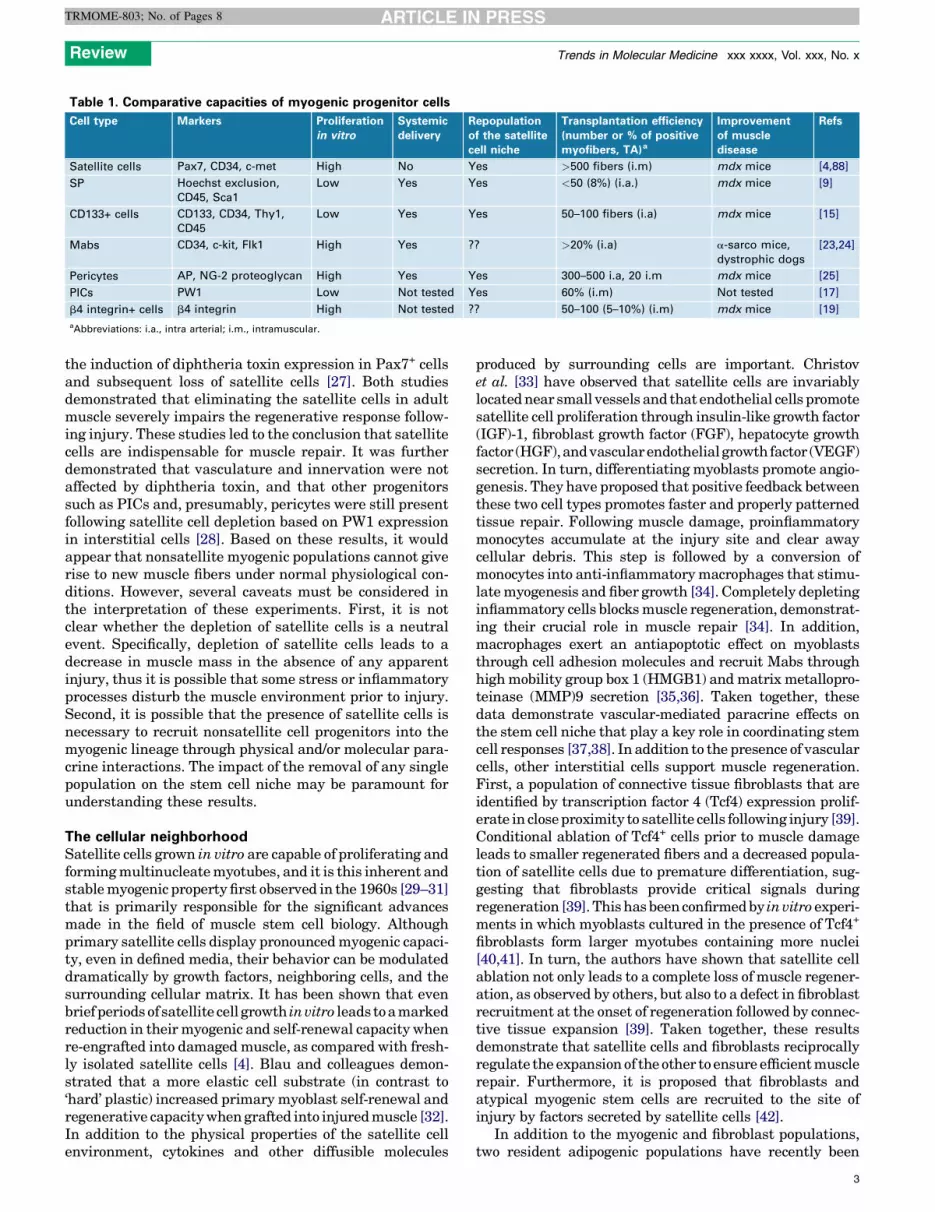
Table 1. Comparative capacities of myogenic progenitor cells
Cell type Markers Proliferation
in vitro
Systemic
delivery
Repopulation
of the satellite
cell niche
Transplantation efficiency
(number or % of positive
myofibers, TA)a
Improvement
of muscle
disease
Refs
Satellite cells Pax7, CD34, c-met High No Yes >500 fibers (i.m) mdx mice [4,88]
SP Hoechst exclusion,
CD45, Sca1
Low Yes Yes <50 (8%) (i.a.) mdx mice [9]
CD133+ cells CD133, CD34, Thy1,
CD45
Low Yes Yes 50–100 fibers (i.a) mdx mice [15]
Mabs CD34, c-kit, Flk1 High Yes ?? >20% (i.a) a-sarco mice,
dystrophic dogs
[23,24]
Pericytes AP, NG-2 proteoglycan High Yes Yes 300–500 i.a, 20 i.m mdx mice [25]
PICs PW1 Low Not tested Yes 60% (i.m) Not tested [17]
b4 integrin+ cells b4 integrin High Not tested ?? 50–100 (5–10%) (i.m) mdx mice [19]
aAbbreviations: i.a., intra arterial; i.m., intramuscular.
Review Trends in Molecular Medicine xxx xxxx, Vol. xxx, No. x
TRMOME-803; No. of Pages 8
the induction of diphtheria toxin expression in Pax7+ cellsand subsequent loss of satellite cells [27]. Both studiesdemonstrated that eliminating the satellite cells in adultmuscle severely impairs the regenerative response follow-ing injury. These studies led to the conclusion that satellitecells are indispensable for muscle repair. It was furtherdemonstrated that vasculature and innervation were notaffected by diphtheria toxin, and that other progenitorssuch as PICs and, presumably, pericytes were still presentfollowing satellite cell depletion based on PW1 expressionin interstitial cells [28]. Based on these results, it wouldappear that nonsatellite myogenic populations cannot giverise to new muscle fibers under normal physiological con-ditions. However, several caveats must be considered inthe interpretation of these experiments. First, it is notclear whether the depletion of satellite cells is a neutralevent. Specifically, depletion of satellite cells leads to adecrease in muscle mass in the absence of any apparentinjury, thus it is possible that some stress or inflammatoryprocesses disturb the muscle environment prior to injury.Second, it is possible that the presence of satellite cells isnecessary to recruit nonsatellite cell progenitors into themyogenic lineage through physical and/or molecular para-crine interactions. The impact of the removal of any singlepopulation on the stem cell niche may be paramount forunderstanding these results.
The cellular neighborhoodSatellite cells grown in vitro are capable of proliferating andforming multinucleate myotubes, and it is this inherent andstable myogenic property first observed in the 1960s [29–31]that is primarily responsible for the significant advancesmade in the field of muscle stem cell biology. Althoughprimary satellite cells display pronounced myogenic capaci-ty, even in defined media, their behavior can be modulateddramatically by growth factors, neighboring cells, and thesurrounding cellular matrix. It has been shown that evenbrief periods of satellite cell growth in vitro leads to a markedreduction in their myogenic and self-renewal capacity whenre-engrafted into damaged muscle, as compared with fresh-ly isolated satellite cells [4]. Blau and colleagues demon-strated that a more elastic cell substrate (in contrast to‘hard’ plastic) increased primary myoblast self-renewal andregenerative capacity when grafted into injured muscle [32].In addition to the physical properties of the satellite cellenvironment, cytokines and other diffusible molecules
produced by surrounding cells are important. Christovet al. [33] have observed that satellite cells are invariablylocated near small vessels and that endothelial cells promotesatellite cell proliferation through insulin-like growth factor(IGF)-1, fibroblast growth factor (FGF), hepatocyte growthfactor (HGF), and vascular endothelial growth factor (VEGF)secretion. In turn, differentiating myoblasts promote angio-genesis. They have proposed that positive feedback betweenthese two cell types promotes faster and properly patternedtissue repair. Following muscle damage, proinflammatorymonocytes accumulate at the injury site and clear awaycellular debris. This step is followed by a conversion ofmonocytes into anti-inflammatory macrophages that stimu-late myogenesis and fiber growth [34]. Completely depletinginflammatory cells blocks muscle regeneration, demonstrat-ing their crucial role in muscle repair [34]. In addition,macrophages exert an antiapoptotic effect on myoblaststhrough cell adhesion molecules and recruit Mabs throughhigh mobility group box 1 (HMGB1) and matrix metallopro-teinase (MMP)9 secretion [35,36]. Taken together, thesedata demonstrate vascular-mediated paracrine effects onthe stem cell niche that play a key role in coordinating stemcell responses [37,38]. In addition to the presence of vascularcells, other interstitial cells support muscle regeneration.First, a population of connective tissue fibroblasts that areidentified by transcription factor 4 (Tcf4) expression prolif-erate in close proximity to satellite cells following injury [39].Conditional ablation of Tcf4+ cells prior to muscle damageleads to smaller regenerated fibers and a decreased popula-tion of satellite cells due to premature differentiation, sug-gesting that fibroblasts provide critical signals duringregeneration [39]. This has been confirmed by in vitro experi-ments in which myoblasts cultured in the presence of Tcf4+
fibroblasts form larger myotubes containing more nuclei[40,41]. In turn, the authors have shown that satellite cellablation not only leads to a complete loss of muscle regener-ation, as observed by others, but also to a defect in fibroblastrecruitment at the onset of regeneration followed by connec-tive tissue expansion [39]. Taken together, these resultsdemonstrate that satellite cells and fibroblasts reciprocallyregulate the expansion of the other to ensure efficient musclerepair. Furthermore, it is proposed that fibroblasts andatypical myogenic stem cells are recruited to the site ofinjury by factors secreted by satellite cells [42].
In addition to the myogenic and fibroblast populations,two resident adipogenic populations have recently been
3

Box 2. Muscle diseases
Muscle diseases are primarily congenital, including most notably
the muscular dystrophies (Duchenne, Emery–Dreyfus and Limb–
Girdle), centronuclear myopathies, and neuromuscular diseases
(amyotrophic lateral sclerosis and spinal muscular atrophy). Mus-
cular dystrophies are characterized by progressive muscle weakness
due to mutations in sarcolemmal proteins (dystrophin, sarcoglycans
and dysferlin), nuclear proteins (emerin and lamin A/C), or extra-
cellular proteins (collagen-6 and a-2 laminin). Although clinical
symptoms are well described, no treatment has yet been fully
successful.
Most muscular dystrophies involve an eventual exhaustion of the
stem cell pool or a change in their fate, in which the muscle tissue
becomes infiltrated with fibrotic and fat tissue. This evolution is
similar to sarcopenia that occurs during physiological aging,
involving a gradual loss of muscle mass and a decline in
regenerative capacity.
In the mdx mouse model, the satellite cell population is reduced,
as compared to healthy mice, and becomes depleted following
repeated cycles of degeneration/regeneration. These repeated
cycles of stem cell activation and proliferation lead to telomere
shortening [87] and have been proposed to lead to the accumulation
of mutations in key regulatory genes required for proper self-
renewal and myogenic competence. Therefore, although Duchenne
muscular dystrophy (DMD) disease progression is driven by
dystrophin deficiency, the pathology exacerbates as a result of
stem cell dysfunction. In this context, therapies designed to promote
stem cell competence may ultimately slow disease progression.
Review Trends in Molecular Medicine xxx xxxx, Vol. xxx, No. x
TRMOME-803; No. of Pages 8
identified in skeletal muscle. Mesenchymal progenitors arecharacterized by platelet-derived growth factor receptor(PDGFR)a expression [43], and fibro/adipogenic progenitors(FAPs) have been isolated based upon stem cell antigen 1(Sca1) expression [44]. These cells display a strong adipo-genic potential in vitro and differentiate into fat whenengrafted into pathological muscle, which is not the casewhen engrafted into healthy muscle. In addition, FAPs areactivated upon injury and promote myoblasts differentia-tion through cell–cell signaling [44], whereas adipogenesisof PDGFRa+ cells is strongly inhibited by myotubes [43]. It istempting to speculate that mesenchymal PDGFRa+ cellsand FAPs are overlapping populations; nonetheless, theseadipogenic progenitors share the properties of adoptingdifferent fates depending on the surrounding environment,as well as promoting differentiation of neighboring myogen-ic progenitors [44]. The authors propose that a balancebetween satellite-cell-dependent myogenesis and PDGFRa+
cell/FAP-dependent adipogenesis regulates muscle homeo-stasis and regeneration. Taken together, these data clearlydemonstrate interactions between multiple resident cellpopulations that promote muscle progenitor activation.The deregulation of any single cell population in muscletissue is therefore likely to have a strong impact on all theresident populations and, therefore, must be taken intoaccount when proposing a ‘central’ role for any single celltype during the regenerative process (Figure 1). Similarly,changes in this local environment may contribute to musclepathologies and age-related loss of muscle stem cell compe-tence (Figure 2).
Age-related loss of regenerative capacity and the stemcell neighborhoodA progressive loss of stem cell competence occurs with ageand is associated with chronic diseases in mammals(Box 2). In diseased and aged muscle, myofibers are
Embryo Juvenile
Pax7-dependent Pax7-independent
A
7wd21d0
Figure 2. Dynamics of stem cell competence with age. At birth, high numbers of stem c
depending on the tissue. In skeletal muscle, this juvenile period is characterized by rapid
and able to contribute to multiple tissue lineages [12,17,43]. In the mouse, Pax7 is require
[2,67,68]; after 3 weeks, regeneration can occur in the absence of Pax7 [68]. During adult
cell competence) continues to decrease due to an increase in inhibitory factors in th
compromised. An additional decline in stem cell competence occurs in aged muscle.
postnatal skeletal muscle stem cell behavior: a juvenile phase (0–3 weeks); adult phase
4
replaced by fat and fibrous tissue and the remaining fibersdecrease in mass. The number of satellite cells in muscledeclines soon after birth from 30% of myonuclei in theneonate to 4% in the adult, followed by a small decrease to2% in the old mouse [45]. It has been demonstrated thatsatellite cells undergo a decrease in levels of Pax7 expres-sion, leading to a loss of myogenicity accompanied byincreased levels of apoptosis [46]. However, not all satellitecells appear to undergo this decrease and retain myogenicand self-renewal capacities comparable to those of young
dult Aged
Stem cell number
18m
Stem cell competence
TRENDS in Molecular Medicine
ells are followed by a dramatic decrease within the first days and/or weeks of life,
postnatal growth and the presence of a variety of stem cells that are highly plastic
d for both postnatal muscle growth and regeneration during the first 3 weeks of life
life, stem cell number is maintained, however, their capacity to repair muscle (stem
eir environment until about 18 months, at which point, regenerative capacity is
Taken together, these results reveal three distinct genetic and cellular phases of
(3 weeks to �18 months); and an aged phase (18 months to end of life).

Review Trends in Molecular Medicine xxx xxxx, Vol. xxx, No. x
TRMOME-803; No. of Pages 8
satellite cells, suggesting that other factors contribute toage-related changes in muscle regenerative capacity [45–47]. An aged-related delay in the early inflammatory re-sponse following muscle injury was reported in mice, al-though this did not impair the regeneration process [48]. Invivo, proliferative and regenerative capacities of satellitecells are restored in old mice when exposed to circulatingfactors from young mice [49]. By contrast, serum obtainedfrom old mice provokes an old age muscle regenerativephenotype when injected into young mice. This old-age-inducing effect has been attributed to elevated circulatingWnt molecules, activating downstream targets such asAxin2 and b-catenin in aged satellite cells, and leadingto conversion to a fibrogenic lineage [50]. Conversely,canonical Wnt signaling inhibition by Frizzled-relatedprotein 3 or Dickkopf-related protein 1 (DKK1) in old micerestores regenerative potential [50]. Wnt molecules may besecreted by tissue-resident endothelial precursors in oldmuscle. Specifically, endothelial precursors are activatedduring neoangiogenesis following muscle injury and inhib-it myoblast proliferation through Wnt3a production[51,52]. In addition, impaired muscle regeneration in agedanimals has been linked to a decline in Notch signaling[53]. Insufficient upregulation of the Notch ligand, Delta-1,in satellite cells following injury in old animals leads to adecrease in their myogenic capacities, which can be re-stored by forced activation of Notch. In turn, in vivoinhibition of Notch signaling reduces muscle regenerationin young mice and results in a phenotype similar to that ofold muscle [53]. In addition, Notch signaling has recentlybeen shown to be necessary for satellite cell maintenance,such that satellite cells undergo accelerated terminal dif-ferentiation without self-renewal in the absence of Notch,resulting in satellite cell depletion [54]. Loss of Notchactivation in aged muscle has been associated with highlevels of transforming growth factor (TGF)b-1 and cyclin-dependent kinase (cdk) inhibitors in satellite cells [55].Abnormally high levels of pSmad-3 in aged satellite cellsimpair regenerative capacity, whereas regenerative com-petence can be restored in vivo by pSmad-3 shRNA block-ade. Activation of Notch blocks the pSmad-3-mediatedupregulation of the cdks p15, p16, p21 and p27, whereasinhibition of Notch induces these cdks [55,56]. Therefore,endogenous Notch and pSmad-3 antagonize each other inthe control of satellite cell proliferation, and deregulationof this balance in old animals is proposed to lead to adecrease in regenerative potential. These data reveal thatthe progressive decrease in regenerative capacity duringmuscle aging is due to a progressive switch from Notch toWnt/TGF-b molecules secretion in the stem cell environ-ment, rather than intrinsic loss of myogenic cell compe-tence.
Can we gain insight from lower vertebrateregeneration?Fish and amphibians retain the capacity to regeneratetissues fully throughout life, whereas progressive loss ofregenerative capacity is common in mammals. Specifically,the adult zebrafish heart can regenerate following ventric-ular resection [57], whereas mammals lose this capacitysoon after birth [58]. A striking demonstration of stem cell
capacity in lower vertebrates was recently reported in newtlens subjected to 15 rounds of injury over 16 years withoutany decline in regenerative capacity [59]. Finally, in addi-tion to cell and organ replacement, amphibians can form ablastema following limb amputation, consisting of dedif-ferentiated mesenchymal cells that will repattern andredifferentiate to generate the entire appendage. Althoughlimb amputation in amphibians activates resident Pax7+
satellite cells [60], a concomitant dedifferentiation of ma-ture fibers is also observed, which give rise to mononucle-ated cells, accounting for approximately 30% of theblastema [61]. Although dedifferentiation is an extremelyrare event in mammals, one recent report suggests that asimilar process occurs in skeletal muscle upon injury, aswitnessed with a Cre-Lox-bgal system based on the ex-pression of muscle creatine kinase to tag specifically dif-ferentiated multinuclear myofibers [62]. In this study,mononuclear bgal+ cells were detected, among which somewere also Pax7+ and capable of redifferentiating into myo-tubes in vitro after injury [62]. Although the contribution ofdedifferentiated myofibers during muscle regenerationremains a subject of scrutiny, this study suggests thatmyofibers are an additional source of myogenic progenitorsfor muscle repair.
Understanding the mechanisms underlying dedifferen-tiation promises tremendous therapeutic applications.Blau and colleagues recently demonstrated that commit-ted myoblasts are reprogrammed by inactivating the tu-mor suppressors retinoblastoma protein (Rb) and ARF[63]. Upon Rb and p16/19 inactivation, myonuclei re-en-tered the cell cycle and myotubes dedifferentiated in mono-nucleated cells. These cells expand under growthconditions and redifferentiate into mature myotubes. Fol-lowing engraftment, these cells are incorporated into pre-existing fibers [63]. Similarly, Conboy and colleagues haveshown that inhibitors of tyrosine phosphatases and apo-ptosis provoke the dedifferentiation of myotubes into myo-genic competent mononucleated cells. They obtained‘reprogrammed’ muscle progenitors expressing Pax7 andMyoD capable of differentiating into skeletal muscle invitro and in vivo [64]. Even if dedifferentiation does notnormally occur during mammalian tissue regeneration,the underlying cellular machinery has not been lost duringmammalian radiation and can be artificially reactivated.Presumably this loss of cellular plasticity and stem cellcompetence arose during evolution, implying that the lossof this regenerative capacity carries some unique advan-tage. Moreover, the observation that genes involved intumor suppression need to be deactivated, at least tran-siently, in order to provoke this process in mammalssuggests that there may have been an evolutionarytrade-off between mechanisms preventing cancer andmechanisms supporting robust regenerative capacities.
Adolescence–a critical period for stem cells?A preprogrammed adult body size, and by consequence,organ size, is a hallmark of mammals, whereas many lowervertebrates display large variations in adult body sizeand, in some cases, postnatal growth is maintained through-out life (reviewed in [65,66]). The continuous growth oflower vertebrates is presumably linked to their enhanced
5

Review Trends in Molecular Medicine xxx xxxx, Vol. xxx, No. x
TRMOME-803; No. of Pages 8
regenerative capacities, and recent studies have demon-strated that mammals possess an enhanced regenerativecapacity during the initial days or weeks after birth. In thecase of the heart, which can regenerate in fish but not inmice, it has recently been shown that partial ventricleresection of the mouse heart performed within the firstpostnatal day provokes complete regeneration through in-duction of heart cardiomyocyte proliferation; this capacity islost by 1 week [58]. In skeletal muscle, genetic evidencehighlights the roles of Pax7 in postnatal skeletal musclegrowth. Mice constitutively lacking Pax7 display a normalpool of satellite cells upon birth but their number rapidlydeclines by 2–3 weeks after birth concomitant with poorpostnatal growth and survival [2]. Furthermore, these miceshow very poor muscle regenerative capacity, which ledinitially to the proposal that Pax7 is required for satellitecell maintenance and self-renewal [2,67]. However, in thecontext of a conditional Pax7 mutant, it was found that Pax7is dispensable for satellite cell function when inactivatedspecifically in the adult [68]. By contrast, when Pax7 isinactivated between birth and 3 weeks of age, postnatalmuscle development and regenerative capacity is abrogated,with the most severe phenotypes corresponding to ablationof Pax7 expression during the first week of life [68]. Theseresults demonstrate that there is a critical period near 3weeks of age when the muscle stem cell niche undergoes acritical change from juvenile to adult. Whether this change isdue to progenitors entering a more stable quiescent state orsome other unidentified event, including changes in the stemcell neighborhood, remains to be determined.
Genomic imprinting – a mammalian feature of stemcellsRecent studies have implicated parentally imprinted genesin the regulation of postnatal growth and adult somatic stemcells. Parental imprinting is an epigenetic control specific tomammals that involves silencing one allele, and it occurs on<1% of the human and mouse genome (reviewed in [69,70]).Several parentally imprinted genes regulate embryonic de-velopment [71,72]. As a group, they are expressed at highlevels in fetal and newborn tissues and decline within thefirst few weeks after birth in mice [73,74]. Our laboratoryidentified the parentally imprinted gene PW1/Peg3 in ascreen designed to isolate early stem cell markers in theskeletal muscle lineage [75]. Subsequent analyses haverevealed PW1/Peg3 expression is restricted to stem cellsin all adult tissues examined, including the skin, blood,bone, testes, and central nervous system (CNS) [76]. Theexpression of a universal adult stem cell marker lendssupport to the idea that common regulatory mechanismsunderlie adult stem cell function regardless of tissue origin; anotion that has been proposed for many years but haspreviously lacked much supporting data [77]. Our findingswere followed by the discovery of Goodell and colleagues thata group of parentally imprinted genes including p57, H19,mesoderm-specific transcript (Mest), Peg3/PW1, Dlk1, andinsulin-like growth factor 2 (IGF2) are expressed in adultsomatic stem cells but not in their differentiated progeny[74]. In addition, two groups have recently demonstratedthat p57, a well-known regulator of the cell cycle, plays a rolein maintaining the hematopoietic stem cell pool [78,79].
6
Similarly, Dlk1 has been shown to be important for skeletalmuscle growth and differentiation [80] and neurogenic pro-gression [81]. Moreover, these authors have demonstratedthat neural stem cells selectively undergo a ‘relaxation ofimprinting’ during postnatal development that is requiredfor proper neurogenesis [81]. This finding raises the inter-esting possibility that biallelic expression of imprinted genesmay constitute an important regulatory event during stemcell progression and differentiation. As most but not allparentally imprinted genes are shared among all verte-brates, it will be of interest to determine whether the sameset of genes are expressed in lower vertebrate stem cells orwhether this set of imprinted genes is specific to mammalianstem cells.
Concluding remarks and future perspectivesMuscle growth and maintenance during the lifetime of ananimal reveals at least three distinct stages after birth –juvenile, adult, and old age – during which stem cells changein behavior and competence. The juvenile period in mice (0–3 weeks) is characterized by pronounced growth and amarked increase in muscle mass and size. In this context,stem cells are required to build the muscle in young mice. Asstated above, the switch from a Pax7-dependent to a Pax7-independent state at 3 weeks old, coinciding with a declinein the rate of postnatal growth, provides direct geneticevidence that the stem cells undergo a change in theirgenetic circuitry during this transition, likely reflecting achange in their role. This change may also reflect therequirement for Pax7 in the myogenicity of PICs duringpostnatal growth [17]. In the context of muscular dystro-phies, this raises the question as to whether it is possible todevelop therapeutics that can induce or maintain juvenilestem cells that may represent a more competent and plasticstem cell population, as compared with adult stem cells.Whether imprinted gene regulation or tumor suppressorgenes underlie the ‘more limited’ potentials of mammalianstem cells as opposed to those in lower vertebrates may alsooffer potential therapeutic solutions. Why mammals andlower vertebrates differ in their regenerative potential cannow be addressed with the advent of high-throughput‘omics’ approaches. Determining how the niche changes,which signals are present during each of the postnatalstages, and what role other cells in the niche play willprovide targets for therapeutic development that promiseto be more tractable than direct stem cell engraftment. Assuch, the field of adult stem cells is entering a largerevolutionary context. Should we seek to induce amphibi-an-like characteristics in mammalian stem cells, we willneed to establish a clearer understanding of what distin-guishes juvenile postnatal tissue from adult and old tissueas well as fully appreciate why mammals have evolution-arily acquired this more limited potential and what Faust-ian bargain may have been made along the way.
AcknowledgmentsWe gratefully thank Drs V. Besson and K. Bismuth for fruitful discussionsand critical reading of the manuscript. This work was supported by theFrench Ministry of Research ‘Chaire d’Excellence’ and the MuscularDystrophy Association of America to D.S. and the European CommunitySeventh Framework Program projects OPTISTEM (Optimization of stemcell therapy for degenerative epithelial and muscle diseases contract

Review Trends in Molecular Medicine xxx xxxx, Vol. xxx, No. x
TRMOME-803; No. of Pages 8
number Health-F5-2009-223098) and ENDOSTEM (Activation ofvasculature associated stem cells and muscle stem cells for the repairand maintenance of muscle tissue-agreement number 241440). TheMyology Group is the beneficiary of a Strategic Plan Support from theAssociation Francaise contre les Myopathies (AFM) and is affiliated withthe Institute of Myology. A.P. was supported by the French Ministry ofResearch and the Fondation pour la Recherche Medicale (Programme‘Espoirs de la Recherche’, number FDT20110922527). The authors have noconflict of interest.
References1 Mauro, A. (1961) Satellite cell of skeletal muscle fibers. J. Biophys.
Biochem. Cytol. 9, 493–4952 Seale, P. et al. (2000) Pax7 is required for the specification of myogenic
satellite cells. Cell 102, 777–7863 Burkin, D.J. and Kaufman, S.J. (1999) The alpha7beta1 integrin in
muscle development and disease. Cell Tissue Res. 296, 183–1904 Montarras, D. et al. (2005) Direct isolation of satellite cells for skeletal
muscle regeneration. Science 309, 2064–20675 Relaix, F. et al. (2005) A Pax3/Pax7-dependent population of skeletal
muscle progenitor cells. Nature 435, 948–9536 Dhawan, J. and Rando, T.A. (2005) Stem cells in postnatal myogenesis:
molecular mechanisms of satellite cell quiescence, activation andreplenishment. Trends Cell Biol. 15, 666–673
7 Zammit, P.S. et al. (2004) Muscle satellite cells adopt divergent fates: amechanism for self-renewal? J. Cell Biol. 166, 347–357
8 Olguin, H.C. and Olwin, B.B. (2004) Pax-7 up-regulation inhibitsmyogenesis and cell cycle progression in satellite cells: a potentialmechanism for self-renewal. Dev. Biol. 275, 375–388
9 Gussoni, E. et al. (1999) Dystrophin expression in the mdx mouserestored by stem cell transplantation. Nature 401, 390–394
10 Ferrari, G. et al. (1998) Muscle regeneration by bone marrow-derivedmyogenic progenitors. Science 279, 1528–1530
11 Goodell, M.A. et al. (1996) Isolation and functional properties of murinehematopoietic stem cells that are replicating in vivo. J. Exp. Med. 183,1797–1806
12 Jackson, K.A. et al. (1999) Hematopoietic potential of stem cellsisolated from murine skeletal muscle. Proc. Natl. Acad. Sci. U.S.A.96, 14482–14486
13 Asakura, A. et al. (2002) Myogenic specification of side population cellsin skeletal muscle. J. Cell Biol. 159, 123–134
14 Torrente, Y. (2004) Human circulating AC133+ stem cells restoredystrophin expression and ameliorate function in dystrophic skeletalmuscle. J. Clin. Investig. 114, 182–195
15 Benchaouir, R. et al. (2007) Restoration of human dystrophin followingtransplantation of exon-skipping-engineered DMD patient stem cellsinto dystrophic mice. Cell Stem Cell 1, 646–657
16 Torrente, Y. et al. (2007) Autologous transplantation of muscle-derivedCD133+ stem cells in Duchenne muscle patients. Cell Transplant. 16,563–577
17 Mitchell, K.J. et al. (2010) Identification and characterization of a non-satellite cell muscle resident progenitor during postnatal development.Nat. Cell Biol. 12, 257–266
18 Young, V.R. (1974) Regulation of protein synthesis and skeletal musclegrowth. J. Anim. Sci. 38, 1054–1070
19 Liadaki, K. et al. (2012) beta4 integrin marks interstitial myogenicprogenitor cells in adult murine skeletal muscle. J. Histochem.Cytochem. 60, 31–44
20 De Angelis, L. et al. (1999) Skeletal myogenic progenitors originatingfrom embryonic dorsal aorta coexpress endothelial and myogenicmarkers and contribute to postnatal muscle growth andregeneration. J. Cell Biol. 147, 869–878
21 Minasi, M.G. et al. (2002) The mesoangioblast a multipotent, self-renewing cell that originates from the dorsal aorta anddifferentiates into most mesodermal tissues. Development 129,2773–2783
22 Tonlorenzi, R. et al. (2007) Isolation and characterization ofmesoangioblasts from mouse, dog, and human tissues. Curr. Protoc.Stem Cell Biol. Chapter 2, Unit 2B 1
23 Sampaolesi, M. et al. (2003) Cell therapy of alpha-sarcoglycan nulldystrophic mice through intra-arterial delivery of mesoangioblasts.Science 301, 487–492
24 Sampaolesi, M. et al. (2006) Mesoangioblast stem cells amelioratemuscle function in dystrophic dogs. Nature 444, 574–579
25 Dellavalle, A. et al. (2007) Pericytes of human skeletal muscle aremyogenic precursors distinct from satellite cells. Nat. Cell Biol. 9,255–267
26 Dellavalle, A. et al. (2011) Pericytes resident in postnatal skeletalmuscle differentiate into muscle fibres and generate satellite cells.Nat. Commun. 2, 499
27 Lepper, C. et al. (2011) An absolute requirement for Pax7-positivesatellite cells in acute injury-induced skeletal muscle regeneration.Development 138, 3639–3646
28 Sambasivan, R. et al. (2011) Pax7-expressing satellite cells areindispensable for adult skeletal muscle regeneration. Development138, 3647–3656
29 Konigsberg, I.R. (1961) Cellular differentiation in colonies derived fromsingle cells platings of freshly isolated chick embryo muscle cells. Proc.Natl. Acad. Sci. U.S.A. 47, 1868–1872
30 Konigsberg, I.R. (1961) Some aspects of myogenesis in vitro.Circulation 24, 447–457
31 Bischoff, R. (1975) Regeneration of single skeletal muscle fibers invitro. Anat. Rec. 182, 215–235
32 Gilbert, P.M. et al. (2010) Substrate elasticity regulates skeletal musclestem cell self-renewal in culture. Science 329, 1078–1081
33 Christov, C. et al. (2007) Muscle satellite cells and endothelial cells:close neighbors and privileged partners. Mol. Biol. Cell 18, 1397–1409
34 Arnold, L. et al. (2007) Inflammatory monocytes recruited after skeletalmuscle injury switch into antiinflammatory macrophages to supportmyogenesis. J. Exp. Med. 204, 1057–1069
35 Sonnet, C. et al. (2006) Human macrophages rescue myoblasts andmyotubes from apoptosis through a set of adhesion molecular systems.J. Cell Sci. 119, 2497–2507
36 Lolmede, K. et al. (2009) Inflammatory and alternatively activatedhuman macrophages attract vessel-associated stem cells, relying onseparate HMGB1- and MMP-9-dependent pathways. J. Leukoc. Biol.85, 779–787
37 Abou-Khalil, R. et al. (2009) Autocrine and paracrine angiopoietin 1/Tie-2 signaling promotes muscle satellite cell self-renewal. Cell StemCell 5, 298–309
38 Mounier, R. et al. (2011) Blood vessels and the satellite cell niche. Curr.Top. Dev. Biol. 96, 121–138
39 Murphy, M.M. et al. (2011) Satellite cells, connective tissue fibroblastsand their interactions are crucial for muscle regeneration. Development138, 3625–3637
40 Mathew, S.J. et al. (2011) Connective tissue fibroblasts and Tcf4regulate myogenesis. Development 138, 371–384
41 Cooper, S.T. et al. (2004) C2C12 co-culture on a fibroblast substratumenables sustained survival of contractile, highly differentiatedmyotubes with peripheral nuclei and adult fast myosin expression.Cell Motil. Cytoskeleton 58, 200–211
42 Wang, Y.X. and Rudnicki, M.A. (2012) Satellite cells, the engines ofmuscle repair. Nat. Rev. Mol. Cell Biol. 13, 127–133
43 Uezumi, A. et al. (2010) Mesenchymal progenitors distinct fromsatellite cells contribute to ectopic fat cell formation in skeletalmuscle. Nat. Cell Biol. 12, 143–152
44 Joe, A.W. et al. (2010) Muscle injury activates resident fibro/adipogenicprogenitors that facilitate myogenesis. Nat. Cell Biol. 12, 153–163
45 Shefer, G. et al. (2006) Satellite-cell pool size does matter: defining themyogenic potency of aging skeletal muscle. Dev. Biol. 294, 50–66
46 Collins, C.A. et al. (2007) A population of myogenic stem cells thatsurvives skeletal muscle aging. Stem Cells 25, 885–894
47 Carlson, M.E. and Conboy, I.M. (2007) Loss of stem cell regenerativecapacity within aged niches. Aging Cell 6, 371–382
48 Smythe, G.M. et al. (2008) Age influences the early events of skeletalmuscle regeneration: studies of whole muscle grafts transplantedbetween young (8 weeks) and old (13-21 months) mice. Exp.Gerontol. 43, 550–562
49 Conboy, I.M. et al. (2005) Rejuvenation of aged progenitor cells byexposure to a young systemic environment. Nature 433, 760–764
50 Brack, A.S. et al. (2007) Increased Wnt signaling during aging altersmuscle stem cell fate and increases fibrosis. Science 317, 807–810
51 Grenier, G. et al. (2007) Resident endothelial precursors in muscle,adipose, and dermis contribute to postnatal vasculogenesis. Stem Cells25, 3101–3110
7

Review Trends in Molecular Medicine xxx xxxx, Vol. xxx, No. x
TRMOME-803; No. of Pages 8
52 Trensz, F. et al. (2010) A muscle resident cell population promotesfibrosis in hindlimb skeletal muscles of mdx mice through the Wntcanonical pathway. Am. J. Physiol. Cell Physiol. 299, C939–C947
53 Conboy, I.M. et al. (2003) Notch-mediated restoration of regenerativepotential to aged muscle. Science 302, 1575–1577
54 Bjornson, C.R. et al. (2012) Notch signaling is necessary to maintainquiescence in adult muscle stem cells. Stem Cells 30, 232–242
55 Carlson, M.E. et al. (2009) Relative roles of TGF-beta1 and Wnt in thesystemic regulation and aging of satellite cell responses. Aging Cell 8,676–689
56 Carlson, M.E. et al. (2008) Imbalance between pSmad3 and Notchinduces CDK inhibitors in old muscle stem cells. Nature 454, 528–532
57 Poss, K.D. et al. (2002) Heart regeneration in zebrafish. Science 298,2188–2190
58 Porrello, E.R. et al. (2011) Transient regenerative potential of theneonatal mouse heart. Science 331, 1078–1080
59 Eguchi, G. et al. (2011) Regenerative capacity in newts is not altered byrepeated regeneration and ageing. Nat. Commun. 2, 384
60 Morrison, J.I. et al. (2006) Salamander limb regeneration involves theactivation of a multipotent skeletal muscle satellite cell population. J.Cell Biol. 172, 433–440
61 Echeverri, K. et al. (2001) In vivo imaging indicates muscle fiberdedifferentiation is a major contributor to the regenerating tailblastema. Dev. Biol. 236, 151–164
62 Mu, X. et al. (2011) Study of muscle cell dedifferentiation after skeletalmuscle injury of mice with a Cre-Lox system. PLoS ONE 6, e16699
63 Pajcini, K.V. et al. (2010) Transient inactivation of Rb and ARF yieldsregenerative cells from postmitotic mammalian muscle. Cell Stem Cell7, 198–213
64 Paliwal, P. and Conboy, I.M. (2011) Inhibitors of tyrosine phosphatasesand apoptosis reprogram lineage-marked differentiated muscle tomyogenic progenitor cells. Chem. Biol. 18, 1153–1166
65 Lui, J.C. and Baron, J. (2011) Mechanisms limiting body growth inmammals. Endocr. Rev. 32, 422–440
66 Johnston, I.A. et al. (2011) Growth and the regulation of myotomalmuscle mass in teleost fish. J. Exp. Biol. 214, 1617–1628
67 Oustanina, S. et al. (2004) Pax7 directs postnatal renewal andpropagation of myogenic satellite cells but not their specification.EMBO J. 23, 3430–3439
68 Lepper, C. et al. (2009) Adult satellite cells and embryonic muscleprogenitors have distinct genetic requirements. Nature 460, 627–631
69 Bartolomei, M.S. and Ferguson-Smith, A.C. (2011) Mammaliangenomic imprinting. Cold Spring Harb. Perspect. Biol. 3, http://dx.doi.org/10.1101/cshperspect.a002592
70 Kaneda, M. (2011) Genomic imprinting in mammals-epigeneticparental memories. Differentiation 82, 51–56
8
71 Ono, R. et al. (2006) Deletion of Peg10, an imprinted gene acquired froma retrotransposon, causes early embryonic lethality. Nat. Genet. 38,101–106
72 Sekita, Y. et al. (2008) Role of retrotransposon-derived imprinted gene.Nat. Genet. 40, 243–248
73 Lui, J.C. et al. (2008) An imprinted gene network that controlsmammalian somatic growth is down-regulated during postnatalgrowth deceleration in multiple organs. Am. J. Physiol. Regul.Integr. Comp. Physiol. 295, R189–R196
74 Berg, J.S. et al. (2011) Imprinted genes that regulate earlymammalian growth are coexpressed in somatic stem cells. PLoSONE 6, e26410
75 Relaix, F. et al. (1996) Pw1, a novel zinc finger gene implicated in themyogenic and neuronal lineages. Dev. Biol. 177, 383–396
76 Besson, V. et al. (2011) PW1 gene/paternally expressed gene 3 (PW1/Peg3) identifies multiple adult stem and progenitor cell populations.Proc. Natl. Acad. Sci. U.S.A. 108, 11470–11475
77 Varrault, A. et al. (2006) Zac1 regulates an imprinted gene networkcritically involved in the control of embryonic growth. Dev. Cell 11,711–722
78 Matsumoto, A. et al. (2011) p57 is required for quiescence andmaintenance of adult hematopoietic stem cells. Cell Stem Cell 9,262–271
79 Zou, P. et al. (2011) p57(Kip2) and p27(Kip1) cooperate to maintainhematopoietic stem cell quiescence through interactions with Hsc70.Cell Stem Cell 9, 247–261
80 Waddell, J.N. et al. (2010) Dlk1 is necessary for proper skeletal muscledevelopment and regeneration. PLoS ONE 5, e15055
81 Ferron, S.R. et al. (2011) Postnatal loss of Dlk1 imprinting in stem cellsand niche astrocytes regulates neurogenesis. Nature 475, 381–385
82 Kassar-Duchossoy, L. et al. (2005) Pax3/Pax7 mark a novel populationof primitive myogenic cells during development. Genes Dev. 19,1426–1431
83 Gros, J. et al. (2005) A common somitic origin for embryonic muscleprogenitors and satellite cells. Nature 435, 954–958
84 Reznik, M. (1969) Thymidine-3H uptake by satellite cells ofregenerating skeletal muscle. J. Cell Biol. 40, 568–571
85 Kuang, S. et al. (2007) Asymmetric self-renewal and commitment ofsatellite stem cells in muscle. Cell 129, 999–1010
86 Menasche, P. (2007) Skeletal myoblasts as a therapeutic agent. Prog.Cardiovasc. Dis. 50, 7–17
87 Sacco, A. et al. (2010) Short telomeres and stem cell exhaustion modelDuchenne muscular dystrophy in mdx/mTR mice. Cell 143, 1059–1071
88 Partridge, T.A. et al. (1989) Conversion of mdx myofibres fromdystrophin-negative to -positive by injection of normal myoblasts.Nature 337, 176–179


