Embed Size (px)
Citation preview
SAGE-Hindawi Access to ResearchParkinson’s DiseaseVolume 2010, Article ID 375462, 9 pagesdoi:10.4061/2010/375462
Research Article
Spinal Cord Pathology in Alpha-Synuclein Transgenic Mice
Sonja Mendritzki,1 Saskia Schmidt,1, 2 Teresa Sczepan,1 Xin-Ran Zhu,1 Daniel Segelcke,1
and Hermann Lubbert1, 2
1 Department of Animal Physiology, Biology, and Biotechnology, Ruhr-University Bochum, ND/5131, D-44780 Bochum, Germany2 International Graduate School of Neuroscience (IGSN), Ruhr-University Bochum, 44780 Bochum, Germany
Correspondence should be addressed to Sonja Mendritzki, [email protected]
Received 18 February 2010; Revised 4 May 2010; Accepted 6 June 2010
Academic Editor: Ted M. Dawson
Copyright © 2010 Sonja Mendritzki et al. This is an open access article distributed under the Creative Commons AttributionLicense, which permits unrestricted use, distribution, and reproduction in any medium, provided the original work is properlycited.
Accumulation of α-synuclein is observed in neurodegenerative diseases like Parkinson’s disease and Multiple System Atrophy.In previous studies with transgenic C57BL/6 mice overexpressing α-synuclein carrying the mutations A53T and A30P foundin Parkinson’s disease or with a parkin-null background, we reported severe mitochondrial impairments in neurons and to alarger extent in glial cells of the mesencephalon. Neuron death was not observed in the brain. Here we show that the mice showsevere motor impairments in behavioral tests. In addition, these mice exhibit astrocytic cell death in the spinal cord, accompaniedby extensive gliosis and microglial activation. This is shown by cell death staining and immunohistochemistry. Ultrastructuralanalyses revealed severe mitochondrial impairments not only in astrocytes, but also in oligodendrocytes and, to a small extent, inneurons. Thus, the transgenic mice show a profound pathology in glial cells of the spinal cord.
1. Introduction
Parkinson’s disease (PD) is a chronic neurodegenerative dis-ease associated with severe motor impairments, substantialmorbidity, and increased mortality. The neuropathology ofPD is characterized by an excessive loss of dopaminergicneurons in the substantia nigra, but other brain regions mayalso be affected. Most cases of PD are sporadic, with lateonset. However, 5–10% of PD cases are familial and canbe attributed to a mutation in genes like α-synuclein (α-syn) and parkin [1]. α-syn is a synaptic molecule and it isassumed that it is involved in membrane vesicle traffickingand cytoskeletal dynamics [2]. Furthermore, aggregations ofα-syn are specific for α-synuncleinopathies like PD, Lewybody disease, or Multiple System Atrophy (MSA) [3, 4]. Itseems to be that α-syn is degraded by Parkin, an E3 ubiquitinligase [5].
Several transgenic mouse models carrying α-syn andparkin mutations have been used to elucidate the molecularmechanisms of PD pathogenesis [6]. In previous studies,we generated mice carrying a deletion in the parkin geneand/or overexpressing the mutated human α-syn transgene
[7, 8]. These mice exhibit ultrastructural mitochondrialalterations in neurons and, interestingly, to a larger extentin glial cells of the mesencephalon, but they do not displaysevere histopathological hallmarks such as cell death in thesubstantia nigra. These data indicate that glial cells are activecontributors to the initiation or progression of PD [9, 10].This is supported by cell culture data suggesting abnormalfunctions of glial cells from mice carrying PD-inducing genemutations [11, 12]. Furthermore, glial cells have been shownto express proteins like superoxide dismutase 1, huntingtin,or ataxin which are linked to neurodegenerative diseases.Recent studies suggest an important role for glial cells alsoin other neurodegenerative diseases. For example, astrocytesplay a central role in the degeneration of spinal motorneurons in amyotrophic lateral sclerosis [13] and oligoden-drocytes are active contributors to neuronal degeneration inMSA [14, 15].
Several clinical studies reported the appearance ofnondopaminergic symptoms like impairments in anxietybehavior in early PD stages [16]. Recent literature suggeststhat pathologic manifestations are first detected in the spinalcord and then progressing from caudal to rostral until

2 Parkinson’s Disease
they reach the mesencephalon. This model, described byBraak and coworkers [17], provides an explanation for theappearance of nondopaminergic symptoms which precedecell death in the substantia nigra. Autonomic dysfunctionslike depression, sleep disturbances, or anxiety behavior arecontrolled by the lower brainstem and spinal ganglia [18].
Based on these reports, we now investigated whether acaudal to rostral progression could also be observed in ourtransgenic mice, since parkin and α-syn are expressed inthe spinal cord [19]. We found cell death, mitochondrialalterations, and severe inflammatory reactions in glial cellsof the spinal cords of the mutated mice. In addition,when looking at the motor abilities of the mice, we foundsevere motor impairments which seem to be related to thepathological changes in the spinal cord.
In summary, our transgenic mice carrying PD-inducinggene mutations exhibit more severe pathological impair-ments in glial cells of the spinal cord than in the mesen-cephalon, similar to the situation in early PD described byBraak and coworkers [17].
2. Methods
2.1. Mouse Strains. Two transgenic mouse lines were ana-lyzed: a homozygous α-syn line (C57BL/6J background)overexpressing doubly mutated (A30P, A53T) human (hm2)α-syn driven by the chicken β-actin (BAsyn) promoter asdescribed previously [7], here we used the BAsyn line 1.5 anda double-mutant mouse line carrying additionally a deletionof exon3 in the parkin gene (BAsyn/PaKO; F10 generation,99% C57BL/6J, and 1% 129SvJ). Control mice were age-matched nontransgenic littermates (LMs).
Housing and breeding of animals were performed inaccordance with the German guidelines of the animal careand use committee. All efforts were made to minimize thenumber of animals used and their suffering.
2.2. Tissue Preparation. Mice were anesthetized by an over-dose of ketamine (100 mg/kg) and rompun (5 mg/kg) andperfused transcardially with phosphate-buffered saline (PBS)followed by 4% paraformaldehyde (PFA) in 0.1 M phosphatebuffer (PB) for immunohistochemistry or 2% PFA plus 2.5%glutaraldehyde (GA) in PB for electron microscopy. Cervicalparts of the spinal cord were immediately removed andpostfixed.
2.3. Immunohistochemistry and Cell Death Staining. Cervi-cal parts of the spinal cord were embedded in paraffin.For sequential double-labeling spinal cords were cut into3 μm-thick sections, for analyses of α-syn aggregations,for cell death staining as well as for the investigations ofinflammatory processes spinal cords were cut into 18 μm-thick sections. The sections were deparaffinized and antigenretrieval (5–10 min cooking in 0.01 M citrate buffer, pH6.0) was performed (except for glial fibrillary acidic protein(GFAP) and myelin basic protein (MBP)). After preincuba-tion (3% normal serum), sections were treated with mouseantihuman α-syn (anti-syn211; 1 : 700, Invitrogen), astrocyte
marker rabbit anticow GFAP (anti-GFAP; 1 : 1000; Acris),oligodendrocyte marker goat antihuman MBP (anti-MBP;1 : 1000; Santa Cruz), microglia marker goat antihumanionized calcium-binding adaptor molecule 1 (anti-Iba1;1 : 250; Abcam), neuronal marker mouse antimouse neu-ronal nuclei (anti-NeuN; 1 : 700; Chemicon), goat antimousecathepsin X (anti-CATX; 1 : 200; R&D Systems), and goatantirat cathepsin S (anti-CATS; 1 : 200; Santa Cruz). Sectionswere stained with the corresponding biotinylated secondaryantibody (1 : 300, Axxora), ABC reagent (1 : 100; Axxora),and silver-gold intensification.
FluoroJade and silver-staining were used to detect degen-erating cells. Deparaffinized and rehydrated sections wereincubated in 0.06% potassium permanganate solution for15 min, rinsed with distilled water, and then incubated in asolution of 0.01% FluoroJade (Histo-Chem) in 0.1% aceticacid for 30 min. Silver staining was performed according tothe protocol of Gallyas et al. [20].
2.4. Transmission Electron Microscopy. Postfixed (2 h, 4◦C)parts of the cervical spinal cord were cut with a vibratome(100 μm thick), anterior horn samples microdissected andplaced in 2% OsO4 for 1 h, dehydrated with gradedconcentrations of ethanol and Epon-propylene oxide, andflat embedded in Epon. Representative ultrathin sectionswere collected on Formvar-coated grids and contrasted withuranyl acetate and lead citrate.
Ultrathin sections were visualized using a Phillips EM-410. Morphology and number of mitochondria were eval-uated in digitized electron microscopic images of 8 m-old animals. In total, 24 somata per cell type (neurons,astrocytes, and oligodendrocytes) taken from two vibratomesections of each animal were independently analyzed bytwo examiners blind with respect to the genotypes of themice. Ultrathin sections were selected at random from themiddle of the vibratome sections to avoid potential structuralirregularities possibly occurring on the surface.
Data are presented as a mean ± standard deviation(SD) of the means of the analyzed animals per genotype.Statistical analyses of ultrastructural changes compared tocorresponding age-matched LMs were carried out using theMann-Whitney Rank Sum Test (U-test, two tailed, alphalevel 0.05).
2.5. Elevated Plus Test. Mice were placed on a cross-shapedelevated maze with two open arms and two closed armswhich stands one meter above the floor. A video camerarecorded for 5 min the horizontal path length, time in closedarms, and duration and number of stops.
2.6. Footprints. To obtain the footprints, the hind paws ofthe mice were dipped in a dye and the mice were placedin a 100 cm long and 4.5 cm wide gangway. The floor waslined with white paper and the footprints were analyzed witha Footprint software. Data are presented as mean ± SD ofthe means of the analyzed animals per genotype. Statisticalanalyses were performed by using the Mann-Whitney RankSum Test (U-test, two tailed, alpha level 0.05).

Parkinson’s Disease 3
2
1
GFAP
(a)
2
1
syn211
(b)
3?
lba1
(c)
3?
syn211
(d)
MBP
(e)
syn211
(f)
NeuN
(g)
syn211
(h)
Figure 1: Light microscopic phenotyping of cells expressing the transgene in BAsyn/PaKO mouse brains. Photomicrographs of serial, 3 μm-thick sections of the spinal cord from BAsyn/PaKO mice ((a)–(h)). The syn211-immunostaining (b) colocalized with the marker GFAP (a),indicating the transgene expression in astrocytes. In the parts of the spinal cord where many Iba1-positive cells (c) are stained, it seemsthat only one cell could be also positive for syn211 (d), indicating that it is not clear if the hm α-syn is expressed in microglia. For MBP-immunopositive oligodendrocytes (e) and NeuN-immunopositive (g) neurons no match with syn211-immunostaining could be detected((f), (h)). Scale bars: (a)–(d): 30 μm; (e)–(h) 50 μm.
3. Results
3.1. Cellular Distribution of the hm α-Syn in the Spinal Cordof BAsyn and BAsyn/PaKO. We investigated and comparedthe distribution of the human α-syn in the transgenic α-syn mouse lines. Sequential immunoperoxidase labeling withα-syn- and cell type-specific antibodies was performed.Examples are shown for 8-month (m) old BAsyn/PaKO inFigures 1(a)–1(h). BAsyn/PaKO expressed the transgene inthe spinal cord almost exclusively in astrocytes (Figures 1(a)and 1(b)). In Figure 2(k), the α-syn distribution is shown fora whole cross section of the spinal cord from BAsyn/PaKO,indicating that the transgene is mostly expressed in whitematter. In this area we also found many GFAP-positive cells
(Figure 2(l)). It is not clear if the transgene is expressed inmicroglia. The parts of the spinal cord with a large amountof microglia showed nearly no α-syn expression (Figures1(c) and 1(d)). However, it cannot be excluded that thetransgene is expressed in few isolated microglia. Neither forimmunopositive oligodendrocytes nor for stained neuronsan expression of the transgene could be detected (Figures1(e)–1(h)). We found the same cellular distribution in BAsynmice (data not shown).
3.2. Severe α-Syn Aggregations, Inflammatory Reactions, andCell Death in the Spinal Cord of α-Syn Transgenics. Weanalyzed the cervical spinal cord of 3- and 8-m old BAsyn and

4 Parkinson’s Disease
Posterior funiculus(PF)
Lateral funiculus(LF)
Anterior funiculus (AF)
Posterior horn(PH)
Zona intermedia(ZI)
Anteriorhorn (AH)
(a)
BAsyn/PaKO
Fluorojade
(b)
Gallyas
(c)
GFAP
(d)
BAsyn/PaKOLM
hm α-asyn− proteinase K
(e) (k) (q)
hm α-asyn+ proteinase K
(f) (l) (r)
GFAP
(g) (m) (s)
Iba1
(h) (n) (t)
CATX
(i) (o) (u)
CATS
(j) (p) (v)
Figure 2: Immunohistochemical analyzes and cell death staining of the cervical spinal cord. Schematic illustration of the transversal spinalcord (a). Cell death staining by FluoroJade and Gallyas are shown in (b) and (c) for BAsyn/PaKO. An example showed a cell positive forGallyas and GFAP in adjacent slices ((c), (d)). Immunopositive structures of the cervical spinal cord for hm α-syn (with or without proteinaseK), GFAP, Iba1, CATX, and CATS are shown for LM in (e)–(j) and for BAsyn/PaKO in (k)–(p). (q)–(v) are higher magnifications of (k)–(p).Scale bars: (e)–(p): 200 μm; (q) and (r): 10 μm, (s)–(v): 100 μm, and details of (s)–(v): 10 μm.

Parkinson’s Disease 5
BAsyn/PaKO mice and their corresponding LM as illustratedin Figure 2. We examined the expression of the hm α-synwithout (Figures 2(s), 2(k), and 2(q)) and with (Figures 2(f),2(l), and 2(r)) pretreatment of the slices with proteinase K.After pretreatment of the slices with proteinase K hm, α-syn aggregations in the anterior horn (AH), zona intermedia(ZI), lateral funiculus (LF), and in the anterior funiculus(AF) of the spinal cord were visible in 8 m-old BAsyn/PaKO(Figures 2(l) and 2(r)). In contrast, no aggregations wereobserved in LM (Figure 2(f)).
Next, we investigated the expression of GFAP and Iba1and found an increased number of GFAP-positive cells(Figures 2(m) and 2(s)) as well as a stronger expression ofIba1 (Figures 2(n) and 2(t)) in the AH, ZI, LF, and in the AFof the spinal cord of 8 m-old BAsyn/PaKO compared to LM(Figures 2(g) and 2(h)), indicating the occurrence of severegliosis.
This glial cell activation in the old BAsyn/PaKO wasaccompanied by a higher expression of the lysosomal cysteinproteases CATX (Figures 2(o) and 2(u)) and CATS (Figures2(p) and 2(v)). Expression of these markers is an earlyindicator of inflammatory reactions. The two proteins werevisible in the same regions where gliosis was observed,different from the LM (Figures 2(i) and 2(j)).
In addition to the severe α-syn aggregations, gliosis andinflammation, FluoroJade (Figure 2(b)) and Gallyas silverstaining (Figure 2(c)) indicated the occurrence of cell deathin the LF and AF of the cervical spinal cord of 8 m-oldBAsyn/PaKO. Analyses of adjacent slices showed that the cellspositive for Gallyas cell death staining were also stained byastrocytes markers (Figures 2(c), 2(d)). No cell death couldbe detected in motor neurons located next to the astrocytes.
The same analyzes were performed with the monomu-tant line BAsyn and in two out of four animals we also foundα-syn aggregations, gliosis, inflammatory reactions as wellas cell death. However, two other animals did not displaythese pathological changes (data not shown). Furthermore,3 m-old mice were investigated and neither young BAsyn norBAsyn/PaKO displayed these alterations (data not shown).
3.3. Ultrastructural Impairments of Mitochondria in the SpinalCord of the Double Mutants. The mitochondrial ultrastruc-ture of neurons, astrocytes, and oligodendrocytes in the cer-vical spinal cord was examined by electron microscopy. Glialcells were counted if they contained a nucleus surroundedby cytoplasm and were classified according to their typicalultrastructural features [21, 22]. Briefly, astrocytes were iden-tified by their electron-lucent cytoplasm and the appearanceof their nuclei (thin rim of heterochromatin adjacent tonuclear membrane). Oligodendrocytes show electron-densecytoplasms and their nuclei are rich in clumped heterochro-matin close to the border of the nucleus. Mitochondriawere classified as structurally damaged when one or moreof the following alterations were observed: distorted ordisrupted cristae, detachment of or protrusions from theouter membrane, electron-lucent domains, or inclusions inthe matrix. At the age of 8 m BAsyn/PaKO showed onlya slight mitochondrial damage in neurons (Figures 3(d)
and 3(g)), whereas LM exhibited nearly no impairments(Figures 3(a) and 3(g)). In contrast, both astrocytes (Figures3(e) and 3(g)) and oligodendrocytes (Figures 3(e) and 3(g))of BAsyn/PaKO displayed a statistically significant increasein mitochondrial damage compared to the correspondingcells in LM (Figures 3(b), 3(c), and 3(g)). Oligodendrocytesexhibited the highest number of altered mitochondria.
3.4. Behavioral Deficits in Mono- and Double-Mutant Mice.The gait of the 8 m-old transgenic mice was investigatedby analyzing their footprints. The following parameterswere measured: stride length, stride width, foot length, areatouched, angle, and spread of toes 1–5 and 2–4 (Figure 4(a)).Examinations revealed a decreased stride length for bothmono- and double-mutants (Figures 4(b)–4(c)).
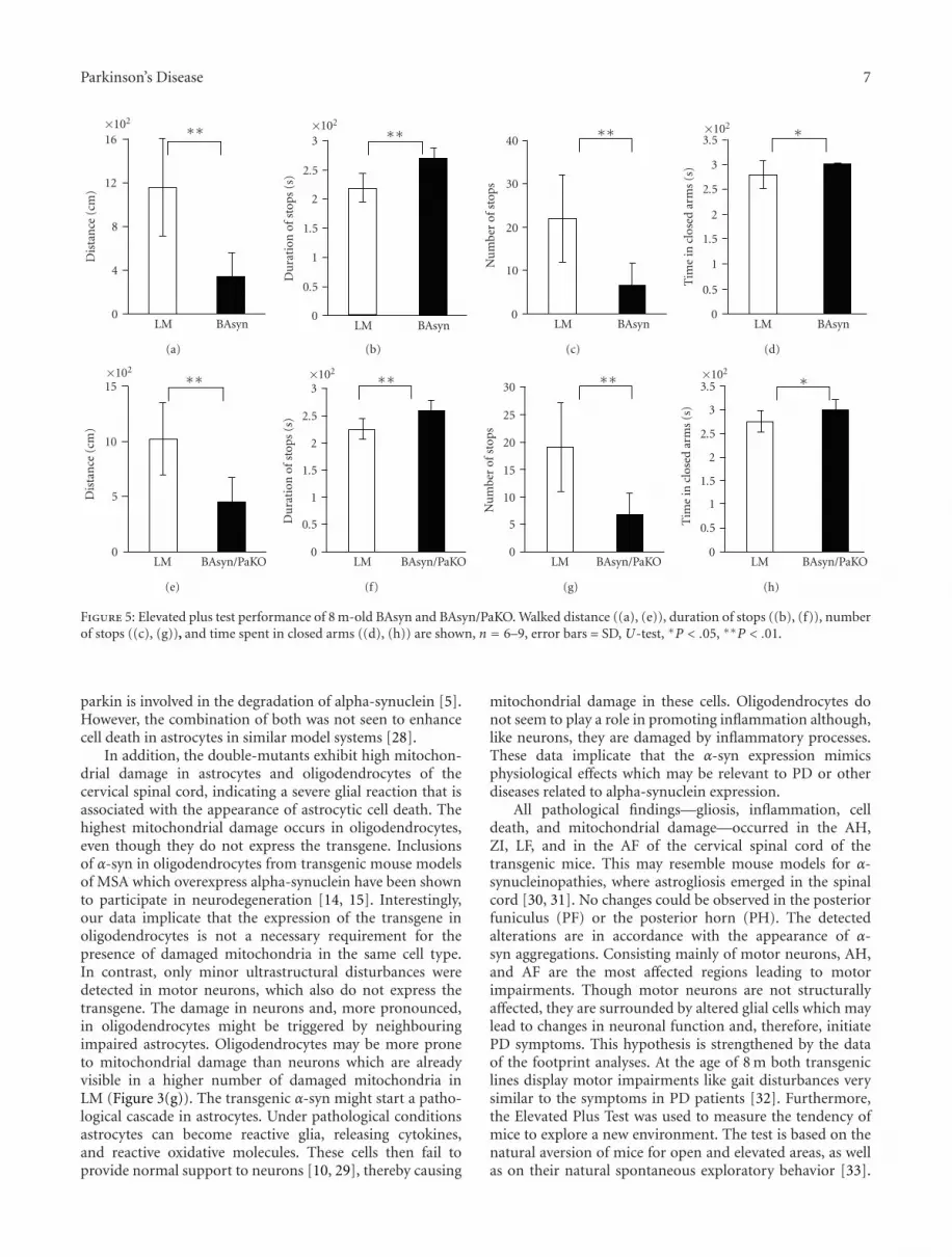
The Elevated Plus Test measures the anxiety as well asthe willingness of mice to explore a new environment. Fourdifferent parameters were analyzed: distance, time in closedarms, number of stops, and the duration of stops. Both 8 m-old BAsyn and BAsyn/PaKO walked shorter distances madefewer stops and longer breaks than LM. The transgenic micespent by trend more time in the closed arms, however, thisdifference is very low (Figures 5(a)–5(h)).
4. Discussion
While the pathological hallmark of PD is the degeneration ofdopaminergic neurons in the substantia nigra, it has recentlybeen suggested that dopaminergic neuron death is precededby pathological changes in more caudal CNS regions [17]. Ithas been proposed that such alterations are accompanied byα-syn aggregations in the spinal cord [17, 23]. These findingsraise new questions about the starting point of PD pathology.
We are analyzing transgenic mouse models overexpress-ing hm2 α-syn alone (BAsyn) or in combination with adeletion of the exon3 of the parkin gene (BAsyn/PaKO). Asthese mice do not exhibit gross pathological impairments inthe mesencephalon we investigated in this study the cervicalpart of the spinal cord according to the staging scheme ofBraak and coworkers [17, 23].
We first demonstrated for both mouse lines the expres-sion of hm2 α-syn in the spinal cord almost exclusively inastrocytes. For neurons, microglia, and oligodendrocytes noα-syn expression could be detected.
Following a proteinase K pretreatment α-syn aggrega-tions were visible in all analyzed double-mutant animals andin two out of four analyzed monomutants. These findingscould only be observed in 8 m- but not in 3 m-old animals.This is in agreement with previous studies of mouse models[24, 25] and humans [26] where α-syn aggregations onlyoccurred in the spinal cord but not in the substantia nigra.These results hint towards an early involvement of the spinalcord in the origination of PD.
In addition, inflammatory processes were increased inthe transgenic mice relative to LM. Transgenic mice exhibithigher numbers of GFAP-positive astrocytes and activatedmicroglia. Furthermore, the lysosomal cystein proteasesCATS and CATX are more abundantly expressed in the

6 Parkinson’s Disease
(a) (b) (c) (d)
(e) (f)
0
5
10
15
20
25
30
35
Mit
och
ondr
iald
amag
e(%
)
∗
∗
Neurons Astrocytes Oligodendrocytes
LMBAsyn/PaKO
(g)
Figure 3: Mitochondrial ultrastructure in the spinal cord of 8 m-old BAsyn/PaKO. Exemplary ultrastructural features of neurons, astrocytes,and oligodendrocytes are shown for LM ((a)–(c)) and BAsyn/PaKO ((d)–(f)). Higher magnifications show examples of healthy or damagedmitochondria. Scale bars: (a)–(f): 2 μm; higher magnification of (a)–(f): 0.5 μm. The quantification of mitochondrial alterations is shown in(g), n = 3, errors bars = SD, U-test, ∗P < .05.
b
(a)
0
5
10
15
Stri
dele
ngt
h(m
m)
∗
LM BAsyn
(b)
0
2
4
6
8
10
12
Stri
dele
ngt
h(m
m)
∗
LM BAsyn/PaKO
(c)
Figure 4: Footprint analyzes of BAsyn and BAsyn/PaKO. Footprints (a) and stride length ((b), (c)). The stride lengths of 8–10 mice pergroup were determined, error bars = SD, U-test, ∗P < .05.
cervical spinal cord of BAsyn and BAsyn/PaKO comparedto LM. These inflammatory processes are accompanied byongoing cell death, as shown by FluoroJade and Gallyasstaining. Analyzes of adjacent slices could identify most cellspositive for cell death staining as astrocytes.
Interestingly, these changes only occur during aging, as3 m-old mice are not affected. This reinforces the hypothesis
that early PD pathology builds up during aging [27]. Allfour BAsyn/PaKO mice analyzed displayed the pathologicalimpairments described above whereas the appearance ofinflammation and cell death occurred only in two out offour BAsyn animals. This suggests that the effect of theoverexpression of α-syn is enhanced by the deletion of theparkin gene. This may possibly be explained by the fact that
Parkinson’s Disease 7
0
4
8
12
16×102
Dis
tan
ce(c
m)
∗∗
LM BAsyn
(a)
0
0.5
1
1.5
2
2.5
3×10²
Du
rati
onof
stop
s(s
)
∗∗
LM BAsyn
(b)
0
10
20
30
40
Nu
mbe
rof
stop
s
∗∗
LM BAsyn
(c)
0
0.5
1
1.5
2
2.5
3
3.5×10²
Tim
ein
clos
edar
ms
(s)
∗
LM BAsyn
(d)
0
5
10
15×102
Dis
tan
ce(c
m)
∗∗
LM BAsyn/PaKO
(e)
0
0.5
1
1.5
2
2.5
3×10²
Du
rati
onof
stop
s(s
)
∗∗
LM BAsyn/PaKO
(f)
0
5
10
15
20
25
30
Nu
mbe
rof
stop
s
∗∗
LM BAsyn/PaKO
(g)
0
0.5
1
1.5
2
2.5
3
3.5×10²
Tim
ein
clos
edar
ms
(s)
∗
LM BAsyn/PaKO
(h)
Figure 5: Elevated plus test performance of 8 m-old BAsyn and BAsyn/PaKO. Walked distance ((a), (e)), duration of stops ((b), (f)), numberof stops ((c), (g)), and time spent in closed arms ((d), (h)) are shown, n = 6–9, error bars = SD, U-test, ∗P < .05, ∗∗P < .01.
parkin is involved in the degradation of alpha-synuclein [5].However, the combination of both was not seen to enhancecell death in astrocytes in similar model systems [28].
In addition, the double-mutants exhibit high mitochon-drial damage in astrocytes and oligodendrocytes of thecervical spinal cord, indicating a severe glial reaction that isassociated with the appearance of astrocytic cell death. Thehighest mitochondrial damage occurs in oligodendrocytes,even though they do not express the transgene. Inclusionsof α-syn in oligodendrocytes from transgenic mouse modelsof MSA which overexpress alpha-synuclein have been shownto participate in neurodegeneration [14, 15]. Interestingly,our data implicate that the expression of the transgene inoligodendrocytes is not a necessary requirement for thepresence of damaged mitochondria in the same cell type.In contrast, only minor ultrastructural disturbances weredetected in motor neurons, which also do not express thetransgene. The damage in neurons and, more pronounced,in oligodendrocytes might be triggered by neighbouringimpaired astrocytes. Oligodendrocytes may be more proneto mitochondrial damage than neurons which are alreadyvisible in a higher number of damaged mitochondria inLM (Figure 3(g)). The transgenic α-syn might start a patho-logical cascade in astrocytes. Under pathological conditionsastrocytes can become reactive glia, releasing cytokines,and reactive oxidative molecules. These cells then fail toprovide normal support to neurons [10, 29], thereby causing
mitochondrial damage in these cells. Oligodendrocytes donot seem to play a role in promoting inflammation although,like neurons, they are damaged by inflammatory processes.These data implicate that the α-syn expression mimicsphysiological effects which may be relevant to PD or otherdiseases related to alpha-synuclein expression.
All pathological findings—gliosis, inflammation, celldeath, and mitochondrial damage—occurred in the AH,ZI, LF, and in the AF of the cervical spinal cord of thetransgenic mice. This may resemble mouse models for α-synucleinopathies, where astrogliosis emerged in the spinalcord [30, 31]. No changes could be observed in the posteriorfuniculus (PF) or the posterior horn (PH). The detectedalterations are in accordance with the appearance of α-syn aggregations. Consisting mainly of motor neurons, AH,and AF are the most affected regions leading to motorimpairments. Though motor neurons are not structurallyaffected, they are surrounded by altered glial cells which maylead to changes in neuronal function and, therefore, initiatePD symptoms. This hypothesis is strengthened by the dataof the footprint analyses. At the age of 8 m both transgeniclines display motor impairments like gait disturbances verysimilar to the symptoms in PD patients [32]. Furthermore,the Elevated Plus Test was used to measure the tendency ofmice to explore a new environment. The test is based on thenatural aversion of mice for open and elevated areas, as wellas on their natural spontaneous exploratory behavior [33].

8 Parkinson’s Disease
We found severe deficits in the performance of both mouselines compared to the LM. The unwillingness of the mice toexplore a new environment, measured by this test, could bedue to their gait disturbances.
Our mouse models express the α-syn under the controlof the BA promoter, leading to the expression of mutated α-syn in astrocytes. Similarly, the parkin gene is knocked outin every cell. Therefore, this model reflects the situation inhumans, as humans express parkin and α-syn in almost everytissue. In spite of this general expression pattern, we foundthe described pathology exclusively in the mouse spinal cord,resembling the situation in early PD patients.
5. Conclusion
We demonstrated that the spinal cord of α-syn transgenicmice is more severely affected than higher brain regions.The results support the hypothesis that α-syn overexpressioninduces astrocyte-derived toxicity, leading to a pathologyof the spinal cord that also involves oligodendrocytes andeventually neurons. This may lead to impaired motor coordi-nation. The nature of the genes involved and the pathologicalchanges indicate a resemblance of the mouse models toMSA or PD. Therefore, it is tempting to speculate thatpathological changes in nonneuronal cells may contribute toα-synucleinopathies.
Acknowledgments
The authors thank Julia Frose, Petra Jergolla, KatjaSchmidtke, Renate Scholl, Katrin Schuster, Silvia Schweer,and Holger Schlierenkamp for excellent technical assistance.This work was supported by the German National AcademicFoundation (SM), the International Graduate School ofNeuroscience Bochum (SS), the Research School of the Ruhr-University Bochum (SM, SS), and Biofrontera BioscienceGmbH.
References
[1] S. Biskup, M. Gerlach, A. Kupsch et al., “Genes associated withParkinson syndrome,” Journal of Neurology, vol. 255, no. 5,supplement, pp. 8–17, 2008.
[2] M. J. Farrer, “Genetics of Parkinson disease: paradigm shiftsand future prospects,” Nature Reviews Genetics, vol. 7, no. 4,pp. 306–318, 2006.
[3] J. E. Duda, B. I. Giasson, T. L. Gur et al., “Immunohistochem-ical and biochemical studies demonstrate a distinct profile ofα-synuclein permutations in multiple system atrophy,” Journalof Neuropathology and Experimental Neurology, vol. 59, no. 9,pp. 830–841, 2000.
[4] M. Baba, S. Nakajo, P.-H. Tu et al., “Aggregation of α-synucleinin Lewy bodies of sporadic Parkinson’s disease and dementiawith Lewy bodies,” American Journal of Pathology, vol. 152, no.4, pp. 879–884, 1998.
[5] A. H. Schapira, “Mitochondria in the aetiology and pathogen-esis of Parkinson’s disease,” The Lancet Neurology, vol. 7, no. 1,pp. 97–109, 2008.
[6] C. A. Ross and W. W. Smith, “Gene-environment interactionsin Parkinson’s disease,” Parkinsonism and Related Disorders,vol. 13, supplement 3, pp. S309–S315, 2007.
[7] L. Maskri, X. Zhu, S. Fritzen et al., “Influence of differentpromoters on the expression pattern of mutated human α-synuclein in transgenic mice,” Neurodegenerative Diseases, vol.1, no. 6, pp. 255–265, 2004.
[8] X.-R. Zhu, L. Maskri, C. Herold et al., “Non-motorbehavioural impairments in parkin-deficient mice,” EuropeanJournal of Neuroscience, vol. 26, no. 7, pp. 1902–1911, 2007.
[9] C. C. Stichel, X.-R. Zhu, V. Bader, B. Linnartz, S. Schmidt,and H. Lubbert, “Mono- and double-mutant mouse modelsof Parkinson’s disease display severe mitochondrial damage,”Human Molecular Genetics, vol. 16, no. 20, pp. 2377–2393,2007.
[10] S. Schmidt, B. Linnartz, S. Mendritzki, T. Sczepan, C. C.Stichel, and H. Lubbert, “Transgenic mouse models forParkinson’s disease display structural and functional pathol-ogy in glial mitochondria,” Submitted to Human MolecularGenetics.
[11] S. A. Austin, A. M. Floden, E. J. Murphy, and C. K. Combs,“α-synuclein expression modulates microglial activation phe-notype,” Journal of Neuroscience, vol. 26, no. 41, pp. 10558–10563, 2006.
[12] R. M. Solano, M. J. Casarejos, J. Menendez-Cuervo, J. A.Rodriguez-Navarro, J. G. De Yebenes, and M. A. Mena, “Glialdysfunction in parkin null mice: effects of aging,” Journal ofNeuroscience, vol. 28, no. 3, pp. 598–611, 2008.
[13] M. Nagai, D. B. Re, T. Nagata et al., “Astrocytes expressingALS-linked mutated SOD1 release factors selectively toxic tomotor neurons,” Nature Neuroscience, vol. 10, no. 5, pp. 615–622, 2007.
[14] C. W. Shults, E. Rockenstein, L. Crews et al., “Neurologicaland neurodegenerative alterations in a transgenic mousemodel expressing human α-synuclein under oligodendrocytepromoter: implications for multiple system atrophy,” Journalof Neuroscience, vol. 25, no. 46, pp. 10689–10699, 2005.
[15] I. Yazawa, B. I. Giasson, R. Sasaki et al., “Mouse model ofmultiple system atrophy α-synuclein expression in oligoden-drocytes causes glial and neuronal degeneration,” Neuron, vol.45, no. 6, pp. 847–859, 2005.
[16] I. Bodis-Wollner, “Neuropsychological and perceptual defectsin Parkinson’s disease,” Parkinsonism and Related Disorders,vol. 9, supplement 2, pp. S83–S89, 2003.
[17] H. Braak, K. del Tredici, U. Rub, R. A. I. De Vos, E. N. H.Jansen Steur, and E. Braak, “Staging of brain pathology relatedto sporadic Parkinson’s disease,” Neurobiology of Aging, vol. 24,no. 2, pp. 197–211, 2003.
[18] J. W. Langston, “The Parkinson’s complex: Parkinsonism isjust the tip of the Iceberg,” Annals of Neurology, vol. 59, no.4, pp. 591–596, 2006.
[19] K. J. Klos, J. E. Ahlskog, K. A. Josephs et al., “α-synucleinpathology in the spinal cords of neurologically asymptomaticaged individuals,” Neurology, vol. 66, no. 7, pp. 1100–1102,2006.
[20] F. Gallyas, J. R. Wolff, H. Boettcher, and L. Zaborszky, “Areliable and sensitive method to localize terminal degenerationand lysosomes in the central nervous system,” Stain Technol-ogy, vol. 55, no. 5, pp. 299–306, 1980.
[21] H. M. Liu and R. M. Bahu, “Ultrastructure of the nervoussystem,” Annals of Clinical and Laboratory Science, vol. 5, pp.348–354, 1975.
[22] A. Peters, S. L. Palay, and H. Webster, The Fine Structure of theNervous System, W.B. Saunders, Philadelphia, Pa, USA, 1976.

Parkinson’s Disease 9
[23] H. Braak, C. M. Muller, U. Rub et al., “Pathology associatedwith sporadic Parkinson’s disease—where does it end?” Jour-nal of Neural Transmission, Supplement, no. 70, pp. 89–97,2006.
[24] L. J. Martin, Y. Pan, A. C. Price et al., “Parkinson’s diseaseα-synuclein transgenic mice develop neuronal mitochondrialdegeneration and cell death,” Journal of Neuroscience, vol. 26,no. 1, pp. 41–50, 2006.
[25] M. Neumann, P. J. Kahle, B. I. Giasson et al., “Misfoldedproteinase K-resistant hyperphosphorylated α-synuclein inaged transgenic mice with locomotor deterioration and inhuman α-synucleinopathies,” Journal of Clinical Investigation,vol. 110, no. 10, pp. 1429–1439, 2002.
[26] S. Sasaki, A. Shirata, K. Yamane, and M. Iwata, “Involvementof spinal motor neurons in parkin-positive autosomal reces-sive juvenile parkinsonism,” Neuropathology, vol. 28, no. 1, pp.74–80, 2008.
[27] D. J. Surmeier, “Calcium, ageing, and neuronal vulnerabilityin Parkinson’s disease,” The Lancet Neurology, vol. 6, no. 10,pp. 933–938, 2007.
[28] R. Von Coelln, B. Thomas, S. A. Andrabi et al., “Inclusion bodyformation and neurodegeneration are parkin independent ina mouse model of α-synucleinopathy,” Journal of Neuroscience,vol. 26, no. 14, pp. 3685–3696, 2006.
[29] M. Vila, V. Jackson-Lewis, C. Guegan et al., “The role of glialcells in Parkinson’s disease,” Current Opinion in Neurology, vol.14, no. 4, pp. 483–489, 2001.
[30] B. I. Giasson, J. E. Duda, S. M. Quinn, B. Zhang, J. Q.Trojanowski, and V. M.-Y. Lee, “Neuronal α-synucleinopathywith severe movement disorder in mice expressing A53Thuman α-synuclein,” Neuron, vol. 34, no. 4, pp. 521–533, 2002.
[31] M. K. Lee, W. Stirling, Y. Xu et al., “Human α-synuclein-harboring familial Parkinson’s disease-linked Ala-53 → Thrmutation causes neurodegenerative disease with α-synucleinaggregation in transgenic mice,” Proceedings of the NationalAcademy of Sciences of the United States of America, vol. 99, no.13, pp. 8968–8973, 2002.
[32] S. Fahn, “Description of Parkinson’s disease as a clinicalsyndrome,” Annals of the New York Academy of Sciences, vol.991, pp. 1–14, 2003.
[33] M. Komada, K. Takao, and T. Miyakawa, “Elevated plus mazefor mice,” Journal of Visualized Experiments, vol. 1088, no. 22,2008.