Embed Size (px)
Citation preview

Delivered by Publishing Technology to: Indian Institute of Technology BombayIP: 103.21.127.79 On: Mon, 17 Mar 2014 09:55:32
Copyright: American Scientific Publishers
Copyright © 2014 American Scientific PublishersAll rights reservedPrinted in the United States of America
ArticleJournal of
Nanoscience and NanotechnologyVol. 14, 4899–4905, 2014
www.aspbs.com/jnn
Spectroscopic Studies of Interaction BetweenBiologically Synthesized Silver Nanoparticles
and Bovine Serum Albumin
Swarup Roy and Tapan Kumar Das∗
Department of Biochemistry and Biophysics, University of Kalyani, Kalyani 741235, West Bengal, India
Binding interaction of biologically synthesized silver nanoparticles with bovine serum albumin (BSA)has been investigated by UV-Vis and fluorescence spectroscopic techniques. UV-Vis analysisimplies the formation of the ground state complex between BSA and silver nanoparticles. The anal-ysis of fluorescence spectrum and fluorescence intensity indicates that silver nanoparticles (SNP)have a strong ability to quench the intrinsic fluorescence of BSA by dynamic quenching mech-anisms. The number of binding sites ‘n’ and binding constants ‘K ’ were determined at differenttemperatures based on fluorescence quenching. The thermodynamic parameters namely �H, �G,and �S were calculated at different temperatures (20, 30, and 40 �C) and the results indicatethat both hydrophobic and electrostatic interactions were predominantly present in the SNP-BSAcomplex. Negative �G values imply that the binding process is spontaneous.
Keywords: Aspergillus foetidus, Silver Nanoparticles, Bovine Serum Albumin, UV AbsorptionSpectroscopy, Fluorescence Quenching.
1. INTRODUCTIONNow a day there has been lot of progress in the synthe-sis of nanomaterials with precise dimensions, geometries,and surface properties.1�2 There is an intensive interestin understanding the interaction between nanomaterialsand biomolecules like proteins.3–7 Proteins have been usedto functionalize nanomaterials and also to influence theirproperties for applications in sensing8 and diagnostics.9–12
Proteins have also been used to design nanocomposites.13–16
Nanomaterials can show a strong effect on the structuraland functional properties of proteins and thereby graduallyan interest has been increasing to understand fundamentaleffects of nanomaterials for interaction with biomolecules.Bovine serum albumin (BSA) has been considered as
a model protein for understanding the interaction betweennanoparticles and BSA. Serum albumin is the most abun-dant plasma protein in humans and other mammals andit is an essential protein for maintenance of osmoticpressure needed for proper distribution of body fluidsbetween intravascular compartments and body tissues. Ter-tiary structures of bovine and human serum albumin arefound to be 70–80% similar; that is, both proteins are
∗Author to whom correspondence should be addressed.
homologous as consistent results were found in all sortsof studies.17 In addition to that BSA is a principal carrierof fatty acids, metabolic products like thyroxin, bilirubin,endogenous and exogenous compounds and BSA contain-ing sulfhydryl groups is scavengers of reactive oxygen andnitrogen species which plays an important role in oxida-tive stress.18 BSA has two tryptophan residues that possessintrinsic fluorescence. Trp 212 located within a hydropho-bic binding pocket in the sub domain IIA and Trp 134,located on the surface of the albumin molecule in thefirst sub domain IB.19�20 Many drugs can bind reversiblyto serum albumin and other serum components, whichthen function as carriers.21�22 Study on the interactionbetween drug/nanoparticles and the protein as protein-drug/nanoparticles binding play an important role in phar-macology and pharmacodynamics. The information onthe interaction of serum albumin with drug/nanoparticleswould reflect to understand the absorption and distributionof the drug/nanoparticles.19
Silver nanoparticles (SNP) have a wide range of appli-cations in different aspects such as in non-linear optics,spectrally selective coating for solar energy absorption,bio-labeling, intercalation materials for electrical batteries,as optical receptors, catalyst in chemical reactions, and
J. Nanosci. Nanotechnol. 2014, Vol. 14, No. 7 1533-4880/2014/14/4899/007 doi:10.1166/jnn.2014.9508 4899

Delivered by Publishing Technology to: Indian Institute of Technology BombayIP: 103.21.127.79 On: Mon, 17 Mar 2014 09:55:32
Copyright: American Scientific Publishers
Interaction Between Biologically Synthesized Silver Nanoparticles and BSA Roy and Das
as antibacterial and antifungal activities.23 Silver nanopar-ticles can be used as a sensitive biosensor to study theelectrochemistry of electron transfer in proteins.24–26 Silvernanoparticles are found to be most effective when size ofthe SNP in the range of 1–50 nm. With decrease of size,the surface area of particles are found to be large comparedto its volume and increases their reactivity. It is reportedthat among all nanoparticles the silver nanoparticle hasbeen used commercially in maximum amount in the med-ical and healthcare sectors. Even at low concentration ofsilver nanoparticles it could show antibacterial and antifun-gal activities. Hence, the demand of SNPs as antimicrobialagents is gradually increasing. The traditional belief is thatexcept for argyria or argyrosis and some minor problems,silver nanoparticles are relatively non-toxic to mammaliancells.27
Therefore it is prime importance to understand the mech-anisms of interaction of biosynthesized SNPs with BSA.The present investigation emphasizes on understandingthe biophysical mechanisms of interactions between silvernanoparticles and BSA using UV-Visible, and fluorescencespectroscopic analysis. Fluorescence is the most usefultechniques to study the interactions between nanoparticlesand plasma proteins because of the high sensitivity, rapid-ity, and ease of implementation of the same technique.28
2. MATERIALS AND METHODS2.1. MaterialsBovine Serum Albumin (Sigma), silver nitrate (Merck)was used in this experiment. The stock solution ofBSA was prepared by dissolving BSA in a Tris–HCl(0.05 mol/L, pH = 7�4� buffer to make the concentrationas 1�0×10−6 mol/L. All the other chemicals were of ana-lytical reagent grade and double distilled water was usedthroughout.
2.2. ApparatusFluorescence spectra were recorded on Agilent Technolo-gies Cary-Eclipse Fluorescence Spectrophotometer wellequipped with attach Cary Temperature controller. Theabsorption spectra were obtained from a Cary 100 UV-VISSpectrophotometer Agilent Technologies.
2.3. Methods2.3.1. Synthesis and Characterization of SNPsSilver nanoparticles were synthesized by using cell filtrateof a fungus, Aspergillus foetidus MTCC8876 and 1 mMfinal concentration of silver nitrate. The formed SNPs werecharacterized by using different biophysical and biochem-ical methods as mentioned earlier.29�30
2.3.2. UV-Visible SpectroscopyThe UV-Visible absorption spectra were measured witha Cary spectrophotometer (Agilent), Spectral changes of
BSA were monitored after adding different concentra-tions of silver nanoparticle (0, 20, 40, 60, 80, 100, 120,140, and 160 �M) in the range of UV-Visible absorption(230–450 nm). All the experiments were run in Tris–HCl(0.05 mol/L, pH= 7�4) buffer in a conventional quartz cellthermo-stat to maintain 20±0�1 �C temperature.
2.3.3. Intrinsic FluorescenceIntrinsic fluorescence intensity measurements were carriedout by using a Cary spectrofluorimeter, well equipped witha thermostatically peltier compartment. The intrinsic emis-sion of protein was seen at the excitation wavelength of279 nm. The experiments were repeated in presence ofdifferent concentrations of silver nanoparticle (0, 20, 40,60, 80, 100, 120, 140, 160,180, and 200 �M). Also theexperiments were carried out at different temperatures as20, 30, and 40 �C.
2.3.4. Synchronous Fluorescence SpectraIn case of synchronous fluorescence measurement, the ini-tial excitation wavelength was set at 250 nm and scannedup to 500 nm, while the wavelength shift �� was equal to15 nm (for tyrosine residues) and 60 nm (for tryptophanresidues).
3. RESULTS AND DISCUSSION3.1. Absorption Characteristics of
BSA-SNP InteractionFigure 1 shows the absorption spectra of BSA and BSAin presence of increasing concentration of biosynthesizedSNP. It was found in Figure 1 that as the nanoparticlesconcentration increases; the intensity at the wavelength of279 nm increases significantly with a blue shift of 4 nm.31
This increases in intensity can reflect the formation of theground state complex between BSA and SNP, as BSA
9
1
Figure 1. UV-visible absorption spectra of BSA and BSA in presenceof SNP×10−5 mol/L; 0, 2, 4, 6, 8, 10, 12, 14 and 16 respectively.
4900 J. Nanosci. Nanotechnol. 14, 4899–4905, 2014

Delivered by Publishing Technology to: Indian Institute of Technology BombayIP: 103.21.127.79 On: Mon, 17 Mar 2014 09:55:32
Copyright: American Scientific Publishers
Roy and Das Interaction Between Biologically Synthesized Silver Nanoparticles and BSA
molecules was adsorbed on the surface of SNP.The equi-librium for the formation of complex between BSA andSNP is given by Eq. (1),
BSA+SNPKapp
� BSA��������SNP (1)
Where Kapp is the apparent association constant. The Kapp
value was calculated by the method as reported by Benesiand Hildebrand32 using Eq. (2)
Aobs = �1−��C0BSAl +�C0Cl (2)
Where Aobs is the absorbance of the BSA solution contain-ing different concentrations of SNP at 279 nm, � is thedegree of association between BSA and SNP, BSA and C
are the molar extinction coefficients at the defined wave-lengths for BSA and the complex formed respectively, C0
is the initial concentration of BSA and l is the optical pathlength. Equation (2) can be expressed as Eq. (3)
Aobs = �1−��A0+�AC (3)
Where A0 and AC are the absorbance of BSA and the com-plex at 279 nm respectively with the concentration of C0.At relatively high SNP concentration, � can be equated to(KappSNP��/�1+KappSNP�� where [SNP] is the concen-tration of silver nanoparticles in mol/L. Thus Eq. (3) nowbecomes
1Aobs−A0
= 1AC −A0
+ 1Kapp�AC −A0�SNP�
(4)
A graph of 1/(Aobs−A0) versus 1/Q� yield a linear plotwith a slope equal to 1/Kapp (AC −A0) and the length ofintercept equal to 1/(AC −A0). From this plot (R value =0�9996) the calculated value of Kapp is found to be 5�8×10² L/mol (Fig. 2). Compare to the interaction betweenchemically synthesized silver nanoparticles and BSA theKapp value is quite low indicating weak complex formationin between BSA and biosynthesized SNP.31
Figure 2. Determination of Kapp of BSA-SNP complex; 1/(Aobs −A0)versus 1/[Q] plot.
(a)
1
11
(b)
1
11
(c)
1
11
Figure 3. The fluorescence quenching spectra of BSA by SNP at 20,30, and 40 �C; (a)–(c) respectively �ex = 279 nm; C�BSA� = 1�00×10−5 mol/L; C�SNP� = �10−5 mol/L) (1–11): 0, 2, 4, 6, 8, 10, 12, 14, 16,18, and 20.
J. Nanosci. Nanotechnol. 14, 4899–4905, 2014 4901

Delivered by Publishing Technology to: Indian Institute of Technology BombayIP: 103.21.127.79 On: Mon, 17 Mar 2014 09:55:32
Copyright: American Scientific Publishers
Interaction Between Biologically Synthesized Silver Nanoparticles and BSA Roy and Das
3.2. Characteristics of Fluorescence SpectraFigure 3 shows the fluorescence emission spectra of BSAand BSA incubated with SNP upon excitation at 279 nm.It can be observed that with the increase of SNP concen-tration the emission intensity of BSA decreases graduallywith a blue shift.33
Fluorescence quenching means a process that decreasesthe fluorescence intensity of a sample solution. Quenchingcan be induced by different mechanisms, which are usu-ally classified into dynamic quenching and static quench-ing. Dynamic means collisional quenching occurs whenexcited state fluorophores deactivated upon contact withthe quencher molecule in solution. Static quenching occursdue to the formation of a non-fluorescent ground statecomplex between the fluorophores and the quencher. Thequenching constant increases with increasing tempera-ture for dynamic quenching, however, it decreases withthe increase of temperature for static quenching.34 Thequenching data can be described by the Stern–Volmerequation.34�35
F0/F = 1+kq�0Q�= 1+KSVQ� (5)
Where F0 and F represent the fluorescence intensities inabsence and presence of quencher, kq is the bimolecu-lar quenching rate constant, KSV is the Stern volmer con-stant, �0 is the average lifetime of the molecule withoutquencher and [Q] is the concentration of the quencher.Since the fluorescence lifetime of a biopolymer is 10−8 s,34
the kq values were calculated using the formula kq =KSV/�o. In case of dynamic quenching the maximum scat-ter quenching collision constant of various quenchers withthe biopolymer is near 1×10¹⁰ L/mol/S.34 Quenching datacan be presented as plots of F0/F versus [Q]. As F0/F isexpected to be linearly dependent upon the concentrationof the quencher, a plot of F0/F versus [Q] yields a slopeequal to stern volmer quenching constant. The values ofKSV and Kq at different temperatures are shown in Table I.Stern–Volmer constants and quenching constants for theinteraction of chemically synthesized SNPs with BSA atdifferent temperatures were comparatively very low com-pare to our findings.31
The linearity of the F0/F versus Q� plots is shown inFigure 4. As shown in Table I, the quenching constantKSV increases with the increase of temperature which indi-cates that the probable quenching mechanism of BSA isa dynamic quenching procedure and it indicates also thatcomplex between nanoparticles and BSA may be formed.
Table I. The Stern—Volmer constants, quenching constants and corre-lation coefficients (R) of BSA by SNP at different temperatures.
T (K) R KSV (L/mol)×102 Kq (L/mol/S)×1010
293 0.9970 3.37 3.37303 0.9979 3.66 3.66313 0.9985 3.7 3.7
Figure 4. Stern Volmer plot for SNP and BSA; (a) at 20 �C, (b) at30 �C and (c) at 40 �C respectively.
3.3. Binding Constant (K) and Number of BindingSites (n)
The number of binding sites (n) and the binding constant(K) between silver nanoparticle and BSA have been cal-culated using the Eq. (6).36
logF0−F
F= logK+n logQ� (6)
A plot of log�F0 − F �/F � versus logQ� makes astraight line, whose slope equals to n (the number of bind-ing sites of SNP on BSA) and the length of intercept onY -axis equals to logK. Figure 5 denotes the double log-arithm plots and Table II gives the corresponding results.The binding constant for the system of chemically synthe-sized SNP and BSA at different temperatures were quite
Figure 5. Plots of the SNP quenching effect on BSA fluorescence atdifferent temperature (20, 30, and 40 �C respectively).
4902 J. Nanosci. Nanotechnol. 14, 4899–4905, 2014

Delivered by Publishing Technology to: Indian Institute of Technology BombayIP: 103.21.127.79 On: Mon, 17 Mar 2014 09:55:32
Copyright: American Scientific Publishers
Roy and Das Interaction Between Biologically Synthesized Silver Nanoparticles and BSA
Table II. Binding constants (K) and number of binding sites (n) ofBSA by SNP at different temperature.
T �K� R K (L/mol)×103 n
293 0.9990 10.23 1.13303 0.9989 8.51 1.10313 0.9995 5.62 1.05
high compare to our case.31 The values of ‘n’ (Table II) atthe experimental temperatures were approximately equalto one which indicates that there is a single binding site inBSA for SNP which is dependent of temperature rangingfrom 293 K to 313 k.
3.4. Thermodynamic Parameters and Nature ofBinding Forces
After calculating binding constant between BSA and SNP,an attempt has been taken to characterize the thermo-dynamic forces responsible for binding between thesetwo molecules. The possible interaction forces betweentwo molecules are hydrogen bonding, van der Waals,hydrophobic and hydrophilic and electrostatic interactions.Determination of parameters such as �H and �S ofbinding interactions provide us to determine the type ofbinding forces and Gibbs free energy (�G) also pro-vides us to determine the spontaneity of the bindingprocess.If the enthalpy change (�H ) does not vary signifi-
cantly over the temperature range studied, then the ther-modynamic parameters of �H (enthalpy change) and �S(entropy change) can be determined using the Van’t Hoffequation (Eq. (7)), where K is the binding constant at thecorresponding temperature (Fig. 6). �H and �S can bedetermined from the slope and intercept of linear Van’t
Figure 6. The Van’t Hoff plot for the interaction of BSA and SNP.
Table III. Thermodynamic parameters for the binding of the SNP toBSA at different temperature.
T �K� R �H (KJ/mol) �S (J/mol/K) �G (KJ/mol)
293 0.9934 −20.78 5.98 −22.54303 −20.78 5.98 −22.60313 −20.78 5.98 −22.65
Hoff plots. The Gibbs free energy (�G) is estimated fromthe Eq. (8).
ln k =−�H/RT +�S/R (7)
�G= �H −T�S =−RT ln k (8)
According to the enthalpy and entropy changes, themodel of interaction between SNP and biomolecules canbe summarized as- �H > 0 and �S > 0: indication ofhydrophobic forces; �H < 0 and �S < 0: indication ofvan der Waals interactions and hydrogen bonds; �H ≈ 0and �S > 0: indication of electrostatic interactions.37
The Eq. (7) yields the values of �H and �S to be−20.78 kJ/mol and 5.98 J/mol/K respectively. The val-ues of thermodynamic parameter and �G are mentionedin Table III. As shown in Table III, both �G and �H werenegative, and �S were positive. Therefore, the formationof biosynthesized SNP-BSA complex was spontaneousand exothermic reaction accompanied by a positive �Svalue. According to previous report of Neméthy andScheraga,38 Ross and Subramananian,37 the positive �Svalue is frequently taken as evidence for hydrophobicinteraction. Furthermore specific electrostatic interactionsbetween ionic species in aqueous solution are character-ized by a positive value of �S and a negative �H value.The negative value of �G revealed that the interaction pro-cess was spontaneous. Accordingly, it was likely that bothhydrophobic and electrostatic interactions were involvedin the binding process. In comparison with interactionof chemically synthesized SNPs and BSA31 this study ofinteraction of biosynthesized silver nanoparticles and BSAhas been similar but in our case not only hydrophobic forcebut also electrostatic interaction plays crucial role for theformation of BSA-SNP complex.
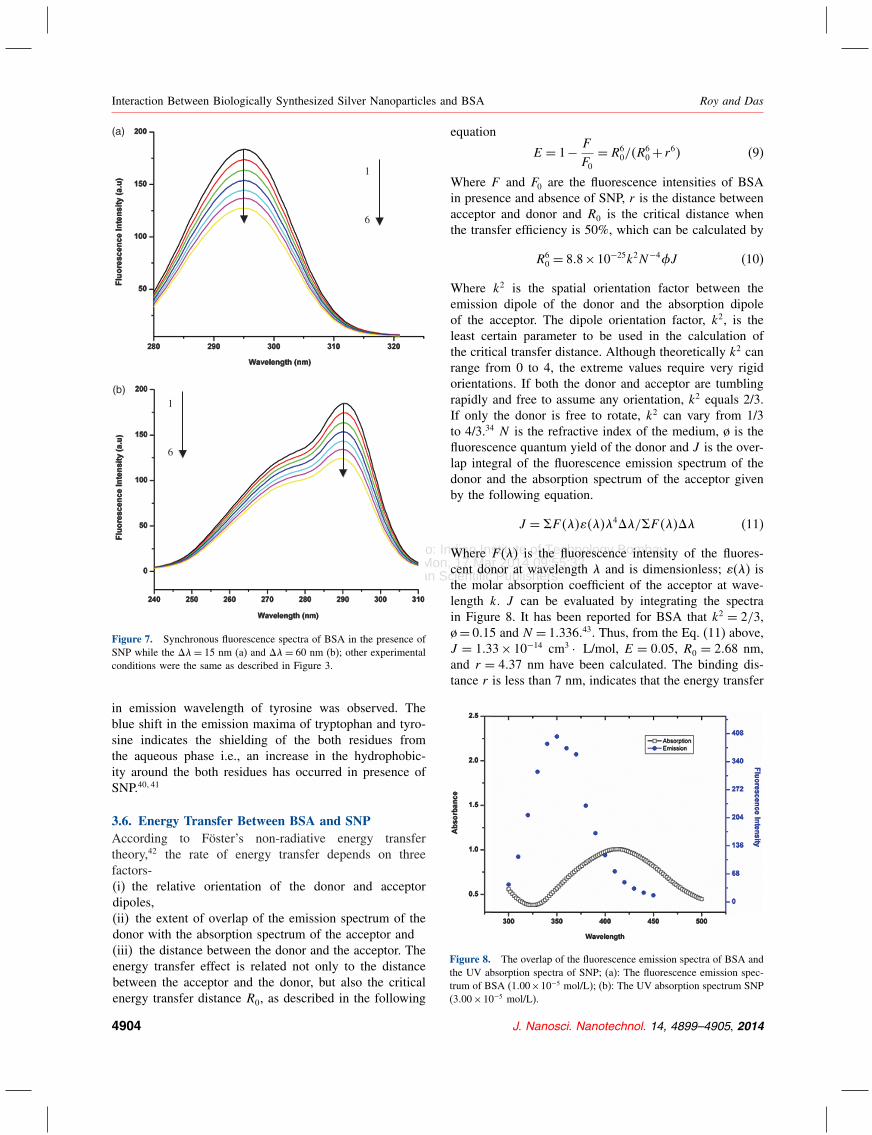
3.5. Synchronous Fluorescence SpectraA synchronous fluorescence spectrum gives the informa-tion on the molecular environment of the fluorophoresfunctional group. The value of �� i.e., difference betweenexcitation and emission wavelengths is an important oper-ating parameter. According to Miller 39 when ��= 15 nm,synchronous fluorescence spectra indicates the changes inthe microenvironment of tyrosine residues and when ��=60 nm, it provides information on the microenvironmentof tryptophan residues. It can be seen from the Figure 7(a)that when �� = 60 nm there is a slight blue shift inthe emission wavelength of tryptophan residues and when�� = 15 nm, (Fig. 7(b)) there is also a slight blue shift
J. Nanosci. Nanotechnol. 14, 4899–4905, 2014 4903
Delivered by Publishing Technology to: Indian Institute of Technology BombayIP: 103.21.127.79 On: Mon, 17 Mar 2014 09:55:32
Copyright: American Scientific Publishers
Interaction Between Biologically Synthesized Silver Nanoparticles and BSA Roy and Das
(a)
(b)
1
6
1
6
Figure 7. Synchronous fluorescence spectra of BSA in the presence ofSNP while the ��= 15 nm (a) and ��= 60 nm (b); other experimentalconditions were the same as described in Figure 3.
in emission wavelength of tyrosine was observed. Theblue shift in the emission maxima of tryptophan and tyro-sine indicates the shielding of the both residues fromthe aqueous phase i.e., an increase in the hydrophobic-ity around the both residues has occurred in presence ofSNP.40�41
3.6. Energy Transfer Between BSA and SNPAccording to Föster’s non-radiative energy transfertheory,42 the rate of energy transfer depends on threefactors-(i) the relative orientation of the donor and acceptordipoles,(ii) the extent of overlap of the emission spectrum of thedonor with the absorption spectrum of the acceptor and(iii) the distance between the donor and the acceptor. Theenergy transfer effect is related not only to the distancebetween the acceptor and the donor, but also the criticalenergy transfer distance R0, as described in the following
equation
E = 1− F
F0= R6
0/�R60+ r6� (9)
Where F and F0 are the fluorescence intensities of BSAin presence and absence of SNP, r is the distance betweenacceptor and donor and R0 is the critical distance whenthe transfer efficiency is 50%, which can be calculated by
R60 = 8�8×10⁻²⁵k2N−4 J (10)
Where k2 is the spatial orientation factor between theemission dipole of the donor and the absorption dipoleof the acceptor. The dipole orientation factor, k2, is theleast certain parameter to be used in the calculation ofthe critical transfer distance. Although theoretically k2 canrange from 0 to 4, the extreme values require very rigidorientations. If both the donor and acceptor are tumblingrapidly and free to assume any orientation, k2 equals 2/3.If only the donor is free to rotate, k2 can vary from 1/3to 4/3.34 N is the refractive index of the medium, ø is thefluorescence quantum yield of the donor and J is the over-lap integral of the fluorescence emission spectrum of thedonor and the absorption spectrum of the acceptor givenby the following equation.
J = �F �������4��/�F ����� (11)
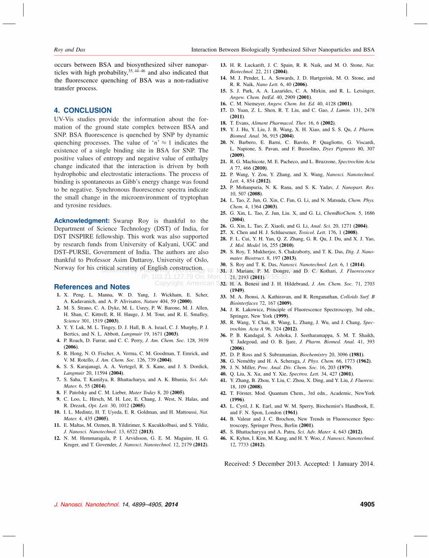
Where F (�) is the fluorescence intensity of the fluores-cent donor at wavelength � and is dimensionless; (�) isthe molar absorption coefficient of the acceptor at wave-length k. J can be evaluated by integrating the spectrain Figure 8. It has been reported for BSA that k2 = 2/3,ø= 0�15 and N = 1�336�43. Thus, from the Eq. (11) above,J = 1�33× 10−14 cm3 · L/mol, E = 0�05, R0 = 2�68 nm,and r = 4�37 nm have been calculated. The binding dis-tance r is less than 7 nm, indicates that the energy transfer
Figure 8. The overlap of the fluorescence emission spectra of BSA andthe UV absorption spectra of SNP; (a): The fluorescence emission spec-trum of BSA (1�00×10−5 mol/L); (b): The UV absorption spectrum SNP(3�00×10−5 mol/L).
4904 J. Nanosci. Nanotechnol. 14, 4899–4905, 2014
Delivered by Publishing Technology to: Indian Institute of Technology BombayIP: 103.21.127.79 On: Mon, 17 Mar 2014 09:55:32
Copyright: American Scientific Publishers
Roy and Das Interaction Between Biologically Synthesized Silver Nanoparticles and BSA
occurs between BSA and biosynthesized silver nanopar-ticles with high probability,35�44–46 and also indicated thatthe fluorescence quenching of BSA was a non-radiativetransfer process.
4. CONCLUSIONUV-Vis studies provide the information about the for-mation of the ground state complex between BSA andSNP. BSA fluorescence is quenched by SNP by dynamicquenching processes. The value of ‘n’ ≈ 1 indicates theexistence of a single binding site in BSA for SNP. Thepositive values of entropy and negative value of enthalpychange indicated that the interaction is driven by bothhydrophobic and electrostatic interactions. The process ofbinding is spontaneous as Gibb’s energy change was foundto be negative. Synchronous fluorescence spectra indicatethe small change in the microenvironment of tryptophanand tyrosine residues.
Acknowledgment: Swarup Roy is thankful to theDepartment of Science Technology (DST) of India, forDST INSPIRE fellowship. This work was also supportedby research funds from University of Kalyani, UGC andDST–PURSE, Government of India. The authors are alsothankful to Professor Asim Duttaroy, University of Oslo,Norway for his critical scrutiny of English construction.
References and Notes1. X. Peng, L. Manna, W. D. Yang, J. Wickham, E. Scher,
A. Kadavanich, and A. P. Alivisatos, Nature 404, 59 (2000).2. M. S. Strano, C. A. Dyke, M. L. Usrey, P. W. Barone, M. J. Allen,
H. Shan, C. Kittrell, R. H. Hauge, J. M. Tour, and R. E. Smalley,Science 301, 1519 (2003).
3. Y. Y. Luk, M. L. Tingey, D. J. Hall, B. A. Israel, C. J. Murphy, P. J.Bertics, and N. L. Abbott, Langmuir 19, 1671 (2003).
4. P. Roach, D. Farrar, and C. C. Perry, J. Am. Chem. Soc. 128, 3939(2006).
5. R. Hong, N. O. Fischer, A. Verma, C. M. Goodman, T. Emrick, andV. M. Rotello, J. Am. Chem. Soc. 126, 739 (2004).
6. S. S. Karajanagi, A. A. Vertegel, R. S. Kane, and J. S. Dordick,Langmuir 20, 11594 (2004).
7. S. Saha, T. Kamilya, R. Bhattacharya, and A. K. Bhunia, Sci. Adv.Mater. 6, 55 (2014).
8. F. Patolsky and C. M. Lieber, Mater Today 8, 20 (2005).9. C. Loo, L. Hirsch, M. H. Lee, E. Chang, J. West, N. Halas, and
R. Drezek, Opt. Lett. 30, 1012 (2005).10. I. L. Medintz, H. T. Uyeda, E. R. Goldman, and H. Mattoussi, Nat.
Mater. 4, 435 (2005).11. E. Maltas, M. Ozmen, B. Yildirimer, S. Kucukkolbasi, and S. Yildiz,
J. Nanosci. Nanotechnol. 13, 6522 (2013).12. N. M. Hemmaragala, P. I. Arvidsson, G. E. M. Maguire, H. G.
Kruger, and T. Govender, J. Nanosci. Nanotechnol. 12, 2179 (2012).
13. H. R. Luckarift, J. C. Spain, R. R. Naik, and M. O. Stone, Nat.Biotechnol. 22, 211 (2004).
14. M. J. Pender, L. A. Sowards, J. D. Hartgerink, M. O. Stone, andR. R. Naik, Nano Lett. 6, 40 (2006).
15. S. J. Park, A. A. Lazarides, C. A. Mirkin, and R. L. Letsinger,Angew. Chem. IntEd. 40, 2909 (2001).
16. C. M. Niemeyer, Angew. Chem. Int. Ed. 40, 4128 (2001).17. D. Yuan, Z. L. Shen, R. T. Liu, and C. Gao, J. Lumin. 131, 2478
(2011).18. T. Evans, Aliment Pharmacol. Ther. 16, 6 (2002).19. Y. J. Hu, Y. Liu, J. B. Wang, X. H. Xiao, and S. S. Qu, J. Pharm.
Biomed. Anal. 36, 915 (2004).20. N. Barbero, E. Barni, C. Barolo, P. Quagliotto, G. Viscardi,
L. Napione, S. Pavan, and F. Bussolino, Dyes Pigments 80, 307(2009).
21. R. G. Machicote, M. E. Pacheco, and L. Bruzzone, Spectrochim ActaA 77, 466 (2010).
22. P. Wang, Y. Zou, Y. Zhang, and X. Wang, Nanosci. Nanotechnol.Lett. 4, 854 (2012).
23. P. Mohanpuria, N. K. Rana, and S. K. Yadav, J. Nanopart. Res.10, 507 (2008).
24. L. Tao, Z. Jun, G. Xin, C. Fan, G. Li, and N. Matsuda, Chem. Phys.Chem. 4, 1364 (2003).
25. G. Xin, L. Tao, Z. Jun, Liu. X, and G. Li, ChemBioChem. 5, 1686(2004).
26. G. Xin, L. Tao, Z. Xiaoli, and G. Li, Anal. Sci. 20, 1271 (2004).27. X. Chen and H. J. Schluesener, Toxicol. Lett. 176, 1 (2008).28. F. L. Cui, Y. H. Yan, Q. Z. Zhang, G. R. Qu, J. Du, and X. J. Yao,
J. Mol. Model 16, 255 (2010).29. S. Roy, T. Mukherjee, S. Chakraborty, and T. K. Das, Dig. J. Nano-
mater. Biostruct. 8, 197 (2013).30. S. Roy and T. K. Das, Nanosci. Nanotechnol. Lett. 6, 1 (2014).31. J. Mariam, P. M. Dongre, and D. C. Kothari, J. Fluorescence
21, 2193 (2011).32. H. A. Benesi and J. H. Hildebrand, J. Am. Chem. Soc. 71, 2703
(1949).33. M. A. Jhonsi, A. Kathiravan, and R. Renganathan, Colloids Surf. B
Biointerfaces 72, 167 (2009).34. J. R. Lakowicz, Principle of Fluorescence Spectroscopy, 3rd edn.,
Springer, New York (1999).35. R. Wang, Y. Chai, R. Wang, L. Zhang, J. Wu, and J. Chang, Spec-
trochim. Acta A 96, 324 (2012).36. P. B. Kandagal, S. Ashoka, J. Seetharamappa, S. M. T. Shaikh,
Y. Jadegoud, and O. B. Ijare, J. Pharm. Biomed. Anal. 41, 393(2006).
37. D. P. Ross and S. Subramanian, Biochemistry 20, 3096 (1981).38. G. Neméthy and H. A. Scheraga, J. Phys. Chem. 66, 1773 (1962).39. J. N. Miller, Proc. Anal. Div. Chem. Soc. 16, 203 (1979).40. Q. Liu, X. Xu, and Y. Xie, Spectros. Lett. 34, 427 (2001).41. Y. Zhang, B. Zhou, Y. Liu, C. Zhou, X. Ding, and Y. Liu, J. Fluoresc.
18, 109 (2008).42. T. Förster, Mod. Quantum Chem., 3rd edn., Academic, NewYork
(1996).43. L. Cyril, J. K. Earl, and W. M. Sperry, Biochemist’s Handbook, E.
and F. N. Spon, London (1961).44. B. Valeur and J. C. Brochon, New Trends in Fluorescence Spec-
troscopy, Springer Press, Berlin (2001).45. S. Bhattacharyya and A. Patra, Sci. Adv. Mater. 4, 643 (2012).46. K. Kyhm, I. Kim, M. Kang, and H. Y. Woo, J. Nanosci. Nanotechnol.
12, 7733 (2012).
Received: 5 December 2013. Accepted: 1 January 2014.
J. Nanosci. Nanotechnol. 14, 4899–4905, 2014 4905


