Embed Size (px)
Citation preview

Solving the r-Conotoxin Folding Problem: EfficientSelenium-Directed On-Resin Generation of More Potent and
Stable Nicotinic Acetylcholine Receptor Antagonists
Markus Muttenthaler,† Simon T. Nevin,‡ Anton A. Grishin,|,‡ Shyuan. T. Ngo,§
Peng T. Choy,§ Norelle L. Daly,† Shu-Hong Hu,† Christopher J. Armishaw,⊥,†
Ching-I. A. Wang,† Richard J. Lewis,† Jennifer L. Martin,† Peter G. Noakes,‡,§
David J. Craik,† David J. Adams,|,‡ and Paul F. Alewood*,†
Institute for Molecular Bioscience, DiVision of Chemistry and Structural Biology, The UniVersityof Queensland, Brisbane, Queensland 4072, Australia; Queensland Brain Institute, The
UniVersity of Queensland, Brisbane, Queensland 4072, Australia; and School of BiomedicalSciences, The UniVersity of Queensland, Brisbane, Queensland 4072, Australia
Received December 16, 2009; E-mail: [email protected]
Abstract: R-Conotoxins are tightly folded miniproteins that antagonize nicotinic acetylcholine receptors(nAChR) with high specificity for diverse subtypes. Here we report the use of selenocysteine in a supportedphase method to direct native folding and produce R-conotoxins efficiently with improved biophysicalproperties. By replacing complementary cysteine pairs with selenocysteine pairs on an amphiphilic resin,we were able to chemically direct all five structural subclasses of R-conotoxins exclusively into their nativefolds. X-ray analysis at 1.4 Å resolution of R-selenoconotoxin PnIA confirmed the isosteric character of thediselenide bond and the integrity of the R-conotoxin fold. The R-selenoconotoxins exhibited similar orimproved potency at rat diaphragm muscle and R3�4, R7, and R1�1δγ nAChRs expressed in Xenopusoocytes plus improved disulfide bond scrambling stability in plasma. Together, these results underpin thedevelopment of more stable and potent nicotinic antagonists suitable for new drug therapies, and highlightthe application of selenocysteine technology more broadly to disulfide-bonded peptides and proteins.
1. Introduction
Nicotinic acetylcholine receptors (nAChRs) are ligand-gatedcationic channels that mediate fast synaptic transmission withinthe peripheral and central nervous system.1,2 The nAChR is afunctional pentamer in which the five transmembrane spanningsubunits are symmetrically arranged around the central pore.1
Channel opening is induced by the endogenous neurotransmitteracetylcholine (ACh) or exogenous agonists such as nicotine.2
The neuromuscular nAChR is made up of combinations of fourdifferent subunits R1, �1, γ (or ε in the adult), and δ, with apredicted (R1)2�1γδ stoichiometry. By contrast, neuronalnAChRs comprise homomeric or heteromeric combinations ofligand binding R subunits (R2-R10) and structural � subunits(�2-�4).2,3 These combinatorial possibilities generate a widediversity of subtypes in different locations in the nervous system
having distinct pharmacological and biophysical properties.3,4
The therapeutic potential of nAChRs is broad, with individualsubtypes being implicated in Alzheimer’s and Parkinson’sdisease, Tourette’s syndrome, pain, myocardial infarction,nicotinic addiction, schizophrenia, attention-deficit hyperactivitydisorder, autism, epilepsy, depression, and anxiety.2,3 Subtypeselectivity of ligands is essential to sharply delineate physi-ological functions and to minimize side effects in therapeutictreatment of such diseases.5
Although much effort has focused on small moleculeantagonist development, little success has been achieved so farin producing viable clinical candidates.2,6 By contrast, recentresearch into marine cone snail venoms has revealed a richsource of competitive, highly potent and selective antagonistsfor a broad range of ion channels, including the nAChRs.7,8
The cone snails have evolved a highly sophisticated cellmachinery over 50 million years that allows the production ofbioactive peptides called conotoxins in a combinatorial fashionfor prey capture and survival strategies.8 Evolutionarily selected,the R-conotoxins have nanomolar activity at target receptors
† Institute for Molecular Bioscience, Division of Chemistry and StructuralBiology, The University of Queensland.
‡ Queensland Brain Institute, The University of Queensland.§ School of Biomedical Sciences, The University of Queensland.| Present Address: Health Innovations Research Institute, RMIT Uni-
versity, PO Box 71, Bundoora, Victoria 3083, Australia⊥ Present Address: Torrey Pines Institute for Molecular Studies, 11350
SW Village Pkwy, Port St Lucie, FL 34987, USA(1) Unwin, N. J. Mol. Biol. 2005, 346, 967–989.(2) Romanelli, M. N.; Gratteri, P.; Guandalini, L.; Martini, E.; Bonaccini,
C.; Gualtieri, F. ChemMedChem 2007, 2, 746–767.(3) Gotti, C.; Clementi, F. Prog. Neurobiol. (Amsterdam, Neth.) 2004,
74, 363–396.
(4) Gotti, C.; Zoli, M.; Clementi, F. Trends Pharmacol. Sci. 2006, 27,482–491.
(5) Bunnelle, W. H.; Dart, M. J.; Schrimpf, M. R. Curr. Top. Med. Chem.(Sharjah, United Arab Emirates) 2004, 4, 299–334.
(6) Arneric, S. P.; Holladay, M.; Williams, M. Biochem. Pharmacol. 2007,74, 1092–1101.
(7) Lewis, R. J.; Garcia, M. L. Nat. ReV. Drug DiscoV. 2003, 2, 790–802.
(8) Terlau, H.; Olivera, B. M. Physiol. ReV. 2004, 84, 41–68.
Published on Web 02/17/2010
10.1021/ja910602h 2010 American Chemical Society3514 9 J. AM. CHEM. SOC. 2010, 132, 3514–3522

and an enormously refined selectivity that can differentiatebetween individual subtypes of nAChRs.8,9 These peptidescontain 12-19 amino acids and have well-defined three-dimensional structures, predominantly due to the bracing of thebackbone by two disulfide bonds in a Cys 1-3 and Cys 2-4connectivity (referred to as the native globular connectivity).Two loops are formed with a short turn-helix-turn backbonefeature that comprises most of the functional groups responsiblefor activity and potency.9 The numbers of residues encompassedwithin these two loops (m/n) represent the basis for a furtherdivision into several structural subfamilies as the loop sizepairings correlate with the nAChR subtype target selectivity.8
Hence the R-conotoxins are ideal probes for receptor studies,and are considered promising therapeutic agents for the treat-ment of pain and other neurological disorders.2,7 However, thechallenge of obtaining correctly folded peptides has ensured thatin-depth investigations of R-conotoxins remain a low-throughputprocess, thus hindering a plethora of potential structure-activitystudies.10
The major challenge is outlined in Figure 1a, where threedistinct folds with different disulfide bond connectivities andfunctions may be obtained from the same linear sequence underoxidative conditions. Although the oxidative folding processmay be partly influenced to give a dominant isomer by adjustingoxidation buffers via variation in organic cosolvents, shufflingagents, solute concentration, temperature and time, this empiricalapproach often fails once non-native structural modificationsare embedded in the synthetic design.10,11 Furthermore, valida-tion of the correct isomer is time intensive, as it involvesstructure and disulfide bond connectivity determination orbiological characterization, if the biological target is known.The purification process also adds to the complexity due to the
similar nature of the isomers, significantly lowering the yields.Given that the primary reason to select chemical approachesover peptide expression is to introduce non-native modificationsfor improved structure-function, fluorescent tagging, ligationchemistry, cyclization or PEGylation, it is essential to haveefficient regioselective control over disulfide bond formation.Currently, this is achieved in a laborious fashion via orthogonalthiol-protecting groups,12 which have long workup times andlow yields. We describe here a methodology that takesadvantage of preferential diselenide bond formation overdisulfide bond formation to selectively generate R-conotoxinsin their native fold either in solution or in a high throughputparallel methodology. This approach gave easy access to allfive classes of nicotinic R-conotoxin antagonists with enhancedstability and potency.
Selenocysteine (Sec, U) is considered to be the mostconservative substitution for cysteine and its isosteric characterhas been exploited in a range of bioactive peptides to eitherelucidate structure activity relationships (SAR) or to improvetheir stability in reducing environments.13 Despite the resem-blance between the elements selenium and sulfur, the aminoacids Sec and Cys exhibit significantly distinct chemicalproperties. While Sec and Cys have similar electronegativity(EN(S) ) 2.58, EN(Se) ) 2.55), Sec is a stronger nucleophileand a better leaving group than its sulfur counterpart.14
Furthermore, Sec exhibits a much higher acidity than Cys (pKa
(Sec) ) 5.24-5.63, pKa (Cys) ) 8.25).15–17 Comparative studieson diselenide, selenylsulfide, and disulfide bonds in linearunconstrained glutaredoxin fragments as well as in foldedglutaredoxin 3 showed a difference of 111-166 mV in redox
(9) Nicke, A.; Wonnacott, S.; Lewis, R. J. Eur. J. Biochem. 2004, 271,2305–2319.
(10) Bulaj, G.; Olivera, B. M. Antioxid. Redox. Signal. 2008, 10, 141–156.
(11) Nielsen, J. S.; Buczek, P.; Bulaj, G. J. Pept. Sci. 2004, 10, 249–256.
(12) Moroder, L.; Musiol, H. J.; Schaschke, N.; Chen, L.; Hargittai, B.;Barany, G. In Houben-Weyl, Synthesis of Peptides and Peptidomi-metics; Goodman, M., Felix, A., Moroder, L., Toniolo, C., Eds.; GeorgThieme Verlag: Stuttgart, 2001; Vol. E22a, p 384-423.
(13) Muttenthaler, M.; Alewood Paul, F. J. Pept. Sci. 2008, 14, 1223–1239.
(14) Moroder, L. J. Pept. Sci. 2005, 11, 187–214.
Figure 1. (a) Standard R-conotoxin synthesis. Assembly on resin using Boc-SPPS, followed by HF cleavage and random oxidation in aqueous buffersolutions, yields all three possible disulfide isomers. (b) Parallel directed-folding strategy using selenocysteine. Parallel chain assembly was performed usingthe HF stable SCAL linker on an amphiphilic ChemMatrix resin. Upon assembly, the conotoxins on solid support are transferred into labeled resin-bags,followed by HF deprotection and oxidation in aqueous 0.1M NH4HCO3 buffers. Regioselective folding is induced upon HF deprotection due to selectiveformation of the diselenide over the disulfide bond. Depending on the synthetic strategy, the peptides can then be separated, cleaved and purified individually,or be cleaved and purified simultaneously via a single RP-HPLC run.
J. AM. CHEM. SOC. 9 VOL. 132, NO. 10, 2010 3515
Solving the R-Conotoxin Folding Problem A R T I C L E S
potential between the disulfide and the diselenide bond.18,19 Thisdifference, in combination with the higher nucleophilicity andlower pKa of Sec, suggests that diselenide or selenylsulfide bondformation is highly favored over disulfide bond formation aswas observed in earlier studies.20 The lower redox potential ofthe diselenide bond should at the same time provide higherstability in a reductive environment.21
2. Results
2.1. Directed Folding using SelenocysteinesIn Solution. Toinvestigate the potential of selenocysteine to direct oxidativefolding in a straightforward and efficient manner, well studiedR-conotoxins from all five existing functional classes withdiffering nAChR selectivity (R3/5-MI, R4/3-ImI, R4/4-BuIA,R4/6-AuIB, R4/7-Vc1.1, R4/7-[A10L]PnIA) were chosen andcomplementary pairs of cysteine residues in each sequence werereplaced by selenocysteine (see Figure 1b and Table 1). TheR-conotoxins AuIB, Sec[1,3]-AuIB, Sec[2,4]-AuIB, MI, Sec[1,3]-MI, Vc1.1, Sec[1,3]-Vc1.1, [A10L]PnIA, Sec[1,3]-[A10L]PnIA,Sec[2,4]-[A10L]PnIA, ImI, Sec[1,3]-ImI, Sec[2,4]-ImI, wereassembled using Boc-SPPS incorporating selenocysteine viaBoc-L-Sec(MeBzl)-OH. This chemical approach was chosenas reports in the literature suggest that Sec racemization mayoccur using Fmoc chemistry strategies. Furthermore, Sec wasshown to have a high tendency to deselenate via �-eliminationduring iterative piperidine Fmoc-deprotection steps that result
in dehydroalanine and consequently piperidyl adducts.22 Boc-L-Sec(MeBzl)-OH was synthesized following the protocol ofArmishaw et al..21 Chain assembly of all R-conotoxin sequencesproceeded smoothly using HBTU-mediated Boc in situ neu-tralization SPPS23 on a 4-methylbenzhydrylamine resin. Thelinear peptides were deprotected and cleaved by hydrofluoricacid (HF) in a single step. Intriguingly, the Sec residues werefound to be fully oxidized directly upon deprotection via HF(pH < 1) and it was not possible to isolate the fully reducedspecies. Attempts to do so in the presence of an excess ofreducing agents such as TCEP, TBP, or NaBH4 led to significantdeselenation. Oxidation of the diselenide, dithiol-containingR-conotoxin intermediates was performed in 0.1M NH4HCO3
at pH 8.4 and the solution was analyzed by RP-HPLC andLCMS (see Supporting Information Figure S2). Additionaloxidation trials on the selenium analogs of AuIB, MI, and[A10L]PnIA revealed that once the diselenide is formed,disulfide bond formation occurred readily at pH 5 in contrastto the native all Cys peptides, which remain as free thiols (seeSupporting Information Figure S3). In general, oxidations (pH8.4, 25 °C) were complete within 10 min without any foldingenhancers and only a single purification step was necessary.This makes Sec-directed oxidative folding far more efficientthan standard oxidation protocols or orthogonal protecting groupstrategies.
2.2. Directed Folding using SelenocysteinesOn Resin. Withregioselective folding established in solution we set out todevelop resin-supported chemistry to underpin high throughputparallel processing of peptides and unify time-intensive taskssuch as side-chain deprotection, oxidation, cleavage, andpurification to give fast and efficient access to correctly foldedR-conotoxins (see Figure 1b). R-Conotoxins MI, Sec[2,4]-MI,Vc1.1, Sec[1,3]-Vc1.1, BuIA, Sec[2,8]-BuIA, and [A10L]PnIAwere assembled using Boc-chemistry on an amphiphilic resin(ChemMatrix) with a C-terminal HF stable safety-catch acid
(15) Huber, R. E.; Criddle, R. S. Arch. Biochem. Biophys. 1967, 122, 164–173.
(16) Nygard, B. Ark. Kemi 1967, 27, 341–361.(17) Arnold, A. P.; Tan, K. S.; Rabenstein, D. L. Inorg. Chem. 1986, 25,
2433–2437.(18) Besse, D.; Siedler, F.; Diercks, T.; Kessler, H.; Moroder, L. Angew.
Chem., Int. Ed. Engl. 1997, 36, 883–885.(19) Metanis, N.; Keinan, E.; Dawson, P. E. J. Am. Chem. Soc. 2006, 128,
16684–16691.(20) Pegoraro, S.; Fiori, S.; Rudolph-Bohner, S.; Watanabe, T. X.; Moroder,
L. J. Mol. Biol. 1998, 284, 779–792.(21) Armishaw, C. J.; Daly, N. L.; Nevin, S. T.; Adams, D. J.; Craik, D. J.;
Alewood, P. F. J. Biol. Chem. 2006, 281, 14136–14143.
(22) Moroder, L.; Besse, D.; Musiol, H.-J.; Rudolph-Bohner, S.; Sideler,F. Biopolymers 1996, 40, 207–234.
(23) Schnolzer, M.; Alewood, P.; Jones, A.; Alewood, D.; Kent, S. B. H.Int. J. Pept. Protein Res. 1992, 40, 180–193.
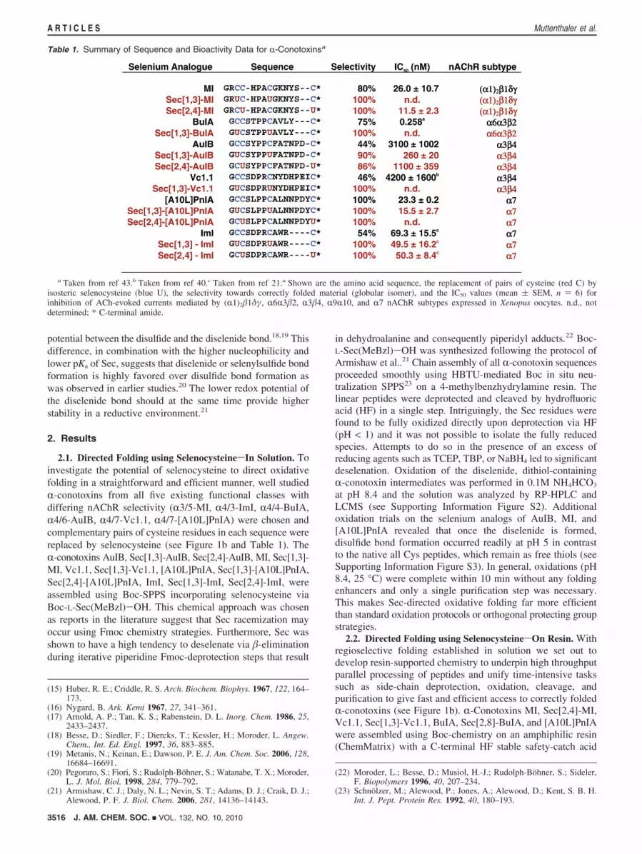
Table 1. Summary of Sequence and Bioactivity Data for R-Conotoxinsa
a Taken from ref 43.b Taken from ref 40.c Taken from ref 21.a Shown are the amino acid sequence, the replacement of pairs of cysteine (red C) byisosteric selenocysteine (blue U), the selectivity towards correctly folded material (globular isomer), and the IC50 values (mean ( SEM, n ) 6) forinhibition of ACh-evoked currents mediated by (R1)2�1δγ, R6R3�2, R3�4, R9R10, and R7 nAChR subtypes expressed in Xenopus oocytes. n.d., notdetermined; * C-terminal amide.
3516 J. AM. CHEM. SOC. 9 VOL. 132, NO. 10, 2010
A R T I C L E S Muttenthaler et al.

labile (SCAL) linker (see Supporting Information Figure S1).24
Simultaneous side-chain deprotection by HF of the conotoxinsseparately packaged in labeled solvent-permeable 74 µm meshpolypropylene bags25 delivered the fully deprotected peptideson-resin and was followed by oxidation in 0.1M NH4HCO3
buffer at pH 8.4. Similar to solution folding outcomes, differentratios of the three possible disulfide bond isomers of the all-Cys analogs could be accessed by changing the foldingconditions (see Supporting Information Table S2 online).26 Fullregioselective control over multiple isomer formation was onlyachieved by incorporation of isosteric selenocysteine into thesynthetic scheme (see Table 1 and Supporting InformationFigure S4). Once the peptides folded on-resin, a chemoselectivereaction (reductive acidolysis by NH4I/TFA) activates the linkerand releases the correctly folded R-conotoxins from the solidsupport (see Supporting Information Figure S1). Depending onthe peptide library strategy, cleavage and purification can eitherbe achieved individually (separated by the labeled teabags) orsimultaneously in an efficient “one-pot” approach. Both alterna-tives were investigated and in both cases the correctly foldedpeptides were isolated in high purity (see Supporting InformationFigure S5).
2.3. Biological Characterization. 2.3.1. Contraction AssayRat Diaphragm. The biological activities of globular MI andSec[2,4]-MI were characterized by their ability to inhibitacetylcholine (ACh)-induced muscular contractions in responseto phrenic nerve stimulation in rat diaphragm preparations. Thepeptides were initially tested at a concentration of 150 nM,
followed by 300 and 500 nM. Over a 2 h period, the globularselenium-analog Sec[2,4]-MI was the only peptide capable of90% inhibition of the ACh-induced muscular contraction at 150nM (see Table 1 and Figure 2a). Both peptides were able tocompletely block the muscle contraction at concentrations of300 and 500 nM.
2.3.2. ElectrophysiologyAssaysusingXenopusOocytes.R-Cono-toxins MI, [A10L]PnIA and AuIB are selective for the muscleR1�1γδ and the neuronal R7 and R3�4 nAChR subtypes,respectively, and the activity of the native peptides a well astheir selenium-analogs was examined on ACh-evoked currentsmediated by recombinant nAChRs expressed in Xenopus oocytes(see Table 1 and Figure 2b). Globular R-conotoxin MI wastested on ACh (1 µM)-evoked currents mediated by muscle(R1)2�1γδ nAChR. Concentration-response curves obtained forthe inhibition of ACh-evoked currents by globular MI exhibitedan IC50 of 26 nM and a Hill coefficient of 1.1 (n ) 6). GlobularSec[2,4]-MI retained the potency with an IC50 of 9 nM and Hillcoefficient of 0.9 (n ) 6). Globular [A10L]PnIA was tested onACh (100 µM) -evoked currents mediated by neuronal R7nAChRs and exhibited an IC50 of 23.3 nM and a Hill coefficientof 2.0 (n ) 6). Globular Sec[1,3]-[A10L]PnIA had a similarIC50 value of 15.5 nM and a Hill coefficient of 1.3 (n ) 5).Neuronal R3�4 nAChR-mediated currents evoked by 50 µMACh were inhibited by globular AuIB giving an IC50 value of3.1 µM and a Hill coefficient of 1.5 (n ) 6). The seleniumsubstituted globular Sec[1,3]-AuIB and globular Sec[2,4]-AuIBexhibited IC50’s of 0.3 µM and 1.1 µM and Hill coefficients of2.0 and 1.0 respectively (n ) 6).
2.4. Structural Characterization. The enhanced potencies ofthe selenium analogs prompted a structural analysis of theisosteric character of selenocysteine. Circular dichroism (CD)
(24) Patek, M.; Lebl, M. Tetrahedron Lett. 1991, 32, 3891–3894.(25) Houghten, R. A.; Pinilla, C.; Blondelle, S. E.; Appel, J. R.; Dooley,
C. T.; Cuervo, J. H. Nature (London) 1991, 354, 84–86.(26) Brust, A.; Tickle, A. E. J. Pept. Sci. 2007, 13, 133–141.
Figure 2. Functional assays of R-conotoxins. (a) Inhibition of nerve-evoked muscle contraction of rat diaphragm by MI and its selenium analog at concentrationsof (i) 150 nM and (ii) 300 nM. Error bars are mean ( SEM (* P ) 0.0123, n ) 3, 2 way ANOVA) (b) Inhibition of ACh-evoked currents mediated bynAChR subtypes expressed in Xenopus oocytes by R-conotoxin (i) MI, (ii) [A10L]PnIA, (iii) AuIB and their Sec-analogs. Error bars are mean ( SEM (n) 6). The concentration-response data are summarized in the Table (iv), where IC50 is the half-maximal inhibitory concentration and nH is the Hill coefficient.
J. AM. CHEM. SOC. 9 VOL. 132, NO. 10, 2010 3517
Solving the R-Conotoxin Folding Problem A R T I C L E S
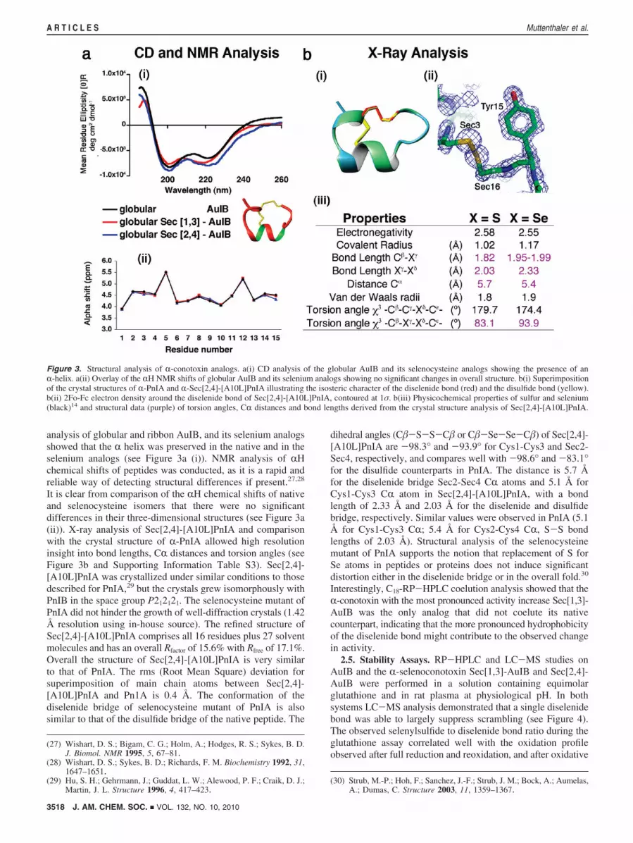
analysis of globular and ribbon AuIB, and its selenium analogsshowed that the R helix was preserved in the native and in theselenium analogs (see Figure 3a (i)). NMR analysis of RHchemical shifts of peptides was conducted, as it is a rapid andreliable way of detecting structural differences if present.27,28
It is clear from comparison of the RH chemical shifts of nativeand selenocysteine isomers that there were no significantdifferences in their three-dimensional structures (see Figure 3a(ii)). X-ray analysis of Sec[2,4]-[A10L]PnIA and comparisonwith the crystal structure of R-PnIA allowed high resolutioninsight into bond lengths, CR distances and torsion angles (seeFigure 3b and Supporting Information Table S3). Sec[2,4]-[A10L]PnIA was crystallized under similar conditions to thosedescribed for PnIA,29 but the crystals grew isomorphously withPnIB in the space group P212121. The selenocysteine mutant ofPnIA did not hinder the growth of well-diffraction crystals (1.42Å resolution using in-house source). The refined structure ofSec[2,4]-[A10L]PnIA comprises all 16 residues plus 27 solventmolecules and has an overall Rfactor of 15.6% with Rfree of 17.1%.Overall the structure of Sec[2,4]-[A10L]PnIA is very similarto that of PnIA. The rms (Root Mean Square) deviation forsuperimposition of main chain atoms between Sec[2,4]-[A10L]PnIA and Pn1A is 0.4 Å. The conformation of thediselenide bridge of selenocysteine mutant of PnIA is alsosimilar to that of the disulfide bridge of the native peptide. The
dihedral angles (C�-S-S-C� or C�-Se-Se-C�) of Sec[2,4]-[A10L]PnIA are -98.3° and -93.9° for Cys1-Cys3 and Sec2-Sec4, respectively, and compares well with -98.6° and -83.1°for the disulfide counterparts in PnIA. The distance is 5.7 Åfor the diselenide bridge Sec2-Sec4 CR atoms and 5.1 Å forCys1-Cys3 CR atom in Sec[2,4]-[A10L]PnIA, with a bondlength of 2.33 Å and 2.03 Å for the diselenide and disulfidebridge, respectively. Similar values were observed in PnIA (5.1Å for Cys1-Cys3 CR; 5.4 Å for Cys2-Cys4 CR, S-S bondlengths of 2.03 Å). Structural analysis of the selenocysteinemutant of PnIA supports the notion that replacement of S forSe atoms in peptides or proteins does not induce significantdistortion either in the diselenide bridge or in the overall fold.30
Interestingly, C18-RP-HPLC coelution analysis showed that theR-conotoxin with the most pronounced activity increase Sec[1,3]-AuIB was the only analog that did not coelute its nativecounterpart, indicating that the more pronounced hydrophobicityof the diselenide bond might contribute to the observed changein activity.
2.5. Stability Assays. RP-HPLC and LC-MS studies onAuIB and the R-selenoconotoxin Sec[1,3]-AuIB and Sec[2,4]-AuIB were performed in a solution containing equimolarglutathione and in rat plasma at physiological pH. In bothsystems LC-MS analysis demonstrated that a single diselenidebond was able to largely suppress scrambling (see Figure 4).The observed selenylsulfide to diselenide bond ratio during theglutathione assay correlated well with the oxidation profileobserved after full reduction and reoxidation, and after oxidative
(27) Wishart, D. S.; Bigam, C. G.; Holm, A.; Hodges, R. S.; Sykes, B. D.J. Biomol. NMR 1995, 5, 67–81.
(28) Wishart, D. S.; Sykes, B. D.; Richards, F. M. Biochemistry 1992, 31,1647–1651.
(29) Hu, S. H.; Gehrmann, J.; Guddat, L. W.; Alewood, P. F.; Craik, D. J.;Martin, J. L. Structure 1996, 4, 417–423.
(30) Strub, M.-P.; Hoh, F.; Sanchez, J.-F.; Strub, J. M.; Bock, A.; Aumelas,A.; Dumas, C. Structure 2003, 11, 1359–1367.
Figure 3. Structural analysis of R-conotoxin analogs. a(i) CD analysis of the globular AuIB and its selenocysteine analogs showing the presence of anR-helix. a(ii) Overlay of the RH NMR shifts of globular AuIB and its selenium analogs showing no significant changes in overall structure. b(i) Superimpositionof the crystal structures of R-PnIA and R-Sec[2,4]-[A10L]PnIA illustrating the isosteric character of the diselenide bond (red) and the disulfide bond (yellow).b(ii) 2Fo-Fc electron density around the diselenide bond of Sec[2,4]-[A10L]PnIA, contoured at 1σ. b(iii) Physicochemical properties of sulfur and selenium(black)14 and structural data (purple) of torsion angles, CR distances and bond lengths derived from the crystal structure analysis of Sec[2,4]-[A10L]PnIA.
3518 J. AM. CHEM. SOC. 9 VOL. 132, NO. 10, 2010
A R T I C L E S Muttenthaler et al.
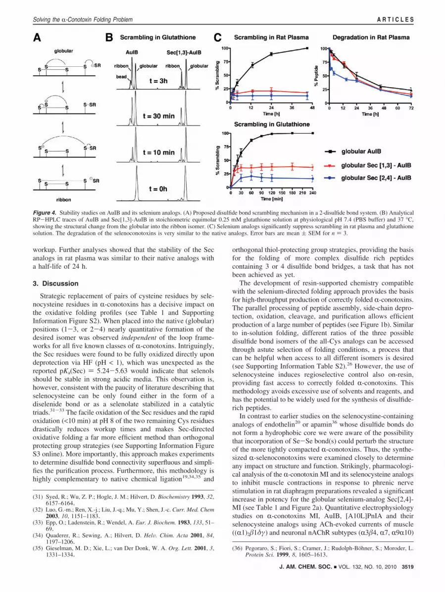
workup. Further analyses showed that the stability of the Secanalogs in rat plasma was similar to their native analogs witha half-life of 24 h.
3. Discussion
Strategic replacement of pairs of cysteine residues by sele-nocysteine residues in R-conotoxins has a decisive impact onthe oxidative folding profiles (see Table 1 and SupportingInformation Figure S2). When placed into the native (globular)positions (1-3, or 2-4) nearly quantitative formation of thedesired isomer was observed independent of the loop frame-works for all five known classes of R-conotoxins. Intriguingly,the Sec residues were found to be fully oxidized directly upondeprotection via HF (pH < 1), which was unexpected as thereported pKa(Sec) ) 5.24-5.63 would indicate that selenolsshould be stable in strong acidic media. This observation is,however, consistent with the paucity of literature describing thatselenocysteine can be only found either in the form of adiselenide bond or as a selenolate stabilized in a catalytictriads.31-33 The facile oxidation of the Sec residues and the rapidoxidation (<10 min) at pH 8 of the two remaining Cys residuesdrastically reduces workup times and makes Sec-directedoxidative folding a far more efficient method than orthogonalprotecting group strategies (see Supporting Information FigureS3 online). More importantly, this approach makes experimentsto determine disulfide bond connectivity superfluous and simpli-fies the purification process. Furthermore, this methodology ishighly complementary to native chemical ligation19,34,35 and
orthogonal thiol-protecting group strategies, providing the basisfor the folding of more complex disulfide rich peptidescontaining 3 or 4 disulfide bond bridges, a task that has notbeen achieved as yet.
The development of resin-supported chemistry compatiblewith the selenium-directed folding approach provides the basisfor high-throughput production of correctly folded R-conotoxins.The parallel processing of peptide assembly, side-chain depro-tection, oxidation, cleavage, and purification allows efficientproduction of a large number of peptides (see Figure 1b). Similarto in-solution folding, different ratios of the three possibledisulfide bond isomers of the all-Cys analogs can be accessedthrough astute selection of folding conditions, a process thatcan be helpful when access to all different isomers is desired(see Supporting Information Table S2).26 However, the use ofselenocysteine induces regioselective control also on-resin,providing fast access to correctly folded R-conotoxins. Thismethodology avoids excessive use of solvents and reagents, andhas the potential to be widely used for the synthesis of disulfide-rich peptides.
In contrast to earlier studies on the selenocystine-containinganalogs of endothelin20 or apamin36 whose disulfide bonds donot form a hydrophobic core we were aware of the possibilitythat incorporation of Se-Se bond(s) could perturb the structureof the more tightly compacted R-conotoxins. Thus, the synthe-sized R-selenoconotoxins were examined closely to determineany impact on structure and function. Strikingly, pharmacologi-cal analysis of the R-conotoxin MI and its selenocysteine analogsto inhibit muscle contractions in response to phrenic nervestimulation in rat diaphragm preparations revealed a significantincrease in potency for the globular selenium-analog Sec[2,4]-MI (see Table 1 and Figure 2a). Quantitative electrophysiologystudies on R-conotoxins MI, AuIB, [A10L]PnIA and theirselenocysteine analogs using ACh-evoked currents of muscle((R1)2�1δγ) and neuronal nAChR subtypes (R3�4, R7, R9R10)
(31) Syed, R.; Wu, Z. P.; Hogle, J. M.; Hilvert, D. Biochemistry 1993, 32,6157–6164.
(32) Luo, G.-m.; Ren, X.-j.; Liu, J.-q.; Mu, Y.; Shen, J.-c. Curr. Med. Chem2003, 10, 1151–1183.
(33) Epp, O.; Ladenstein, R.; Wendel, A. Eur. J. Biochem. 1983, 133, 51–69.
(34) Quaderer, R.; Sewing, A.; Hilvert, D. HelV. Chim. Acta 2001, 84,1197–1206.
(35) Gieselman, M. D.; Xie, L.; van Der Donk, W. A. Org. Lett. 2001, 3,1331–1334.
(36) Pegoraro, S.; Fiori, S.; Cramer, J.; Rudolph-Bohner, S.; Moroder, L.Protein Sci. 1999, 8, 1605–1613.
Figure 4. Stability studies on AuIB and its selenium analogs. (A) Proposed disulfide bond scrambling mechanism in a 2-disulfide bond system. (B) AnalyticalRP-HPLC traces of AuIB and Sec[1,3]-AuIB in stoichiometric equimolar 0.25 mM glutathione solution at physiological pH 7.4 (PBS buffer) and 37 °C,showing the structural change from the globular into the ribbon isomer. (C) Selenium analogs significantly suppress scrambling in rat plasma and glutathionesolution. The degradation of the selenoconotoxins is very similar to the native analogs. Error bars are mean ( SEM for n ) 3.
J. AM. CHEM. SOC. 9 VOL. 132, NO. 10, 2010 3519
Solving the R-Conotoxin Folding Problem A R T I C L E S

expressed in Xenopus oocytes (see Table 1 and Figure 2b)confirmed observed results obtained using the hemidiaphragmpreparation, with selenium analogs Sec[2,4]-MI, Sec[1,3]-AuIB,Sec[2,4]-AuIB, and Sec[1,3]-[A10L]PnIA exhibiting up to a 10-fold increase in inhibition compared to their respective nativeR-conotoxins.
With selenium having a slightly larger atomic radius (S 1.02Å, Se 1.17 Å) and longer bond length than sulfur (C�-Sγ )1.82 Å, C�-Seγ ) 1.95-1.99 Å)14 we assessed the isostericcharacter of selenocysteine more rigorously to determine ifstructural differences could explain the increased biologicalactivity. CD and RH chemical shift NMR analysis of the AuIBanalogs showed that the secondary structure was highlypreserved in the selenium analogs, and no significant differencesin their three-dimensional structures from the native peptideswere observed (see Figure 3a). This conclusion was furthersupported by the crystal structure of the R-selenoconotoxin,Sec[2,4]-[A10L]PnIA (resolution 1.42 Å), which was nearidentical to the native R-conotoxin PnIA (rmsd of 0.4 Å for themain-chain atoms).29 Further structural analysis of Sec[2,4]-
[A10L]PnIA indicated that torsion angles, bond lengths, CR-CRdistances and electron densities of the diselenide and thehomologous disulfide bridge from the native PnIA were verysimilar and no distortion was observed in the overall fold (seeFigure 3b and Supporting Information Table S3). To determinethe role of increased hydrophobicity of the Se-Se bond inbinding with the nAChR, the cocrystal structure of PnIA andthe acetylcholine binding protein (AChBP), a structurally andfunctionally homologous protein of nAChR was examined.37
The cocrystal structure suggests that R-conotoxin PnIA bindsto AChBP via complementary hydrophobic patches, with the[1,3]-disulfide bond flanked by two tyrosine residues (3.6-3.9Å) (see Figure 5 a and b). This hydrophobic binding motif(YNCCEEIY) (see Figure 5b) is also highly conserved in theR3 subunit (YSCCPEPY), the subunit important for Sec[1,3]-AuIB recognition, which has the most pronounced enhancement
(37) Celie, P. H. N.; Kasheverov, I. E.; Mordvintsev, D. Y.; Hogg, R. C.;van Nierop, P.; van Elk, R.; van Rossum-Fikkert, S. E.; Zhmak, M. N.;Bertrand, D.; Tsetlin, V.; Sixma, T. K.; Smit, A. B. Nat. Struct. Mol.Biol. 2005, 12, 582–588.
Figure 5. Surface analysis and binding model (a) Surface hydrophobicity plot of the cocrystal structure of AChBP and R-conotoxin PnIA with the hydrophobicresidues colored in orange and the disulfide bonds in yellow. R,�,γ indicate the complementary hydrophobic interaction of PnIA with the binding pocket.(b) Close up of R-conotoxin PnIA binding into the hydrophobic pocket, revealing that the first disulfide bond1–3 is flanked by two tyrosine residues (3.6-3.9Å). (c) NMR surface analysis of R-conotoxin AuIB, showing that the1–3 disulfide bond is in close proximity to the conserved hydrophobic residues that areimportant for binding. The surface exposure of the disulfide/diselenide bonds of PnIA, Sec[2,4]-[A10L]PnIA and AuIB are analyzed in the table, showingthat the surface exposure of the diselenide bond is twice as large as the disulfide bond counterpart. (d) Co-elution study of AuIB its selenium analogs.Analytical C18-RP-HPLC traces of the coelution of (i) globular AuIB with globular Sec[2,4]-AuIB and (ii) globular Sec[2,4]-AuIB with globular Sec[1,3]-AuIB, revealed that Sec[1,3]-AuIB is more hydrophobic than its analogs.
3520 J. AM. CHEM. SOC. 9 VOL. 132, NO. 10, 2010
A R T I C L E S Muttenthaler et al.
of activity (10-fold) and hydrophobicity (see Table 1 and Figure5 c and d). Such a change in hydrophobicity in combinationwith small changes in bond length and torsion angle are likelyto strengthen hydrophobic interactions between the diselenidebond and the two tyrosine residues or in changing in the bindingorientation, which subsequently increases the hydrophobiccontacts with conserved aromatic residues in the binding site.Another anticipated advantage of selenoconotoxins is theirhigher stability in reducing environments due to the lower redoxpotential of the diselenide.18,19,21 RP-HPLC and LC-MSstudies on AuIB and the R-selenoconotoxin Sec[1,3]-AuIB andSec[2,4]-AuIB performed in solutions containing equimolarglutathione or rat plasma at physiological pH demonstrated thata single diselenide bond was able to largely suppress scrambling(see Figure 4). However, the selenium analogs showed a similarhalf-life to the native peptides in plasma, confirming that themain degradation occurs mostly via enzymatic digestion. Inorder to improve the plasma stability of this class of peptideseven more, N- to C-terminal cyclization is currently the methodof choice and significant progress has been achieved in this fieldrecently.38,39
In summary, we have developed a novel, highly scalablemethod amenable to high throughput R-conotoxin synthesis thatemploys resin-supported selenium-chemistry to solve a long-standing folding problem of an important class of subtype-selective nAChR antagonists. Stability studies and electrophys-iological analysis revealed that the selenium analogs ofR-conotoxins are more potent and stable than their nativecounterparts. Comprehensive structural analysis showed that theslightly larger atomic radius of selenium has no significantimpact on the overall structure. Surface analysis and investiga-tion of the binding pocket of AChBP revealed that the increasein hydrophobicity of the diselenide bond is likely to be thereason for observed increase in potency of this toxin class. Thismethodology is highly complementary to native chemicalligation and orthogonal thiol-protecting group strategies and isanticipated to provide access to more complex disulfide richpeptides or proteins with similar folding challenges.19,35
4. Methods
4.1. Peptide Synthesis. All peptides were assembled by manualBoc-SPPS using HBTU-mediated in situ neutralization protocolwith DMF as solvent.23 Deprotection of the 2,4-dinitrophenyl (Dnp)group of histidine was carried out prior to HF treatment with 20%2-mercaptoethanol/ 10% DIEA/ DMF. HF deprotection or cleavagewas performed by treatment of the dried peptide resin (300 mg)with 10 mL HF/p-cresol/p-thio-cresol (18:1:1, v/v/v) for 2 h at 0°C. Following evaporation of the HF, the peptides were precipitatedand washed with cold ether, filtered, and either redissolved in 30mL of 50% ACN/1% TFA and lyophilized, or redissolved directlyin 0.1 M NH4HCO3 (pH 8.4, c ) 0.1 µM) for direct oxidation.Oxidation was monitored by RP-HPLC, LC-MS and MS, andthe peptides were isolated using preparative C18 RP-HPLC.
4.2. On-Resin Folding using a Safety-Catch Acid Labile(SCAL) Linker. Peptide assembly was achieved by manual Boc-SPPS23 using the Fmoc-SCAL linker and a three glycine spacerbetween the linker and the aminomethyl ChemMatrix resin.Deprotection of the Fmoc group of the SCAL linker was performedwith 2× 1 min treatment of 50% piperidine/DMF. 100-500 mgof the individual peptides on-resin were transferred into labeled
74 µm mesh polypropylene bags with the dimension of 5 × 5 cm.Side-chain deprotection was achieved by 10 mL HF treatment at 0°C for 2 h with p-cresol as scavengers [9:1 (v/v) HF:scavenger].HF was evaporated and the solid support was directly transferredinto TFA to maintain the swelling properties. The resins werewashed with DCM, DMF, and H2O before being placed into a 0.1M NH4HCO3 solution (100 mg /10 mL) at pH 8.4. The finalcleavage was performed either by reductive acidolysis with NH4I/TFA/DMS for 1 h at 0 °C and 1 h at room temperature.26 TFAwas then evaporated by nitrogen purging, the peptides wereprecipitated and the scavengers removed with cold ethyl acetate.The peptides were redissolved in H2O, 0.1% TFA and lyophilized.After MS and analytical RP-HPLC analysis, the peptides werepurified by C18-RP-HPLC.
4.3. Contraction Bioassay Rat Diaphragm and Electrophys-iologysXenopus Oocytes. RNA preparation, oocyte preparation,and expression of nAChRs in Xenopus oocytes were performed aspreviously described.40 The methods and experimental protocolsfor the rat diaphragm contraction bioassay and the electrophysi-ological assays using Xenopus oocytes can be found in the Sup-porting Information.
4.4. Circular Dichroism (CD) Spectroscopy. CD spectroscopywas performed on a Jasco J-810 spectropolarimeter. Spectra wererecorded at room temperature under nitrogen atmosphere. Peptideswere dissolved in 20 mM phosphate buffer, containing 30%trifluoroethanol at pH 7. The peptide concentration was determinedby quantitative RP-HPLC. The peptides were transferred into a0.01 cm path length demountable cell and data were recorded over5 scans, from 260 to 185 at 10 nm/min, with a resolution of 1 nmand a response time of 0.25 s. CD data in ellipticity was convertedto mean residue ellipticity ([θ]R) using the equation: [θ]R ) θ/(10× C × Np × l) where θ is the ellipticity in millidegrees, C is thepeptide molar concentration (M), l is the cell path length (cm), andNp is the number of peptide residues.
4.5. NMR Spectroscopy. NMR spectra were recorded at 290K on a Bruker Avance 600 MHz spectrometer and processed usingTopspin (Bruker Corp. Billerica, MA, USA) software. The con-centration for the 1H NMR measurements was ∼1 mM peptide in90% H2O/ 10% D2O (v/v) at pH 3. 2D NMR spectra were recordedin phase-sensitive mode using time-proportional phase incremen-tation for quadrature detection in the t1 dimension.41 The 2Dexperiments consisted of a TOCSY using a MLEV-17 spin locksequence42 with a mixing time of 80 ms, and NOESY with a mixingtime of 250 ms. Solvent suppression was achieved using a modifiedWATERGATE sequence. Spectra were acquired over 6024 Hz with4096 complex data points in F2 and 512 increments in the F1
dimension. The t1 dimension was zero-filled to 1024 real data points,and 90° phase-shifted sine bell window functions were applied priorto Fourier transformation. Chemical shifts were referenced tointernal 2, 2-dimethyl-2-silapentane- 5-sulfonate.
4.6. Glutathione Stability Assay. Peptide samples (0.3 mM)were dissolved in a solution containing 0.3 mM reduced glutathione(Sigma Aldrich) in 100 mM phosphate buffer, pH 7.2 and incubatedat 37 °C. Aliquots (30 mL) were taken at different time points,quenched with extraction buffer (70 mL) consisting of 50% aqueousacetonitrile, 100 mM NaCl and 1% TFA, and analyzed byRP-HPLC and LC-MS.21
4.7. Rat Plasma Stability Assay. Rat plasma (Sigma Aldrich)was incubated at 37 °C for 30 min and 300 mL of plasma was addedto 50 mL of 0.3 mM peptide sample in 100 mM phosphate buffer,pH 7.2. The samples were incubated at 37 °C and aliquots (30 mL)were taken at different time points, quenched with extraction buffer
(38) Clark, R. J.; Fischer, H.; Dempster, L.; Daly, N. L.; Rosengren, K. J.;Nevin, S. T.; Meunier, F. A.; Adams, D. J.; Craik, D. J. Proc. Natl.Acad. Sci. U.S.A. 2005, 102, 13767–13772.
(39) Craik, D. J.; Adams, D. J. ACS Chem. Biol. 2007, 2, 457–468.
(40) Clark, R. J.; Fischer, H.; Nevin, S. T.; Adams, D. J.; Craik, D. J.J. Biol. Chem. 2006, 281, 23254–23263.
(41) Marion, D.; Wuthrich, K. Biochem. Biophys. Res. Commun. 1983, 113,967–974.
(42) Bax, A.; Davis, D. G. J. Magn. Reson. 1985, 65, 355–360.(43) Azam, L.; Dowell, C.; Watkins, M.; Stitzel, J. A.; Olivera, B. M.;
McIntosh, J. M. J. Biol. Chem. 2005, 280, 80–87.
J. AM. CHEM. SOC. 9 VOL. 132, NO. 10, 2010 3521
Solving the R-Conotoxin Folding Problem A R T I C L E S

(70 mL) consisting of 50% aqueous acetonitrile, 100 mM NaCl, and1% TFA, chilled on ice for 5 min prior to centrifugation at 14 000rpm for 10 min and analyzed by RP-HPLC and LC-MS.21
Acknowledgment. We acknowledge Dr. Aline Dantas deAraujo for her contribution to the synthetic work and Alun Jonesfor the help with MS. This research was financially supported byan Australian Research Discovery Project Grant and a NationalHealth and Medical Research Council Program Grant. D.J.C. is anNHMRC Professorial Fellow.
Supporting Information Available: Synthetic procedures andanalytical data; general materials and methods; details on thesafety-catch acid labile (SCAL) linker; additional experimentsand conditions on selenium-directed folding; X-ray analysis;details on biological analysis. This material is available free ofcharge via the Internet at http://pubs.acs.org.
JA910602H
3522 J. AM. CHEM. SOC. 9 VOL. 132, NO. 10, 2010
A R T I C L E S Muttenthaler et al.


