Embed Size (px)
Citation preview

Report
Site-Dependent Cysteine
Lipidation Potentiates theActivation of Proapoptotic BAXGraphical Abstract
C126
Inactive BAX
BH3
O
N H
S OH N
R
R
t-2-hex
Cell Death
Active BAX
Oligomerization &Cyto c Release
Mitochondrion
C126
Highlights
d Trans-2-hexadecenal (t-2-hex) regulates BAX through direct
covalent reaction
d NMR and HXMS reveal the conformational consequences of
BAX derivatization
d Covalent lipidation potentiates BAX activation and requires
C126
d C126 lipidation is an apoptotic control point with therapeutic
implications
Cohen et al., 2020, Cell Reports 30, 3229–3239March 10, 2020 ª 2020 The Author(s).https://doi.org/10.1016/j.celrep.2020.02.057
Authors
Daniel T. Cohen, Thomas E. Wales,
Matthew W. McHenry, John R. Engen,
Loren D. Walensky
In Brief
Cohen et al. show that trans-2-
hexadecenal (t-2-hex) potentiates BAX-
mediated apoptosis by non-enzymatic
covalent lipidation of BAX C126. t-2-hex
derivatization induces BAX-activating
conformational changes and enhances
BH3-triggered BAX poration of liposomal
and mitochondrial membranes in a C126-
dependent manner. This mechanism
informs a therapeutic strategy for
modulating BAX-mediated apoptosis by
targeting C126.
Cell Reports
Report
Site-Dependent Cysteine LipidationPotentiates the Activation of Proapoptotic BAXDaniel T. Cohen,1,2 Thomas E. Wales,3 Matthew W. McHenry,1,2,4 John R. Engen,3 and Loren D. Walensky1,2,5,*1Department of Pediatric Oncology, Dana-Farber Cancer Institute, 450 Brookline Avenue, Boston, MA 02215, USA2Linde Program in Cancer Chemical Biology, Dana-Farber Cancer Institute, 450 Brookline Avenue, Boston, MA 02215, USA3Department of Chemistry and Chemical Biology, Northeastern University, Boston, MA 02115, USA4Department of Chemistry and Chemical Biology, Harvard University, Cambridge, MA 02138, USA5Lead Contact*Correspondence: [email protected]
https://doi.org/10.1016/j.celrep.2020.02.057
SUMMARY
BCL-2 family proteins converge at the mitochondrialouter membrane to regulate apoptosis and maintainthe critical balance between cellular life and death.This physiologic process is essential to organism ho-meostasis and relies on protein-protein and protein-lipid interactions among BCL-2 family proteins in themitochondrial lipid environment. Here, we find thattrans-2-hexadecenal (t-2-hex), previously implicatedin regulating BAX-mediated apoptosis, does so bydirect covalent reaction with C126, which is locatedon the surface of BAX at the junction of its a5/a6core hydrophobic hairpin. The application of nuclearmagnetic resonance spectroscopy, hydrogen-deuterium exchange mass spectrometry, special-ized t-2-hex-containing liposomes, and BAX muta-tional studies in mitochondria and cells revealsstructure-function insights into the mechanism bywhich covalent lipidation at the mitochondria sensi-tizes direct BAX activation. The functional role ofBAX lipidation as a control point of mitochondrialapoptosis could provide a therapeutic strategy forBAX modulation by chemical modification of C126.
INTRODUCTION
BAX is a pro-apoptotic BCL-2 family protein that transforms
from a latent cytosolic protein into a homo-oligomer that perme-
abilizes the mitochondrial membrane in response to a host of
cellular stress signals (Walensky and Gavathiotis, 2011). Mito-
chondrial outer membrane permeabilization (MOMP) represents
a ‘‘point of no return’’ for apoptosis induction, with the release of
key signaling factors, such as cytochrome c and Smac/Diablo,
triggering the caspase cascade that systematically destroys
the cell from within (Chipuk et al., 2010). Given the attendant
risks of renegade BAX activation, the conformational and func-
tional transformation of BAX is tightly regulated at a series of
control points. For example, the initiation step of BAX activation
is mediated by direct interaction between select members of
the ‘‘BH3-only’’ subclass of BCL-2 proteins and a trigger site
Cell RThis is an open access article under the CC BY-N
that we previously identified at the N-terminal face of BAX
(Gavathiotis et al., 2008). Engagement of the trigger site by the
BH3 a-helix of BIM, for example, displaces the a1-a2 loop of
BAX, resulting in a series of key conformational changes,
including release of the BAX a9 helix and exposure of the BAX
BH3 domain (Gavathiotis et al., 2010). Release of the BAX a9
helix facilitates the translocation of BAX from the cytosol to the
mitochondrial outer membrane, whereas BAX BH3 exposure is
critical to the process of propagating the BAX activation signal
and inducing homo-oligomerization. Importantly, anti-apoptotic
members can trap the BAX BH3 a-helix in a surface groove
and, thereby, block BAX-mediated apoptosis (Korsmeyer
et al., 1993; Sattler et al., 1997), a mechanism hijacked by cancer
cells to thwart cell death.
A series of stimuli, both physiologic and pharmacologic, have
been shown to activate BAX, including select BH3-only proteins
(Kim et al., 2009), stapled BH3 peptides (Walensky et al., 2006),
heat (Pagliari et al., 2005), detergents (Hsu and Youle, 1997),
small molecules (Walensky, 2019), and endogenous lipids
(Chipuk et al., 2012). In 2012, Chipuk and colleagues reported
the seminal observation that BH3-triggered, BAX-mediated
MOMP of purified mitochondria was enhanced by endoplasmic
reticulummicrosomes, leading to the discovery that specific lipid
species derived from the sphingolipid metabolic pathway,
namely trans-2-hexadecenal (or (E)-hexadec-2-enal, hereafter
referred to as t-2-hex) and sphingosine-1-phosphate, could
selectively facilitate BAX- and BAK-driven MOMP, respectively
(Chipuk et al., 2012). Not only were neutral sphingomyelinases
implicated as the enzymatic drivers of the sensitization phenom-
enon but also pharmacologic inhibition of sphingolipid meta-
bolism was shown to block MOMP and preserve cell survival in
the face of apoptotic stimuli. Pro-apoptotic BAX, but not anti-
apoptotic BCL-XL, bound to t-2-hex, but not other sphingolipids
spotted on nitrocellulose membranes, supporting a specific as-
sociation between BAX and t-2-hex (Chipuk et al., 2012). The au-
thors hypothesized that a direct interaction between t-2-hex and
BAX might lower the thermodynamic constraints on the acti-
vating conformational changes of BAX and, thereby, enhance
BH3-triggered BAX activation. Here, we report that t-2-hex
directly, covalently, and functionally modifies BAX at C126,
providing an explicit mechanism for the activity of t-2-hex.
Nuclear magnetic resonance (NMR) spectroscopy and
hydrogen-deuterium exchange mass spectrometry (HXMS)
eports 30, 3229–3239, March 10, 2020 ª 2020 The Author(s). 3229C-ND license (http://creativecommons.org/licenses/by-nc-nd/4.0/).

A B
C
E GF
(E)-hexadec-2-enal (t-2-hex)
10x t-2-hex
100x t-2-hex
1000x t-2-hex
0x t-2-hex
BAX + t-2-hexBAX
1000x t-2-hex-H2
100x t-2-hex-H2
10x t-2-hex-H2
0x t-2-hex-H2
BAX + t-2-hex-H2BAX
5
20
0
40
60
80
100
6 7 8 9 1011121316Carbon Chain Length
Perc
ent L
abel
ing
17
28
38
49
62
98
198
17
28
38
49
62
98
198
Hexadecanal (t-2-hex-H2)
Vehicle
t-2-he
x-H2168 9 10 11 12 13765
100x
t-2-he
x-H2
Vehicle
10x t
-2-he
x
30x t
-2-he
x
100x
t-2-he
x
D
O
Cysteine
O
N H
H SH N
R
R
O
N H
S OH N
R
R
O
Cysteine
O
N H
H SH N
R
R
21200 21400 21600Molecular Weight
Nor
mal
ized
Inte
nsity
21200 21400 21600Molecular Weight
Nor
mal
ized
Inte
nsity
No Reaction
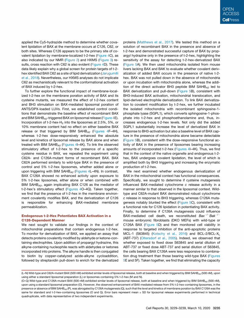
Figure 1. Covalent Lipidation of Recombi-
nant BAX by t-2-Hex
(A) Chemical reaction of the lipid electrophile trans-
2-hexadecenal (t-2-hex) with a nucleophilic
cysteine residue.
(B) Dose-responsive lipidation of recombinant BAX
(molecular weight [MW] 21,320 Da; 5 mM) by t-2-
hex to yield a covalent adduct that incorporates 1
equivalent of lipid (MW 21,558 Da).
(C) The reduced form of t-2-hex, hexadecanal (t-2-
hex-H2), does not chemically react with cysteine.
(D) Exposure of recombinant BAX (MW 21,320 Da;
5 mM) to increasing doses of t-2-hex-H2 causes no
covalent lipidation, as reflected by the absence of a
MW change in BAX.
(E) Incubation of BAX (5 mM) with increasing
amounts of t-2-hex (red; 103, 303, and 1003) in-
duces a prominent laddering effect consistent with
the activation and oligomerization of BAX, whereas
treatment with the highest dose of t-2-hex-H2
(blue) has little to no effect.
(F) Percent labeling of BAX (5 mM) by lipid-derived
electrophiles (a,b-unsaturated aldehydes; 500 mM)
of varying carbon length, as monitored by intact
mass spectrometry. 5, 2-pentenal; 6, 2-hexenal; 7,
2-heptenal; 8, 2-octenal; 9, 2-nonenal; 10, 2-de-
cenal; 11, 2-undecenal; 12, 2-dodecenal; 13, 2-
tridecenal; 16 (red), t-2-hex. Data are mean ± SD of
three independent biological replicates.
(G) Effect of incubating lipid-derived electrophiles
(500 mM) of varying carbon chain length on the
oligomerization of BAX (5 mM), as assessed by the
laddering of BAX upon gel electrophoresis and
BAX western analysis.
analyses of the reaction provided structural insights into the ef-
fect of C126 lipidation on the conformational activation of BAX,
informing both a fundamental apoptotic process and an oppor-
tunity for therapeutic modulation of cell death.
RESULTS
t-2-Hex Covalently Lipidates BAXWe noted that the molecular structure of t-2-hex contains an
a,b-unsaturated aldehyde and is, therefore, susceptible to
Michael addition in the presence of nucleophiles, such as the
sulfhydryl group of cysteines (Figure 1A). Thus, we tested the
reactivity of t-2-hex toward recombinant, full-length BAX to
monitor for direct covalent lipidation. t-2-Hex was added to
BAX (5 mM) at molar ratios of 10:1, 100:1, and 1000:1, followed
by dialysis and analysis by intact protein mass spectrometry
(MS). Covalent derivatization of BAX was detectable at 10-fold
3230 Cell Reports 30, 3229–3239, March 10, 2020
molar excess, progressed to 50%conver-
sion with 100-fold excess, and was
completely derivatized by 1,000-fold
molar excess of t-2-hex (Figure 1B).
Although a large excess of t-2-hex was
required to achieve complete conversion
to the fully conjugated form, we reasoned
that 100% lipidation would not be
required to achieve a functional effect
given the catalytic nature of BAX activation, as reflected by the
capacity of sub-stoichiometric BH3-only protein to trigger
BAX, followed by its robust autoactivation (Tan et al., 2006). Of
note, the generation of an m+238 Da adduct suggested that
BAX reacts with t-2-hex by Michael addition because imine for-
mation would have yielded a mass of m+222, which was not
observed. Interestingly, the saturated form of t-2-hex, hexade-
canal (hereafter referred to as t-2-hex-H2) (Figure 1C), was previ-
ously shown to have no functional effect on BAX activation (Chi-
puk et al., 2012). Here, we find that even at 1,000-fold molar
excess, incubating t-2-hex-H2 with BAX yielded no covalent
adduct (Figure 1D), reinforcing the point that BAX reacts with
t-2-hex by Michael addition and that the capacity for covalent
reactivity with BAX biochemically distinguishes t-2-hex from
t-2-hex-H2.
To link covalent lipidation of BAX to its functional activation,
we first compared the effects of t-2-hex and t-2-hex-H2 on

6521 43 87 9
6521 43 87 9
A
B 100x t-2-hex
30x t-2-hex
L45
R134
E131K128
V121K119
F114
W188
I80
V177
Core Residues Core Residue ContactsC D
0 5 10 15 20 25 30 35 40 45 50 55 60 65 70 75 80 85 90 95 100
105
110
115
120
125
130
135
140
145
150
155
160
165
170
175
180
185
190
0.00
0.01
0.02
0.03
0.04
0.05
(ppm
)
0 5 10 15 20 25 30 35 40 45 50 55 60 65 70 75 80 85 90 95 100
105
110
115
120
125
130
135
140
145
150
155
160
165
170
175
180
185
190
0.00
0.01
0.02
0.03
(ppm
)
60°
2
1
39
4
67
8
C126
C62
5
2
1
39
4
67
8
C126
C62
52
1
3
9
6
78
C62
5
C126
BAX Residue Number
BAX Residue Number
Figure 2. Influence of t-2-Hex on the Struc-
ture of BAX in Solution
(A–D) NMR analysis of 15N-BAX (50 mM) upon in-
cubation with 30:1 (A) and 100:1 (B) t-2-hex:BAX
revealed chemical shift changes in residues of the
a5/a6 hairpin adjacent to C126, in addition to res-
idues of the a1-a2 loop, a2, a3, and a9 that engage
the a5/a6 core and are implicated in the initiation
and translocation of BAX (C). Residues with sig-
nificant chemical shift changes (1 SD threshold of
R0.0109 ppm for 30:1, 0.0094 ppm for 100:1) are
mapped onto the ribbon structure of BAX (PDB:
1F16) (C and D). The proximity of affected a5/a6
core residues (cyan) and their contacts (magenta) is
further highlighted by the stick representations (D).
Cysteines 62 and 126 are colored orange.
See also Figures S1 and S2.
inducing BAX oligomerization, as assessed by denaturing gel
electrophoresis of the reaction samples, followed by anti-BAX
western blotting. The covalent reactivity feature of t-2-hex
indeed correlated with dose-responsive production of notably
increased levels of higher-order BAX oligomeric species, which
even persisted after heating to 95�C in 100 mM HCl, quenching
with lithium dodecyl sulfate (LDS) loading dye containing
250 mM DTT, and performing electrophoresis under denaturing
conditions (Figure 1E). The saturated formof t-2-hex, t-2-hex-H2,
had comparatively little effect when examined at equivalent
high-dosing ratios (e.g., 100:1). To further characterize the struc-
ture-activity relationship for the reaction between t-2-hex (16
carbons) and BAX, we screened commercially available a,b-un-
saturated aldehydes ranging from 5 to 13 carbons long in
BAX covalent labeling and oligomerization assays. The shorter-
chain electrophiles were more reactive toward BAX (Figure 1F),
whereas induction of higher-order oligomerization correlated
instead with increasing chain length, with the physiologic lipid
electrophile, t-2-hex, demonstrating the most robust effect
(Figure 1G). In general, induction of BAX oligomerization was
enhanced as a function of carbon chain length; yet, even the
shortest species, such as 2-pentenal, 2-hexenal, and 2-
heptenal, demonstrated greater activation of BAX than t-2-
hex-H2, highlighting the critical role of the Michael addition
reaction. Thus, the combination of covalent reactivity and the
Cell Re
hydrophobic surface area afforded by
the 16-carbon t-2-hex appeared to maxi-
mize the stimulatory effect.
NMR Analysis of the t-2-Hex/BAXInteractionTo characterize the effect of t-2-hex
conjugation on BAX conformation, we
performed 1H-15N heteronuclear single
quantum coherence (HSQC) NMR anal-
ysis upon t-2-hex titration. First, we found
that pre-incubation of BAX with t-2-hex
for greater than 30 min, or t-2-hex con-
centrations over 5 mM, resulted in severe
line broadening that precluded analysis
but is consistent with a major conformational change, such as
the oligomerization described above (Figures 1E and 1G).
Upon applying 30- and 100-fold molar excess of t-2-hex, the
most prominent chemical shift changes occurred in the a5/a6
hairpin residues that are adjacent to C126 (F114, K119, L120,
V121, K128, E131, and R134) and local residues that engage
the a5/a6 core, including select amino acids from the a1-a2
loop, a2 (C62), a3 (I80), and a9 (V177, W188) (Figures 2A–2D).
Interestingly, a pair of residues that juxtapose in the ‘‘closed-
loop’’ form of inactive BAX, L45 of the a1-a2 loop and R134 of
a6 (a residue within the N-terminal trigger site), were among
those residues that undergo significant chemical shift change
upon t-2-hex interaction. These data suggest that when t-2-
hex is applied to BAX in solution, the dominant effect is confor-
mational perturbation of the a5/a6 hairpin, which represents the
hydrophobic core of the BAX protein and displays C126 at the
protein surface, with attendant effects on key adjacent regions
involved in the initiation (a1-a2 loop, a2) and translocation (a9)
of BAX (Figure 2D). Of note, BAX has two cysteine residues,
C62 and C126, with the latter more exposed than the former
in the monomeric BAX structure (Figure S1) (Suzuki et al.,
2000). Interestingly, the a5/a6 junction where C126 resides is
a conserved cysteine-containing region across BCL-2 family
proteins (Figure S2). Based on the predominance of chemical
shift changes centered around C126 upon t-2-hex titration,
ports 30, 3229–3239, March 10, 2020 3231
A
B
C
D
3
94
6
7
8
52
11
2
9
7
8
5
6
3
4
(legend on next page)
3232 Cell Reports 30, 3229–3239, March 10, 2020
the NMR data suggested that C126 is the likely site of regulation
by the lipid electrophile.
C126-Dependent Conformational Activation of BAXby t-2-HexTo further characterize the conformational consequences and
cysteine-specific dependency of t-2-hex lipidation of BAX, we
performed comparative HXMS (Table S1). HXMS probes protein
structure by measuring changes in the deuterium incorporation
of backbone amide hydrogens (Engen, 2009). When diluted
into deuterium buffer, backbone hydrogens of flexible and/or
exposed protein regions rapidly exchange with deuterium,
whereas buried domains and/or those regions that contain
hydrogen-bonding involving backbone amide hydrogens (such
as in a helices) demonstrate slowed or suppressed deuterium
exchange (Laiken et al., 1969; Printz et al., 1972; Shi et al.,
2013). When we compared the deuterium exchange profiles of
BAX in the presence and absence of t-2-hex, we observed
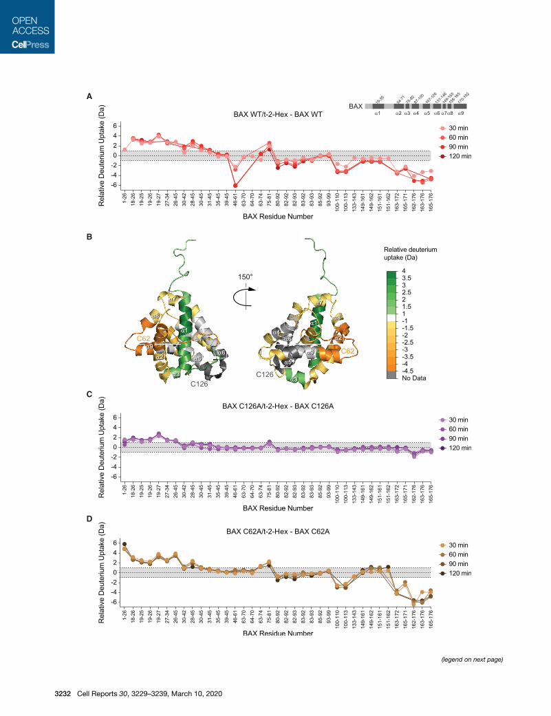
time-responsive changes in discrete functional regions consis-
tent with the induction of oligomerization (Figures 3A and 3B).
Specifically, we observed deprotection of the N-terminal struc-
tures of BAX, including a1 and the a1-a2 loop, a region impli-
cated in the initiating conformational changes of BAX activation
(Gavathiotis et al., 2010), in addition to a3. Prominent regions of
protection included the BH3 domain (a2) and the C-terminal
helix (a9), which are believed to mobilize upon BAX activation
and then engage in self-association interactions in the resultant
oligomerized BAX species (Czabotar et al., 2013; Gahl et al.,
2014; Gavathiotis et al., 2010). Proximal portions of a4 and a5,
the a4-a5 loop, a7, and a8 also showed lipid-induced protection
from deuterium exchange.
To interrogate the requirement of a specific cysteine residue
in the conformational changes induced by t-2-hex, we generated
full-length BAX protein bearing C62A or C126A mutations
(Figure S3) and repeated the HXMS analyses. Strikingly,
C126A mutagenesis abrogated essentially all of the t-2-hex-
induced conformational changes, with residual—yet markedly
reduced—exchange evident in the N-terminal region alone
(Figure 3C). Importantly, a comparison of the baseline deuterium
exchange profiles for C126A and wild-type BAX demonstrated
no differences (Figure S4A). Thus, C126A mutagenesis has no
apparent independent effect on the overall structure of BAX
but instead directly impacts the conformational responsiveness
of BAX to t-2-hex (Figure 3C). In contrast, C62A mutagenesis
phenocopied the response of wild-type BAX to t-2-hex. Indeed,
the only difference observed between the C62A and wild-type
Figure 3. t-2-Hex Promotes the Conformational Activation and Oligom
(A) A plot of relative deuterium differences obtained by subtracting the relative
incorporation of wild-type BAX (60 pmoles) in the presence of t-2-hex (100:1, t-2-
represents changes in the plot that are below a threshold of 1 Da in magnitude
representative of two independent biological replicates per BAX construct for ea
(B) The differences in deuterium exchange above the threshold induced by incuba
The color scale represents the extent of deprotection (green) and protection (ora
(C) The deuterium uptake difference plot indicates the relative deuterium incorpor
C126A) over time (30-, 60-, 90-, and 120-min time points) minus the relative deu
(D) The deuterium uptake difference plot shows the relative deuterium incorpora
C62A) over time (30-, 60-, 90-, and 120-min time points) minus the relative deute
See also Figures S3 and S4 and Table S1.
BAX responses was apparent elimination of t-2-hex-induced
protection of the BH3 region in the C62A mutant. However,
comparison of the deuterium exchange profiles of C62A and
wild-type BAX revealed that C62Amutagenesis caused focal de-
protection within the BH3 region, which is correspondingly
reduced (to zero exchange) upon exposure to t-2-hex (Fig-
ure S4B). Thus, the C62A mutation alone alters conformational
dynamics in the vicinity of C62, but the overall influence of
t-2-hex on the deuterium exchange profiles of C62A and wild-
type BAX is the same. Taken together, the HXMS analyses
demonstrate that exposure of BAX to t-2-hex in solution can
directly induce a major conformational reorganization of mono-
meric BAX, consistent with oligomerization, and that this phe-
nomenon specifically requires C126.
t-2-Hex-Containing Model Membranes Enhance BAXActivationTo develop a model system that best recapitulates the physio-
logic context for covalent lipidation while allowing for direct
analysis of the individual components that comprise the BAX
activation mechanism, we developed a liposomal system that
incorporates lipid electrophiles. Large unilamellar vesicles
(LUVs) reflecting the lipid composition of the mitochondrial outer
membrane and containing 0%, 5%, or 10% t-2-hex were incu-
bated with recombinant, full-length BAX in the presence or
absence of a triggering stapled-BH3 helix (BIM SAHBA2) (Gava-
thiotis et al., 2008). First, to determine if t-2-hex in the liposomal
membrane covalently labeled BAX, we subjected the reaction
mixture to Cy5 hydrazide, which selectively reacts with alde-
hydes and ketones and, thus, serves as a direct and selective
in-gel probe for t-2-hex-conjugated BAX, as detected by Cy5
scan of the SDS-PAGE gel of electrophoresed samples (Boone
et al., 2016). We observed t-2-hex dose-dependent increases
in the amount of generated t-2-hex/BAX conjugate upon treat-
ment with BIM SAHBA2 (Figure 4A), demonstrating that
membrane-embedded t-2-hex was indeed capable of covalently
reacting with BAX once it was triggered to undergo membrane
translocation. We corroborated this result using an orthogonal
method, whereby alkynylated t-2-hex ((E)-hexadec-2-en-
15-ynal) was incorporated into liposomes so that t-2-hex/
BAX conjugate could be isolated by streptavidin pull-down after
biotinylating t-2-hex with the biotin-PEG-azide reagent and click
chemistry (Jarugumilli et al., 2018). This alternative analysis pro-
duced essentially the same results as our Cy5 method, confirm-
ing that t-2-hex, as a liposomal membrane component, can
directly and covalently react with BAX (Figure 4B). We further
erization of BAX
deuterium incorporation of wild-type BAX alone from the relative deuterium
hex:BAX) over time (30-, 60-, 90-, and 120-min time points). Light gray shading
, whereas the white region highlights changes above the threshold. Data are
ch time point.
ting BAXwith t-2-hex aremapped onto the ribbon structure of BAX (PDB: 1F16).
nge) of BAX in response to t-2-hex.
ation of BAX C126A (60 pmoles) in the presence of t-2-hex (100:1, t-2-hex:BAX
terium incorporation of BAX C126A alone.
tion of BAX C62A (60 pmoles) in the presence of t-2-hex (100:1, t-2-hex:BAX
rium incorporation of BAX C62A alone.
Cell Reports 30, 3229–3239, March 10, 2020 3233
A C E
D
GF
B
I J
L M
H
K
N
QPO
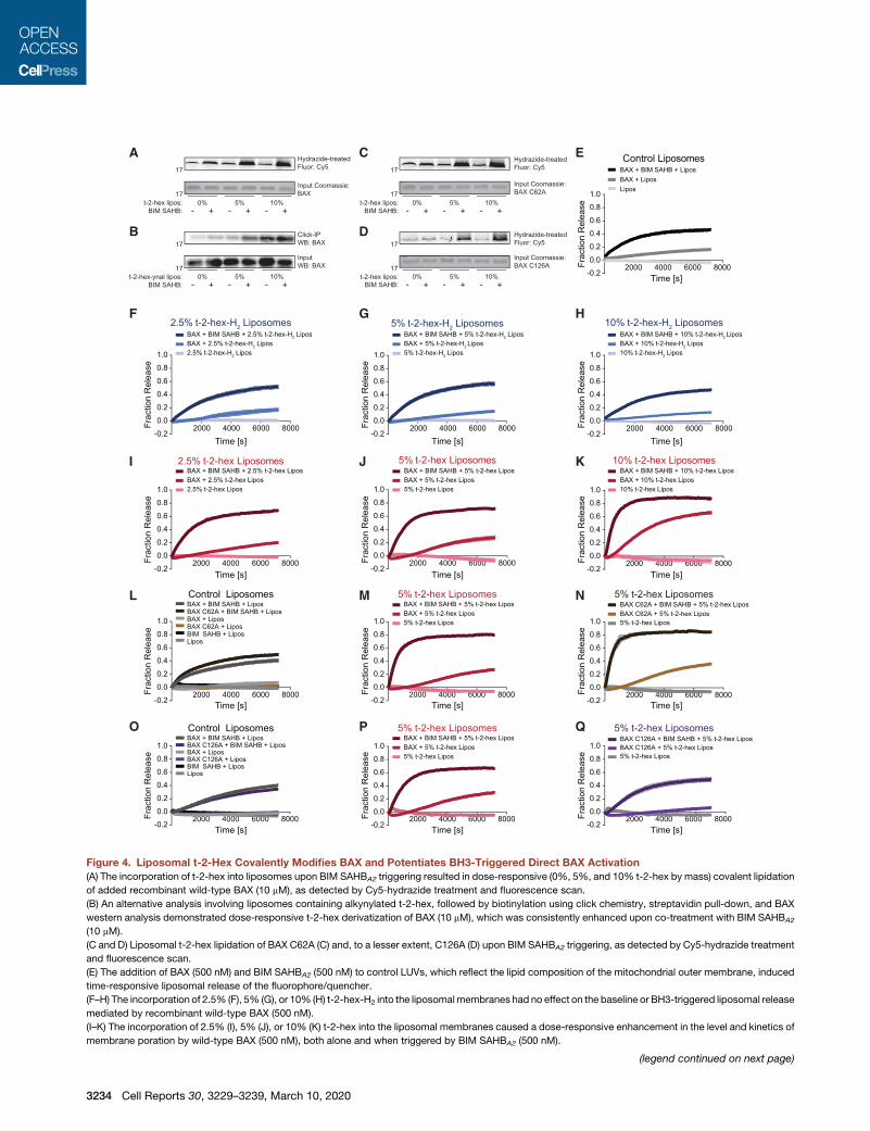
Figure 4. Liposomal t-2-Hex Covalently Modifies BAX and Potentiates BH3-Triggered Direct BAX Activation
(A) The incorporation of t-2-hex into liposomes upon BIM SAHBA2 triggering resulted in dose-responsive (0%, 5%, and 10% t-2-hex by mass) covalent lipidation
of added recombinant wild-type BAX (10 mM), as detected by Cy5-hydrazide treatment and fluorescence scan.
(B) An alternative analysis involving liposomes containing alkynylated t-2-hex, followed by biotinylation using click chemistry, streptavidin pull-down, and BAX
western analysis demonstrated dose-responsive t-2-hex derivatization of BAX (10 mM), which was consistently enhanced upon co-treatment with BIM SAHBA2
(10 mM).
(C and D) Liposomal t-2-hex lipidation of BAX C62A (C) and, to a lesser extent, C126A (D) upon BIM SAHBA2 triggering, as detected by Cy5-hydrazide treatment
and fluorescence scan.
(E) The addition of BAX (500 nM) and BIM SAHBA2 (500 nM) to control LUVs, which reflect the lipid composition of the mitochondrial outer membrane, induced
time-responsive liposomal release of the fluorophore/quencher.
(F–H) The incorporation of 2.5% (F), 5% (G), or 10% (H) t-2-hex-H2 into the liposomal membranes had no effect on the baseline or BH3-triggered liposomal release
mediated by recombinant wild-type BAX (500 nM).
(I–K) The incorporation of 2.5% (I), 5% (J), or 10% (K) t-2-hex into the liposomal membranes caused a dose-responsive enhancement in the level and kinetics of
membrane poration by wild-type BAX (500 nM), both alone and when triggered by BIM SAHBA2 (500 nM).
(legend continued on next page)
3234 Cell Reports 30, 3229–3239, March 10, 2020
applied the Cy5-hydrazide method to determine whether cova-
lent lipidation of BAX at the membrane occurs at C126, C62, or
both sites. Whereas C126 appears to be the primary site of co-
valent lipidation by membrane-localized t-2-hex (Figure 4C), as
also indicated by our NMR (Figure 2) and HXMS (Figure 3) re-
sults, cross reaction with C62 is also evident (Figure 4D). These
data likely explain why a global screen for protein targets of t-2-
hex identified BAXC62 as a site of lipid derivatization (Jarugumilli
et al., 2018). Nevertheless, our HXMS analyses do not implicate
C62 as mechanistically relevant to the conformational activation
of BAX induced by t-2-hex.
To further explore the functional impact of membrane-local-
ized t-2-hex on the membrane poration activity of BAX and its
cysteine mutants, we measured the effect of t-2-hex content
and BH3 stimulation on BAX-mediated liposomal poration of
ANTS/DPX-loaded LUVs. First, we established control condi-
tions that demonstrate the baseline effect of recombinant BAX
andBIMSAHBA2-triggeredBAXon liposomal release (Figure 4E).
Incorporation of t-2-hex-H2 into the liposomes at 2.5%, 5%, or
10% membrane content had no effect on either baseline BAX
release or that triggered by BIM SAHBA2 (Figures 4F–4H),
whereas t-2-hex dose-responsively enhanced the absolute
level and kinetics of liposomal release by BAX alone and when
treated with BIM SAHBA2 (Figures 4I–4K). To link the observed
stimulatory effect of t-2-hex to the presence of a specific
cysteine residue in BAX, we repeated the experiment using
C62A- and C126A-mutant forms of recombinant BAX. BAX
C62A performed similarly to wild-type BAX in the presence of
control and 5% t-2-hex liposomes, whether added alone or
upon triggering with BIM SAHBA2 (Figures 4L–4N). In contrast,
BAX C126A showed no enhanced activity upon exposure to
5% t-2-hex liposomes, either alone or when combined with
BIM SAHBA2, again implicating BAX C126 as the mediator of
t-2-hex’s stimulatory effect (Figures 4O–4Q). Taken together,
we find that the presence of t-2-hex in the membrane environ-
ment covalently modifies BAX, and the derivatization of C126
is responsible for enhancing BAX-mediated membrane
permeabilization.
Endogenous t-2-Hex Potentiates BAX Activation in aC126-Dependent MannerWe next sought to validate our findings in the context of
mitochondrial preparations that contain endogenous t-2-hex.
To monitor for derivatization of BAX, we applied an assay that
detects proteins covalently modified by aldehyde or ketone-con-
taining electrophiles. Upon addition of propargyl hydrazine, this
alkyne-containing nucleophile reacts with aldehydes or ketones
incorporated into proteins. The alkyne handle is then conjugated
to biotin by copper-catalyzed azide-alkyne cycloaddition,
followed by streptavidin pull-down to enrich for the derivatized
(L–N) Wild-type and C62A-mutant BAX (500 nM) exhibited similar levels of liposom
using either a standard liposomal preparation (L) or liposomes containing 5% t-2
(O–Q) Wild-type and C126A-mutant BAX (500 nM) exhibited similar levels of lipo
upon using a standard liposomal preparation (O). However, the observed enhanc
presence or absence of BIMSAHBA2 (P), was abrogated by C126Amutagenesis (Q
same for standard and t-2-hex-containing liposomes (O and Q). Error bars rep
quadruplicate, with data representative of two independent experiments.
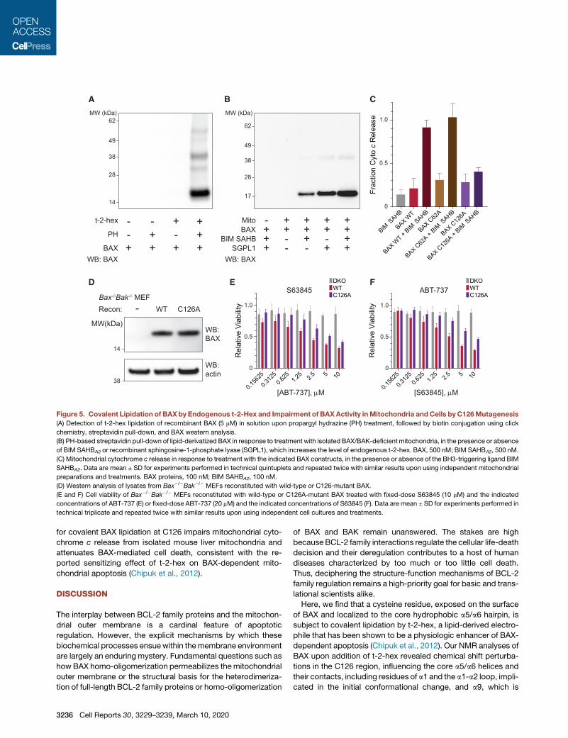
proteins (Matthews et al., 2017). We tested this method on a
solution of recombinant BAX in the presence and absence of
t-2-hex and demonstrated successful capture of BAX by prop-
argyl hydrazine only in the presence of t-2-hex, highlighting the
sensitivity of the assay for detecting t-2-hex-derivatized BAX
(Figure 5A). We then used mitochondria isolated from mouse
livers lacking BAX and BAK to evaluate whether covalent deriv-
atization of added BAX occurs in the presence of native t-2-
hex. BAX was not pulled down in the absence of mitochondria
or upon incubation with mitochondria alone, whereas the addi-
tion of the direct activator BH3 peptide BIM SAHBA2 led to
BAX derivatization and pull-down (Figure 5B), consistent with
BH3-induced BAX activation, mitochondrial translocation, and
lipid-derived electrophile derivatization. To link BAX derivatiza-
tion to covalent modification by t-2-hex, we further incubated
the isolated mitochondria with recombinant sphingosine-1-
phosphate lyase (SGPL1), which converts sphingosine-1-phos-
phate into t-2-hex and phosphoethanolamine and, thus, in-
creases endogenous t-2-hex levels. Not only did the added
SGPL1 substantially increase the level of derivatized BAX in
response to BH3-activation but also a baseline level of BAX cap-
ture in the presence of mitochondria alone became detectable
(Figure 5B), consistent with the dose-responsive functional ac-
tivity of BAX in the presence of liposomes bearing increasing
amounts of incorporated t-2-hex (Figures 4I–4K). Thus, we find
that in the context of the native organelle and endogenous t-2-
hex, BAX undergoes covalent lipidation, the level of which is
amplified both by BH3 triggering and increasing the enzymatic
production of t-2-hex.
We next examined whether endogenous derivatization of
BAX in the mitochondrial context has functional consequences.
Specifically, we sought to determine if cysteine mutagenesis
influenced BAX-mediated cytochrome c release activity in a
manner similar to that observed in the liposomal context. Wild-
type and C62A-mutant BAX demonstrated robust cytochrome
c release in response to BH3 triggering, whereas C126A muta-
genesis notably blunted the effect (Figure 5C), consistent with
a functional role for C126 lipidation in potentiating BAX activity.
Finally, to determine if C126A mutagenesis could influence
BAX-mediated cell death, we reconstituted Bax�/�Bak�/�
mouse embryonic fibroblasts (DKO MEFs) with wild-type or
C126A BAX (Figure 5D) and then monitored cell viability in
response to targeted inhibition of the anti-apoptotic proteins
MCL-1 (S63845) (Kotschy et al., 2016) and BCL-2/BCL-XL
(ABT-737) (Oltersdorf et al., 2005). Indeed, we observed that
whether exposed to fixed dose S63845 and serial dilution of
ABT-737 or fixed dose ABT-737 and serial dilution of S63845,
the cells bearing BAX C126A were less responsive to combina-
tion drug treatment than those bearing wild-type BAX (Figures
5E and 5F). Taken together, we find that eliminating the capacity
al release, both at baseline and when triggered by BIM SAHBA2 (500 nM), upon
-hex (M and N).
somal release, both at baseline and when triggered by BIM SAHBA2 (500 nM),
ement of BAX-mediated release from 5% t-2-hex-containing liposomes, in the
), such that the level and kinetics ofmembrane poration by BAXC126Awas the
resent mean ± SD for liposomal release experiments performed in technical
Cell Reports 30, 3229–3239, March 10, 2020 3235
A B C
D
WB: BAX
+++
BAXBIM SAHB
SGPL1
+--
++-
+-+
-Mito + + + ++-- +PH++- -t-2-hex
WB: BAX+++ +BAX
BIM SAHB
BAX WT
BAX WT + BIM
SAHB
BAX C62
A
BAX C62
A + BIM SAHB
BAX C12
6A
BAX C12
6A + BIM
SAHB
62
49
38
28
14
+++
38
14
Bax-/-Bak-/- MEF- WT C126ARecon:
WB: BAX
WB: actin
MW(kDa)
0
0.5
1.0
Frac
tion
Cyt
o c
Rel
ease
5
0.156
25
0.312
50.6
25 1.25 2.5 10
[ABT-737], M
0
0.5
1.0
ABT-737
Rel
ativ
e Vi
abilit
y
DKOWTC126A
0
0.5
1.0
S63845
Rel
ativ
e Vi
abilit
y
E F DKOWTC126A
62
49
38
28
17
5
0.156
25
0.312
50.6
25 1.25 2.5 10
[S63845], M
MW (kDa) MW (kDa)
Figure 5. Covalent Lipidation of BAX by Endogenous t-2-Hex and Impairment of BAXActivity inMitochondria and Cells by C126Mutagenesis
(A) Detection of t-2-hex lipidation of recombinant BAX (5 mM) in solution upon propargyl hydrazine (PH) treatment, followed by biotin conjugation using click
chemistry, streptavidin pull-down, and BAX western analysis.
(B) PH-based streptavidin pull-down of lipid-derivatized BAX in response to treatment with isolated BAX/BAK-deficient mitochondria, in the presence or absence
of BIM SAHBA2 or recombinant sphingosine-1-phosphate lyase (SGPL1), which increases the level of endogenous t-2-hex. BAX, 500 nM; BIM SAHBA2, 500 nM.
(C) Mitochondrial cytochrome c release in response to treatment with the indicated BAX constructs, in the presence or absence of the BH3-triggering ligand BIM
SAHBA2. Data are mean ± SD for experiments performed in technical quintuplets and repeated twice with similar results upon using independent mitochondrial
preparations and treatments. BAX proteins, 100 nM; BIM SAHBA2, 100 nM.
(D) Western analysis of lysates from Bax�/�Bak�/� MEFs reconstituted with wild-type or C126-mutant BAX.
(E and F) Cell viability of Bax�/�Bak�/� MEFs reconstituted with wild-type or C126A-mutant BAX treated with fixed-dose S63845 (10 mM) and the indicated
concentrations of ABT-737 (E) or fixed-dose ABT-737 (20 mM) and the indicated concentrations of S63845 (F). Data are mean ± SD for experiments performed in
technical triplicate and repeated twice with similar results upon using independent cell cultures and treatments.
for covalent BAX lipidation at C126 impairs mitochondrial cyto-
chrome c release from isolated mouse liver mitochondria and
attenuates BAX-mediated cell death, consistent with the re-
ported sensitizing effect of t-2-hex on BAX-dependent mito-
chondrial apoptosis (Chipuk et al., 2012).
DISCUSSION
The interplay between BCL-2 family proteins and the mitochon-
drial outer membrane is a cardinal feature of apoptotic
regulation. However, the explicit mechanisms by which these
biochemical processes ensuewithin themembrane environment
are largely an enduring mystery. Fundamental questions such as
how BAX homo-oligomerization permeabilizes themitochondrial
outer membrane or the structural basis for the heterodimeriza-
tion of full-length BCL-2 family proteins or homo-oligomerization
3236 Cell Reports 30, 3229–3239, March 10, 2020
of BAX and BAK remain unanswered. The stakes are high
because BCL-2 family interactions regulate the cellular life-death
decision and their deregulation contributes to a host of human
diseases characterized by too much or too little cell death.
Thus, deciphering the structure-function mechanisms of BCL-2
family regulation remains a high-priority goal for basic and trans-
lational scientists alike.
Here, we find that a cysteine residue, exposed on the surface
of BAX and localized to the core hydrophobic a5/a6 hairpin, is
subject to covalent lipidation by t-2-hex, a lipid-derived electro-
phile that has been shown to be a physiologic enhancer of BAX-
dependent apoptosis (Chipuk et al., 2012). Our NMR analyses of
BAX upon addition of t-2-hex revealed chemical shift perturba-
tions in the C126 region, influencing the core a5/a6 helices and
their contacts, including residues of a1 and the a1-a2 loop, impli-
cated in the initial conformational change, and a9, which is
allosterically released for membrane translocation upon direct
BH3-induced BAX activation (Gavathiotis et al., 2010). Compar-
ative HXMSanalyses, conducted over time, demonstrated that t-
2-hex triggered amajor conformational change in both wild-type
and C62A BAX, consistent with a monomer-to-oligomer transi-
tion. Strikingly, C126Amutagenesis abrogated this effect, further
implicating C126 as the functionally relevant site of t-2-hex deriv-
atization. Exposure of BAX to liposomal membranes containing
t-2-hex not only resulted in the covalent modification of BAX
but also directly enhanced the absolute level and kinetics of
BAX-mediated membrane poration, an activity that specifically
required C126. BAX derivatization was also observed in the
context of mitochondrial preparations containing endogeneous
t-2-hex, wherein covalent modification was specifically induced
by BH3 triggering, which translocates BAX to the mitochondrial
membrane and effectively concentrates BAX in a two-dimen-
sional bilayer with t-2-hex. Boosting the levels of endogenous
t-2-hex by adding SGPL1 to the mitochondrial preparation
enhanced BAX derivatization even further. Importantly, both
BAX-mediated mitochondrial cytochrome c release and BAX-
mediated cell death were selectively impaired by C126A muta-
genesis, further highlighting the functional relevance of C126 lip-
idation. Based on these data, we expand the mechanistic model
of BH3-triggered direct BAX activation to include covalent reac-
tion of the a5/a6 hydrophobic core with the target membrane, a
biochemical process that enhances BAX activation and mem-
brane poration. Indeed, a three-dimensional model of active
BAX at the membrane based on double electron-electron reso-
nance spectroscopy predicts opening of the a5/a6 hairpin to
form a clamp-like conformation on the membrane surface
(Bleicken et al., 2014), which would ideally position C126
(located at the junction of the a5/a6 hairpin) for interaction with
lipid headgroups, such as the reactive a,b-unsaturated aldehyde
of t-2-hex.
Post-translational modification has emerged as a key mecha-
nism for regulating the functional activities of BCL-2 family pro-
teins. For example, myristoylation of the BH3-only protein tBID
targets it to the mitochondrial outer membrane (Zha et al.,
2000), phosphorylation of BAD shifts its binding affinity from
BCL-2 family anti-apoptotic proteins to glucokinase (Danial
et al., 2008), and ubiquitination of anti-apoptotic members
such as MCL-1 controls protein half-life (Zhong et al., 2005). De-
pending on the physiologic context and experimental setup, a
variety of cysteine modifications have been reported for BAX,
including S-palmitoylation of C126 (Frohlich et al., 2014), oxida-
tion of C62 (Nie et al., 2008), and most recently t-2-hex derivati-
zation of C62 (Jarugumilli et al., 2018). Although in this study we
do not detect a functional role for C62 in the activity of t-2-hex, it
remains plausible that under specific conditions that affect
accessibility of the relatively buried C62 residue, covalent conju-
gation may occur. Here, we demonstrate that the functionally
relevant site of BAX derivatization by t-2-hex is C126. Notably,
in contrast to the post-translational modifications described
above that require enzymatic reactions (e.g., myristoylation,
phosphorylation, and ubiquitination), the covalent lipidation of
BAX C126 reported here is non-enzymatic and instead involves
a direct biochemical reaction between BAX and t-2-hex at the
mitochondrial membrane.
Of the two cysteines in human BAX, the more exposed C126
residue is located at the a5/a6 junction, where a variety of
BCL-2 family proteins also contain a cysteine residue (Fig-
ure S2). Interestingly, our prior screen for small molecule inhib-
itors of anti-apoptotic MCL-1 yielded select compounds that
covalently modified C286 and, in doing so, induced an allosteric
conformational change in its canonical surface groove that
impaired BH3 interaction (Lee et al., 2016). Thus, characterizing
the structure-function consequences of post-translational
cysteine derivatization not only is critical to our understanding
of the physiologic properties of BCL-2 family proteins but also
informs approaches to selectively target reactive cysteines by
Michael addition to modulate their pro- and anti-apoptotic ac-
tivities for therapeutic benefit. Indeed, cysteine modification
by selective, electrophilic molecules has become an increas-
ingly appealing approach to drugging recalcitrant targets, as
exemplified by the success of ibrutinib, a covalent inhibitor of
Bruton’s tyrosine kinase (Zhang et al., 2019). The presence of
a unique cysteine residue within the canonical BH3-binding
groove of A/A1 inspired our development of selective,
cysteine-reactive stapled peptide inhibitors of this anti-
apoptotic member implicated in the development and chemo-
resistance of leukemia, lymphoma, and melanoma (Guerra
et al., 2018; Harvey et al., 2018; Huhn et al., 2016). Here, our
finding that t-2-hex-containing membranes can covalently
derivatize and sensitize BAX activation reveals a pharmacologic
opportunity to influence cell fate by alternatively simulating or
blocking C126 lipidation. Small molecules that covalently target
C126 and contain hydrophobic moieties that engage the site of
t-2-hex interaction have the potential to directly modulate BAX-
mediated apoptosis. Intriguingly, the small molecule OICR766A
that emerged from a screen for compounds that enhance BAX-
mediated liposomal poration was found to bind and activate
BAX in a C126-dependent manner (Brahmbhatt et al., 2016).
Thus, whether by physiologic or pharmacologic targeting, the
C126 region may represent an important control point for
modulating the pro-apoptotic functionality of BAX.
STAR+METHODS
Detailed methods are provided in the online version of this paper
and include the following:
d KEY RESOURCES TABLE
d LEAD CONTACT AND MATERIALS AVAILABILITY
d EXPERIMENTAL MODEL AND SUBJECT DETAILS
B Microbe Strains
B Cell Lines and Culture
d METHOD DETAILS
B BAX Protein Preparation
B Intact Protein Mass Spectrometry
B BAX Laddering Assay
B NMR Spectroscopy
B Hydrogen-Deuterium Exchange Mass Spectrometry
B Cy5-Hydrazide Labeling of Lipidated BAX
B Streptavidin Pull-down of Lipidated BAX
B Liposomal Preparation and Release Assay
B Chemical Synthesis of Labeling Reagents
Cell Reports 30, 3229–3239, March 10, 2020 3237
B Detection of Lipidated BAX by Propargyl Hydrazine in
Solution
B Detection of Lipidated BAX by Propargyl Hydrazine in
Isolated Mitochondria
B Cytochrome c Release Assay
B Retroviral Transduction of Mouse Embryonic Fibro-
blasts
B Cell Viability
d QUANTIFICATION AND STATISTICAL ANALYSIS
d DATA AND CODE AVAILABILITY
SUPPLEMENTAL INFORMATION
Supplemental Information can be found online at https://doi.org/10.1016/j.
celrep.2020.02.057.
ACKNOWLEDGMENTS
We thank G. Bird and T. Oo for BIM SAHBA2 production, J. Lee for performing
intact MS analysis at the Dana-Farber Molecular Biology Core, C. Sheahan
and G. Heffron for assistance with NMR spectroscopy, and E. Smith for
graphics support and figure preparation. This research was supported by
NIH grant R35CA197583 to L.D.W.; a Landry Cancer Biology Research Fellow-
ship and Chleck Family Scholarship to D.T.C.; NIH grant R01GM101135 to
J.R.E.; a research collaboration between J.R.E. and the Waters Corporation;
and a National Science Foundation Predoctoral Fellowship and a Landry Can-
cer Biology Research Fellowship to M.W.M..
AUTHOR CONTRIBUTIONS
L.D.W. and D.T.C. designed the study. D.T.C. generated BAX protein and per-
formed the chemical syntheses and the biochemical, cellular, and NMR exper-
iments. D.T.C. and T.E.W. performed the HXMS analyses under the guidance
of J.R.E.M.W.M. conducted the structure-activity relationship analysis of lipid-
derived electrophiles. All authors analyzed the data, and L.D.W. and D.T.C.
wrote the manuscript, which was reviewed by all co-authors.
DECLARATION OF INTERESTS
L.D.W. is a scientific co-founder and shareholder in Aileron Therapeutics. The
composition of a stapled peptide used in the study has been patented by the
Dana-Farber Cancer Institute.
Received: May 13, 2019
Revised: December 23, 2019
Accepted: February 11, 2020
Published: March 10, 2020
REFERENCES
Barclay, L.A., Wales, T.E., Garner, T.P., Wachter, F., Lee, S., Guerra, R.M.,
Stewart, M.L., Braun, C.R., Bird, G.H., Gavathiotis, E., et al. (2015). Inhibition
of pro-apoptotic BAX by a noncanonical interaction mechanism. Mol. Cell
57, 873–886.
Bleicken, S., Jeschke, G., Stegmueller, C., Salvador-Gallego, R., Garcıa-Saez,
A.J., and Bordignon, E. (2014). Structural model of active Bax at the mem-
brane. Mol. Cell 56, 496–505.
Boone, C.H., Grove, R.A., Adamcova, D., Braga, C.P., and Adamec, J. (2016).
Revealing oxidative damage to enzymes of carbohydrate metabolism in yeast:
An integration of 2D DIGE, quantitative proteomics, and bioinformatics. Prote-
omics 16, 1889–1903.
Brahmbhatt, H., Uehling, D., Al-Awar, R., Leber, B., and Andrews, D. (2016).
Small molecules reveal an alternative mechanism of Bax activation. Biochem.
J. 473, 1073–1083.
3238 Cell Reports 30, 3229–3239, March 10, 2020
Chipuk, J.E., Moldoveanu, T., Llambi, F., Parsons, M.J., and Green, D.R.
(2010). The BCL-2 family reunion. Mol. Cell 37, 299–310.
Chipuk, J.E., McStay, G.P., Bharti, A., Kuwana, T., Clarke, C.J., Siskind, L.J.,
Obeid, L.M., andGreen, D.R. (2012). Sphingolipidmetabolism cooperateswith
BAK and BAX to promote the mitochondrial pathway of apoptosis. Cell 148,
988–1000.
Czabotar, P.E., Westphal, D., Dewson, G., Ma, S., Hockings, C., Fairlie, W.D.,
Lee, E.F., Yao, S., Robin, A.Y., Smith, B.J., et al. (2013). Bax crystal structures
reveal how BH3 domains activate Bax and nucleate its oligomerization to
induce apoptosis. Cell 152, 519–531.
Danial, N.N.,Walensky, L.D., Zhang, C.Y., Choi, C.S., Fisher, J.K., Molina, A.J.,
Datta, S.R., Pitter, K.L., Bird, G.H., Wikstrom, J.D., et al. (2008). Dual role of
proapoptotic BAD in insulin secretion and beta cell survival. Nat. Med. 14,
144–153.
Engen, J.R. (2009). Analysis of protein conformation and dynamics by
hydrogen/deuterium exchange MS. Anal. Chem. 81, 7870–7875.
Frohlich, M., Dejanovic, B., Kashkar, H., Schwarz, G., and Nussberger, S.
(2014). S-palmitoylation represents a novel mechanism regulating the mito-
chondrial targeting of BAX and initiation of apoptosis. Cell Death Dis. 5, e1057.
Gahl, R.F., He, Y., Yu, S., and Tjandra, N. (2014). Conformational rearrange-
ments in the pro-apoptotic protein, Bax, as it inserts into mitochondria: a
cellular death switch. J. Biol. Chem. 289, 32871–32882.
Gavathiotis, E., Suzuki, M., Davis, M.L., Pitter, K., Bird, G.H., Katz, S.G., Tu,
H.C., Kim, H., Cheng, E.H., Tjandra, N., and Walensky, L.D. (2008). BAX acti-
vation is initiated at a novel interaction site. Nature 455, 1076–1081.
Gavathiotis, E., Reyna, D.E., Davis, M.L., Bird, G.H., and Walensky, L.D.
(2010). BH3-triggered structural reorganization drives the activation of proap-
optotic BAX. Mol. Cell 40, 481–492.
Guerra, R.M., Bird, G.H., Harvey, E.P., Dharia, N.V., Korshavn, K.J., Prew,
M.S., Stegmaier, K., and Walensky, L.D. (2018). Precision targeting of BFL-
1/A1 and an ATM co-dependency in human cancer. Cell Rep. 24, 3393–
3403.e5.
Harvey, E.P., Seo, H.S., Guerra, R.M., Bird, G.H., Dhe-Paganon, S., and Wa-
lensky, L.D. (2018). Crystal structures of anti-apoptotic BFL-1 and its complex
with a covalent stapled peptide inhibitor. Structure 26, 153–160.e154.
Hsu, Y.T., and Youle, R.J. (1997). Nonionic detergents induce dimerization
among members of the Bcl-2 family. J. Biol. Chem. 272, 13829–13834.
Huhn, A.J., Guerra, R.M., Harvey, E.P., Bird, G.H., and Walensky, L.D. (2016).
Selective covalent targeting of anti-apoptotic BFL-1 by cysteine-reactive sta-
pled peptide inhibitors. Cell Chem. Biol. 23, 1123–1134.
Jarugumilli, G.K., Choi, J.R., Chan, P., Yu, M., Sun, Y., Chen, B., Niu, J., De-
Ran, M., Zheng, B., Zoeller, R., et al. (2018). Chemical probe to identify the
cellular targets of the reactive lipid metabolite 2-trans-hexadecenal. ACS
Chem. Biol. 13, 1130–1136.
Kim, H., Rafiuddin-Shah,M., Tu, H.C., Jeffers, J.R., Zambetti, G.P., Hsieh, J.J.,
and Cheng, E.H. (2006). Hierarchical regulation of mitochondrion-dependent
apoptosis by BCL-2 subfamilies. Nat. Cell Biol. 8, 1348–1358.
Kim, H., Tu, H.C., Ren, D., Takeuchi, O., Jeffers, J.R., Zambetti, G.P., Hsieh,
J.J., and Cheng, E.H. (2009). Stepwise activation of BAX and BAK by tBID,
BIM, and PUMA initiates mitochondrial apoptosis. Mol. Cell 36, 487–499.
Korsmeyer, S.J., Shutter, J.R., Veis, D.J., Merry, D.E., and Oltvai, Z.N. (1993).
Bcl-2/Bax: a rheostat that regulates an anti-oxidant pathway and cell death.
Semin. Cancer Biol. 4, 327–332.
Kotschy, A., Szlavik, Z., Murray, J., Davidson, J., Maragno, A.L., Le Toumelin-
Braizat, G., Chanrion, M., Kelly, G.L., Gong, J.N., Moujalled, D.M., et al. (2016).
TheMCL1 inhibitor S63845 is tolerable and effective in diverse cancer models.
Nature 538, 477–482.
Laiken, S.L., Printz, M.P., and Craig, L.C. (1969). Tritium-hydrogen exchange
studies of protein models. I. Gramicidin S-A. Biochemistry 8, 519–526.
Lee, S., Wales, T.E., Escudero, S., Cohen, D.T., Luccarelli, J., Gallagher, C.G.,
Cohen, N.A., Huhn, A.J., Bird, G.H., Engen, J.R., and Walensky, L.D. (2016).
Allosteric inhibition of antiapoptotic MCL-1. Nat. Struct. Mol. Biol. 23,
600–607.
Lovell, J.F., Billen, L.P., Bindner, S., Shamas-Din, A., Fradin, C., Leber, B., and
Andrews, D.W. (2008). Membrane binding by tBid initiates an ordered series of
events culminating in membrane permeabilization by Bax. Cell 135, 1074–
1084.
Marintchev, A., Frueh, D., and Wagner, G. (2007). NMR methods for studying
protein-protein interactions involved in translation initiation. Methods Enzy-
mol. 430, 283–331.
Matthews, M.L., He, L., Horning, B.D., Olson, E.J., Correia, B.E., Yates, J.R.,
3rd, Dawson, P.E., and Cravatt, B.F. (2017). Chemoproteomic profiling and
discovery of protein electrophiles in human cells. Nat. Chem. 9, 234–243.
Murray, V., Chen, J., Huang, Y., Li, Q., and Wang, J. (2010). Preparation of
very-high-yield recombinant proteins using novel high-cell-density bacterial
expression methods. Cold Spring Harb. Protoc. 2010, pdb.prot5475.
Nie, C., Tian, C., Zhao, L., Petit, P.X., Mehrpour, M., and Chen, Q. (2008).
Cysteine 62 of Bax is critical for its conformational activation and its proapo-
ptotic activity in response to H2O2-induced apoptosis. J. Biol. Chem. 283,
15359–15369.
Oltersdorf, T., Elmore, S.W., Shoemaker, A.R., Armstrong, R.C., Augeri, D.J.,
Belli, B.A., Bruncko, M., Deckwerth, T.L., Dinges, J., Hajduk, P.J., et al.
(2005). An inhibitor of Bcl-2 family proteins induces regression of solid tu-
mours. Nature 435, 677–681.
Pagliari, L.J., Kuwana, T., Bonzon, C., Newmeyer, D.D., Tu, S., Beere, H.M.,
and Green, D.R. (2005). The multidomain proapoptotic molecules Bax and
Bak are directly activated by heat. Proc. Natl. Acad. Sci. USA 102, 17975–
17980.
Pitter, K., Bernal, F., Labelle, J., and Walensky, L.D. (2008). Dissection of the
BCL-2 family signaling network with stabilized a-helices of BCL-2 domains.
Methods Enzymol. 446, 387–408.
Printz, M.P., Williams, H.P., and Craig, L.C. (1972). Evidence for the presence
of hydrogen-bonded secondary structure in angiotensin II in aqueous solution.
Proc. Natl. Acad. Sci. USA 69, 378–382.
Sattler, M., Liang, H., Nettesheim, D., Meadows, R.P., Harlan, J.E., Eberstadt,
M., Yoon, H.S., Shuker, S.B., Chang, B.S., Minn, A.J., et al. (1997). Structure of
Bcl-xL-Bak peptide complex: recognition between regulators of apoptosis.
Science 275, 983–986.
Shi, X.E., Wales, T.E., Elkin, C., Kawahata, N., Engen, J.R., and Annis, D.A.
(2013). Hydrogen exchange-mass spectrometry measures stapled peptide
conformational dynamics and predicts pharmacokinetic properties. Anal.
Chem. 85, 11185–11188.
Studier, F.W. (2005). Protein production by auto-induction in high density
shaking cultures. Protein Expr. Purif. 41, 207–234.
Suzuki, M., Youle, R.J., and Tjandra, N. (2000). Structure of Bax: coregulation
of dimer formation and intracellular localization. Cell 103, 645–654.
Tan, C., Dlugosz, P.J., Peng, J., Zhang, Z., Lapolla, S.M., Plafker, S.M., An-
drews, D.W., and Lin, J. (2006). Auto-activation of the apoptosis protein Bax
increases mitochondrial membrane permeability and is inhibited by Bcl-2.
J. Biol. Chem. 281, 14764–14775.
Walensky, L.D. (2019). Targeting BAX to drug death directly. Nat. Chem. Biol.
15, 657–665.
Walensky, L.D., and Gavathiotis, E. (2011). BAX unleashed: the biochemical
transformation of an inactive cytosolic monomer into a toxic mitochondrial
pore. Trends Biochem. Sci. 36, 642–652.
Walensky, L.D., Pitter, K., Morash, J., Oh, K.J., Barbuto, S., Fisher, J., Smith,
E., Verdine, G.L., and Korsmeyer, S.J. (2006). A stapled BID BH3 helix directly
binds and activates BAX. Mol. Cell 24, 199–210.
Wales, T.E., and Engen, J.R. (2006). Hydrogen exchange mass spectrometry
for the analysis of protein dynamics. Mass Spectrom. Rev. 25, 158–170.
Zha, J., Weiler, S., Oh, K.J., Wei, M.C., and Korsmeyer, S.J. (2000). Posttrans-
lational N-myristoylation of BID as a molecular switch for targeting mitochon-
dria and apoptosis. Science 290, 1761–1765.
Zhang, T., Hatcher, J.M., Teng, M., Gray, N.S., and Kostic, M. (2019). Recent
advances in selective and irreversible covalent ligand development and valida-
tion. Cell Chem. Biol. 26, 1486–1500.
Zhong, Q., Gao, W., Du, F., and Wang, X. (2005). Mule/ARF-BP1, a BH3-only
E3 ubiquitin ligase, catalyzes the polyubiquitination of Mcl-1 and regulates
apoptosis. Cell 121, 1085–1095.
Cell Reports 30, 3229–3239, March 10, 2020 3239

STAR+METHODS
KEY RESOURCES TABLE
REAGENT or RESOURCE SOURCE IDENTIFIER
Antibodies
Mouse monoclonal anti-BAX 2D2 Santa Cruz Cat# SC-20067; RRID: AB_626726
Sheep anti-mouse HRP Bio-Rad Cat# AAC10P; RRID: AB_321929
Mouse monoclonal anti-b-Actin AC-15 Sigma-Aldrich Cat# A1978; RRID: AB_476692
Bacterial and Virus Strains
One Shot BL21(DE3) Competent Cells Invitrogen Cat# C600003
Chemicals, Peptides, and Recombinant Proteins
Rink Amide AM Resin LL 100-200 mesh Millipore Cat# 855120
(S)-N-Fmoc-a-(4-pentenyl) alanine Nagase & Co Cat# 365023
Grubbs Catalyst 1st Generation Sigma-Aldrich Cat# 579726
8-aminonapthalene-1,3,6-trisulfonic acid (ANTS) Thermo Fisher Scientific Cat# A350
p-xylene-bis-pyridinium bromide (DPX) Thermo Fisher Scientific Cat# X1525
IPTG Gold Biotechnology Cat# I2481C
Chitin bead resin New England Biolabs Cat# S6651S
Dithiothreitol (DTT) Gold Biotechnology Cat# DTT25
1-palmitoyl-2-oleoyl-sn-glycero-3-phosphocholine Avanti Cat# 850457C
1-palmitoyl-2-oleoyl-sn-glycero-3-
phosphoethanolamine
Avanti Cat# 850757C
L-a-phosphatidylinositol (Liver, Bovine) (sodium salt) Avanti Cat# 840042C
1,2-dioleoyl-sn-glycero-3-phospho-L-serine
(sodium salt)
Avanti Cat# 840035C
Cardiolipin (Heart, Bovine) (sodium salt) Avanti Cat# 840012C
3-((3-cholamidopropyl)dimethyl ammonio-
1-propanesulfonate (CHAPS)
Thermo Fisher Scientific Cat# 28300
Trans-2-hexadecenal Cayman Chemical Cat# 17566
Trans-2-pentenal Sigma-Aldrich Cat# 269255
Trans-2-hexenal Sigma-Aldrich Cat# 132659
Trans-2-heptenal Sigma-Aldrich Cat# 316504
Trans-2-octenal Sigma-Aldrich Cat# 269956
Trans-2-nonenal Sigma-Aldrich Cat# 255653
Trans-2-decenal Sigma-Aldrich Cat# 30658
Trans-2-undecenal Sigma-Aldrich Cat# W342300
Trans-2-dodecenal Sigma-Aldrich Cat# W240206
Trans-2-tridecenal Sigma-Aldrich Cat# W308218
Hexadecanal Cayman Chemical Cat# 9001996
Trans-2-hexadecenal alkyne Cayman Chemical Cat# 20714
Cy5 hydrazide Kerafast Cat# FLP158
Ammonium chloride (15N, 99%) Cambridge Isotope Labs Cat# NLM-467-PK
Factor XIII Protease from Aspergillus saitoi Sigma Aldrich Cat# P2143
Pepsin from porcine gastric mucosa Sigma Aldrich Cat# P6887
Tetrabutylammonium hydrogensulfate Sigma-Aldrich Cat# 791784
Propargyl bromide solution 80 wt. % in toluene Sigma-Aldrich Cat# P51001
Di-tert-butyl hydrazodiformate Sigma-Aldrich Cat# 140465
Hydrogen chloride solution 4.0 M in dioxane Sigma-Aldrich Cat# 345547
Azide-PEG4-biotin conjugate Sigma-Aldrich Cat# 762024
Copper Sulfate Sigma-Aldrich Cat# PHR1477
(Continued on next page)
e1 Cell Reports 30, 3229–3239.e1–e6, March 10, 2020
Continued
REAGENT or RESOURCE SOURCE IDENTIFIER
Tris[(1-benzyl-1H-1,2,3-triazol-4-yl)methyl]amine Sigma-Aldrich Cat# 678937
(+)-Sodium L-ascorbate Sigma-Aldrich Cat# A7631
S63845 MedChemExpress Cat# HY-100741
ABT-737 Cayman Chemical Cat# 11501
BIM SAHBA2 Walensky Lab N/A
Recombinant BAX WT Walensky Lab N/A
Recombinant BAX C62A Walensky Lab N/A
Recombinant BAX C126A Walensky Lab N/A
SGPL1 Human Recombinant Protein Origene Cat# TP308705
Critical Commercial Assays
Q5 Site Directed Mutagenesis Kit New England Biolabs Cat# E0554S
CellTiter-Glo� Luminescent Cell Viability Assay Promega Cat# G7571
Pierce BCA Protein Assay Kit ThermoFisher Scientific Cat# 23225
Rat/Mouse Cytochrome c Quantikine ELISA Kit R&D Systems Cat# MCTC0
Experimental Models: Cell Lines
Bax�/�Bak�/� SV40 MEF ATCC Cat# CRL-2913
Bak�/� BAX WT SV40 MEF Walensky Lab N/A
Bak�/� BAX C126A SV40 MEF Walensky Lab N/A
Recombinant DNA
Plasmid PTYB1 New England Biolabs Cat#E6901
Plasmid PTYB1_BAX_WT Walensky Lab N/A
Plasmid PTYB1_BAX_C62A Walensky Lab N/A
Plasmid PTYB1_BAX_C126A Walensky Lab N/A
Plasmid pMIG_BAX_WT Genewiz N/A
Plasmid pMIG Addgene 52107
Plasmid pMIG_BAX_C126A Walensky Lab N/A
Software and Algorithms
Prism Graphpad Software Inc. https://www.graphpad.com/
scientific-software/prism/
Pymol The PyMol Molecular Graphics
System, Version 1.7.4.0
Schrodinger, LLC
ProteinLynx Global Server (PLGS) 3.0.1 Waters Corporation https://www.waters.com/waters/en_US/
ProteinLynx-Global-SERVER-(PLGS)/
nav.htm?cid=513821&locale=en_US
DynamX 3.0 Waters Corporation https://www.waters.com/waters/
library.htm?cid=511436
&lid=134832928&locale=en_US
Other
Superdex 75 10/300 GL size exclusion column GE Healthcare Life Sciences Cat# 29148721
LEAD CONTACT AND MATERIALS AVAILABILITY
Further information and requests for resources and reagents should be directed to and will be fulfilled by the lead contact Loren
Walensky ([email protected]). Plasmids and mouse cell lines generated in this study are available upon request to
the lead contact.
EXPERIMENTAL MODEL AND SUBJECT DETAILS
Microbe StrainsRecombinant proteins were expressed in BL21 (DE3) E. coli bacteria, which were grown in Luria Broth (LB) at 37�C with shaking at
220 rpm.
Cell Reports 30, 3229–3239.e1–e6, March 10, 2020 e2
Cell Lines and CultureMouse embryonic fibroblasts (Bax�/�Bak�/� andBAX-reconstituted variants) weremaintained in Dulbecco’sModified EagleMedium
(GIBCO) with 10% FBS, 100 U\mL penicillin and streptomycin, and 2 mM glutamine. Cells were verified as mycoplasma-negative
using the MycoAlert mycoplasma detection kit (Lonza Biologics).
METHOD DETAILS
BAX Protein PreparationRecombinant full-length, wild-type BAX was expressed using the pTYB1 vector in BL21 (DE3) E. coli, which were grown in LB-car-
benicillin and induced with 1 mM IPTG for 4 h at 30�C. Bacterial pellets were resuspended in lysis buffer (20 mM Tris, 250 mM NaCl,
pH 7.2) containing protease inhibitor tablets (Roche) and lysed over three passages through a microfluidizer (Microfluidics) on ice.
The soluble fraction was isolated by centrifugation at 20,000 rpm for 45 min at 4�C. BAX protein was purified by chitin affinity
chromatography using chitin resin (New England Biolabs) on a gravity flow column. The intein and affinity tag were cleaved using
10 mg/mL dithiothreitol at 4�C for 12-36 h. The full-length, tagless protein was eluted, concentrated, and purified by size-exclusion
chromatography in FPLC buffer (20 mM HEPES-KOH, 150 mM KCl, pH 7.2) using a Superdex 75 10/300 Global column on an
FPLC system (AKTA Pure, GE Healthcare Life Sciences).
The C62A and C126A mutants of BAX were generated by PCR-based site directed mutagenesis (Q5 Site-Directed Mutagenesis
Kit, NEB) and confirmed by DNA sequencing. Overnight cultures were grown in MDG-carbenicillin and bacterial colonies plated onto
MDG-carbenicillin agar plates (Studier, 2005). Highly expressing clonal populations were selected by double colony selection (Mur-
ray et al., 2010). The resulting colonies were grown overnight in MDG-carbenicillin, inoculated into LB, grown to OD600 0.6-0.8, and
induced with 1 mM IPTG overnight at 16�C. Recombinant protein was then isolated as described above for wild-type BAX. All BAX
protein constructs were verified by SDS-PAGE, BAX western analysis, and intact and peptic digest MS (see Intact Protein Mass
Spectrometry and Hydrogen-Deuterium Exchange Mass Spectrometry sections).
Intact Protein Mass SpectrometryStock solutions of a,b-unsaturated aldehydes (200mM in ethanol) were incubated with recombinant BAX (5 mM) in FPLC buffer for 2 h
at 37�C, followed by dialysis. For each MS analysis, protein was injected onto a self-packed reversed phase column (1/32’’ O.D. x
500 mm I.D., 5 cm of POROS 50R2 resin). After desalting for 4min, protein was eluted using anHPLC gradient of 0%–100%solution B
over 1minute at a flow rate of 30 mL/min (Solution A, 0.2M acetic acid inwater, Solution B, 0.2M acetic acid in acetonitrile) into an LTQ
ion trap mass spectrometer (ThermoFisher Scientific, San Jose, CA) that acquired profile MS spectra (m/z 300-2000). Mass spectra
were deconvoluted using MagTran1.03b2 software.
BAX Laddering AssayRecombinant BAX (5 mM in FPLC buffer) was incubated with a solution of a,b-unsaturated aldehydes or 2.5% ethanol (vehicle) for 2 h
at 37�C. The reaction was quenched by adding 3x LDS sample loading buffer containing 250mMDTT and heated to 95�C for 10 min.
In order to reverse aldehyde-mediated crosslinks, HCl (100 mM) was then added and the sample heated to 95�C for an additional
10 min. Samples were run on a 4%–12% Bis-Tris gel and analyzed by western blotting using the HRP-BAX 2D2 antibody (Santa
Cruz).
NMR SpectroscopyUniformly 15N-labeled recombinant BAX was generated as previously described (Gavathiotis et al., 2008; Suzuki et al., 2000). Briefly,
BL21 (DE3) E. coli were grown in LB-carbenicillin media to OD600 of 0.6-0.8, pelleted by centrifugation, and resuspended in
M9 minimal media (3.2 g/L KH2PO4, 12.8 g/L Na2HPO4-7H2O, 0.5 g/L NaCl, 0.4% glucose, 1x trace minerals [Teknova], 1 mM
MgSO4, 100 mM CaCl2) containing 1 g/L of 15NH4Cl (Cambridge Isotope Laboratories). Resuspended cultures were shaken at
37�C for 30 min, and then 1 mM IPTG was added to induce 15N-BAX expression at 30�C for 4 h. 15N-labeled protein was purified
as described above for recombinant BAX. A 50 mM solution of 15N-BAX in 25 mM sodium phosphate, 50 mM NaCl, pH 6.2, 10%
D2O was treated with vehicle (2.5% ethanol) or 1.5 mM or 5 mM t-2-hex. 1H-15N HSQC experiments were conducted at 32�Con a 600MHz NMR spectrometer equipped with a cryogenic probe. Spectra were processed in Topspin (Bruker) and analyzed using
CcpNmr software. Chemical shift perturbations were calculated in ppmby applying the formula
ffiffiffiffiffiffiffiffiffiffiffiffiffiffiffiffiffiffiffiffiffiffiffiffiffiffiffiffiffiffiffiffiffiffiffiffiffiffiffiffiffiffiffiffiffiffiffiffiffiffiffiffiffiffiffið1=2Þ � ððDHÞ2 + ðDN=5Þ2Þ
qto the 1H
and 15N differences for each cross peak. The absence of a bar indicates no chemical shift difference, or the presence of a proline or
residue that is overlapped or not assigned. BAX cross-peak assignments were applied as previously reported (Suzuki et al., 2000).
The significance threshold for the chemical shift changes was calculated based on the average chemical shift across all residues plus
the standard deviation, in accordance with standard methods (Marintchev et al., 2007).
Hydrogen-Deuterium Exchange Mass SpectrometryHXMS experiments were performed as described (Barclay et al., 2015; Lee et al., 2016) and in accordance with the conditions sum-
marized in Table S1. BAX (30 mM) was incubated at 37�Cwith shaking in the presence of 3 mM t-2-hex or vehicle (1.5% ethanol), and
e3 Cell Reports 30, 3229–3239.e1–e6, March 10, 2020
2 mL aliquots were removed at 30-min intervals and labeled in D2O buffer. Deuterium labeling was initiated with an 18-fold dilution into
room-temperature D2O buffer (20mMHEPES, 150mMKCl, 1mMMgCl2, pD 7.2) of a pre-equilibratedmixture of BAX protein (2 mL at
30 mM) containing vehicle (1.5% ethanol) or t-2-hex (3 mM). After 10 s, the labeling reaction was quenched with the addition of an
equal volume of quench buffer (0.8 M guanidinium chloride, 0.8% formic acid [v/v]). Each deuterium labeling experiment was per-
formed in biological duplicate (Wales and Engen, 2006). Proteolysis was performed by incubation for 5 min on ice with 40 mg pepsin
and 20 mg aspergillopepsin (protease type XIII), both prepared at 10 mg/mL in water. Digested samples were then processed and
analyzed as described previously (Barclay et al., 2015). Briefly, the peptides were trapped and desalted on a VanGuard Pre-Column
trap (2.1 3 5 mm, ACQUITY UPLC BEH C18, 1.7 mm) for 3 min, eluted from the trap using a 5%–35% gradient of acetonitrile over
10.5 min at a flow rate of 50 mL/min, and then separated using an ACQUITY UPLC HSS T3, 1.8 mm, 1.0 3 50 mm column. Peptides
from an unlabeled protein were identified using ProteinLynx Global Server (PLGS) searches of a protein database including analyte
protein. All mass spectra were acquired using aWaters SYNAPTG2Simass spectrometer. Relative deuterium levels for each peptide
were calculated by subtracting the averagemass of the undeuterated control sample from that of the deuterium-labeled sample, and
identified peptides common to all evaluated conditions were plotted.
Cy5-Hydrazide Labeling of Lipidated BAXRecombinant BAX (10 mM)was incubatedwith liposomes containing 0%, 5%, or 10% t-2-hex in the presence of BIM SAHBA2 (10 mM)
or vehicle (1% DMSO) for 2 h at room temperature. Cyanine-5-hydrazide (10 mM; Kerafast) was then added and the solution incu-
bated for 1 h at room temperature. The labeling reaction was quenched by adding Tris base (437.5 mM), followed by treatment
with NaCNBH3 (12 mM) to stabilize the hydrazide adduct. Samples were prepared for electrophoresis by adding 3x LDS loading
dye containing 250 mM DTT and run on a 4%–12% Bis-Tris gel. Cy5 fluorescence (excitation 635 nm, emission 662 nm, 50 mm pixel
size) was detected using a Typhoon FLA 9500 (GE Healthcare Life Sciences).
Streptavidin Pull-down of Lipidated BAXRecombinant BAX (10 mM) was incubated with liposomes containing 0%, 5%, or 10% (E)-hexadec-2-en-15-ynal (Cayman Chemical)
in the presence of BIM SAHBA2 (10 mM) or vehicle (1% DMSO) for 2 h at room temperature. A click-chemistry reaction was then per-
formed by adding 0.8 mM of premixed 1:1 CuSO4:Tris((1-benzyl-4-triazolyl)methyl)amine (TBTA) complex, followed by 125 mM
Azide-PEG3-biotin conjugate (Aldrich) and 1 mM sodium ascorbate. The reaction was incubated for 1 h at room temperature with
gentle mixing. High-capacity streptavidin agarose beads (30 mL) were washed three times in 1 mL of liposomal release assay buffer
containing 1%CHAPS. The beads were blocked in 3%BSA in PBS for 1 h at 4�C, and then washed 3 times. The click reaction (1 mM)
was added to the beads and rotated for 1 h at 4�C. The beads were sequentially washed 3 times with liposomal buffer containing 1%
CHAPS and then 3 times with PBS. Bound protein was eluted from the beads by adding 10 mg/mL biotin in 10% SDS and heating
to 95�C for 10 min. Samples were prepared by adding 3x LDS loading dye containing 1M DTT to the elution mixture, followed by
electrophoresis on a 4%–12% Bis-Tris gel and BAX western analysis using the HRP-BAX 2D2 antibody (Santa Cruz).
Liposomal Preparation and Release AssayLarge unilamellar vesicles (LUVs) that reflect the lipid composition of the mitochondrial outer membrane were prepared as previously
described (Barclay et al., 2015; Lovell et al., 2008). Briefly, a chloroform solution containing 48:28:10:10:4 molar ratio of phosphati-
dylcholine, phosphatidylethanolamine, phosphatidylinositol, dioleoyl phosphatidylserine, and tetraoleolyl cardiolipin (Avanti Polar
Lipids) was batch-prepared, aliquoted, and dried in glass tubes using a stream of nitrogen, placed under vacuum overnight, and
then stored at �80�C. The film was redissolved in chloroform, and then ethanol alone or ethanolic solutions of (E)-hexadec-2-
enal, (E)-hexadec-2-en-15-ynal, or hexadecanal (Cayman Chemical) were added at the appropriate molar ratio to achieve the indi-
cated compositions of 2.5%, 5%, or 10%, and then dried again under nitrogen and by vacuum. Dried filmswere resuspended in 1mL
of liposomal assay buffer (20 mM HEPES-KOH, 150 mM KCl, 5 mMMgCl2, pH 7.2) containing 8-aminonaphthalene-1,3,6-trisulfonic
acid (ANTS; 12.5mM) and p-xylene-bis-pyridinium bromide (DPX; 45mM), and incubated at room temperature for 10min. Liposomal
suspensions were then freeze-thawed using liquid nitrogen and hot water (>70�C) five times and incubated at room temperature
for an additional 90 min. The resulting turbid, yellow solution was extruded 11 times through a 100 nm polycarbonate membrane
(Whatman), and the liposomes purified by size-exclusion chromatography using Sepharose CL-2B resin on a gravity column. The
liposomal release assay was performed by monitoring ANTS fluorescence (355 nm excitation, 540 nm emission, 20 nm slit width)
every 60 s after the addition of vehicle (0.05% DMSO), recombinant BAX (500 nM), or the combination of BAX (500 nM) and BIM
SAHBA2 (500 nM). Maximal release was determined by the addition of Triton X-100 to a final concentration of 0.625% (v/v). Percent
release was calculated as ((F�F0)/(F100�F0))3 100, where F is the observed fluorescence at a given time, and F0 and F100 represent
baseline and maximal fluorescence, respectively.
Chemical Synthesis of Labeling ReagentsPropargyl hydrazine was synthesized as described (Matthews et al., 2017) and detailed below.
Di-tert-butyl 1-(prop-2-yn-1-yl)hydrazine-1,2-dicarboxylate
Di-tert-butylhydrazine-1,2-dicarboxylate (5g, 22 mmol) was reacted with 3-bromoprop-1-yne (6 mL of 80% solution in
toluene, 67 mmol) in toluene (33 mL) in the presence of sodium hydroxide (40 mL of 5% aqueous solution, 43 mmol, 2 eq) and
Cell Reports 30, 3229–3239.e1–e6, March 10, 2020 e4
the phase-transfer catalyst tetrabutylammonium hydrogensulfate (170mg, 0.5mmol). Themixturewas diluted inwater, washed three
times with ethyl acetate, the organic layer dried over sodium sulfate, concentrated, and the resulting mixture subjected to column
chromatography (30% ethyl acetate/hexanes) to afford 3.293 g of white solid (58% yield). 1H and 13C NMR spectra conformed to
the published data (Matthews et al., 2017).
Prop-2-yn-1-ylhydrazine
Di-tert-butyl 1-(prop-2-yn-1-yl)hydrazine-1,2-dicarboxylate (1.9 g, 7.1 mmol) was dissolved in an HCl solution in dioxane (4N,
8.75 mL, 5 eq) and allowed to react at 0�C for 24 h. The resulting mixture was diluted in diethyl ether and concentrated in vacuo,
and the resulting solids were dissolved in methanol. Cold diethyl ether was added until precipitation occurred, the precipitate was
isolated by filtration, and then recrystallized three times from methanol and diethyl ether to afford 247 mg of white solid (32% yield).1H and 13C NMR spectra conformed to the published data (Matthews et al., 2017). The product was dissolved in 1 N sodium hydrox-
ide at 1 M, after which the pH was determined to be �7. Aliquots were stored at 1 M and 10 mM at �80�C.
Detection of Lipidated BAX by Propargyl Hydrazine in SolutionRecombinant BAX (100 mL of a 5 mM solution) was incubated with t-2-hex (500 mM) or vehicle (0.25% ethanol) at 37�C for 2 h in the
presence of 1% CHAPS. Propargyl hydrazine was added to a final concentration of 500 mM and reacted for 30 min at 37�C. Biotin-PEG4-azide (5 mL of 10 mM DMSO stock), sodium ascorbate (10 mL of 50 mM solution), copper sulfate (1.75 mL of 50 mM solution),
and TBTA (Tris[(1-benzyl-1H-1,2,3-triazol-4-yl)methyl]amine, 5.25 mL of 1.7 mM solution, 3:1 DMSO:tert-butanol) were then added,
and the reaction allowed to proceed for 1 h at room temperature with vigorous mixing. The reactions were quenched by the addition
of tris base (14 mL of 2 M stock), sodium cyanoborohydride (4 mL of 0.2 M solution), dithiothreitol (5 mL of 1 M stock), and BAX FPLC
buffer containing 1% CHAPS (900 mL). The quenched click reactions were applied to 50 mL of high-capacity streptavidin agarose
beads (Thermo Scientific), which had been pre-washed three times with PBS, blocked for 1 h at 4�C with 3% bovine serum albumin
in PBS, and then washed three times with BAX FPLC buffer containing 1% CHAPS. The samples were allowed to bind to the beads
for 1 h at 4�C, followed by washing three times with BAX FPLC buffer containing 1% CHAPS, three times with PBS, and then eluted
with 10 mg/mL biotin in 10% SDS for 10 min at 95�C. The bead eluates were then subjected to electrophoresis and BAX western
analysis.
Detection of Lipidated BAX by Propargyl Hydrazine in Isolated MitochondriaLiver mitochondria were isolated from AlbCreBaxf/fBak�/� mice as previously described (Pitter et al., 2008). Recombinant full-length
BAX (500 nM) was incubated inmitochondrial assay buffer (25 mL; 200mMmannitol, 70mMsucrose, 10mMHEPES, 1mMEDTA, pH
7.4, complete protease inhibitor cocktail [Roche]) with mitochondria (1 mg/mL), BIM SAHBA2 (500 nM) or vehicle (0.05% DMSO), in
the presence or absence of recombinant SGPL1 (1 mg, Origene), for 2 h at 37�C. Reactions were diluted in an equal volume of BAX
FPLC buffer containing 2% CHAPS, followed by addition of propargyl hydrazine (1.25 mL of 10 mM solution) and 30 min incubation
at 37�C. A premixed solution of Biotin-PEG4-azide (1.25 mL of 10 mM DMSO stock), copper sulfate (1.2 mL of 50 mM solution), and
TBTA (Tris[(1-benzyl-1H-1,2,3-triazol-4-yl)methyl]amine, 2.4 mL of 1.7mM solution) was addedwith sodium ascorbate (5 mL of 50mM
solution) to the reaction, which was incubated for 1 h at room temperature with vigorous mixing. The reaction was quenched as
above and diluted to 1 mL in BAX FPLC buffer containing 1% CHAPS. The samples were then subjected to streptavidin pulldown,
elution, and BAX western analysis as described above.
Cytochrome c Release AssayCytochrome c release assays were performed as previously described (Pitter et al., 2008). Briefly, BAX/BAK-deficient mouse liver
mitochondria were incubated with the indicated recombinant BAX protein (100 nM) and either BIM SAHBA2 (100 nM) or vehicle
(0.01% DMSO) in mitochondrial assay buffer for 1 h at room temperature. The supernatant fraction was then isolated from the
mitochondrial pellet by centrifugation. Maximal release was determined by incubating mitochondria with 2% Triton X-100. Superna-
tants were subjected to anti-rat/mouse cytochrome c ELISA (R&D Systems) according to the manufacturer’s protocol. Fractional
release was determined by the formula (R-R0)/(Rmax-R0), where R, R0, and Rmax correspond to the colorimetric intensities of wells
treated with the indicated condition, vehicle, or Triton X-100.
Retroviral Transduction of Mouse Embryonic FibroblastsHuman BAX constructs (wild-type, C126A) were cloned into the pMIG (MSCV-IRES-GFP) vector, and the presence of insert and the
indicated mutation were confirmed by DNA sequencing. Transfection of the packaging cell line GPG-293 yielded amphotropic retro-
viral particles, which were collected by filtration and ultracentrifugation, and then used to reconstitute Bax�/�Bak�/� MEFs with the
indicated BAX proteins, as described (Gavathiotis et al., 2008; Kim et al., 2006). The reconstituted MEFs were sorted for GFP-posi-
tivity over two rounds of flow cytometry to ensure comparable levels of expression. Transduction was verified by anti-BAX western
analysis using the 2D2 antibody (Santa Cruz).
Cell ViabilityBax�/�Bak�/� MEFs reconstituted with wild-type or C126A mutant BAX were seeded at 5,000 cells per well in a white, flat-bottomed
96-well plate (Costar) and grown overnight, followed by the indicated co-treatments with ABT-737 (Cayman Chemical) and S63845
e5 Cell Reports 30, 3229–3239.e1–e6, March 10, 2020
(MedChemExpress), or vehicle (0.1% or 0.05% DMSO, respectively). Viability was measured after 24 h using Cell-TiterGlo in
accordance with the manufacturer’s protocol and expressed as a fraction of the corresponding vehicle treatment.
QUANTIFICATION AND STATISTICAL ANALYSIS
The number of technical and biological replicates for each experiment are indicated in the corresponding figure legend. Mean ± SD
values were calculated using Prism software (Graphpad).
DATA AND CODE AVAILABILITY
The data supporting the findings of this study are available within the article and its supplementary materials. This study did not
generate deposited datasets or code.
Cell Reports 30, 3229–3239.e1–e6, March 10, 2020 e6


