Embed Size (px)
Citation preview
Single-domain antibody fragments with highconformational stability
MIREILLE DUMOULIN, 1,5 KATJA CONRATH,2 ANNEMIE VAN MEIRHAEGHE,2
FILIP MEERSMAN,3 KAREL HEREMANS,3 LEON G.J. FRENKEN,4
SERGE MUYLDERMANS,2 LODE WYNS,2 AND ANDRE MATAGNE11Laboratoire d’Enzymologie, Centre d’Ingénerie des Protéines, Institut de Chimie B6, Université de Liège, B-4000Liège (Sart Tilman), Belgium2Department Ultrastructure, Vrije Universiteit Brussel, Paardenstraat 65, B-1640 St. Genesius Rode, Belgium3Department of Chemistry, Katholieke Universiteit Leuven, Celestijnenlaan 200D, B-3001 Leuven, Belgium4Unilever Nederland B.V., Weena 455, NL-3013 AL Rotterdam, The Netherlands
(RECEIVED August 15, 2001; FINAL REVISION November 13, 2001; ACCEPTEDNovember 16, 2001)
Abstract
A variety of techniques, including high-pressure unfolding monitored by Fourier transform infrared spec-troscopy, fluorescence, circular dichroism, and surface plasmon resonance spectroscopy, have been used toinvestigate the equilibrium folding properties of six single-domain antigen binders derived from camelidheavy-chain antibodies with specificities for lysozymes,�-lactamases, and a dye (RR6). Various denaturingconditions (guanidinium chloride, urea, temperature, and pressure) provided complementary and indepen-dent methods for characterizing the stability and unfolding properties of the antibody fragments. With allbinders, complete recovery of the biological activity after renaturation demonstrates that chemical-inducedunfolding is fully reversible. Furthermore, denaturation experiments followed by optical spectroscopicmethods and affinity measurements indicate that the antibody fragments are unfolded cooperatively in asingle transition. Thus, unfolding/refolding equilibrium proceeds via a simple two-state mechanism (N↼⇁U),where only the native and the denatured states are significantly populated. Thermally-induced denaturation,however, is not completely reversible, and the partial loss of binding capacity might be due, at least in part,to incorrect refolding of the long loops (CDRs), which are responsible for antigen recognition. Mostinterestingly, all the fragments are rather resistant to heat-induced denaturation (apparentTm� 60–80°C),and display high conformational stabilities (�G(H2O)� 30–60 kJ mole−1). Such high thermodynamicstability has never been reported for any functional conventional antibody fragment, even when engineeredantigen binders are considered. Hence, the reduced size, improved solubility, and higher stability of thecamelid heavy-chain antibody fragments are of special interest for biotechnological and medical applica-tions.
Keywords: Camel heavy-chain antibodies; protein stability; protein folding; circular dichroism; fluores-cence; Fourier transform infrared spectroscopy; surface plasmon resonance; high pressure
Reprint requests to: André Matagne, Laboratoire d’Enzymologie, Centred’Ingénerie des Protéines, Institut de Chimie B6, Université de Liège,B-4000 Liège (Sart Tilman), Belgium; e-mail: [email protected]; fax:32 (0)4 3663364.
5Present address: Department of Chemistry, University of Cambridge,Lensfield Road, Cambridge CB2 1EW, UK.Abbreviations:ANS, 8-anilino-1-naphtalene-sulfonic acid; BSA, bovine
serum albumin; CD, circular dichroism; CDR, complementary determiningregion; csm, center of the spectral mass; Fab, Fv, scFv, and dsFv, antigen-binding fragment, variable fragment, single-chain variable fragment, and
disulphide stabilized variable fragment of conventional antibodies, respec-tively; FTIR, Fourier transform infrared; GdmCl, guanidinium chloride;HEPES, N-(2-hydroxyethyl)piperazine-N�-2-ethanesulfonic acid; IPTG,isopropyl �-D-thiogalactopyranoside; IR, infrared; MOPS, 3-N-morpho-linopropanosulfonic acid; RU, resonance units; SPR, surface plasmon reso-nance; VH, variable domain of immunoglobulin heavy chain; VL, variabledomain of immunoglobulin light chain; VHH, variable domain of camelidheavy-chain antibody.Article and publication are at http://www.proteinscience.org/cgi/doi/
10.1110/ps.34602.
Protein Science(2002), 11:500–515. Published by Cold Spring Harbor Laboratory Press. Copyright © 2002 The Protein Society500

Antibodies and their derivative fragments have long beenused as tools in a variety of applications, in fundamentalresearch work, biotechnology, diagnosis, and even humantherapy. Not surprisingly, immunoglobulins constitute atleast 25% of the proteins in clinical trials (Hudson 1998;Glennie and Johnson 2000). Utilization of antibodies asdrug delivery vehicles, or as triggers for human immuneresponse in cancer therapy, are clearly successful applica-tions (Green et al. 2000). Antibodies might also becomeuseful in the treatment of amyloidosis associated with arange of debilitating conditions such as Alzheimer’s andCreutzfeldt-Jakob diseases. Monoclonal antibodies can pre-vent the in vitro aggregation of the Alzheimer�-amyloidpeptide, and also induce the solubilization of its aggregatedpathological component (Solomon et al. 1996, 1997). Formost applications, high-yield production, solubility, stabil-ity, and small size (when efficient biodistribution or reducedimmunogenicity is required) are critical factors. Thus, manyattempts to reduce the size of the conventional heterote-trameric IgG molecule (Mr ∼ 160 kD), while retaining itsantigen-binding properties, have been reported. This re-sulted in a series of antibody fragment constructs, such asFabs (Better et al. 1988), Fvs (Skerra and Plückthun 1988),scFvs (Bird et al. 1988), dsFvs (Reiter et al. 1996), and evensingle-domain VHs (Ward et al. 1989; Cai and Garen 1996),which can be expressed inE. coli, yeast (Horwitz et al.1988) or myeloma cells (Riechmann et al. 1988).Camels, dromedaries, and llamas (camelids) generate an-
tibodies formed by two heavy chains, but no light chains(Hamers-Casterman et al. 1993). These immunoglobulins(Mr ∼ 95 kD), referred to as heavy-chain antibodies, consti-tute a major fraction of the functional antibodies in theserum of camelids (up to∼ 50% in dromedaries). Refinedstructural changes (Muyldermans et al. 1994; Muyldermansand Lauwereys 1999; Muyldermans et al. 2001) in the vari-able domain of the naturally occurring heavy-chain antibod-ies (referred to as VHH) compensate for the absence ofassociation with the light chain variable domain.Following the immunization of dromedaries (Ghahroudi
et al. 1997) and llamas (Frenken et al. 2000), recombinantantibody fragments (VHHs) can be isolated, which consistof a single domain only (118–136 residues). The X-raystructures of several of these minimum-sized antigen bind-ers directed against various haptens or proteins are nowavailable (Desmyter et al. 1996; Spinelli et al. 1996, 2000;Decanniere et al. 1999; Muyldermans et al. 2001). The VHHscaffold adopts the common immunoglobulin fold of con-ventional variable domains (VH), but the antigen-bindingloops (CDRs) often deviate from the predicted canonicalstructures (Decannierre et al. 2000). The modifications inthe VHH domain that compensate for the absence of a VL
domain can be seen. In particular, three hydrophobic resi-dues at positions 44, 45, and 47 (the Kabat numbering [Ka-bat et al. 1991] is used throughout the text) of the VHH
surface, which interacts with the VL in conventional anti-bodies, are substituted by more hydrophilic amino acids.The single-domain VHH antibody fragments display uniqueproperties (Muyldermans and Lauwereys 1999; Muylder-mans et al. 2001), including their reduced size, good solu-bility and stability. They display a high level of specificityand affinity for their antigens (Lauwereys et al. 1998), withvalues of the dissociation constant (KD) in the nanomolarrange, which is very similar to the affinity of most conven-tional antibodies. Remarkably, it appears that a significantfraction of heavy-chain antibodies raised against enzymesinteract directly with the active site (Lauwereys et al. 1998;Conrath et al. 2001), indicating that the catalytic cleft of anenzyme is immunodominant for this class of immunoglobu-lins (Muyldermans and Lauwereys 1999). Thus, camelidantibodies recognize novel epitopes, such as enzyme activesites, that are not accessible to classical antibodies becauseof the size of the VH-VL binding site (Lauwereys et al. 1998;Transue et al. 1998). The enzyme inhibitory properties ofVHHs offer high expectations for biotechnological andmedical applications (Riechmann and Muyldermans 1999;Muyldermans 2001).VHH fragments have been reported to be more stable than
most conventional antibody fragments (Ghahroudi et al.1997), even at temperatures as high as 90°C (van der Lindenet al. 1999), and thermal unfolding was shown to be revers-ible (Perez et al. 2001), which also contrasts with conven-tional antibody fragments. No detailed information on theconformational stability of these fragments have, however,been reported to date. In the present work, the denaturant-induced unfolding transitions of six VHHs, elicited fromdromedaries or llamas, were studied by using a combinationof spectroscopic techniques and affinity measurements. Avariety of denaturants were used, including chemicals(GdmCl and urea), temperature, and pressure. This surveygives insights into the mechanism of equilibrium unfolding,and provides independent estimates of the thermodynamicparameters.
Results
Characterization of cAb-HuL6 and cAb-NmcA2
Two new VHHs, cAb-HuL6 and cAb-NmcA2 (Fig. 1), withspecificity for human lysozyme and the NmcA�-lactamase,respectively, have been selected from libraries of VHHgenes cloned from immunized dromedaries. Following ex-pression inE. coli, these antibody fragments were purifiedto homogeneity, together with four VHHs previously char-acterized, that is, cAb-Lys3, cAb-TEM2, cAb-BcII10, andcAb-R2, respectively, with hen lysozyme, TEM-1, and BcII�-lactamases, and azo dye RR6 specificity. Thus, five out ofsix VHHs have specificities for proteins, whereas cAb-R2 isa hapten binder. The kinetic rate constants for antigen–
Highly stable single-domain antibody fragments
www.proteinscience.org 501
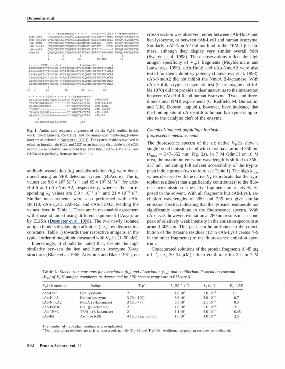
antibody association (ka) and dissociation (kd) were deter-mined using an SPR detection system (BIAcore). Thekavalues are 8.6 × 10⁵ M−1s−1 and 65 × 105 M−1s−1 for cAb-HuL6 and cAb-NmcA2, respectively, whereas the corre-spondingkd values are 5.9 × 10⁻⁴ s−1 and 21 × 10−4 s−1.Similar measurements were also performed with cAb-BcII10, cAb-Lys3, cAb-R2, and cAb-TEM2, yielding thevalues listed in Table 1. These are in reasonable agreementwith those obtained using different equipment (IAsys), orby ELISA (Desmyter et al. 1996). The two newly isolatedantigen binders display high affinities (i.e., low dissociationconstants; Table 1) towards their respective antigens, in thetypical order of magnitude measured with VHHs (1–50 nM).Interestingly, it should be noted that, despite the high
similarity between the hen and human lysozyme X-raystructures (Blake et al. 1965; Artymiuk and Blake 1981), no
cross-reaction was observed, either between cAb-HuL6 andhen lysozyme, or between cAb-Lys3 and human lysozyme.Similarly, cAb-NmcA2 did not bind to the TEM-1�-lacta-mase, although they display very similar overall folds(Swarén et al. 1998). These observations reflect the highantigen specificity of VHH fragments (Muyldermans andLauwereys 1999). cAb-HuL6 and cAb-NmcA2 were alsotested for their inhibitory potency (Lauwereys et al. 1998).cAb-NmcA2 did not inhibit the NmcA�-lactamase. WithcAb-HuL6, a typical enzymatic test (Charlemagne and Jol-lès 1970) did not provide a clear answer as to the interactionbetween cAb-HuL6 and human lysozyme. Two- and three-dimensional NMR experiments (C. Redfield, M. Dumoulin,and C.M. Dobson, unpubl.), however, have indicated thatthe binding site of cAb-HuL6 to human lysozyme is oppo-site to the catalytic cleft of the enzyme.
Chemical-induced unfolding: Intrinsicfluorescence measurements
The fluorescence spectra of the six native VHHs show asingle broad emission band with maxima at around 350 nm(�max� 347–352 nm; Fig. 2a). In 7 M GdmCl or 10 Murea, the maximum emission wavelength is shifted to 356–357 nm, indicating full solvent accessibility of the trypto-phan indole groups (two to four; see Table 1). The high�maxvalues observed with the native VHHs indicate that the tryp-tophan residue(s) that significantly contribute(s) to the fluo-rescence emission of the native fragments are relatively ex-posed to the solvent. With all fragments but cAb-Lys3, ex-citation wavelengths of 280 and 295 nm give similaremission spectra, indicating that the tyrosine residues do notsignificantly contribute to the fluorescence spectra. WithcAb-Lys3, however, excitation at 280 nm results in a secondpeak of relatively weak intensity in the emission spectrum ataround 305 nm. This peak can be attributed to the contri-bution of the tyrosine residues (12 in cAb-Lys3 versus 4–9in the other fragments) to the fluorescence emission spec-trum.Concentrated solutions of the protein fragments (0.45 mg
mL−1, i.e., 30–34�M) left to equilibrate for 2 h in 7 M
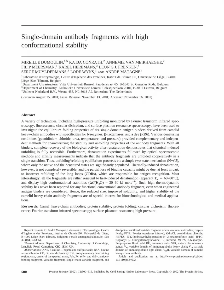
Fig. 1. Amino acid sequence alignment of the six VHHs studied in thiswork. The fragments, the CDRs, and the amino acid numbering (bottomline) are as defined in Kabat et al. (1991). The cystein residues involved ineither an intradomain (C22 and C92) or an interloop disulphide bond (C33and C100e in cAb-lys3) are in bold type. Note that in cAb-TEM2, C33, andC100a also probably form an interloop link.
Table 1. Kinetic rate constants for association (ka) and dissociation (kd), and equilibrium dissociation constant(KD) of VHH-antigen complexes as determined by SPR spectroscopy with a BIAcore X
VHH fragments Antigen Trpa ka (M−1 s−1) kd (s
−1) KD (nM)
cAb-Lys3 Hen lysozyme 2 1.8 105 2.0 10−3 11cAb-HuL6 Human lysozyme 3 (Trp-100) 8.6 105 5.9 10−4 0.7cAb-NmcA2 NmcA (�-lactamase) 3 (Trp-47) 6.5 106 2.1 10−3 0.3cAb-BcII10 BcII (�-lactamase) 2 1.8 106 5.6 10−3 3cAb-TEM2 TEM-1 (�-lactamase) 2 1.1 106 5.0 10−4 0.45cAb-R2 Azo dye RR6 4 (Trp-52a; Trp-58) 1.6 105 4.0 10−4 2.5
The number of tryptophan residues is also indicated.a Two tryptophan residues are strictly conserved, namely Trp-36 and Trp-103. Additional tryptophan residues are indicated.
Dumoulin et al.
502 Protein Science, vol. 11
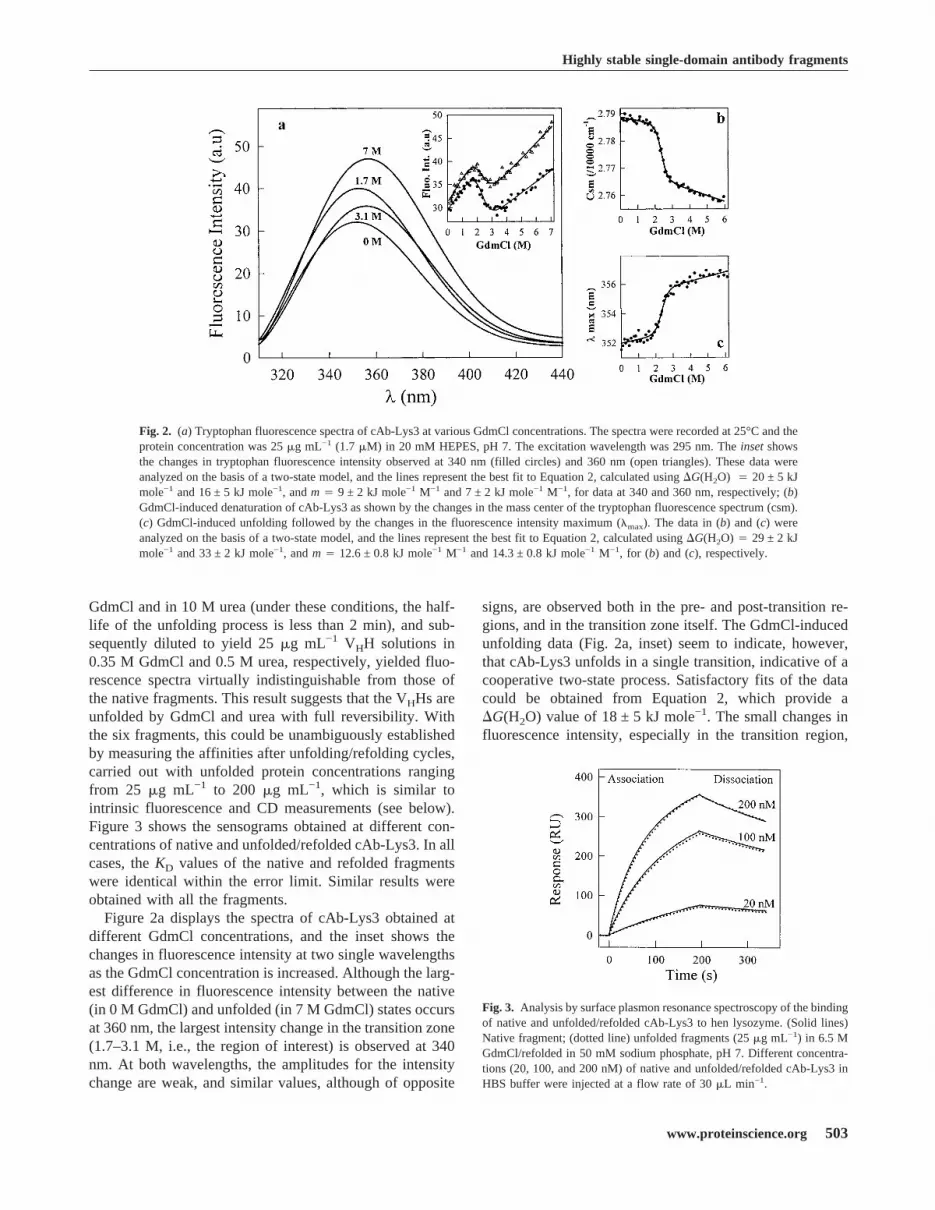
GdmCl and in 10 M urea (under these conditions, the half-life of the unfolding process is less than 2 min), and sub-sequently diluted to yield 25�g mL−1 VHH solutions in0.35 M GdmCl and 0.5 M urea, respectively, yielded fluo-rescence spectra virtually indistinguishable from those ofthe native fragments. This result suggests that the VHHs areunfolded by GdmCl and urea with full reversibility. Withthe six fragments, this could be unambiguously establishedby measuring the affinities after unfolding/refolding cycles,carried out with unfolded protein concentrations rangingfrom 25 �g mL−1 to 200 �g mL−1, which is similar tointrinsic fluorescence and CD measurements (see below).Figure 3 shows the sensograms obtained at different con-centrations of native and unfolded/refolded cAb-Lys3. In allcases, theKD values of the native and refolded fragmentswere identical within the error limit. Similar results wereobtained with all the fragments.Figure 2a displays the spectra of cAb-Lys3 obtained at
different GdmCl concentrations, and the inset shows thechanges in fluorescence intensity at two single wavelengthsas the GdmCl concentration is increased. Although the larg-est difference in fluorescence intensity between the native(in 0 M GdmCl) and unfolded (in 7 M GdmCl) states occursat 360 nm, the largest intensity change in the transition zone(1.7–3.1 M, i.e., the region of interest) is observed at 340nm. At both wavelengths, the amplitudes for the intensitychange are weak, and similar values, although of opposite
signs, are observed both in the pre- and post-transition re-gions, and in the transition zone itself. The GdmCl-inducedunfolding data (Fig. 2a, inset) seem to indicate, however,that cAb-Lys3 unfolds in a single transition, indicative of acooperative two-state process. Satisfactory fits of the datacould be obtained from Equation 2, which provide a�G(H2O) value of 18 ± 5 kJ mole−1. The small changes influorescence intensity, especially in the transition region,
Fig. 2. (a) Tryptophan fluorescence spectra of cAb-Lys3 at various GdmCl concentrations. The spectra were recorded at 25°C and theprotein concentration was 25�g mL−1 (1.7�M) in 20 mM HEPES, pH 7. The excitation wavelength was 295 nm. Theinsetshowsthe changes in tryptophan fluorescence intensity observed at 340 nm (filled circles) and 360 nm (open triangles). These data wereanalyzed on the basis of a two-state model, and the lines represent the best fit to Equation 2, calculated using�G(H2O) � 20 ± 5 kJmole−1 and 16 ± 5 kJ mole−1, andm� 9 ± 2 kJ mole−1 M−1 and 7 ± 2 kJ mole−1 M−1, for data at 340 and 360 nm, respectively; (b)GdmCl-induced denaturation of cAb-Lys3 as shown by the changes in the mass center of the tryptophan fluorescence spectrum (csm).(c) GdmCl-induced unfolding followed by the changes in the fluorescence intensity maximum (�max). The data in (b) and (c) wereanalyzed on the basis of a two-state model, and the lines represent the best fit to Equation 2, calculated using�G(H2O)� 29 ± 2 kJmole−1 and 33 ± 2 kJ mole−1, andm� 12.6 ± 0.8 kJ mole−1 M−1 and 14.3 ± 0.8 kJ mole−1 M−1, for (b) and (c), respectively.
Fig. 3. Analysis by surface plasmon resonance spectroscopy of the bindingof native and unfolded/refolded cAb-Lys3 to hen lysozyme. (Solid lines)Native fragment; (dotted line) unfolded fragments (25�g mL−1) in 6.5 MGdmCl/refolded in 50 mM sodium phosphate, pH 7. Different concentra-tions (20, 100, and 200 nM) of native and unfolded/refolded cAb-Lys3 inHBS buffer were injected at a flow rate of 30�L min−1.
Highly stable single-domain antibody fragments
www.proteinscience.org 503

and the relatively large amplitudes of the pre- and post-transition baselines in comparison with that of the transitionzone itself, might, however, lead to a wrong estimation ofthe thermodynamic parameters. To increase the confidencein the determination of these parameter values, both themaximum in fluorescence intensity (�max), and the center ofthe spectral mass of the fluorescence spectrum (csm; Eq. 1)were calculated at varying GdmCl concentrations. Follow-ing this analysis, it can be seen in Figure 2b and c that betterdata could be obtained, where the relative amplitudes of thepre- and post-transition regions are significantly reduced.According to the two-state model hypothesis and Equation2, analysis of the data in Figure 2b and c provided a�G(H2O) value of 31 ± 5 kJ mole−1. Noticeably, this valueis significantly larger than that obtained from the analysis offluorescence intensity changes at single emission wave-lengths. The unfolding data obtained in a protein concen-tration range from 25 to 90�g mL−1 (i.e., 1.7–6.1�M)yielded identical values of the thermodynamic parameters,within the error limit, indicating that no significant aggre-gation of the protein fragment takes place in this concen-tration range. These findings are consistent with the SPRanalysis (see above), which demonstrates that the biologicalactivity of the VHHs fragments is fully recovered after acomplete unfolding/refolding cycle.With four out of the six VHH fragments, the change in
fluorescence intensity within the transition zone proved tobe more important than with cAb-Lys3. In those instances,the analysis of the data obtained by fluorescence measure-ments at a single emission wavelength yielded values of thethermodynamic parameters similar to those derived from�max and csm measurements. Finally, each experimentalunfolding curve was analyzed using at least two biophysicalparameters; that is (a) the intensity at a given wavelength(not reliable with cAb-Lys3 and cAb-BcII10); (b) the wave-length at the maximum in fluorescence intensity (�max; donewith all fragments); and (c) the center of the spectral massof the fluorescence spectrum (csm; done with all frag-ments). With the six fragments, the analysis of the sameexperiment based on two or three different fluorescenceparameters resulted in similar values of the thermodynamicparameters, within the error limit, which were averaged toyield the data in Table 2.The unfolding curves in Figures 4 and 5 indicate that with
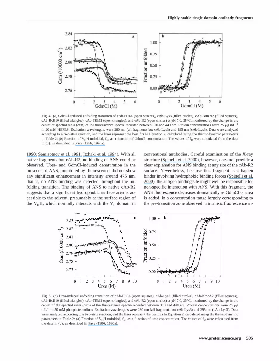
the six VHH fragments, single transitions between the initialand final states are observed in both GdmCl and urea. WhenGdmCl (Fig. 4) was used as denaturant, the characteristicthermodynamic parameters of all VHHs (Table 2) could becomputed with the help of Equation 2, assuming a two-statemodel for the unfolding transitions. With urea (Fig. 5), thevery highCm values of cAb-NmcA2 (∼ 8 M) and cAb-HuL6(>9 M) preclude any quantitative analysis of the data. WithcAb-Lys3, cAb-BcII10, cAbTEM2, and cAb-R2, however,a two-state model analysis was performed and the fitting
parameters are given in Table 2. Consistent data were ob-tained with the two denaturants, and the�G(H2O) values ofthe six fragments are comprised between 30 and 60 kJmol−1, whereas rather highCm values are calculated (�2.3M in GdmCl and�5.8 M in urea).Note that with cAb-Lys3, comparison of the parameter
values obtained with the (His)6 tag-containing cAb-Lys3,and the same fragment devoid of poly-histidine tag indi-cated that the short C-terminal extension has no significanteffect on the conformational stability of the fragment. Simi-lar conclusions have been reached with various histidinetagged proteins (see, e.g. Milla et al. 1993; Reid et al. 1998),and this is assumed to be valid for all six VHHs as well.
Chemical-induced unfolding: ANS-boundfluorescence measurements
The enhancement of ANS fluorescence upon binding toexposed hydrophobic regions of partially unfolded proteinmolecules has been extensively used to detect intermediatespecies in protein folding (Kuwajima 1989; Ptitsyn et al.
Table 2. Thermodynamic parameters of unfolding of VHHantibody fragments at pH 7, 25°C, as obtained from theanalysis of the equilibrium transitions
VHH fragments�G(H2O)(kJ mol−1) m (kJ mol−1 M−1) Cm (M)
cAb-Lys3GdmCla 31 ± 5 13.5 ± 2 2.3 ± 0.5Urea 36 ± 4 5.3 ± 0.6 6.7 ± 1
cAb-HuL6GdmClFluorescence 40 ± 3 12.7 ± 1 3.1 ± 0.3CD (209 nm) 43 ± 6 14.5 ± 2 3 ± 0.5CD (229 nm) 37 ± 4 12 ± 1.5 3 ± 0.6Ureaa (—) (—) >9
cAb-NmcA2GdmCla 34 ± 5 11 ± 1.5 3.0 ± 0.6Ureaa (—) (—) ∼ 8
cAb-BCII 10GdmCla 47 ± 2 14.2 ± 0.7 3.3 ± 0.2Ureaa 49 ± 6 6.6 ± 0.8 7.5 ± 1
cAb-TEM2GdmCla 55 ± 3 17.6 ± 0.7 3.1 ± 0.2Ureaa 61 ± 7 8.3 ± 1 7.5 ± 1
cAb-R2GdmClFluorescence 36 ± 5 16 ± 2.5 2.3 ± 0.5CD (212 nm) 30 ± 5 13 ± 2.5 2.3 ± 0.6CD (222 nm) 33 ± 10 14 ± 5 2.3 ± 1CD (268 nm) 42 ± 10 18 ± 4 2.3 ± 0.7Ureaa 38 ± 3 6.5 ± 0.5 5.8 ± 0.4
a Fluorescence measurements. Errors are calculated at the 95% confidencelimit.— � not accessible.
Dumoulin et al.
504 Protein Science, vol. 11
1990; Semisotnov et al. 1991; Itzhaki et al. 1994). With allnative fragments but cAb-R2, no binding of ANS could beobserved. Urea- and GdmCl-induced denaturation in thepresence of ANS, monitored by fluorescence, did not showany significant enhancement in intensity around 475 nm,that is, no ANS binding was detected throughout the un-folding transition. The binding of ANS to native cAb-R2suggests that a significant hydrophobic surface area is ac-cessible to the solvent, presumably at the surface region ofthe VHH, which normally interacts with the VL domain in
conventional antibodies. Careful examination of the X-raystructure (Spinelli et al. 2000), however, does not provide aclear explanation for ANS binding at any site of the cAb-R2surface. Nevertheless, because this fragment is a haptenbinder involving hydrophobic binding forces (Spinelli et al.2000), the antigen binding site might well be responsible fornon-specific interaction with ANS. With this fragment, theANS fluorescence decreases dramatically as GdmCl or ureais added, in a concentration range largely corresponding tothe pre-transition zone observed in intrinsic fluorescence in-
Fig. 4. (a) GdmCl-induced unfolding transition of cAb-HuL6 (open squares), cAb-Lys3 (filled circles), cAb-NmcA2 (filled squares),cAb-BcII10 (filled triangles), cAb-TEM2 (open triangles), and cAb-R2 (open circles) at pH 7.0, 25°C, monitored by the change in thecenter of spectral mass (csm) of the fluorescence spectra recorded between 310 and 440 nm. Protein concentrations were 25�g mL−1
in 20 mM HEPES. Excitation wavelengths were 280 nm (all fragments but cAb-Lys3) and 295 nm (cAb-Lys3). Data were analyzedaccording to a two-state reaction, and the lines represent the best fits to Equation 2, calculated using the thermodynamic parametersin Table 2; (b) Fraction of VHH unfolded, fU, as a function of GdmCl concentration. The values of fU were calculated from the datain (a), as described in Pace (1986, 1990a).
Fig. 5. (a) Urea-induced unfolding transition of cAb-HuL6 (open squares), cAb-Lys3 (filled circles), cAb-NmcA2 (filled squares),cAb-BcII10 (filled triangles), cAb-TEM2 (open triangles), and cAb-R2 (open circles) at pH 7.0, 25°C, monitored by the change in thecenter of the spectral mass (csm) of the fluorescence spectra recorded between 310 and 440 nm. Protein concentrations were 25�gmL−1 in 50 mM phosphate sodium. Excitation wavelengths were 280 nm (all fragments but cAb-Lys3) and 295 nm (cAb-Lys3). Datawere analyzed according to a two-state reaction, and the lines represent the best fits to Equation 2, calculated using the thermodynamicparameters in Table 2; (b) Fraction of VHH unfolded, fU, as a function of urea concentration. The values of fU were calculated fromthe data in (a), as described in Pace (1986, 1990a).
Highly stable single-domain antibody fragments
www.proteinscience.org 505

tensity measurements (∼ 90% of the fluorescence intensity islost in the presence of 0.1 M GdmCl). At higher concen-trations, no ANS-bound fluorescence is observed. Thus,with the six VHH fragments, unfolding experiments carriedout in the presence of ANS confirm that no partially struc-tured species are significantly populated, in good agreementwith the intrinsic fluorescence data.
Chemical-induced unfolding: CD measurements
The GdmCl-induced unfolding transitions of cAb-R2 andcAb-HuL6 have been followed by far UV CD measure-
ments that monitor the backbone secondary structures (212and 209 nm, respectively) and, presumably the side-chaintertiary structures (222 and 229 nm, respectively). The CDspectra of cAb-R2 and cAb-HuL6 in the far UV region areshown in Figure 6a. Both native fragments show negativemaxima at 215 and 229 nm. The CD signal near 222 nm(positive with cAb-R2) and above (negative) is probablycaused by the aromatic residues of the fragments. Indeed,Phe, Tyr, and Trp are known to contribute to the CD signalin this spectral region, especially when disulphide bonds arepresent (Venyaminov and Yang 1996). Interestingly, theconserved tryptophan residue at position 36 in the VHHs is
Fig. 6. (a) CD spectra in the far UV region of cAb-R2 and cAb-HuL6 (inset), at pH 7.0, 25°C. (Solid line) Native cAb-R2 and cAb-HuL6in 10 mM MOPS and in 10 mM HEPES, respectively; (broken line) in 4 M GdmCl, same buffers. The protein concentrations were 0.2 mgmL−1 (14�M) in a 0.1 cm cell; (b) GdmCl-induced unfolding transition of cAb-HuL6 followed by far UV CD measurements at 209 and229 nm (inset). The protein concentrations were 1mgmL−1 (72�M) in a 0.01-cm cell. Data were analyzed on the basis of a two-state model,and the lines represent the best fit to Equation 2, calculated using�G(H2O)� 43 ± 6 kJ mole−1 and 37 ± 4 kJ mole−1, andm� 14.5 ± 2kJ mole−1 M−1 and 12 ± 1.5 kJ mole−1 M−1, at 209 and 229 nm, respectively; (c) CD spectra in the near UV region of cAb-R2, at pH 7.0,25°C. Solid line, in 10 mM HEPES; broken line, in 10 mM HEPES and 5.5 M GdmCl. The protein concentration was 0.2 mg mL−1 in a1-cm cell. The equilibrium unfolding transition of cAb-R2 followed by CDmeasurements at 268 nm is shown as aninset.The lines representthe best fit to Equation 2, calculated using�G(H2O)� 42 ± 10 kJ mole−1 andm� 18 ± 4 kJ mole−1 M−1; (d) Fraction of cAb-HuL6unfolded, fU, as a function of GdmCl concentration, at pH 7.0, 25°C. The fU values were calculated from data obtained by fluorescence (filledcircles), far UV CD at 209 nm (open triangles), and far UV CD at 229 nm (filled triangles). Theinsetshows the unfolded fraction of cAb-R2as monitored by intrinsic fluorescence (filled circles), far UV CD at 212 nm (open triangles), far UV CD at 222 nm (filled triangles), andnear UV CD at 268 nm (open diamonds). The continuous lines were drawn using the parameters obtained in CD (at 229 nm; cAb-HuL6)and intrinsic fluorescence (cAb-R2) experiments.
Dumoulin et al.
506 Protein Science, vol. 11

in close proximity to the conserved disulphide bridge (C22–C92), and thus might well be responsible (at least in part)for the far UV CD positive band observed with some frag-ments around 220–230 nm. Note that in helical proteins,these non-peptidic contributions are masked by the strongnegative ellipticity of the�-helices (Woody 1994). By con-trast, the positive CD band near 203 nm and the negativeband at around 215 nm are most likely due to the peptidebackbone, that is, to the antiparallel�-sheet structure(Schindler et al. 1995; Venyaminov and Yang 1996).In the presence of 4 M GdmCl, both fragments are un-
folded, the positive band near 222 nm and the negative bandnear 229 nm in cAb-R2 and cAb-HuL6, respectively, areabsent (Fig. 6a), and the signal below 220 nm is character-istic of random coil structures. The denaturation of the twoVHH fragments was followed at both 209 nm (cAb-HuL6)or 212 nm (cAb-R2), and 222 nm (cAb-R2) or 229 nm(cAb-HuL6), thus monitoring secondary and, presumablytertiary structure melting, respectively. In both cases, singletransition curves were obtained (Fig. 6b) and analyzed ac-cording to a two-state model. After calculation of the un-folded fraction at all GdmCl concentrations, the data ob-tained at 212 (or 209) and 222 (or 229) nm are superim-posable. Furthermore, the values of the thermodynamicparameters (Table 2) derived from far UV CD and intrinsicfluorescence measurements are in good agreement. Finally,the CD spectrum of cAb-R2 in the aromatic (i.e., near UV)region (Fig. 6c) shows several bands, the disappearance ofwhich can easily be followed upon unfolding. Thus, asshown in Figure 6c (inset), the cooperative unfolding of thetertiary structure of cAb-R2 could unambiguously be fol-lowed by CD measurements at 268 nm, and yielded thethermodynamic parameters in Table 2. The transition curvesof cAb-HuL6 and cAb-R2, obtained by intrinsic fluores-cence and CD measurements at two and three differentwavelengths, respectively, are shown in Figure 6d. Withboth fragments, the various data superimpose, clearly indi-cating that secondary and tertiary structures melt simulta-neously, that is that unfolding in the transition zone occursbetween the native (N) and the unfolded (U) states, withoutpopulation of any intermediate structural state (Kuwajima1996; Fersht 1999).Similar CD measurements could not be performed with
the other four fragments, because either the spectral prop-erties were not compatible, or not enough material wasavailable.
Chemical-induced unfolding: SPR measurements
Using a BIAcore equipment, the GdmCl-induced unfoldingof cAb-HuL6 could also be analyzed by measuring the af-finity of the antibody fragment towards its antigen (humanlysozyme; Fig. 7). At low GdmCl concentrations (0–2 M),the dissociation constant (KD) gradually increases with the
denaturant concentration. This limited increase is mostlikely due to the effect of GdmCl on the antigen–antibodyinteraction, rather than to any significant conformationalchange of either of the proteins (Cm� 3 M and 3.5 M forcAb-HuL6 and human lysozyme, respectively). Then, atGdmCl concentrations above 2.5 M, a dramatic increase inKD occurs as the denaturant concentration is raised, clearlyindicating that the protein fragment is destabilized in a co-operative transition reminiscent of the GdmCl-induced un-folding curves obtained by intrinsic fluorescence and CDmeasurements. This result suggests that unfolding of theloops (CDRs) responsible for antigen binding occurs con-currently with the destabilization of the whole protein core,and it brings further experimental evidence for two-stateunfolding of cAb-HuL6.
Pressure-induced unfolding
Fourier transform infrared (FTIR) spectroscopy, in combi-nation with temperature- and pressure-induced unfolding, isa powerful technique for determining conformationalchanges in proteins under equilibrium conditions (Wongand Heremans 1988; Jackson and Mantsch 1995; Panick etal. 1998; Torrent et al. 2001). In particular, bandfitting ofthe Fourier-deconvoluted amide I� spectrum (Byler and Susi1986; Smeller et al. 1995a) allows the changes of the sec-ondary structure elements to be followed on protein unfold-
Fig. 7. GdmCl-induced unfolding of cAb-HuL6 followed by SPR spec-troscopy, using a BIAcore X instrument. Protein fragments (0.07 mg mL−1,i.e., 5�M in HBS buffer) were incubated overnight at 25°C in the presenceof varying concentrations of GdmCl. VHHs were then diluted to yieldconcentrations in the range from 0.6 to 4800 nM, in varying GdmCl con-centrations. Ninety microliters of each solution were injected at a flow rateallowing the equilibrium to be reached (i.e., 2–30�L min−1), usingGdmCl-containing HBS buffer. For each GdmCl concentration, at leasteight protein concentrations were injected. Under these equilibrium con-ditions, theKD value corresponds to the concentration of VHH leading tohalf-saturation.
Highly stable single-domain antibody fragments
www.proteinscience.org 507

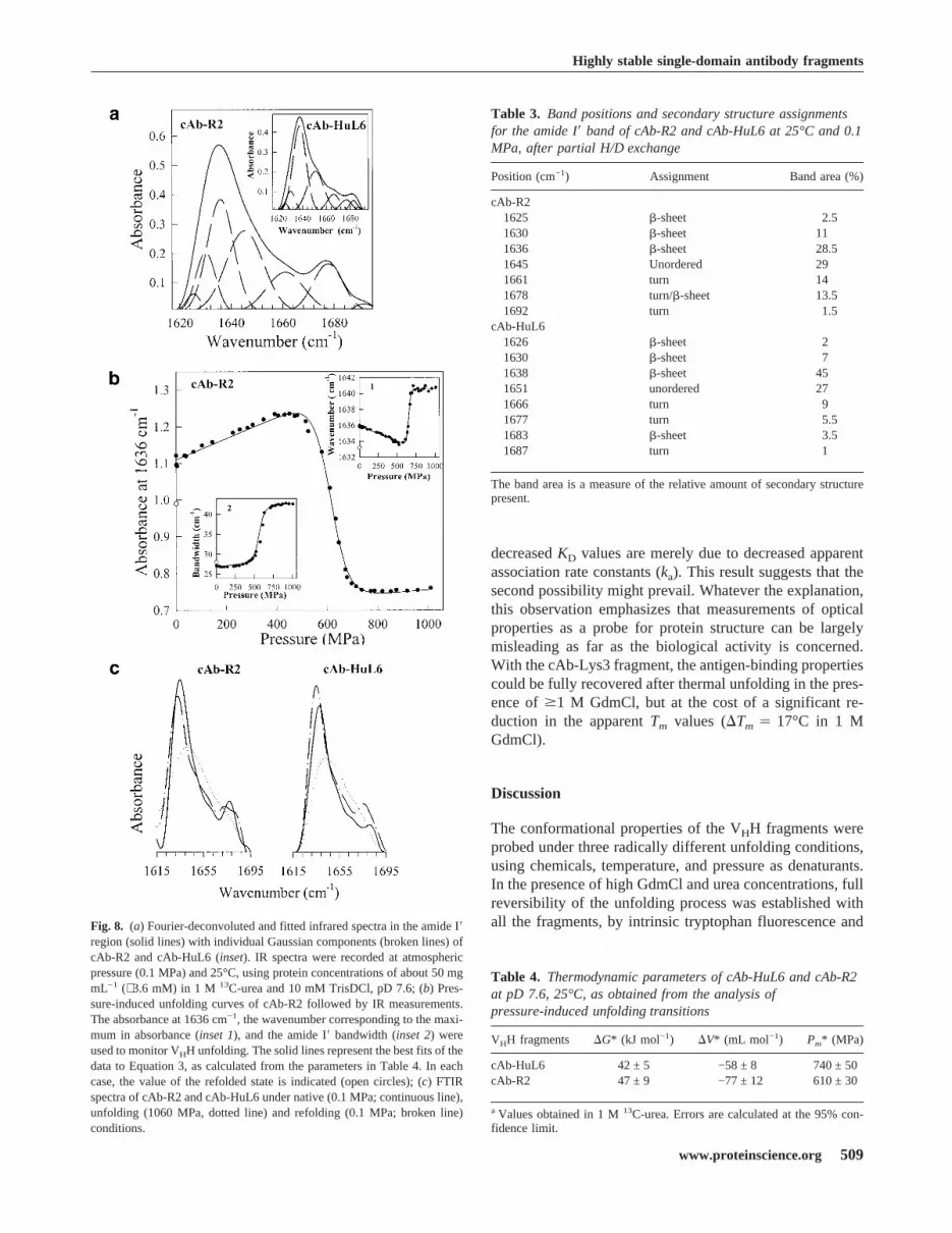
ing. The deconvoluted IR spectra of the amide I� region ofnative cAb-R2 and cAb-HuL6 are shown in Figure 8a.Spectral features are emphasized under Fourier self-decon-volution. The assignments of the component bands at dif-ferent wavenumbers (Byler and Susi 1986; Jackson andMantsch 1995; Haris and Chapman 1995) are shown inTable 3. In the case of cAb-R2, the band at 1678 cm−1 islikely due to an overlap of the turn and�-sheet structures,whereas for cAb-HuL6 the contribution of these bandscould be separated (Table 3). The occurrence of a�-sheetabsorption band at high wavenumber values is indicative ofantiparallel�-sheet (Haris and Chapman 1995; Jackson andMantsch 1995). The lack of�-helix, which is normally ex-pected to absorb around 1650 cm−1 (Byler and Susi 1986),and the high�-sheet content are consistent with the X-raystructures of VHHs (Desmyter et al. 1996; Spinelli et al.1996, 2000; Decanniere et al. 1999).A possible limitation of the method arises from the very
high protein concentration (∼ 50 mg mL−1, i.e., ∼ 3.6 mM)required. With both cAb-HuL6 and cAb-R2, this could beachieved without significant aggregation of the fragments,providing that 1 M13C-labeled urea was added to the buffer.Considering the highCm values (>9 and 5.8 M for cAb-HuL6 and cAb-R2, respectively) determined with urea, nosignificant effect of the denaturant is expected.Pressure-induced changes in the deconvoluted amide I�
region of the IR spectra were followed to obtain informationabout structural changes in the protein fragments. Threeparameters were considered (Fig. 8b), that is, the absor-bance at a fixed wavenumber, the wavenumber correspond-ing to the absorbance maximum of the amide I� band, andthe width of the band. With both VHHs, no significant modi-fication in the IR spectrum is observed as the pressure israised up to 400 MPa (Fig. 8b). In this pressure range, onlya limited decrease in the wavenumber of the band maximumoccurs (inset 1), which results from the effect of compres-sion (and hence strengthening) of the hydrogen bonds (San-droff et al. 1984), and also from H/D exchange (Haris andChapman 1995) due to forced water penetration inside theprotein structure. The latter is clear from the decompressiondata for the amide I� band maximum at pressures below 400MPa (not shown). Above 400 MPa, a cooperative displace-ment of the band maximum towards higher wavenumbervalues (Fig. 8b, inset 1) is observed, together with a broad-ening of the band (Fig. 8b, inset 2) and significant intensitychanges. Thus, a cooperative decrease in band intensities ismeasured around 1636 cm−1 and 1678 cm−1, indicating thedisappearance of the native structure, whereas the increasein band intensities observed between 1645 cm−1 and 1675cm−1 are consistent with an increased amount of unorderedstructures. No indication of VHH aggregation could be ob-served, and the changes in the IR spectra described aboveproved to be largely reversible after pressure release (Fig.8b,c). Indeed, Figure 8b (inset 2) shows that the width of the
amide I� band returns to its native value. In contrast, theabsorbance at 1636 cm−1 (Fig. 8b) and the wavenumber ofthe band maximum (Fig. 8b, inset 1) characteristic of thenative state are not completely restored. This phenomenonis due to the significant H/D exchange occurring on proteinunfolding, which causes a slight shift of the amide I� bandcomponents towards lower wavenumbers (Haris and Chap-man 1992). Thus, the pressure-induced unfolding transitions(Fig. 8b) followed by IR intensity measurements at fourdifferent wavenumbers, and also by measuring bandwidthchanges and band maximum displacements indicate thatboth fragments unfold reversibly, according to a coopera-tive two-state model. On this basis, the data in Figure 8bwere analyzed by use of Equation 3, yielding the thermo-dynamic parameters in Table 4. ThePm values are∼ 600MPa and∼ 750 MPa for cAb-R2 and cAb-HuL6, respec-tively, and the�G values measured in 1 M urea are∼ 47 kJmole−1 and ∼ 42 kJ mole−1 for cAb-R2 and cAb-HuL6, re-spectively. These values are in reasonable agreement withthose found for chemical-induced unfolding (Table 2).
Heat-induced unfolding
In the absence of denaturant, the strong positive CD band atabout 200 nm (Fig. 6a) can be used as a sensitive probe for�-sheet unfolding. Thus, thermal unfolding of the six pro-tein fragments was followed by CD measurements at 201–203 nm, using concentrations of 0.17 mg mL−1 (11–13�M). With all fragments, apparently two-state unfoldingcurves were observed, with rather high melting temperaturevalues (Tm � 60°C; Table 5). With cAb-BcII10, heat-in-duced unfolding was followed at 202 and 223 nm for sec-ondary and tertiary structure unfolding, respectively (seeabove). The unfolding curves at both wavelengths arelargely superimposable, with high midpoint values(Tm� 68°C). These data suggest that thermal unfoldingoccurs with a simple two-state transition. In all cases, how-ever, aggregation was observed at temperatures above theTm values. Despite this phenomenon, a large proportion ofthe native far UV CD signal (80 to 90%) was recovered withfour out of the six antibody fragments (Table 5) after cool-ing down the samples. This high regain in native ellipticitycontrasts, however, with the twofold decrease in the appar-ent antigen-binding affinity measured with the correspond-ing samples (Table 5). TheKD(heated) values in Table 5were calculated, however, by assuming an homogeneouspopulation of refolded molecules. Hence, this experimentdoes not discriminate between the situation in which thestructure of all the VHH molecules is slightly modified(yielding an increasedKD), and a second in which∼ 50% ofthe fragments have fully refolded to the native state (with anunchangedKD), while the other∼ 50% are incorrectly foldedand no longer bind the antigen. However, the dissociationrate constants (kd) appear to be unchanged, and hence, the
Dumoulin et al.
508 Protein Science, vol. 11
decreasedKD values are merely due to decreased apparentassociation rate constants (ka). This result suggests that thesecond possibility might prevail. Whatever the explanation,this observation emphasizes that measurements of opticalproperties as a probe for protein structure can be largelymisleading as far as the biological activity is concerned.With the cAb-Lys3 fragment, the antigen-binding propertiescould be fully recovered after thermal unfolding in the pres-ence of�1 M GdmCl, but at the cost of a significant re-duction in the apparentTm values (�Tm� 17°C in 1 MGdmCl).
Discussion
The conformational properties of the VHH fragments wereprobed under three radically different unfolding conditions,using chemicals, temperature, and pressure as denaturants.In the presence of high GdmCl and urea concentrations, fullreversibility of the unfolding process was established withall the fragments, by intrinsic tryptophan fluorescence andFig. 8. (a) Fourier-deconvoluted and fitted infrared spectra in the amide I�
region (solid lines) with individual Gaussian components (broken lines) ofcAb-R2 and cAb-HuL6 (inset). IR spectra were recorded at atmosphericpressure (0.1 MPa) and 25°C, using protein concentrations of about 50 mgmL−1 (∼ 3.6 mM) in 1 M 13C-urea and 10 mM TrisDCl, pD 7.6; (b) Pres-sure-induced unfolding curves of cAb-R2 followed by IR measurements.The absorbance at 1636 cm−1, the wavenumber corresponding to the maxi-mum in absorbance (inset 1), and the amide I� bandwidth (inset 2) wereused to monitor VHH unfolding. The solid lines represent the best fits of thedata to Equation 3, as calculated from the parameters in Table 4. In eachcase, the value of the refolded state is indicated (open circles); (c) FTIRspectra of cAb-R2 and cAb-HuL6 under native (0.1 MPa; continuous line),unfolding (1060 MPa, dotted line) and refolding (0.1 MPa; broken line)conditions.
Table 3. Band positions and secondary structure assignmentsfor the amide I� band of cAb-R2 and cAb-HuL6 at 25°C and 0.1MPa, after partial H/D exchange
Position (cm−1) Assignment Band area (%)
cAb-R21625 �-sheet 2.51630 �-sheet 111636 �-sheet 28.51645 Unordered 291661 turn 141678 turn/�-sheet 13.51692 turn 1.5
cAb-HuL61626 �-sheet 21630 �-sheet 71638 �-sheet 451651 unordered 271666 turn 91677 turn 5.51683 �-sheet 3.51687 turn 1
The band area is a measure of the relative amount of secondary structurepresent.
Table 4. Thermodynamic parameters of cAb-HuL6 and cAb-R2at pD 7.6, 25°C, as obtained from the analysis ofpressure-induced unfolding transitions
VHH fragments �G* (kJ mol−1) �V* (mL mol−1) Pm* (MPa)
cAb-HuL6 42 ± 5 −58 ± 8 740 ± 50cAb-R2 47 ± 9 −77 ± 12 610 ± 30
aValues obtained in 1 M13C-urea. Errors are calculated at the 95% con-fidence limit.
Highly stable single-domain antibody fragments
www.proteinscience.org 509
affinity measurements. Under these genuine equilibriumconditions, we demonstrated that cAb-HuL6 and cAb-R2unfold cooperatively in a single two-state transition, whereonly the native and unfolded states are significantly popu-lated. Thus, following the normalization of the fluorescenceand CD (both in the far and near UV regions in the case ofcAb-R2) data to give the fraction of unfolded protein atvarious denaturant concentrations (Fig. 6d), it can be seenthat coincident unfolding curves are obtained. This is usu-ally taken as a stringent test for the absence of populatedintermediate species under equilibrium conditions (Ku-wajima 1989). Furthermore, the affinity of cAb-HuL6 forhuman lysozyme, measured by SPR spectroscopy at varyingGdmCl concentrations suggests that the antigen-bindingloops (CDRs) are disorganized coincidentally with the pro-tein core (Fig. 7). Although no CD and SPR experimentswere performed with the other four VHHs, the two-statemodel is most probably valid for all the fragments studied inthis work. This assumption is supported by several lines ofevidence. Thus, all fluorescence-detected unfolding curvesare adequately described by Equation 2. With each VHH,identical free energy values (within the error limit; Table 2)were obtained for urea- and GdmCl-induced unfolding, andall these values are quite high (�G(H2O)� 30–60 kJmole−1). In addition, them values (Table 2), which accountfor the dependence of�G on the denaturant concentration(m� ��G/�[denaturant]), that is, for the slope of the tran-sition, are found to be very similar for the six fragments,either in GdmCl or in urea. Interestingly, with both dena-turants, them values (11–18 kJ mole−1 M−1 and 5.3–8.3 kJmole−1 M−1, respectively) are as high, or even higher thanpredicted (12–14 kJ mole−1M−1 and 5.8–6.7 kJ mole−1M−1,respectively) from their change in solvent-accessible sur-face area upon unfolding (estimated from their sizes; Myerset al. 1995). These findings are consistent with a two-stateunfolding mechanism, from which any deviation shouldlower them values (Pace 1986; Myers et al. 1995). Finally,
the absence of ANS binding in the presence of denaturant isalso a good indication of the lack of intermediate species atequilibrium.Considering the analysis carried out with the six antibody
fragments, the values of their thermodynamic parameters(Table 2) can be considered with good confidence. Interest-ingly, simple two-state unfolding is observed, and all frag-ments exhibit high conformational stability. By contrast, thechemical-induced unfolding of conventional single-chainantibody fragments (scFv) is usually more complex, withseveral unfolding transitions (Wörn et al. 2000). Whethertwo or more transitions occur, the functionality of the scFvfragments is lost with the first transition, theCm value ofwhich is often in the range of 1–2 M and 2–3 M in GdmCland urea, respectively (Proba et al. 1997; Wörn and Plück-thun 1998, 1999; Wörn et al. 2000), that is, at denaturantconcentrations significantly lower than with single-domainVHHs (2.3–3.3 M and�6 M in GdmCl and urea, respec-tively; Table 2). In comparison with antibody fragmentsderived from conventional immunoglobulin, VHH frag-ments from camelids combine excellent antigen-bindingproperties, with remarkably high conformational stabilities.In particular, such a high value (�G(H2O) ∼ 60 kJ mole−1,with Cm� 7.5 M urea) as measured with cAb-TEM2 hasnever been reported to date, even when tailored conven-tional antibody fragments (Jung and Plückthun 1997) areconsidered.The exceptional stability of VHHs is also clearly evident
from pressure- and heat-induced unfolding experiments.FTIR measurements reveal that high pressures (>400 MPa)are needed to unfold cAb-HuL6 and cAb-R2, and reversibleunfolding of the two fragments allows high�G values (∼ 40kJ mole−1 in 1 M urea) to be calculated, very close to thoseobtained from denaturant-induced unfolding. In contrast tothe high values necessary to unfold VHHs, relatively lowpressure values (∼ 50–250 MPa) are required to dissociatenoncovalent protein complexes (Silva et al. 1986, 1992;Erijman et al. 1993). Therefore, in this pressure range, itshould be feasible to disrupt antigen–VHH complexes with-out unfolding either molecule. This strategy can be advan-tageously used for immunoaffinity separation, where a spe-cifically bound antigen is eluted from the immunoadsorbentafter a controlled pressure increase (Olson et al. 1989;Sudaram et al. 1998). Compared with the harsh conditionsusually used, such a “hyperbaric elution” extends the im-munoadsorbent lifetime (Olson et al. 1989). The use ofVHHs, which are made up of a single domain, and conse-quently are easier to express, purify, and handle, will renderthis technique even more valuable.Although in our hands thermal unfolding of the six pro-
tein fragments did not appear to be fully reversible (�60%of activity recovered; Table 5), high apparentTm valuescould be estimated (60–80°C). These are similar to the val-ues measured with a llama heavy-chain antibody fragment
Table 5. Overview of the heat-induced unfolding experiments
VHHsTemp. range
(°C)Tm(°C)
Reversibility
CD201–203 nm
(%)KD (native)/KD (heated)
cAb-Lys3 25–80 62 90 0.5cAb-HuL6 25–92 78 91 0.63cAb-NmcA2 25–80 62 20 NDcAb-BcII10 25–80 68 30 NDcAb-TEM2 25–84 76 77 0.48cAb-R2 25–70 60 80 0.54
Tm is the temperature at the midpoint of the denaturation curve, i.e., themelting temperature. ApparentTm values are given with standard devia-tions below 10%.KD values were measured using SPR spectroscopy, asdescribed in the text, assuming a homogeneous population of refoldedVHHs.ND: not determined.
Dumoulin et al.
510 Protein Science, vol. 11

(VHH-H14, Tm� 60°C; Perez et al. 2001), but also withvarious Fab, Fv, and scFv fragments (Yasui et al. 1994;Shimba et al. 1995; Young et al. 1995; Welfle et al. 1999).These results confirm that scFv and VHH fragments maydisplay comparable thermostabilities, but that the latter re-fold more efficiently following heat-induced denaturation,although not always completely, to the native conformation(van der Linden et al. 1999; Perez et al. 2001). Incubation ofconventional antibody fragments atT > Tm results in disso-ciation of the native structures, with subsequent exposure ofthe hydrophobic interfaces of both the heavy and the lightchains. These exposed “sticky” areas induce aggregationand precipitation, ultimately resulting in nonfunctional mol-ecules. By contrast, the high solubility and thermostabilityof the VHH fragments are most probably largely due tospecific amino acid substitutions at the VH–VL interface,which confer much more hydrophilicity to this normallyvery hydrophobic area (Muyldermans et al. 2001). This hy-pothesis is strongly supported by the results (i.e., enhancedsolubility and thermostability) obtained by Davies andRiechmann (1994, 1996), with camelized human antibodyVH domains.Another distinct feature of VHHs is the occurrence of
enlarged CDR1 and CDR3 loops, which is thought to com-pensate for both the lack of the antigen-binding surfacecontributed by the three hypervariable loops (CDRs) of theVL domain, and the absence of the VH-VL combinatorialdiversity (Muyldermans and Lauwereys 1999; Muylder-mans et al. 2001). Thus, the average length of the CDR3sequence is 17, 12, and 9 amino acids in camelid VHHs,human VHs, and mouse VHs, respectively (Wu et al. 1993;Muyldermans et al. 1994; Vu et al. 1997), and remarkably,in the cAb-Lys3 fragment with specificity for hen lysozymeit is made up of 24 amino acids (Desmyter et al. 1996). Thisvery long antigen-binding loop is constrained by an inter-loop disulphide bond, which is expected to impose confor-mational restraints on the loop flexibility in the absence ofantigen (Desmyter et al. 1996; Muyldermans et al. 2001).Beside the strictly conserved intradomain disulphide bridge(C22–C92), which is characteristic for the immunoglobulinfold, the presence of a second interloop disulphide bond isof common occurrence in VHHs of the dromedary. Al-though this bond has been shown to stabilize camelizedhuman antibody VH domains with long CDR3 (Davies andRiechmann 1996), it is clear that the high stability of cam-elid VHHs cannot be merely attributed to this additionallinkage. Indeed, all the VHH antibody fragments studied byothers (van der Linden et al. 1999; Perez et al. 2001), or inthe present work display very high stabilities, whether theycontain one or two disulphide bridges. A comparison of theavailable VHH sequences indicates that the sequences withthe longest CDR3 have most frequently two additional cys-teine residues, one within the CDR1 (or at position 45 in theframework 2 region) and one in the CDR3, which most
probably form a disulphide bridge. Interestingly, knockingout this interloop bond yields nonfunctional VHHs, at leastin case of cAb-Lys3 (K.B. Vu and S. Muyldermans, unpubl.obs.). Thus, it seems that the role of this tightening disul-phide link is to compensate for the possible drawback oflong CDR3 loops, which might affect the affinity, the speci-ficity, and the stability of VHH antigen binders. In conse-quence, the net stability of VHHs with long CDR3 loops andone extra-disulphide bond is neither reduced nor enhancedwhen compared with other VHHs. The interloop disulphidebridge is especially common in dromedary VHHs, whichshow longer CDR3 in comparison with llama sequences(Vu et al. 1997). It is noteworthy that with some fragments(Table 5), only∼ 50% of the antigen affinity could be re-stored after heat-induced unfolding, whereas a large propor-tion of the native far UV CD signal (80 to 90%) was re-covered. This phenomenon might be due to efficient refold-ing of the core of the protein fragments, which wouldexplain the large recovery of native far UV CD signal, andincorrect refolding of the long CDR loops, which are re-sponsible for antigen binding.The present results highlight the remarkable stability of
VHHs, and confirm their superiority to conventional anti-gen-binders in a number of biotechnological and clinicalapplications because of their smaller size, higher solubility,broad and unique antigen binding capacity, and increasedstability.
Materials and methods
Enzymes and chemicals
Guanidinium chloride (GdmCl) (>99%), bovine serum albumin(BSA), and 8-anilino-1-naphtalene-sulfonic acid (ANS) were pur-chased from Sigma Chemical Co. Urea (>99%) and13C-urea(>99%) were from Merck and Isotec IMC, respectively. Ni2+
NTA-agarose was obtained from Affiland. Azo-dye reactive red-6(RR6, Procion Rubine MX-B) was from ICI. All solutions wereprepared with milli-Q water, and filtered through 0.22-�m filtersbefore use.Hen egg white lysozyme was purchased from Sigma Chemical
Co., and the human lysozyme preparation was kindly given byprofessor C.M. Dobson (O.C.M.S., New Chemistry Laboratory,University of Oxford, UK). The TEM-1, NmcA andBacillus ce-reus569H (BcII) �-lactamases were purified as described in Ra-quet et al. (1994), Swarén et al. (1998), and Carfi et al. (1995),respectively. The human lysozyme and NmcA preparations wereused for dromedary immunization, as described in Conrath et al.(2001).
Selected VHH fragments
The various single-domain antibody fragments studied in the pre-sent work are listed in Table 1. Five VHHs (cAb-Lys3, cAb-HuL6,cAb-TEM2, cAb-NmcA2, and cAb-BcII10) were derived fromdromedary heavy-chain antibodies, and one (cAb-R2) originatedfrom llama heavy-chain antibodies. In the case of dromedaries,
Highly stable single-domain antibody fragments
www.proteinscience.org 511
specific VHHs were isolated by biopanning from phage librariescontaining the genes coding for the variable domains of the heavy-chain antibodies. The phage libraries were generated using themRNA extracted from the lymphocytes of immunized dromedaries(Ghahroudi et al. 1997). According to this procedure, VHHs thatbind specifically to hen (cAb-Lys3; Desmyter et al. 1996) andhuman (cAb-HuL6) lysozyme, the TEM-1 (cAb-TEM2; Conrath etal. 2001) and NmcA (cAb-NmcA2)�-lactamases, and the BcIImetallo-�-lactamase (cAb-BcII10; Conrath et al. 2001) were iso-lated. The cAb-R2 fragment, with specificity for the azo dye re-active red-6 (RR6) was constructed and selected as described inSpinelli et al. (2000).
Expression and purification of VHH fragments
cAb-TEM2 was produced and purified as previously described(Conrath et al. 2001). cAb-R2 was produced inSaccharomycescerevisiaeas a fusion protein (Spinelli et al. 2000), and purifiedfrom the crude yeast culture broth by combining ion exchange(Q-Sepharose, Amersham Pharmacia Biotech) and gel filtration(Sephadex S75, Amersham Pharmacia Biotech) chromatographytechniques. The VHH genes of the other four selected binders wererecloned into an expression vector pHEN6 (Conrath et al. 2001),and were transformed into the WK6 nonsuppressor strain ofE.coli. The recombinant VHHs were expressed and purified as fol-lows: a 15 L fermentor (BioFlo 4500, New Brunswick), con-taining 12 L of SB-ampicillin medium (32 g L−1 tryptone, 20 gL−1 yeast extract, 5 g L−1 NaCl, 1 g L−1 glucose, and 100�g L−1
of ampicillin) was inoculated with 750 mL of an approximately 5h preculture. The culture was grown at 37°C, pH 7.2, and celldevelopment was followed by monitoring both the oxygen con-sumption and the turbidimetry. At the end of the exponentialphase, expression was induced by the addition of 1 mM IPTG, andcell growth was allowed for an additional 3–6 h at 25°C. Afterharvesting the cells by centrifugation, the periplasmic proteinswere extracted according to the procedure described in Skerra andPlückthun (1988). The fusion fragments containing a C-terminal(His)6 tag were then purified in a single step, by metal chelateaffinity chromatography on a Ni2+ NTA–agarose matrix. The VHHfragments were eluted with a linear imidazole gradient (0–240mM) in 50 mM potassium phosphate, pH 7. The fractions in themajor peak were pooled, and imidazole was removed by dialysis.With all six fragment preparations, no trace of contaminating pro-tein was found, either by SDS-PAGE or by mass spectrometryanalysis, indicating a purity higher than 98%. The VHH concen-trations were determined spectrophotometrically using their com-puted extinction coefficients andMr (PC Gene, IntelliGenetics),and a yield in the range of 1 to 5 mg of purified protein per litreof bacterial culture was calculated. The final cAb-HuL6 and cAb-R2 preparations were stored lyophilized at −80 °C, whereas theother fragments were conserved at −80 °C in 50 mM sodiumphosphate, pH 7. Under these conditions, the six VHH fragmentswere shown to be fully stable, at least for the duration of this work.
Chemical-induced unfolding transitions
Samples of VHH fragments were incubated overnight at 25°C inthe presence of various concentrations of urea or guanidiniumchloride (GdmCl). Unfolding curves were determined by monitor-ing the intrinsic fluorescence emission or circular dichroism (CD)at 25°C. The pH was checked to ensure a constant value through-out the whole transition, and the denaturant concentration was
determined from refractive index measurements (Pace 1986), us-ing a R5000 hand refractometer from Atago.
Fluorescence measurements
Both intrinsic fluorescence and ANS-bound fluorescence emissionspectra were recorded on a Perkin-Elmer LS50B spectrofluorim-eter. Excitation and emission slit widths were 3 and 5 nm, respec-tively, and the scan speed was 350 nm min−1. Cuvettes with 1-cmpathlength were used.Intrinsic fluorescence measurements were performed using a
protein concentration of 25�g mL−1 (1.7–1.9�M), with excitationwavelength at either 280 or 295 nm, and emission spectra recordedfrom 310 to 440 nm. The buffers used were 50 mM phosphatesodium, pH 7, and 20 mM HEPES, pH 7, in the presence of ureaand GdmCl, respectively. With all samples, fluorescence spectrawere corrected for the background fluorescence of the solution(buffer + denaturant). Three fluorescence parameters have beenconsidered in this work: the fluorescence intensity at single exci-tation and emission wavelengths, the wavelength corresponding tothe maximum in fluorescence intensity (�max), and the center ofthe spectral mass of the fluorescence spectrum (csm). A five-parameter weibull function (provided with the software SigmaPlot5.0) was fitted to the fluorescence intensity spectra to obtain theassociated�max values. The csm values were computed accordingto the following equation (Royer 1995):
csm= i × Fi �Fi (1)
wherei is the wavenumber (i.e., inverse wavelength) andFi thefluorescence intensity ati. The csm values were calculated be-tween 310 and 440 nm.ANS-bound fluorescence measurements were performed with
the samples used for intrinsic fluorescence measurements, withexcitation at 350 nm, and emission spectra recorded from 420 to600 nm. The fluorescence spectra were corrected for the back-ground fluorescence of ANS. The ANS concentration (determinedfrom the molar extinction coefficient of 4950 M−1 cm−1 at 350 nm;Merck Index, Merck & Co.) was 255–360�M and hence, [ANS]/[VHH] ≈ 140–230.
Circular dichroism measurements
CD measurements were performed with a Jobin-Yvon CD6 spec-tropolarimeter, either in the far UV (205–250 nm) or in the nearUV (250–350 nm) regions, using a protein fragment concentrationof 0.2 mg mL−1 (13–15�M), and 0.1 cm or 1 cm cell pathlengths,respectively. With cAb-HuL6, experiments in the far UV were alsoperformed at a 1 mg mL−1 (70 �M) concentration, in a 0.01-cmcell pathlength. The buffers used were 10 mM MOPS, pH 7, and10 mM HEPES, pH 7, with cAb-R2 and cAb-HuL6, respectively.The instrument was calibrated with d-10-camphorsulfonic acid(Schmid 1997). Spectra were acquired at a scan speed of 12 nmmin−1, with a 2-nm bandwidth and a 1-s integration time. Thespectra were measured five times, averaged, and corrected by sub-traction of the solvent spectrum obtained under identical condi-tions.GdmCl and urea unfolding curves were recorded at fixed wave-
lengths of 212, 222, and 268 nm with cAb-R2, and 209 and 229 nmwith cAb-HuL6, using a 2-nm bandwidth. At all denaturant con-centrations, at least 30 data points were acquired with a readingfrequency of 1/15 s−1 and a 2-s integration time, and averaged. Theresulting values were corrected for the contribution of the solvent.
Dumoulin et al.
512 Protein Science, vol. 11

Heat-induced unfolding transitions were monitored at 201–203and 223 nm, using a protein concentration of 0.17 mg mL−1 (11–13 �M) in 50 mM sodium phosphate, pH 7, and a 0.1-cm cellpathlength. The temperature was increased monotonically in therange from 25 to 65–84°C, at a rate of 0.55°C min−1. The revers-ibility of the phenomenon was assayed by cooling the sampledown to 25°C, at a rate of 0.6°C min−1. Data were acquired witha reading frequency of 1/20 s−1, a 1-s integration time and a 2-nmbandwidth.
Affinity measurements
The kinetics of binding were analyzed by surface plasmon reso-nance (SPR) spectroscopy, using a BIAcore X instrument (BiacoreAB). For immobilization, the azo dye RR6 was coupled to BSA, asdescribed in van der Linden et al. (2000). The individual antigenicproteins (RR6-BSA, lysozymes, and�-lactamases) were immobi-lized on a carboxymethylated dextran-coated sensor chip (CM5,Biacore AB), using the amine coupling chemistry (EDC/NHS)according to the instructions of the manufacturer. In general, 250–350 resonance units (RU) were immobilized, except with RR6-BSA (5000 RU immobilized). Hen lysozyme was immobilized asthe reference protein for measurements with human lysozyme, andvice versa, whereas NmcA was taken as the reference for BcII, andvice versa. BcII and BSA were used as references for TEM-1 andRR6-BSA, respectively. Hence, blank sensograms could be ob-tained for subtraction of bulk refractive index background. Thebinding/regeneration cycles were performed at 25°C in HBS (10mM HEPES, pH 7.4, 150 mM NaCl, 3 mM EDTA, 0.005% sur-factant P20), at a constant flow rate of 30�L min−1. Under theseconditions, the mass transfer effects proved to be negligible. Re-generation of the surfaces was achieved by injection of 30�L of10 mM NaOH or 5 M GdmCl. Binding traces were recorded induplicate, with at least six different concentrations. The sen-sograms were analyzed by nonlinear least-squares fitting, with thehelp of the BIAevaluation 3.0 software (Amersham PharmaciaBiotech), on the basis of a homogeneous 1:1 association model,with simultaneous fitting of the dissociation (kd) and association(ka) rate constants. The equilibrium dissociation constant (KD) wasthen calculated from the ratio of the individual rate constants (kd/ka). Note that the concentration of antibody in the test solutionmust be introduced as a known parameter to perform the fitting.
Enzymatic assays
The inhibitory capacity of cAb-NmcA2 was assayed by measuringthe rate of hydrolysis of nitrocefin, after preincubation of theNmcA �-lactamase with varying concentrations of antibody(Conrath et al. 2001).
Pressure-induced unfolding
Lyophilized cAb-HuL6 and cAb-R2 fragments were dissolved indeuterated 10 mM TrisDCl, pD 7.6, at a protein concentration of50 mg mL−1 (3.6 mM), in the presence of 1 M13C-labeled urea tominimize aggregation. The samples were stored overnight at 25°C,hence ensuring complete H/D exchange of all solvent-accessibleprotons, and then mounted into the well of a stainless steel gasketof a diamond anvil cell (Diacell Products). The cell was placed intoa cell holder, and the pressure was built up by means of a screwmechanism. The pressure in the cell was measured with BaSO4 byfollowing the shift of the 983 cm−1 sulphate peak in the deconvo-
luted spectrum (Wong and Moffat 1989). The infrared spectrawere recorded on a Bruker IFS66 FTIR spectrometer, equippedwith a liquid nitrogen cooled broad band mercury–cadmium–tel-luride solid-state detector. A total of 250 interferograms were co-added at a resolution of 2 cm−1.The secondary structure of the protein fragments was deter-
mined by fitting the resolution enhanced amide I� band of thespectrum (Byler and Susi 1986; Smeller et al. 1995a). The over-lapping components of the amide I� band were narrowed by theFourier self-deconvolution developed by Kauppinen et al. (1981).The optimal parameters were determined from the observation ofthe power spectrum, as described in Smeller et al. (1995b). Aresolution enhancement factor (Kauppinen et al. 1981) of 1.5 wasreached using the Lorentzian band shape of 20-cm−1 bandwidth. Atriangular square apodization function was used. The deconvolutedspectra were then fitted with Gaussian functions. The fitting ofcomponent peaks was performed by a program developed in theLeuven laboratory, using the Levenberg-Marquard algorithm(Press et al. 1986).Three variable parameters were considered, that is, the amide I�
bandwidth, the wavenumber of the band maximum, and the ab-sorbance at given wavenumbers.
Data analysis
The thermodynamic parameters for chemical unfolding were com-puted on the assumption of a two-state model for the unfoldingreaction (N↼⇁U). On this basis, the transition curves were analyzedaccording to the following equation (Santoro and Bolen 1988;Pace 1990a):
yobs= ��yN + p × �D�� + �yU + q × �D��× exp�−a����1 + exp�−a�� (2)
where
a � (�G(H2O) − m × [D])/RT,
and whereyobs is the measured variable parameter at a givendenaturant concentration, andyN and yU represent the values ofthis parameter for the native and denatured states, respectively.�G(H2O) is the difference in free energy between the folded andunfolded conformations under physiological conditions (also de-fined as the conformational stability of a globular protein; Pace1990b);m is a measure of the dependence of the free energy on thedenaturant concentration, and [D] is the denaturant concentration.p and q are the slopes of the pre- and post-unfolding baselines,respectively,R is the gas constant, andT is the absolute tempera-ture. The midpoint of the denaturation curve ([U]/[N] � 1) isgiven byCm� �G(H2O)/m.Similarly, the pressure-induced unfolding experiments were
analyzed assuming an all-or-none transition between the unfoldedand native states (N↼⇁U), and the corresponding thermodynamicparameters were determined using the following equation (Du-moulin et al. 1999):
yP= ��yN + p × P� + �yU + q × P� × exp�−b����1 + exp�−b�� (3)
where
b � (�G(H2O) + P �V)/RT,
and where P is the pressure,�V is the volume change, and�G(H2O), yN, yU, p, q, R and T are the same parameters as defined
Highly stable single-domain antibody fragments
www.proteinscience.org 513

in Equation (2). The melting pressure of half transition ([U]/[N]�1) is given byPm � −�G(H2O)/�V.The programs GraFit 3.09 (Erithacus software Ltd.) and Sigma-
plot 5.0 (SPSS Inc.) were used to carry out nonlinear least-squaresfitting of the data. Errors are calculated at the 95% confidencelimit.
Acknowledgments
We thank Klaas Decanniere, Aline Desmyter, Christopher M.Dobson, Jean-Marie Frère, Marc Lauwereys, Annabelle Lejeune,and Roger H. Pain for many helpful discussions. We acknowledgeBart Devreese for performing the mass spectrometry experiments,and Cees van Vliet for producing cAb-R2 inSaccharomyces cer-evisiae. We are grateful to Jean-Marie Frère and Roger H. Pain forcritical reading of the manuscript. A.M. and M.D. also wish toacknowledge C.M. Dobson for his encouragement and support.This work was supported by the RTD programme on Biotechnol-ogy of the 4th EC framework program, grant number BIO4-98-0048. The purchase of BIAcore X equipment was supported in partby a grant from the Fonds de la Recherche Fondamentale et Col-lective (contract number 3.4589.96). A.M. is a Research Associateof the National Fund for Scientific Research (F.N.R.S., Belgium).The publication costs of this article were defrayed in part by
payment of page charges.This article must therefore be herebymarked “advertisement” in accordance with 18 USC section 1734solely to indicate this fact.
References
Artymiuk, P.J. and Blake, C.C.F. 1981. Refinement of human lysozyme at 1.5Å resolution.J. Mol. Biol. 152: 737–762.
Better, M., Chang, C.P., Robinson, R.R., and Horwitz, A.H. 1988.Escherichiacoli secretion of an active chimeric antibody fragment.Science240: 1041–1043.
Bird, R.E., Hardman, K.D., Jacobson, J.W., Johnson, S., Kaufman, B.M., Lee,S.M., Lee, T., Pope, S.H., Riordan, G.S., and Whitlow, M. 1988. Single-chain antigen-binding proteins.Science242: 423–426.
Blake, C.C.F., Koenig, D.F., Mair, G.A., and Sarma, R. 1965. Crystal structureof lysozyme by X-ray diffraction.Nature206: 757–761.
Byler, D.M. and Susi., H. 1986. Examination of the secondary structure ofproteins by deconvolved FTIR spectra.Biopolymers25: 469–487.
Cai, X. and Garen, A. 1996. A melanoma-specific VH antibody cloned from afusion phage library of a vaccinated melanoma patient.Proc. Natl. Acad.Sci.93: 6280–6285.
Carfi, A., Pares, S., Duée, E., Galleni, M., Duez, C., Frère, J.M., and Dideberg,O. 1995. The 3-D structure of a zinc metallo-�-lactamase fromBacilluscereusreveals a new type of protein fold.EMBO J.14: 4914–4921.
Charlemagne, D. and Jollès, P. 1970. Inhibition par des polymères de la N-acetylglucosamine de l’activité lysante à pH 6,2 de lysozymes d’originesdifférentes vis-à-vis deMicrococcus lysodeikticus. C.R. Acad. Sci. Hebd.Seances Acad. Sci. D270: 2721–2723.
Conrath, K.E., Lauwereys, M., Galleni, M., Matagne, A., Frère, J.M., Kinne, J.,Wyns, L., and Muyldermans, S. 2001.�-Lactamase inhibitors derived fromsingle-domain antibody fragments elicited in Camelidae.Antimicrob.Agents Chemother.45: 2807–2812.
Davies, J. and Riechmann, L. 1994. “Camelising” human antibody fragments:NMR studies on VH domains.FEBS Lett.339: 285–290.
Davies, J. and Riechmann, L. 1996. Single antibody domains as small recog-nition units: Design and in vitro antigen selection of camelized, human VHdomains with improved protein stability.Protein Eng.6: 531–537.
Decanniere, K., Desmyter, A., Lauwereys, M., Ghahroudi, M.A., Muyldermans,S., and Wyns, L. 1999. A single-domain antibody fragment in complex withRNase A: Non-canonical loop structures and nanomolar affinity using twoCDR loops.Struct. Fold. Des.7: 361–370.
Decanniere, K., Muyldermans, S., and Wyns, L. 2000. Canonical antigen-bind-ing loop structures in immunoglobulins: More structures, more canonicalclasses?J. Mol. Biol. 300: 83–91.
Desmyter, A., Transue, T.R., Ghahroudi, M.A., Thi, M.H., Poortmans, F., Ham-
ers, R., Muyldermans, S., and Wyns, L. 1996. Crystal structure of a camelsingle-domain VH antibody fragment in complex with lysozyme.Nat.Struct. Biol.3: 803–811.
Dumoulin, M., Ueno, H., Hayashi, R., and Balny, C. 1999. Contribution of thecarbohydrate moiety to conformational stability of the carboxypeptidase Y.High pressure study.Eur. J. Biochem.262: 475–483.
Erijman, L., Lorimer, G.H., and Weber, G. 1993. Reversible dissociation andconformational stability of dimeric ribulose bisphosphate carboxylase.Bio-chemistry32: 5187–5195.
Fersht, A.R. 1999. Protein stability. InStructure and mechanism in proteinscience—A guide to enzyme catalysis and protein folding, pp. 508–539.W.H. Freeman and Co., New York.
Frenken, L.G.J., van der Linden, R.H.J., Hermans, P.W., Bos, J.W., Ruuls, R.C.,de Geus, B., and Verrips, C.T. 2000. Isolation of antigen specific llama VHantibody fragments and their high level secretion bySaccharomyces cer-evisiae. J. Biotechnol.78:11–21.
Ghahroudi, M., Desmyter, A., Wyns, L., Hamers, R., and Muyldermans, S.1997. Selection and identification of single domain antibody fragments fromcamel heavy-chain antibodies.FEBS Lett.414: 521–526.
Glennie, M.J. and Johnson, P.W. 2000. Clinical trials of antibody therapy.Immunol. Today21: 403–410.
Green, M.C., Murray, J.L., and Hortobagyi, G.N. 2000. Monoclonal antibodytherapy for solid tumors.Cancer Treat. Rev.26: 269–286.
Hamers-Casterman, C., Atarhouch, T., Muyldermans, S., Robinson, G., Hamers,C., Songa, E.B., Bendahman, N., and Hamers, R. 1993. Naturally occurringantibodies devoid of light chains.Nature363: 446–448.
Haris, P.I. and Chapman, D. 1992. Does Fourier-transform infrared-spectros-copy provide useful information on protein structures.Trends Biochem. Sci.17: 328–333.
———. 1995. The conformational analysis of peptides using Fourier transformIR spectroscopy.Biopolymers37: 251–263.
Horwitz, A.H., Chang, C.P., Better, M., Hellstrom, K.E., and Robinson, R.R.1988. Secretion of functional antibody and Fab fragment from yeast cells.Proc. Natl. Acad. Sci.85: 8678–8682.
Hudson, P.J. 1998. Recombinant antibody fragments.Curr. Opin. Biotechnol.9:395–402.
Itzhaki, L.S., Evans, P.A., Dobson, C.M., and Radford, S.E. 1994. Tertiaryinteractions in the folding pathway of hen lysozyme: Kinetic studies usingfluorescent probes.Biochemistry33: 5212–5220.
Jackson, M. and Mantsch, H.H. 1995. The use and misuse of FTIR spectroscopyin the determination of protein structure.Crit. Rev. Biochem. Mol. Biol.30:95–120.
Jung, S. and Plückthun, A. 1997. Improving in vivo folding and stability of asingle-chain Fv antibody fragment by loop grafting.Protein Eng.10: 959–966.
Kabat, E., Wu, T.T., Perry, H.M., Gottesman, K.S. and Foeller, C. 1991. Se-quence of proteins of immunological interest.U.S. Public Health Sevices,NIH Bethesda, MD, Publication No. 91-3242.
Kauppinen, J.K., Moffat, D.J., Mantsch, H.H., and Cameron, D.G. 1981. Fourierself-deconvolution—A method for resolving intrinsically overlapped bands.Appl. Spectrosc.35: 271–276.
Kuwajima, K. 1989. The molten globule state as a clue for understanding thefolding and cooperativity of globular–protein structure.Protein Struct.Funct. Genet.6: 87–103.
Kuwajima, K. 1996. Stopped-flow circular dichroism. InCircular dichroismand the conformational analysis of biomolecules(ed. G.D. Fasman), pp.159–182. Plenum, New York.
Lauwereys, M., Ghahroudi, M.A., Desmyter, A., Kinne, J., Holzer, W., DeGenst, E., Wyns, L., and Muyldermans, S. 1998. Potent enzyme inhibitorsderived from dromedary heavy-chain antibodies.EMBO J.17: 3512–3520.
Milla, M.E., Brown, B.M., and Sauer, R.T. 1993. P22 Arc repressor: Enhancedexpression of unstable mutants by addition of polar C-terminal sequences.Protein Sci.2: 2198–2205.
MuyldermansS. 2001. Single-domain camel antibodies: Current status.Rev.Mol.Biotechnol.74: 277–302.
Muyldermans, S. and Lauwereys, M. 1999. Unique single-domain binding frag-ments derived from naturally occuring camel heavy-chain antibodies.J.Mol. Recognit.12: 131–140.
Muyldermans, S., Atarhouch, T., Saldanha, J., Barbosa, J.A., and Hamers, R.1994. Sequence and structure of VH domain from naturally occurring camelheavy chain immunoglobulins lacking light chains.Protein Eng.7: 1129–1135.
Muyldermans, S., Cambillau, C., and Wyns, L. 2001. Recognition of antigensby single-domain antibody fragments: The superfluous luxury of paireddomains.Trends Biochem. Sci.26: 230–235.
Myers, J.K., Pace, C.N., and Scholtz, J.M. 1995. Denaturantm values and heat
Dumoulin et al.
514 Protein Science, vol. 11
capacity changes: Relation to changes in accessible surface areas of proteinunfolding.Protein Sci.4: 2138–2148.
Olson, W.C., Leung, S.K., and Yarmush, M.L. 1989. Recovery of antigens fromimmunoadsorbents using high pressure.Biotechnology7: 369–373.
Pace, C.N. 1986. Determination and analysis of urea and guanidine hydrochlo-ride denaturation curves.Methods Enzymol.131: 266–280.
———. 1990a. Measuring and increasing protein stability.Trends Biotechnol.8: 93–98.
———. 1990b. Conformational stability of globular proteins.Trends Biochem.Sci.15: 14–17.
Panick, G., Malessa, R., Winter, R., Rapp, G., Frye, K.J., and Royer, C.A. 1998.Structural characterization of the pressure-unfolded state of staphylococcalnuclease by synchrotron small-angle X-ray scattering and Fourier-transforminfrared spectroscopy.J. Mol. Biol. 275: 389–402.
Perez, J.M., Renisio, J.G., Prompers, J.J., van Platerink, C.J., Cambillau, C.,Darbon, H., and Frenken, L.G. 2001. Thermal unfolding of a llama antibodyfragment: A two-state reversible process.Biochemistry40: 74–83.
Press, W.H., Flannery, B.P., Teukolsky, S.A., and Vetterling, W.T. 1986. Op-timal (Wiener) filtering with the FFT. InNumerical recepies in pascal: Theart of scientific computing, pp. 459–462. Cambridge University Press, Cam-bridge.
Proba, K., Honegger, A., and Plückthun, A. 1997. Natural antibody missing acysteine in VH: Consequences for thermodynamic stability and folding.J.Mol. Biol. 265: 161–172.
Ptitsyn, O.B., Pain, R.H., Semisotnov, G.V., Zerovnik, E., and Razgulyaev, O.I.1990. Evidence for a molten globule state as a general intermediate inprotein folding.FEBS Lett.262: 20–24.
Raquet, X., Lamotte-Brasseur, J., Fonzé, E., Goussard, S., Courvalin, P., andFrère, J.M. 1994. TEM�-lactamase mutants hydrolysing third-generationcephalosporins. A kinetic and molecular modelling analysis.J. Mol. Biol.244: 625–639.
Reid, K.L., Rodriguez, H.M., Hillier, B.J., and Gregoret, L.M. 1998. Stabilityand folding properties of a model�-sheet protein,Escherichia coliCspA.Protein Sci.7: 470–479.
Reiter, Y., Brinkmann, U., Lee, B., and Pastan, I. 1996. Engineering antibodyFv fragments for cancer detection and therapy: Disulfide-stabilized Fv frag-ments.Nat. Biotechnol.14: 1239–1245.
Riechmann, L. and Muyldermans, S. 1999. Single domain antibodies: Compari-son of camel VH and camelised human VH domains.J. Immunol. Methods231: 25–38.
Riechmann, L., Foote, J., and Winter, G. 1988. Expression of an antibody Fvfragment in myeloma cells.J. Mol. Biol. 203: 825–828.
Royer, C. 1995. Fluorescence spectroscopy. InMethods in molecular biology,vol. 40, protein stability and folding(ed. B.A. Shirley), pp. 65–90. HumanaPress Inc., Totowa, NJ.
Sandroff, C.J., King, H.E., and Herschbach, D.R. 1984. High pressure study ofthe liquid/solid interface: Surface enhanced raman scattering from adsorbedmolecules.J. Phys. Chem.88: 5647–5653.
Santoro, M.M. and Bolen, D.W. 1988. Unfolding free energy changes deter-mined by the linear extrapolation method. 1. Unfolding of phenylmethane-sulfonyl alpha-chymotrypsin using different denaturants.Biochemistry27:8063–8068.
Schindler, T., Herrler, M., Marahiel, M.A., and Schmid, F.X. 1995. Extremelyrapid protein folding in the absence of intermediates.Nat. Struct. Biol.2:663–673.
Schmid, F.X. 1997. Optical spectroscopy to characterize protein conformationand conformational changes. InProtein structure, a practical approach, 2nded. (ed. T.E. Creighton), pp. 299–321. Oxford University Press, Oxford.
Semisotnov, G.V., Rodionova, N.A., Razgulyaev, O.I., Uversky, V.N., Gripas,A.F., and Gilmanshin, R.I. 1991. Study of the “molten globule” intermediatestate in protein folding by a hydrophobic fluorescent probe.Biopolymers31:119–128.
Shimba, N., Torigoe, H., Takahashi, H., Masuda, K., Shimada, I., Arata, Y., andSarai, A. 1995. Comparative thermodynamic of the Fv, Fab*, and Fabfragments of anti-dansyl mouse monoclonal antibody.FEBS Lett.360:247–250.
Silva, J.L., Miles, E.W., and Weber, G. 1986. Pressure dissociation and con-formational drift of the beta dimer of tryptophan synthetase.Biochemistry25: 5780–5786.
Silva, J.L., Silveira, C.F., Correia, J.A., and Pontes, L. 1992. Dissociation of anative dimer to a molten globule monomer. Effects of pressure and dilutionon the association equilibrium of arc repressor.J. Mol. Biol.223:545–555.
Skerra, A. and Plückthun., A. 1988. Assembly of a functional immunoglobulinFv fragment inEscherichia coli. Science240: 1038–1041.
Smeller, L., Goossens, K., and Heremans, K. 1995a. Determination of thesecondary structure of proteins at high pressure.Vib. Spectrosc.8: 199–203.
———. 1995b. How to minimize certain artifacts in Fourier self-deconvolution.Appl. Spectrosc.49: 1538–1542.
Solomon, B., Koppel, R., Frankel, D., and Hanan-Aharon, E. 1997. Disaggre-gation of Alzheimer beta-amyloid by site-directed mAb.Proc. Natl. Acad.Sci.94: 4109–4112.
Solomon, B., Koppel, R., Hanan, E., and Katzav, T. 1996. Monoclonal anti-bodies inhibit in vitro fibrillar aggregation of the Alzheimer beta-amyloidpeptide.Proc. Natl. Acad. Sci.93: 452–455.
Spinelli, S., Frenken, L.G., Bourgeois, D., de Ron, L., Bos, W., Verrips, T.,Anguille, C., Cambillau, C., and Tegoni, M. 1996. The crystal structure ofa llama heavy chain variable domain.Nat. Struct. Biol.9: 752–757.
Spinelli, S., Frenken, L.G., Hermans, P., Verrips, T., Brown, K., Tegoni, M.,and Cambillau, C. 2000. Camelid heavy-chain variable domains provideefficient combining sites to haptens.Biochemistry39: 1217–1222.
Sudaram, S., Roth, C.M., and Yarmush, M.L. 1998. Pressure-induced dissocia-tion of antigen-antibody complexes.Biotechnol. Prog.14: 773–781.
Swarén, P., Maveyraud, L., Raquet, X., Cabantous, S., Duez, C., Pédelacq, J.D.,Mariotte-Boyer, S., Labia, R., Nicolas-Chanoine, M.H., Mourey, L., Frère,J.M., and Samama, J.P. 1998. X-ray analysis of the NMC-A�-lactamase at1.64-Å resolution, a class A carbapenemase with broad substrate specificity.J. Biol. Chem.273: 26714–26721.
Torrent, J., Rubens, P., Ribo, M., Heremans, K., and Vilanova, M. 2001. Pres-sure versus temperature unfolding of ribonuclease A: An FTIR spectro-scopic characterization of 10 variants at the carboxy-terminal site.ProteinSci.10: 725–734.
Transue, T.R., De Genst, E., Ghahroudi, M.A., Wyns, L., and Muyldermans, S.1998. Camel single-domain antibody inhibits enzyme by mimicking carbo-hydrate substrate.Protein Struct. Funct. Genet.32: 515–522.
van der Linden, R.H., Frenken, L.G., de Geus, B., Harmsen, M.M., Ruuls, R.C.,Stok, W., de Ron, L., Wilson, S., Davis, P., and Verrips, C.T. 1999. Com-parison of physical chemical properties of llama VHH antibody fragmentsand mouse monoclonal antibodies.Biochim. Biophys. Acta1431:37–46.
van der Linden, R.H.J., de Geus, B., Stok, W., Bos, W., van Wassenaar, D.,Verrips, T., and Frenken, L. 2000 Induction of immune responses andmolecular cloning of the heavy chain antibody repertoire ofLama glama. J.Immunol. Methods240: 185–195.
Venyaminov, S.Y. and Yang, J.T. 1996. Determination of protein secondarystructure. InCircular dichroism and the conformational analysis of biomol-ecules(ed. G.D. Fasman), pp. 69–108. Plenum, New York.
Vu, K.B., Ghahroudi, M.A., Wyns, L., and Muyldermans, S. 1997. Comparisonof llama VH sequences from conventional and heavy chain antibodies.Mol.Immunol.34: 1121–1131.
Ward, E.S., Gussow, D., Griffiths, A.D., Jones, P.T., and Winter, G. 1989.Binding activities of a repertoire of single immunoglobulin variable do-mains secreted fromEscherichia coli. Nature341: 544–546.
Welfle, K., Misselwitz, R., Hausdorf, G., Höhne, W., and Welfle, H. 1999.Conformation, pH-induced conformational changes and thermal unfoldingof anti-p25 (HIV-1) monoclonal antibody CB4–1 and its Fab and Fc frag-ments.Biochim. Biophys. Acta1431:120–131.
Wong, P.T. and Heremans, K. 1988. Pressure effects on protein secondarystructure and hydrogen deuterium exchange in chymotrypsinogen: A Fou-rier transform infrared spectroscopic study.Biochim. Biophys. Acta956:1–9.
Wong, P.T.T. and Moffat, D.J. 1989. A new internal pressure calibrant forhigh-pressure infrared spectroscopy of aqueous systems.Appl. Spectrosc.43: 1279–1281.
Woody, R.W. 1994. Contributions of tryptophan side chains to the far-ultravio-let circular dichroism of proteins.Eur. Biophys. J.23: 253–262.
Wörn, A. and Plückthun, A. 1998. Mutual stabilisation of VL and VH insingle-chain antibody fragments, investigated with mutants engineered forstability.Biochemistry37: 13122–13127.
———. 1999. Different equilibrium stability behavior of scFv fragments: Iden-tification, classification, and improvement by protein engineering.Biochem-istry 38: 8739–8750.
Wörn, A., Auf der Maur, A., Escher, D., Honegger, A., Barberis, A., andPlückthun, A. 2000. Correlation between in vitro stability and in vivo per-formance of anti-GCN4 intrabodies as cytoplasmic inhibitors.J. Biol.Chem.275: 2795–2803.
Wu, T.T., Johnson, G., and Kabat, E.A. 1993. Length distribution of CDR H3in antibodies,Protein Struct. Funct. Genet.16: 1–7.
Yasui, H., Ito, W., and Kurosawa, Y. 1994. Effects of substitutions of aminoacids on the thermal stability of the Fv fragments of antibodies.FEBS Lett.353: 143–146.
Young, N.M., MacKenzie, C.R., Narang, S.A., Oomen, R.P., and Baenziger,J.E. 1995. Thermal stabilisation of a single-chain Fv antibody fragment byintroduction of a disulfide bond.FEBS Lett.377: 135–139.
Highly stable single-domain antibody fragments
www.proteinscience.org 515


