Embed Size (px)
Citation preview

International Journal of Pharmaceutics 266 (2003) 3–16
Shifting paradigms: biopharmaceuticalsversus low molecular weight drugs
Daan J.A. Crommelina,b,∗, Gert Storma, Ruud Verrijkb,Leo de Leedeb, Wim Jiskoota, Wim E. Henninka
a Department of Pharmaceutics, Utrecht Institute for Pharmaceutical Sciences, UIPS, Utrecht TB 3508, The Netherlandsb OctoPlus, Leiden, The Netherlands
Received 27 January 2003; received in revised form 22 April 2003; accepted 24 April 2003
To celebrate and commemorate Prof. Dr. H.E. Junginger’s 60th birthday
Abstract
Biopharmaceuticals are pharmaceutical products consisting of (glyco)proteins. Nowadays a substantial part of the FDA-approved drugs belong to this class of drugs. Biopharmaceuticals deserve special attention as they have a number of characteristicsthat set them aside from low molecular weight drugs.
Their activity depends on their complicated shape based on secondary, tertiary and (sometimes) quaternary structures. Thesestructures cannot be fully defined with our present set of analytical techniques and approaches for potency testing. They often arethe same as (or closely resemble) endogenous proteins. This means that in safety testing and clinical test programs questions haveto be addressed regarding species specific responses, selection of dosing schedules and route of administration, and the possibleoccurrence of immunogenicity. As the conformational structure of a protein is easily disturbed, formulation and handling ofbiopharmaceuticals needs special attention in order to optimize the therapeutic effect and minimize adverse reaction, amongwhich immune responses.
The issue of biogenerics is gaining more and more interest and different critical elements in the development of biogenericsare touched upon.
In conclusion, biopharmaceuticals cannot be characterized fully in terms of their structure like low molecular weight drugs.The performance of biopharmaceuticals relies on strict production protocols and close monitoring of their activity in the clinicalsituation.© 2003 Published by Elsevier B.V.
Keywords:Biopharmaceuticals; Biologicals; Biogenerics; Immunogenicity; PK–PD
Abbreviations: ADA, adenosine deaminase; CHO, Chinese hamster ovary; ELISA, enzyme-linked immunosorbent assay; FSH,follicle stimulating hormone; G-CSF, granulocyte colony-stimulating factor; HAMA, human anti mouse antibodies; hGH, humangrowth hormone; i.m., intramuscular; IEF, iso-electric focusing; IEP, iso-electric point; LHRH, luteinizing hormone releasing hormone;NESP, novel erythropoiesis stimulating protein; PEG-ADA, pegylated adenosine deaminase; PEG-G-CSF, pegylated G-CSF; PK–PD,pharmacokinetics–pharmacodynamics; PLGA, polylactic-coglycolic acid; QCM, quartz crystal microbalance; rhuEPO, recombinant humanerythropoeitin; s.c., subcutaneous; SDS–PAGE, sodium dodecylsulfate polyacrylamide-gel electrophoresis; TPO, thrombopoeitin
∗ Corresponding author. Tel.:+31-302536973; fax:+31-302517839.E-mail address:[email protected] (D.J.A. Crommelin).
0378-5173/$ – see front matter © 2003 Published by Elsevier B.V.doi:10.1016/S0378-5173(03)00376-4

4 D.J.A. Crommelin et al. / International Journal of Pharmaceutics 266 (2003) 3–16
1. Introduction
Biopharmaceuticals are defined as ‘pharmaceuticalproducts consisting of (glyco)proteins and/or nucleicacids’ (Schellekens, 2002). In this contribution theterm only applies to (glyco)protein products usedfor therapeutic purposes and vaccines. Biopharma-ceuticals still make up only a small part of the totalarsenal of medicines that is used by mankind. But, thegrowth of this category of medicines is much fasterthan the introduction and growth of ‘conventional’,low molecular weight medicines. Recent statisticsshow that the FDA approved 130 biotechnology de-rived protein medicines and vaccines (BiotechnologyIndustry Organization Site, 2002), 70% of whichwere approved in the last 6 years. Currently, over 350biopharmaceuticals are in clinical trials.
These biopharmaceuticals deserve specific attentionby pharmaceutical scientists as they have a number ofcharacteristics that set them aside from low molecu-lar weight drugs. These specific characteristics centeraround a number of issues listed inTable 1. The issueson this list will be discussed in more detail below.
2. Molecular characteristics
Biopharmaceuticals are (glyco)proteins. The build-ing blocks for these molecules arel-amino acids anddifferent sugar molecules. They form three-dimen-sional structures based on secondary structures (alfa-
Table 1Specific characteristics of biopharmaceuticals
Molecular characteristicsHigh molecular weight:Mw > a few thousand Daltons
(glyco)proteinsPharmacology/therapeutic use
Life threatening/severe diseasesSafety/clinical testing
Species specificityDosing schedulesRoute of administration/PK–PD relationshipImmunogenicity
FormulationStabilityRate controlled delivery
HandlingBiogenerics: the possibility to launch biogenerics?
VL
CLC
HC
H
VH
IgG
CH
Fv
antigen binding site
macrophage interaction
complement binding
isotype Fc
Fab idiotype
Fig. 1. Antibody structure. Cartoon of IgG antibody structure.Antibodies bind antigen via their variable region (VL and VH).The CH2 and CH3 domains of heavy chains make up the Fc partand determine the biological activity. N-linked glycosylation sitesare indicated as small circles. L stands for light chain, H for heavychain. The small circles represent the glycosylation sites and sugarsegments. Adapted fromvan Dijk and Vidarsson (2002).
helices, beta-sheets and random coil areas), tertiarystructures (folding of the secondary structures intocomplicated three-dimensional structures) and insome cases quaternary structures (where differentmonomers interact). Such special structures do notexist in low molecular weight drug molecules. Dif-ferent areas in protein drug molecules have differentfunctions. For instance, in a monoclonal IgG type an-tibody different parts clearly have different functions.A schematic of a monoclonal antibody is depicted inFig. 1. About 1400 amino acids make up the proteinbackbone of an IgG antibody. The antigen bindingsite (responsible for site specific docking on the anti-gen) is located at the beta-plated N-terminal ends ofthe Fab part. The sites for complement binding andmacrophage interactions are found on the Fc part.N-Glycosylation sites are located at the Fc part as well.Small molecules do not possess such sophisticatedstructures harboring several different functions on onemolecule.
Small molecules like aspirin and acetaminophencan be fully described in terms of their molecularstructure. For full identification a limited set of ana-lytical assays can be used. Different pharmacopoeialsources describe this in detail. Assays to describe im-purity profiles can be found there as well. Typically,98 to >99% purity is required and suppliers are able
D.J.A. Crommelin et al. / International Journal of Pharmaceutics 266 (2003) 3–16 5
to reach that quality level consistently. What aboutbiopharmaceuticals? Later in this contribution wewill discuss biopharmaceutical quality in more detail.Here, two differences between biopharmaceuticalsand low molecular weight compounds will be dealtwith first: these relate to (a) protein production andpurity and (b) protein characterization.
2.1. Protein purity
The choice of the expression system and growthconditions for the production of biopharmaceuticals iscritical. Only a few expression systems are regularlyused for the production of biopharmaceuticals, as bothmanufacturers and authorities are familiar with thespecifics of these production systems (e.g.Escherichiacoli as a prokaryotic and yeast or CHO cells as eu-karyotic production cells). General patterns emergedfor the optimization of growth conditions and the se-lection of proper downstream processing protocols topurify the desired protein, removing all host cell-,process- and product-related impurities.Table 2, takenfrom Kadir and Hamers (2002), lists typical produc-tion contaminants both for prokaryotic and eukaryoticproduction cells that have to be rigorously removed.Here, as with low molecular weight drugs, the ulti-mate goal is 99+% purity. Meeting that purity levelin terms of amino acid sequence may be feasible forsmaller proteins (such as insulin or calcitonin:Mw inthe range of 5 kDa); sequencing methodology togetherwith chromatographic techniques provide the meansto establish those purity levels.
Many biopharmaceuticals like monoclonal antibod-ies, Factor VIII and FSH are glycoproteins producedin eukaryotic cells. Glycosylation patterns vary, not
Table 2List of typical production contaminants both for prokaryotic and eukaryotic production cells (taken fromKadir and Hamers (2002))
Type of nutrient Example(s)
Sugars Glucose, lactose, sucrose, maltose, dextrinsFat Fatty acids, triglyceridesWater (high quality, sterilized) Water for injectionAmino acids GlutamineElectrolytes Calcium, sodium, potassium, phosphateVitamins Ascorbic acid,�-tocopherol, thiamine, riboflavine, folic acid, pyridoxinSerum (fetal calf serum, synthetic serum) Albumin, transferrinTrace minerals Iron, manganese, copper, cobalt, zincHormones Growth factors
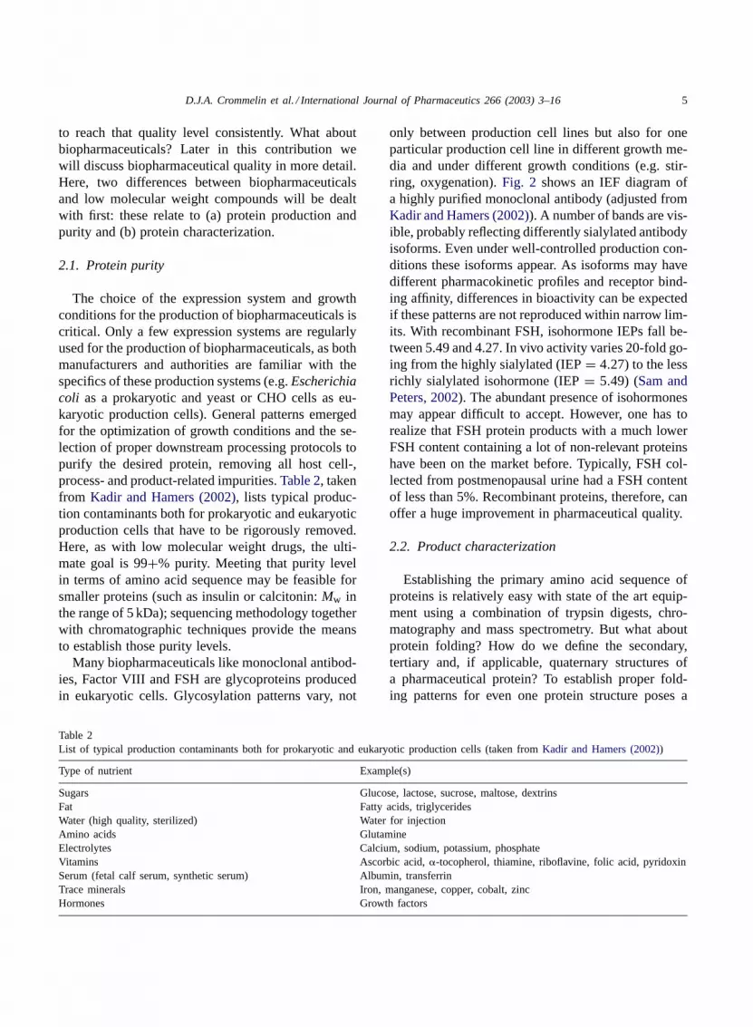
only between production cell lines but also for oneparticular production cell line in different growth me-dia and under different growth conditions (e.g. stir-ring, oxygenation).Fig. 2 shows an IEF diagram ofa highly purified monoclonal antibody (adjusted fromKadir and Hamers (2002)). A number of bands are vis-ible, probably reflecting differently sialylated antibodyisoforms. Even under well-controlled production con-ditions these isoforms appear. As isoforms may havedifferent pharmacokinetic profiles and receptor bind-ing affinity, differences in bioactivity can be expectedif these patterns are not reproduced within narrow lim-its. With recombinant FSH, isohormone IEPs fall be-tween 5.49 and 4.27. In vivo activity varies 20-fold go-ing from the highly sialylated (IEP= 4.27) to the lessrichly sialylated isohormone (IEP= 5.49) (Sam andPeters, 2002). The abundant presence of isohormonesmay appear difficult to accept. However, one has torealize that FSH protein products with a much lowerFSH content containing a lot of non-relevant proteinshave been on the market before. Typically, FSH col-lected from postmenopausal urine had a FSH contentof less than 5%. Recombinant proteins, therefore, canoffer a huge improvement in pharmaceutical quality.
2.2. Product characterization
Establishing the primary amino acid sequence ofproteins is relatively easy with state of the art equip-ment using a combination of trypsin digests, chro-matography and mass spectrometry. But what aboutprotein folding? How do we define the secondary,tertiary and, if applicable, quaternary structures ofa pharmaceutical protein? To establish proper fold-ing patterns for even one protein structure poses a

6 D.J.A. Crommelin et al. / International Journal of Pharmaceutics 266 (2003) 3–16
Fig. 2. Silver-stained IEF patterns of three types of purified mon-oclonal antibodies. The right lane represents marker proteins.Adapted fromJiskoot et al. (1991).
challenge. This challenge increases in magnitude ifprotein products contain mixtures of proteins, e.g.different isoforms.
Table 3 lists analytical techniques to characterizethe structure or function of proteins, but none of thesetechniques allows us to fully define protein foldingpatterns. They all provide pieces of information. Com-bining the different pieces of evidence allows us tobuild a picture of the protein. But this picture will beincomplete even if all assays are used. One might thinkthat the ultimate test is the potency test. However, po-tency tests have limited value because of the inher-ently large variability in outcome of these tests. Theyalso provide little information about immunogenicity.
Table 3(Analytical) techniques for monitoring protein structure
UV absorptionCircular dichroism spectroscopyFourier transform IRFluorescence spectroscopyNMR spectroscopyCalorimetric approaches
Bio-assaysImmunochemical assays
ELISAImmunoprecipitationBiosensor (SPR, QCM)
Potency testingIn cell linesIn animals
Chromatographic techniquesRP-HPLCSEC-HPLCHydrophobic interaction HPLCIon-exchange HPLCPeptide mapping
Electrophoretic techniquesSDS–PAGEIEFCZE
Field flow fractionactionUltracentifugationStatic and dynamic light scatteringElectron microscopyX-ray techniquesMass spectrometry
Some of these analytical approaches (e.g. X-raycrystallography and NMR) are quite complex andmay only be relevant to consider in (early) develop-ment stages. Other analytical assays (e.g. SDS–PAGE,ELISA and HPLC analyses) are used routinely in re-lease tests. A full discussion of the pros and cons ofall listed assays inTable 3is beyond the scope of thiscontribution. Extensive reviews on these analyticalapproaches can be found, for example inHerron et al.(1995)or Metz et al. (2002).
Some reflection on two selected spectrometric ap-proaches: X-ray diffraction provides information onthe full protein, but the information is obtained for acrystallized product in a milieu that is different fromthe protein environment in the formulated product.Moreover, not all proteins crystallize easily, in partic-ular glycosylated proteins and those that are present indifferent isoforms offer problems. Crystallization anddata analysis can take months to achieve. Therefore,

D.J.A. Crommelin et al. / International Journal of Pharmaceutics 266 (2003) 3–16 7
such approaches would never work as a routine batchrelease assay. In principle, NMR spectrometric anal-ysis would also be an option to provide full informa-tion on protein folding. However, at present structureelucidation of proteins is restricted to structures up to25 kDa in size. And, for many proteins the requiredconcentrations for NMR analysis exceed those in theformulated material. With NMR, as with X-ray crys-tallography, the technique lacks sensitivity for minorfractions of (misfolded) impurities and full data anal-ysis takes too much time to be used for batch releasedecisions.
3. Pharmacology/therapeutic use
3.1. Life threatening and serious diseases
Table 4shows a list of biopharmaceuticals marketedin the USA in early 2002 (Evens and Sindelar (2002)).When considering the therapeutic indications, it isclear that these biopharmaceuticals were introduced totreat severe and/or life threatening diseases. The list in-cludes monoclonal antibodies for immune modulationand treatment of cancer; biological response modifiersto stimulate cell growth; hormones such as insulinand hGH; enzymes such as alteplase to remove bloodclots; and last but not least, a number of vaccines.The list of indications for the different molecules isexpanding rapidly as well. Interestingly, many bio-pharmaceuticals are endogenous proteins or slightlymodified endogenous proteins. Their therapeutic indi-cation can be derived from their mode of action andphysiological effects in humans. However, one has tobe careful in directly deriving the therapeutic potentialof endogenous substances from physiological insights.Upon parenteral administration, severe, dose limitingside effects can be observed because resulting pharma-cokinetic profiles are different from those of endoge-nous proteins. For example, many cytokines work in aparacrine mode: the cytokine is secreted as a mediatingmolecule by a cell to signal other, neighboring ‘target’cells. Upon intravenous injection, however, a cytokinesuch as IL-2 enters the body far from its target site andmay cause severe, dose limiting side effects. It is clearthat drug targeting strategies should be considered forall biopharmaceuticals that work in a paracrine modeand which lack intrinsic targeting properties.
Table 4Marketed biotechnology-produced pharmaceuticals (status early2002)
Product Classa
Abciximab MAbAcelluvax VaccineAldesleukin BRMAlteplase EnzymeAntihemophilic factor VIII (2)b EnzymeBasiliximab MAbDaclizumab MAbDenileukin diftitox BRMDnase EnzymeEntanercept BRMEpoetin Alfa (6) HGFEptifibatide EnzymeFilgrastim HGFFactor VII EnzymeFactor IX EnzymeFollitropin (2) HormoneGanirelix PeptideGemtuzumab MAbGlatiramer BRMGlucagon HormoneGrowth hormone releasing hormone HormoneHepatitis B vaccine (2) VaccineImiglucerase EnzymeInfliximab MAbInsulin (3) HormoneInterferon Alfa (5) BRMInterferon Alfa con BRMInterferon Beta (2) BRMInterferon Gamma BRMLenograstim HGFLyme disease VaccineMolgramostim HGFMuromonab-CD3 MAbNartograstim HGFOprelvekin BRMPanorex MAbPalivizumab MAbPeg-Interferon (2) BRMPolymer-BCNU Drug deliveryRituximab MAbReteplase EnzymeSargramostim HGFSomatropin (6) HormoneTenecteplase EnzymeTirobifan EnzymeTrastuzumab MAb
Adjusted fromEvens and Sindelar, 2002.a Abbreviations include: MAb: monoclonal antibody; HGF:
hematopoietic growth factor; BRM: biological response modifier.b The numbers in parentheses indicate the approximate number
of products of the same molecule manufactured by different com-panies.

8 D.J.A. Crommelin et al. / International Journal of Pharmaceutics 266 (2003) 3–16
Endocrine hormones may also act differently whenadministered parenterally compared to their normalphysiological actions. In standard insulin therapythe insulin need of the body of a diabetic cannot befully mimicked as in a non-diabetic. Maximum andminimum glucose plasma concentration levels covera wider range in diabetics in spite of the controlledinsulin release regimens that are presently available,because of the lack of the ‘natural’ bio-feedbackmechanism. Moreover, the dose is injected s.c. ori.m. in extremities and, consequently, the physio-logical high liver first pass extraction of pancreaticinsulin is not mimicked. This leads to relatively highsystemic insulin levels and a disbalance between theintra-hepatic effects and extra-hepatic effects whencompared with pancreatic insulin action.
3.2. Safety/clinical testing: species specificity
Non-clinical drug safety programs are an intrinsicpart of a dossier that is submitted to the regulatoryauthorities. Over the years a certain routine protocolhas been agreed upon. However, for biopharmaceuti-cals there are reasons to reconsider the use of a stan-dard protocol. First of all, some biopharmaceuticalsare species specific. Interferons are an example of aclass of biopharmaceuticals well known for its speciesspecificity in terms of pharmacological action. Humaninterferon does not show the same pharmacologicaleffects as mouse interferon in mice. It may even lackall activity in animals. Secondly, unlike low molecularweight drugs, biopharmaceuticals rarely yield metabo-lites that are pharmacologically or toxicologically ac-tive; they are simply degraded to non-active products.Thirdly, human biopharmaceuticals may readily in-duce immune reactions in animals. For example, inchronic daily dosing programs antibodies will be neu-tralizing whatever the effects the biopharmaceutical isexpected to have. This induction of antibody forma-tion has no relevance to what may happen in patients.A debatable solution is the use of the murine versionof the biopharmaceutical instead of the human versionin mice or rats. Or, to use transgenic animals that ex-press the human version of the biopharmaceutical tobe tested.
Regularly, clinically relevant discrepancies betweenthe reactions occurring in humans and mice to the ad-ministered proteins were reported. Thus, the question
may be raised: what test program should be used? Im-portant information is provided by the comparison ofthe in vitro binding characteristics and functional ac-tivity between the biopharmaceutical and its receptoron (relevant) animal and human cells and pharmacoki-netic data. In the early 1990s, industry and authoritiesrealized these typical biopharmaceutical-related prob-lems. Safety testing programs on a case-to-case basiswere advocated (Bass et al., 1992; Claude, 1992;Cavagnaro, 2002). This led to the ICH document‘Guidance (and not guidelines!) for Industry; S6 Pre-clinical Safety Evaluation of Biotechnology-DerivedPharmaceuticals’ (FDA (1997)). In the introduction ofthis document the following statement can be foundwhich reflects the spirit of this document:
‘All three (EU, USA and Japan) regions haveadopted a flexible, case-by-case, science-based ap-proach to preclinical safety evaluation needed tosupport clinical development and marketing au-thorization. In this rapidly evolving scientific area,there is a need for common understanding and con-tinuing dialogue among the regions. The primarygoals of pre-clinical safety evaluation are: (1) toidentify an initial safe dose and subsequent doseescalation schemes in humans; (2) to identify po-tential target organs for toxicity and for the studyof whether such toxicity is reversible; and (3) toidentify safety parameters for clinical monitor-ing. Adherence to the principles presented in thisdocument should improve the quality and consis-tency of the preclinical safety data supporting thedevelopment of biopharmaceuticals’.
This document guides the industrial scientist andregulator through the many specific challenges safetytesting of biopharmaceuticals offers.
3.3. Safety/clinical testing: dosing schedules
Biopharmaceuticals are administered parenterally(except for oral vaccines and those cases where alocal action is aimed for). In spite of tireless effortsof a number of groups, such as Junginger and hisgroup (Thanou et al., 2001) oral delivery of proteinsand peptides never became a success. Fractions of(intact) peptide and protein absorbed in the GI tractjust remain very low and not easy to reproduce.Bell-shaped dose response curves often occur in an-
D.J.A. Crommelin et al. / International Journal of Pharmaceutics 266 (2003) 3–16 9
imal studies with biopharmaceuticals, in particularwith cytokines. These curves have been encounteredin clinical settings as well (Thomas, 2002). The phar-macological action of cytokines is pleiotropic. Thesesubstances influence many different processes in theimmune response. Dose increase may thus lead to afull disappearance of the desired effect, because ofdown-regulation of key receptors, or a signal trans-duction mechanism where the cells become refractoryto subsequent receptor mediated augmentation. Dosefinding for drugs with a proven bell-shaped dose re-sponse relationship in animals increases the level ofuncertainty when starting a clinical trial, in particularwhen long-term therapeutic effects are defined (e.g.in tumor therapy) and reliable therapeutic markers aredifficult to identify (seeTalmadge, 1998).
3.4. Safety/clinical testing: route ofadministration/PK–PD relationship
The most common route of administration forbiopharmaceuticals is the s.c. injection route. Whathappens with the protein drug upon s.c. injection?Bioavailability upon s.c. injection varies. It may beclose to 100%, but also may be much lower. The fateof the protein depends on a number of factors listedin Table 5, derived fromSwartz (2001)and Porteret al. (2000).
Supersaxo et al. (1990)reported that a subcu-taneously injected protein can diffuse through theblood endothelial wall entering the blood capillariesat the site of injection, or enter the lymphatic sys-tem and reach the blood mainly via the thoracic duct(Supersaxo et al., 1990; Porter et al., 2000). For pro-teins over 16,000Mw the lymphatic route of uptakeis the primary one. Lower molecular weight proteinsare predominantly absorbed in the blood circulationvia the local blood capillaries. As lymphatic transport
Table 5Factors influencing the pharmacokinetics of biopharmaceuticalsupon s.c. injection (derived fromSwartz, 2001and Porter et al.,2000)
Molecular weightAnimal modelSite of s.c. injectionMuscular activityPathological conditions
takes time and the protein is exposed to degrading en-zymes the absorption rate will be lower and bioavail-ability is far from complete. Interestingly, there arereports claiming that this lower bioavailability (e.g.60% for G-CSF, 40% for NESP, rHUEPO) may becompensated for by the prolonged presence of theprotein drug in the blood circulation (Kuwabara et al.,1996). But, prolonged presence at the site of injection,as with insulin formulations, may lead to substantialenzymatic degradation of proteins. Insulin resistancein diabetic patients has been described because ofhigh peptidase activity at the site of injection.
Pharmacokinetic profiling of biopharmaceuti-cals poses specific challenges. Often the protein is125I-radiolabeled through its tyrosine or lysine groupsto monitor its fate upon injection. However, degra-dation takes place and the measured radioactivitymay no longer exclusively represent the intact pro-tein. Besides, there is always the question whetherradiolabeling with iodine changes the pharmacoki-netic properties of the protein. Alternatively, im-munoassays such as ELISA can be used. Validationof these assays is of prime importance. The issue ofcross-reactivity needs to be addressed whenever usingimmunoassays.
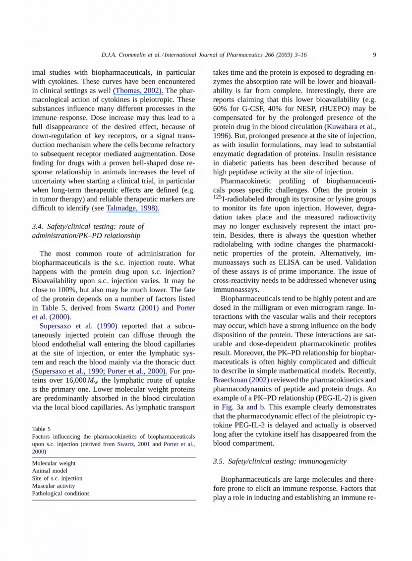
Biopharmaceuticals tend to be highly potent and aredosed in the milligram or even microgram range. In-teractions with the vascular walls and their receptorsmay occur, which have a strong influence on the bodydisposition of the protein. These interactions are sat-urable and dose-dependent pharmacokinetic profilesresult. Moreover, the PK–PD relationship for biophar-maceuticals is often highly complicated and difficultto describe in simple mathematical models. Recently,Braeckman (2002)reviewed the pharmacokinetics andpharmacodynamics of peptide and protein drugs. Anexample of a PK–PD relationship (PEG-IL-2) is givenin Fig. 3a and b. This example clearly demonstratesthat the pharmacodynamic effect of the pleiotropic cy-tokine PEG-IL-2 is delayed and actually is observedlong after the cytokine itself has disappeared from theblood compartment.
3.5. Safety/clinical testing: immunogenicity
Biopharmaceuticals are large molecules and there-fore prone to elicit an immune response. Factors thatplay a role in inducing and establishing an immune re-

10 D.J.A. Crommelin et al. / International Journal of Pharmaceutics 266 (2003) 3–16
K01 K10PlasmaVAbsorption Elimination
Blood Lymphocytes
Tissue Lymphocytes
Production Trafficking In Trafficking Out
Rprod Rin Rout
[1 +EmaxpCE
EC50 p+]
Stimulation
Stimulation
Delay 1
Delay 2
Delay 3
CE
CE
C
Kd
Kd
Kd Kd
[1 +E C
EC50 o+]
Cmaxo
10
100
1000
0
10
20
30
40
50
0 3 6 9 12 15
PE
G IL
-2 P
lasm
a C
on
c.n
g/m
L
Blo
od
Lym
ph
ocytes
Cells/m
m3 x 10
3
Days After SC Administration
(a)
(b)
Fig. 3. (a) PK/PD model for changes in blood lymphocytes after s.c. administration of PEG IL-2 in rats. The PK model is a one-compartmentalmodel with first-order absorption (rate constantK01) and elimination (rate constantK10). PEG IL-2 stimulates the trafficking out of bloodand/or catabolism of lymphocytes (first-order rateRout) according to anEmax model (parametersEmaxo and EC50o), which is a function ofthe PEG IL-2 plasma concentration (C). The delayed increase of blood lymphocytes is modeled in two consecutive ways: (1) three delaycompartments with first-order input and output rates (rate constantKd) resembling distribution and transduction delays; (2) stimulation oflymphocyte production in tissues according to anEmax model (parametersEmaxp and EC50p), which is a function of the effect concentrationCe. Tissue lymphocytes traffic into the blood pool (first-order rateRin) (from Braeckman, 2002). (b) PEG IL-2 pharmacokinetics andpharmacodynamics (changes in blood lymphocyte count) after subcutaneous administration of 10 MIU/kg in rats, modeled according tothe PK–PD model depicted in (a) (fromBraeckman, 2002).

D.J.A. Crommelin et al. / International Journal of Pharmaceutics 266 (2003) 3–16 11
Table 6Factors influencing immune reactions induced by biopharmaceu-ticals (adapted fromBraeckman, 2002and Schellekens, 2002)
Nature of the protein (endogenous/non-endogenous)Contaminants (e.g. host cell material)Route of administrationDose and regimenFormulationDisease and concomitant medicationBioassay design
sponse are listed inTable 6(adapted fromSchellekens(2002) and Braeckman (2002)). Endogenous pro-teins are less immunogenic than non-endogenousmolecules. That does not mean that endogenousmolecules are non-immunogenic per se. Route ofadministration (immunogenicity s.c. > i.v.), body dis-tribution (e.g. endogenous protein active in paracrinemode versus recombinant protein systemically active),dose (immunogenicity high dose> low dose), dura-tion of dosing (immunogenicity long> short dosingschedules), presence of aggregates and contami-nants: all these factors may turn a non-immunogenicmolecule into an immunogenic one. Another impor-tant factor is the molecule itself. What is claimedto be an endogenous protein may actually not be anendogenous one. Small sequence differences mayhave been introduced, for instance, for technical rea-sons (replacement of a cysteine). Or, the endogenousmolecule is glycosylated and the recombinant proteinis non-glycosylated (as produced inE. coli). Or, theglycosylation pattern of the biotech product is differ-ent from the endogenous product. These differencesmay lead to different ‘handling patterns’ of thesebiotech molecules in the patient compared to the en-dogenous molecules, subsequently leading to immuneresponses. Immunogenic responses occurring with thefirst generation of therapeutic monoclonal antibodies(HAMA) first led to the introduction of much lessimmunogenic humanized and, recently, human anti-bodies, or to the use of only fragments of monoclonalantibodies (e.g. chimeric Fab fragments for abcix-imab). Another approach to reduce immune responsesis to covalently attach polyethylene glycol polymersto the protein (as with PEG-G-CSF, PEG-ADA). Thiseffect is presumably the result of reduced exposureof hydrophobic parts of the protein in the presence ofPEG.
Surveying the literature on immunogenicity of re-combinant endogenous proteins, makes it clear thatimmune responses do not occur in all patients. Some-times only a few percent of the patients develop a re-sponse after a year or more of regular exposure. Onecan only guess what the reason is that not all patientsdevelop antibodies.
Antibody formation can have quite different effectson the therapeutic performance of a biopharmaceuti-cal (Schellekens, 2002; Braeckman, 2002). Reducedtherapeutic potency is observed when neutralizing an-tibodies are formed. Then, the antibody binds to a partof the protein close to or at the receptor binding site.Non-neutralizing antibodies may have an indirect ef-fect on the performance as they may change the phar-macokinetics of the protein drug. Both increases anddecreases in therapeutic effect have been observed. Aspecifically unwanted situation occurs when neutral-izing antibodies are formed that cross-react with anendogenous protein and if there is no alternative path-way to substitute for loss of action of that particularendogenous protein. Cases of long-term thrombocy-topenia were observed after the use of recombinantthrombopoeitin. Neutralizing antibodies blocked en-dogenous TPO, which is a crucial factor in the pro-duction of platelets.
Formulation design also affects immune responseinduction. In particular, the presence of aggregatesor contaminants (e.g. lipopolysaccharides) has beenshown to elicit an immune reaction. During the for-mulation design stage specific attention should be paidto the stability of the formulation with regard to theprevention of protein aggregation.
Finally, the bioassay used to detect antibodiesshould be well-validated and internationally stan-dardized, as non-immunogenicity was claimed in thepast on the basis of non-validated bioassays (usuallyELISAs) (Schellekens, 2002).
In conclusion, antibody formation is highly unde-sirable and should be avoided as much as possible,but at the present only general rules exist and im-munogenicity in clinical situations is highly unpre-dictable. Several approaches have been developed topredict immunogenicity in man by using animal mod-els. Carefully designed transgenic animals that areimmunotolerant to the human protein are presentlyconsidered useful predictors, if properly validated(Schellekens, 2002).

12 D.J.A. Crommelin et al. / International Journal of Pharmaceutics 266 (2003) 3–16
4. Formulation
Proper protein formulation development is crucialfor the optimal therapeutic performance of biophar-maceuticals. As mentioned above, immunogenicityis in some way related to the presence of aggre-gates and contaminants. As all systemically activeproteins are administered parenterally, sterility andnon-pyrogenicity are standard requirements for theseproducts. Removal of viruses and other contaminantsshould be an integral part of the downstream process(Crommelin, 2002). To reach the desired shelf life oftwo years for biopharmaceuticals, a number of spe-cific challenges have to be met that will be discussedin the following paragraphs.
4.1. Stability
On storage, proteins are exposed to a number of dif-ferent chemical degradation pathways that are listed inTable 7a. This table also provides a selection of tech-niques from our analytical ‘toolbox’ to monitor thesedegradation processes. Apart from chemical degrada-tion, physical degradation can occur in the form ofchanges in secondary or tertiary structure, and aggre-
Table 7aCommon chemical degradation reactions affecting the stability of proteins and methods of analysis (adapted fromArakawa and Philo, 2002)
Physical property effected Method of analysis
OxidationCys
Disulfide Hydrophobicity RP-HPLC, SDS–PAGEIntrachain Size Size exclusion chromatographyIntrachain Hydrophobicity Mass spectrometry
Met, Trp, Tyr
Peptide bondHydrolysis Size Size exclusion chromatography, SDS–PAGE
N to O migration Hydrophobicity RP-HPLCSer, Thr Chemistry inactive in Edman reaction
�-Carboxy to�-carboxy migration Hydrophobicity RP-HPLCAsp, Asn Chemistry Inactive in Edman reaction
Deamidation Charge Ion exchange chromatographyAsn, Gln
Acylation Charge Ion exchange chromatography�-Animo group,ε-amino group Mass spectrometry
Esterification/carboxylation Charge Ion exchange chromatographyGlu, Asp, C-terminal Mass spectrometry
gate formation, which can ultimately lead to precipi-tation (Table 7b) (Arakawa and Philo, 2002).
Formulation design should be geared to avoiddegradation. Proper selection of excipients, physicalstate and storage conditions are critical to avoid lossof therapeutic value and induction of immune re-sponses (Crommelin, 2002). In principle, the selectionof excipients for protein formulations is no differentthan for low molecular weight formulations. Osmoticagents, buffers and, if necessary, lyoprotectants andantioxidants are found in formulations. Proteins oftenare highly potent and low doses (milligram or evenmicrogram range) are administered. Low concentra-tions of anti-adsorbents are added to avoid proteinloss or protein denaturation upon contact with thesurface of vials, syringes or infusion tubing. Albuminis used (but not everywhere recommended!) as ananti-adsorbent. Low concentrations of surfactants canalso be added for this purpose.
If aqueous solutions of the biopharmaceutical arenot sufficiently stable, then freeze-drying is an alter-native stabilizing technique. But, freeze-drying canreadily damage the protein structure. Proper lyopro-tection is necessary. Non-reducing sugars can be usedfor that purpose. Freeze-drying conditions (pressure-,

D.J.A. Crommelin et al. / International Journal of Pharmaceutics 266 (2003) 3–16 13
Table 7bCommon physical reactions affecting the stability of proteins andmethods of analysis (adapted fromArakawa and Philo, 2002)
Unfolding Hydrophobicity RP-HPLCSecondarystructure changes
CD
FTIRAnalyticalultracentrifugation
Aggregation/precipitation
Size SEC
Light scatteringAnalyticalultracentrifugation
temperature–time profiles) have to be chosen such thatno collapse of the cake occurs during the drying pro-cess and the residual water content is low enough tohave a glass transition temperature in the freeze driedstate exceeding 40◦C.
4.2. Rate controlled delivery
Biopharmaceuticals often have to be administeredfrequently, a number of times daily or a few times perweek. To improve patient friendliness several differentapproaches can be chosen. Modification of the proteinis one option. With NESP (darpoeitin) a hypersialy-
Fig. 4. ‘All-water-system’ for the formation of protein-loaded microspheres on the basis of biodegradable, cross-linked dex-tran-hydroxyethyl-metacrylate (dextran-HEMA). Typically, protein yield is >80% and protein loads up to 15% have been described (Stenekes,2000).
lated erythropoeitin is used. At twoN-glycosylationsites of the protein that do not interact with the recep-tor carbohydrates with terminal sialic acid groups areattached. This modification increases thet1/2 in bloodfrom 9 to 21 h. Similar effects have been describedfor PEGylation of biopharmaceuticals. PEG-G-CSF(pegfilgrastim, a long circulating form of G-CSF)has recently been approved. Six milligrams pegfil-grastim in one injection proved to be therapeuticallyequivalent to five daily doses of filgrastim (PhysicianPackage Insert Neulasta® (Pegfilgrastim)). Manyyears ago, different formulations of insulin weredeveloped that provide controlled release patterns.Zinc or protamine interactions with insulin lead tothe preferred blood level of insulin. And a span ofduration of action between 6 h for soluble insulin to28 h for ‘ultralente extended insulin zinc suspension’can be achieved. For low molecular weight drugs,release duration may be extended to a few days orto a few weeks by using oil suspensions or solutions(with antipsychotics or hormones). Biodegradablemicroparticle systems based on polylactic-coglycolicacid (PLGA) have also been described. PLGA in theform of a rods or microspheres has been successfullyused for the sustained release (up to six months) ofsmall peptide drugs (e.g. LHRH analogs such as le-uprolide). Only if the drug molecule is highly potent

14 D.J.A. Crommelin et al. / International Journal of Pharmaceutics 266 (2003) 3–16
with a daily required dose of<1 mg and if prolongedrelease for over 1 week is required, does this PLGAtechnology (or other prolonged release technologies)offer advantages. But, PLGA technology suffers froma number of disadvantages (Crommelin, 2002): (a)strong burst effects are regularly observed (van deWeert et al., 2000a); (b) several reports show thatinside the device a substantial pH drop occurs dur-ing degradation of the PLGA, affecting the integrityof the PLGA-device associated protein; (c) standardmicrosphere preparation technology involves a w/oemulsification step, while the protein is dissolvedin the aqueous phase. As proteins readily denatureat w/o interfaces this process may not be optimal(van de Weert et al., 2000b). There is a clear needfor more protein friendly technologies without bursteffects. Several hydrogel technologies for controlledrelease of biopharmaceuticals have been describedover the last few years (Hennink and van Nostrum,2002).
Here we will focus on the ‘all-water-hydrogel-microsphere technology’ as developed by Henninket al. (Fig. 4, Stenekes, 2000). When hGH is used,a single injection of hGH gives a similar growthincrease as 10 subsequent working days injectionscovering the same total dose in a dwarf mouse model.The encapsulation efficiency is >90% and the loadingcapacity up to 15%. No burst release is observed.Some of these controlled release hydrogel technolo-gies have entered clinical test phases, others are inadvanced stage of animal testing. Improving patientfriendliness of many of the present biopharmaceu-ticals by reducing the frequency of injection of thechronic dosing schedules through controlling releaserates is a challenge taken up by a number of academicgroups and delivery companies.
5. Handling
During manufacturing and transport to the (hos-pital) pharmacy the manufacturers should take careof ‘Good Transport Practices’. That means that coldchain conditions should be maintained, if so dictated.The receiving pharmacist should do his/her utmost toensure that these delicate, often expensive, biophar-maceuticals are stored and used as stipulated at theward by medical staff, or at the patient’s home by the
patient him/herself. There is a general lack of aware-ness with regard to the proper use of protein drugs.For example, forced heating and excessive shakingshould be avoided at all times as this readily inducesdenaturation and aggregation, increasing chances forinducing immune reactions. Recently, guidelines forthe proper storage and handling of biopharmaceuti-cals in hospital pharmacies were issued (Crommelinet al., 2003). For conventional, low molecularweight parenterals such strict rules are rather excep-tional.
6. Generics: the possibility to launchbiogenerics(?)
In the production of biopharmaceuticals, seem-ingly minor changes in production conditions maylead to subtle changes in molecular structure of theprotein. Folding might be different, or glycosylationpatterns might change. For relatively small proteinssuch as insulin, equivalence might be establishedusing a set of assays from our ‘analytical toolbox’(Table 3): this is the category of the ‘well character-ized biopharmaceutical’. But, for larger proteins, it isalmost impossible to guarantee full equivalence of theprotein product characteristics in the clinic, includingequivalent efficacy and safety (cf. immune response),when using the full content of our present range ofsophisticated analytical techniques. The maximumdegree of product equivalency can be reached bykeeping all conditions in the manufacturing processconstant from batch to batch, starting with the work-ing cell bank, fermentation conditions, downstreamprocessing, filling, finishing and storage conditionsof the biopharmaceutical. Besides, a set of analyti-cal techniques, including bioassays, should be run tofurther support equivalence claims. Over the years,the FDA has developed a ‘Guidance for Industry.Changes to an Approved Application for SpecifiedBiotechnology and Specified Synthetic BiologicalProducts’. For minor changes in production protocolsa limited comparability program has to be run, butfor major changes additional clinical tests have to beperformed to establish comparability. Considering therestrictions regarding ‘in house’ changes of produc-tion processes, it is very unlikely that generic versionsof biopharmaceuticals will enter the market along the

D.J.A. Crommelin et al. / International Journal of Pharmaceutics 266 (2003) 3–16 15
same regulatory pathways as low molecular weightgeneric products do. Actually, there are already anumber of hGH products on the market developedindependently by different companies (e.g.Marian,2002). Considering the above analysis, the questioncan be raised whether they ever will be launched forthe more complicated, ‘not-so-well-defined’, biophar-maceuticals.
7. Conclusions
Biopharmaceuticals are very different from lowmolecular weight drugs. The complicated protein pro-duction processes and structures ask for a paradigmshift in thinking compared to low molecular weightdrugs. No absolute description of drug and drug prod-uct is possible with these materials. Our analyticaltoolbox content and bioassays, including animal test-ing, are important in ensuring drug quality, efficacyand safety issues in the development phase. But, thebiopharmaceuticals rely critically on strict produc-tion protocols, clinical expertise and performancemonitoring in the clinical situation.
References
Arakawa, T., Philo, J.S., 2002. Biophysical and biochemicalanalysis of recombinant proteins. In: Crommelin, D.J.A.,Sindelar, R.D. (Eds.), Pharmaceutical Biotechnology. Taylor &Francis, London, pp. 25–52.
Bass, R., Kleeberg, U., Schröder, H., Scheibner, E., 1992. Currentguidelines for the preclinical safety assessment of therapeuticproteins. Toxicol. Lett. 64–65, 339–347.
Biotechnology Industry Organization Site, 2002.http://www.bio.org/er/statistics.asp.
Braeckman, R., 2002. Pharmacokinetics and pharmacodynamicsof peptide and protein drugs. In: Crommelin, D.J.A., Sindelar,R.D. (Eds.), Pharmaceutical Biotechnology. Taylor & Francis,London, pp. 105–131.
Cavagnaro, J.A., 2002. Preclinical safety testing of biotechnology-derived pharmaceuticals. Nat. Rev. Drug Disc. 1, 469–475.
Claude, J.-R., 1992. Difficulties in conceiving and applyingguidelines for the safety evaluation of biotechnologically-produced drugs: some examples. Toxicol. Lett. 64–65, 349–355.
Crommelin, D.J.A., Bissig, Gouveia, M., W., Tredree, R.L., 2003.Storage and handling of biopharmaceuticals: problems andsolutions. A workshop discussion. Eur. J. Hosp. Pharm. 8,89–93.
Crommelin, D.J.A., 2002. Formulation of biotech products,including biopharmaceutical considerations. In: Crommelin,D.J.A., Sindelar, R.D. (Eds.), Pharmaceutical Biotechnology.Taylor & Francis, London, pp. 73–104.
van Dijk, M.A., Vidarsson, G., 2002. Monoclonal antibody-basedpharmaceuticals. In: Crommelin, D.J.A., Sindelar, R.D. (Eds.),Pharmaceutical Biotechnology. Taylor & Francis, London,pp. 283–299.
Evens, R.P., Sindelar, R.D., 2002. Biotechnology products inthe pipeline. In: Crommelin, D.J.A., Sindelar R.D. (Eds.),Pharmaceutical Biotechnology. Taylor & Francis, London,pp. 389–406.
FDA, 1997. Sitehttp://www.fda.gov/cder/guidance/1859fnl.pdf.Hennink, W.E., van Nostrum, C.F., 2002. Novel crosslinking
methods to design hydrogels. Adv. Drug Deliv. Rev. 54, 13–36.Herron, J., Jiskoot, W., Crommelin, D.J.A., 1995. Physical
Methods to Characterize Pharmaceutical Proteins. PlenumPress, NewYork.
Jiskoot, W., van Hertooij, J.J.C.C., Hoven, A.M.V., KleinGebbinck, J.W.T.M., van der Velden-de Groot, T., Crommelin,D.J.A., Beuvery, E.C., 1991. Preparation of clinical grademonoclonal antibodies from serum-containing cell culturesuspensions. J. Immunol. Methods 138, 273–283.
Kadir, F., Hamers, M., 2002. Production an downstream processingof biotech compounds. In: Crommelin, D.J.A., Sindelar,R.D. (Eds.), Pharmaceutical Biotechnology. Taylor & Francis,London, pp. 53–71.
Kuwabara, T., Kobayashi, S., Sugiyama, Y., 1996. Pharmaco-kinetics and pharmacodynamics of a recombinant humangranulocyte colony-stimulating factor. Drug Metab. Rev. 28,625–658.
Marian, M., 2002. Growth hormones. In: Crommelin, D.J.A.,Sindelar, R.D. (Eds.), Pharmaceutical Biotechnology. Taylor &Francis, London, pp. 245–258.
Metz, B., Hendriksen, C.F.M., Jiskoot, W., Kersten, G.F.A., 2002.Reduction of animal use in human vaccine quality control:opportunities and problems. Vaccine 321, 1–20.
Physician Package Insert Neulasta® (Pegfilgrastim).Porter, C.J.H., Edwards, G.A., Charman, S.A., 2000. Lymphatic
transport of proteins after s.c. injection: implications of animalmodel selection. Adv. Drug Deliv. Rev. 50, 157–171.
Sam, T., Peters, M., 2002. Follicle-stimulating hormone. In:Crommelin, D.J.A., Sindelar, R.D. (Eds.), PharmaceuticalBiotechnology. Taylor & Francis, London, pp. 359–364.
Schellekens, H., 2002. Bioequivalence and the immunogenicity ofbiopharmaceuticals. Nature Rev. Drug Disc. 1, 457–462.
Stenekes, R., 2000. Nanoporous dextran microspheres for drugdelivery, Ph.D. Thesis, Utrecht University, The Netherlands.
Supersaxo, A., Hein, W.R., Steffen, H., 1990. Effect ofmolecular weight on the lymphatic absorption of water-solublecompounds following subcutaneous administration. Pharm.Res. 7, 167–169.
Swartz, M.A., 2001. The physiology of the lymphatic system. Adv.Drug Deliv. Rev. 50, 3–20.
Talmadge, J.E., 1998. Pharmacodynamic aspects of peptideadministration biological response modifiers. Adv. Drug Deliv.Rev. 33, 241–252.

16 D.J.A. Crommelin et al. / International Journal of Pharmaceutics 266 (2003) 3–16
Thanou, M., Verhoef, J.C., Junginger, H.E., 2001. Chitosan andits derivatives as intestinal absorption enhancers. Adv. DrugDeliv. Rev. 50, 91–101.
Thomas, P.T., 2002. Non-clinical evaluation of therapeuticcytokines: immunotoxicologic issues. Toxicology 174, 27–35.
van de Weert, M., Hoechstetter, J., Hennink, W.E., Crommelin,D.J.A., 2000a. The effect of a water/organic solvent interface
on the structural stability of lysozyme. J. Control Rel. 68,351–359.
van de Weert, M., van’t Hof, R., van der Weerd, J., Heeren, R.M.A.,Posthuma, G., Hennink, W.E., Crommelin, D.J.A., 2000b.Lysozyme distribution and conformation in a biodegradablepolymer matrix as determined by FTIR techniques. J. ControlRel. 68, 31–40.


