Embed Size (px)
Citation preview

Shark Bay Stromatolites: Microfabrics and Reinterpretation of Origins
R. Pamela Reid, Miami; Noel P. James, Kingston; lan G. Macintyre, Washington D.C.; Christophe P. Dupraz, Neuchfitel; Robert V. Burne, Canberra
KEYWORDS: STROMATOLITES MARINk MICROBIAL PRI{CIP[TATION MICRII'I', t:',~<TOI'tl's?~IL],<, " St4AP, K BAY (ALJS-I RAI,IA)
Summary
Detailed analysis of microfabrics in Hamelin Pool stromatolites leads to reinterpretation of the origins of these structures. Previous studies have concluded that Shark Bay stromatolites form primari ly as a result oF sediment trapping and binding by microorganisms. Our results suggest that microbial precipitation af microcrys- lalline carbonate (micritc), as both franlcwork and ce- ment in these stromatolites, is also a fundamental, here- tofore unrecognized, process in their formation. Micro- bial trapping and binding is the primary mechanism of stromatolite accretion in the intertidal zone, forming grainy, calcarenite structures. Microbial precipital ion is the primary accretionary mechanism in the subtidal zone, forming muddy, micritic stromatoliles. Microbial pre- cipitation also lithifies trapped and bound sediment in the calcarenite stromatolites. Recognition o lmic rob ia l ly precipitated micritc in Shark Bay stromatolites is i m p e l rant, as many ancient stromatolites are micritic.
1 INTRODUCTION
The most abundant and diverse modern stromalolilcs in the modern world ocean occur in Hamelin Pool, a hypersaline embayment of Shark Bay, Western Australia (Fig. 1 ). These stromatolites were discovered in 1954 by D. Johnstone, P.E. Playford and R.L. Chase of West Australian Petroleum Pty. Lid (Playford and Cockbain 1976). They were the first known modern microbial build- ups with sizes and shapes analogous to Precambrian stro- matolites, which dominated the fossil record Ior much o I Earth history. As such, Hamelin PoaI stromatoliles have had a profound impact on stromatolite research and, for many years, they were used as a basis for comparison with all fossi l examples (Playford 1979: Riding. 1991: McNamara. 1999).
Microorganisms construct strontalolitcs by /rapping and binding sediment and/or precipitating a framework of calcium carbonate. Based on previous studies, which haxc emphasized the coarse grained, detrital nalure of Hamelin
Pool strolnatoliles (e.g. l,ngan 1961: Logan ct el. 1974: Playford and Cockbain 1976), these !;tructurcs are widel,< believed to have accrctcd lhrough sediment trapping and binding (Playlk~rd 1990; Golubie 1992: McNamara 1999). Indeed, in the past decade, it has been suggested lhal lhc coarse grained Shark Bay buildups are inappropriate analogs Ior ancient stromatolites, which are typically composed of micra- crystalline carbonate (micrile) (e.g. Av, ramik and P, iding 1988; Riding 1994). The potential role of microbial precipita- tion in the accretion and tithificafion of Shark Bay stromalo- lfies (Burne and Moore (1987) Inns received relatively little attention and, in ttne words of Play ford (I 990, p. 2 9 ) , "furlher work needs Io be done to more closely link dclails of the imernal tabrics of lhe siromatolites to lhe microbial C{/lnmu-
nities flint build Ihenf'. Geonlicrobiological studies linking micro fabric to micro-
bial activity have recently been applied ba the study of modern Ballami:m stromatolites. Detailed corrclalions of subsurface petrographic features, as seen in thin seclions~ with analysis of microbial popula!ions and microbial processes in surface mats show the importance of prokaryotic microbial c~mmmnilies in the accretion, lamination and early lithification e l marine stromatolites in Exuma Cays (e.g. Reid et al. 2000: Visscher et al. ]998, 2000: Kawaguchi and Decho 2001. 2(1(12}. Al- though Shark Bay stromalolites were discovered more than 40 years ago, previous sludies have emphasized morphology in relation to environmental setting (e.g. Playford 198(.)) and there are very few delailed descriptions of thin sections or published pholomicrographs (Monly 1976).
The aim of this paper is to provide an analysis of microfabrics in Shark Bay slromatolites. This is a critical first step in defining the role of microbial activity in stromatotitc accretion alld lifldl'icafion.
2 BACK(;ROI!NI) 2.1 Termiuoh~gy
"To be consistent with earlier studies (e.g. Pla\,lord 1990). the term 'stromatolite' is used in this paper fl~r ell ~r~ano- scdimenlary structures formed by Ihe sediment-binding and/ or carbonate precipitating activities of microoiganisnls, re-
Addresses: P. Reid, University of Miami, 4600 Rickenbacker Causewa),, Miami, FI, 33149 t iSA: prcidCa~ rsnms.nfiami.edu: N. P. James. Dept. Geological Sciences, Queen's University. Kingston Ont.. Canada K7L3N6: I.G. Macintyrc. ,";,mithsonian lnsfiltni~m. Nalional Museum of natural H~story, Washington D.C., 20013, USA: CP. l)upraz, l, Jni'>crsitd de Neuchfitel, I 1 Rue Emile Argand. Ca'~e Posmlc 2, CH-2007 Neuchfitel, Switzerland: R.V. Berne. Dept_ Geology. Australian National Unix crsity Canberra. ACT ()20(1 Auslralia

300
Fig. 1. Location map of Shark Bay, Western Australia, showing Hamelin Pool and the sampling sites at Flag- pole Landing and Carbla Point. Dashed lines are roads.
gardless of whether or not they are laminated. In this usage, the term is equivalent to 'microbialite' of Burne and Moore (1987). We use the term 'micrite' formicrocrystalline car- bonate, < 4 iam in size, without regard to mineralogy or inferred origin.
2.2 Setting
Stromatolites cover ~ 100 km of shoreline to water depths of 3-4 meters around the margins of Hamelin Pool, a hypersaline embayment at the southern end of Shark Bay (Fig. I). The environment is well-documented by Logan and Cebulski (1970) and Logan et al. (1974). In general, Hamelin Pool (Fig. 1 ) is a shallow (<10 m deep) broad embayment, surrounded on three sides by strandline environments rang- ing from rocky headlands to beaches to wide intertidal flats. Hamelin Pool is separated from most of Shark Bay by a grass bank, called the Faure Sill, which impedes water circulation. Salinities range from 55-70%0 throughout the year (Playford 1990). The stromatolites are believed to have developed because of these elevated salinities, which restrict the growth of microbial grazers (cf. Garrett 1970) and other organisms that compete for the same ecological niches (cf. Monty 1973). The tidal range is 1-1.5 m; tides are mainly wind driven and are not predictable (Playford 1990).
The environment over the last 2700 years (Chivas et al. 1990) has been shallow and energetic. Today southerly winds prevail with maximum velocity of 45km/hr; cyclonic
winds up to 180km/hr occur approximately once every 6 years. The domal to columnar stromatolites are found from the supratidal to shallow subtidal settings. They are devel- oped on hard substrates in carbonate sand. Substrates are either hardgrounds or fragments of Pleistocene to Miocene limestone. Sediment in the stromatolites is mainly ooids, bivalves (including large Fragum shells) and benthic foraminfers, with minor detrital quartz and dolomite. Stro- matolite growth is restricted to the last 4,000 years, during which time sea level receded from - l - 2 m above its present level (Logan et al. 1970; Hagan and Logan 1974).
2.3 Previous Work
Previous studies (e.g. Logan et al. 1974; Playford and Cockbain 1976; Hoffman 1976; Playford 1990) recognize three types of living stromatolites at Hamelin Pool based on macroscopic descriptions of internal structures and position relative to sea level (Fig. 2). Construction of these stromatolites has been attributed to three mat types: pus- lular, smooth, and colloform. Each mat type has a charac- teristic microbial assemblage whose distribution is tidally controlled. Reported scenarios of stromatolite construc- tion are as follows:
Upper to mid intertidal zone: Pustular mats (mamillate mats of Golubic 1983, 1992) form large columns and mounds up to I m wide and 40 cm high. These buildups are unlaminated,

30I
with irregular coarse fenestral networks and irregular outer surfaces (Logan et al. 1974; Playiord 1990). "l"he pustular mats, which have a characteristic brown color, arc domi- nated by the coccoid cyanobacterium E~ltophysali.~ mai~:r. The brown color results from the presence of se.vtonemine. a light induced extracellular pigment that is cmbedded in polysaccharide gel envelopes surrounding the cells. Entophysalis major is thought to be a descendent of the Precambr ian s t romatol i te-bt , i lding cyanobac te r ium Eoentophysalis (Golubic 1976, 1983). E. mqior mats at Hamelin Pool function as sediment traps, overgrowing and incorporating sand and silt that accumulates in their irregular surfaces (Logan et al. 1974; Goh, bic 1983).
Rapid lithification of pustular ntats ,,',,'as documented by Golubic and colleagues during the peak of the austral sum met in 1980 (Golubic 1983, 1992). Lilhificathm occurs as a result of precipitation of C a C Q within the polysaccharide envelopes surrounding E.major cells. Pockets of shriveled cells can be found encased within the prccipitates, which retain the characteristic golden to red-brown color ofthc live mats. SEM observations show that the gel precipitates have a globular morphology that is characteristic of amorphous carbonate formed in biological systems (Golubic 1983. 1992). These amorphous precipitates are in turn coated v, ith abiotic crusts of aragonite needle cement.
Mature, desiccated columns in thc upper intertidal lo supratidal zones may be colomzed by dark colored, fihn mats (Logan et al. 1974; Golubic, 1992). l+ogan et al. (I 974. p. 150) described these mats as "ecomorphs of pustular mats', comprised of 1 to 2 mm thick pelletoid rinds coh>- nized by desiccated E. major. Golubic (I 992) reported that the film mats were dominated by the endolithic cyanobacte- ria Hormathonema luteobrunneum and H. violaceonigrum, which penetrate and weaken the stromatolites (Golubic 1992). Playford and Cockbain (1976) suggested that lhese desiccated structures were no longer active, having died as a result of emergence due to Holocene upli ft.
Lower intertidal and shallowest suhtMal zones: Smooth mats construct smaller stromatolite columns and mounds. These structures are well laminated, with fine laminoid fenestral fabrics and smooth outer surfaces (Play ford 1990). The smooth mats are dominated by filamenlous cyanobac- teria; major species are reported as Schimth rix helva (t.ogan et al. 1974, Hoffman 1976) or Microcoleus chthom:plasres ( B a u l d 1984; Golubic 1985; Golubic 1992). Strornatolite accretion results from the trapping and binding of sediment by the filamentous cyanobacteria.
Lamination was thought to be related to variation in sediment influx, with stabilization of scdirncnt by precipi- tation of aragonite ceinent during periods of desiccation when interstitial water evaporated (l.ogan el al. 1974). During these exposure periods, Logan ct al. (1974)re- ported that Schizothrix reverts to a boring role, producing pellets and micro-unconformities. Fencstrac develop a[ter burial by oxidation of the mat (Logan et al. 1974). These laminated structures have been considered true stromato- lites in the original sense of Kalkowsky (1908).
Sub:Ma/zrme. Col lo lorm mats Ic, rn~ el l ipt ical to circular coltunns and cornpound tnasses t-ron~ a (eve centin~ctcrs to several reelers across and up to I m high hi depth.', of about 4 m (Logan el al. 1974; Pla',, ford, 1990). These structures are weakly to poorly larninaled, wi lh coarse, hregular fencstrae and hTegular outer surfaces (Playford 1990). Culhfform mats have been variously described as gelatinous tt) leath- ery. pale ycllov,,ish-brown to gray, 1-2 mm thick with hollow contiguous convexities. I-3 cm in diameter (l.ogan et al 1974) and as sof! mats that arc coherent at tile surface, with lithification a few mm belo,,',, the surface (Playford 1990). The reals are ephcmcral, bcing widely established al limes and absent at others (1.mgan el aI. 1974).
The microbial communilv of the coJtoform nlats is diverse and includcs nutllClOtts prokaryntic and eukarvotic species. Logan et al. (1974) reported gclatintms mats domi- nated by filamentous Mierocoleu.~ tener,imus and SvmpIr:ca laete viridis and thin leathery mats dominated by 5,'chi:othrix heh'a. Goluhic (1992) described gelatinous col hfform mats dominated hy a coccoid enlophysalidecean cyanohacterium that is he,,,, to science, with precipilalion of liny hollow aragonitc spheres within these mats (Golubic 1992). Bauld reported a diverse diatom flora, whose role in mat construc- tion is at least as import as cyanobacteria (rcporled in Playford I990). Colloform mats are typically associated with the green algae Acetabularia sp. and ephemeral popu- lations of encrusting foraminifers, molluscs, and ,~ct-pulids.
Major factors in the grov,th of colloform rnal stromato- lites arc rcporled as ( I ) dotnirlg of mat>., t2) binding (~I sand. (3) precipitation and hardening o1: el)'ptocrystalJine arago- nite. and (4) oxidation of organic material (Logan et al. 1974). Lithificd layers, I-3 mm thick and conq~osed of cryptocrystallinc aragonilc and variable quantities o f pcloidal grains anti skeletal fragments, rninlic domal undulations in the mat; tt~ese layers arch over elongalc fenestrac, which tend to be" elongated following the mat surface. The laminae are brokcn by Acetabuh~ria holdfasly;, which [~rm cross cutting \.oids. Fcncstrac tnay be open or infilled by dcl.rilal carbonate sediment (Logan et al. 1974).
Some stromatolite structures arc reported lo show a progression from one mat lype to another. /ks sublidal stromatolites grow upward into tile shallow subtidal or interti,.tal range, o.)llolbrnl mats may bc replaced by stnc, oth mats. Similarly, as h',\',cr intertidal stromatolilcs grow upward into the upper intertidal ranges, smooth mats may be replaced by pustular mats. Fcnestral fabrics, which arc characteristic of all the stromatolite types, arc thought to be developed largely through dccon~positicm ~l the en- closed mat material of by microbi;tl mats bridging inden- tations on the slromatolitc surfaces ((h)lubic 198,3).
The growth history of slromaloliles, in Hamelin Pool is controxersial. The struclures were originally en\is0.oed :_is primarily intertidal phenomena (Logan 1961 l,ogan et al. 1964). Subsequently. sublidal forms, with even more exlcn- sive distribution than the intertidal forms, w'ere recognized ( Play ford and Cockbain 1976, Play ford 1979, Waller and Bauld 1986). Playford (l 9901 argued Ihat many of existing intertidal slrotnatolites hax, c grown wh~l ly in the intertidal

302
Fig. 2. Stromatolite distribution in Hamelin Pool as summarized by Playford (1990).
zone; others have suggested that present-day intertidal forms originated as subtidal stromatolites, which were veneered with intertidal growth as sea level fell (Burne and James 1986; Burne and Hunt 1990; Burne 1992
Radiocarbon dates by Chivas et al. (1990) indicate the following ages: a supratidal stromatolite was dated at 1250 -1010 years BP; one mid intertidal stromatolite was dated at 540-170 yrs BP, and another ranged fi'om 930 yrs BP at the base to 520 yrs BP at the top; two subtidal stromatolites gave ages of 550-470 yrs BP at the base and 70-modern yrs BP at the top. These results suggest that subtidal stromatolite growth is presently active but that intertidal growth involves both past and modern accretion. Estimates of growth rates based on the radiocarbon dates suggest long-term vertical accretion is < 0.4 ram/year. Thus individual stromatolites are estimated to have taken about 1000 years to reach their present growth heights of -350 ram.
3 MATERIALS AND METHODS
Samples were collected from intertidal and subtidal environments at Carbla Point and Flagpole Landing, Hamelin Pool (Fig. 1, P1.45, Table 1). Most samples were collected during a Bureau of Mineral Resources, Baas Becking Labo- ratory expedition to Shark Bay in November 1985. The stromatolites were studied in situ and representative ex- amples were sampled and returned to Canberra for analysis. These stromatolites were slabbed, and one half was impreg- nated with epoxy resin and saved as an archive specimen (P1. 45/I d). A suite of thin sections was made from the other hal f, with 7-15 thin sections (2.5 cm x 4.5 cm) per stromatolite. Eight stromatolites and corresponding thin sections from the Baas Becking collection were analyzed for this study (Table 1).
The 1985 sample set was augmented by four smaller stromatolite samples collected by R.N. Ginsburg in 1979 and R.P. Reid in 1999. These stromatolites were slabbed and impregnated with epoxy; 1-3 large thin sections (5 cm x 7 cm) were made for each stromatolite. In addition to the
primary set of 12 stromatolites (Table 1 ), we examined thin sections from from two additional samples in the mid intertidal zone (SB-99-4 and SB-99-8). SB-99-4 is a piece of pustular mat with a distinct knobby morphology (PI. 45/lb); this characteristic mat type is referred to in this paper as knobby pustular mat.
Microfabrics were described by systematic mapping and photography of slabs and thin sections using binocular and petrographic microscopes. Selected thin sections were cho- sen for analysis using a scanning electron microscope (Philips XL-30 field emission SEM) equipped with Oxford Link energy-dispersive spectrometer (EDS) for elemental analy- sis. Sections were etched in 1% HC1 for 5-10 seconds and lightly sputter coated (- 10 nm) with palladium prior to SEM analysis.
Mineralogy was dctermiamd using acombination of light microscopy of petrographic thin sections, EDS analysis of SEM samples, and x-ray diffraction (XRD) of bulk samples Calcium carbonate with 1-2% strontium, as determined by EDS, was assumed to be aragonite. Aragonite in bulk samples was verified by XRD using a Scintag diffractometer with Cu Ka radiation over the range 250-35 ~ 20 at a scan rate of 1 ~
4 MICROFABRICS
In contrast to previous studies, which emphasized the sandy nature of Shark Bay stromatolites, we recognize two distinct microfabrics: grainy (or sandy), here called calcarenite, and muddy, here called micrite. The calcarenite and micritic groupings are further subdivided based on the presence or absence of lamination. In addition, a distinct micritic microfabric that fbrms a massive knobby caprock at the tops of some stromatolites is classified separately as Massive Micrite. An additional microfabric that is transi- tional between the calcarenite and micrite end members is also recognized. Characteristic features of each microfabric are described below: colors refer to appearance in plane- polarized light.

303
4.1 Calcarenite
Carbonate sand grains are the dominant colnponenl of this microfabric, forming grainstonc, packstone, and. less commonly wackestone, textures. The sand is typically well sorted, ranging in size trom 100-200 btm. Peloids (microc- rystalline grains of unknown origin), foran3inifera, shell fragments, and grains with superficial oolitic coatings arc the major grain types; oolitic nuclei are mainly shell frag- ments and peloids. Larger foraminfera and bivalve shells up to several mm long am locally abundant, mainly in cavities. In addition to carbonate, quartz, and dolonfite typically comprise 5-10% of the grains and con/nlonly ft)rnl ooid nuclei. In some samples, the calcarenite is laminated; m other samples, it forms a heterogeneous, t,nlaminated fabric. as follows:
4.1.1 Laminated Calcarenite (LC, PI. 46 and 47)
Description: Millimeter-scale lamination is defined by couplets of cemented and uncemented sand layers. The cemented laminae, typically 0.5-2 mm thick, appear as dense white bands in hand samples (PI. 46/I a. PI. 47/1 a), antt dark brown or gray bands in thin section, plane polarized light (Pl. 46/1 b, l c; Pl. 47/I b, 1 c). Laminae are often con vex upwards to wavy, and are laterally continuous for distances of several centimeters; in some cases, adjacent cemented horizons merge into a single band. Lamination is often enhanced by laminoid fenestrae or aligned pores. Elongate sand grains are also commonly aligned parallel to bedding. Two types of laminated calcarenite arc recognized based on characteristic features of the cemented layers.
(a) Type 1 (LC-I, PI. 46)." LC-1 is the dominant tbrm of laminated calcarenite. The characteristic feature of this microfabric is the presence (/t" dark micritic coatings on carbonate sand grains in the cementcd layers. These micritic coatings commonly have micropeloidal textures (peloids ~20 btm in diameter) and encompass silt-sized patches of translucent golden micrite (Pl. 46/1d-1 t). The rnicritic coat- ings are in turn fringed with aragonite needle cement (PI. 46/ 117). In some cases, cemented laminae show an upward gradation from wackestone consisling predominantly of golden micrite with a granular texture t(~ packstone or grainstone of micrite-coated sand grains (PI. 46/le). Layer thickness is irregular and varies between and within layers, generally ranging from 200 bt, m - 2 m e , but up Io 5 mm thick.
Sand grains in LC-I are typically fresh, with well pre- served internal structure (Pl. 46lid-f). In some cemented layers, however, grains are extensively altered, or micritizcd. The altered grains appear as peloids with a uniform grey appearance in plane polarized light, showing no internal structure (PI. 46/1 g); altered grains are cominonly "fused" or 'welded together' at point contacts (PI. 46/1 h). As in ce- mented layers of freshly preserved grains, the micritizcd grains are surrounded by dark micritic coati ngs wilh em bed- ded golden granules and fringing aragonile needle cement (PI. 46/1g, lh). Some cemented layers show an upward gradation from fresh grains to micritizcd grains (P1.46/I g).
Upper surfaces of the micritized horizons are lypically abrupt and in some cases are defined by red-brown micritic crusts, also rinnned w:ith dark micrilie coatings (f'l. 46/I g).
Fenestsae (unsupported voids)are comm(m in LC-I. Small irregular holes (< I cm) arc abundant in cemented and LmCcmentcd layers and may be aligned along a layer( Pl. 46/ l b). Elongate fcnestrae, 1-2 cm long, are common, particu- larly in the cemented bands: poorly sorted geopetal sand, with large I\)raminifcra and bi;,alve shells, often partially fills these cavities. Some larger cavities ( 1-4 cm) containing pc<My sorted coarse-grained sand cut across the lamination. Truncated grains can bc found at the margins of all cavity types, although many of the small pores and l)nesirac exhibit no truncation.
(b) Type 2 (LC-2, PI. 47). The second, and less common. laminated calcarcnile micrnfabric. LC-2. is difl)i-cntiated from LC-I b} a lack of dark inicritic coatings and aragonitc needle cement on sand grains in lhe cemented lavers of the lithified/unlithi fled couplels. Cemented layers in I.C-2 cnn- sist of horizons of extensively micritizcd grains. The mieritizcd grains appear as slructurelcss grey peloids that are [used together al point contacts (Pl. 47/I a-d). These layers of grey. welded grains form well-cemented layers 200-500 ~_tm lhick. Dolomite and quarlz within lhese bands appear while in thin section, and stand mJt in sharp contrast to the sur- rounding micritizcd, gray grains (Pl. 47/ld). The upper boundaries of the micritized horizons are abrupt (Pl. 47/1 d). and arc cmnmonly defined by micriiic crusts. 20-50 btm thick (Pl. 47/1 c). Upper surlacch tlt" mieritizcd grains below the micrilic crusts arc often truncated (PI. 47/1 f). Fcnestrae and other small cavities are con3mon in IC-2, and arc often aligned with a single layer. Particles at the margins o1' these cavities are generally not truncated
SEM pholon3icrographs show lhat micri t ized horizons in both l,C-2 and LC- l form as a result o1' microboring, in 1s micritized grains are l-~crmealed by bore holes that are infillcd with micritic cement (Pl. 48/Ib). The bore lloles arc approximately 10 lanl in diameter; tbcy (ornl elongate tracks, which fo l low arain oull incs, ]c'avin<g Ihe inafgins e l the grains intact (PI. 48,/Id). ln f i l l ing consists ot nccdlc-shaped aragonile crystals that Iornl scalloped patterns perpetldicular to the direction of boring (PI. 48t I d. I c). ln f i l l ing of bore holes that cl-oss between grains al poi nt contacls (Pl. 48/1c)results in grain fusioiL Advanced fusion lcad~> to obliteration of orieinal grain nullincs. The aragonile crystals in the bore holes arc about 0.5 inicrons wide anti 2 ,urn wide. The crystals ha~,e a chal-~.lclorislic granular structure, bein 7 composed hi tiny ci-y~,iallites about 50 nnl in size tPI. 48tl l).
Micritizcd horizons ill [,C- I (Pl. 48/2) show inicroborilig l)alures similar to those in Is Micritized grains are permeated with infiIIed bore hole<; and grains arc Fused as result of microboring tracks crossing between ~rain~, at point contacts. The micropcloidal coatings and Iringing aragoniie needle CClllcnts, which characterize [.('- 1 but are absent in LC--], arc not microborcd (Pl. 48;/210.
Di.vtribution: Laminated calcarenite is reslricted Io the inter- tidal zone (Fig. 3). LC-I is the domirlanl microfabrie of the

304
Table 1. Samples mmlyzed for the present study; depth refers to height of water above top of stromatolite at time of collection.
large stronmtolite heads 866 and 867, from the upper inter- tidal zone, and in the snmller stromatolite 79-136 from the lower intertidal zone. Field descriptions at the time of collection (Table 1 ) classified surface mats on these stroma- tolites as film (866, 867) and pustular (79-136). LC-2 microfabric is the dominant component of small, topo- graphically low stromatolites (e.g. SB-99-5), which occur in the lower intertidal to shallow subtidal zone between the larger stromatolites. These stromatolites are colonized by smooth mats. LC-2 also forms the base of mid intertidal stromatolite SB-99-1.
Origin: Laminated calcarenite is interpreted as a primary depositional microfabric, with couplets of cemented and uncemented sand representing discrete accrctionary events. Fenestrae in the lanfinated calcarenite could represent buck- ling of a microbial mat due to air bubbles (Monty 1976), degradation of a mat, holdfasts, or holes from boring organ- isms. Truncated gains at cavity margins indicate that at least some of the holes are diagenetic products resulting from boring activities.
Insight into the role of organisms in the formation of laminated calcarenite comes from comparison with Baha- mian stromatolites Exuma stromatolites (Lee Stocking Is.) exhibit many features that are identical to those observed in LC-2. Indeed, except for the presence of minor amounts of quartz and dolomite in LC-2, which are lacking in the
Bahamian structures, Pls. 3/1 and 4/1 could be photomicro- graphs of Exuma stromatolites. The succession of couplets of well preserved, well sorted, uncemented sand alternating with cemented bands of grey micritized fused grains capped with micritic crusts in LC-2 (P1. 47/1) is identical to the pattern in Exuma stromatolites. In addition, the dimensions and patterns of the infilled bore holes, within and crossing between grains, in the grey micritized horizons (P1.48/1) are identical to the microboring patterns observed in the micritized horizons of Exuma stromatolites (Macintyre et al. 2000).
Previous work has shown that lamination in Exuma stromatolites results from a cycling of surface microbial communities (Reid et al 2000). The binding and trapping of well sorted, fine grained sand by the filamentous cyano- bacterium, Schizothrix sp., forms unlithified grain layers. During a hiatus in sedimentation, a surface biofilm develops, with abundant heterotrophic bacteria. Thin micritic crusts (20-50 t-tin) precipitate in the biofilm as a result of het- erotrophic activity (Visscber et al. 1998, 2000). During longer periods of hiatus, the coccoid endolithic cyano- bacterium Solentia sp. infests carbonate grains below the biofilm. Carbonate precipitation within an organic gel ma- trix in the bore holes concurrent with endolithic activity welds grains together, forming cemented horizons of fused, micritized grains (Macintyre et al. 2000). A characteristic feature of sand accreted by Schizothrix is the absence of
P l a t e
Fig. 1 a.
Fig. I b.
Fig. 1 c.
Fig. I d.
45 Hamelin Pool stromatolites (Western Australia).
Field photographs at Carbla Point showing upper to mid intertidal stromatolites with pustular surface mats; stromatolites arc approximately 40 cm high. Mid to lower intertidal stromatolites at Carbla Point with knobby pustular (k), pustular (p) and smooth (s) surface mats; fihn mat (f) caps the tallest stromatolite (-0.5 m), which extends into the upper intertidal zone. Subtidal stromatolites, 20-50 cm high, approximately 1 m water depth, Carbla Point; upper surfaces are coated with loosely bound sediment that has been referred to as colloform mat; sides are colonized by Acetabularia. Vertical sections through slabbed field specimens; 866, supra to upper intertidal; 842, mid intertidal; 79-136, lower intertidal; SB-99-5, lower intertidal; 857, subtidal zone.

P l a t e 45 305

306
interstitial submarine fringe cement, because pore-filling polymeric secretions of Schi=othrix inhibit precipitation (Kawaguchi and Decho 2002).
The similarities in microfabrics of LC-2 and Exuma stromatolites suggest that lamination in LC-2 results fi'om a cycling of surface communities similar to that documented in the Bahamian examples. Thus the uncemented grain layers in LC-2 are interpreted to accrete through the binding and trapping activities of SchizothrLr or similar filamentous cyanobacteria such as Microcoleus. The micritic crusts of LC-2 (e.g. P1, 47/le) are interpreted to precipitate within bacterial biofilms formed during short hiatuses in sedimen- tation. Micritized horizons (e.g. P1.47/ld) develop during longer hiatal periods, when grains become infested with the endolithic cyanobacterium Solentia sp.; carbonate cemented precipitated in bore holes crossing between grains welds them together (PI. 48/1b, lc). This interpretation is consis- tent with the presence of smooth mats, which previous authors reported are dominated by SchizothrLr or Micmcoleus (e.g. Logan et al 1974, Golubic 1985), on the surfaces of lower intertidal and shallow subtidal stromatolites com- prised mainly of LC-2 (e.g. SB-99-5, Fig. 3, Table I ).
In contrast to LC-2, features in LC-I are distinctly different from those in Exuma stromatolites. In particular, cemented layers with micritic coatings on sand grains and aragonite needle cement are lacking in Exuma stromatolites. Indeed, as noted previously, syndepositional precipitation in interstitial spaces between grains accreted by Schizothrix is inhibited by Schizothrix polymers (Kawaguchi and Decho
2002). Thus, the cemented layers of LC-1 did not form in Schi~othrir mats.
So what is the origin of LC- 1, the dominant microfabric of laminated stromatolites in the intertidal zone? Our inter- pretation is limited by a lack of detailed microbiological studies. The dark micropeloidal coatings with enclosed patches of golden precipitate and fringing aragonite needle cement (PI. 46/1d, l f) suggest, however, that cementation occurred in mats dominated by Entophysalis major. Micropeloidal texture is a characteristic feature of micrite precipitated in microbial mucous or organic gel (Dupraz and Strasser, 1999, 2002; Reitner, 1993; Riding, 1991,2000). In addition, precipitates formed by lithification of E. major have a characteristic golden to red-brown color and these biological precipitates are typically coated with abiotic aragonite fi'inge cement (Golubic 1985, 1992). Finally, dark micrilic coatings, golden micritic granules, and fringing aragonite needle cement are characteristic features of the massive micrite fabric, described in Ch. 4.2.1, which is a product of E. major lithification. We therefore interpret that sand in the cemented layers of LC- 1 was trapped and bound by soft Entophysalis mats and subsequently cemented dur- ing episodic mat calcification. The tq.at upper surfaces of the cemented layers (e.g. P1.46/1 d) indicate that the Entophysalis mats in which they formed were relatively smooth.
The micritized horizons that characterize some cemented layers in LC-I (e.g. Pl. 46/1g, lh) are thought to represent periods of Solentia infestation of grains trapped by Entophysalis prior to mat lithification. This is based on the
P l a t e 46
Fig. I a.
Fig. lb.
Fig. I c.
Fig. ld.
Fig. 1 e.
Fig. 1 f.
Fig. lg.
Fig. lh.
Hamelin Pool stromatolites (Western Australia). Laminated Calcarenite Type 1 (LC-1 ) microfabric. Unless otherwise indicated, figures in all plates are thin sections photographed using a petrographic microscope with plane-polarized light: color descriptions refer to observations in plane-polarized light.
Vertical section through a slabbed field specimen with laminated structure defined by dense, white bands. Fenestrae of variable shapes and sizes are common: some are elongate parallel to lamination. Sample 867. Low magnification photomicrograph showing a distinct banded pattern, emphasized by an alignment of pores. Thin section SB-99-I-B. Higher magnification view showing alternating layers of loose and cemented sand; the cemented layers (arrows), which appeal dark in thin section, are the white bands of Fig. I a. Thin section SB-99-9A. Detailed view of cemented layer with abrupt upper surface (arrow), showing well preserved oolitic sand grains embedded in golden brown micrite: sand grains above and below this cemented layer lack conspicuous pore filling. Thin section SB-99-8. Cemented layer that grades upward from a wackestone of grains floating in golden micrite (m) to a packstone or grainstone of oolitic grains with micritic coatings and aragonite needle cement. Thin section 867-D. Detailed view of cemented layer showing fi'esh sand grains, with well preserved internal structure, rimmed by dark micritic coatings (d) with embedded golden granules (gr); aragonite needle cement (an) is infilling remaining pore space. Thin section SB-99-I-A. Cemented layer showing upward gradation from fi'esh grains (fg) to grey micritized grains (rag); both fresh and altered grains are rimmed with dark inicritic coatings. The sharp upper boundary of the micritized horizon is defined by a micritic crust (anow); the crust has a red brown color and is also rimmed with dark micrite. Thin section SB-99-1-B. Higher magnification view ofa micritized horizon showing fusion of grains at point contacts (arrows); quartz (q) and dolomite (d) grains are not microbored. Micritized grains are coated with dark micropeloidal micrite (arrow, peloids ~ 20 lain), in turn coated with aragonite needle cement. Thin section 866-C 1.

P l a t e 46 307

308
similarity of microboring patterns and infilling cements in these grains (PI. 48/2a,2b) to micritized grains in LC-2 (Pl. 48/la- l f). In the case of LC- 1, micritized grains were subse- quently encased by micritic coatings and fringe cement, which were not microbored (Pl. 48/2a, 2b), during episodic lithification of Entol)hysalis.
Uncemented layers in LC-I could have accreted through trapping and binding by Entophysalis mats during periods of non lithification, or through binding and trapping by smooth mats dominated by filamentous cyanobacteria such as Schizothrix or Microcoleus. The sand, which is well sorted and fine grained, is texturally similar to sand in Exuma stromatolites that is accreted by Schi~.othrix. Couplets of cemented-uncemcnted layers in LC- 1 could, therefore, rep- resent a cycling of surface microbial communities between Entophysalis-dominated mats and Schi~othrix-dominated smooth mats, with cementation resulting fi'om calcification ofEntophysalis. A shift fi'om coccoid to filamentous cyano- bacteria could occur in response to sedimentation or burial.
The distribution of LC-1 as the dominant component of stromatolites in the upper intertidal zone (866 and 867, Fig. 3) and some stromatolites in the lower intertidal zone (79- 136, Fig. 3) is noteworthy. Stromatolites 866 and 867 were colonized primarily by black film mat at the time of collec- tion. Film mat was described by Logan et al. (1974) as a desiccated ecomorph of Entophysalis, and by Golubic (1983) as consisting ofHonnathonema endoliths. Previous authors (e.g. Playford and Cockbain 1976, Playford 1990) have suggested that desiccated stromatolites above the high-tide level are dead. In contrast, the lower intertidal zone, where stromatolite 79-136 was collected, is colonized by actively growing smooth and pustular mats (Pl. 45/Ib), and alterna- tion between these mat types is a likely origin of the LC-I microfabric. Thus LC-I stromatolites that are presently in the upper intertidal zone are interpreted to be inactive, relict
structures, which formed in lower intertidal zones when sea level was higher.
4.1.2 Unlaminated Calcarenite (UC, PI. 49)
Unlaminated calcarenite is a heterogeneous sandy mi- crofabric; couplets of cemented-uncemented layers are not present. Slabbed field samples show a mottled network of dense white limestone and irregular fenestrae (PI. 49/la). Low magnification views of thin sections show a disorga- nized pattern of limestone and fenestrae (Pl. 49/1b). Fenes- trae, which are equidimensional to elongate and typically l - 3 mm in size, occupy -20% of the bulk volume. Larger, cm- scale vugs that are commonly filled with chalky sediment are also present.
Examination with a petrographic microscope indicates that this microfabric is characterized by the same cemented textures present in LC-1. These textures, however, are arranged in patchy, rather than banded, patterns (Pl. 49/lc, d). Fresh sand grains rimmed by dark crusts of micropeloidal micrite in turn coated by acicular aragonite form grainstone patches; well preserved sand grains are also found floating in golden mottled microcrystalline matrix, forming wack- estones. Patches of dark micrite with golden granule inclu- sions (P1.49/ld), golden granular micrite (Pl. 49/le) and sand grains rimmed with dark micrite (PI. 49/1l-') are com- mon. Areas of grey micritized grains or clotted micrite lacking distinct grain outlines (Pl. 49/1f, lg) are also preva- lent: occasional skeletal grains within these grey areas are typically micritized and losing definition.
Many small fenestrae in the mottled calcm-enite are filled with aragonite needle cement (PI. 49/lc). Margins of open fenestrae and cm-scale vugs commonly show truncated grains (PI. 49/1h). Some large cavities me partially filled with poorly sorted, coarse-grained sediment, including large
P l a t e
Part 1 Fig. 1 a.
Fig. 1 b.
Fig. 1 c.
Fig. ld.
Fig. I e. Fig. I f.
Part 2 Fig. 2a.
Fig. 2b.
47 Hamelin Pool strornatolites (Western Australia).
Laminated Calcarenite Type 2 (LC-2) microfabric. Vertical section through a slabbed field specimen showing layered structure defined by dense white bands. Sample SB-99-5. Low magnification photomicrograph showing couplets of cemented and uncemented sand; the cemented layers (arrows), which appear grey in plane-polarized light, are the white bands of Fig. la. Thin section SB- 99-5. Higher magnification view showing several micritized horizons composed of grey structureless grains that are fusing together. Detail of white boxed area is shown in Fig. 1 d; SEM view of area in dashed black box is shown in PI. 48/la. Thin section SB-99-1B. Detail of micritized horizon indicated in Fig. I c. Peloidal grains are extensively fused so that grain outlines are barely visible. Dolomite (d) and quartz (,q) appear white, in sharp contrast to the grey fused peloids. Thin section SB-99-1B Micritic crust (arrow) overlying grey micritized grains. Thin section SB-99-3b. Micritized grains with truncated tipper surfaces (an'ows) below the micritic crust. Thin section SB-99-5.
Micrite-Calcarenite (M-C) transitional microfabric. Early stage: Initial infilling of pore space between micritic bands with calcarenite. Sand grains are coated with dark, micropeloidal micrite and aragonite needle cement. Thin section 858-E Late stage: Complete infilling of pore space between micritic bands with calcarenite. Sand grains in a matrix of dark micropeloidal micrite fill pore space between micritic bands. The original structure of laminated micrite is almost lost and the fabric is gradational to unlaminated calcarenite. Thin section 841-E.

P l a t e 47 309

310
foraminifera ( I-2 mm) and bivalves (3-5 mm): cavity-filling internal sediments often have a golden to red brown micritic matrix (P1.49/1g).
Distribution: Unlaminated calcmenite is the dominant mi- crofabric in stromatolites 842 and 841 in the mid to lower intertidal zone (Fig. 3). This fabric also forms the base of subtidal stromatolites 857 and 843 (Fig. 3).
Origin: Unlaminated calcarenite in the intertidal zone is interpreted as a primary depositional microfabric. Compo- nents comprising UC are basically the same as those in the cemented layers of LC-I : fresh grains rimmed with micropeloidal micrite and aragonite needle cement (PI. 49/ ld, I f, I h), golden granular micrite (PI. 49/1 e), and micritized grains (P1.49/lf). In UC, however, these components have patchy, rather than laminar distributions. UC is further differentiated from LC- 1 by the lack of layers ofuncemented sand. A probable explanation for such similarities and dif- ferences is that. in contrast to cement layers of LC-I , envisaged to tk)rm in fiat econ~orphs of Entophysalis mat, UC formed in pustular Entol)lusalis mats with in'egular, warty surfaces. The irregular surface morphology inhibits layer formation. The common occurrence of truncated grains at cavity margins is evidence tk)r at least some post deposi- tional boring and cavity fommtion.
An alternative interpretation is that UC is a reworked microfabric formed as a result of multicyclic boring and infilling of a preexisting sti'omatolite. This is a possible origin of UC at the base of subtidal stromatolites 857 and 843, where 2 ''d generation infilling of cavities with cemented sediment is a dominant petrographic feature (e.g. PI. 49/1g).
4.2 M i c r i t e
The rnicritic microfabrics are composed mainly of mi- crocrystalline aragonite, which COl-nmonly displays a clotted texture. Three varieties of are recognized: massive micrite (MaM), laminated micrite (LM), and unlaminated micrite (UM). as described below.
4.2.1 Massive Micrite (MaM, Pl. 50)
Descro~tion: In slabbed hand specimens, the massive mi- crite microfabric appears as a dense, mottled mixture of greenish to light brown limestone (PI. 50/la). The surface morphology has a characteristic knobby appearance result- ing flom the growth of convex upwards accretionary sur- faces (P1.50/1 a). Bivalve fragments up to several millime- ters in length are locally abundant (P1.50/1 a). Voids space, including irregular pores, and circum convex and radial cracks comprise ~30% of the volume.
Thin section observations show a micritic framework with a second generation of inl"illing sediment (PI. 50/lb). The micrite has a characteristic golden to red-brown color; XRD analyses of five bulk samples indicate that it is arago- nite. Dense homogeneous micrite grades to more open equant clusters, 20-50 btm in size (P1.50/1c, ld). Filalnent molds are sometimes present and small dark inclusions (10 lam) are locally abundant (PI. 50/le). Dense micrite may grade to micrite with a granular appearance: 'granules' are 20-100 btm in size, surrounded by dark rims and acicular aragonite cement (P1.50/113.
Sand grains, including superficial ooids, foraminifera, and bivalves, with minor amounts of quartz and dolomite
P l a t e 48
Part 1. Fig. 1 a.
Fig. I b.
Fig. 1 c.
Fig. ld.
Fig. I e.
Fig. 1 f.
Part 2. Fig. 2a.
Fig. 2b.
Hamelin Pool stromatolites (Western Australia). Scanning electron photomicrographs of features observed in petrographic thin sections lightly etched in 1% HCI for 5-10 seconds and coated with palladium; sample SB-99- I b.
Laminated calca.enite. Type 2 (LC-2) Low magnification SEM photomicrograph of micritized horizons shown in boxed area of P1.47/1 c. Box and arrow on this figure indicate locations of PI. 48/1 b, l c. Boxed area in Fig. I a showing grains permeated with bore holes that are infilled with cement; note the fusion of grains, which results in loss of original grain outlines. Quartz grains (q) m'e not microbored. Detailed view of point contact between two fused grains showing infilled bore hole that crosses from one grain to the other (arrow). Carbonate precipitation within the bore holes concurrent with endolithic activity welds grains together. Typical pattern of bore hole infilling. Bands of aragonite needles are precipitated across the hole, forming a scalloped pattern. Note that the borehole follows the grain perimeter, but does not penetrate the grain margin. Higher magnification view highlighting the banded nature of the elongate crystals that in fill the boreholes. This infilling pattern is characteristic of the endolithic cyanobacterium, Solentia sp., which titans layers of fused micritized grains m Bahamian stromatolites. Detailed view of the elongate crystals within the boreholes: the granular appearance is characteristic of Solentia infilling in Bahamian stromatolites.
Laminated calcarenite, Type 1 (LC- 1 ) Low magnification view of a micritized horizon in an LC-1 microfabric similar to that shown in PI. 46/lg; micritized grains are surrounded by dark micropeloidal coatings and fringe cement. Detail of boxed area is shown in Fig. 2b. Detail of 2a. Note that the micritized grains (m) are permeated with infiUed microborings. The micropeloidal coatings and fringe cement (arrow) sun'ounding the micritized grains are not microbored.

P l a t e 48 311
312
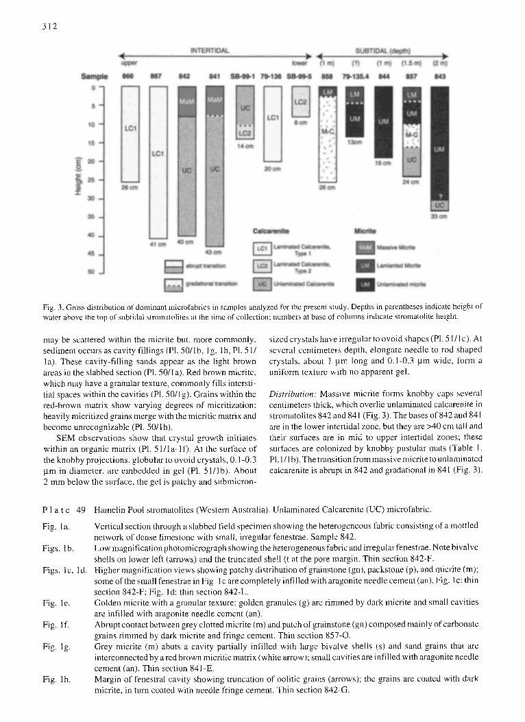
Fig, 3. Gross distribution of dominant microfabrics in samples analyzed for the present study. Depths in parentheses indicate height of water above the top of subtidal stromatolites at the time of collection; numbers at base of colunms indicate stromatolite height.
may be scattered within the micrite but, more commonly. sediment occurs as cavity fillings (Pl. 50/1b. I g. 1 h, Pl. 51 / la). These cavity-filling sands appear as the light brown areas in the slabbed section (Pl. 50/la). Red brown micrite, which may have a granular texture, commonly fills intersti- tial spaces within the cavities (Pl. 50/lg). Grains within the red-brown matrix show varying degrees of micritization: heavily micritized grains merge with the micritic matrix and become unrecognizable (Pl. 50/I h).
SEM observations show that crystal growth initiates within an organic matrix (PI. 51 / la - l f ) . At the surface of the knobby projections, globular to ovoid crystals, 0.1-0.3 ftm in diameter, are embedded in gel (P1. 51/lb). About 2 mm below the surface, the gel is patchy and submicron-
sized crystals have irregular to ovoid shapes (P1.51/1 c). At several centimeters depth, elongate needle to rod shaped crystals, about 1 }am long and 0.I-0.3 btm wide, form a tmiform texture with no apparent gel.
Distribution: Massive micrite forms knobby caps several centimeters thick, which overlie unlaminated calcarenite in stromatolites 842 and 841 (Fig. 3). The bases of 842 and 841 are in the lower intertidal zone, but they are >40 cm tall and their surfaces are in mid to upper intertidal zones; these surfaces are colonized by kmobby pustular mats (Table 1. PI. 1/I b). The transition from massive micrite to unlaminated calca,'enite is abrupt in 842 and gradational in 841 (Fig. 3).
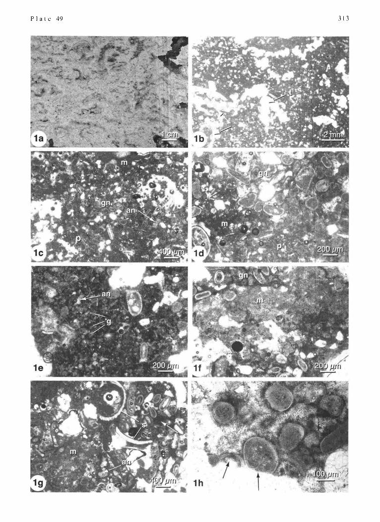
P l a t e 49
Fig. la.
Figs. I b.
Figs. I c, I d.
Fig. le.
Fig. if.
Fig. 1 g.
Fig. lh.
Hamelin Pool stromatolites (Western Australia). Unlaminated Calcarenite (UC) microfabric.
Vertical section through a slabbed field specimen showing the heterogeneous fabric consisting of a mottled network of dense limestone with small, irregular fenestrae. Sample 842. Low magnification photomicrograph showing the heterogeneous fabric and irregular fenestrae. Note bivalve shells on lower left (arrows) and the truncated shell (t at the pore margin. Thin section 842-F. Higher magnification views showing patchy distribution of grainstone (gn), packstone (p), and micrite (m); some of the small fenestrae in Fig. l c are completely infilled with aragonite needle cement (an). Fig. l c: thin section 842-F; Fig. l d: thin section 842-L. Golden micrite with a granular texture; golden granules (g) are rimmed by dark micrite and small cavities are infilled with aragonite needle cement (an). Abrupt contact between grey clotted micrite (m) and patch of grainstone (gn) composed mainly of carbonate grains r immed by dark micrite and fringe cement. Thin section 857-0. Grey micrite (m) abuts a cavity partially infilled with large bivalve shells (s) and sand grains that are interconnected by a red brown micritic matrix (white arrow); small cavities are i nfilled with aragonite needle cement (an). Thin section 841-E. Margin of fenestral cavity showing truncation of oolitic grains (arrows): the grains are coated with dark micrite, in mrn coated with needle fringe cement. Thin section 842-G.
P l a t e 49 313

314
Origin: Massive micrite is interpreted as a primary deposi- tional microfabric, with infilling of original cracks with a second generation of internal sediment. This knobby micro- fabric, which caps mid-intertidal stromatolites composed mainly of UC (Fig. 3), represents calcified knobby Entophysalis mat (P1.45/lb) with little to no trapped and bound sediment. MaM has the characteristic red-brown to golden color (Pl. 50/lb-lg) of Entophysalis precipitates (Golubic 1983, 1992). The black inclusions (Pl. 50/1c)may be shriveled Entophysalis cells or bacteria entombed by the calcification process (cf. Golubic 1983). Intimate relation- ships between organic gel and precipitated crystals at the uppermost surface of the accreting knobs (Pl. 51/lb) is further evidence of a biologic origin. Precipitation of CaCO, within the pusmlar mats is reported to begin in polysaccha- ride envelopes sun'ounding E.major cells (Golubic 1983, 1992). Globular crystal shape, as observed in the initial precipitates (PI. 51/1 b), is typical of amorphous carbonate formed in biological systems (Golubic 1983). Progression from globular to irregular ovoid (Pl. 51/Ic) and finally elongate, needle and rod-shaped crystals (Pl. 51/ld) may accompany the development of an organized aragonite crys- tal structure. Biologic precipitation of golden micrite within the Entophysalis mats is lk)llowed by abiotic precipitation of crusts of aragonite needle cement (P1.50/1 t) as described by Golubic ( 1983, 1992).
Convex upward and radial cracks within the massive micrite (PI. 50/lb, Pl. 51/la) are interpreted to form as a result of desiccation of the pustular mat. Sediment that filters into these cracks forms geopetal infilling. The red-brown micritic matrix of the internal sediment (P1. 50/lg, lh) is likely formed by calcification of Entophysalis cells within these cavities. Micritization of skeletal grains within the cavities (Pl. 50/I h) may result from microboring by Solentia, as described by Reid and Macintyre (2000).
4.2.2 Laminated Micrite (LM, P1.52)
Millimeter scale lamination in the LM microfabric is defined by bands of open space alternating with bands of
microcrystalline aragonite. This banding pattern is seen in slabbed hand specimens (Pl. 52/1a) and in low magnifica- tion thin section photomicrographs (Pl. 52/1b). The micritic bands are 200 gm to 2 mm wide and most are laterally continuous for distances of several centimeters. Pore spaces between the micritic bands also range from about 200 tam to 2 mm in height.
Higher magnification photomicrographs show p,'ogres- sive development of micritic bands with clotted texture in the upper few centhneters of specific samples (P1.52/1 c- 1 h). Band formation begins by the aggregation of grey clotted micrite in surface mats (Pl. 52/I c, I d). The clotted micrite is confined to specific portions of the mat: an organic mat framework defines the intervening spaces between clotted laminae. Below the surface, the micritic bands lack an organic framework, but retain the open pore spaces origi- nally occupied by mat (PI. 52/1 e, I f). These laminae may be heavily encrusted with worm tubes.
Eventually, open spaces within the clotted laminae are infilled with additional micritic carbonate, to form dense bands (Pl. 52/1g, lh). Infilling carbonate often has a dark, micropeloidal texture (PI. 52/lh) and may include red- brown clotted micrite. Small cavities are commonly infilled with aragonite needle cement. Foraminifera and worm tubes in inter-band pore spaces may be incorporated into the micritic bands as infilling progresses.
SEM observations (PI. 51/2) indicate that the micrite in the uppermost layers, where evidence of a microbial mat is seen in thin section, consists of equant to elongate crystals less than I bun in size; these crystals are embedded in organic gel matrix (P1.51/2b). Crystals m the lower, denser layers are similar in size and shape, but organic films are minimal to absent. Microborings are locally abundant, but much of the banded micrite shows no evidence of boring.
Distribution: LM forms the upper caps of subtidal stroma- tolites 858, 79-135.4 and 857 (Fig. 3). In 858, LM grades downward to a transitional micrite-calcarenite microfabric. In 79-135.4 and 857, LM grades downward to unlaminated micrite. As previously noted, although no mat was observed
P l a t e 50
Fig. I a.
Fig. I b.
Figs. lc, ld.
Fig. 1 e.
Fig. I f.
Fig. 1 g.
Fig. 1 h.
Hamelin Pool stromatolites (Western Australia). Massive Micrite (MaM) microfabric.
Vertical section through a slabbed field specimen showing characteristic dense, massive texture, mottled greenish to light brown color, and knobby upper surface (arrows). Bivalve shells (s) are common. Sample 842. Low magnification photomicrograph showing dense microcrystalline carbonate with irregular pores and radial and convex upward cracks; some cracks are filled or partially filled with internal sediment (arrows). Thin section SB-99-4. Gradation tu dense, homogenous micrite to more open, equant clusters. Note the characteristic golden to red-brown color of the micrite. Detail of boxed area in I c is shown in I d. Thin section 842-B. Golden micrite with small black inclusions: filament molds (arrows) are scattered throughout the micrite. Thin section SB-99-4. Golden micrite with granular texture; outlines of granules are defined by dark micritic coatings (d) and overlying aragonite needle cement (an). Thin section 842-D. Carbonate sand grains (right) infilling a cavity within a massive micrite framework (left); a red-brown miclitic matrix extends between the grains of internal sediment (arrow). These sediment-rich areas form the light brown patches seen in hand specimens (Fig. l a). Thin section 842-C. Micritization of foraminifera in cavity' fi 11; note progression from fresh (1), to partly altered (2), to completely micritized (3) skeletons. Thin section 842-C.

P l a t e 50 315

316
at the top of 857 during field collection, thin section obser- vations indicate the presence of an organic mat embedded with micrite in the uppermost 2 mm of this sample.
Origin: Laminated micrite is interpreted as a primary depo- sitional inicrofabric formed in the subtidal zone by an unknown mat type. Observations of clotted micrite within an organic mat framework (PI. 52/1 c, I d) at the surface of some stromatolites exhibiting the LM texture are evidence that microbial mats are involved in micrite accretion. Interband porosity, present as large laminoid fenestrae (P1.52/1 a, I b), rep,'esents space originally occupied by the organic fl'alne- work of the accreting lnat (PI. 52/lc, [d). Preservation of the organic matter as observed in thin sections is poor and the composition of the accreting mats is unknown.
SEM observations showing a lack of grain outlines and a paucity of microboring in surface micrite bands (PI. 52/2a) are evidence that the clotted rnicrite is not trapped and bound peloids or microbored grains, but instead is precipitated within the mat. Further evidence for organically induced precipitation of micrite within the surface bands is the close association between crystals and organic gel (P1. 51/2b), with a downward loss of gel in the subsurface (PI. 51/2d).
The increasing density of micrite, fi'om relatively open surface bands (PI. 52/1e, If) to dense subsurface bands (PI. 52/1 g, I h) indicates that precipitation of micrite continues in the subsurface. The mechanism of subsurface precipitation is unknown, but the dark micropeloidal textures of the infilling micrite (P1.52/1h) are suggestive of precipitation in an organic matrix (e.g. Dupraz and Strasser 1999, 2002).
4.2.3 Unlaminated Micrite (UM, PI. 53)
Unlaminated micrite is characterized by ilvegular dense limestone with an inegular fenestral fabric. Slabbed speci- mens show a network of dense limestone with abundant cm- to mm-sized open fenestrae (PI. 53/1 a): some fenestrae are filled with chalky sediment. Low magnification photomi- crographs show mottled carbonate with irregular cavities (PI. 53/Ib). Chalky areas of loose sediment may have been lost during the thin sectioning process.
Higher magnification photomicrographs reveal a pat- tern of micritic bands (P1. 53/lc, ld). These bands are identical to those described in the laminaled micrite fabric above. The banded structure is altered to an unlaminated fabric by partial in filling of pore spaces between the bands. Pore spaces are infilled with dark peloidal micrite, red- brown clotted micrite, and aragonite needle cements (P1. 53/1 c- I g); needle cements commonly radiate in fan-shaped bundles from cavity walls.
Skeletal grains and ooids in the pore spaces are incorpo- rated into the enveloping micrite network. These grains are often extensively micritized, eventually loosing all defini- tion of their origin (PI. 53/lh).
Distribution: Unlaminated micrite is the dominant micro- fabric in subtidal stromatolites 79-135.4, 844, and 843 and is present in 857 (Fig. 3). UM grades upward to LM in 79- 135.4 and 857. In 857, UM grades downward to micrite- calcarenite (M-C) transitional microfabric. In 843, UM overlies unlaminated calcarenite; the nature of this transition is unknown, as it was not sampled in thin section and is not apparent in hand sample.
P l a t e
Part 1. Fig. I a.
Fig. lb.
Fig. lc. Fig. I d.
Part 2 Fig. 2a.
Fig. 2b. Fig. 2c.
Fig. 2d.
51 Hamelin Pool stromatolites (Western Australia).
Massive Micrite (MaM) microfabric; all except Fig. l a are SEMS images. Scanned image of a vertical thin section through a knobby surface crust in transinitted light; a lnicrocrystalline carbonate framework is dissected by convex upward and radial cracks filled or partially filled with internal sediment. Arrows indicate the location of Figs. l b, l c and l d. Thin section SB-99-4. Globular to ovoid sub-micron crystals embedded in organic gel at the uppermost surface of the knobby crust. Irregular elongate sub-micron crystals with patches of film two millimeters below the surface. Rod and needle-shaped crystals up to 1 ~am long lacking organic gel 2.5 cm below the surface of the knobby crust; EDS analyses showing 1-2% Sr in these crystals support XRD analysis of bulk samples indicating aragonite.
SEM images of Laminated Micrite (LM) microfabric. Micritic lamina near the surface of st, btidal stromatolite 858, a petrographic microscope view of this area is shown in P1.52/ld. An organic mat framework (ore) defines laminar pore space on either side of the micritic band. The micrite has a clotted texture with open po,'e spaces between the clots. Arrow indicates the location of Fig. 2b. Thin section 858-A St,b-micron irregular crystals (< 1 lum), embedded in organic gel. EDS analyses st, ggest aragonite. Dense micritic lamina 1 cm below the surface of subtidal strolnatolite 858. An'ow marks the location of Fig. 2d. Thin section 858-A Heterogeneous mixture of sub-micron irregular crystals with little to no orgaqic gel. EDS analyses suggest an aragonite composition.

P l a t e 5l 317

318
Origin: Unlaminated micrite is interpreted as a diagenetic microfabric formed by alteration of laminated micrite. Alteration results from aragonite precipitation in the large laminoid fenestrae that comprise LM inter-band porosity (PI. 53/lc-lf) . This precipitation obliterates pore space and transforms the banded structure to an irregular fenes- tral fabric (PI. 53/1 b). Petrographic features of the interband precipitates, such as dark micropeloidal micrile (P1.53/1 d, l f) and golden granules (PI. 53/1g) fringed with aragonite needle cement (P1.53/1 d, l f, l g, t h), are characteristic of precipitates forlned in Entophysalis mats, as described in MaM and LC-1. E. ma/or, the dominant component of the intertidal pustular mats forming MaM and LC-1 does not grow well in subtidal environments (Golubic 1983). However, a subtidal coccoid entophysalidecean cyano- bacterium that is new to science has been reported as a dominant component of gelatinous colloform mats (Golubic 1992); this species, which has not yet been described (S. Golubic, pers. comm. 2003), may be important in the precipitation of interband micrite and thus the lk~rmation of the UM microfabric. Gradual obliteration of internal struc- ture of skeletal grains within UM (e.g. Pl. 53/1h) may be the result of microboring and micritization by Solentia.
The continuum from laminated to unlaminated micrite in samples 79-135.4 and 857 (Fig. 3) is consistent with the interpretation that the UM micro fabric fomls by progressive alteration of a primary banded smicture. The occun'ence of UM at the top of samples 843 and 844 suggests that these stromatolites are not presently colonized by mats precipitat- ing banded micrite.
4.3 Micrite-Calcarenite Transitional Microfabric (M-C)
The M-C microfabric is transitional between the end- members described above. It is similar to unlaminated micrite, but calcarenite, rather than micrite infills the micrite bands (Pl. 47/2a). With progressive infilling by calcarenite, the micritic bands become increasing difficult to recognize, and the microfabric grades to unlaminated calcarenite (Pl. 47/2b). Sand grains in the calcarenite infillings are charac- terized by dark, micropeloidal coatings, golden granules, and aragonite needle cement (P1.47/2a, 2b)
Distribution: M-C microfabric forms the dominant compo- nent of stromatolite 858 in the shallow subtidal zone (Fig. 3). At the top of this stromatolite, M-C grades upward to laminated micrite (LM). In addition, in subtidal stromatolite 857, unlaminated micrite (UM) grades to M-C, which in turn grades downward to unlaminated calcarenite (UC).
Ori<~in: Like UM, the transitional micrite-calcarenite micro- fabric is interpreted as a diagenetic alteration of laminated micrite. Observation of micritic bands in some thin sections (Pl. 47/2a) is evidence of the original laminated structure. Infilling of laminoid fenestrae with sand (Pl. 47/2a. 2b) rather than micritic precipitates suggests frequent sediment influx, or possible burial. Dark, micropeloidal coatings and golden granules fringed with aragonite needle cement in the calcarenite infillings (Pl. 47/2a, 2b), which are characteristic of Entophysalis precipitation, suggest an active presence by a subtidal entophysalidecean community in the interband porosity. Stromatolite 858, composed mainly of M-C, is in a sense a hybrid between subtidal stromatolites, which are predominantly micritic, and intertidal stromatolites, com- posed mainly of calcarenite.
5 SYNTHESIS 5.1 Role of Microbes
The microfabrics in Shark Bay stromatolites are mani- festations of four basic components: carbonate sand accu- nmlated through microbial trapping and binding, and three products of microbial precipitation: (a) fused, micritized grains, (b) a 'triumvirate' of dark, micropeloidal micrite, golden to red-brown micrite, and fringing aragonite needle cement, referred to henceforth as the 'golden micrite trium- virate" and (c) clotted micrite laminae. These components combine to form distinct microfabrics, which in turn form a variety of stromatolite types (Fig. 4). Accumulation of each component is a function of microbial activity as follows:
Carbonate sand is trapped and bound by smooth mats composed of filamentous cyanobacteria, such as Schizothrir or Microcoleus, and by pustular coccoid cyanobacterial mats formed by Entophysalis major. Grains are typically fresh, with well-preserved internal structure. Trapped and
P l a t e 52
Fig. la.
Fig. lb.
Figs. Ic, ld.
Fig. 1 e.
Fig. I f. Fig. lg. Fig. lh.
Hamelin Pool stromatotites (Western Australia). Laminated Micrite (LM) microfabric.
Vertical section through a slabbed field specimen showing bands of dense, light colored limestone and interband porosity. Sample 858. Low magnification photomicrograph showing laterally continuous micritic bands 200/am to 2 nml thick separated by open pore space. Thin section 79-135.4. Upper surf'ace of strornatolite 858 shown in Fig. 1 a. Grey clotted micrite forms distinct bands or laminae within a microbial mat. Interband spaces are defined by organic components of the mat (om). Thin section 858-A. Near surface band, lacking organic framework; note the complex open structure. Boxed area is shown in Fig. 1 f. Thin section 79-135.4. Detail of Fig. le showing the clotted texture of the micritic layer. Thin section 79-135.4. Subsurface layer of dense, clotted micrite. Boxed area is shown in Fig. l h, Thin section 79-135.4. Detail of Fig. l g showing the dense clotted micritic texture of this band; the color patterns suggest infilling of an open structure, such as that in Fig. 1 f, by a second generation of darker clotted to micropeloidal micrite (arrow). Thin section 79-135.4.

P l a t e 52 319

320
bound sand forms the accretionary framework of LC- 1, LC- 2, and UC (Fig. 4).
Fused micritized sand grains are formed as a result of endolithic activity by the coccoid cyanobacterium Solentia sp. Syndepositional precipitation of micrite in organic gel within Solentia boreholes crossing between grains leads to grain fusion. Grain fusion by micritization lithifies trapped and bound sediment, forming cemented layers in LC-2 and irregular patches in UC; fused micritized grain layers may also be present in LC-t.
The 'golden micrite triumvirate" of dark micropeloidal micrite and golden to red brown micrite coated with arago- nite needle cement is formed as a result of calcification of Entophysalis mats (intertidal Entophysalis major and prob- ably also a subtidal entophysalidecean species). The arago- nite needles represent abiotic precipitation templated on Entophysalis precipitates and are a characteristic feature of these mat precipitates. Golden micrite triumvirate lithifies trapped and bound sediment, forming cemented layers in LC-I and irregular cemented patches in UC. In addition, the golden micrite element of the triumvirate forms the accre- tionary framework of MaM, and triumvirate infills laminoid fenestrae in UM.
Clotted micrite laminae are formed by precipitation within an unknown subtidal mat type. These laminae form the primary accretionm2r framework of LM and UM.
Interactions between microbial communities result in the combination of components to form distinct microfabrics as follows:
LC-I is formed by a cycling between smooth mats (Schizothrix or other filamentous cyanobacteria) and pus- tular mats composed mainly of Enlophysatis major. The smooth mats trap and bind sand; Entophysalis mats trap and bind sand and calcify, precipitating golden triumvirate in interstial pore space. The result is layered couplets of uncemented sand and sand cemented by triumvirate.
LC-2 is formed by a cycling between smooth mats (Schizothrix or other filamentous cyanobacteria) and en- dolithic communities of Solentia sp. The smooth mats trap and bind sand: Solentia micritizes sand grains, which become welded together. The result is layered couplets of uncemented sand and sand cemented by grain fusion.
UC is formed by sediment trapping and binding and episodic calcification of pusmlar Entot)hysalis mats. The result is an irregular mixture of sand and golden micrite triumvirate.
MaM is formed by lithification of mats comprised solely of E. major, with little to no incorporated sediment. The result is a framework of golden micritic aragonite.
LM is formed by an unknown mat type that precipitates bands of clotted micritic aragonite. The result is a laminated micritic framework.
UM is formed by precipitation of golden triumvirate by an undescribed entophysalidecean community within the laminoid fenestrae of LM. The result is obliteration of lamination to form an unlaminated micritic framework.
Distributions of microbes within the peritidal environ- ment determine the spatial patterns of microfabric and stromatolite structure as follows:
Upper intertidal to supratidal zone: This zone is charac- terized by laminated stromatolites composed mainly of LC- 1 (Fig. 4). LC-1 is interpreted to form by alternation of Schizothrix and Entophysalis mats in the lower intertidal zone, suggesting that these stromatolites formed when sea level was higher than it is today.
Mid to lower intertidal zone: Unlaminated stromato- lites composed mainly of UC, with caps of MaM, form in this zone. This reflects the formation of UC in pustular Entophysalis mats in mid to lower tidal zones and MaM in topographically higher Entophysalis mats, with reduced sediment influx.
Lower intertidal to shallow subtidal zone: Two types of laminated stromatolite form in this zone. Larger buildups consist of LC- I, representing an alternation between fila- mentous and Entophysalis mats. Smaller buildups consist of LC-2, formed by cycling between filamentous and endolithic cyanobacteria. These differences may be re- lated to exposure tYequency.
Subtidal: Laminated to unlaminated stromatolites in this zone are composed of LM grading to UM. Lamination results from precipitation of micritic laminae in subtidal microbial mats of unknown composition; diagenetic infill- ing of open fenestrae between the micritic bands by a subtidal entophysalidecean species obliterates original lamination.
P l a t e 53
Fig. 1 a.
Fig. I b.
Figs. lc-lf .
Fig. lg.
Fig. lh.
Hamelin Pool stromatolites (Western Australia). Unlaminated Micrite (UM) microfabric.
Vertical section through a slabbed field specimen showing a network of dense carbonate with abundant small fenestrae. Sample 844. Low magnification photomicrograph of the specimen in Fig. 1 a. showing the dense micritic network and small irregular pores. Thin section 844-A. Higher magnification photomicrographs showing that the dense carbonate networks are comprised of micritic bands infilled with dark, micropeloidal micrite (rap) and aragonite needle cement (an). Boxed area in 1 c is shown in 1 d; boxed area in 1 g is shown in 1 f. Figs. I c, I d: thin section 79-135.4; Figs I e, I f: thin section 844- D. High magnification photomicrograph of a cavity fill showing golden granular micrite (arrows) infilling pore space between bivalve shells (s): granules are rimed with dark micrite and coated with fringe cement. Thin section 843-B. Micritized foraminifera (t') and micropeloidal micrite (arrow) infilling interband porosity: extensive micritization can obliterate evidence of skeletal origin, forming micritic peloids. Thin section 844-B.

P l a t e 53 321

322
5.2 Comparison with Previous Ideas
Our detailed study of microfabric sheds new light on the origins of Shark Bay stromatolites. In contrast to previous reports, which asclibe accretion of Hamelin Pool stromato- lites to trapping and binding of sediment (Playford 1990; Golubic 1992; McNamara 1999), we recognize two funda- mentally different structures: ( I ) grainy, calcarenite stroma- tolites, with primary accretion by sediment binding and trapping and (2) muddy, micritic stromatolites with primary accretion resulting from in situ precipitation of microcrys- talline aragonite (Fig. 4). Recognition of micritic stromato- lite buildups in Shark Bay is of fundamental importance as many ancient stromatolites are micritic.
Comparison of Figs. 2 and 4 shows some similarities but also significant differences between our results, based on microfabric, and previous interpretations, based mainly on stromatolite macrostructure. Previous studies have described stromatolites in the upper to mid intertidal zone (Pustular- Mat Stromatolites of Playford 1990) as unlaminated struc- tures built by Entophysalis mats (Logan et al. 1974; Hoffman 1976; Playford and Cockbain 1976). This is, in part, consis- tent with our interpretation that Entophysalis mats form unlaminated buildups of UC capped with MaM in the mid intertidal zone (Fig. 4). Our results, however, also show laminated (rather than unlaminated) stromatolites in the upper intertidal zone. These emergent laminated forms are interpreted as relict structures formed in the lower intertidal zone during higher sea level stands. These results support previous speculation that some intertidal forms are relict (Playford and Cockbain 1976; Burne and James 1986; Chivas et al. 1990).
Laminated stromatolites in the lower intertidal to shal- low subtidal zones, designated 'Smooth-Mat Stromatolites' by Playford (1990), have previously been interpreted as products of trapping and binding by smooth filamentous cyanobacterial mats, which were cemented a few millime- ters below the surface (Logan 1961, Logan et al. 1974: Hoffman 1976; Playford and Cockbain 1976). Logan ( 1961 ) suggested that lithification occurred by abiotic cementation when the structures were exposed. Others (e.g. Playford 1979, 1990) speculated that it was likely that algal or bacterial action were involved in lithification. Our analysis of microfabric in lower intertidal to shallowest subtidal stromatolites provides evidence for two types of laminated calcarenite buildups, both of which form by an alternation between microbial trapping and binding and microbial pre- cipitation (Fig. 4). The most common type, the LC- 1 stroma- tolites of Fig. 4, are formed by a cycling between smooth mats and pustular Entophysalis mats; early lithification results fiom calcification of pustular mats. The second type, which forms the smaller laminated LC-2 stromatolites of Fig. 4, are formed by a cycling between filamentous cyano- bacteria and endolithic communities: early lithification re- sults from fusion of micfitized grains. Recognition of micro- bial micritization as a mechanism of of stromatolite lithifi- cation contrasts with previous ideas that endolithic activity serves only to weaken and destroy the stromatolites (e.g. Golubic 1992).
Subtidal stromatolites were previously described as weakly laminated and designated as 'Colloform Mat Stro- matolites' (Playford 1990). Accretion was attributed to trapping and binding of sediment in soft, coherent colloform mats composed of diverse cyanobacteria and diatoms, with lithification a few millimeters or centimeters below the surface (Playford and Cockbain 1976; Hoffman 1976, Playford 1990). Subtidal stromatolites with hard surfaces were considered to be inactive (Playford and Cockbain 1976). Our results are quite different. Microfabric analyses indicate that the dominant accretionary framework in the subtidal strornatolites examined for this study is aragonitic micrite formed by microbial precipitation (Fig. 4). This micrite is precipitated in a microbial mat of unknown com- position, forming a laminated structure of aragonite bands and open pore space. The laminoid fenestrae, representing pore space originally occupied by the mat, are subsequently infilled with micrite precipitated by a subtidal entophysal- idecean species, leading to a gradual loss of lamination.
6 CONCLUSIONS
I. Hamelin Pool stromatolites are formed by (a) microbial trapping and binding and (b) microbial precipitation. Micro- bial trapping and binding is the primary accretionary mecha- nism in the intertidal zone, forming grainy, calcarenite stromatolites. Microbial precipitation is the primary accre- tionary mechanism in the subtidal zone, forming muddy, micritic stromatolites. Microbial precipitation also cements trapped and bound sediment in the calcarenite stromatolites.
2. Micritic stromatolites in the subtidal zone have an initial laminated fabric, formed by precipitation of bands of mi- critic aragonitic in surface microbial mats of unknown composition. Additional precipitation of micritic aragonite by a subtidal entophysalidecean cyanobacterium infills laminoid pore space originally occupied by the mat and obliterates lamination.
3. Calcarenite stromatolites in the lower intertidal zone include two types of laminated structures; in both types, lamination represents layered couplets of cemented and uncemented sand. In Type 1, cemented layers are formed by calcification of thin Entophysalis mats. In Type 2, cemented layers are formed by grain fusion resulting from microbial micritization by Solentia sp. Uncemented layers in both types are accreted by microbial trapping and binding. Type 2 laminated calcarenite stromatolites are similar to Baha- mian stromatolites in Exuma Cays.
4. Calcarenite stromato]ites in mid to upper intertidal zones are unlaminated buildups formed by trapping and binding of sand by soft pustular Entophysalis mats; accreted sediment is lithified by precipitation of micritic cement during inter- vals of mat calcification.
5. Laminated calcarenite stromatolites presently occupying the upper intertidal zone are relict structures that formed in the lower intertidal zone when sea level was higher than present.
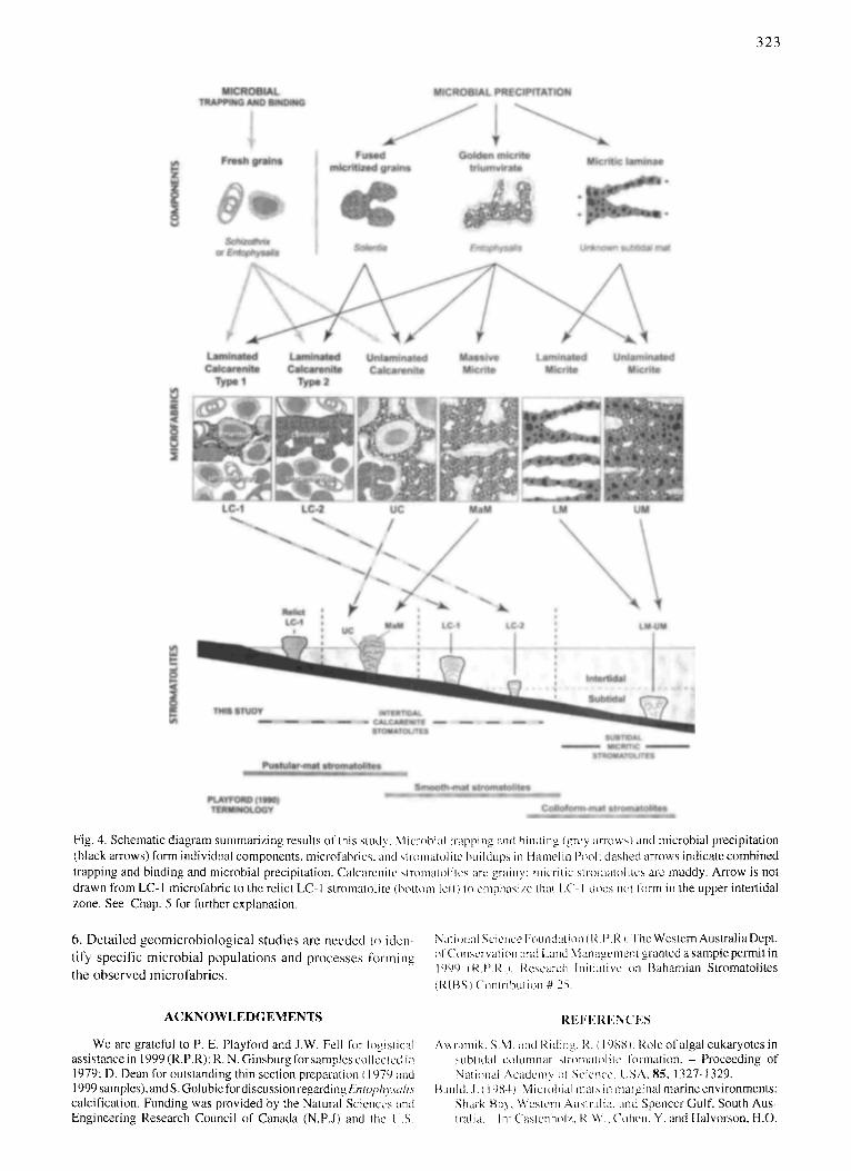
323
Fig. 4. Schematic dia~am summarizing results of this study. Micr(~bial trapping and binding (grey arrows) and microbial precipitation (black anows) form individual components, microfabrics, and slmmatolite buildups in Hamelin I->r dashed arrows indicate combined trapping and binding and microbial precipitation. Calcarenite slromai(Hilc<; are grainy: micrilic stromalolitcs are muddy. Arrow is not drawn from LC- I microfabfic lo the relict LC- 1 siromalolite (bott~m/[ell ) lo emphasize thai [,C- I dr not li~rm in the upper intertidal zone. See Chap. 5 for furiher explanation.
6. Detai led geomicrobio logica l studies are needed to idcn-
tify specific microbial populat ions and processes forming
the observed microfabrics .
National Science Fr (14. P.l ), I. -l'he Western Australia Dept. ~lConscrvation and Land Management granted a sample permit in 1999 (t4.1'.14.1. Research Initiative on Bahamian Siromatolites (14[BS) ('~mtrilr~cition # 25.
ACKNOWLEDGEMENTS R E F E R E N C E S
We are grateful to P. E. Playford and J.W. Fell for logistical assistance in 1999 (R.P.R); R. N. Ginsburg for samples collcclcd ia 1979; D. Dean for outstanding thin section preparation (I 979 m~d 1999 samples), and S. Golubic for discussion regarding L77tol~hysa/i.~ calcification. Funding was provided by the Natural Sciences and Engineering Research Council of Canada (N.P.J) and the t .S.
Awranaik. SM. and Riding. 14. ( 198H1: Role of algal eukaryotes in subtidal ctHumnar stromai~Hitc formation. - Proceeding of Nalflmal Academy tH- Science, USA, 85, 1327-1329.
Baulcl, .I. ( 19841: Microbial mats in marginal marine environments: ,Shark P, aS, Western Australia. and Spenccr Gulf. South Aus- Iral ia. - In: Castcnhr I 4. W., Cohen, Y. and 1 lalvorson, H.O.

3 2 4
(eds.): Microbial Mats: Stromatolites - 9-58, New York (Liss). Bume, R.V. (1992): Lilliput's Castles - Stromatolites of Hamelin
Pool . - Landscope-WA's Conservation, Forrests and Wildlife Magazine, 772, 34-40, CALM. WA
Bume, R.V. and Hunt, G. (1990): The Geobiology of Hamelin Pool. - Bureau of Mineral Resources, Australia. Record 1990/ 89.288 pp.
Bume, R. V and James, N.P. ( 1986): Subtidal origin o f club-shaped stromatolites, Shark Bay. Abstracts 12 u' Int. Sedimentol. Cong., p. 49, Canberra.
Burne, R.V. and Moore, L. S. (1987): Microbialites: organo- sedimentary deposits of benthic microbial communities. - Palaios 2, 241-254.
Chivas, A.R., Torgersen, T. and Polach, H.A. ( 1990): Growth rates and Holocene development of stromatolites from Shark Bay, Western Australia. - Australian Journal of Earth Science, 37, 113-I21.
Dupraz, C. and Strasser, A. (1999): Microbialites and micro- encmsters in shallow coral bioherms (Middle to Late Oxford- Jan, Swiss Jura Mountains). - Facies, 411, 101-130.
Dupraz, C. and Strasser, A. (2002): Nutritional Modes in Coral- Microbialite Reefs (Jurassic, Oxfordian, Switzerland): Evolu- tion of Trophic Structure as a Response to Environmental Change. - Palaios, 17,449-471.
Garret, P. (1970): Phanerozoic stromatolites: noncompetitive eco- logic restriction by grazing and burrowing organisms. Science, 169, 171-173.
Golubic, S. (1976): Comparison of Holocene and mid-Precam- brian Entophysalidaceae (Cyanophyta) in stromatolitic algal mats: cell division and degradation. - Jour. Paleontology, 50/ 6, 1074-1082.
Golubic, S. (1983): Stromatolites, fossil and Recent: a case history'. - In: Westbroek, P. and de Jong. E.W. (eds): Biomineralization and Biological Metal Accumulation. - 313-326, Dordmcht (Reidel).
Golubic, S. (1985): Microbial mats and modem stromatolites in Shark Bay, Western Australia. - In: Cardwell, I).E., Brierley, J.A., and Bricrley, C.L. (eds): Planetary Ecology. - 3-16, New York (van Nostrand ).
Golubic, S. (1992): Stromatolites of Shark Bay . - In: Margulis, L. and Olendzenski, L. (eds.): Environmental Evolution: Effects of the Origin and Evolution of Life on Planet Earth. - 131 - 147, Cambridge (MIT Press).
Hoffman, P. (1976): Stromatolite morphogenesis in Shark Bay, Western Australia. - In: Walter, M.R. (ed.): Stromatolites. - Developments in Sedimentology, 20, 261-271, Amsterdam (Elsevier).
Kalkowsky, V.H.E. (1908): Oolith und stromatolith im nord- deutschen Buntsandstein. - Z. Deut. Geol. Ges., 60, 231-242.
Kawaguchi, T. and Decho, A.W. (2001): Potential roles of extra- cellular polymeric secretions (EPS) in regulating calcification: a study of marine stromatolites, Bahamas.-Thalassas, 17, 11 -19.
Kawaguchi T. and Decho, A.W. (2002): Isolation and biochemical characterization of extracellular polymers (EPS) from modern soft marine stromatolites: inhibitory effects on CaCO~ precipi- tation.- Preparative BioChemistry BioTechnology, 32, 51-63.
Logan, B.W. ( 1961 ): Cp)7~tozoon and associate stromatolites from the Recent Shark Bay, Western Australia. - Joum. Geology, 69, 517-533.
Logan, B.W. and Cebulski, D.E. (1970): Sedimentary environ- ments of Shark Bay, Western Australia. - In: Carbonate Sedimentation and Environments, Shark Bay, Western Austra- lia. - Amer. Assoc. Petrol. Geol. Mem., 13, 1-37.
Logan, B.W., Hoffman, P. and Gebelein, C.D. ( 1974): Algal mats, cryptalgal fabrics and structures, Hamelin Pool, Western Aus- tralia. - In: Evolution and l)iagenesis of Quaternary Carbonate Sequences, Shark Bay, Western Australia. - Amer. Assoc. Petrol. Geol. Mem., 22, 140-194.
Logan, B.W., Read, J.F, and Davies, G.R. (1970): ltistory of carbonate sedimentation, Quaternary Epoch, S hark Bay, West- em Australia. - In: Carbonate Sedimentation and Environ-
ments, Shark Bay, Western Australia. - Amcr. Assoc. Petrol. Geol. Mere., 13, 38-84.
Logan, B.W., R. Rezak, and R.N. Ginsburg (1964): Classification and environmental significance of algal stromatolites. - J our- nal of Geology, 72, 68-83.
Hagan, G.M. and Logan, B.W. (1974): Development of carbonate banks and hypersaline basins, Shark Bay, Western Australia. In: Evolution and Diagenesis of Quaternary Carbonate Se- quences, Shark Bay, Western Australia. - Amer. Assoc. Petrol. Geol. Mere., 22, 61-139.
Macintyre, I.G., Pru fert-Bebout, L. and Reid, R.P. (2000): The role of endolithic cyanobacteria in the formation of lithified lami- nae in Bahamian stromatolites. - Sedi mentology, 47, 915-92 I.
McNamara, K. (1999): Stromatolites. - 28p. Western Australian Museum, Perth.
Monty, C.L.V. (1973): Precambrian background and Phanerozoic history of stromatolitic communities, an overview. - Annales de la Socidtd Gdologique de Belgique, 96, 585-624.
Monty, C.L.V. (1976): The origin and development of cryptalgal fabrics. - In: Walter, M.R., (ed.), Stromatolites.- Develop- merits in Sedimentology, 20, 193-259, Amsterdam (Elsevier).
Playford, P.E. (1979): Stromatolite research in Western Australia. - J . Royal Soc. Western Australia, 62/I-4, 13-20.
Playford, P.E. (1980): Environmental controls on morphology of modem stromatolites at Hamelin Pool, Western Australia. - West. Aust. Geol. Survey Ann. Rept. (for 1979), 73-77.
Playford, P.E, (1990): Geology of the Shark Bay area, Western Australia. - In: Berry, P.F., Bradshaw, S.D., and Wilson, B.R., (eds.): Research In Shark Bay. - 13-31, West Australian Museum.
Playford, P.E., and Cockbaln, A. E., ( 1976): Modem algal stroma- tolites at Hamelin Pool, a hypersaline barred basin in Shark Bay. Western Australia. - In: Walter, M.R., (ed.): Stromato- lites. - Developments in Sedimentology, 20, 389-411, Amsterdam (Elsevier).
Reid, R.P., Macintyre, I.G. and Steneck, R.S. (1999): A MicroN- alite/algal ridge fringing reef complex, Highbome Cay, Baha- mas. - Atoll Res. Bull., 466, 1-18.
Reid, R. P., Visscher, P.T., Decho, A.W., Stolz, J.F., Bebout, B.M., Dupraz, C., Macintym, I.G.. Paerl, H.W., Pinckney, J.L., Prufert-Bebout, L., Steppe, T.F. and DesMarais, D.J. (2000): The role of microbes in accretion, lamination and early lithifi- cation of modem marine stromatolites. - Nature, 406,989-992.
Reid, R.P. and Macintyre, I.G. (2000): Microboring versus recrys- tallization: Further insight into the micritization process. - Journal of Sedimentary Research, 70, 24-28.
Reitner, J. (1993): Modem cryptic microbialite/metazoan facies fi'om Lizard Island (Great Barrier Reef, Australia) formation and concepts. - Facies, 29, 2-40.
Riding, R. (1991): Classification of microbial carbonate, - In: Riding, R. (ed.), Calcareous Algae and Stromatol i tes .- 21- 5 I, New York (Springer).
Riding, R. (1994): Stromatolite survival and change: The signifi- cance of Shark Bay and Lee Stocking lsland subtidal columns. - In: Krumbein, W.E., Paterson, DM.. and Stal, L.J. (eds.) Biostabilization of Sediments. - 183-202, Oldenburg.
Riding, R. (2000): Microbial carbonates: the geological record of calcified bacterial-algal mats and biofilms. - Sedimentology, 47, 179-214.
Visscher. P.T., Reid. R.P. and Bebout, B.M. (2000) Microscale observations of sulfate reduction: con'eiation of microbial activity with [ithified micritic laminae in modem marine stro- matoli tes.- Geology, 28, 919-922.
Visscher, P.T., Reid, R.P., Bebout, B.M., Hoeft, S.E., Macintyre, I.G., and Thompson, J. Jr., 1998, Formation oflithified n'ficritic laminae in modem marine stromatolites (Bahamas): the role of sulfur cycling. - American Mineralogist, 83, 1482-1491.
Walter, M.R. and Bauld, J. (1986): Subtidal stromatolites of Shark Bay. - Abstracts 12 ~h Int. Sedimentol. Cong., p. 315, Canberra.
Manuscript accepted September 18, 2003