Embed Size (px)
Citation preview

�����������������
Citation: Ali, S.; Saeed, U.; Rizwan,
M.; El-Adawy, H.; Mertens-Scholz, K.;
Neubauer, H. Serological Prevalence
of and Risk Factors for Coxiella
burnetti Infection in Women of Punjab
Province, Pakistan. Int. J. Environ.
Res. Public Health 2022, 19, 4576.
https://doi.org/10.3390/
ijerph19084576
Academic Editor: Maria Antonia De
Francesco
Received: 9 January 2022
Accepted: 7 April 2022
Published: 11 April 2022
Publisher’s Note: MDPI stays neutral
with regard to jurisdictional claims in
published maps and institutional affil-
iations.
Copyright: © 2022 by the authors.
Licensee MDPI, Basel, Switzerland.
This article is an open access article
distributed under the terms and
conditions of the Creative Commons
Attribution (CC BY) license (https://
creativecommons.org/licenses/by/
4.0/).
International Journal of
Environmental Research
and Public Health
Article
Serological Prevalence of and Risk Factors for Coxiella burnettiInfection in Women of Punjab Province, PakistanShahzad Ali 1,* , Usama Saeed 1 , Muhammad Rizwan 1, Hosny El-Adawy 2,3 , Katja Mertens-Scholz 2
and Heinrich Neubauer 2
1 Wildlife Epidemiology and Molecular Microbiology Laboratory (One Health Research Group), Discipline ofZoology, Department of Wildlife & Ecology, University of Veterinary and Animal Sciences, Lahore, RaviCampus, Pattoki 55300, Pakistan; [email protected] (U.S.); [email protected] (M.R.)
2 Friedrich-Loeffler-Institute, Institute of Bacterial Infections and Zoonoses,Naumburger Str. 96a, 07743 Jena, Germany; [email protected] (H.E.-A.);[email protected] (K.M.-S.); [email protected] (H.N.)
3 Faculty Medicine of Veterinary, Kafrelsheikh University, Kafr El-Sheikh 33516, Egypt* Correspondence: [email protected]
Abstract: Background: Coxiella burnetii, the etiological agent of Q (query) fever, provokes abortions inruminants and is suspected to cause adverse pregnancy outcomes in women. Infection of pregnantwomen is linked with high mortality and morbidity of the fetus and the mother is at high risk to acquirechronic Q fever. This research was conducted to evaluate the prevalence of Q fever in women and todetect associated risk factors in four districts of Punjab Province, Pakistan. Methods: A total of 297 bloodsamples were obtained from 147 pregnant and 150 non-pregnant women of the districts Okara, Jhang,Chiniot and Faisalabad of Punjab, Pakistan. Data related to risk factors and demographic parameterswere collected using a questionnaire. Serum samples were screened for phase I and phase II specificIgG antibodies for antigens of phase I and phase II using ELISA tests. Univariate and binary regressionwere used to analyze important risk factors of Q fever. Results: Twenty-five serum samples (8.4%)were found seropositive for Q fever. Seventeen women were positive for Phase-I and twenty-onewere positive for phase-II antibodies. Highest and statistically significant (p < 0.05) seroprevalence of17.1% was observed in Faisalabad. Age, urbanicity, living status, pregnancy status, abortion history,occupation, and consumption of tap water were positively correlated (p < 0.05) with Q fever, whilebeing aged, urbanity, low income, contact with animals and consumption of tap water was identifiedas potential risk factors. Conclusions: Q fever is prevalent in women of Pakistan. There is a need for anawareness program about the importance of C. burnetii infections and prevention strategies in womenduring pregnancy to minimize adverse pregnancy outcomes.
Keywords: Q-fever; risk assessment; women; pregnancy; Pakistan
1. Introduction
Human population is at high risk of acquiring emerging infectious diseases partic-ularly those of zoonotic nature [1]. Among zoonotic diseases, Q fever is of great signif-icance with special reference to human public health. The causative agent of Q fever isCoxiella (C.) burnetii which is a Gram-negative bacterium [2]. The transmission of infectionto human beings occurs through direct and indirect routes. Ruminants are consideredas the main reservoir for human infections [3,4]. In these animals an infection is oftenasymptomatic but can lead to abortions and weak offspring. The bacteria are shed in urine,feces, milk and in tremendously amounts within birth products [5,6]. The direct routes oftransmission of infection from infected animals to humans are contact with unattendedbirth products and body fluids [7].
Consumption of unpasteurized milk and its products is at least associated with aseroconversion [3,8,9]. The most common indirect source of infection are aerosols from
Int. J. Environ. Res. Public Health 2022, 19, 4576. https://doi.org/10.3390/ijerph19084576 https://www.mdpi.com/journal/ijerph

Int. J. Environ. Res. Public Health 2022, 19, 4576 2 of 10
infected farm animals because C. burnetii can remain in the environment over long periodsof time and is transported by winds over long distances [10–12]. Other animals such as cats,dogs, rabbits, wild animals and birds have also been described as hosts [13–18]. In urbansettings, outbreaks of Q fever in humans were linked with serological-positive pet cats [19].
Acute Q fever can be symptomatic or asymptomatic in human beings. The clinicalconditions of an acute infection with C. burnetii are diverse: hepatitis, atypical pneumoniaor flu-like illness [15–18]. Moreover, clinical outcomes of patients with acute infectionvary from country to country (i.e., The Netherlands, Spain, France, Kenya) which includesabdominal pain, cough, chest pain, diarrhoea, dyspnoea, fatigue, fever, headache, joint pain,muscle pain, night sweating, nausea, and vomiting [3,20–27]. Chronic Q fever may developfrom an acute infection. Possible predisposing factors are preexisting vascular grafts,cardiac valvulopathy, immunosuppression, and aneurysms. However, a combinationof serological testing and clinical presentation helps in accurate identification of chronichuman Q fever cases [28,29].
The incidence of adverse pregnancy outcomes (APOs) (both acute and persistentinfection) has been investigated using seropositivity as marker and was inconsistentlyassociated with low birth weight, preterm birth, congenital malformations, abortions or fetaldeath [30–33]. Most people of Pakistan live in rural areas. These under-privileged personsmainly depend on livestock for livelihood and are often involved in food production (i.e.,milk, butter, meat, etc.). Women play an important role in the management of livestock atthe small household level.
Only few studies related to Q fever seroprevalence in the human population have beenconducted previously in Pakistan. A seroprevalence of 10.19% phase-I and 11.8% phase-IIwas reported using microtiter complement fixation in humans of Northern Pakistan [34].However, 26.8% individuals were found positive for Q fever by complement fixation test inKarachi, Sindh, Pakistan [35]. The present study was conducted for serological detection of aC. burnetii infection and risk factors for women of four districts of Punjab Province, Pakistan.
2. Materials and Methods2.1. Sampling Area
The four districts Chiniot, Okara, Faisalabad and Jhang of Punjab were includedin the study. District Chiniot is famous for its agricultural production, and it is locatedon the left side of the Chenab River. Jhang is located at the junction of the two riversChenab and Jhelum. Okara city is well known for its military dairy farms and cotton mills,and it is situated adjacent the river Ravi. District Faisalabad is the third largest city ofPakistan, and it is famous all around the world for its textile industry. The people of thesedistricts live in urban and rural areas. Livestock keeping is an important source of food andincome of the rural communities. Management of livestock is done by women at householdlevel especially in rural areas. The important animal species of these districts are cattle,buffaloes, sheep and goats [36]. According to the 2017 census, the total number of thehuman population of all four districts was 15,026,205 [37].
2.2. Data Collection
This cross-sectional study was designed with no follow-up investigation. Data relatedto demography and epidemiology were collected on blood sampling day using a ques-tionnaire covering location/district, urbanicity, living status, pregnancy status, abortionhistory, occupation, age, contact with animals (livestock), consumption of raw milk, andconsumption of tap water.
2.2.1. Collection of Blood Sample and Processing
A total of 297 blood samples were obtained from women (147 pregnant and 150 non-pregnant). In total, 4 mL of blood was collected from each woman using sterile syringesand transferred to blood collection tubes (non-EDTA). These samples were centrifuged at

Int. J. Environ. Res. Public Health 2022, 19, 4576 3 of 10
3000 rpm for 5 min for separation. The sera were collected in 1.5 mL reaction tubes andkept at −40 ◦C for further analysis.
2.2.2. Serological and Interpretation
Serum samples were examined for IgG antibodies using phase I and phase II serionELISA kits (Virion\Serion, Würzburg, Germany) and assessed according to the manufac-turer’s instructions. Briefly, for phase-I, sera with an optical density (OD) > 10% abovethe OD from the cut off samples were recorded as positive, those with ODs < 10% belowthe OD from the cut off sera were recorded as negative. Samples between these valueswere considered as the borderline. For phase II, the cut off range was determined on thebase of the standard curve fixed by the mean of the extinction of the standard serum as perthe instructions of manufacturers. An outcome of <20 U/mL was considered as negative,20–30 U/mL borderline, whereas >30 U/mL was considered positive [3].
2.3. Statistical Analysis
For the statistical analysis, individuals were considered seropositive based on parallelinterpretation of phase-I and phase-II ELISA results. Chi-square test was performedto detect the significance of association between seropositive samples. The significancelevel (p-value) was ≤0.05%. Multivariable logistic regression was used to determine,corresponding 95% CIs, odds ratios (ORs) and p-values to analyze the relationship betweenthe demographic data, risk factors and seropositivity by using statistical software (SPSSVersion 21.0) [38].
3. Results
Twenty-five serum samples (8.4%) were found seropositive for Q fever based on theparallel interpretation of the two ELISA tests (Table 1). Among these, 17 (5.7%) individualswere seropositive for Q fever in phase-I, while 21 (7.07%) were seropositive for Q feverin phase-II. The highest seroprevalence (17.1%) was documented in Faisalabad district ascompared to Chiniot (7.7%), Okara (5.6%), and Jhang (2.8%). This finding was significant(p = 0.01) (Table 1).
The seroprevalence of Q fever was higher in women from urban areas (12.4%) whencompared to women from rural areas (5%). The seroprevalence of Q fever was much higher inwomen with lower living status (13.7%). This variation was statistically (p = 0.026) significant.The prevalence of C. burnetii was 11.6% (n = 17) in pregnant women and 5.3% (n = 8) innon-pregnant. Seroprevalence of Q fever appeared to be higher in those females that had ahistory of abortion (12.2%) while lower in females without a history of abortion (5.1%).
The prevalence of Q fever could be correlated with women’s professions. Thosewomen who were livestock farmers and had direct contact with animals or animal samplesshowed a higher (18.5%; n = 12) seroprevalence for Q fever when compared to house-wives (7.6%; n = 5) and teachers (5.3%; n = 3) (Table 1). The prevalence of C. burnetiiwas 8.9% in those who had close contact with animals and 7.9% in those who had nodirect contact with animals. A higher seroprevalence was shown for women who reportedto not consume raw milk (10.2%) as compared to those who consume raw milk (5.4%).Seroprevalence of C. burnetii was greater (11.9%) in those women with access to tap water ifcompared to those who consumed drinking water from sewerage channels and/or stagnantwater (4.3%). In terms of age groups, 19.5% positive samples were found in the age group≥41 years. Seroprevalence was 6.2%, 5.4% and 2.5% for women of age groups ≤ 20, 31–40and 21–30 years, respectively. These data show that age group was statistically significantfor prevalence’s of Q fever (p = 0.001) (Table 2).

Int. J. Environ. Res. Public Health 2022, 19, 4576 4 of 10
Table 1. Seroprevalence of C. burnetii antibodies based on epidemiological and demographic factorsin women of Punjab, Pakistan, by using Chi-square analysis.
Category Variable Examined Positive Prevalence (%) X2 p-Value
District
Okara 71 4 5.6
11.179 0.01Jhang 72 2 2.8
Chiniot 78 6 7.7Faisalabad 76 13 17.1
Urbanicity Urban 137 17 12.45.255 0.022Rural 160 8 5
Living statusHigh 97 6 6.2
7.3 0.026Medium 83 3 3.6Low 117 16 13.7
Pregnancy status Pregnant 147 17 11.63.739 0.05Non-pregnant 150 8 5.3
Abortion history Yes 139 17 12.24.92 0.03No 158 8 5.1
Occupation
Housewife 66 5 7.6
11.380 0.023Teacher 57 3 5.3Students 67 3 4.5
Livestock Farmer 65 12 18.5Business Women 42 2 4.8
Contact with animal (livestock *)Yes 157 14 8.9
0.108 0.7No 140 11 7.9
Consumption of raw milk Yes 111 6 5.42.086 0.149No 186 19 10.2
Consumption of tap water Yes 159 19 11.95.538 0.02No 138 6 4.3
Livestock * = sheep, goat, cattle, buffalo.
Table 2. Seroprevalence of C. burnetii antibodies in different age group of women of Punjab, Pakistanby using Chi-square analysis.
Category Variables Examined Seropositive Prevalence (%) X2 p-Value
Age Group
≤20 65 4 6.2
17.246 0.00121–30 81 2 2.531–40 74 4 5.4≥41 77 15 19.5
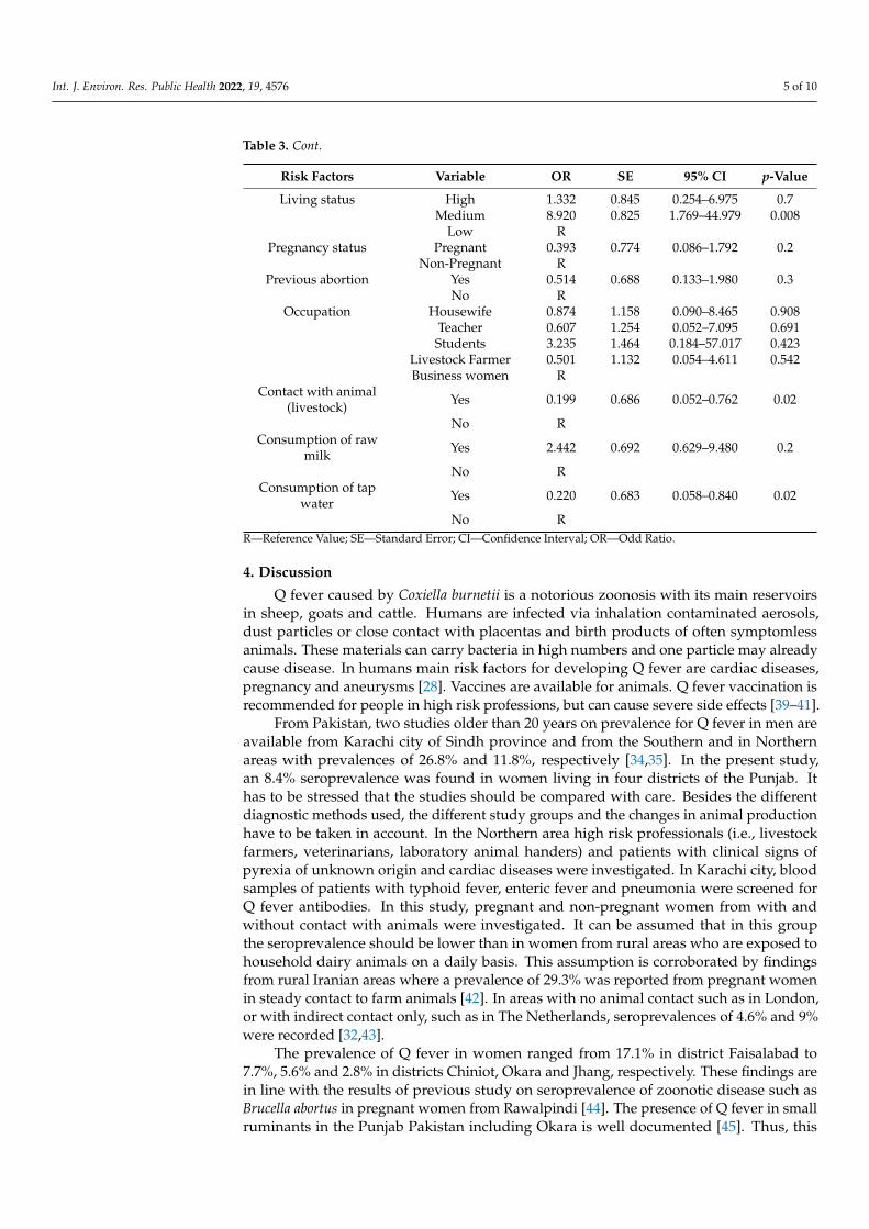
Based on binary logistic regression, being aged, urbanity, low-income status, contact withanimals and consumption of tap water are potential risk factors (p < 0.05) for the seropositivitywhile other risk factors were found non-significant for this setting (p > 0.05) (Table 3).
Table 3. Identification of Risk factors associated with Q fever based on seroprevalence in womenfrom four districts of Punjab, Pakistan, by using binary logistic regression.
Risk Factors Variable OR SE 95% CI p-Value
Age ≤20 1.908 0.829 0.376–9.697 0.43621–30 14.375 0.953 2.220–93.071 0.00531–40 12.255 0.912 2.051–73.221 0.006≥41 R
Location Okara 4.9 0.890 0.857–28.024 0.07Jhang 17.94 1.027 2.396–134.33 0.005
Chiniot 4.9 0.762 1.102–21.84 0.037Faisalabad R
Urbanicity Urban 7.002 0.696 1.788–27.415 0.005Rural R
Int. J. Environ. Res. Public Health 2022, 19, 4576 5 of 10
Table 3. Cont.
Risk Factors Variable OR SE 95% CI p-Value
Living status High 1.332 0.845 0.254–6.975 0.7Medium 8.920 0.825 1.769–44.979 0.008
Low RPregnancy status Pregnant 0.393 0.774 0.086–1.792 0.2
Non-Pregnant RPrevious abortion Yes 0.514 0.688 0.133–1.980 0.3
No ROccupation Housewife 0.874 1.158 0.090–8.465 0.908
Teacher 0.607 1.254 0.052–7.095 0.691Students 3.235 1.464 0.184–57.017 0.423
Livestock Farmer 0.501 1.132 0.054–4.611 0.542Business women R
Contact with animal(livestock) Yes 0.199 0.686 0.052–0.762 0.02
No RConsumption of raw
milk Yes 2.442 0.692 0.629–9.480 0.2
No RConsumption of tap
water Yes 0.220 0.683 0.058–0.840 0.02
No RR—Reference Value; SE—Standard Error; CI—Confidence Interval; OR—Odd Ratio.
4. Discussion
Q fever caused by Coxiella burnetii is a notorious zoonosis with its main reservoirsin sheep, goats and cattle. Humans are infected via inhalation contaminated aerosols,dust particles or close contact with placentas and birth products of often symptomlessanimals. These materials can carry bacteria in high numbers and one particle may alreadycause disease. In humans main risk factors for developing Q fever are cardiac diseases,pregnancy and aneurysms [28]. Vaccines are available for animals. Q fever vaccination isrecommended for people in high risk professions, but can cause severe side effects [39–41].
From Pakistan, two studies older than 20 years on prevalence for Q fever in men areavailable from Karachi city of Sindh province and from the Southern and in Northernareas with prevalences of 26.8% and 11.8%, respectively [34,35]. In the present study,an 8.4% seroprevalence was found in women living in four districts of the Punjab. Ithas to be stressed that the studies should be compared with care. Besides the differentdiagnostic methods used, the different study groups and the changes in animal productionhave to be taken in account. In the Northern area high risk professionals (i.e., livestockfarmers, veterinarians, laboratory animal handers) and patients with clinical signs ofpyrexia of unknown origin and cardiac diseases were investigated. In Karachi city, bloodsamples of patients with typhoid fever, enteric fever and pneumonia were screened forQ fever antibodies. In this study, pregnant and non-pregnant women from with andwithout contact with animals were investigated. It can be assumed that in this groupthe seroprevalence should be lower than in women from rural areas who are exposed tohousehold dairy animals on a daily basis. This assumption is corroborated by findingsfrom rural Iranian areas where a prevalence of 29.3% was reported from pregnant womenin steady contact to farm animals [42]. In areas with no animal contact such as in London,or with indirect contact only, such as in The Netherlands, seroprevalences of 4.6% and 9%were recorded [32,43].
The prevalence of Q fever in women ranged from 17.1% in district Faisalabad to7.7%, 5.6% and 2.8% in districts Chiniot, Okara and Jhang, respectively. These findings arein line with the results of previous study on seroprevalence of zoonotic disease such asBrucella abortus in pregnant women from Rawalpindi [44]. The presence of Q fever in smallruminants in the Punjab Pakistan including Okara is well documented [45]. Thus, this

Int. J. Environ. Res. Public Health 2022, 19, 4576 6 of 10
existing animal reservoir is expected source of the transmission to humans. The prevalenceof C. burnetti DNA in soil samples was successfully linked to seroconversion of smallruminants in a previous study conducted in different districts of the Punjab, includingFaisalabad [46]. The presence of Coxiella bacteria in the soil is considered a threat for thehuman population of that area [47].
In the selected study area, women from low-income families were more often seropos-itive (13.7%) than those from families with medium and high living standards (3.6–6.2%).Quite an opposite situation was observed in a previous study in which women from middlestandard families were significantly more often seropositive (54.0%) when compared towomen from low- and high-income families in Turkey [48]. The result of this study is inagreement with that of a study on brucellosis in which women from families with lowerincome had also a high risk of disease in Iran [49]. This might be due to the fact that bothdiseases (Q fever, brucellosis) are of zoonotic nature and poor women handling animalsusually have no adequate access to personal protection and medical care due to poverty.Hence, it is a well-known fact that zoonotic diseases are more prevalent in low-incomefamilies in general. Hence, the results from different countries should always be comparedwith care.
Coxiella burnetii can cause complications (i.e., premature birth, abortions, intrauterinegrowth retardation, etc.) in pregnant women and their unborn children. Thus, well-timeddiagnosis of disease can be important to avoid pregnancy associated complications [45].Our study revealed that prevalence of Q fever was higher (12.2%) in women who had ahistory of abortion as compared to those who did not (5.1%). These findings are compa-rable to the results of Abushahba et al., [50] who also described a higher seroprevalencein women who had an abortion history (32.2%). A much higher prevalence was alsoreported in women with abnormal pregnancy history (39.8%) and a lower one in womenwith normal pregnancy (23.8%) in Parsabad and Ahvaz regions of Iran [42]. Pregnantwomen may also be more prone to infection as we found a higher prevalence of Q fever inpregnant woman (11.6%) when compared with a non-pregnant woman (5.3%). Hence, thisassumption needs further research to be proven. However, antibodies against C. burnetiiduring pregnancy do not necessarily mean that C. burnetii is sole cause of reproductiveabnormalities. Infection with other pathogens associated with animal contacts such asbrucellae and Toxoplasma gondii can be possible confounders [49].
In previous studies conducted in Ilam province of Iran (37.93%) and in Egypt (46.15%)the seroprevalences of Q fever were found to be much higher in women with animalcontact than in this study [50,51]. Hence, the variable “contact with animals” was provedby binary logistic regression as a potential risk factor for Q fever (95% CI = 0.052–0.762,OR = 0.199, p = 0.02) in this study. The possible reason is that most women shared theirliving environments with animals. Moreover, poor farmers cannot afford to cull or disposediseased animals which are often then the source of the continuing spread of diseases.It is well known from many studies that the most frequent vehicle for transmission areaerosols caused by infected farm animals. C. burnetii can survive in the environment overlong periods and is transmitted by winds over long distances [10–12]. Thus, it poses apermanent threat for the human population of Punjab, Pakistan [44].
In this study not all seropositive women were rearing animals or had contact withanimals. However, occupational risk of infection had a great impact on the seroprevalenceof Q fever. Women working as livestock farmers had a higher seroprevalence (18.5%) thanothers, i.e., housewives (7.6%), teachers (5.3%), businesswomen (4.8%) or students (4.5%).Even in highly industrialized countries such as The Netherlands, 3.2% seroprevalence ofQ fever was observed in women of the agriculture sector [26] but only 0.5% women ofthe meat processing industry including abattoirs and 0.7% of the general population werepositive. The observed elevated seroprevalence in our study can possibly be linked to thetime of exposure, poor handling techniques and missing protective means while dealingwith infected animals.

Int. J. Environ. Res. Public Health 2022, 19, 4576 7 of 10
The prevalence of Q fever in this study was higher in women who did not consumeraw milk (9.3%) than in those who consumed raw milk (6.2%). In contrast, a significantrelationship was detected in consumption of raw milk (p < 0.05) and seropositivity for Qfever in Jordan [52]. Consumption of unpasteurized cheese and raw milk was related toQ fever with human disease in United Kingdom also. The local geographical situationmay have contributed to our findings, e.g., that herds were grazed hillside and main winddirection was directed towards near cities as transmission via consumption of contaminatedproducts is less likely than transmission via contaminated aerosols from infected livestockor their parturient products [9].
C. burnetti DNA was detected from sewage water and wastewater of Q-fever-positivegoat farms which may had been the source of environmental contamination in The Nether-lands [53]. Moreover, detection of C. burnetii in river water from Rome suggested thatcontaminated water can be a contributing factor to Q fever seropositivity in humans [54].A low level of risk was determined for transmission of C. burnetti via inhalation of drink-ing water aerosols during showering in a previous study [55]. Consequently, the role ofsome additional factors like contaminated tap water, raw vegetables and fruits as possiblesources for C. burnetti were investigated in this study. Indeed, a higher prevalence wasobserved in those women who consumed tap water (11.9%) when compared with thosewho took water from other sources (4.3%). Hence, these findings are not conclusive andneed further studies as tap water use simply may reflect poor living conditions of womenof the agriculture sector of Punjab.
Among different age groups, women > 40 years were more at risk (19.5%) to bearanti-Coxiella antibodies. These results are comparable to that of previous study conductedin Kurdistan, Iran [49]. Therefore, the chances of infection with pathogens or contactwith their antigens may increase with age because of the longer exposure times due totraditionally sharing of the living environments with animals. However, the present studyresults are not comparable with a study conducted in Switzerland in which the age group<15 years were at high risk of infection with Q fever [56].
Finally, phase-specific serology allows differentiation of an acute or recent Q feverinfection from chronic disease. During an acute infection, antibodies against phase IIantigens are predominant with IgG levels higher than IgM. Chronic Q fever is indicatedby a high IgG titer to phase I antigen [57]. There were used ELISA tests allowed onlyqualitative interpretation of the results for IgG against phase II and quantitative results forIgG against phase I. Therefore, positive serological results indicate exposure to C. burnetii,but does not allow differentiation of an acute, past or chronic infection. Additional testingfor IgM would have been beneficial, since IgM antibodies against phase II are detectableduring the acute phase of Q fever.
5. Conclusions
These data indicate that a zoonotic disease such as Q fever may become a serious threatto public health and especially to pregnant women, when there is a lack of control strategies.An educational program related to pregnancy and the risk of zoonotic diseases, and theirprevention is needed. For pregnant women with close contact to animals and signs of aninfectious disease, a routine serological examination covering zoonotic diseases includingQ fever should be made available in Pakistan. Moreover, there is a need for a more generalcontrol strategy for Q fever in animals involving the use of a vaccine to prevent spread of Qfever from animals to humans. Foodborne contamination of C. burnetii can be minimized bythe proper boiling of water and milk before human use in remote settings.
Author Contributions: Conceptualization, S.A., H.E-A., K.M.-S. and H.N.; Data curation, S.A., U.S.,M.R. and K.M.-S.; Formal analysis, S.A., U.S. and K.M.-S.; Methodology, S.A., U.S., M.R., H.E.-A.and H.N.; Supervision, K.M.-S. and H.N.; Writing—original draft, S.A., U.S., M.R., K.M.-S. and H.N.;Writing—review and editing, S.A., U.S., M.R., H.E.-A., K.M.-S. and H.N. All authors have read andagreed to the published version of the manuscript.
Int. J. Environ. Res. Public Health 2022, 19, 4576 8 of 10
Funding: This research was funded by German Federal Foreign Office, funded project “Building anetwork of laboratories in Pakistan to enhance biosafety and biosecurity in Pakistan” Grant Number:AA-OR12-370:43 BIOS FLIPAK.
Institutional Review Board Statement: The procedures of the present study were approved fromthe Institutional Committee for Biomedical Research, University of Veterinary and Animal Sciences,Lahore, Pakistan (No. 068/IRC/BMR). Informed consent was obtained from each participant beforeblood and data collection.
Informed Consent Statement: Informed consent was obtained from each participant before bloodand data collection.
Data Availability Statement: The data presented in this study are available within the article.
Acknowledgments: Shahzad Ali is thankful to “DAAD (German Academic Exchange Service) underResearch Stays for University Academics and Scientists, 2018 funding programme” for funding hisvisit to Friedrich Loeffler Institute, Jena, Germany for training and samples analysis.
Conflicts of Interest: The authors declare no conflict of interest.
References1. Watkins, K. A Review. Current Emergency and Hospital Medicine Reports. Emerg. Infect. Dis. 2018, 6, 86–93.2. Shaw, E.I.; Voth, D.E. Coxiella burnetii: A Pathogenic Intracellular Acidophile. Microbiology 2019, 165, 1–3. [CrossRef] [PubMed]3. Njeru, J.; Henning, K.; Pletz, M.W.; Heller, R.; Forstner, C.; Kariuki, S.; Fèvre, E.M.; Neubauer, H. Febrile patients admitted to
remote hospitals in Northeastern Kenya: Seroprevalence, risk factors and a clinical prediction tool for Q-Fever. BMC Infect. Dis.2016, 16, 244. [CrossRef] [PubMed]
4. Neare, K.; Janson, M.; Hütt, P.; Lassen, B.; Viltrop, A. Coxiella burnetii Antibody Prevalence and Risk Factors of Infection in theHuman Population of Estonia. Microorganisms 2019, 7, 629. [CrossRef]
5. Roest, H.J.; Gelderen, B.; Dinkla, A.; Frangoulidis, D.; Zijderveld, F.; Rebel, J.; Keulen, L. Q fever in pregnant goats: Pathogenesisand excretion of Coxiella burnetii. PLoS ONE 2012, 7, e48949. [CrossRef]
6. Roest, H.I.J.; Conny, B.; Solt, V.; Jeroen, J.H.C.; Tilburg, C.H.W.; Klaassen, E.K.; Frank, H.T.F.; Vellema, R.P.; Brom, R.V.D.;Zijderveld, F.G.V. Search for Possible Additional Reservoirs for Human Q Fever, the Netherlands. Emerg. Infect. Dis. 2013, 19,843–845. [CrossRef]
7. Eldin, C.; Melenotte, C.; Mediannikov, O.; Ghigo, E.; Million, M.; Edouard, S. From Q fever to Coxiella burnetii infection: Aparadigm change. Clin. Microbiol. Rev. 2017, 30, 115–190. [CrossRef]
8. Porter, S.R.; Czaplicki, G.; Mainil, J.; Guattéo, R.; Saegerman, C. Q Fever: Current state of knowledge and perspectives of researchof a neglected zoonosis. Int. J. Microbiol. 2011, 2011, 248418. [CrossRef]
9. Gale, P.; Kelly, L.; Mearns, R.; Duggan, J.; Snary, E.L. Q fever through consumption of unpasteurized milk and milk products—Arisk profile and exposure assessment. J. Appl. Microbiol. 2015, 118, 1083–1095. [CrossRef]
10. Boden, K.; Brasche, S.; Straube, E.; Bischof, W. Specific risk factors for contracting Q fever: Lessons from the outbreak Jena. Int. J.Hyg. Environ. Health 2014, 217, 110–115. [CrossRef]
11. Nusinovici, S.; Frössling, J.; Widgren, S.F.; Beaudeau, F.; Lindberg, A. Q fever infection in dairy cattle herds: Increased risk withhigh wind speed and low precipitation. Epidemiol. Infect. 2015, 143, 3316–3326. [CrossRef] [PubMed]
12. Heppell, C.W.; Egan, J.R.; Hall, I.A. Human time dose response model for Q fever. Epidemics 2017, 21, 30–38. [CrossRef]13. Dalton, H.R.; Dreier, J.; Rink, G.; Hecker, A.; Janetzko, K.; Juhl, D.; Bieback, K.; Steppat, D.; Görg, S.; Hennig, H.; et al. Coxiella
burnetii-pathogenic agent of Q (query) fever. Transfus. Med. Hemother. 2014, 41, 60–72.14. Mousapour, M.; Oveisi, A.; Key, Y.A.; Mikaeili, E.; Rahimi, F.; Shademan, B.; Bedoustani, A.B.; Fattahi, S.; Fasaei, M.S.;
Abbasnezhad, A.D.; et al. First Serological and Molecular Study of Coxiella burnetii in Stray, Domestic Cats, and Their Owners inIran. Top. Companion Anim. Med. 2020, 41, 100471. [CrossRef]
15. Shapiro, A.J.; Norris, J.M.; Heller, J.; Brown, G.; Malik, R.; Bosward, K.L. Seroprevalence of Coxiella burnetii in Australian dogs.Zoonoses Public Health 2016, 63, 458–466. [CrossRef] [PubMed]
16. Sanchez, M.; Valcarcel, F.; Gonzalez, J.; González, M.G.; Martin-Hernandez, R.; Tercero, J.M.; Gonzalez-Jara, P.; Olmeda, A.S.Seasonality of Coxiella burnetii among Wild Rabbits (Oryctolagus cuniculus) and the Hyalomma lusitanicum (Acari: Ixodidae) in aMeso-Mediterranean Ecosystem. Pathogens 2022, 11, 36. [CrossRef]
17. González-Barrio, D.; Ruiz-Fons, F. Coxiella burnetii in wild mammals: A systematic review. Transbound. Emerg. Dis. 2019, 66,662–671. [CrossRef]
18. Ebani, V.V.; Nardoni, S.; Giani, M.; Rocchigiani, G.; Archin, T.; Altomonte, I.; Poli, A.; Mancianti, F. Molecular survey on theoccurrence of avian haemosporidia, Coxiella burnetii and Francisella tularensis in waterfowl from central Italy. Int. J. Parasitol. ParasitesWildl. 2019, 10, 87–92. [CrossRef]

Int. J. Environ. Res. Public Health 2022, 19, 4576 9 of 10
19. Malo, J.A.; Colbran, C.; Young, M.; Vasant, B.; Jarvinen, K.; Viney, K.; Lambert, S.B. An outbreak of Q fever associated withparturient cat exposure at an animal refuge and veterinary clinic in southeast Queensland. Aust. N. Z. J. Public Health 2018, 42,451–455. [CrossRef]
20. Maurin, M.; Raoult, D. Q fever. Clin. Microbiol. Rev. 1999, 12, 518–553. [CrossRef]21. Raoult, D.; Marrie, T.J.; Mege, J.L. Natural history and pathophysiology of Q fever. Lancet Infect. Dis. 2005, 5, 219–226. [CrossRef]22. Parker, N.R.; Barralet, J.H.; Bell, A.M. Q fever. Lancet 2006, 367, 679–688. [CrossRef]23. Wielders, C.C.H.; Wuister, A.M.H.; Visser, V.L.; Jager-Leclercq, M.G.; Groot, C.A.R. Characteristics of Hospitalized Acute Q Fever
Patients during a Large Epidemic, The Netherlands. PLoS ONE 2014, 9, e91764. [CrossRef]24. Tissot-Dupont, H.; Raoult, D.; Brouqui, P.; Janbon, F.; Peyramond, D. Epidemiologic features and clinical presentation of acute Q
fever in hospitalized patients: 323 French cases. Am. J. Med. 1992, 93, 427–434. [CrossRef]25. Raoult, D.; Tissot-Dupont, H.; Foucault, C.; Gouvernet, J.; Fournier, P.E. Q fever 1985–1998. Clinical and epidemiologic features of
1383 infections. Medicine 2000, 79, 109–123. [CrossRef]26. Dijkstra, F.; Van der Hoek, W.; Wijers, N.; Schimmer, B.; Rietveld, A.; Wijkmans, C.J.; Vellema, P.; Schneeberger, P.M. The 2007–2010
Q fever epidemic in The Netherlands: Characteristics of notified acute Q fever patients and the association with dairy goatfarming. FEMS Immunol. Med. Microbiol. 2012, 64, 3–12. [CrossRef] [PubMed]
27. Hoek, V.D.W.; Morroy, G.; Renders, N.H.M.; Wever, P.C.; Hermans, M.H.A. Epidemic Q fever in humans in the Netherlands. Adv.Exp. Med. Biol. 2012, 984, 329–364.
28. Kampschreur, L.M.; Dekker, S.; Hagenaars, J.C.; Lestrade, P.J.; Renders, N.H.; Jager-Leclercq, M.G.; Hermans, M.H.; Groot, C.A.;Groenwold, R.H.; Hoepelman, A.I.; et al. Identification of risk factors for chronic Q fever, the Netherlands. Emerg. Infect. Dis.2012, 18, 563–570. [CrossRef]
29. Wegdam-Blans, M.C.A.; Kampschreur, L.M.; Delsing, C.E.; Bleeker-Rovers, C.P.; Sprong, T. Chronic Q fever: Review of theliterature and a proposal of new diagnostic Criteria. J. Infect. 2012, 64, 247–259. [CrossRef]
30. Langley, J.M.; Marrie, T.J.; Leblanc, J.C.; Almudevar, A.; Resch, L.; Raoult, D. Coxiella burnetii seropositivity in parturient womenis associated with adverse pregnancy outcomes. Am. J. Obstet. Gynecol. 2003, 189, 228–232. [CrossRef]
31. Million, M.; Roblet, F.; Carles, D.; D’Amato, F.; Protopopescu, C.; Carrieri, M.P. Reevaluation of the risk of fetal death andmalformation after Q fever. Clin. Infect. Dis. 2014, 59, 256–260. [CrossRef] [PubMed]
32. Hoek, V.D.W.; Meekelenkamp, J.C.; Leenders, A.C.; Wijers, N.; Notermans, D.W.; Hukkelhowen, C.W. Antibodies against Coxiellaburnetii and pregnancy outcomes during the 2007–2008 Q fever outbreaks in The Netherlands. BMC Infect. Dis. 2011, 11, 44.
33. Mboussou, Y.; Jaubert, J.; Larrieu, S.; Atiana, L.; Naze, F.; Folio, C.; Randrianaivo, H.; Bertolotti, A.; Picot, S.; Robillard, P.Y.; et al.Pregnancy outcomes of Q fever: Prospective follow-up study on Reunion island. BMC Infect. Dis. 2019, 19, 1001. [CrossRef][PubMed]
34. Ayaz, M.; Bari, A.; Humayun, A. Coxiellosis in man and animals in northern parts of Pakistan. Proc. Pak. Congr. Zool. 1993, 13,425–431.
35. Ahmad, I.P. A serological investigation of Q fever in Pakistan. J. Pak. Med. Assoc. 1987, 37, 126–129.36. Madariaga, M.G.; Rezai, K.; Trenholme, G.M.; Weinstein, R.A. Q fever: A biological weapon in your backyard. Lancet Infect. Dis.
2003, 3, 709–721. [CrossRef]37. Angelakis, E.; Raoult, D. Q fever. Vet. Microbiol. 2010, 140, 297–309. [CrossRef]38. Duron, O.; Sidi-Boumedine, K.; Rousset, E.; Moutailler, S.; Jourdain, E. The importance of ticks in Q fever transmission: What has
(and has not) been demonstrated. Trends Parasitol. 2015, 31, 36–52. [CrossRef]39. Khameneie, M.K.; Asadi, J.; Khalili, M.; Abiri, Z. The first serological study of Coxiella burnetii among pregnant women in Iran.
Iran. J. Public Health 2016, 45, 523–530.40. Ghobadi, E.A.; Jaydari, A.; Akbari, S.; Anbari, K. First Seroprevalence Study of Coxiella burnetii in Rural Pregnant Women in
Contact with Livestock in Khorramabad. Int. J. Infect. 2019, 6, 1–5.41. Schimmer, B.; Schegget, T.R.; Wegdam, M. The use of a geographic information system to identify a dairy goat farm as the most
likely source of an urban Q-fever outbreak. BMC Infect. Dis. 2010, 10, 69. [CrossRef] [PubMed]42. Baud, D.; Peter, O.; Langel, C.; Regan, L.; Greub, G. Seroprevalence of Coxiella burnetii and Brucella abortus among pregnant
women. Clin. Microbiol. Infect. 2009, 15, 499–501. [CrossRef] [PubMed]43. De Lange, M.M.; Hukkelhoven, C.W.P.M.; Munster, J.M.; Schneeberger, P.M.; Van der Hoek, W. Nationwide registry-based
ecological analysis of Q fever incidence and pregnancy outcome during an outbreak in the Netherlands. BMJ Open 2015, 5,e006821. [CrossRef] [PubMed]
44. Ali, S.; Shamim, A.; Neubauer, H.; Scherag, A.; Kesselmeier, M.; Melzer, F.; Khan, I.; Adawy, H.E.; Azam, A.; Qadeer, S.; et al.Brucellosis in pregnant women from Pakistan: An observational study. BMC Infect. Dis. 2016, 16, 468. [CrossRef]
45. Ullah, Q.; Adawy, H.E.; Jamil, T.; Jamil, H.; Qureshi, Z.I.; Saqib, M.; Ullah, S.; Shah, M.K.; Khan, A.Z.; Zubair, M.; et al. Serologicaland molecular investigation of C. burnetti in small ruminants and ticks in Punjab, Pakistan. Int. J. Environ. Res. Public Health 2019,16, 4271. [CrossRef]
46. Shabbir, M.Z.; Akram, S.; Hassan, Z.U.; Hanif, K.; Rabbani, M.; Muhammad, J.; Chaudhary, M.H.; Abbas, T.; Ghori, M.T.; Rashid,H.; et al. Evidence of Coxiella burnetii in Punjab province, Pakistan. Acta Trop. 2016, 163, 61–69. [CrossRef]
47. Quijada, S.G.; Teran, B.M.; Murias, P.S.; Anitua, A.A.; Cermeno, J.L.B.; Frıas, A.B. Q fever and spontaneous abortion. Clin.Microbiol. Infect. 2012, 18, 533–538. [CrossRef]

Int. J. Environ. Res. Public Health 2022, 19, 4576 10 of 10
48. Arserim, N.B.; Yesilmen, S.; Tel, O.Y.; Ozekinci, T.; Keskin, O.; Pulat, H.; Vural, A. Seroprevalence of Coxiellosis in cows, sheep,goats and humans in Diyarbakir region of Turkey. Afr. J. Mirobiol. Res. 2011, 5, 2041–2043.
49. Esmaeili, S.; Pourhossein, B.; Gouya, M.M.; Amiri, F.B.; Mostafavi, E. Seroepidemiological survey of Q fever and brucellosis inKurdistan province, western Iran. Vector-Borne Zoonotic Dis. 2014, 14, 41–45. [CrossRef]
50. Abushahba, M.F.N.; Abdelbaset, A.E.; Rawy, M.S.; Ahmed, S.O. Cross sectional study for determining the prevalence of Q feverin small ruminants and humans at El Minya Governorate, Egypt. BMC Res. Notes 2017, 10, 538. [CrossRef]
51. Mostafavi, E.; Molaeipoor, L.; Esmaeili, S.; Ghasemi, A.; Kamalizad, M.; Behzadi, M.Y.; Naserifar, R.; Rohani, M.; Shahraki, A.H.Seroprevalence of Q fever among high-risk occupations in the Ilam province, the west of Iran. PLoS ONE 2019, 14, e0211781.[CrossRef] [PubMed]
52. Obaidat, M.M.; Malania, L.; Imnadze, P.; Roess, A.A.; Salman, A.E.B.; Arner, R.J. Seroprevalence and Risk Factors for Coxiellaburnetii in Jordan. Am. Soc. Trop. Med. Hyg. 2019, 101, 40–44. [CrossRef] [PubMed]
53. Schets, F.M.; Heer, L.D.A.M.; Husman, D.R. Coxiella burnetii in sewage water at sewage water treatment plants in a Q feverepidemic area. Int. J. Hyg. Environ. Health 2013, 216, 698–702. [CrossRef] [PubMed]
54. D’Ugo, E.; Sdanganelli, M.; Grasso, C.; Magurano, F.; Marcheggiani, S.; Boots, B.; Baggieri, M.; Mancini, L. Detection of Coxiellaburnetii in Urban River Water. Vector-Borne Zoonotic Dis. 2017, 17, 514–516. [CrossRef] [PubMed]
55. Ortells, S.H.; Medema, G. Screening-Level Risk Assessment of Coxiella burnetii (Q fever) Transmission via Aeration of DrinkingWater. Environ. Sci. Technol. 2012, 46, 4125–4133. [CrossRef] [PubMed]
56. Dupuis, G.; Vouilloz, M.; Peter, O.; Mottiez, M. Incidence of Q fever in Valais. Rev. Méd. Suisse Romande 1985, 105, 949–954.57. Dupont, H.T.; Thirion, X.; Raoult, D. Q fever serology: Cutoff determination for microimmunofluorescence. Clin. Diagn. Lab.
Immunol. 1994, 1, 189–196. [CrossRef]

![[Serological survey on the prevalence of arboviruses in man in forest and periforest environments of the region of Lobaye (Central African Republic)]](https://img.dokumen.tips/doc/110x75/634c52401983efcda6055bc0/serological-survey-on-the-prevalence-of-arboviruses-in-man-in-forest-and-periforest.jpg)


