Embed Size (px)
Citation preview
Serine-71 Phosphorylation of Rac1 ModulatesDownstream SignalingJanett Schwarz1, Julia Proff1, Anika Havemeier2, Markus Ladwein3, Klemens Rottner3,4, Britta Barlag1,
Andreas Pich1, Helma Tatge1, Ingo Just1, Ralf Gerhard1*
1 Department of Toxicology, Hannover Medical School, Hannover, Germany, 2 Department of Virology, Hannover Medical School, Hannover, Germany, 3 Cytoskeleton
Dynamics Group, Helmholtz Centre for Infection Research, Braunschweig, Germany, 4 Institut fur Genetik, Rheinische Friederich-Wilhelms-Universitat, Bonn, Germany
Abstract
The Rho GTPases Rac1 and Cdc42 regulate a variety of cellular functions by signaling to different signal pathways. It isbelieved that the presence of a specific effector at the location of GTPase activation determines the route of downstreamsignaling. We previously reported about EGF-induced Ser-71 phosphorylation of Rac1/Cdc42. By using the phosphomimeticS71E-mutants of Rac1 and Cdc42 we investigated the impact of Ser-71 phosphorylation on binding to selected effectorproteins. Binding of the constitutively active (Q61L) variants of Rac1 and Cdc42 to their specific interaction partners Sra-1and N-WASP, respectively, as well as to their common effector protein PAK was abrogated when Ser-71 was exchanged toglutamate as phosphomimetic substitution. Interaction with their common effector proteins IQGAP1/2/3 or MRCK alphawas, however, hardly affected. This ambivalent behaviour was obvious in functional assays. In contrast to Rac1 Q61L,phosphomimetic Rac1 Q61L/S71E was not able to induce increased membrane ruffling. Instead, Rac1 Q61L/S71E allowedfilopodia formation, which is in accordance with abrogation of the dominant Sra-1/Wave signalling pathway. In addition, incontrast to Rac1 transfected cells Rac1 S71E failed to activate PAK1/2. On the other hand, Rac1 Q61L/S71E was as effective inactivation of NF-kappaB as Rac1 Q61L, illustrating positive signal transduction of phosphorylated Rac1. Together, these datasuggest that phosphorylation of Rac1 and Cdc42 at serine-71 represents a reversible mechanism to shift specificity ofGTPase/effector coupling, and to preferentially address selected downstream pathways.
Citation: Schwarz J, Proff J, Havemeier A, Ladwein M, Rottner K, et al. (2012) Serine-71 Phosphorylation of Rac1 Modulates Downstream Signaling. PLoS ONE 7(9):e44358. doi:10.1371/journal.pone.0044358
Editor: Robert Alan Arkowitz, Institute of Developmental Biology and Cancer Research, France
Received May 16, 2011; Accepted August 3, 2012; Published September 10, 2012
Copyright: � 2012 Schwarz et al. This is an open-access article distributed under the terms of the Creative Commons Attribution License, which permitsunrestricted use, distribution, and reproduction in any medium, provided the original author and source are credited.
Funding: This study was supported in part by the Deutsche Forschungsgemeinschaft, SFB621, project B5. The funders had no role in study design, datacollection and analysis, decision to publish, or preparation of the manuscript.
Competing Interests: The authors have declared that no competing interests exist.
* E-mail: [email protected]
Introduction
The small Rho GTPases are monomeric GTP-binding proteins
that play a role in a variety of cellular processes that depend on the
actin cytoskeleton, such as morphogenesis, endocytosis, phagocy-
tosis, cytokinesis and migration. They act as nucleotide-dependent
switches cycling between an active, GTP-bound state and an
inactive, GDP-bound state. In their GTP-bound state, they
interact with downstream effectors to initiate downstream
signaling. The binding of Rho GTPases to their effector proteins
occurs by distinct binding motifs. Most effector proteins harbor the
common Cdc42/Rac1 interactive binding motif (CRIB), which is
present in the p21-activated kinase (PAK) and the Wiskott-Aldrich
syndrome protein (WASP) [1]. The myotonic dystrophy related
Cdc42 kinase (MRCK) also contains a CRIB-related binding
domain (PBD) [2]. In addition Rac1/Cdc42 bind to the RasGAP-
homology domain which is present in IQGAP [3]. The specifically
Rac1-associated protein (Sra-1), a subunit of the pentameric
WAVE complex, does not harbor a CRIB-motif and can directly
bind to Rac1 but not to Cdc42 [4]. Each of these effectors
contributes to the cytoskeletal reorganization downstream of Rac1
and Cdc42, driving the formation of a diverse array of actin
structures, e. g. membrane ruffles and lamellipodia. Formation of
these structures is stimulated by Rac1, whereas Cdc42 induces the
formation of filopodia and microspikes [5]. Additionally, Rho
GTPases influence gene expression by regulating signaling
pathways involving the transcription factor NF-kB, c-Jun N-
terminal kinase (JNK), and p38 mitogen-activated protein kinase,
and they drive G1 cell cycle progression, apoptosis and cell
transformation. Activation of Rho GTPases is regulated by three
types of proteins: The guanine nucleotide dissociation inhibitors
(GDIs), the guanine nucleotide exchange factors (GEFs), and
GTPase activating proteins (GAPs) [6]. The GDIs stabilize the
inactive, GDP-bound form of the Rho GTPases in the cytosol
(Rho-GDI complex) and thus prevent association with the
membrane [7]. The GEFs catalyze the exchange of GDP for
GTP, which is thought to be coordinated with membrane
targeting of Rho GTPases [8]. The GAPs stimulate the intrinsic
GTPase activity and convert the GTP-bound form of Rho
GTPases to the inactive, GDP-bound form [9].
Phosphorylation of Rho GTPases is an additional mechanism to
modulate the activity of these proteins, mainly leading to their
functional inactivation. Phosphorylation was first described for
RhoA, which can be phosphorylated by PKA/PKG at Ser-188
[10–12] resulting in cytosolic relocalization due to an increased
binding to Rho-GDI. Cdc42 also harbors a PKA phosphorylation
site at Ser-185 [11]. Additionally, EGF treatment of cells can
induce the phosphorylation of Cdc42 at tyrosine-64 by Src kinase.
PLOS ONE | www.plosone.org 1 September 2012 | Volume 7 | Issue 9 | e44358
This specific phosphorylation does not affect interaction with
effector proteins but leads to increased interaction with Rho-GDI
[13]. Very recently, the same was reported for Rac1, where
phosphorylation of Tyr-64 affects interaction with PAK and cell
spreading as shown by transfection experiments with the
phosphomimetic mutant Rac1 Y64D [14]. Rac1 can also be
phosphorylated by the Akt kinase at Ser-71, which is embedded in
the consensus sequence 64ydRIRplSYp73. Most Rho GTPases, e. g.
Cdc42 and RhoA/B/C/G share this sequence. The Ser-71-
phosphorylation of Rac1 results in reduced GTP-binding without
affecting GTPase activity [15–16]. Ser-71-phosphorylated Rac1/
Cdc42 appear to be in their active conformation according to pull
down assays with PAK CRIB-domain and Rho-GDI. RhoE can
be phosphorylated by ROCK I at Ser-11 resulting in the cytosolic
relocalization and an increased stability of the GTPase [17]. In
summary, phosphorylation is not the key event in activation/
inactivation of Rho GTPases, but it predominantly modulates
affinity to GDI and subcellular localization of the GTPase, thereby
negatively affecting its activity. In the present study we show that
phosphorylation of Rac1 and Cdc42 at Ser-71, in addition,
modulates downstream signaling by inhibiting interaction with
some effectors but allowing interaction with others. This is the first
description of the functional regulation of effector coupling by
phosphorylation of Rac1/Cdc42 at Ser-71 as an additional
mechanism to specify downstream signaling of the activated
GTPases.
Results
Ser-71-Phosphorylation of Rac1 Induces a Cdc42-likePhenotype
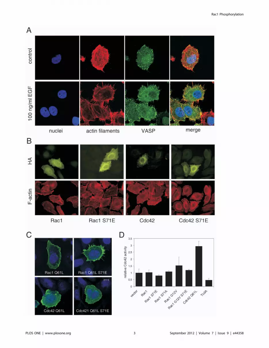
Phosphorylation of Rac1/Cdc42 induces a specific phenotype
of cells. Treatment of cells with the epidermal growth factor (EGF)
induces Rac1/Cdc42 phosphorylation which is accompanied by
the formation of filopodia [16]. EGF-induced filopodia formation
is illustrated in Fig. 1A, showing staining of the actin cytoskeleton
and the localization of the vasodilator-stimulated phosphoprotein
(VASP). VASP was visualized as marker for filopodia [18] to
dissect these structures from retraction fibers. Morphological
effects of Rac1 and Cdc42 as well as their S71E mutants are
shown in Fig. 1B. Only cells transfected with Rac1 S71E showed
increased formation of filopodia, whereas Rac1, Cdc42, and
Cdc42S71E transfected cells showed phenotype of non-transfected
controls. The effect was also obvious when Rac1 S71E with
constitutive active (Q61L) background was used. As shown in
Fig. 1C, Rac1 Q61L induced formation membrane ruffles,
whereas Rac1 S71E Q61L strongly induced formation of
filopodia. Rac1 S71E Q61L-induced filopodia were compared
with those induced by constitutive active Cdc42 (Q61L).
Formation of filopodia by Cdc42 Q61L was less pronounced
than in Rac1 Q61L/S71E transfected cells. The phenotype of cells
expressing the double mutant Cdc42 Q61L/S71E corresponded
to the Cdc42 Q61L phenotype with more microspike like
structures. It is of importance to investigate morphological effects
of the Q61L mutants of Rac1/Rac1 S71E and Cdc42/Cdc42
S71E because these mutants were used later on for pull down
experiments to characterize effector coupling.
To investigate the mechanism leading to filopodia formation in
more detail, we checked whether overexpression of Rac1 S71
somehow induces activation of Cdc42. Neither overexpression of
wildtype Rac1 nor of Rac1 S71E nor the control mutant Rac1
S71A induced activation of Cdc42 (Fig. 1D). The constitutive
active mutant Rac1 G12V showed weak but not significant effect
on Cdc42 activation. In Rac1 G12V/S71E transfected cells,
however, no sign of Cdc42 activation was observed at all.
Transfection experiments with Cdc42 Q61L were performed as
positive control for experimental setup. C. difficile TcdA, which
catalyzes inactivation of Rho GTPases including Cdc42 by mono-
glucosylation served as negative control. These results indicate that
the Rac1 S71E-induced filopodial phenotype is not caused by
concomitant activation of Cdc42. It is well established that
filopodia formation can be enforced by suppression of Rac1
signaling, either directly through microinjection of mixtures of
active Cdc42 and inactive Rac1 [5], or more indirectly when
interfering with Rac effector function [19]. We suppose this
principal as reason for the Rac1 S71E-induced changes in cell
morphology.
Phosphorylation of Rac1 and Cdc42 at Ser-71 ModulatesEffector Binding
Unfortunately, it is not possible to dissect between Ser-71
phosphorylation of Rac1 and Cdc42. To date there is no antibody
that differentiates between pSer-71-Rac1 and pSer-71-Cdc42. We
therefore took advantage of murine fibroblasts lacking the Rac1
gene (rac12/2) to check for pSer-71 staining of Rac1 and Cdc42.
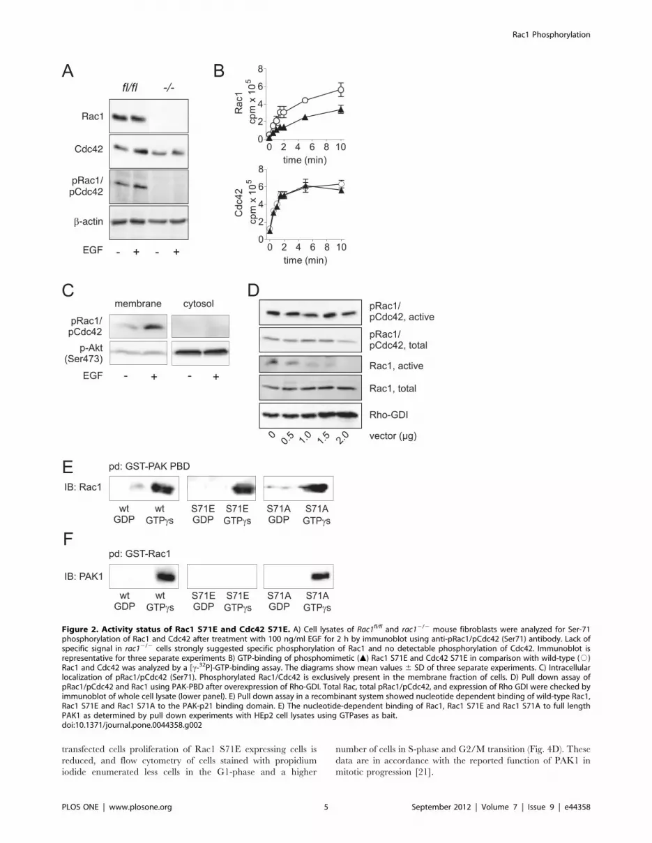
As shown in Fig. 2A, rac12/2 fibroblasts do not express detectable
levels of Rac1. Cdc42 expression, however, was comparable to the
parental cell line rac1fl/fl fibroblasts. Beta-actin served as loading
control. In contrast to control fibroblasts, Rac1-deficient cells were
completely devoid of pSer-71-Rac1 and pSer-71-Cdc42 staining,
even after treatment of cells with 100 ng/ml EGF. This strongly
argues that EGF mainly drives phosphorylation of Rac1, at least in
fibroblasts. With respect to this finding, we assumed that the
observed effects in the present study are mainly due to Rac1
phosphorylation.
To better understand the outcome of Ser-71 phosphorylation at
the molecular level, we explored nucleotide-binding of the wild
type forms of Rac1 and Cdc42 in comparison with their
phosphomimetic mutants (Fig. 2B). GTP-binding of the phospho-
mimetic Rac1 mutant (S71E) was markedly reduced compared to
wild-type Rac1 (upper panel). This data is in line with results
published by Kwon et al [15], who also found reduced GTP-
binding of phosphorylated Rac1. We previously reported that
Rac1 S71E is able to bind GTP [16], and it is noteworthy that
GTP-binding of the phosphomimetic mutant was only reduced to
some extent, but not abolished. This finding is in accordance with
previously reported pull down assays showing active conformation
of Ser-71 phosphorylated Rac1. Mutation of serine to alanine
(S71A) did not alter the GTP-binding, indicating a specific effect of
the phosphomimetic mutation (data not shown). In contrast to
Rac1, no difference in GTP-binding between Cdc42 wild-type and
phosphomimetic Cdc42 was observed (Fig.2B, lower panel). We
also provided indirect evidence of active state of Ser-71
phosphorylated Rac1: Immunoblot analyses showed that consti-
tutive and EGF-induced pRac1/pCdc42 exclusively locates at the
membranes (,100,0006g fraction) of HEp2 cells (Fig. 2C). No
signal was detected within the cytosol (.100,0006g fraction). The
majority of activated Akt kinase was detected within the cytosol
but also to some extend within the membrane fraction.
Furthermore, Overexpression of Rho-GDI in HEp2 cells did not
reduce level of active pRac1 as tested in pull down assay with PAK
p21 binding domain (PAK-PBD) (Fig. 2D). In contrast, non-
phosphorylated active Rac1 was reduced with increasing amount
of overexpressed Rho-GDI. Previous studies showed binding of
pRac1 (S71) to the PAK-PBD [16]. We thus initially analyzed
binding of Rac1 S71E to PAK1 to show specificity of precipitation
experiments. By using immobilized PAK1-PBD as bait in a
recombinant system we were able to show specific and nucleotide-
Rac1 Phosphorylation
PLOS ONE | www.plosone.org 2 September 2012 | Volume 7 | Issue 9 | e44358
Rac1 Phosphorylation
PLOS ONE | www.plosone.org 3 September 2012 | Volume 7 | Issue 9 | e44358
dependent interaction with Rac1 and Rac1 S71E (Fig. 2E).
Unspecific binding of Rac1 S71E was excluded by comparison
with an alternative mutant, where S71 was exchanged to alanine
(S71A). Rac1 S71A showed identical binding to PAK-PBD as
wild-type Rac1. When performing the contrary experiment, where
Rac1, Rac1 S71E and Rac1 S71A were used as bait, surprisingly
only wild-type and the control mutant S71A of Rac1 were able to
bind and to precipitate full length PAK1 from HEp2 cell lysates
(Fig. 2F). Rac1 S71E did not interact with PAK1, neither in its
active (GTPcS-loaded) nor inactive (GDP-bound) form. This is
interesting because similar results were published by Matos et al,
who showed that Rac1b, a splice variant of Rac1 with 19
additional amino acids following switch 2 region, also interacts
with the PAK-PBD domain but not with full length PAK1 [20].
This result emphasizes the importance of the switch 2 region for
effector interaction.
Based on these findings, we performed further pull down
experiments from HEp2 cell lysates where GST-fusion proteins of
wild type and mutant (S71E) Rac1 and Cdc42 (data not shown)
were used as baits to identify possible interacting proteins.
Figure 3A shows a Coomassie-stained SDS-gel of precipitates
from HEp2 cell lysates where the nucleotide-dependent binding of
interacting proteins was analyzed. An approximately 190 kDa-
sized band was visible that showed increased binding to GTP-
loaded Rac1 and Rac1 S71E. Constitutively active Rac1 Q61L
was additionally used as positive control. Mass spectrometry
analysis identified IQGAP1 as interacting protein within this range
of about 190 kDa. Since it was not clear whether the prominent
coomassie-stained band definitely reflects IQGAP, all further
precipitates were analyzed by immunoblots to specifically detect
IQGAP and additional effector proteins such as PAK1, N-WASP,
Sra-1 and MRCK alpha.
Therefore, the specificity of binding to effector proteins was
checked by using GTPcS-loaded wild-type GTPases. Additionally,
constitutive active (Q61L) mutants of Rac1 and Cdc42 were
compared with respect to their specificity. Comparison of GTPcS
loaded GTPases with constitutive active ones was done to evaluate
the Q61L mutants for further experiments. In fact, GTPcS-loaded
Rac1 and the constitutively active (Q61L) mutant interacted with
the Rac1-specific effector protein Sra-1 but not with the Cdc42-
specific effector N-WASP (Fig. 3B). In contrast, active Cdc42
interacted with N-WASP but not with Sra-1. Both GTPases bound
to their common effectors PAK1, IQGAP1 and 2 and the alpha
form of MRCK. Unspecific precipitation of effectors was excluded
by using GST-bound glutathione beads as control. The constitu-
tively active (Q61L) mutants of Rac1 S71E and Cdc42 S71E were
further on used for analysis of effector interaction in comparison
with wild-type GTPases. The Q61L/S71E double mutants of
Rac1 and Cdc42 were chosen to overcome incomplete GTPcS-
loading of either GTPase. Double mutants allowed semi-quanti-
fication of the phosphomimetic mutants in comparison with the
constitutively active wild-type GTPases and warranted exclusion
of false negative results.
Pull down experiments with constitutively active GTPases
were performed in triplicate. Figure 3C shows representative
input controls of GST-GTPases and Fig. 3D shows represen-
tative immunoblots of precipitates from pull downs: The
phosphorylation of Rac1 and Cdc42 at serine-71 as represented
by the S71E mutants fully abrogated binding of Cdc42 to N-
WASP (the Cdc42 effector), of Rac1 to Sra-1 (the Rac1 effector)
and of both to their common effector PAK 1. The interaction
with the isoform 1 and 2 of IQGAP and the alpha isoform of
MRCK was only reduced to some extent or even unchanged.
Since cell lysates were adjusted to same protein concentrations,
MRCK also serves as loading control. Densitometrical evalua-
tion of immunoblots (Fig. 3D left panel) revealed that binding of
Rac1 Q61L/S71E to IQGAP1, IQGAP2 and MRCK alpha
was reduced to about 60–70% of Rac1 Q61L binding. Cdc42
S71E showed an approximately 30% reduced binding to both
IQGAP isoforms. The interaction of Cdc42 Q61L with MRCK
alpha was not affected by exchange of Ser-71 to Glu-71. In
conclusion, these data clearly show that phosphorylation of
Rac1 and Cdc42 interferes with the binding to their GTPase-
specific effectors Sra-1 and N-WASP as well as to their
common effector PAK1. In contrast, the binding to IQGAP1/
2 and MRCK alpha is only reduced or not even affected. The
effect of the serine-71 phosphorylation on the binding to their
effectors was slightly different between both GTPases. Whereas
Rac1 showed the strongest binding with IQGAP2, Cdc42
showed strongest interaction with MRCK alpha. With respect
to the consequences on the molecular level phosphorylation on
Ser-71 of each GTPase seems to minimize differences of effector
binding between Rac1 and Cdc42.
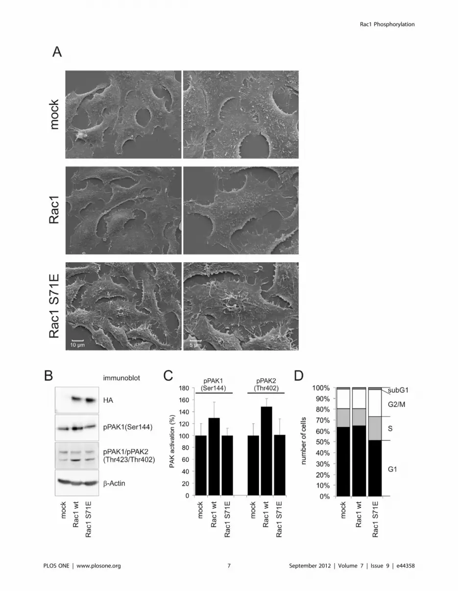
Phosphorylation-induced Loss of Function of Rac1Results shown in figure 2 and 3 show, that Ser-71
phosphorylation interfered with binding of Rac1 to full length
PAK1. We performed immunoblot analyses to check PAK
activation by Rac1 and Rac1 S71E and possible functional
consequences. To ensure homogenous expression of GTPases we
generated stable transfected HEp2 cells expressing Rac1 and
Rac1 S71E. A brief characterization of these cell lines is shown
in figure 4. Scanning electron microscopy graphs reveal different
surface topology of Rac1 S71E expressing cells compared with
mock transfected cells or cells expressing wild type Rac1
(Fig. 4A). Representative figures show filopodia like protrusions
in Rac1 S71E cells whereas cell surface of Rac1 cells is as smooth
as in mock transfected cells. Expression of HA-tagged GTPases is
shown in immunoblot (Fig. 4B). The Rac1 expressing cell line
showed increased Ser-144 phosphorylation of PAK1 and Thr-
402 phosphorylation of PAK2. In contrast, expression of Rac1
S71E failed to induce PAK1 or PAK2 phosphorylation.
Densitometrical evaluation of three separate immunoblots is
shown in Fig.4C. These data are in line with the precipitation
experiment where Rac1 S71E was shown not to interact with full
length PAK1. Additionally, compared to mock or wild type Rac1
Figure 1. Phosphomimetic Rac1 S71E induces filopodia formation. A) Treatment with 100 ng/ml EGF for 2 h induces pronounced formationof filopodia. Cells were stained for nuclei (DAPI, blue), actin cytoskeleton (rhodamin-phalloidin, red), and VASP (Alexa-488, green). B) HEp2 cellstransfected with HA-tagged Rac1, Rac1 S71E, Cdc42, and Cdc42 S71E. Expression of GTPases was visualized by HA-staining, the actin cytoskeletonwas stained with rhodamin-phalloidin. Only Rac1 S71E induced morphotype that is comparable with EGF-induced alterations. C) Phenotypes of HEp2cells transfected with HA-tagged constitutive active mutants of Rac1 and Cdc42 as well as their phosphomimetic mutants S71E. Constitutively active(Q61L) Rac1 induced membrane ruffling whereas Rac1 S71E induced formation of filopodia. Filopodia formation is less pronounced in Cdc42 Q61Land Cdc42 Q61L/S71E transfected cells. Stained are nuclei (blue) and HA-tag (green); bar represents 10 mm. D) Active, GTP-bound form of Cdc42 wasdetermined by G-LISA 24 h post transfection with constructs as indicated. Cdc42 Q61L was used for transfection experiments as positive control forexperimental setup. Additionally, C. difficile toxin A (TcdA) was used as negative control for inactivation of Cdc42. The bar chart shows mean values 6
SD of three (for TcdA) or four separate experiments.doi:10.1371/journal.pone.0044358.g001
Rac1 Phosphorylation
PLOS ONE | www.plosone.org 4 September 2012 | Volume 7 | Issue 9 | e44358
transfected cells proliferation of Rac1 S71E expressing cells is
reduced, and flow cytometry of cells stained with propidium
iodide enumerated less cells in the G1-phase and a higher
number of cells in S-phase and G2/M transition (Fig. 4D). These
data are in accordance with the reported function of PAK1 in
mitotic progression [21].
Figure 2. Activity status of Rac1 S71E and Cdc42 S71E. A) Cell lysates of Rac1fl/fl and rac12/2 mouse fibroblasts were analyzed for Ser-71phosphorylation of Rac1 and Cdc42 after treatment with 100 ng/ml EGF for 2 h by immunoblot using anti-pRac1/pCdc42 (Ser71) antibody. Lack ofspecific signal in rac12/2 cells strongly suggested specific phosphorylation of Rac1 and no detectable phosphorylation of Cdc42. Immunoblot isrepresentative for three separate experiments B) GTP-binding of phosphomimetic (m) Rac1 S71E and Cdc42 S71E in comparison with wild-type (#)Rac1 and Cdc42 was analyzed by a [c-32P]-GTP-binding assay. The diagrams show mean values 6 SD of three separate experiments. C) Intracellularlocalization of pRac1/pCdc42 (Ser71). Phosphorylated Rac1/Cdc42 is exclusively present in the membrane fraction of cells. D) Pull down assay ofpRac1/pCdc42 and Rac1 using PAK-PBD after overexpression of Rho-GDI. Total Rac, total pRac1/pCdc42, and expression of Rho GDI were checked byimmunoblot of whole cell lysate (lower panel). E) Pull down assay in a recombinant system showed nucleotide dependent binding of wild-type Rac1,Rac1 S71E and Rac1 S71A to the PAK-p21 binding domain. E) The nucleotide-dependent binding of Rac1, Rac1 S71E and Rac1 S71A to full lengthPAK1 as determined by pull down experiments with HEp2 cell lysates using GTPases as bait.doi:10.1371/journal.pone.0044358.g002
Rac1 Phosphorylation
PLOS ONE | www.plosone.org 5 September 2012 | Volume 7 | Issue 9 | e44358

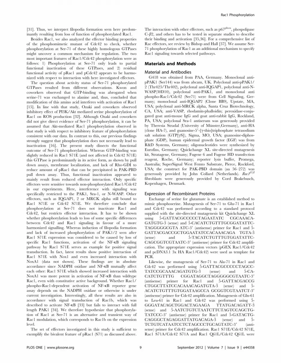
Phosphorylation-insensitive Function of Rac1Since Rac1 S71E showed active conformation as tested by
PAK-PBD pull downs and bound to effector proteins IQGAP and
MRCK, we investigated whether phosphomimetic Rac1 and
Cdc42 are able to activate specific pathways. We choose the NF-
kB pathway because the activation by Rac1 and Cdc42 is well
described [22] and can easily be monitored by a reporter gene
assay. Activation of the transcription factor NF-kB regulates the
expression of genes required for inflammatory responses, cell
growth and suppression of apoptosis. To measure the effect of Ser-
71-phosphorylated Rac1 and Cdc42 on NF-kB activity, HEK-293
cells were transfected with corresponding Rac1 and Cdc42
expression vectors and a luciferase reporter plasmid containing
three NF-kB-responsive sites. Fig. 5 shows the activation of the
NF-kB-reporter after transfection of cells with the indicated
GTPases. Expression of Rac1 induced two-fold and Rac1 S71E
induced four-fold NF-kB-reporter gene activation compared to
mock transfected cells. Constitutively active Rac1 Q61L and Rac1
Q61L/S71E were even more potent, leading to 200-fold and 300-
fold activation, respectively. These results nicely demonstrate the
ability of phosphomimetic Rac1 to activate effectors to the same
magnitude as wildtype Rac1. Expression of Cdc42 and Cdc42
S71E as well as Cdc42 Q61L and Cdc42 Q61L/S71E showed
activation of NF-kB that was in accordance to the set of Rac1
experiments. The expression level of the Cdc42 and Rac1 mutants
was validated by immunoblots against HA-tag of ectopically
expressed GTPases (Fig. 5B).
Figure 3. GTPase-specific binding to their effectors. A) Coomassie-stained SDS-gel of precipitates of pull down experiments from HEp2 celllysate. Arrows indicate GST-Rac1/2Rac1 S71E (48 kDa) used as bait and a coprecipitated 190 kDa protein that was identified as IQGAP1 by MALDI-TOF/TOF analysis. B) The interaction of active, GTP-bound Rac1/Cdc42 and their active forms (Q61L) with specific effectors was analyzed byimmunoblot analyses of precipitates from pull down assays. Non-specific binding was tested by GST-loaded glutathione beads as control. C)Representative input control of pull down analyses using constitutively active (Q61L) mutants of Rac1 and Cdc42 and their S71E mutants. D)Representative immunoblots of pull down precipitates showing the interaction of constitutively active Rac1 and Cdc42 and constitutively active S71Emutants with their effector proteins. Rac1 Q61L/S71E and Cdc42 Q61L/S71E did hardly bind to their specific effectors Sra-1 and N-WASP, respectivelyand to their common effector protein PAK1. Both phosphomimetic GTPases, however bound to their common effectors IQGAP and MRCK alpha,although to a lesser extent. The bars show the arithmetic mean value 6 SD of densitometrical evaluation of 3 independent experiments.doi:10.1371/journal.pone.0044358.g003
Rac1 Phosphorylation
PLOS ONE | www.plosone.org 6 September 2012 | Volume 7 | Issue 9 | e44358
Rac1 Phosphorylation
PLOS ONE | www.plosone.org 7 September 2012 | Volume 7 | Issue 9 | e44358
Discussion
Rho GTPases are involved in various signaling pathways that
regulate diverse cellular functions [23]. Especially Rac1 has
diverse functions, but the predominant role of Rac1 is the
regulation of actin-based processes such as membrane ruffling and
lamellipodia formation or cell migration [24]. Rac1 is also
involved in further actin-dependent processes like phagocytosis
[25,26]. In addition, Rac1 is part of signaling cascades, e.g. that of
MAP kinases or NF-kB, that initiate gene expression, or in
activation of the NADPH oxidase which results in production of
reactive oxygen species [27,28]. It is an unsolved question how
activated Rac1 addresses specific pathways at a given time whilst
being capable in principle of activating all of them simultaneously.
In one theory, co-localization of Rac1 with specific effectors
defines the functional outcome of Rac1 activation. Thus, the
microenvironment might be decisive for Rac1 function. We here
describe a potential, alternative regulatory mechanism, based on
phosphorylation of Ser-71, by which Rac1 can be directed towards
specific effectors, and which might also be applicable for Cdc42.
We previously reported that Ser-71 phosphorylation of Rac1/
Cdc42 is not accompanied with general loss of function [16]. In
the present study we clarified the outcome of Ser-71 phosphor-
ylation of Rac1 and Cdc42 with respect to interaction with
downstream effectors. One essential tool for investigating phos-
phorylation of Rac1 and Cdc42 is the phosphospecific antibody.
To date, there is no phosphospecific antibody available that
specifically recognizes pSer-71 Rac1 or pSer-71 Cdc42. Thus, it is
not clear whether an observed effect accounts for Rac1 or Cdc42
phosphorylation. We took advantage of Rac1-deficient murine
fibroblasts to show that EGF induces phosphorylation of Rac1 but
not of Cdc42. The absence of Rac1 expression in these cells
confirmed that anti-phosphospecific antibody staining observed in
Rac1-expressing control cells mostly corresponded to phosphor-
ylated Rac1, and not Cdc42. It is reasonable to assume that this is
also true for other cell lines, although this remains to be
experimentally validated. Focus of the present study is the
characterization of phosphorylated Rac1 (S71) by using the
phosphomimetic mutant. Cdc42 S71E was also applied since we
cannot exclude Cdc42 phosphorylation. Thus, Cdc42 was
additionally investigated in key experiments to emphazise the
finding of phosphorylation as principal mechanism to modulate
interaction with effector proteins which, theoretically might also
apply for Cdc42. According to amino acid sequence, RhoG also
possesses consensus sequence for Akt-mediated phosphorylation.
However, since overexpression of Rac1 S71E induced the specific
phenotype of cells, we assume that Rac1 is crucial for EGF-
induced filopodia.
This hypothesis is substantiated by the predominant morpho-
type of cells with increased pSer-71 Rac1 showing massive
formation of filopodia (see Fig. 1B) - a morphotype that is also seen
in HEp2 cells stably expressing phosphomimetic Rac1 S71E.
Fig. 1B also shows that formation of filopodia is less pronounced in
Cdc42 S71E transfected cells compared to Rac1 S71E. Key
experiments with transient expression of Rac1 S71E showed that
formation of filopodia was not accompanied with increased
activity of Cdc42. Due to cross talk between Cdc42 and Rac1
signaling a predominant Cdc42 morphotype can also be achieved
by reducing Rac1 activity. For instance, filopodia formation
downstream of Cdc42 could be enhanced by concomitant
inhibition of Rac1 signaling [29,30]. Furthermore, Cdc42-induced
filopodia formation was described to be increased rather than
decreased upon inhibition of Rac1 signaling to Arp2/3-mediated
actin assembly through WAVE-complex. Dominant filopodia
formation might thus very likely result from decreased signaling of
Rac1S71E to WAVE and the Arp2/3-complex due to defective
interaction with the WAVE-complex subunit Sra-1, as shown here
by pull down experiments. Filopodia formation can also be
induced by specific inactivation of Rac1 by C. sordellii Lethal Toxin
TcsL-82 from strain IP82, which glucosylates Rac1 but not Cdc42
Figure 4. Activation of PAK by Rac1 and Rac1 S71E. The effect of Rac1 and Rac1 S71E on PAK phosphorylation was shown in HEp2 cells stablyexpressing either GTPase. A) A brief characterization of these stable transfected cell lines was done by scanning electron microscopy showing surfacetopology of the cells. B) Immunoblot analysis revealed comparable ectopic expression of HA-tagged Rac1 and Rac1 S71E and concomitant Ser-144phosphorylation of PAK1 and Thr-402 phosphorylation of PAK2. C) densitometrical evaluation of three separate Immunoblots showingphosphorylation of PAK 1/2. Shown are mean values 6 SD. D) Propidium iodide staining of stable cell lines indicates populations of cells with 2n (G1phase) or 4n (G2/M phase) set of chromosomes. Shown are percentages of cells within different cell cycle phases (mean values of five separateexperiments).doi:10.1371/journal.pone.0044358.g004
Figure 5. NF-kB is activated by phosphorylated Rac1. HEK 293cells were co-transfected with NF-kB luciferase reporter plasmid as wellas with Rac1 and Cdc42 mutants. Cells were lysed after 40 hours andanalyzed for luciferase activity. Relative fold activity of mock-transfectedcells is shown (arithmetic means6SD, n = 4). B) Expression of HA-taggedGTPases of quadruplicate samples from reporter gene assays wasvisualized by immunoblot using an anti-HA antibody.doi:10.1371/journal.pone.0044358.g005
Rac1 Phosphorylation
PLOS ONE | www.plosone.org 8 September 2012 | Volume 7 | Issue 9 | e44358
[31]. Thus, we interpret filopodia formation seen here predom-
inantly resulting from loss of function of phosphorylated Rac1.
Besides Rac1, we also analyzed the effector binding properties
of the phosphomimetic mutant of Cdc42 to check, whether
phosphorylation at Ser-71 of these highly homologous GTPases
might uncover a common mechanism for regulation. The two
most important features of Rac1/Cdc42 phosphorylation were as
follows: 1) Phosphorylation at Ser-71 only leads to partial
functional inactivation of these GTPases, and 2) residual
functional activity of pRac1 and pCdc42 appears to be harmo-
nized with respect to interaction with here investigated effectors.
The question about activity status of Ser-71 phosphorylated
GTPases resulted from different observations. Kwon and
coworkers observed that GTP-binding was abrogated when
serine-71 was exchanged to alanine and, thus, concluded that
modification of this amino acid interferes with activation of Rac1
[15]. In line with that study, Ozaki and coworkers observed
inhibitory effect of PI3K/Akt mediated serine phosphorylation of
Rac1 on ROS production [32]. Although Ozaki and coworkers
did not give direct evidence of Ser-71 phosphorylation, it can be
assumed that Akt-mediated Rac1 phosphorylation observed in
that study is with respect to inhibitory feature of phosphorylation
consistent with our data. In contrast to this, our previous findings
strongly suggest that phosphorylation does not necessarily lead to
inactivation [16]. The present study dissects the functional
outcome of Ser-71 phosphorylation. Whereas GTP-binding was
slightly reduced in Rac1 S71E (and not affected in Cdc42 S71E)
this GTPase is predominantly in its active form, as shown by pull
down assays, membrane localization, and lack of Rho-GDI to
reduce amount of pRac1 that can be precipitated in PAK-PBD
pull down assay. Thus, functional inactivation appeared to
mainly result from reduced effector interaction. Only specific
effectors were sensitive towards non-phosphorylated Rac1/Cdc42
in our experiments. Here, interference with signaling was
specifically restricted to the PAK1, Sra-1, or N-WASP. Other
effectors, such as IQGAP1, 2 or MRCK alpha still bound to
Rac1 S71E or Cdc42 S71E. We therefore conclude that
phosphorylation at Ser-71 does not inactivate Rac1 and
Cdc42, but restricts effector interaction. It has to be shown
whether phosphorylation leads to loss of some specific differences
between Cdc42 and Rac1 signaling in favour of a more
harmonized signalling. Whereas induction of filopodia formation
and lack of increased phosphorylation of PAK1/2 seen after
Rac1 S71E expression was considered to be caused by loss of
specific Rac1 functions, activation of the NF-kB signaling
pathway by Rac1 S71E serves as example for positive signal
transduction. In fact, latest results show positive interaction of
Rac1 S71E with Nox1 and even increased interaction with
NoxA1 (data not shown). These findings are in absolute
accordance since NADPH oxidase and NF-kB mutually affect
each other: Rac1 S71E which showed increased interaction with
NoxA1 was more potent in activation of NF-kB than wildtype
Rac1, even with constitutive active background. Whether Rac1/
phospho-Rac1-dependent activation of NF-kB reporter gene
assay depends on the NADPH oxidase or otherwise is under
current investigation. Interestingly, all these results are also in
accordance with signal transduction of Rac1b, which was
described to activate NF-kB [33] but fails to interact with full
length PAK1 [34]. We therefore hypothesize that phosphoryla-
tion of Rac1 at Ser-71 is an alternative and transient way of
Rac1 modulation, which corresponds to Rac1b on the expression
level.
The set of effectors investigated in this study is sufficient to
exemplify the bivalent feature of pRac1 (S71) as discussed above.
The interaction with other effectors, such as p67phox, phospolipase
C-b2, and others has to be tested in separate studies to describe
their binding and activation [35,36]. For a comprehensive list of
Rac effectors, see review by Bishop and Hall [37]. We assume Ser-
71 phosphorylation of Rac1 as an additional mechanism to specify
Rac1 signaling towards selected pathways.
Materials and Methods
Material and AntibodiesG418 was obtained from PAA, Germany. Monoclonal anti-
pPAK1 (Ser144) was from abcam, UK. Polyclonal anti-pPAK1/
2 (Thr423/Thr402), polyclonal anti-IQGAP1, polyclonal anti-N-
WASP(30D10), polyclonal anti-PAK1, and monoclonal anti
phospho-Rac1/Cdc42 (Ser71) were from Cell Signaling, Ger-
many; monoclonal anti-IQGAP2 (Clone BB9), Upstate, MA,
USA; polyclonal anti-MRCK alpha, Santa Cruz Biotechnology,
CA, USA; anti-VASP, rhodamin-phalloidin; peroxidase-conju-
gated goat anti-mouse IgG and goat anti-rabbit IgG, Rockland,
PA, USA; polyclonal Sra-1 antiserum was generously provided
by Theresia Stradal (University of Munster,Germany), anti-HA
(clone HA-7), and guanosine-59-[c-thio]triphosphate tetrasodium
salt solution (GTP[cS]), Sigma, MO, USA; guanosine-diphos-
phate (GDP), human epidermal growth factor (EGF) was from
R&D Systems, Germany; oligonucleotides were synthesized by
Eurofins, Germany; Quickchange XL site-directed mutagenesis
kit, Stratagene, Germany; Fugene 6 and Fugene HD transfection
reagent, Roche, Germany; reporter lysis buffer, Promega,
Australia; SuperSignal West Femto Substrate, Pierce, Rockford,
USA; the construct for PAK-PBD domain (aa 56–272) was
generously provided by John Collard (Netherlands). Rac1fl/fl
fibroblasts were generously provided by Cord Brakebusch,
Kopenhagen, Denmark.
Expression of Recombinant ProteinsExchange of serine for glutamate is an established method to
mimic phosphoserine. Mutagenesis of Ser-71 to Glu-71 in Rac1
and Cdc42 was performed according to a standard protocol
supplied with the site-directed mutagenesis kit Quickchange XL
using 5-GATTACGCCCCCTAGAATATC CGCAAACA-
GATGTG-3 (sense) and 5-CACATCTGTTTGCGGATATTC-
TAGGGGGCGTA ATC-39 (antisense) primer for Rac1 and 5-
GATTACGACCGCTGGAATATCCACAAACAGA TGTA-3
(sense) and 5-TACATCTGTTTGTGGATATTC-
CAGCGGTCGTAATC-39 (antisense) primer for Cdc42 amplifi-
cation. The appropriate expression vectors (pGEX Rac1/Cdc42
and pcDNA3.1 3x HA Rac1/Cdc42) were used as template for
PCR.
Likewise, the mutagenesis of Ser-71 to Ala-71 in Rac1 and
Cdc42 was performed using 5-GATTACGCCCCCTAGCC-
TATCCGCAAACAGATGTG-3 (sense) and 5-CA-
CATCTGTTTG CGGATAGGCTAGGGGGCGTAATC-39
(antisense) primer for Rac1 and 5-GATTACGACCG
CTGGCTTATCCACAAACAGATGTA-3 (sense) and 5-
ACATCTGTTTGTGGATAAGCCA GCGGTCGTAATCT-39
(antisense) primer for Cdc42 amplification. Mutagenesis of Gln-61
to Leu-61 in Rac1 and Cdc42 was performed using 5-
GGGATACAGCTGGACTAGAAGA TTATGACAGATT-3
(sense) and 5-AATCTGTCTAATCTTCTAGTCCAGCTG-
TATCCC-39 (antisense) primer for Rac1 and 5-GATACTG-
CAGGGCTAGAGGATTATGACAGA-3 (sense) and 5-
TCTGTCATAATCCTCTAGCCCTGCAGTATC-39 (anti-
sense) primer for Cdc42 amplification. Rac1 S71E/Cdc42 S71E,
Rac1 S71A/Cdc42 S71A and Rac1 Q61L/Cdc42 Q61L were
Rac1 Phosphorylation
PLOS ONE | www.plosone.org 9 September 2012 | Volume 7 | Issue 9 | e44358
expressed in E. coli after standard protocol for glutathione-S-
transferase fusion proteins.
GTPases for fishing experiments were expressed in Escherichia
coli and purified following standard procedures for glutathione-S-
transferase fusion proteins. GST-GTPase fusion proteins bound to
glutathione-beads were used for pull down experiments.
GTP Binding AssayRecombinant Rac1, Rac1 S71E, Cdc42 and Cdc42 S71E
(2.5 mg of each) were incubated with 13,5 mCi [c-32P]GTP in
700 ml binding buffer (50 mM Hepes pH 7.6, 0.2 mg/ml BSA
and 0.5 mM EDTA) at 15uC for indicated times. The samples
were then applied to a nitrocellulose membrane that had been
rinsed with 3 times with 1 ml wash buffer. The filters were
immediately washed 3 times with 1 ml of ice-cold wash buffer
(50 mM Hepes pH 7.6, 150 mM NaCl and 10 mM MgCl2) and
soaked in scintillation solution for 30 min. GTPase bound
radioactivity was counted by liquid scintillation spectrometry.
Cell Culture and TransfectionHEK293 cells were cultured in DMEM with sodium pyruvate
(PAA, Germany), supplemented with 10% (v/v) fetal bovine
serum, 100 U/ml penicilline, 100 mg/ml streptomycine at 37uC in
humified air with 5% CO2 [38]. For transfection, cells were grown
to subconfluence in 6 well plates and transfected with Fugene 6
transfection reagent according to manufactorer’s protocol. HEp2
cells were grown in MEM Eagle’s medium, supplemented with
10% (v/v) FBS, 100 U/ml penicilline and 100 mg/ml streptomy-
cine at 37uC in humified air with 5% CO2 [16]. Stable transfected
HEp2 cells were generated by transfection with pcDNA3.1 Rac1
or pcDNA3.1 Rac1 S71E constructs or with empty vector and
cultured in the presence of 750 mg/ml G418 for six weeks. G418-
resistant clones were selected and expression of HA-tagged Rac1/
Rac1 S71E was monitored. After selection of positive clones
further cell culture of stable transfected cells was performed in the
presence of 400 mg/ml G418.
Rac1fl/fl fibroblasts were kindly provided by Cord Brakebusch
and are homozygous for the loxP-flanked Rac1 allele described
before [39]. Generation of Rac2/2 fibroblasts will be described
elsewhere. Rac1fl/fl and Rac12/2 fibroblasts were maintained in
DMEM containing 10% (v/v) fetal bovine serum, 2 mM
glutamine, 1 mM Na+ pyruvate and 1% non-essential amino
acids (Invitrogen, Germany).
ImmunoblotsCell lysates or precipitates from pull down assays were subjected
to SDS-PAGE and subsequent transfer onto nitrocellulose. After
blocking with Tris-buffered saline containing 3% (w/v) milk
powder and 0.2% (v/v) Tween-20, the nitrocellulose membrane
was incubated with the appropriate primary antibody diluted
1:1.000 in TBS-T supplemented with 3% (w/v) bovine albumine
over night at 4uC. The membranes were washed three times with
TBS-T and incubated with the corresponding HRP-conjugated
secondary antibody diluted 1:5.000 in TBS-T for 30 min. Bound
antibodies were visualized by incubation with SuperSignal West
femto chemiluminescence substrate (Pierce).
Flow CytometryFlow cytometry was performed to estimate number of cells
within different cell cycle phases. The DNA content was measured
using the fluorescent nucleotide acid dye propidium iodide.
Therefore, cells were suspended by trypsinization and app.
56105 cells were fixed in ice cold ethanol (70%) for 30 min.
After washing once with 1% bovine serum albumin in PBS, the
total DNA content was stained with propidium iodide (150 mg/ml
in Tris/HCl, pH 7.4, containing 1% BSA and 1% Triton X-100).
RNA was removed by incubating cells with 0.5% RNase for
30 min. Subsequently, cells were subjected to FACS-analysis
(FACScan flow cytometer, Becton Dickinson). A fluorescence area
(FL2) of 400 was set to correlate with a 2n-set of chromosomes
within the G1-phase. Cells found in the sub-G1-phase were
considered as apoptotic and necrotic due to the decrease in DNA
content.
Western Blot AnalysisProtein samples were separated by SDS-PAGE and transferred
onto nitrocellulose membrane. After blocking with 5% (w/v)
nonfat dry milk in TBS-T (50 mM Tris HCl pH 7.2, 150 mM
NaCl, 0.05% (v/v) Tween 20) the membrane was incubated
overnight with the primary antibody at 4uC. After washing with
TBS-T it was incubated for 45 min at room temperature with the
appropriate horseradish peroxidase-conjugated secondary anti-
body. Detection was performed by means of enhanced chemilu-
minescence.
Luciferase-based Reporter AssayHEK-293 cells were transiently cotransfected with 50 ng of NF-
kB luciferase reporter plasmid p3ENhkBcona-Luc containing
three tandem repeats of NF-kB sites from the immunoglobulin Gkpromoter and 1 mg of the HA-tagged Rho GTPases. At 40 h post
transfection, cells were washed with PBS and lysed in reporter lysis
buffer. Luciferase activities were measured in cleared lysates with a
luciferase assay system in accordance with manufacturer’s
instructions. NF-kB activity was calculated as per cent of induction
compared to that of pcDNA3.1_Cdc42 Q61L (constitutively active
Cdc42) transfected cells.
Pull Down ExperimentsPull down experiments with the PAK-PBD domain to detect
active Rac1, Cdc42 or P-Ser-71 Rac1/Cdc42 were performed as
previously described by Schoentaube [16]. Fishing experiments
were either performed with constitutive active GTPases or with
GDP/GTP-loaded GTPases. The nucleotide exchange of
GTPases was performed at 30uC using 10 mM EDTA in
20 mM Tris pH 7.4 and 25 mM NaCl to extract bound
nucleotide from the GTPase. After loading with either 1 mM
GDP or 1 mM GTP[cS] for 15 min, the complex of GTPase and
nucleotide was stabilized by addition of 50 mM MgCl2.
For pull down experiments HEp2 cells grown in 75 cm2 flasks
were lysed in 2 ml ice-cold Fish-buffer (50 mM Tris pH 7.4,
2 mM MgCl2, 10% glycerine, 100 mM NaCl, 1% NP40, 0.5 mg/
ml BSA). After 5 min incubation on ice the lysates were
centrifugated at 16,000 g for 5 min. The supernatant was split
into four samples of 0.5 ml each and used for precipitation of
desired proteins in parallel to guarantee identical protein load in
all samples. Therefore, 20 ml of bead slurry with bound GST-
fusion protein of the respective bait (app. 15 mg each) were added
to each sample and rotated at 4uC for 60 min. The beads were
collected by centrifugation at 10.000 g and washed twice with
Fish-buffer and subjected for SDS-PAGE.
Cdc42 Activity AssayThe G-LISA assay from Cytoskeleton was used to assess active
GTP bound Cdc42 in cell lysates. Subconfluent HEp2 cells were
transfected with different Rac1 constructs. At 24 h post transfec-
tion the cells were analyzed according to manufacturer’s revised
Rac1 Phosphorylation
PLOS ONE | www.plosone.org 10 September 2012 | Volume 7 | Issue 9 | e44358
instructions. The level of activation was measured by reading the
absorbance at 490 nm.
MALDI-TOF/TOF AnalysisWashed precipitates from pull down experiments were separat-
ed by SDS-PAGE and stained with Coomassie brilliant blue.
Specific bands were cut out, destained, digested with trypsin
(12.5 ng/ml) and the generated peptides were extracted and solved
in 10% acetonitril containing 0.2% trifluoroacetic acid. Peptides
were cocrystalized with alpha-Cyano-4-hydroxycinnamic acid
(4 mg/ml) in 50% acetonitril and 0.2% trifluoroacetic acid on
an anchor target (Bruker Daltonic). After crystallization the
peptides were analyzed in an MALDI-TOF/TOF mass spec-
trometer (Ultraflex I, Bruker Daltonic). Masses were determined
by external calibration using suitable standard peptides (Bruker
Daltonic). MS and MS/MS spectra were generated and analyzed
with the BioTools (Bruker Daltonik) and MASCOT (Matrix
Science, UK) software package.
Acknowledgments
We thank Karin Agternkamp for excellent technical assistance in MALDI-
TOF/TOF analyses, and Cord Brakebusch, University of Kopenhagen,
for reagents.
We also thank Stefanie Groos, Institute for Cellular Biology in the
Centre for Anatomy, MHH, for excellent performance of scanning
electron microscopy of HEp2 cells.
Author Contributions
Conceived and designed the experiments: JS ML KR IJ RG AP.
Performed the experiments: JS JP AH ML BB HT AP. Analyzed the
data: JS ML KR RG. Contributed reagents/materials/analysis tools: AH.
Wrote the paper: JS RG. Critical reading of manuscript: IJ.
References
1. Burbelo PD, Drechsel D, Hall A (1995) A conserved binding motif definesnumerous candidate target proteins for both Cdc42 and Rac GTPases. J Biol
Chem 270: 29071–29074.2. Leung T, Manser E, Tans L, Lim L (1995) A novel serine/threonine kinase
binding the Ras-related RhoA GTPase which translocates the kinase to
peripheral membranes. J Biol Chem 270: 29051–29054.3. Briggs MW, Sacks DB (2003) IQGAP proteins are integral components of
cytoskeletal regulation. EMBO Rep 4: 571–574.4. Kobayashi K, Kuroda S, Fukata M, Nakamura T, Nagase T, et al. (1998)
p140Sra-1 (Specifically Rac1-associated protein) is a novel specific target for
Rac1 small GTPase. J Biol Chem 273: 291–295.5. Nobes CD, Hall A (1995) Rho, Rac, and Cdc42 GTPases regulate the assembly
of multimolecular focal complexes associated with actin stress fibers, lamellipo-dia, and filopodia. Cell 81: 53–62.
6. Etienne-Manneville S, Hall A (2002) Rho GTPases in cell biology. Nature 420:629–635.
7. Olofson B (1999) Rho guanine dissociation inhibitors: pivotal molecules in
cellular signalling. Cell Signal 11: 545–554.8. Zheng Y (2001) Dbl family guanine nucleotide exchange factors. Trends
Biochem Sci 26: 724–732.9. Lamarche N, Hall A (1994) GAPs for rho-related GTPases. Trends Genet 10:
436–440.
10. Rolli-Derkinderen M, Sauzeau V, Boyer L, Lemichez E, Baron C, et al. (2005)Phosphorylation of serine 188 protects RhoA from ubiquitin/proteasome-
mediated degradation in vascular smooth muscle cells. Circ Res 96: 1152–1160.11. Forget MA, Desrosiers RR, Gingras D, Beliveau R (2002) Phosphorylation states
of Cdc42 and RhoA regulate their interactions with Rho GDP dissociationinhibitor and their extraction from biological membranes. Biochem J 361: 243–
254.
12. Ellerbroek SM, Wennerberg K, Burridge K (2003) Serine phosphorylationnegatively regulates RhoA in vivo. J Biol Chem 278: 19023–19031.
13. Tu H, Wigler M (1999) Genetic evidence for Pak1 autoinhibition and its releaseby Cdc42. Molecular and Cellular Biology 19: 602–611.
14. Chang F, Lemmon C, Lietha D, Eck M, Romer L (2011) Tyrosine
phosphorylation of Rac1: a role in regulation of cell spreading. PLoS One 6:e28587.
15. Kwon T, Kwon DY, Chun J, Kim JH, Kang SS (2000) Akt protein kinaseinhibits Rac1-GTP binding through phosphorylation at serine 71 of Rac1. J Biol
Chem 275: 423–428.16. Schoentaube J, Olling A, Tatge H, Just I, Gerhard R (2009) Serine-71
phosphorylation of Rac1/Cdc42 diminishes the pathogenic effect of Clostridium
difficile toxin A. Cell Microbiol 11: 1816–1826.17. Riento K, Totty N, Villalonga P, Garg R, Guasch R, et al. (2005) RhoE function
is regulated by ROCK I-mediated phosphorylation. EMBO J 24: 1170–1180.18. Breitsprecher D, Kiesewetter AK, Linkner J, Urbanke C, Resch GP, et al. (2008)
Clustering of VASP actively drives processive, WH2 domain-mediated actin
filament elongation. EMBO J 27: 2943–2954.19. Steffen A, Faix J, Resch GP, Linkner J, Wehland J, et al. (2006) Filopodia
formation in the absence of functional. Mol Biol Cell 17: 2581–2591.20. Matos P, Collard J, Jordan PW (2003) Tumor-related alternative spliced Rac1b
is not regulated by Rho-GDI and exhibits selective downstream signaling. J Biol
Chem 278: in press.
21. Maroto B, Ye MB, von LK, Schnelzer A, Knaus UG (2008) P21-activated kinase
is required for mitotic progression and regulates Plk1. Oncogene 27: 4900–4908.
22. Perona R, Montaner S, Saniger L, Sanchez-Perez I, Bravo R, et al. (1997)
Activation of the nuclear factor-kB by Rho, Cdc42, and Rac-1 proteins. Genes &
Development 11: 463–475.
23. Bosco EE, Mulloy JC, Zheng Y (2009) Rac1 GTPase: a ‘‘Rac’’ of all trades. Cell
Mol Life Sci 66: 370–374.
24. Fukata M, Nakagawa M, Kuroda S, Kaibuchi K (1999) Cell adhesion and Rho
small GTPases. J Cell Sci 112: 4491–4500.
25. Hoppe AD, Swanson JA (2004) Cdc42, Rac1, and Rac2 display distinct patterns
of activation during phagocytosis. Mol Biol Cell 15: 3509–3519.
26. Michaelson D, Silletti J, Murphy G, D’Eustachio P, Rush M, et al. (2001)
Differential Localization of Rho GTPases in Live Cells. Regulation by
hypervariable regions and rhogdi binding. J Cell Biol 152: 111–126.
27. Jin S, Ray RM, Johnson LR (2006) Rac1 mediates intestinal epithelial cell
apoptosis via JNK. Am J Physiol Gastrointest Liver Physiol 291: G1137–G1147.
28. Miyano K, Sumimoto H (2007) Role of the small GTPase Rac in p22phox-
dependent NADPH oxidases. Biochimie 89: 1133–1144.
29. Nobes CD, Hawkins P, Stephens L, Hall A (1995) Activation of the small GTP-
binding proteins rho and rac by growth factor receptors. J Cell Sci 108: 225–
233.
30. Hall A (1998) G proteins and small GTPases: distant relatives keep in touch.
Science 280: 2074–2075.
31. Geny B, Popoff MR (2009) Activation of a c-Jun-NH2-terminal kinase pathway
by the lethal toxin from Clostridium sordellii, TcsL-82, occurs independently of
the toxin intrinsic enzymatic activity and facilitates small GTPase glucosylation.
Cell Microbiol 11: 1102–1113.
32. Ozaki M, Haga S, Zhang HQ, Irani K, Suzuki S (2003) Inhibition of hypoxia/
reoxygenation-induced oxidative stress in HGF-stimulated antiapoptotic signal-
ing: role of PI3-K and Akt kinase upon rac1. Cell Death Differ 10: 508–515.
33. Matos P, Jordan P (2005) Expression of Rac1b stimulates NF-kappaB-mediated
cell survival and G1/S progression. Exp Cell Res 305: 292–299.
34. Matos P, Collard JG, Jordan P (2003) Tumor-related alternatively spliced Rac1b
is not regulated by Rho-GDP dissociation inhibitors and exhibits selective
downstream signaling. J Biol Chem 278: 50442–50448.
35. Prigmore E, Ahmed S, Best A, Kozma R, Manser E, et al. (1995) A 68-kDa
kinase and NADPH oxidase component p67phox are targets for Cdc42Hs and
Rac1 in neutrophils. J Biol Chem 270: 10717–10722.
36. Jezyk MR, Snyder JT, Gershberg S, Worthylake DK, Harden TK, et al. (2006)
Crystal structure of Rac1 bound to its effector phospholipase C-beta2. Nat
Struct Mol Biol 13: 1135–1140.
37. Bishop AL, Hall A (2000) Rho GTPases and their effector proteins. Biochem J
348: 241–255.
38. Pietrek M, Brinkmann MM, Glowacka I, Enlund A, Havemeier A, et al. (2010)
Role of the Kaposi’s sarcoma-associated herpesvirus K15 SH3 binding site in
inflammatory signaling and B-cell activation. J Virol 84: 8231–8240.
39. Chrostek A, Wu X, Quondamatteo F, Hu R, Sanecka A, et al. (2006) Rac1 is
crucial for hair follicle integrity but is not essential for maintenance of the
epidermis. Mol Cell Biol 26: 6957–6970.
Rac1 Phosphorylation
PLOS ONE | www.plosone.org 11 September 2012 | Volume 7 | Issue 9 | e44358


