Embed Size (px)
Citation preview
Hart et al. BMC Musculoskeletal Disorders 2014, 15:139http://www.biomedcentral.com/1471-2474/15/139
RESEARCH ARTICLE Open Access
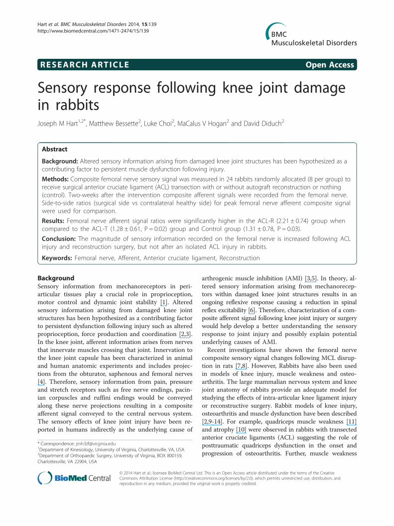
Sensory response following knee joint damagein rabbitsJoseph M Hart1,2*, Matthew Bessette2, Luke Choi2, MaCalus V Hogan2 and David Diduch2
Abstract
Background: Altered sensory information arising from damaged knee joint structures has been hypothesized as acontributing factor to persistent muscle dysfunction following injury.
Methods: Composite femoral nerve sensory signal was measured in 24 rabbits randomly allocated (8 per group) toreceive surgical anterior cruciate ligament (ACL) transection with or without autograft reconstruction or nothing(control). Two-weeks after the intervention composite afferent signals were recorded from the femoral nerve.Side-to-side ratios (surgical side vs contralateral healthy side) for peak femoral nerve afferent composite signalwere used for comparison.
Results: Femoral nerve afferent signal ratios were significantly higher in the ACL-R (2.21 ± 0.74) group whencompared to the ACL-T (1.28 ± 0.61, P = 0.02) group and Control group (1.31 ± 0.78, P = 0.03).
Conclusion: The magnitude of sensory information recorded on the femoral nerve is increased following ACLinjury and reconstruction surgery, but not after an isolated ACL injury in rabbits.
Keywords: Femoral nerve, Afferent, Anterior cruciate ligament, Reconstruction
BackgroundSensory information from mechanoreceptors in peri-articular tissues play a crucial role in proprioception,motor control and dynamic joint stability [1]. Alteredsensory information arising from damaged knee jointstructures has been hypothesized as a contributing factorto persistent dysfunction following injury such as alteredproprioception, force production and coordination [2,3].In the knee joint, afferent information arises from nervesthat innervate muscles crossing that joint. Innervation tothe knee joint capsule has been characterized in animaland human anatomic experiments and includes projec-tions from the obturator, saphenous and femoral nerves[4]. Therefore, sensory information from pain, pressureand stretch receptors such as free nerve endings, pacin-ian corpuscles and ruffini endings would be conveyedalong these nerve projections resulting in a compositeafferent signal conveyed to the central nervous system.The sensory effects of knee joint injury have been re-ported in humans indirectly as the underlying cause of
* Correspondence: [email protected] of Kinesiology, University of Virginia, Charlottesville, VA, USA2Department of Orthopaedic Surgery, University of Virginia, BOX 800159,Charlottesville, VA 22904, USA
© 2014 Hart et al.; licensee BioMed Central LtdCommons Attribution License (http://creativecreproduction in any medium, provided the or
arthrogenic muscle inhibition (AMI) [3,5]. In theory, al-tered sensory information arising from mechanorecep-tors within damaged knee joint structures results in anongoing reflexive response causing a reduction in spinalreflex excitability [6]. Therefore, characterization of a com-posite afferent signal following knee joint injury or surgerywould help develop a better understanding the sensoryresponse to joint injury and possibly explain potentialunderlying causes of AMI.Recent investigations have shown the femoral nerve
composite sensory signal changes following MCL disrup-tion in rats [7,8]. However, Rabbits have also been usedin models of knee injury, muscle weakness and osteo-arthritis. The large mammalian nervous system and kneejoint anatomy of rabbits provide an adequate model forstudying the effects of intra-articular knee ligament injuryor reconstructive surgery. Rabbit models of knee injury,osteoarthritis and muscle dysfunction have been described[2,9-14]. For example, quadriceps muscle weakness [11]and atrophy [10] were observed in rabbits with transectedanterior cruciate ligaments (ACL) suggesting the role ofposttraumatic quadriceps dysfunction in the onset andprogression of osteoarthritis. Further, muscle weakness
. This is an Open Access article distributed under the terms of the Creativeommons.org/licenses/by/2.0), which permits unrestricted use, distribution, andiginal work is properly credited.

Hart et al. BMC Musculoskeletal Disorders 2014, 15:139 Page 2 of 6http://www.biomedcentral.com/1471-2474/15/139
induced by botox injections caused increased cartilage de-generation in rabbits [13]. However, a model to studycomposite afferent signals arising from the femoral nervein the presence of ACL injury or reconstruction has notbeen developed.Therefore the purpose of this study was to compare
femoral nerve afferent signal using a whole nerve re-cording technique in rabbits 2-weeks following kneejoint trauma. We hypothesized that the damage causedby injury and surgery would cause an increase in afferentactivity from sensory endings located in tissues withinand surrounding the knee joint thereby increasing com-posite signal measured from the femoral nerve in rabbitswith knee joint injury and reconstruction surgery com-pared to controls due to increase afferent activity fromsensory endings.
MethodsThis study was reviewed and approved by the Institu-tional Animal Care and Use Committee at the Universityof Virginia. Twenty-four adult, male New Zealand Whiterabbits were randomized into one of 3 intervention groups.Eight of the rabbits received unilateral ACL transection, 8of the rabbits received an ACL reconstruction and theremaining 8 were control rabbits who received no inter-vention. The side receiving the surgical intervention wasrandomly allocated.
Surgical interventionAnimals receiving ACL transection (ACL-T) were fullyinduced with inhaled isoflurane. A knee joint arthrotomywas performed and the anterior cruciate ligament wasidentified and carefully transected. Care was taken toavoid damage to cartilage or other intra- or peri- articu-lar structures. The joint capsule and skin were closedwith absorbable sutures. For the animals in the ACL re-construction (ACL-R) group the medial third of the pa-tellar tendon was removed and prepared as an ACLautograft. The graft was passed through small diametertunnels drilled through the tibia and femur and fixed tothe tibial and femoral periosteum with suture. Controlanimals received no surgical intervention and remainedon the procedure table for a period of time similar tothat of the surgical procedures. The animals receivingunilateral knee surgery (ACL transection with or withoutreconstruction) were given buprenorphine and a fentanylpatch for pain control and kept in recovery cages onheating pads under warm light post operatively. Animalswere housed in a cage for repeat observation on the firstday and then daily for signs of pain. Rabbits were allhoused for 2 weeks with daily monitoring at which pointthe terminal measurements were recorded.After 2 weeks we performed whole nerve recordings
on the femoral nerve in each animal bilaterally. This
technique was performed in a similar manner to previouspublished research in rats that utilizing a direct nerve re-cording technique to characterize changes in compositeafferent nerve signal arising from stimulated gustatory re-ceptors [15]. Animals were first prepared by shaving hairaround the thighs and groin. Rabbits were fully inducedwith inhaled isoflurane. We wrapped the lower extremitydistal to the knee joint with self-adhesive elastic tape tominimize afferent input from tissues distal to the knee. Ananterior incision on the proximal thigh was used to care-fully dissect the down to the femoral nerve. The femoralnerve was identified, isolated from surrounding vascularstructures and transected to remove efferent signal fromnerve recordings. The distal portion of the nerve was de-sheathed using a sharp probe and attached to a platinumrecording electrode. A second electrode was placed innearby muscle tissue to serve as a ground. Signals werepassed through a high impedance headstage, amplifiedand digitized (ADInstruments, Colorado Springs, CO).We recorded neural activity from the femoral nerve dur-ing passive knee extension trials. During each passive kneeextension trial the knee was extended manually at a con-stant rate from a flexed position until fully extended.Afferent signal recorded from the femoral nerve was fil-
tered and integrated and displayed on a computer screen inreal time. We continuously monitored visual and auditorysignal to assure a consistent baseline level of afferent activ-ity as we manually held the limb to initiate passive move-ment. Then, the limb was slowly and passively extended toend range and held for 5–10 seconds. The peak signal mea-sured in the extended position was used for analyses.
Data analysisSignal was integrated and filtered (50 Hz low pass). Theaverage from 5 trials was calculated then, a ratio be-tween the affected and unaffected side was calculatedand used for statistical analysis between surgery groups.The affected side was the numerator and the unaffectedside the denominator therefore, ratios higher than 1.0indicated higher femoral nerve afference on the affectedside. For the control group, affected and unaffected sideswere selected at random.
Statistical methodsA 1X3 ANOVA was used to compare femoral nerve affer-ent ratios among the 3 treatment groups. Tukey’s LSD testwas used for post hoc analysis if appropriate. Tests wereconsidered statistically significant if the p-value was 0.05or less. SPSS version 17.0 (SPSS, Inc., Chicago, IL) wasused for all statistical analyses.
ResultsThe ratio of peak, integrated femoral nerve afference be-tween the involved and uninvolved sides was significantly
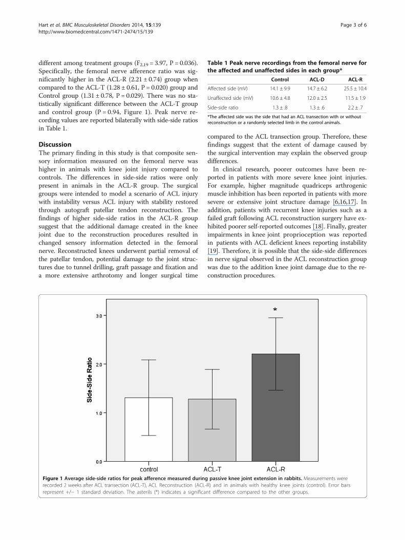
Table 1 Peak nerve recordings from the femoral nerve forthe affected and unaffected sides in each group*
Control ACL-D ACL-R
Affected side (mV) 14.1 ± 9.9 14.7 ± 6.2 25.5 ± 10.4
Unaffected side (mV) 10.6 ± 4.8 12.0 ± 2.5 11.5 ± 1.9
Side-side ratio 1.3 ± .8 1.3 ± .6 2.2 ± .7
*The affected side was the side that had an ACL transection with or withoutreconstruction or a randomly selected limb in the control animals.
Hart et al. BMC Musculoskeletal Disorders 2014, 15:139 Page 3 of 6http://www.biomedcentral.com/1471-2474/15/139
different among treatment groups (F2,19 = 3.97, P = 0.036).Specifically, the femoral nerve afference ratio was sig-nificantly higher in the ACL-R (2.21 ± 0.74) group whencompared to the ACL-T (1.28 ± 0.61, P = 0.020) group andControl group (1.31 ± 0.78, P = 0.029). There was no sta-tistically significant difference between the ACL-T groupand control group (P = 0.94, Figure 1). Peak nerve re-cording values are reported bilaterally with side-side ratiosin Table 1.
DiscussionThe primary finding in this study is that composite sen-sory information measured on the femoral nerve washigher in animals with knee joint injury compared tocontrols. The differences in side-side ratios were onlypresent in animals in the ACL-R group. The surgicalgroups were intended to model a scenario of ACL injurywith instability versus ACL injury with stability restoredthrough autograft patellar tendon reconstruction. Thefindings of higher side-side ratios in the ACL-R groupsuggest that the additional damage created in the kneejoint due to the reconstruction procedures resulted inchanged sensory information detected in the femoralnerve. Reconstructed knees underwent partial removal ofthe patellar tendon, potential damage to the joint struc-tures due to tunnel drilling, graft passage and fixation anda more extensive arthrotomy and longer surgical time
Figure 1 Average side-side ratios for peak afference measured duringrecorded 2 weeks after ACL transection (ACL-T), ACL Reconstruction (ACL-represent +/− 1 standard deviation. The asterils (*) indicates a significa
compared to the ACL transection group. Therefore, thesefindings suggest that the extent of damage caused bythe surgical intervention may explain the observed groupdifferences.In clinical research, poorer outcomes have been re-
ported in patients with more severe knee joint injuries.For example, higher magnitude quadriceps arthrogenicmuscle inhibition has been reported in patients with moresevere or extensive joint structure damage [6,16,17]. Inaddition, patients with recurrent knee injuries such as afailed graft following ACL reconstruction surgery have ex-hibited poorer self-reported outcomes [18]. Finally, greaterimpairments in knee joint proprioception was reportedin patients with ACL deficient knees reporting instability[19]. Therefore, it is possible that the side-side differencesin nerve signal observed in the ACL reconstruction groupwas due to the addition knee joint damage due to the re-construction procedures.
passive knee joint extension in rabbits. Measurements wereR) and in animals with healthy knee joints (control). Error barsnt difference compared to the other groups.

Hart et al. BMC Musculoskeletal Disorders 2014, 15:139 Page 4 of 6http://www.biomedcentral.com/1471-2474/15/139
In the current study, we observed higher magnitudecomposite afferent signal in knees that underwent ACLreconstruction compared to control animals. The signalmeasured from the femoral nerve may include severalsources of afferent information. For example, articularstructures within and around the knee joint containnerve receptors such as free nerve endings, pacinian cor-puscles and ruffini endings [20,21]. These receptors areinnervated by articular branches from the femoral, sa-phenous, obturator, tibial, common peroneal, and recur-rent peroneal nerves [22]. In feline models, it has beenreported that the anterior and posterior knee joint capsuleis densely innervated with Ruffini endings [23] which areslowly adapting mechanorecptors that respond to capsularstretching [24]. The innervation of the knee joint is dis-persed among the various nerve branches. Knee jointinnervation has been previously divided into anterior(articular branches from the femoral, common peronealand saphenous nerve) and posterior (articular branchesfrom the tibial and obturator nerves) [4]. The exact articu-lar distribution is unknown in rabbits, but in other mam-malian systems, components of the anterior group ofnerve fibers, including the femoral and saphenous nervearticular branches, terminate on structures around the an-terior, medial and lateral aspects of the joint capsule andanterior cruciate ligament [4]. Posterior group afferentsterminate on posterior structures and the posterior cruci-ate ligament. Interestingly, branches from the saphenousnerve and the obturator nerve have been reported to forma nerve plexus innervating the posterior capsular struc-tures. In the current study, we certainly did not captureall of the sensory information because we did not recordfrom obturator, tibial or common peroneal nerves. Thecomposite information recorded in the current study likelyincluded information from the femoral nerve and its sen-sory branch, the saphenous nerve. Therefore femoral nervesignal measured in the current study is most likely attrib-uted to afferent signal arising from articular structures in-nervated by the femoral and saphenous nerves.In the current study, we observed higher magnitude
composite signal from the femoral nerve in the recon-structed group only. This finding highlights the fact thatthe anterior cruciate ligament may play an importantrole in conveying sensory information [4]. Mechanorecep-tors such as pacinian corpuscles, golgi tendon organs, andruffini endings are heavily clustered at the proximal anddistal poles of the anterior cruciate ligament [22,25-27]giving rise to afferent proprioceptive information. Duringan ACL reconstruction, it is likely that terminal branchesof sensory nerves are severed as a natural consequence ofthe surgical procedure so its not clear what sources play arole in conveying sensory information following ACL in-jury and reconstruction. In the post-amputee literature ec-topic afferent, nociceptive signaling has been hypothesized
to arise from nerve sprouting from severed nerves [28].In the current study there may be potential relation-ship between the loss of tissue and afferent input. Forexample, reconstruction techniques where the ACL rem-nants are preserved [29-31] have been described as hav-ing good outcomes [32]; attributed to improved improvedvascularization and re-innervation due to the ACL remnant[33,34]. The presence of a remnant in the ACL-T group isone differentiating factor that may partially explain whythis group did not have increased composite afferent sig-nal. The role of tissue preservation and sensory input is anarea for future research.Joint damage often leads to arthrogenic muscle inhib-
ition in the quadriceps musculature [35]. Arthrogenicmuscle inhibition is a unique phenomenon because itexists despite no injury or pathology to the efferentnerve or target muscle. In theory, arthrogenic muscle in-hibition is a reflexive response to aberrant sensory infor-mation arising from damaged joint structures resultingin a failure to voluntarily activate motor units. The re-sponse of Ruffini endings to capsular stretching due tolaxity or joint effusion has been implicated in reflexivemuscular inhibition that is commonly seen in patientswith extensive knee injuries [3,5]. In humans, this mani-fests as persistent muscle weakness, altered gait patternsand joint degeneration [36]. While this may be a protect-ive response in the acutely injured knee, the long termoutcome in persistently inhibited musculature can resultin dysfunction. Therefore, if arthrogenic muscle inhib-ition is persistent following joint injury, recovery maybe impeded. In clinical populations, the quadriceps [16]muscle is commonly affected by arthrogenic muscle inhib-ition which often leads to impaired movement duringwalking gait [37]. Increased afferent information is cur-rently hypothesized to contribute to post-traumatic muscleinhibition. The findings from the current study may bethe basis of future investigations into the potential re-lationships among increased afferent information andquadriceps muscle dysfunction and osteoarthritis in thepost-traumatic knee.There are some limitations to the study due to the pos-
siblity that other factors associated with knee joint injury,such as inflammation and associated chemical mediators,sensitize afferent nociceptive neurons, which may alsocontribute to altered sensory information in the post-traumatic knee [38]. However, we feel that the potentialinfluence of chronic inflammation was minimal becauseupon examining the medical records, none of the rabbitswere showing any outward signs of inflammation at thetime of follow up evaluations nor any changes in behaviorthat would indicate the animals were in pain. All were ac-tive and healthy at the time of terminal measurementstherefore our conclusions are made based on measure-ments recorded when rabbits were in good health and
Hart et al. BMC Musculoskeletal Disorders 2014, 15:139 Page 5 of 6http://www.biomedcentral.com/1471-2474/15/139
recovered from their knee joint surgery. Another limita-tion is the lack of comparison to baseline measurement.Unfortunately, due to the terminal nature of the measure-ment technique, baseline measurements were not able tobe recorded prior to surgery.
ConclusionWe observed higher side-to-side ratios of peak, compositeafferent signal measured directly from the femoral nerveduring passive knee extension in rabbits, 2 weeks follow-ing ACL reconstruction. This difference suggests highermagnitude sensory information from damaged knee jointstructures. This increase in sensory afference may play arole in reflex quadriceps muscle inhibition that is com-monly observed in the post traumatic knee.
AbbreviationsACL: Anterior cruciate ligament; ACL-R: Anterior cruciate ligamentreconstruction group; ACL-T: Anterior cruciate ligament transected group.
Competing interestsWe have no financial disclosures or competing interests relevant to the datapresented in this study. The study was supported by internal universityfunds. All authors declare that they have no competing interests.
Authors’ contributionsJH designed the study, performed all outcome measures and drafted themanuscript. MB assisted with outcome measures and surgeries, assisted withdesign and drafting the mnusctript, LC performed the surgeries, interpret dataand draft the manuscript, MH assisted with study design, data interpretationand manuscript drafting, DD helped with study design, data interpretation andmanuscript drafting. All authors read and approved the final manuscript.
AcknowledgementsWe acknowledge David Hill, PhD from the University of Virginia for hisinsightful comments and guidance throughout this project.
Received: 27 May 2013 Accepted: 10 April 2014Published: 28 April 2014
References1. Nyland J, Brosky T, Currier D, Nitz A, Caborn D: Review of the afferent
neural system of the knee and its contribution to motor learning.J Orthop Sports Phys Ther 1994, 19(1):2–11.
2. Herzog W, Longino D, Clark A: The role of muscles in joint adaptation anddegeneration. Langenbecks Arch Surg 2003, 388(5):305–315.
3. Rice DA, McNair PJ: Quadriceps arthrogenic muscle inhibition: neuralmechanisms and treatment perspectives. Semin Arthritis Rheum 2010,40(3):250–266.
4. Hirasawa Y, Okajima S, Ohta M, Tokioka T: Nerve distribution to the humanknee joint: anatomical and immunohistochemical study. Int Orthop 2000,24(1):1–4.
5. Hopkins JT, Ingersoll CD: Arthrogenic muscle inhibition: a limiting factorin joint rehabilitation. J Sport Rehabil 2000, 9:135–159.
6. Hurley MV, Jones DW, Newham DJ: Arthrogenic quadriceps inhibition andrehabilitation of patients with extensive traumatic knee injuries. Clin Sci(Lond) 1994, 86(3):305–310.
7. Laurin J, Dousset E, Mesure S, Decherchi P: Neuromuscular recoverypattern after medial collateral ligament disruption in rats. J Appl Physiol2009, 107(1):98–104.
8. Laurin J, Dousset E, Mesure S, Decherchi P: Neuromuscular recovery aftermedial collateral ligament disruption and eccentric rehabilitationprogram. Med Sci Sports Exerc 2011, 43(6):1032–1041.
9. Anderson DR, Weiss JA, Takai S, Ohland KJ, Woo SL: Healing of the medialcollateral ligament following a triad injury: a biomechanical andhistological study of the knee in rabbits. J Orthop Res 1992, 10(4):485–495.
10. Kilic BA, Dingil O, Erkula G, Elmas C, Erdogan D, Atik OS: Evaluation of themuscles around the knee in rabbits whose anterior cruciate and/ormedial collateral ligaments were dissected. Arch Orthop Trauma Surg2004, 124(9):626–630.
11. Longino D, Frank C, Herzog W: Acute botulinum toxin-induced muscleweakness in the anterior cruciate ligament-deficient rabbit. J Orthop Res2005, 23(6):1404–1410.
12. Longino D, Frank C, Leonard TR, Vaz MA, Herzog W: Proposed model ofbotulinum toxin-induced muscle weakness in the rabbit. J Orthop Res2005, 23(6):1411–1418.
13. Rehan Youssef A, Longino D, Seerattan R, Leonard T, Herzog W: Muscleweakness causes joint degeneration in rabbits. Osteoarthritis Cartilage2009, 17(9):1228–1235.
14. Wada Y, Takahashi T, Michinaka Y, Morisawa Y, Yamamoto H:Mechanoreceptors of patellar tendon used for ACL reconstruction.Rabbit experiments. Acta Orthop Scand 1997, 68(6):559–562.
15. Hill DL, Phillips LM: Functional plasticity of regenerated and intact tastereceptors in adult rats unmasked by dietary sodium restriction. J Neurosci1994, 14(5 Pt 1):2904–2910.
16. Hart JM, Pietrosimone B, Hertel J, Ingersoll CD: Quadriceps activationfollowing knee injuries: a systematic review. J Athl Train 2010, 45(1):87–97.
17. Hurley MV, Newham DJ: The influence of arthrogenous muscle inhibitionon quadriceps rehabilitation of patients with early, unilateralosteoarthritic knees. Br J Rheumatol 1993, 32(2):127–131.
18. Hart JM, Turman KA, Diduch DR, Hart JA, Miller MD: Quadriceps muscleactivation and radiographic osteoarthritis following ACL revision. Knee SurgSports Traumatol Arthrosc 2011, 19(4):634–640.
19. Roberts D, Friden T, Zatterstrom R, Lindstrand A, Moritz U: Proprioceptionin people with anterior cruciate ligament-deficient knees: comparison ofsymptomatic and asymptomatic patients. J Orthop Sports Phys Ther 1999,29(10):587–594.
20. Friden T, Roberts D, Ageberg E, Walden M, Zatterstrom R: Review ofknee proprioception and the relation to extremity function after ananterior cruciate ligament rupture. J Orthop Sports Phys Ther 2001,31(10):567–576.
21. Solomonow M, Krogsgaard M: Sensorimotor control of knee stability.A review. Scand J Med Sci Sports 2001, 11(2):64–80.
22. Zimny ML: Mechanoreceptors in articular tissues. Am J Anat 1988,182(1):16–32.
23. Bastani A, Hadian MR, Talebian S, Bagheri H, Olyaie GR: Modulation of theipsilateral and contralateral H reflexes following ipsilateral mechanicalpressure of the foot in normal subjects. Electromyogr Clin Neurophysiol2010, 50(5):251–256.
24. Grigg P, Hoffman AH: Properties of Ruffini afferents revealed by stressanalysis of isolated sections of cat knee capsule. J Neurophysiol 1982,47(1):41–54.
25. Schutte MJ, Dabezies EJ, Zimny ML, Happel LT: Neural anatomy ofthe human anterior cruciate ligament. J Bone Joint Surg Am 1987,69(2):243–247.
26. Zimny ML, Schutte M, Dabezies E: Mechanoreceptors in the humananterior cruciate ligament. Anat Rec 1986, 214(2):204–209.
27. Kennedy JC, Alexander IJ, Hayes KC: Nerve supply of the human knee andits functional importance. Am J Sports Med 1982, 10(6):329–335.
28. Hsu E, Cohen SP: Postamputation pain: epidemiology, mechanisms, andtreatment. J Pain Res 2013, 6:121–136.
29. Locherbach C, Zayni R, Chambat P, Sonnery-Cottet B: Biologically enhancedACL reconstruction. Orthop Traumatol Surg Res 2010, 96(7):810–815.
30. Lee BI, Min KD, Choi HS, Kim JB, Kim ST: Arthroscopic anterior cruciateligament reconstruction with the tibial-remnant preserving techniqueusing a hamstring graft. Arthroscopy 2006, 22(3):340 e341–347.
31. Zhang Q, Zhang S, Cao X, Liu L, Liu Y, Li R: The effect of remnantpreservation on tibial tunnel enlargement in ACL reconstruction withhamstring autograft: a prospective randomized controlled trial. Knee SurgSports Traumatol Arthrosc 2014, 22(1):166–173.
32. Ahn JH, Wang JH, Lee YS, Kim JG, Kang JH, Koh KH: Anterior cruciateligament reconstruction using remnant preservation and a femoraltensioning technique: clinical and magnetic resonance imaging results.Arthroscopy 2011, 27(8):1079–1089.
33. Xie GM, Huang Fu XQ, Zhao JZ: The effect of remnant preservation onpatterns of gene expression in a rabbit model of anterior cruciateligament reconstruction. J Surg Res 2012, 176(2):510–516.

Hart et al. BMC Musculoskeletal Disorders 2014, 15:139 Page 6 of 6http://www.biomedcentral.com/1471-2474/15/139
34. Papalia R, Franceschi F, Vasta S, Di Martino A, Maffulli N, Denaro V: Sparingthe anterior cruciate ligament remnant: is it worth the hassle? Br MedBull 2012, 104:91–111.
35. Snyder-Mackler L, De Luca PF, Williams PR, Eastlack ME, Bartolozzi AR 3rd:Reflex inhibition of the quadriceps femoris muscle after injury orreconstruction of the anterior cruciate ligament. J Bone Joint Surg Am1994, 76(4):555–560.
36. Palmieri-Smith RM, Thomas AC: A neuromuscular mechanism ofposttraumatic osteoarthritis associated with ACL injury. Exerc Sport SciRev 2009, 37(3):147–153.
37. Hart JM, Ko JW, Konold T, Pietrosimone B: Sagittal plane knee jointmoments following anterior cruciate ligament injury and reconstruction:a systematic review. Clin Biomech (Bristol, Avon) 2010, 25(4):277–283.
38. Gold MS, Flake NM: Inflammation-mediated hyperexcitability of sensoryneurons. Neurosignals 2005, 14(4):147–157.
doi:10.1186/1471-2474-15-139Cite this article as: Hart et al.: Sensory response following knee jointdamage in rabbits. BMC Musculoskeletal Disorders 2014 15:139.
Submit your next manuscript to BioMed Centraland take full advantage of:
• Convenient online submission
• Thorough peer review
• No space constraints or color figure charges
• Immediate publication on acceptance
• Inclusion in PubMed, CAS, Scopus and Google Scholar
• Research which is freely available for redistribution
Submit your manuscript at www.biomedcentral.com/submit