Embed Size (px)
Citation preview
Sensitization of meningeal nociceptors: inhibition by naproxen
Dan Levy, Xi-Chun Zhang, Moshe Jakubowski, and Rami BursteinDepartments of Anesthesia, Critical Care and Pain Medicine, Beth Israel Deaconess Medical Centerand Harvard Medical School, Boston, MA 02115, USA
AbstractMigraine attacks associated with throbbing (manifestation of peripheral sensitization) and cutaneousallodynia (manifestation of central sensitization) are readily terminated by intravenous administrationof a non-selective cyclooxygenase (COX) inhibitor. Evidence that sensitization of rat centraltrigeminovascular neurons was also terminated in vivo by non-selective COX inhibition has led usto propose that COX inhibitors may act centrally in the dorsal horn. In the present study, we examinedwhether COX inhibition can also suppress peripheral sensitization in meningeal nociceptors. Usingsingle-unit recording in the trigeminal ganglion in vivo, we found that intravenous infusion ofnaproxen, a non-selective COX inhibitor, reversed measures of sensitization induced in meningealnociceptors by prior exposure of the dura to inflammatory soup (IS): ongoing activity of Aδ- and C-units and their response magnitude to mechanical stimulation of the dura, which were enhanced afterIS, returned to baseline after naproxen infusion. Topical application of naproxen or the selectiveCOX-2 inhibitor N-[2-(cyclohexyloxy)-4-nitrophenyl]-methanesulfonamide (NS-398) onto thedural receptive field of Aδ- and C-unit nociceptors also reversed the neuronal hyper-responsivenessto mechanical stimulation of the dura. The findings suggest that local COX activity in the dura couldmediate the peripheral sensitization that underlies migraine headache.
Keywordsheadache; inflammation; migraine; NSAID; pain; trigeminal
IntroductionHaving studied the rat trigeminovascular system as a model for neural pathways underlyingmigraine pain, we have shown that chemical irritation of the cerebral dura mater can activateand sensitize meningeal nociceptors in the trigeminal ganglion and central trigeminovascularneurons in the medullary dorsal horn (Strassman et al., 1996; Burstein, 2001; Levy &Strassman, 2002). We proposed that sensitization of meningeal nociceptors mediates thethrobbing nature of migraine headache, and that sensitization of central trigeminovascularneurons mediates cephalic allodynia and muscle tenderness that develop during migraine(Burstein, 2001). When we asked patients to distinguish between pain intensity and thethrobbing of migraine headache, we found that triptan therapy consistently terminated thethrobbing aspect of the pain; on the other hand, rendering the patient pain free was successfulin the absence, but not in the presence, of well-established allodynia at the time of treatment(Burstein et al., 2004).
Correspondence: Dr D. Levy, as above. E-mail: E-mail: [email protected]'s Disclaimer: Copyright of European Journal of Neuroscience is the property of Blackwell Publishing Limited and its contentmay not be copied or emailed to multiple sites or posted to a listserv without the copyright holder's express written permission. However,users may print, download, or email articles for individual use.
NIH Public AccessAuthor ManuscriptEur J Neurosci. Author manuscript; available in PMC 2009 May 6.
Published in final edited form as:Eur J Neurosci. 2008 February ; 27(4): 917–922. doi:10.1111/j.1460-9568.2008.06068.x.
NIH
-PA Author Manuscript
NIH
-PA Author Manuscript
NIH
-PA Author Manuscript
To understand this discrepancy, we studied the effects of triptans on the induction andmaintenance of peripheral and central sensitization in the trigeminovascular pathway. Wefound that the treatment inhibited neither the sensitized meningeal nociceptors nor thesensitized central neurons, and concluded that triptan action is exerted through blockade ofsynaptic transmission between the two types of neurons (Burstein & Jakubowski, 2004; Levyet al., 2004). In search of drugs capable of directly inhibiting both the peripheral and centralneurons, we focused on non-steroidal anti-inflammatory drugs (NSAIDs) because we foundthat one of them (ketorolac) was effective in terminating migraine pain in allodynic patientswho had failed to respond to triptan therapy earlier in the same attack (Jakubowski et al.,2005).
The analgesic effect of NSAIDs in many inflammatory pain conditions is generally thought tobe exerted through inhibition of the two cyclooxygenase (COX) isoforms (Zeilhofer & Brune,2006). Our working hypothesis was that NSAIDs might inhibit meningeal nociceptors andcentral trigeminovascular neurons through local inhibition of COX activity in the meningesand dorsal horn, respectively. Hitherto, we have found that naproxen, a non-selective COXinhibitor, was extremely effective in suppressing ongoing activity as well as mechanical andthermal hypersensitivity in sensitized central trigeminovascular neurons. We concluded thatthe dorsal horn constitutes at least one critical site of action for this class of drugs (Jakubowskiet al., 2007). The present study focused on NSAID action at the peripheral side of thetrigeminovascular system, meningeal nociceptors. Using extracellular single-unit recordingwe tested whether ongoing sensitization of rat meningeal nociceptors can be reversed byadministration of the non-selective COX inhibitor naproxen, or the more selective COX-2inhibitor N-[2-(cyclohexyloxy)-4-nitrophenyl]-methanesulfonamide (NS-398).
Materials and methodsAnimals
Sprague–Dawley male rats (250–300 g) were used in compliance with the experimentalprotocol approved by the institutional Animal Care and Use Committee of the Harvard MedicalSchool and in accordance with the Ethical Guidelines of the International Association for theStudy of Pain.
ElectrophysiologyRats were deeply anesthetized with an initial intraperitoneal dose of 1.8 g/kg urethane plus 0.2g/kg supplemental doses as needed, and fitted with an intravenous plastic cannula for drug orsaline infusions through the femoral vein. Single-unit recording of meningeal nociceptors inthe trigeminal ganglion was recorded as described before (Strassman et al., 1996; Levy et al.,2005). Briefly, the rat's head was mounted in a stereotaxic apparatus (David Kopf), and a 2 ×2-mm craniotomy was performed to expose the dura over the mid-sagittal and left transversesinuses. The exposed dura was bathed with a modified synthetic interstitial fluid (SIF; in mM:NaCl, 135; KCl, 5; MgCl2, 1; CaCl2, 5; glucose, 10; HEPES, 10; pH 7.2). A platinum-coatedtungsten microelectrode (impedance 500 kΩ; FHC, Bowdoin, ME, USA) was lowered into thetrigeminal ganglion through a small circular opening (2 mm wide) that was drilled in the leftparietal bone, about 2 mm caudal to the Bregma suture and 2 mm left to the midline. Usingrepeated electrical stimulation of the dura overlying the ipsilateral transverse sinus (0.5 mspulse, 5 mA, 0.5 Hz), a neuron exhibiting constant response latencies was identified as ameningeal nociceptor. The site on the dura from which electrical stimulation produced theshortest response latency was identified and used to determine the conduction velocity (CV)of the nociceptor, as described previously (Strassman & Raymond, 1999). Assuming a 12.5-mm conductance distance between the stimulation site on dura and the recording site in thetrigeminal ganglion, a neuron was classified as a C-unit (CV ≤ 1.5 m/s) or an Aδ-unit (CV >
Levy et al. Page 2
Eur J Neurosci. Author manuscript; available in PMC 2009 May 6.
NIH
-PA Author Manuscript
NIH
-PA Author Manuscript
NIH
-PA Author Manuscript
1.5 m/s). A waveform of the action potential evoked by the electrical stimuli was stored as atemplate using a real-time waveform discriminator (Spike 2, CED, Cambridge, UK), whichwas used to acquire experimental data and perform on- and off-line analyses. In all experiments,only one unit was tested in each animal.
Mechanical receptive fields of meningeal nociceptors were mapped initially by stroking thedura with blunt forceps. The lowest threshold point was determined using a series of calibratedvon Frey monofilaments ranging from 0.39 to 58.82 mN (exerting pressure stimuli in the rangeof 38-443 kPa). Response magnitudes to mechanical stimulation were determinedquantitatively, using a servo force-controlled mechanical stimulator (Aurora Scientific,Aurora, ON, Canada) fitted with a flat-ended plastic cylinder (0.5 mm diameter) aimed at thelowest threshold point on the dura. Stimulus trials for testing changes in mechanical sensitivityconsisted of a graded series of three square-wave stimuli (100 ms rise time, 2 s width, 60 sinterstimulus interval) delivered in ascending order, which included a threshold and twosuprathreshold stimuli. Stimuli that evoked 1–2 Hz afferent discharge were considered asthreshold. Suprathreshold stimuli were usually two and four times greater than threshold.Responses to these stimuli, as well as ongoing spontaneous activity, were recorded every 15min throughout the experiment. Baseline measurements of spontaneous and mechanicallyevoked activity were obtained prior to drug administration. Only units that exhibited consistentresponses (changes of less than 0.5 and 2.5 Hz for the threshold and suprathreshold responses,respectively, and 0.3 Hz for the ongoing activity level) in at least three consecutive baselinetrials were tested further.
Experimental designActivation and sensitization of meningeal nociceptors was induced by topically applying aninflammatory soup (IS) containing 0.1 mM prostaglandin E2, 1 mM histamine, serotonin,bradykinin, pH 5.5 (Steen et al., 1992, 1995, 1996). The exposed dura was bathed in IS for 15min followed by wash with SIF as described before (Strassman et al., 1996; Levy & Strassman,2002; Levy et al., 2004). Neurons were tested further only if they showed clear signs ofactivation and sensitization.
Given that sensitization induced by this method persists for at least 2 h (Levy et al., 2004; seealso Fig. 2), the ability of the NSAIDs to reverse peripheral sensitization was tested 1 h afterIS using naproxen sodium, a water-soluble, non-selective COX inhibitor (Sigma, St Louis,MO, USA), or NS-398, a selective COX-2 inhibitor (Cayman Chemicals, Ann Arbor, MI,USA). Naproxen was administered either systemically or directly onto the dural receptive fieldof the neuron. Systemic naproxen administration (1 mg/kg) was performed using 0.25–0.30mL of a 1 mg/mL solution (made using a sterile saline solution) and infused intravenously over5 min. Direct naproxen application was performed by bathing the exposed area of the dura for1 h in 30 μL of a 0.1 mg/mL solution made in SIF. NS-398 was first dissolved indimethylsulfoxide (DMSO) and then SIF (final concentration of DMSO 0.1%), and was appliedtopically as described for naproxen, by bathing the exposed area of the dura for 1 h in 30 μLof a 3 μg/mL solution. Drug doses chosen for this study were derived from previous reports(Morioka et al., 2002; Huntjens et al., 2005) and our own preliminary studies. A control groupwas treated with IS as described above, while receiving an infusion of 0.3 mL saline insteadof naproxen plus SIF (which was the final vehicle for the topically applied naproxen andNS-398) on the dura. At the end of the experiment rats were killed with an intravenous bolusof 1 M KCl.
Data analysisData are presented as mean ± SEM. Aδ- and C-units showed comparable response profiles tothe various treatments and were therefore combined for the purpose of statistical analysis.
Levy et al. Page 3
Eur J Neurosci. Author manuscript; available in PMC 2009 May 6.
NIH
-PA Author Manuscript
NIH
-PA Author Manuscript
NIH
-PA Author Manuscript
Neuronal ongoing activity and responses magnitude to mechanical stimulation of the dura wereanalysed using the Friedman test. The test was first applied on all six time points: baseline, 60min after IS (= time 0) and 15, 30, 45 and 60 min after treatment with NSAID or vehicle. Todissect the effect of NSAID or vehicle treatment, the Friedman test was repeated using the fivetime points from 0 to 60 min. To determine whether the resulting measurements returned tothe initial baseline values, the test was repeated using the 0, 30, 45 and 60 min time points. Forthe first overall analysis, the level of significance was set at 0.05. For the subsequent two posthoc repetitions of the test, the Bonferroni correction was applied to lower the level ofsignificance to 0.025 (i.e. 0.05/2).
ResultsEffects of systemic infusion of naproxen on sensitized meningeal nociceptors
Fourteen meningeal nociceptors were tested for the effects of naproxen infusion: six wereAδ-units (mean CV: 4.09 ± 1.37; range 1.67–10.42 m/s) and eight were C-units (mean CV:0.93 ± 0.13; range 0.48–1.47 m/s; see example Fig. 1A). The control group consisted of 18neurons: nine were Aδ-units (mean CV: 3.87 ± 0.86; range 1.76–10.00 m/s) and nine were C-units (mean CV: 0.83 ± 0.39; range 0.39–1.47 m/s).
In the control group, application of IS on the dura produced significant, long-lasting increasesin neuronal responses to mechanical stimulation of the dura and ongoing firing rate thatremained heightened after vehicle infusion (Friedman test: comparing baseline, 30, 45 and 60min: P ≤ 0.002; Fig. 2A–C). In contrast, responses to mechanical stimuli as well as ongoingactivity, which increased after IS, were significantly suppressed in the group that receivednaproxen infusion (Figs 1B and 3). The resulting threshold response and ongoing activity from30 to 60 min after naproxen infusion were comparable to baseline values (Friedman test:comparing baseline, 30, 45 and 60 min: P ≥ 0.4; Fig. 3A; P ≥ 0.1; Fig. 3C). The resultingsuprathreshold responses from 30 to 60 min were significantly lower than baseline (P ≤ 0.001;Fig. 3B; Friedman test).
Effects of topical application of naproxen on sensitized meningeal nociceptorsEight meningeal nociceptors were studied: four were Aδ-units (mean CV: 3.84 ± 0.81; range2.84-6.50 m/s); four were C-units (mean CV: 0.82 ± 0.16; range 0.52–1.25 m/s). IS-inducedenhanced threshold and suprathreshold response magnitudes were significantly suppressedafter topical application of naproxen on the dura (Fig. 4A and B). The enhanced ongoingactivity also dropped progressively over the same interval, though this change fell short ofreaching statistical significance (Fig. 4C). The resulting threshold and suprathreshold valuesfrom 30 to 60 min after topical application of naproxen on the dura were either comparable tothe baseline values (threshold: P > 0.1; Fig. 4A; Friedman test) or lower than baseline values(suprathreshold: P < 0.025; Fig. 4B; Friedman test).
Effects of topical application of NS-398 on sensitized meningeal nociceptorsTen meningeal nociceptors were studied: five were Aδ-units (mean CV: 2.17 ± 0.29; range1.52–3.13 m/s); five were C-units (mean CV: 0.88 ± 0.18; range 0.46–1.47 m/s). All threemeasures of neuronal sensitization were significantly suppressed after topical application ofNS-398 onto the dura (Fig. 5). The resulting values from 30 to 60 min after NS-398 infusionwere comparable to the respective baseline values (P ≥ 0.2; Fig. 5A; P ≥ 0.1; Fig. 5B and C;Friedman test).
Levy et al. Page 4
Eur J Neurosci. Author manuscript; available in PMC 2009 May 6.
NIH
-PA Author Manuscript
NIH
-PA Author Manuscript
NIH
-PA Author Manuscript
DiscussionSystemic infusion of naproxen, a non-selective COX inhibitor, reversed ongoing sensitizationin meningeal nociceptors. Ongoing neuronal activity and response magnitudes to mechanicalstimulation of the dura, which increased significantly after stimulating the dura with IS,returned to baseline within 60 min of naproxen infusion. Topical application of naproxen (anon-selective COX inhibitor) or NS-398 (a selective COX-2 inhibitor) onto the dural receptivefield of the nociceptor also resulted in reversal of sensitization to mechanical stimulation ofthe dura. In the control group, all measures of sensitization remained elevated throughout theexperiment, confirming that NSAID treatment indeed inhibited sensitization, which wouldhave continued otherwise.
The finding that direct application of naproxen on the dura was as effective as systemicnaproxen infusion in suppressing IS-induced neuronal hyper-responsiveness suggests thatnaproxen action was exerted, at least in part, at the level of the dura mater. That directapplication of NS-398 (10 μm) on the dura produced similar suppression of neuronal hyper-responsiveness as topical application of naproxen (400 μm), likely indicates that naproxenaction was mediated, at least in part, through local inhibition of COX-2 activity in the dura.Such local inhibition of the nociceptor could be mediated potentially through local blockadeof COX-2 in adjacent components of the dura, such as in resident macrophages, blood vessels(Schiltz & Sawchenko, 2002) or even within the nociceptive nerve fibers themselves (Mayeret al., 2007; Levy, D., unpublished results), resulting in decreased levels of prostaglandins inthe vicinity of the nociceptor terminals.
Although blockade of COX activity is generally believed to underlie the analgesic effect ofNSAIDs, the relatively short latency (within 30 min) for inhibition of meningeal nociceptorsmechanosensitivity by both naproxen and NS-398 in our study would be more consistent witha COX-independent mechanism, such as direct inhibition of neuronal excitability through themodulation of sodium currents (Lee et al., 2003; Acosta et al., 2007; Park et al., 2007). Forexample, both tetrodotoxin (TTX)-sensitive and TTX-resistant currents were shown to beinhibited in dissociated dorsal root ganglion neurons by 14 and 97 μm of diclofenac,respectively (Lee et al., 2003), and by 6 μm and 19 μm of celecoxib, respectively (Park et al.,2007). While these in vitro studies cannot be easily compared with our in vivo study, we cannotrule out that naproxen may act similarly by blocking both TTX-sensitive and TTX-resistantsodium channels in meningeal nociceptors.
Intravenous infusion of naproxen, as well as other NSAIDs, is extremely effective insuppressing both sensitized (Jakubowski et al., 2005, 2007) and non-sensitized (Kaube et al.,1993; Ellrich et al., 1999) trigeminovascular neurons in the dorsal horn. The present evidencefor inhibition of sensitized meningeal nociceptors suggests that the inhibitory effect observedin the central neurons is mediated, at least in part, by a peripheral action of NSAIDs in themeninges. Thus, NSAID-mediated inhibition of sensitized peripheral and centraltrigeminovascular neurons is likely to account for their abortive action in migraine therapy.
AcknowledgementsThis study was supported by NIH grants NS46502 (D.L.), NS051484 (R.B.), and grants from Glaxo-Smith-Kline(R.B.) and the National Headache Foundation (D.L.). We thank Dr John M. Kelly for his valuable comments on thestatistical analysis.
Levy et al. Page 5
Eur J Neurosci. Author manuscript; available in PMC 2009 May 6.
NIH
-PA Author Manuscript
NIH
-PA Author Manuscript
NIH
-PA Author Manuscript
AbbreviationsCOX, cyclooxygenase; CV, conduction velocity; DMSO, dimethylsulfoxide; IS, inflammatorysoup; NSAID, non-steroidal anti-inflammatory drug; NS-398, N-[2-(cyclohexyloxy)-4-nitrophenyl]-methanesulfonamide; SIF, synthetic interstitial fluid; TTX, tetrodotoxin.
ReferencesAcosta MC, Luna C, Graff G, Meseguer VM, Viana F, Gallar J, Belmonte C. Comparative effects of the
nonsteroidal anti-inflammatory drug nepafenac on corneal sensory nerve fibers responding to chemicalirritation. Invest. Ophthalmol. Vis. Sci 2007;48:182–188. [PubMed: 17197531]
Burstein R. Deconstructing migraine headache into peripheral and central sensitization. Pain2001;89:107–110. [PubMed: 11166465]
Burstein R, Collins B, Jakubowski M. Defeating migraine pain with triptans: a race against thedevelopment of cutaneous allodynia. Ann. Neurol 2004;55:19–26. [PubMed: 14705108]
Burstein R, Jakubowski M. Analgesic triptan action in an animal model of intracranial pain: a race againstthe development of central sensitization. Ann. Neurol 2004;55:27–36. [PubMed: 14705109]
Ellrich J, Schepelmann K, Pawlak M, Messlinger K. Acetylsalicylic acid inhibits meningeal nociceptionin rat. Pain 1999;81:7–14. [PubMed: 10353488]
Huntjens DR, Danhof M, Della Pasqua OE. Pharmacokinetic-pharmacodynamic correlations andbiomarkers in the development of COX-2 inhibitors. Rheumatology (Oxford) 2005;44:846–859.[PubMed: 15855183]
Jakubowski M, Levy D, Goor-Aryeh I, Collins B, Bajwa Z, Burstein R. Terminating migraine withallodynia and ongoing central sensitization using parenteral administration of COX1/COX2 inhibitors.Headache 2005;45:850–861. [PubMed: 15985101]
Jakubowski M, Levy D, Kainz V, Zhang XC, Kosaras B, Burstein R. Sensitization of centraltrigeminovascular neurons: blockade by intravenous naproxen infusion. Neuroscience 2007;148:573–583. [PubMed: 17651900]
Kaube H, Hoskin KL, Goadsby PJ. Intravenous acetylsalicylic acid inhibits central trigeminal neuronsin the dorsal horn of the upper cervical spinal cord in the cat. Headache 1993;33:541–544. [PubMed:8294191]
Lee HM, Kim HI, Shin YK, Lee CS, Park M, Song JH. Diclofenac inhibition of sodium currents in ratdorsal root ganglion neurons. Brain Res 2003;992:120–127. [PubMed: 14604780]
Levy D, Burstein R, Strassman AM. Calcitonin gene-related peptide does not excite or sensitizemeningeal nociceptors: implications for the pathophysiology of migraine. Ann. Neurol 2005;58:698–705. [PubMed: 16240341]
Levy D, Jakubowski M, Burstein R. Disruption of communication between peripheral and centraltrigeminovascular neurons mediates the antimigraine action of 5HT 1B/1D receptor agonists. Proc.Natl Acad. Sci. USA 2004;101:4274–4279. [PubMed: 15016917]
Levy D, Strassman AM. Distinct sensitizing effects of the cAMP-PKA second messenger cascade on ratdural mechanonociceptors. J. Physiol. (Lond.) 2002;538:483–493. [PubMed: 11790814]
Mayer S, Izydorczyk I, Reeh PW, Grubb BD. Bradykinin-induced nociceptor sensitisation to heat dependson cox-1 and cox-2 in isolated rat skin. Pain 2007;130:14–24. [PubMed: 17196338]
Morioka N, Inoue A, Hanada T, Kumagai K, Takeda K, Ikoma K, Hide I, Tamura Y, Shiomi H, Dohi T,Nakata Y. Nitric oxide synergistically potentiates interleukin-1 beta-induced increase ofcyclooxygenase-2 mRNA levels, resulting in the facilitation of substance P release from primaryafferent neurons: involvement of cGMP-independent mechanisms. Neuropharmacology2002;43:868–876. [PubMed: 12384172]
Park SY, Kim TH, Kim HI, Shin YK, Lee CS, Park M, Song JH. Celecoxib inhibits Na+ currents in ratdorsal root ganglion neurons. Brain Res 2007;1148:53–61. [PubMed: 17359944]
Schiltz JC, Sawchenko PE. Distinct brain vascular cell types manifest inducible cyclooxygenaseexpression as a function of the strength and nature of immune insults. J. Neurosci 2002;22:5606–5618. [PubMed: 12097512]
Levy et al. Page 6
Eur J Neurosci. Author manuscript; available in PMC 2009 May 6.
NIH
-PA Author Manuscript
NIH
-PA Author Manuscript
NIH
-PA Author Manuscript
Steen KH, Issberner U, Reeh PW. Pain due to experimental acidosis in human skin: evidence for non-adapting nociceptor excitation. Neurosci. Lett 1995;199:29–32. [PubMed: 8584219]
Steen KH, Reeh PW, Anton F, Handwerker HO. Protons selectively induce lasting excitation andsensitization to mechanical stimulation of nociceptors in rat skin, in vitro. J. Neurosci 1992;12:86–95. [PubMed: 1309578]
Steen KH, Steen AE, Kreysel HW, Reeh PW. Inflammatory mediators potentiate pain induced byexperimental tissue acidosis. Pain 1996;66:163–170. [PubMed: 8880837]
Strassman AM, Raymond SA. Electrophysiological evidence for tetrodotoxin-resistant sodium channelsin slowly conducting dural sensory fibers. J. Neurophysiol 1999;81:413–424. [PubMed: 10036248]
Strassman AM, Raymond SA, Burstein R. Sensitization of meningeal sensory neurons and the origin ofheadaches. Nature 1996;384:560–564. [PubMed: 8955268]
Zeilhofer HU, Brune K. Analgesic strategies beyond the inhibition of cyclooxygenases. TrendsPharmacol. Sci 2006;27:467–474. [PubMed: 16876882]
Levy et al. Page 7
Eur J Neurosci. Author manuscript; available in PMC 2009 May 6.
NIH
-PA Author Manuscript
NIH
-PA Author Manuscript
NIH
-PA Author Manuscript
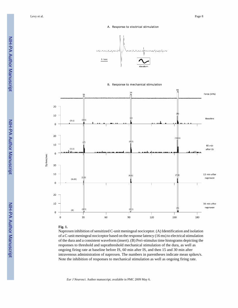
Fig. 1.Naproxen inhibition of sensitized C-unit meningeal nociceptor. (A) Identification and isolationof a C-unit meningeal nociceptor based on the response latency (16 ms) to electrical stimulationof the dura and a consistent waveform (insert). (B) Peri-stimulus time histograms depicting theresponses to threshold and suprathreshold mechanical stimulation of the dura, as well asongoing firing rate at baseline before IS, 60 min after IS, and then 15 and 30 min afterintravenous administration of naproxen. The numbers in parentheses indicate mean spikes/s.Note the inhibition of responses to mechanical stimulation as well as ongoing firing rate.
Levy et al. Page 8
Eur J Neurosci. Author manuscript; available in PMC 2009 May 6.
NIH
-PA Author Manuscript
NIH
-PA Author Manuscript
NIH
-PA Author Manuscript
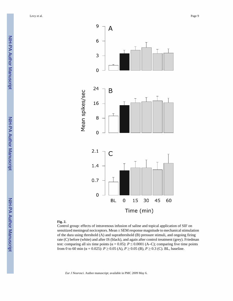
Fig. 2.Control group: effects of intravenous infusion of saline and topical application of SIF onsensitized meningeal nociceptors. Mean ± SEM response magnitude to mechanical stimulationof the dura using threshold (A) and suprathreshold (B) pressure stimuli, and ongoing firingrate (C) before (white) and after IS (black), and again after control treatment (grey). Friedmantest: comparing all six time points (α = 0.05): P ≤ 0.0001 (A–C); comparing five time pointsfrom 0 to 60 min (α = 0.025): P ≥ 0.05 (A), P ≥ 0.05 (B), P ≥ 0.3 (C). BL, baseline.
Levy et al. Page 9
Eur J Neurosci. Author manuscript; available in PMC 2009 May 6.
NIH
-PA Author Manuscript
NIH
-PA Author Manuscript
NIH
-PA Author Manuscript
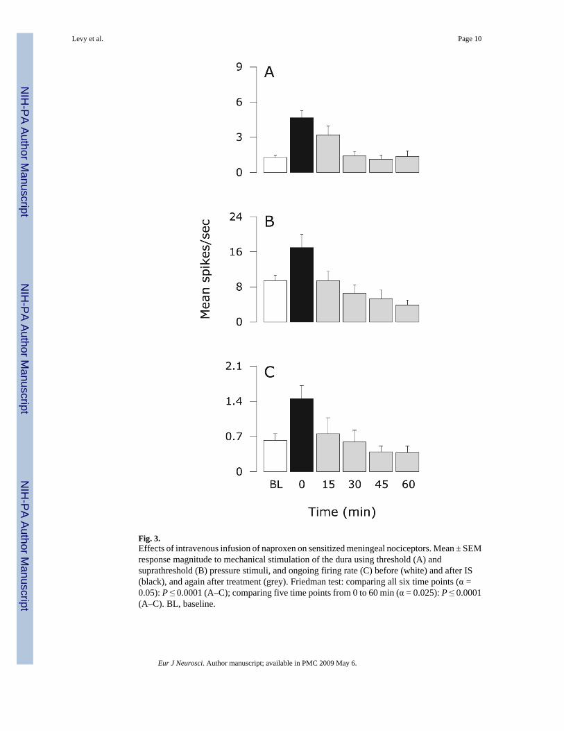
Fig. 3.Effects of intravenous infusion of naproxen on sensitized meningeal nociceptors. Mean ± SEMresponse magnitude to mechanical stimulation of the dura using threshold (A) andsuprathreshold (B) pressure stimuli, and ongoing firing rate (C) before (white) and after IS(black), and again after treatment (grey). Friedman test: comparing all six time points (α =0.05): P ≤ 0.0001 (A–C); comparing five time points from 0 to 60 min (α = 0.025): P ≤ 0.0001(A–C). BL, baseline.
Levy et al. Page 10
Eur J Neurosci. Author manuscript; available in PMC 2009 May 6.
NIH
-PA Author Manuscript
NIH
-PA Author Manuscript
NIH
-PA Author Manuscript
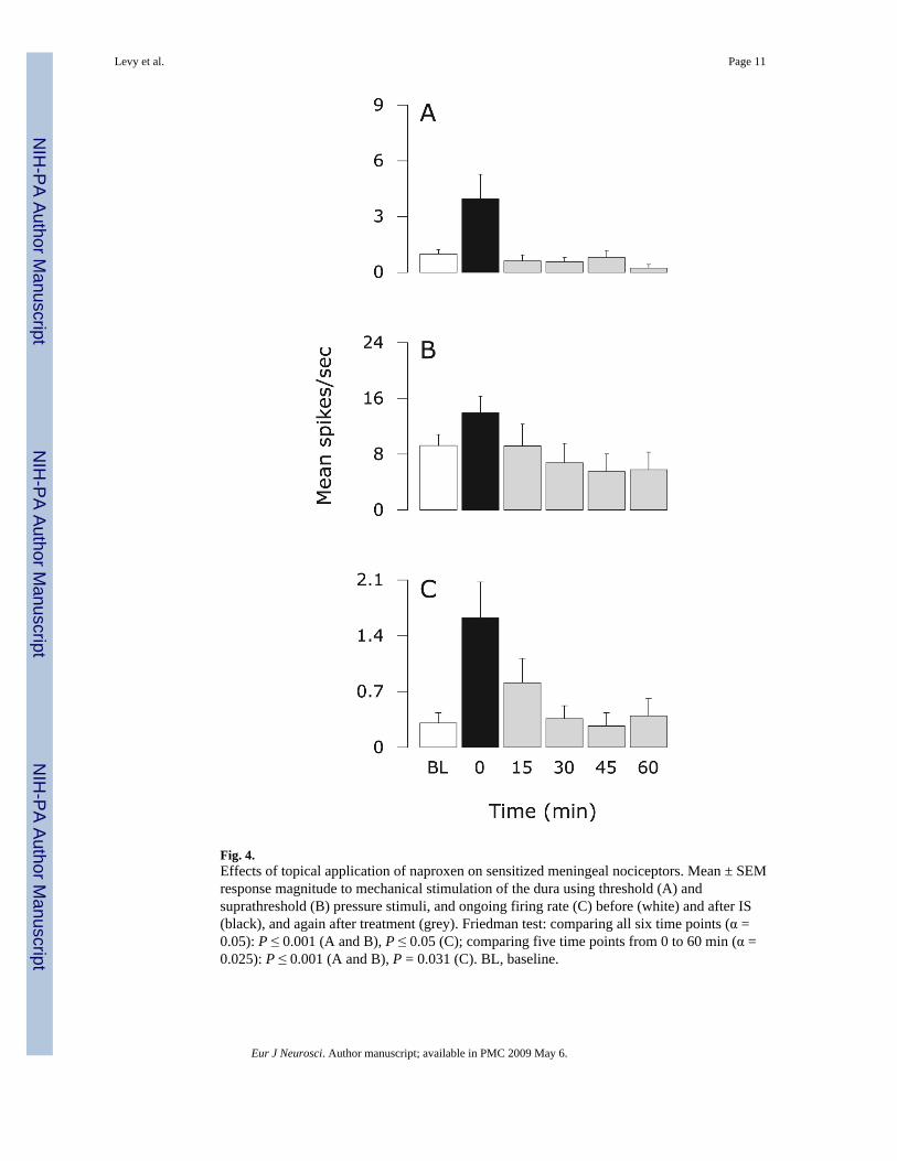
Fig. 4.Effects of topical application of naproxen on sensitized meningeal nociceptors. Mean ± SEMresponse magnitude to mechanical stimulation of the dura using threshold (A) andsuprathreshold (B) pressure stimuli, and ongoing firing rate (C) before (white) and after IS(black), and again after treatment (grey). Friedman test: comparing all six time points (α =0.05): P ≤ 0.001 (A and B), P ≤ 0.05 (C); comparing five time points from 0 to 60 min (α =0.025): P ≤ 0.001 (A and B), P = 0.031 (C). BL, baseline.
Levy et al. Page 11
Eur J Neurosci. Author manuscript; available in PMC 2009 May 6.
NIH
-PA Author Manuscript
NIH
-PA Author Manuscript
NIH
-PA Author Manuscript
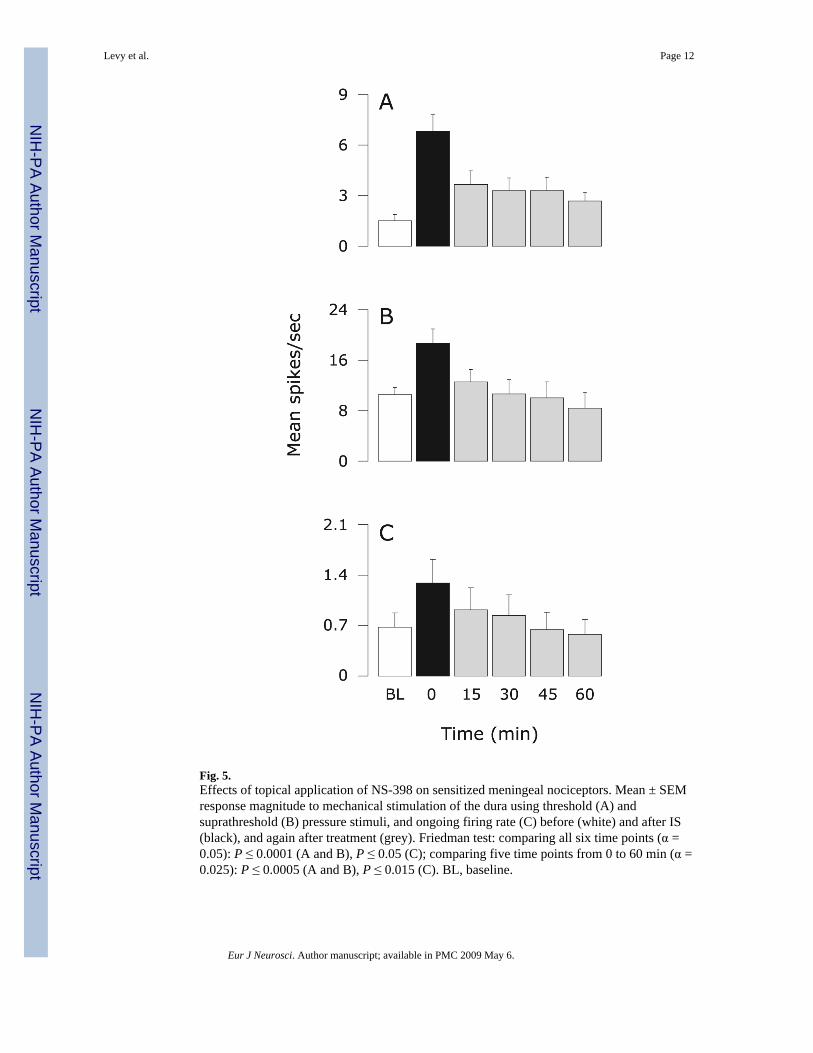
Fig. 5.Effects of topical application of NS-398 on sensitized meningeal nociceptors. Mean ± SEMresponse magnitude to mechanical stimulation of the dura using threshold (A) andsuprathreshold (B) pressure stimuli, and ongoing firing rate (C) before (white) and after IS(black), and again after treatment (grey). Friedman test: comparing all six time points (α =0.05): P ≤ 0.0001 (A and B), P ≤ 0.05 (C); comparing five time points from 0 to 60 min (α =0.025): P ≤ 0.0005 (A and B), P ≤ 0.015 (C). BL, baseline.
Levy et al. Page 12
Eur J Neurosci. Author manuscript; available in PMC 2009 May 6.
NIH
-PA Author Manuscript
NIH
-PA Author Manuscript
NIH
-PA Author Manuscript


