Embed Size (px)
Citation preview
RSC Advances
PAPER
Selective G-quad
aDipartimento di Scienze e Tecnologie Biol
delle Scienze, Edicio 17, 90128 Palermo,
it; Fax: +39 091 596825bSwedish Medical Nanoscience Center, D
Institutet, Stockholm, SwedencIstituto EuroMediterraneo di Scienza e Tec
Palermo, Italy
† Electronic supplementary information(Fig. S1–S3) and tables (Tables S1 and S2)energies, in vacuo and in solution, thermVideo le showing the molecular dynamiand c-myc G4-DNA. See DOI: 10.1039/c4ra
Cite this: RSC Adv., 2014, 4, 33245
Received 5th June 2014Accepted 15th July 2014
DOI: 10.1039/c4ra05355a
www.rsc.org/advances
This journal is © The Royal Society of C
ruplex stabilizers: Schiff-basemetal complexes with anticancer activity†
Alessio Terenzi,a Riccardo Bonsignore,a Angelo Spinello,a Carla Gentile,a
Annamaria Martorana,a Cosimo Ducani,b Bjorn Hogberg,b Anna Maria Almerico,a
Antonino Lauriaa and Giampaolo Barone*ac
The affinity of three square-planar nickel(II) (1), copper(II) (2) and zinc(II) (3) Schiff-base complexes for wild-
type human telomeric (h-Telo) and protooncogene c-mycG-quadruplex (G4) DNAwas investigated by UV-
visible absorption spectroscopy and circular dichroism. DNA-binding constants (Kb) were determined by
spectrophotometric titrations for both G4-DNA and B-DNA. The results obtained point out that the
three metal complexes selectively bind G4-DNA with higher affinity, up to two orders of magnitude, with
respect to B-DNA. The nickel(II) complex 1 was found to be the most effective G4-DNA stabilizer and the
Kb values decrease in the order 1 > 2 z 3. Innovative computational investigations, consisting of
molecular dynamics (MD) simulations followed by density functional theory/molecular mechanics
(DFT/MM) calculations, provide atomistic support for the interpretation of the binding mechanism to
G4-DNA by end stacking and also of the experimental affinity order. Interestingly, 1 is able to induce
G4-DNA formation of h-Telo sequences, also in the absence of K+ cations. This last result is nicely
confirmed and highlighted by polymerase chain reaction (PCR) stop assays, which show the ability of the
title compounds to induce and stabilize G4 structures inhibiting the amplification of PCR products.
Finally, compounds 1–3 showed concentration and time-dependent cytotoxicity towards HeLa and
MCF-7 human cancer cell lines, inducing significant effects on cell cycle distribution with G2/M arrest in
HeLa cells and G0/G1 arrest in MCF-7 cells. Overall, the PCR inhibition and anticancer activity of the
three compounds decreases in the same order 1 > 2 z 3, in excellent correlation with the G4-DNA-
binding affinity, implying that G4-DNA is the biotarget for their biological activity.
1. Introduction
Since the discovery by Rosenberg et al. that cisplatin is a highlyeffective anticancer agent,1 DNA has been considered a majortarget for anticancer drugs, and actually is the target of the mostcommon clinically used platinum-based drugs.2 Unluckily,platinum anticancer drugs, which covalently bind the poly-nucleotide, oen present serious side-effects. For such reason,in the last 40 years there was a growing research interest in thestudy of DNA non-covalent recognition by small molecules.3–6
ogiche, Chimiche e Farmaceutiche, Viale
Italy. E-mail: giampaolo.barone@unipa.
epartment of Neuroscience, Karolinska
nologia, Via Emerico Amari 123, 90139
(ESI) available: Additional gures, reporting PCR inhibition assays, DFTal corrections, Cartesian coordinates.
cs of the binding between compound 305355a
hemistry 2014
Despite many of these compounds have been extensivelystudied and some of them clinically used,3,7 their serious sideeffects and lack of selectivity resulted in a gradual loss ofinterest. DNA has lost his initial “appeal” as target mainly due tothe discover of more specic cellular targets like proteins,enzymes and cell surface receptors, among others.8–12
Nevertheless, new ndings in DNA non-canonical struc-tural arrangements with possible roles in carcinogenicevents gave to DNA-based drugs a new impetus.13–15 Telo-meres, for instance, are able to organize themselves in four-stranded DNA structures, termed guanine-quadruplexes (G4).G4s, in general terms, can be dened as G-rich sequencescapable of forming highly polymorphic 4-stranded structuresorganized in stacked guanine tetrads connected by loopingDNA bases and stabilized by a central alkali ion channel.16
The propensity of a sequence to fold into a particularsecondary structure is inuenced by a number of factorsincluding the nature of the central ion, the relative directionof the strands, the syn or anti glycosidic conformation,the length of the sequence connecting the strands (i.e. theloops) and, in general terms, by the folding experimentalconditions.16–18
RSC Adv., 2014, 4, 33245–33256 | 33245

Fig. 1 Structure of the ML2+ complex (1: M¼ Ni, 2: M¼ Cu; 3: M¼ Zn;H2L
2+ ¼ N,N’-bis-5-(triethyl ammonium methyl)-salicylidene-2,3-naphthalendiimine).
RSC Advances Paper
G4s in telomeres were found to be involved in maintainingchromosome stability through the inhibition of telomerase, aribonucleoprotein complex with reverse transcriptase activity,19
which turns on to elongate the telomeric overhangs, with acorresponding extension of the cell life. Indeed, telomerase isover-expressed in ca. 80–85% of cancer cells and is responsibleof their immortalization. Hence, the inhibition of telomerase,through the folding of its substrate in G4 conformation, isnowadays considered a smart and selective anticancerstrategy.20
As human genome presents approximately 350 000 guanine-rich sequences,21 it is not strange the nding that G4-DNAstructures are over-represented not only in telomeres but also ingene promoter regions, making them even more attractive astherapeutic targets in oncology.8 For example, the proto-onco-gene c-myc presents a putative G4-DNA in the nuclease hyper-sensitive element (NHE).22,23 The aberrant overexpression ofc-myc is associated with a variety of malignant cancers.22 Foldingpatterns of several G4s motifs in promoter regions, described aspossible molecular switch in transcriptional regulation,8 havebeen proposed, including c-myc, c-kit, KRAS, PDGF-A, hTERT andHIF.8,21,24 Futhermore, recent works emphasize that G4 struc-tures were found also in RNA G-rich sequences, and that theyseem to play a key role in post-transcriptional control of geneexpression.25
Many research groups have worked to identify or designsmall-molecule ligands, which specically bind to the G4-DNAinhibiting cell proliferation.16–18,26 To date, molecules able tostabilize a G4 structure present specic features, like a p-delo-calised system in order to p-stack with the terminal G-quartetsand positively charged substituent able to interact with thegrooves. It has been recently reported that planar aromaticorganic molecules complexed with transition metal ions areattractive systems for quadruplex binding.20,27,28 The presence ofa metal ion, due to an electronwithdrawing effect, reduces theelectron density on the coordinated aromatic ligands andinduces stronger p interactions with the G-quartets.20 Further-more, the metal ion increases the electrostatic G4 stabilizationby positioning at the center of a G-tetrad and ideally continuingthe central ion channel normally created by alkali metalcations.27
The principal effort in G4-DNA binders design concernstarget selectivity. The ideal ligand should bind a G4-DNAstructure with high affinity and recognize specically theG4-DNA in preference to the duplex B-DNA.29
Cationic Salphen-like metal complexes, already known to beB-DNA binders,30–32 represent a powerful class of G4-DNAstabilizers.20 For istance, Vilar et al. reported the synthesis of aseries of square planar transition metal complexes with sal-phen-like N,N0-bridged tetradentate ligands with a surprisinglyability to stabilize human telomeric DNA with considerableaffinity and selectivity.33,34
With the aim to extend the library of Schiff-base G4-bindingmetal complexes and to increase their selectivity over B-DNA,three square-planar cationic complexes, ML2+ (M ¼ Ni, Cu, andZn), recently synthesized and characterized by an extendednearly planar area (Fig. 1),35 have been tested as G4 stabilizers
33246 | RSC Adv., 2014, 4, 33245–33256
and their binding affinity compared to that toward B-DNA.Circular dichroism (CD) and UV-visible (UV-vis) absorptionspectroscopy allowed us to monitor the metal complex-G4interaction and to discriminate the quadruplex fold from otherarchitectures. Computational chemistry methods have beenused to provide atomistic models of the supramolecular metalcomplex-G4 binding complexes.
We have taken our studies further to demonstrate the effectof the selected compounds on the DNA processing through invitro polymerase chain reaction (PCR) assays. Moreover, we haveevaluated the related antiproliferative activity towards HeLa andMCF-7 cancer cell lines.
2. Results and discussion2.1. Absorption spectroscopy
To investigate the DNA recognition properties of compounds1–3 and, in particular, their selectivity towards G4-DNA, UV-vistitrations with 50-(AGGGTT)3AGGG-30 (h-Telo G4), 50-GGGAGGGTGGGGAGGGTGGG-30 (c-myc G4) and ct-DNA wereperformed (Fig. 2 and S1 of the ESI†).
Compounds 1–3 present a metal center in a +2 oxidationstate but differing for the number of d electrons and share anintense absorption band at about 250 nm (black solid lines inFig. 2a–c). Moreover, characteristic absorption bands arenoticeable in 1 (346 and 467 nm), 2 (316 and 406 nm) and 3 (304and 384 nm).
Such spectra are signicantly modied by the addition ofincreasing amounts of the selected h-Telo and c-myc G4 oligo-nucleotides (Fig. 2). The addition of increasing amounts of G4-DNA produces a considerable hypochromic and bathochromiceffect of the metal complex intraligand p–p* band. In detail, ahypochromic effect of about 24% for compounds 1–2, and of14% for 3 is observed, with a red shi of about 4 nm for thethree metal complexes. The results, almost identical for h-Teloand c-myc, are in agreement with an end-stacking bindingmode.36 Structural details of the metal complex-G4 interaction,nicely explaining the observed spectroscopic properties, wereobtained by the computational studies discussed below.
The B-DNA binding abilities by intercalation of the synthe-sized metal complexes are already known from our studiesrecently published.35 However, to be quantitatively compared,
This journal is © The Royal Society of Chemistry 2014

Fig. 2 Absorption spectra of 1 (a and b), 2 (c and d) and 3 (e and f), inpresence of increasing amounts of h-Telo G4-DNA (left column) andc-myc G4-DNA (right column) in Tris-HCl buffer 50 mM and KCl100 mM. (a) [1] ¼ 13.4 mM, (b) [1] ¼ 13.7 mM, (c) [2] ¼ 35.1 mM, (d) [2] ¼19.4 mM, (e) [3]¼ 21.8 mM, (f) [3]¼ 27.3 mM. Ratios R¼ [DNA]/[ML2+] arein the range 0.00–0.30 for all the titrations. The arrows indicate thechange upon G4-DNA addition.
Table 1 DNA-binding constants, Kb (M�1), of the three metalcomplexes 1–3 with G4-DNA (h-Telo and c-myc) and B-DNA (ct-DNA)
ct-DNA h-Telo c-myc
1 (4.43 � 0.37) � 104 (2.16 � 0.57) � 106 (1.54 � 0.20) � 106
2 (1.68 � 0.13) � 104 (2.04 � 0.12) � 105 (4.46 � 0.42) � 105
3 (1.33 � 0.14) � 104 (1.98 � 0.21) � 105 (1.16 � 0.28) � 105
Paper RSC Advances
the titrations with ct-DNA and G4-DNA have been performed byusing the same experimental conditions. In particular, it isknown that the ionic strength of the medium strongly affectsthe interaction of the negatively charged double helical polymerand the positively charged molecules.37 The effect of ionicstrength on the binding constant can be rationalized by theRecord equation,38 in which the decrease of the bindingconstant, D(log K) versus the incremental ionic strength,D(�log I), must be linear.
In details, the absorption band of 1 at 346 nm (black line inFig. S1a†) is red shied by about 5 nm and shows hypo-chromism of about 26%. The absorption band of 2 at 316 nm(black line in Fig. S1b†) is red shied by about 5 nm and showshypochromism of about 22%. Finally, the absorption band of 3at 304 nm (black line in Fig. S1c†) is red-shied by about 3 nmand shows hypochromism of about 11.9%. These results,mainly caused by stacking interaction between the extendedaromatic rings of the Schiff-base metal complexes and the basepairs of DNA,37,39,40 collectively conrm that 1–3 act as DNAintercalators also at high ionic strength conditions.
To determine the intrinsic binding constant (Kb) of the ML2+/DNA systems, the quantity [DNA]/|3a� 3f| at 346 nm for 1, at 407nm for 2 and at 304 nm for 3 nm has been plotted, as a functionof themolar concentration of DNA (insets in Fig. 2 and S1†). Thebinding constants were obtained by tting the data to a
This journal is © The Royal Society of Chemistry 2014
reciprocal plot of [DNA]/|3a � 3f| versus [DNA] using thefollowing equation:20
[DNA]/|3a � 3f| ¼ [DNA]/|3b � 3f| + 1/(|3b � 3f| � Kb) (1)
where the concentration of DNA is expressed in terms ofmonomer units. In details, 3a ¼ Aobserved/[ML2+], 3b is theextinction coefficient of the DNA bound complex, and 3f is theextinction coefficient of the free complex determined by a cali-bration curve of the isolated metal complexes in aqueoussolution, following the Beer–Lambert law. The Kb valuesobtained by the linear ts of the experimental data using eqn (1)are reported in Table 1.
These results conrm that eachmetal complex interacts withboth B- and G4-DNA secondary structures and that the bindingof NiII complex is tighter than CuII which is tighter than ZnII,following the order 1 > 2 z 3. Most importantly, the threecompounds show binding selectivity for G4 structures. In fact,while the binding constant of compounds 2 and 3 for both h-Telo and c-myc G4-DNA is about 10 times higher than that for ct-DNA, this value increases to about 100 times higher for thenickel(II) compound 1. In this respect, it has been recentlyreported that, to achieve sequence-specic DNA targeting, theideal binding affinity between specic and nonspecic sitesshould be approximately 1000 times.41 However, such selectivitywas up to date not yet reached. For example, highly activetelomerase inhibitors bind to human quadruplex DNA only 30–40 times more strongly than to duplex DNA.42 By a comparisonwith the binding data so far reported, this means that thebinding selectivity reached by the nickel(II) compound is greaterthan that obtained for most selective G4-binders known up todate.
2.2. Circular dichroism
CD is an essential method for the structural characterizationof G4-DNA in solution. This technique is highly sensitive tolittle variations of the chiral conformation of an opticallyactive biomolecule.43–45 In G4-DNA the chromophoresabsorbing in the UV-Vis region are represented by guanines,with two well-isolated absorption bands which are connectedto two well characterized short and long axis polarised p–p*
transitions at ca. 279 nm and 248 nm. The fact that thestacked G-tetrads are rotated one with respect to the otherscauses chiral exciton coupling between transition dipolemoments located in near-neighbour guanines. This chiralDNA structure is thus active for CD studies and drug–DNAinteractions can be monitored.46
RSC Adv., 2014, 4, 33245–33256 | 33247
RSC Advances Paper
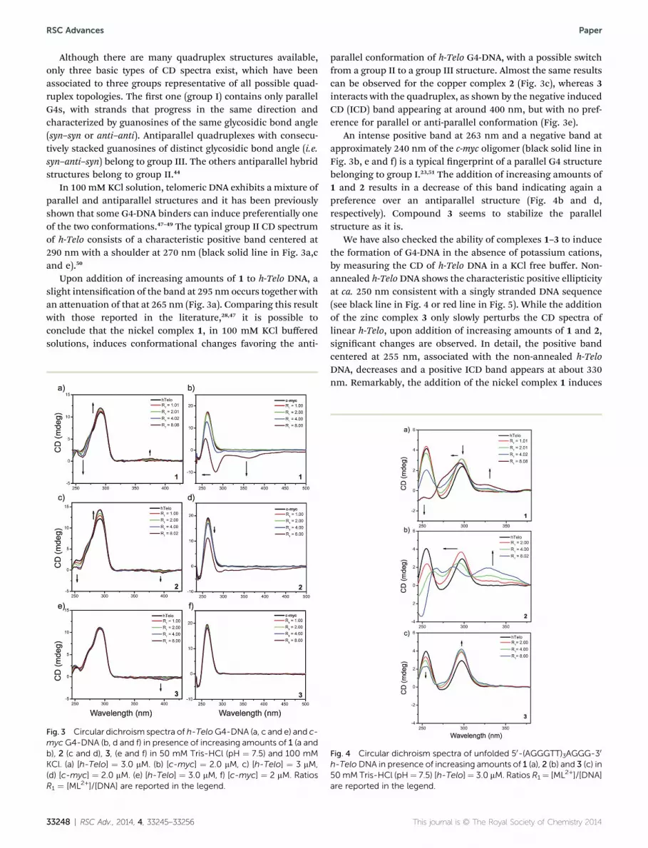
Although there are many quadruplex structures available,only three basic types of CD spectra exist, which have beenassociated to three groups representative of all possible quad-ruplex topologies. The rst one (group I) contains only parallelG4s, with strands that progress in the same direction andcharacterized by guanosines of the same glycosidic bond angle(syn–syn or anti–anti). Antiparallel quadruplexes with consecu-tively stacked guanosines of distinct glycosidic bond angle (i.e.syn–anti–syn) belong to group III. The others antiparallel hybridstructures belong to group II.44
In 100 mM KCl solution, telomeric DNA exhibits a mixture ofparallel and antiparallel structures and it has been previouslyshown that some G4-DNA binders can induce preferentially oneof the two conformations.47–49 The typical group II CD spectrumof h-Telo consists of a characteristic positive band centered at290 nm with a shoulder at 270 nm (black solid line in Fig. 3a,cand e).50
Upon addition of increasing amounts of 1 to h-Telo DNA, aslight intensication of the band at 295 nm occurs together withan attenuation of that at 265 nm (Fig. 3a). Comparing this resultwith those reported in the literature,28,47 it is possible toconclude that the nickel complex 1, in 100 mM KCl bufferedsolutions, induces conformational changes favoring the anti-
Fig. 3 Circular dichroism spectra of h-TeloG4-DNA (a, c and e) and c-myc G4-DNA (b, d and f) in presence of increasing amounts of 1 (a andb), 2 (c and d), 3, (e and f) in 50 mM Tris-HCl (pH ¼ 7.5) and 100 mMKCl. (a) [h-Telo] ¼ 3.0 mM. (b) [c-myc] ¼ 2.0 mM, c) [h-Telo] ¼ 3 mM,(d) [c-myc] ¼ 2.0 mM. (e) [h-Telo] ¼ 3.0 mM, f) [c-myc] ¼ 2 mM. RatiosR1 ¼ [ML2+]/[DNA] are reported in the legend.
33248 | RSC Adv., 2014, 4, 33245–33256
parallel conformation of h-Telo G4-DNA, with a possible switchfrom a group II to a group III structure. Almost the same resultscan be observed for the copper complex 2 (Fig. 3c), whereas 3interacts with the quadruplex, as shown by the negative inducedCD (ICD) band appearing at around 400 nm, but with no pref-erence for parallel or anti-parallel conformation (Fig. 3e).
An intense positive band at 263 nm and a negative band atapproximately 240 nm of the c-myc oligomer (black solid line inFig. 3b, e and f) is a typical ngerprint of a parallel G4 structurebelonging to group I.23,51 The addition of increasing amounts of1 and 2 results in a decrease of this band indicating again apreference over an antiparallel structure (Fig. 4b and d,respectively). Compound 3 seems to stabilize the parallelstructure as it is.
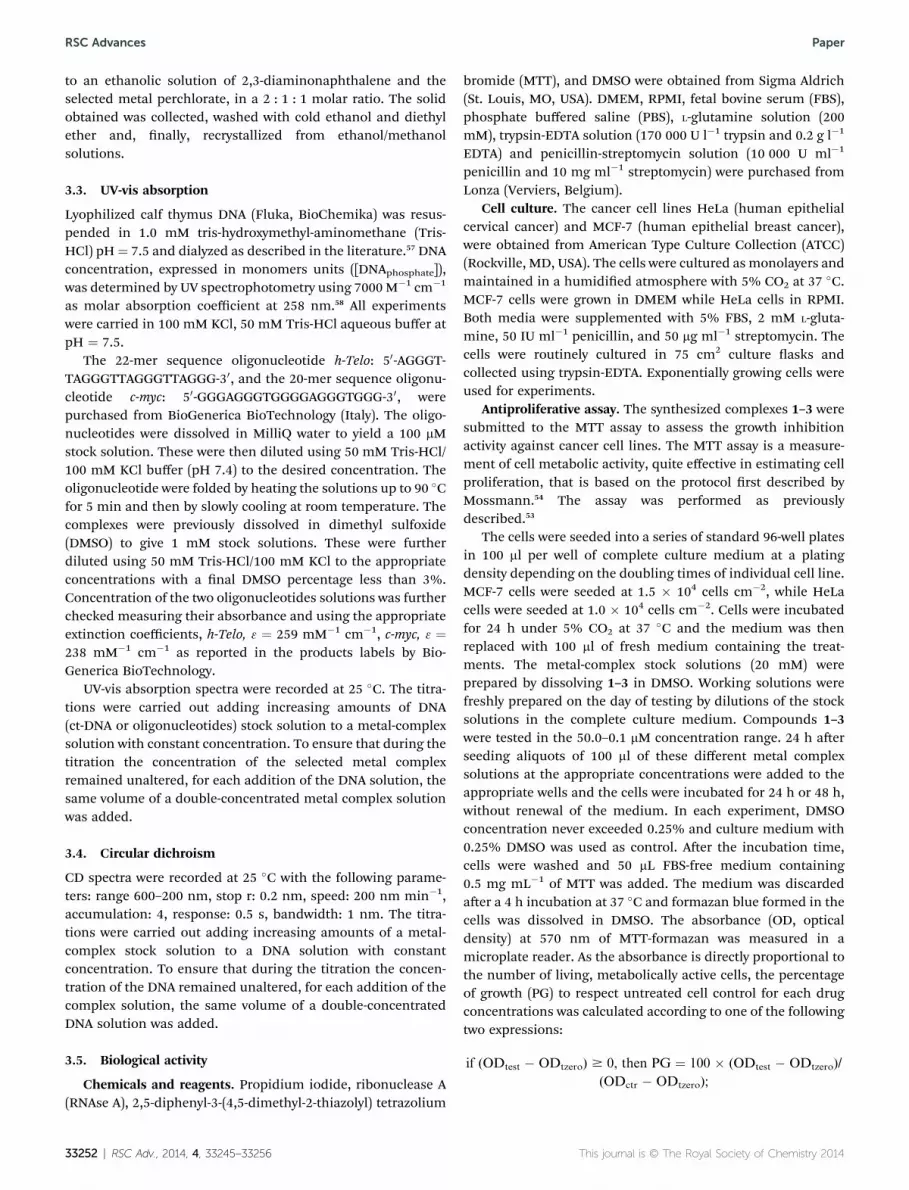
We have also checked the ability of complexes 1–3 to inducethe formation of G4-DNA in the absence of potassium cations,by measuring the CD of h-Telo DNA in a KCl free buffer. Non-annealed h-Telo DNA shows the characteristic positive ellipticityat ca. 250 nm consistent with a singly stranded DNA sequence(see black line in Fig. 4 or red line in Fig. 5). While the additionof the zinc complex 3 only slowly perturbs the CD spectra oflinear h-Telo, upon addition of increasing amounts of 1 and 2,signicant changes are observed. In detail, the positive bandcentered at 255 nm, associated with the non-annealed h-TeloDNA, decreases and a positive ICD band appears at about 330nm. Remarkably, the addition of the nickel complex 1 induces
Fig. 4 Circular dichroism spectra of unfolded 50-(AGGGTT)3AGGG-30
h-TeloDNA in presence of increasing amounts of 1 (a), 2 (b) and 3 (c) in50mM Tris-HCl (pH¼ 7.5) [h-Telo]¼ 3.0 mM. Ratios R1¼ [ML2+]/[DNA]are reported in the legend.
This journal is © The Royal Society of Chemistry 2014

Fig. 5 Circular dichroism spectra of unfolded 50-(AGGGTT)3AGGG-30
h-Ttelo 3 mM (red line), G4 folded in presence of K+ 100mM (black line)and G4 folded in presence of 20 mM 1. All spectra were recorded in 50mM Tris-HCl.
Paper RSC Advances
also an increase of the positive peaks at 295 nm and of theshoulder at 265 nm, indicative of the formation of antiparalleland of parallel G4-DNA, respectively (see Fig. 4 and 5). Thisresult indicates that complex 1 is able to induce the formationof G4-DNA even in the absence of K+ cations.
2.3. MD simulations and DFT/MM calculations
MD simulations have been performed on the two complexesbetween 3 and both c-myc and h-Telo sequences in G4-DNAconformation. The root mean square deviations (RMSD) alongthe simulation, for all non-hydrogen atoms and for the guaninebases, are shown in Fig. 6 and S2 of the ESI,† respectively. Theresults of the MD simulations show that the strong interactionbetween the zinc(II) complex 3 and both G4-DNA models isdriven by the strong electrostatic attraction between thepositively charged triethylammoniummethyl groups of the
Fig. 6 Plot of the RMSD obtained for 3/c-myc up to 150 ns of MDsimulations.
This journal is © The Royal Society of Chemistry 2014
Schiff-base ligand and the negatively charged phosphate groupsof the biomolecule. This long-range interaction allows the metalcomplex to easily approach the biomolecule. Moreover, a strongp–p stacking interaction occurs at the equilibrium, between thenaphthalene moiety of the Schiff-base ligand and the terminalG-tetrad. The three snapshots reported in the RMSD plot inFig. 6 nicely describe the dynamics of the approaching of thezinc(II) complex toward the c-myc G4 structure. In particular, it isworth noting that the guanine basis lying above the terminalG-tetrad, colored in green, performs a rotation of about 90�
around its glycosidic bond and that the larger rotation isabruptly obtained at about 100 ns, as highlighted by the step inthe RMSD of the guanine bases (red line in Fig. 6). This rotationallows a suitable stacking interaction between the naphthalenemoiety of the metal complex and the G-tetrad of c-myc G4-DNA.The binding mechanism can be appreciated also by looking atthe ESI video le video.mpg,† with a hyperlink in the onlineversion of this paper, which shows a movie of the moleculardynamics described in Fig. 6.
Concerning the interaction with the h-Telo G4, the RMSDplot in Fig. S2† shows that the equilibrium is quickly reached atabout 5 ns and the stacked metal complex remains tightlybound to the biomolecule up to the end of the MD simulation.The equilibrium geometry, aer about 50 ns, has been used asstarting point for further geometry optimizations, by hybridtwo-layer QM/MM calculations, using DFT as QM method andthe Amber99 force eld as MM method, as recently reported,52
of the intercalation complexes of the three metal complexes 1–3
Fig. 7 Two different views of the binding site of the supramolecularcomplexes between the three metal complexes 1–3 with h-Telo G4,highlighting the interatomic distances of the metal ion with one of thefour O6 keto atoms of guanine (in red, 3.12 A (1), 2.46 A (2) and 2.12 A(3)). DFT andMM regions are represented by “ball and stick” and “sticks”styles, respectively.
RSC Adv., 2014, 4, 33245–33256 | 33249

RSC Advances Paper
with h-Telo in G4 conformation (Fig. 7). The higher layer of theDFT/MM structures involves the four guanine bases of theG-tetrad and the metal complex. The optimized structuresshown in Fig. 7 provide interesting atomistic details of thebinding complexes, explaining the strong DNA-binding experi-mentally detected. In particular, the metal ion of the threeSchiff-base complexes is almost in line with the two potassiumcations in the central channel formed by the three stacked G-tetrads. Moreover, metal coordination occurs in 2/h-Telo and 3/h-Telo, by one of the four O6 keto oxygen atoms of the guaninebases, as reported in Fig. 7. Such metal coordination and theconcomitant distortion of the square planar geometry of thecomplexes, that decreases in the order Zn > Cu > Ni, togetherwith consideration on solvent and thermodynamic contribu-tions, provide an explanation of the decreasing affinity order,Ni > Cu z Zn, experimentally detected, between the threecomplexes and G4-DNA.
In fact, standard enthalpy and Gibbs free energy values,calculated at 298.15 K, were used to evaluate, in vacuo and insolution, the formation energy of the supramolecularcomplexes between 1, 2 and 3 with h-Telo G4-DNA (Table 2). Thetabulated data allow us to make interesting considerations ofthe energetic contributions involved in the G4-DNA binding ofthe title metal complexes, analogous to that recently reportedfor the binding of the three metal complexes with B-DNA.35
First, the binding with the biomolecule is always accompa-nied by a strong exothermic contribution, both in vacuo and insolution. However, both entropy and solvation play a destabi-lizing effect on the DNA-binding energy. The role of the polarsolvent can be rationalized taking into account that there is aconsiderable electrostatic character in the interaction energy,which is screened going from the gas phase to water solution.The solvent destabilization decreases in the order Zn > Cu > Ni,in parallel with the decrease of the calculated APT charges, invacuo, of the three ions in the binding complexes shown inFig. 7, 1.61, 1.34 and 1.04, for Zn, Cu and Ni, respectively. Theformation free energy is always smaller than the formationenthalpy, both in vacuo and in solution, indicating that theentropic contribution, in the equation DG� ¼ DH� � TDS�, isalways negative. However, such entropic destabilization is lowerfor the complexes of h-Telowith the nickel complex 1 and higherfor that with the copper and zinc complexes 2 and 3. The latterresult is in our opinion related to the existence of a chemicalbond between the exocyclic keto oxygen O6 and both the copper
Table 2 Formation energy,a in kJ mol�1, in terms of standard enthalpy(DH�) and Gibbs free energy (DG�), calculated at 298.15 K for thecomplexes of 1, 2 and 3 with h-Telo G4-DNA
Model system DH� (vacuo) DG� (vacuo) DG� (water)
1/h-Telo �233.9 �122.6 �34.62/h-Telo �242.5 �107.6 �14.43/h-Telo �290.9 �154.9 �20.9
a The formation energy was evaluated by the following equation, whereE can be either H or G:E� ¼ E�(ML2+/G4-DNA)� E�(G4-DNA)� E�(ML2+).
33250 | RSC Adv., 2014, 4, 33245–33256
and zinc ions in 2/h-Telo and 3/h-Telo, while this coordinationbond does not form with the nickel ion in 1/h-Telo (see Fig. 7).Finally, the calculated formation free energy values in solution,�34.6, �14.4 and �20.9 kJ mol�1, are in good agreement withthe experimental values, �36.2, �30.3 and �30.2 kJ mol�1,obtained by the equation DG� ¼ -RT ln (Kb) and using the Kb
values reported in Table 1 for the interaction of 1, 2 and 3 withh-Telo G4-DNA.
2.4. Biological activity
Spectroscopic analysis of the interaction of ML2+ complexeswith DNA showed that the complexes behave as typical inter-calators and bind effectively to G4. Thus, to elucidate theconsequences of the cell exposure to those DNA-binders, thethree ML2+ complexes were tested for the antiproliferativeactivity and in cell cycle perturbation experiments on HeLa andMCF-7 cancer cell lines.53,54
Antiproliferative activity. Antiproliferative activity ofcompounds 1–3 was tested using MTT based cell viability assay.All compounds showed concentration-dependent and time-dependent growth inhibition activity toward both cancer celllines, but the effect was achieved with diverse efficacy. Ingeneral, 2 and 3 showed modest antiproliferative effectswhereas 1 was the most active.
The GI50 values of 1–3 tested at 24 h and 48 h are shown inTable 3.
3 showed low cytotoxic effects against both tested cell linesand displayed at 24 h GI50 values > 50 mM and at least 80% cellviability. At 48 h no signicant difference in GI50 values wasobserved between 2 and 3 on HeLa cells (10.15 � 0.94 and13.04 � 1.42 mM respectively), whereas 2 was more active onMCF-7 cells (13.58 � 1.22 and 21.94 � 2.04 mM respectively). 1showed very strong cytotoxic effect with GI50 at 24 h in the lowmicromolar range and at 48 h in sub-micromolar range.Moreover at 48 h the MCF-7 cell line displayed higher resistancethan the HeLa cells to both 1 and 3. Higher cytotoxicity ofcompound 1 may indicate that its mode of action might differfrom those of the other active compounds and this result is inagreement with DNA interaction studies in which 1 resulted thebest G4-DNA stabilizer.
Cell cycle perturbation experiments. Anticancer effects canbe achieved by cell death and/or cell cycle arrest. The treatmentwith compounds 1–3 in the range 25–0.1 mM did not yieldnecrosis in MCF7 and HeLa cells, as demonstrated by negative
Table 3 Time-dependent anticancer activity – GI50 � SE (mM).Cytotoxicity expressed as GI50 values of ZnL2+, CuL2+ and NiL2+ inHeLa and MCF-7 cell lines
HeLa MCF-7
1 24 h 16.54 � 1.72 9.80 � 0.8148 h 0.31 � 0.07 1.42 � 0.08
2 24 h 22.32 � 1.36 29.26 � 2.3648 h 10.15 � 0.94 13.58 � 1.22
3 24 h >50 >5048 h 13.04 � 1.42 21.94 � 2.04
This journal is © The Royal Society of Chemistry 2014

Fig. 9 PCR inhibition of Pu22myc with compounds 1, 2 and 3.
Paper RSC Advances
response to LDH activity assay (data not shown). A ow cyto-metric analysis was carried out to clarify the inuence of thetitle compounds on cell-cycle distribution, and in addition tothe cell viability studies. Cell cycle analysis was performed aer48 h of incubation and the working concentration of 1–3 wereset at 1� and 2� their GI50 values. All tested compounds ach-ieved signicant effects (p < 0.05) on cell cycle distribution(Fig. 8).
Strong suppression of the G1/G0 phase with cell cycle arrestin the G2/M phase was observed in HeLa cells. In contrast, inMCF-7 compounds induced early arrest with accumulation ofcells in the G0/G1 or/and S phases with a G2/M phase reduction.Moreover, a signicant cell population increase in the sub-G1phase was observed, which is indicative of apoptotic cells. Thedistinct cell cycle arrest phase observed in cells treated with 1–3might be due to the different consequences of their DNA-binding properties in different cancer cells.53
2.5. Stabilizing the G4 structures by polymerase chainreaction (PCR) stop assay
A PCR stop assay was performed to further establish whetherthe synthesized compounds could induce G4 structures. A testoligonucleotide Pu22myc, corresponding to the NHE III1sequence able to form biologically relevant chair G4s but notbasket one,55 was chosen. An induction of its G4 secondarystructure mediated by 1–3 would prevent its annealing to acomplementary strand overlapping the last G repeat, impedingconsequently the elongation of the 30 ends of the oligonucleo-tides and therefore inhibiting the nal double stranded DNAPCR product. The test oligonucleotide and its partiallycomplementary strand were incubated with increasingconcentrations of NiII, CuII and ZnII complexes for 35 PCRcycles. The nal products were visualized on native
Fig. 8 Effects of 1, 2 and 3, at 2� and 1�, of their GI50 values on thecell cycle distribution of MCF-7 and HeLa cells at 48 h of treatment.The histograms represent the percentages of cells in the respectivecell cycle phase (G1, S, and G2/M), along with the percentage of cells inthe subG1 (dead cells) obtained by flow cytometry. Results areexpressed as the mean of two independent experiments, performed induplicate �SE. Statistical analyses were performed using the Student'st test to determine the differences between the datasets. *p < 0.05,denotes significant differences from untreated control cells.
This journal is © The Royal Society of Chemistry 2014
polyacrylamide gels and they show how all the complexes,though with different efficiency, inhibit the amplicationreactions in a dose dependent manner (Fig. 9). In particular themost effective compound is 1, which is able to achieve aninhibition close to 50% already at 0.2 mM. Also remarkable is theinhibition of 2, which at only 1 mM induces an inhibition ofalmost 70%. Finally the last tested compound, 3, has a lowereffect of G4 structure induction, in fact a signicant inhibitioncan be appreciated only at higher concentration (10 and 30 mM).It is important to remark that the PCR inhibition in this assayroughly follows the same trend of the binding constant foundby UV-vis experiments performed on the same test oligonucle-otides: 1 > 2 > 3. These results remark the ability of these metalcomplexes to induce and stabilize G4 structures inhibiting theamplication of PCR products. In addition this assay, differ-ently from previous similar studies,56 was performed in absenceof KCl showing how the tested compounds can induce the G4structure even without potassium ions in solution.
To further demonstrate that the inhibition induced by thetitle compounds was mainly due to G4 stabilization of thePu22myc oligonucleotide, the same assay was performed byreplacing the test oligonucleotide Pu22myc with a modied testoligonucleotide, Pu22mu which contains two mutations in oneof the guanine repeats. In that case, much higher concentra-tions were required for inducing an inhibition of the DNA PCRproducts (see ESI, Fig. S3†). In detail by using Pu22mu, a 30 mMconcentration of 1 is necessary for a complete inhibition whilein the previous assay performed with Pu22myc, at 1 mM the DNAPCR product is already barely detectable (Fig. 9). Compound 2similarly induces signicant non-specic PCR inhibition only athigher concentrations whereas 3 does not interfere with theamplication even at highest concentrations.
3. Experimental section3.1. General
Solvents and reagents (reagent grade) were all commercial andused without further purication. UV-vis absorption spectrawere collected on a Varian Cary 1E double beam spectropho-tometer. Circular dichroism spectra were recorded on a Jasco J-715 spectropolarimeter, using 1 cm path-length quartz cells.
3.2. Synthesis
Compounds 1–3 were synthesized and characterized as recentlyreported.35 Briey, 5-(triethylammoniummethyl) salicylalde-hyde chloride in EtOH/H2O basic solution was added dropwise
RSC Adv., 2014, 4, 33245–33256 | 33251
RSC Advances Paper
to an ethanolic solution of 2,3-diaminonaphthalene and theselected metal perchlorate, in a 2 : 1 : 1 molar ratio. The solidobtained was collected, washed with cold ethanol and diethylether and, nally, recrystallized from ethanol/methanolsolutions.
3.3. UV-vis absorption
Lyophilized calf thymus DNA (Fluka, BioChemika) was resus-pended in 1.0 mM tris-hydroxymethyl-aminomethane (Tris-HCl) pH¼ 7.5 and dialyzed as described in the literature.57 DNAconcentration, expressed in monomers units ([DNAphosphate]),was determined by UV spectrophotometry using 7000 M�1 cm�1
as molar absorption coefficient at 258 nm.58 All experimentswere carried in 100 mM KCl, 50 mM Tris-HCl aqueous buffer atpH ¼ 7.5.
The 22-mer sequence oligonucleotide h-Telo: 50-AGGGT-TAGGGTTAGGGTTAGGG-30, and the 20-mer sequence oligonu-cleotide c-myc: 50-GGGAGGGTGGGGAGGGTGGG-30, werepurchased from BioGenerica BioTechnology (Italy). The oligo-nucleotides were dissolved in MilliQ water to yield a 100 mMstock solution. These were then diluted using 50 mM Tris-HCl/100 mM KCl buffer (pH 7.4) to the desired concentration. Theoligonucleotide were folded by heating the solutions up to 90 �Cfor 5 min and then by slowly cooling at room temperature. Thecomplexes were previously dissolved in dimethyl sulfoxide(DMSO) to give 1 mM stock solutions. These were furtherdiluted using 50 mM Tris-HCl/100 mM KCl to the appropriateconcentrations with a nal DMSO percentage less than 3%.Concentration of the two oligonucleotides solutions was furtherchecked measuring their absorbance and using the appropriateextinction coefficients, h-Telo, 3 ¼ 259 mM�1 cm�1, c-myc, 3 ¼238 mM�1 cm�1 as reported in the products labels by Bio-Generica BioTechnology.
UV-vis absorption spectra were recorded at 25 �C. The titra-tions were carried out adding increasing amounts of DNA(ct-DNA or oligonucleotides) stock solution to a metal-complexsolution with constant concentration. To ensure that during thetitration the concentration of the selected metal complexremained unaltered, for each addition of the DNA solution, thesame volume of a double-concentrated metal complex solutionwas added.
3.4. Circular dichroism
CD spectra were recorded at 25 �C with the following parame-ters: range 600–200 nm, stop r: 0.2 nm, speed: 200 nm min�1,accumulation: 4, response: 0.5 s, bandwidth: 1 nm. The titra-tions were carried out adding increasing amounts of a metal-complex stock solution to a DNA solution with constantconcentration. To ensure that during the titration the concen-tration of the DNA remained unaltered, for each addition of thecomplex solution, the same volume of a double-concentratedDNA solution was added.
3.5. Biological activity
Chemicals and reagents. Propidium iodide, ribonuclease A(RNAse A), 2,5-diphenyl-3-(4,5-dimethyl-2-thiazolyl) tetrazolium
33252 | RSC Adv., 2014, 4, 33245–33256
bromide (MTT), and DMSO were obtained from Sigma Aldrich(St. Louis, MO, USA). DMEM, RPMI, fetal bovine serum (FBS),phosphate buffered saline (PBS), L-glutamine solution (200mM), trypsin-EDTA solution (170 000 U l�1 trypsin and 0.2 g l�1
EDTA) and penicillin-streptomycin solution (10 000 U ml�1
penicillin and 10 mg ml�1 streptomycin) were purchased fromLonza (Verviers, Belgium).
Cell culture. The cancer cell lines HeLa (human epithelialcervical cancer) and MCF-7 (human epithelial breast cancer),were obtained from American Type Culture Collection (ATCC)(Rockville, MD, USA). The cells were cultured as monolayers andmaintained in a humidied atmosphere with 5% CO2 at 37 �C.MCF-7 cells were grown in DMEM while HeLa cells in RPMI.Both media were supplemented with 5% FBS, 2 mM L-gluta-mine, 50 IU ml�1 penicillin, and 50 mg ml�1 streptomycin. Thecells were routinely cultured in 75 cm2 culture asks andcollected using trypsin-EDTA. Exponentially growing cells wereused for experiments.
Antiproliferative assay. The synthesized complexes 1–3 weresubmitted to the MTT assay to assess the growth inhibitionactivity against cancer cell lines. The MTT assay is a measure-ment of cell metabolic activity, quite effective in estimating cellproliferation, that is based on the protocol rst described byMossmann.54 The assay was performed as previouslydescribed.53
The cells were seeded into a series of standard 96-well platesin 100 ml per well of complete culture medium at a platingdensity depending on the doubling times of individual cell line.MCF-7 cells were seeded at 1.5 � 104 cells cm�2, while HeLacells were seeded at 1.0 � 104 cells cm�2. Cells were incubatedfor 24 h under 5% CO2 at 37 �C and the medium was thenreplaced with 100 ml of fresh medium containing the treat-ments. The metal-complex stock solutions (20 mM) wereprepared by dissolving 1–3 in DMSO. Working solutions werefreshly prepared on the day of testing by dilutions of the stocksolutions in the complete culture medium. Compounds 1–3were tested in the 50.0–0.1 mM concentration range. 24 h aerseeding aliquots of 100 ml of these different metal complexsolutions at the appropriate concentrations were added to theappropriate wells and the cells were incubated for 24 h or 48 h,without renewal of the medium. In each experiment, DMSOconcentration never exceeded 0.25% and culture medium with0.25% DMSO was used as control. Aer the incubation time,cells were washed and 50 mL FBS-free medium containing0.5 mg mL�1 of MTT was added. The medium was discardedaer a 4 h incubation at 37 �C and formazan blue formed in thecells was dissolved in DMSO. The absorbance (OD, opticaldensity) at 570 nm of MTT-formazan was measured in amicroplate reader. As the absorbance is directly proportional tothe number of living, metabolically active cells, the percentageof growth (PG) to respect untreated cell control for each drugconcentrations was calculated according to one of the followingtwo expressions:
if (ODtest � ODtzero) $ 0, then PG ¼ 100 � (ODtest � ODtzero)/
(ODctr � ODtzero);
This journal is © The Royal Society of Chemistry 2014

Paper RSC Advances
if (ODtest � ODtzero) < 0,
then PG ¼ 100 � (ODtest � ODtzero)/ODtzero,
where ODtzero is the average of optical density measurementsbefore exposure of cells to the test compound, ODtest is theaverage of optical density measurements aer the desiredperiod of time, and ODctr is the average of optical densitymeasurements aer the desired period of time with no exposureof cells to the test compound.
The concentration necessary for 50% of growth inhibition(GI50) for each metal-complex was calculated from concentra-tion–response curves using linear regression analysis by ttingthe test concentrations that give PG values above and below thereference value (i.e. 50%). If, however, for a given cell line all ofthe tested concentrations produced PGs exceeding the respec-tive reference level of effect (PG value of 50), then the highesttested concentration was assigned as the default value,preceded by a “>” sign. Each result was the mean value of threeseparate experiments performed in quadruplicate.
Cell-cycle analysis. Effects of 1–3 exposure on cell-cycle wereassessed by DNA staining with propidium iodide (PI) and owcytometry analysis. MCF7 and HeLa cells were seeded on 6 wellplates at a density of 2.5 104 cells cm�2, and treated 24 hoursaer seeding with or without test compounds for 48 h.Following the treatments, cells were collected, washed in PBS,xed in ice-cold 70% ethanol and kept at �20 �C. Fixed cellswere centrifuged, resuspended in PBS and incubated withstaining solution (20 mg ml�1 propidium iodide, 200 mg ml�1
RNAse A and Triton X-100 in PBS) for 30 min at 37 �C. The DNAcontents of more than 10 000 cells were subjected to uores-cence-activated cell sorting (FACS) analysis (Coulter® Epics®XL™, Beckman) and the percentage of cells belonging to thedifferent compartments of the cell cycle was determined. Allexperiments were performed in duplicate and reproduced atleast two times.
Statistical analysis. Statistical analyses were performed usingthe Student's t-test to determine the differences between thedatasets. Values of p lower than 0.05 were consideredsignicant.
3.6. PCR stop assay
The stabilization of G4 structures by Salnaph complexes wasinvestigated by PCR Stop Assay. The last G repeat of a testoligonucleotide (Pu22myc, GAGGGTGGGGAGGGTGGGGAAG)hybridizes with a partially complementary oligonucleotide(RevPu22, ATCGCTTCTCGTCTTCCCCA).
Assay reactions were performed in a nal volume of 25 ml, 1�PCR buffer (Thermoscientic, 75 mM Tris-HCl, 20 mM(NH4)2SO4, 0.1% (v/v) Tween 20), 1.5 mMMgCl2, dNTPs 0.5 mM(each) 7.5 pmol of each oligonucleotide, 1.5 U of Taq DNApolymerase (recombinant) (Thermoscientic) and increasingconcentrations of the tested ligand. Reaction mixtures wereincubated in a thermocycler (MJ Research PTC-225-Tetrad PCRSystem) with the following cycle conditions: 94 �C for 5minutes,followed by 35 cycles of 94 �C for 30 s, 58 �C for 30 s and 72 �Cfor 1 min, then a nal step 72 �C for 10 minutes was run. The
This journal is © The Royal Society of Chemistry 2014
same reactions were performed by replacing the test oligonu-cleotide Pu22myc with a modied test oligonucleotide(Pu22mu, GAGGGTGGAAAGGGTGGGGAAG). Amplied prod-ucts were loaded on 15% native polyacrylamide gels in 1� TBEbuffer and run for 45 min at 180 V. Aer staining for 10 min thegels in SYBR gold (Invitrogen), images were acquired by UVtrans-illumination (UVITEC) and analyzed by the sowareImage J.
3.7. Computational details
Molecular dynamics simulations. The interaction of thezinc(II) complex 3 with two different G4-DNA models, i.e. theh-Telo DNA (PDB ID 1KF1)59 and the human c-myc promoter(PDB ID 1XAV),60 was investigated by molecular dynamics(MD) simulations, by following a recently reported proce-dure.35 In detail, four MD simulations were carried out, twofor the two G4 models and two for the 3/G4 complexes,through the GROMACS 4.5.3 soware package,61,62 of 50 nsand 150 ns in the case of 3/c-myc complex, using the Amber99force eld63 with Parmbsc0 nucleic acid torsions.64,65 Thezinc(II) complex 3 was positioned about 7 A far over the 30
G-quartet in order to simulate the recognition process. Thestarting geometry and the partial atomic charges of the metalcomplex were obtained by DFT calculations (see below), whileother intramolecular force-eld parameters were generatedwith the ACPYPE soware.66–68 Triclinic box of TIP3P watermolecules was added around the quadruplex to a depth of1.5 nm on each side of the solutes to obtain a solution densityof about 1.02 g ml�1 21 K+ counterions (19 in the presence ofthe metal complex) were added to neutralize the negativecharges of the phosphate groups, while other 17 K+ and Cl�
ions were added to set the solution ionic strength to about0.15 M (see ESI, Fig. S2†). Van der Waals parameters for zinc(¼0.195998 nm ¼ 0.05230 kJ mol�1) and chlorine (¼0.440104nm, ¼ 0.418400 kJ mol�1) ions were taken from the Amber99force eld implemented in GROMACS, while those forpotassium cation (¼0.3410 nm, ¼ 0.81091 kJ mol�1) weretaken from the literature.69 Explicit solvent simulations wereperformed in the isothermal-isobaric NPT ensemble, at atemperature of 300 K, under control of a velocity rescalingthermostat.70,71 The particle mesh Ewald method was used todescribe long-range electrostatic interactions.72 The timestepfor integration was 2 fs and all covalent bonds, including thefour bonds between the metal ion and the tetracoordinateSchiff-base ligand, constrained with the LINCS algorithm.There were two temperature coupling groups in these simu-lations, the rst for the quadruplex and, if present, for themetal complex, the second for water and ions. PreliminaryMD simulations showed that the structure of the isolatedmetal complex is maintained in solution. Preliminary energyminimizations were run for 5000 steps with the steepestdescend algorithm. During the equilibration, the quadruplexand the metal complex/quadruplex system were harmonicallyrestrained with a force constant of 1000 kJ mol�1 nm�2,gradually relaxed into ve consecutive steps of 100 ps each, to500, 200, 100 and 50 kJ mol�1 nm�2.
RSC Adv., 2014, 4, 33245–33256 | 33253

RSC Advances Paper
DFT/MM calculations. The relaxed geometries of the h-TeloG4 model and its complex with 3 were used as starting struc-tures to investigate the interaction of G4 with 1, 2 and 3, by two-layer quantum mechanics/molecular mechanics (QM/MM)hybrid calculations, as implemented in the ONIOMmethod,73,74
with the aim to perform a high-level calculation on the complexstacked on the 30 G-quartet and to take account of the con-straining effects of the quadruplex structure at a lower level oftheory. Full geometry optimizations were performed, by usingthe M06-2X DFT functional75 and the dzvp basis set,76 in thehigher QM layer77 and the Amber99 force eld in the lower MMlayer of the DFT/MM calculations. The highest layer of themodel includes the four guanine bases of the 30 G-quartet andthe cationic complex, with charge set to +2 and spin multiplicity1. Default atomic partial charges were used for the quadruplexatoms, implicitly included in the force eld parameters.
Vibration frequency calculations, within the harmonicapproximation, were performed on the optimized geometries byusing the same DFT/MMmethod. Solvent effects were evaluatedby performing M06-2X/dzvp single point calculations on thehigh layer model extracted by the DFT/MM optimized geometry,with the implicit water solvent reproduced by the polarizablecontinuum model (PCM),78,79 using default settings for PCMcavities and an ultrane integration grid. Standard enthalpyand Gibbs free energy values, at 298.15 K, of each energyminimum structure, both in vacuo and in solution, werecalculated by adding the thermal correction obtained by vibra-tion frequency analysis of the DFT/MM systems to the DFTenergy calculated for the high layers (see ESI, Table S1†). ThePCM energy data contain also non-electrostatic effects. Carte-sian coordinates of the optimized structures are reported in theESI (Table S2†). All calculations were performed by the Gaussian09 program package.80
4. Conclusions
Detailed information on the binding of three square planarnickel(II) (1), copper(II) (2) and zinc(II) (3) Schiff-base complexeswith G4-DNA was obtained through the complementary appli-cation of absorption, circular dichroism and gel electrophoresisinvestigations in water solution and computational studies,consisting of molecular dynamics simulations (MD) andquantum mechanics/molecular mechanics (DFT/MM)calculations.
The results obtained conrmed that 1, 2 and 3 are strong G4-binders, with affinity decreasing in the order Ni > Cu z Zn andwith selective affinity of the three metal complexes toward G4-DNA compared to B-DNA. In particular, the nickel compound 1binds G4-DNA 100 times stronger that B-DNA and that thisvalue is among the highest reported in the literature.
MD simulations provided a possible interaction mechanismbetween the zinc complex 3 with both c-myc and h-Telo G4-DNA,while DFT/MM calculations provided detailed local informationon the DNA-binding site and an explanation of the solvent andthermodynamics contributions in the binding with thebiomolecules. In particular, the higher entropic destabilizationfollowing the formation of both 2/h-Telo and 3/h-Telo, compared
33254 | RSC Adv., 2014, 4, 33245–33256
to the 1/h-Telo complex, follows the coordination of the apicalempty site of the copper and zinc ions by the exocyclic keto-oxygen of a guanine base in the terminal G-tetrad, while thenickel ion maintains its square planar coordination geometry ofthe isolated Schiff-base complex. The values of the DNA-bindingconstants and their decreasing trend in the order 1 > 2 z 3, arecorrectly reproduced by the calculated formation Gibbs freeenergy values of the supramolecular complexes of 1, 2 and 3with h-Telo G4-DNA in solution.
CD and PCR experiments strongly suggest that complex 1 isable to induce the formation of G4-DNA at low concentrationeven in the absence of K+ cations, conrming the possibledifferent behavior of this compound as indicated by bothspectroscopic and computational studies. Finally, the DNAbinding results of the tested complexes nicely agree with theirbiological activity against HeLa and MCF-7 cancer cell lines. Indetails, the nickel complex 1 showed effective antiproliferativeproperties that decreases by following the same trend found inthe G4-DNA binding studies.
Abbreviations
CD
circular dichroism c-myc avian myelocytomatosis virus oncogene cellularhomolog
ct-DNA calf thymus DNA DFT/MMdensity functional theory/molecular mechanics
DMEM
Dulbecco's Modied Eagle Medium G4 G-quadruplex Hela: human epithelial cervical cancer h-Telo human Telomeric MCF-7 human epithelial breast cancer MD molecular dynamics PCR polymerase chain reaction RPMI Roswell Park Memorial Institute medium Tris-Hcl tris-hydroxymethyl-aminomethaneAcknowledgements
We gratefully acknowledge University of Palermo for nancialsupport, through the FFR 2012/2013 grant, and the CINECAaward N. IsB07, year 2013, under the ISCRA initiative, for theavailability of high performance computing resources andsupport. We thank members of the European COST ActionCM1105 for stimulating discussions.
Notes and references
1 B. Rosenberg, L. Van Camp, E. B. Grimley and A. J. Thomson,J. Biol. Chem., 1967, 242, 1347–1350.
2 V. Brabec and J. Kasparkova, Drug Resist. Updates, 2005, 8,131–146.
3 M. J. Hannon, Chem. Soc. Rev., 2007, 36, 280–295.4 B. M. Zeglis, V. C. Pierre and J. K. Barton, Chem. Commun.,2007, 4565–4579.
This journal is © The Royal Society of Chemistry 2014

Paper RSC Advances
5 C. Ducani, A. Leczkowska, N. J. Hodges and M. J. Hannon,Angew. Chem., Int. Ed., 2010, 49, 8942–8945.
6 A. Lauria, A. Terenzi, C. Gentile, A. Martorana, G. Gennaro,G. Barone and A. Almerico, Lett. Drug Des. Discovery, 2013,11, 15–26.
7 S. Neidle, Nat. Prod. Rep., 2001, 18, 291–309.8 S. Balasubramanian, L. H. Hurley and S. Neidle, Nat. Rev.Drug Discovery, 2011, 10, 261–275.
9 P. Cohen, Nat. Rev. Drug Discovery, 2002, 1, 309–315.10 A. Pace, G. Barone, A. Lauria, A. Martorana, A. P. Piccionello,
P. Pierro, A. Terenzi, A. M. Almerico, S. Buscemi,C. Campanella, F. Angileri, F. Carini, G. Zummo, E. C. deMacario, F. Cappello and A. J. L. Macario, Curr. Pharm.Des., 2013, 19, 2757–2764.
11 A. Lauria, I. Abbate, C. Gentile, F. Angileri, A. Martorana andA. M. Almerico, J. Med. Chem., 2013, 56, 3424–3428.
12 A. Lauria, M. Ippolito and A. M. Almerico, Comput. Biol.Chem., 2009, 33, 386–390.
13 D. Sen and W. Gilbert, Nature, 1988, 334, 364–366.14 W. I. Sundquist and A. Klug, Nature, 1989, 342, 825–829.15 A. M. Zahler, J. R. Williamson, T. R. Cech and D. M. Prescott,
Nature, 1991, 350, 718–720.16 D. Yang and K. Okamoto, Future Med. Chem., 2010, 2, 619–
646.17 G. W. Collie and G. N. Parkinson, Chem. Soc. Rev., 2011, 40,
5867–5892.18 T. M. Bryan and P. Baumann,Mol. Biotechnol., 2011, 49, 198–
208.19 A. G. Bodnar, Science, 1998, 279, 349–352.20 N. H. Campbell, N. H. A. Karim, G. N. Parkinson,
M. Gunaratnam, V. Petrucci, A. K. Todd, R. Vilar andS. Neidle, J. Med. Chem., 2012, 55, 209–222.
21 J. L. Huppert and S. Balasubramanian, Nucleic Acids Res.,2005, 33, 2908–2916.
22 C. V. Dang, L. M. S. Resar, E. Emison, S. Kim, Q. Li,J. E. Prescott, D. Wonsey and K. Zeller, Exp. Cell Res., 1999,253, 63–77.
23 T.-M. Ou, Y.-J. Lu, C. Zhang, Z.-S. Huang, X.-D. Wang,J.-H. Tan, Y. Chen, D.-L. Ma, K.-Y. Wong, J. C.-O. Tang,A. S.-C. Chan and L.-Q. Gu, J. Med. Chem., 2007, 50, 1465–1474.
24 T. A. Brooks, S. Kendrick and L. Hurley, FEBS J., 2010, 277,3459–3469.
25 S. Millevoi, H. Moine and S. Vagner, Wiley Interdiscip. Rev.:RNA, 2012, 3, 495–507.
26 H. Han and L. H. Hurley, Trends Pharmacol. Sci., 2000, 21,136–142.
27 S. N. Georgiades, N. H. Abd Karim, K. Suntharalingam andR. Vilar, Angew. Chem., Int. Ed., 2010, 49, 4020–4034.
28 A. Arola-Arnal, J. Benet-Buchholz, S. Neidle and R. Vilar,Inorg. Chem., 2008, 47, 11910–11919.
29 S. M. Haider, G. N. Parkinson and S. Neidle, J. Mol. Biol.,2003, 326, 117–125.
30 G. Barone, N. Gambino, A. Ruggirello, A. Silvestri, A. Terenziand V. T. Liveri, J. Inorg. Biochem., 2009, 103, 731–737.
31 A. Terenzi, C. Ducani, L. Male, G. Barone and M. J. Hannon,Dalton Trans., 2013, 42, 11220.
This journal is © The Royal Society of Chemistry 2014
32 A. Silvestri, G. Barone, G. Ruisi, D. Anselmo, S. Riela andV. T. Liveri, J. Inorg. Biochem., 2007, 101, 841–848.
33 J. E. Reed, A. Arola-Arnal, S. Neidle and R. Vilar, J. Am. Chem.Soc., 2006, 128, 5992–5993.
34 N. H. Abd Karim, O. Mendoza, A. Shivalingam,A. J. Thompson, S. Ghosh, M. K. Kuimova and R. Vilar,RSC Adv., 2014, 4, 3355–3363.
35 A. Lauria, R. Bonsignore, A. Terenzi, A. Spinello, F. Giannici,A. Longo, A. M. Almerico and G. Barone, Dalton Trans., 2014,43, 6108–6119.
36 R. Kieltyka, J. Fakhoury, N. Moitessier and H. F. Sleiman,Chem.–Eur. J., 2008, 14, 1145–1154.
37 G. Barone, A. Terenzi, A. Lauria, A. M. Almerico, J. M. Leal,N. Busto and B. Garcıa, Coord. Chem. Rev., 2013, 257,2848–2862.
38 M. T. Record, C. F. Anderson and T. M. Lohman, Q. Rev.Biophys., 1978, 11, 103–178.
39 T. Uno, K. Hamasaki, M. Tanigawa and S. Shimabayashi,Inorg. Chem., 1997, 36, 1676–1683.
40 A. Terenzi, L. Tomasello, A. Spinello, G. Bruno, C. Giordanoand G. Barone, J. Inorg. Biochem., 2012, 117, 103–110.
41 B. R. Vummidi, J. Alzeer and N. W. Luedtke, ChemBioChem,2013, 14, 540–558.
42 M. Read, R. J. Harrison, B. Romagnoli, F. A. Tanious,S. H. Gowan, A. P. Reszka, W. D. Wilson, L. R. Kelland andS. Neidle, Proc. Natl. Acad. Sci. U. S. A., 2001, 98, 4844–4849.
43 M. J. Waring, J. Mol. Biol., 1965, 13, 269–274.44 A. I. Karsisiotis, N. M. Hessari, E. Novellino, G. P. Spada,
A. Randazzo and M. Webba da Silva, Angew. Chem., Int.Ed., 2011, 50, 10645–10648.
45 S. Paramasivan, I. Rujan and P. H. Bolton,Methods, 2007, 43,324–331.
46 S. Masiero, R. Trotta, S. Pieraccini, S. De Tito, R. Perone,A. Randazzo and G. P. Spada, Org. Biomol. Chem., 2010, 8,2683–2692.
47 K. Suntharalingam, A. J. P. White and R. Vilar, Inorg. Chem.,2009, 48, 9427–9435.
48 J. Zhou and G. Yuan, Chem.–Eur. J, 2007, 13, 5018–5023.49 D. P. N. Gonçalves, S. Ladame, S. Balasubramanian and
J. K. M. Sanders, Org. Biomol. Chem., 2006, 4, 3337–3342.50 E. M. Rezler, J. Seenisamy, S. Bashyam, M.-Y. Kim, E. White,
W. D. Wilson and L. H. Hurley, J. Am. Chem. Soc., 2005, 127,9439–9447.
51 P. Wu, D.-L. Ma, C.-H. Leung, S.-C. Yan, N. Zhu, R. Abagyanand C.-M. Che, Chem.–Eur. J, 2009, 15, 13008–13021.
52 A. Spinello, A. Terenzi and G. Barone, J. Inorg. Biochem.,2013, 124, 63–69.
53 M. Aleksic, B. Bertosa, R. Nhili, L. Uzelac, I. Jarak, S. Depauw,M.-H. David-Cordonnier, M. Kralj, S. Tomic andG. Karminski-Zamola, J. Med. Chem., 2012, 55, 5044–5060.
54 T. Mosmann and J. Immunol, Methods, 1983, 65, 55–63.55 A. Siddiqui-Jain, C. L. Grand, D. J. Bearss and L. H. Hurley,
Proc. Natl. Acad. Sci. U. S. A., 2002, 99, 11593–11598.56 T. Lemarteleur, D. Gomez, R. Paterski, E. Mandine,
P. Mailliet and J.-F. Riou, Biochem. Biophys. Res. Commun.,2004, 323, 802–808.
57 P. McPhie, Methods Enzymol., 1971, 22, 23–32.
RSC Adv., 2014, 4, 33245–33256 | 33255
RSC Advances Paper
58 S. D. Kennedy and R. G. Bryant, Biophys. J., 1986, 50, 669–676.
59 G. N. Parkinson, M. P. H. Lee and S. Neidle, Nature, 2002,417, 876–880.
60 A. Ambrus, D. Chen, J. Dai, R. A. Jones and D. Yang,Biochemistry, 2005, 44, 2048–2058.
61 D. Van Der Spoel, E. Lindahl, B. Hess, G. Groenhof,A. E. Mark and H. J. C. Berendsen, J. Comput. Chem., 2005,26, 1701–1718.
62 B. Hess, C. Kutzner, D. van der Spoel and E. Lindahl, J. Chem.Theory Comput., 2008, 4, 435–447.
63 J. Wang, P. Cieplak and P. A. Kollman, J. Comput. Chem.,2000, 21, 1049–1074.
64 A. Perez, I. Marchan, D. Svozil, J. Sponer, T. E. Cheatham III,C. A. Laughton and M. Orozco, Biophys. J., 2007, 92, 3817–3829.
65 A. T. Guy, T. J. Piggot and S. Khalid, Biophys. J., 2012, 103,1028–1036.
66 A. W. Sousa Da Silva, W. F. Vranken and E. D. Laue, ACPYPE -Antechamber Python Parser Interface, http://code.google.com/p/acpype.
67 J. Wang, R. M. Wolf, J. W. Caldwell, P. A. Kollman andD. A. Case, J. Comput. Chem., 2004, 25, 1157–1174.
68 J. Wang, W. Wang, P. A. Kollman and D. A. Case, J. Mol.Graphics Modell., 2006, 25, 247–260.
69 I. S. Joung and T. E. Cheatham, J. Phys. Chem. B, 2008, 112,9020–9041.
70 G. Bussi, D. Donadio and M. Parrinello, J. Chem. Phys., 2007,126, 014101.
71 M. Parrinello and A. Rahman, J. Appl. Phys., 1981, 52, 7182–7190.
72 T. Darden, D. York and L. Pedersen, J. Chem. Phys., 1993, 98,10089–10092.
33256 | RSC Adv., 2014, 4, 33245–33256
73 M. Svensson, S. Humbel, R. D. J. Froese, T. Matsubara,S. Sieber and K. Morokuma, J. Phys. Chem., 1996, 100,19357–19363.
74 T. Vreven and K. Morokuma, J. Comput. Chem., 2000, 21,1419–1432.
75 Y. Zhao and D. G. Truhlar, Theor. Chem. Acc., 2008, 120, 215–241.
76 N. Godbout, D. R. Salahub, J. Andzelm and E. Wimmer, Can.J. Chem., 1992, 70, 560–571.
77 A. Spinello, A. Terenzi and G. Barone, J. Inorg. Biochem.,2013, 124, 63–69.
78 J. Tomasi, B. Mennucci and R. Cammi, Chem. Rev., 2005,105, 2999–3093.
79 G. Scalmani and M. J. Frisch, J. Chem. Phys., 2010, 132,114110.
80 M. J. Frisch, G. W. Trucks, H. B. Schlegel, G. E. Scuseria,M. A. Robb, J. R. Cheeseman, G. Scalmani, V. Barone,B. Mennucci, G. A. Petersson, H. Nakatsuji, M. Caricato,X. Li, H. P. Hratchian, A. F. Izmaylov, J. Bloino, G. Zheng,J. L. Sonnenberg, M. Hada, M. Ehara, K. Toyota,R. Fukuda, J. Hasegawa, M. Ishida, T. Nakajima, Y. Honda,O. Kitao, H. Nakai, T. Vreven, J. A. Montgomery, Jr,J. E. Peralta, F. Ogliaro, M. Bearpark, J. J. Heyd,E. Brothers, K. N. Kudin, V. N. Staroverov, R. Kobayashi,J. Normand, K. Raghavachari, A. Rendell, J. C. Burant,S. S. Iyengar, J. Tomasi, M. Cossi, N. Rega, J. M. Millam,M. Klene, J. E. Knox, J. B. Cross, V. Bakken, C. Adamo,J. Jaramillo, R. Gomperts, R. E. Stratmann, O. Yazyev,A. J. Austin, R. Cammi, C. Pomelli, J. W. Ochterski,R. L. Martin, K. Morokuma, V. G. Zakrzewski, G. A. Voth,P. Salvador, J. J. Dannenberg, S. Dapprich, A. D. Daniels,O. Farkas, J. B. Foresman, J. V. Ortiz, J. Cioslowski andD. J. Fox, Gaussian 09 Revis. A1, Gaussian Inc, WallingfordCT, 2009.
This journal is © The Royal Society of Chemistry 2014


