Embed Size (px)
Citation preview

ORIGINAL PAPER
Scoring of dual fluorescein and ICG inflammatoryangiographic signs for the grading of posterior segmentinflammation (dual fluorescein and ICG angiographicscoring system for uveitis)
Ilknur Tugal-Tutkun Æ Carl P. Herbort ÆMoncef Khairallah Æ The Angiography Scoring
for Uveitis Working Group (ASUWOG)
Received: 12 March 2008 / Accepted: 21 August 2008 / Published online: 16 September 2008
� Springer Science+Business Media B.V. 2008
Abstract Purpose To propose a semiquantitative
dual fluorescein angiography (FA) and indocyanine
green angiography (ICGA) scoring system for uveitis
that would assist in the follow-up of disease progres-
sion and monitoring response to treatment. Methods
The scoring system was based on the FA scoring
systems, the standardized ICGA protocol, and sche-
matic interpretation of ICGA findings in posterior
uveitis that have been previously published. We
assigned scores to the fluorescein and ICG angio-
graphic signs that represent ongoing inflammatory
process in the posterior segment. We rated each
angiographic sign according to the impact it has on our
appreciation of active intraocular inflammation. In
order to permit direct comparison between FA and
ICGA, we multiplied the total ICGA score by a
coefficient of 2 to adjust to the total score of FA.
Results A total maximum score of 40 was assigned to
the FA signs, including optic disc hyperfluorescence,
macular edema, retinal vascular staining and/or leak-
age, capillary leakage, retinal capillary nonperfusion,
neovascularization of the optic disc, neovasculariza-
tion elsewhere, pinpoint leaks, and retinal staining
and/or subretinal pooling. A total maximum score of
20 was assigned to the ICGA signs, including early
stromal vessel hyperfluorescence, choroidal vasculitis,
dark dots or areas (excluding atrophy), and optic disc
hyperfluorescence. Conclusion The combined fluores-
cein and ICG angiographic scoring system proposed
herein may help estimate the magnitude of retinal
versus choroidal inflammation, monitor disease pro-
gression and response to treatment, and provide
comparable data for clinical studies. The applicability
of the proposed system needs to be tested in clinical
The Angiography Scoring for Uveitis Working Group
(ASUWOG)—Pia Allegri (Genova, Italy), Barbara Biziorek
(Lublin, Poland), Bahram Bodaghi (Paris, France), Nadia
Bouchenaki (Lausanne, Switzerland), Luca Cimino (Reggio
Emilia, Italy), Christine Fardeau (Paris, France), Amod Gupta
(Chandigarh, India), Vishali Gupta (Chandigarh, India),
Philippe Kestelyn (Ghent, Belgium), Alessandro Mantovani
(Como, Italy), Manabu Mochizuki (Tokyo, Japan), Piergiorgio
Neri (Ancona, Italy), Carlos Pavesio (London, UK) (in
alphabetical order).
This study was presented in part at the 9th Congress of
International Ocular Inflammation Society (IOIS), Paris,
France, 17–20 September, 2007.
I. Tugal-Tutkun (&)
Department of Ophthalmology, Istanbul Faculty
of Medicine, Istanbul University, Capa, 34390 Istanbul,
Turkey
e-mail: [email protected]
C. P. Herbort
Inflammatory Eye Diseases, Center for Specialized
Ophthalmic Care, Lausanne, Switzerland
M. Khairallah
Department of Ophthalmology, Fattouma Bourguiba
University Hospital, Monastir, Tunisia
The Angiography Scoring for Uveitis Working Group
(ASUWOG)
Lausanne, Switzerland
123
Int Ophthalmol (2010) 30:539–552
DOI 10.1007/s10792-008-9263-x

settings, and intra- and interobserver variations need to
be determined.
Keywords Uveitis � Intraocular inflammation �Fluorescein angiography � Indocyanine green �Angiography � Angiographic scoring
Introduction
Angiographic imaging of the posterior segment is
essential in the appraisal of intraocular inflammation.
Fluorescein angiography (FA) and indocyanine green
angiography (ICGA) are well-established techniques
that are widely used in the clinic and in studies of
various intraocular inflammatory diseases. They are
useful in assessing the activity of the inflammatory
process, in identifying the primary focus of inflam-
mation, extent, and complications of inflammation, in
elucidating the cause(s) of visual loss, and in
monitoring response to treatment. Furthermore, char-
acteristic angiographic patterns may assist in
diagnosis of some conditions [1–3].
Fluorescein angiography is especially useful in
evaluating retinal inflammation, which is prominent
in diseases such as Behcet’s disease, idiopathic
retinal vasculitis, birdshot retinochoroidopathy, and
intermediate uveitis where the retinal vessels are
primarily inflamed. The hallmark of retinal disease
activity is breakdown of the inner blood-retinal
Fig. 1 Fundus diagram showing how the fundus is divided
into four quadrants by a horizontal and a vertical line passing
through the optic disc. The shaded area between the temporal
vascular arcades is defined as the posterior pole
Table 1 Fluorescein angiographic scoring system
Angiographic sign Score
Optic disc hyperfluorescence at 5–10 min
Normal staining of the scleral rim 0
Staining of the disc with distinct margins
Partial 1
Diffuse 2
Leakage at the optic disc with blurring of margins and
papillary vasculature
3
Macular edema at 10 min
Faint hyperfluorescence 1
Incomplete ring of leakage 2
Complete ring of leakage 3
Pooling of dye in cystic spaces 4
Retinal vascular staining and/or leakage at 5–10 min
Posterior pole arcades
Focal 1
More extended or multifocal but limited area 2
Diffuse 3
For each quadrant 1
Capillary leakage at 5–10 min
Posterior pole (excluding perifoveal ring of leakage)
Limited 1
Diffuse 2
For each quadrant
Foci of leakage limited in area or intensity 1
Diffuse leakage 2
Retinal capillary nonperfusion
Macular ischemia (enlargement of foveal avascular
zone)
1
Posterior pole (excluding macular ischemia) 1
For each quadrant 1
Neovascularization of the optic disc (NVD) 2
Neovascularization elsewhere (NVE)
At one focus 1
Multiple 2
Pinpoint leaks
Limited area or at one focus (B3 DD) 1
Extensive ([3 DD) 2
Retinal staining and/or subretinal pooling at 5–10 min
Limited or at one focus (B3 DD) 1
Extensive ([3 DD) 4
DD, disc diameter
540 Int Ophthalmol (2010) 30:539–552
123

barrier demonstrated by leakage of dye from retinal
vessels and capillaries. Fluorescein angiography is
essential in the recognition of primary retinal
inflammatory signs or secondary retinal involvement
caused by uveitis, including retinal vascular leakage,
retinal vascular occlusions, retinal ischemia, neovas-
cularizations, cystoid macular edema, macular
ischemia, and serous retinal detachment [1–3]. Fluo-
rescein angiography is especially useful in the
evaluation of optic disc involvement in uveitis.
Because of the limitations of FA in imaging the
choroidal circulation and associated pathologies,
ICGA has become a more useful tool in studying
choroidal inflammatory disorders. Several reports
have shown that ICGA revealed choroidal lesions that
were not detected by ophthalmoscopy or FA in patients
Table 2 Total maximum score for fluorescein angiography
Angiographic sign Maximum score
Optic disc hyperfluorescence 3
Macular edema 4
Retinal vascular staining/leakage 7
Capillary leakage 10
Retinal capillary nonperfusion 6
Neovascularization of the optic disc (NVD) 2
Neovascularization elsewhere (NVE) 2
Pinpoint leaks 2
Retinal staining/pooling 4
Total 40
Fig. 2 Fluorescein angiogram of patient with Behcet’s disease
showing staining of the optic disc in the right eye (a)
(score = 1), and normal staining of the scleral rim in the left
eye (b)
Fig. 3 Fluorescein angiography shows leakage at the optic
disc with blurring of margins (score = 3)
Fig. 4 Fluorescein angiography of a patient with severe
occlusive retinal vasculitis showing hyperfluorescence of
neovascularization at the superior nasal margin of the optic
disc in late frame. Note that there is no blurring of the remaining
margins of the disc (staining of disc score = 1 ? NVD
score = 2, giving a total score of 3 for the optic disc)
Int Ophthalmol (2010) 30:539–552 541
123

with a variety of chorioretinal inflammatory disorders,
including ocular sarcoidosis [4, 5], ocular tuberculosis
[6], ocular syphilis [7], ocular toxoplasmosis [8–10],
sympathetic ophthalmia [11], Vogt–Koyanagi–Harada
(VKH) disease [12], multifocal choroiditis [13–15],
acute posterior multifocal placoid pigment epitheliop-
athy (APMPPE) [13, 16], multiple evanescent white
dot syndrome (MEWDS) [13, 17, 18], serpiginous
choroiditis [19, 20], the choroidal involvement in
birdshot chorioretinopathy [21], and lupus choroidop-
athy [22–24]. The use of ICGA has also made it
possible to better understand the pathogenesis of
several choroidal inflammatory disorders and to clas-
sify these entities based on either predominant
inflammation of the choriocapillaris or predominant
inflammation of the stromal choroidal vessels with or
without secondary choriocapillaritis [25].
Standard angiographic protocols have been devel-
oped for both FA and ICGA [1, 2, 26]. In clinical
practice, interpretation of angiographic findings is
mainly subjective and descriptive. Lack of level I
evidence that supports the use of FA or ICGA in the
management of uveitis may be partly due to the
inherent limitations in the use of angiographic data.
Most of the studies in the literature describe
Fig. 5 Fluorescein angiogram of a patient with Behcet’s
disease showing neovascularization of the optic disc (a), and
staining of vessel walls, diffuse capillary leakage, and marked
hyperfluorescence at the optic disc with blurring of margins in
late frame (b) (disc score = 3 ? NVD score = 2 ? vascular
staining = 3 ? capillary leakage at posterior pole = 2 ? mac-
ular edema score = 1, giving a total score for this frame of 11)
Fig. 6 Late-phase fluorescein angiographic frame showing
disc staining, faint hyperfluorescence at the macula, and foci of
capillary leakage at the posterior pole (disc score = 1 ? mac-
ular edema score = 1 ? capillary leakage score = 1, giving a
total score for this frame of 3)
Fig. 7 Late-phase fluorescein angiographic frame showing a
complete ring of leakage around the fovea (score = 3)
542 Int Ophthalmol (2010) 30:539–552
123

angiographic findings in series of patients with
certain uveitic conditions [4–10, 12–15, 17, 19–22,
25–31]. Although there are also nonrandomized
clinical trials that have used angiography to monitor
therapeutic intervention, angiographic data were
presented in a qualitative manner [12, 15, 20, 21,
32]. We are not aware of any ICGA scoring system
that has been used in uveitis patients. A FA scoring
system for uveitis was published in 1991 [33].
However, different FA scoring systems have been
used in subsequent clinical studies [34–36]. Common
use of a standard system would enhance compara-
bility of reported data. For this purpose, the
Standardization of Uveitis Nomenclature (SUN)
Working Group held an international workshop and pub-
lished standard methods for clinical grading of intra-
ocular inflammation [37]. However, angiographic
Fig. 8 Fluorescein angiography showing pooling of dye in
cystic spaces at the macula and foci of capillary leakage at the
posterior pole (macular edema score = 4 ? foci of capillary
leakage score = 1, giving a total score of 5)
Fig. 9 Fluorescein angiography shows staining/leakage of
posterior pole arcade and peripheral retinal vessel [score = 2
(posterior pole) ? 1 (periphery) = total score for vascular
staining/leakage in this frame = 3]
Fig. 10 Fluorescein angiography showing diffuse staining and
leakage of both arcades (vascular staining = 3 ? disc
score = 2, giving a total score for this frame of 5)
Fig. 11 Fluorescein angiography showing diffuse capillary
leakage at the posterior pole (capillary leakage score = 2 for
posterior pole)
Int Ophthalmol (2010) 30:539–552 543
123

grading in uveitis was not included in that
workshop.
The purpose of the present report is to propose a
dual fluorescein and ICG angiographic scoring sys-
tem for uveitis. We believe that a combined grading
system is required in order to estimate the magnitude
of retinal versus choroidal inflammation. A semi-
quantitative grading system would assist in the
follow-up of disease progression and monitoring
response to treatment. It would also enhance the use
of quantitative and comparable data for clinical
research. The scoring system was primarily devel-
oped for angiographic systems using traditional
fundus cameras coupled to a digitizing system.
Methods
The design of the system was based on the FA
scoring systems, the standardized ICGA protocol, and
schematic interpretation of ICGA findings in poster-
ior uveitis that have been previously published [26,
33, 34]. It was considered a prerequisite for interpre-
tation and scoring of FA or ICGA findings that the
assessor is provided with the ophthalmoscopic
Fig. 12 Fluorescein angiography showing foci of capillary
leakage in the peripheral retina (score for this quadrant = 1)
Fig. 13 Fluorescein angiography showing diffuse capillary
leakage and staining of vessel walls (capillary leakage
score = 2 ? vascular staining score = 1, giving a total score
for this quadrant of 3)
Fig. 14 Fluorescein angiography showing peripheral retinal
capillary nonperfusion
Fig. 15 Fluorescein angiography showing enlargement of
foveal avascular zone and retinal capillary nonperfusion at
the posterior pole (nonperfusion score = 2)
544 Int Ophthalmol (2010) 30:539–552
123

findings, i.e., color fundus photographs, and all
angiographic frames obtained throughout the proce-
dure; for example, hyperfluorescence on FA can be
attributed to a window defect by comparing the
intensity of hyperfluorescence in early and late
frames as well as making correlations with the color
fundus photographs. Similarly, dark dots on ICGA
can be attributed to atrophy only by direct compar-
ison to the color fundus photographs. In classical
teaching of FA, staining refers to the deposition of
fluorescein into involved tissues [38]. Increased
visibility of scleral staining due to severe chorioret-
inal atrophy or staining of fibrotic scars can be
distinguished from staining of inflamed retina by
direct comparison with the fundus photographs and
evaluation of early and late frames. Retinal staining is
defined as the deposition of dye in the retina in late
frames of FA. The angiographic systems used to
establish this scoring system were systems using a
traditional fundus camera coupled to a digitizing
system, including instruments such as the Topcon 50
IA fundus camera (Tokyo, Japan) coupled to a
Topcon Imagenet digitizing system.
We first outlined the fluorescein and ICG angio-
graphic signs that represent ongoing inflammatory
process in the posterior segment. Angiographic
findings related to structural damage such as window
defects on FA were excluded. Then, we rated each
angiographic sign according to the impact it has on
our appreciation of active intraocular inflammation;
for example, capillary leakage on FA was considered
to have the highest significance for active inflamma-
tion, and thus was given the highest score. After
determining a maximum score for each sign, we
graded the severity and/or extent of that particular
sign. Grading of optic disc hyperfluorescence and
macular edema were based on severity. To determine
the extent of other signs such as capillary leakage we
divided the fundus into four quadrants by a horizontal
and a vertical line passing through the optic disc. The
area between the temporal vascular arcades was
defined as the posterior pole, and angiographic signs
in the posterior pole were graded separately (Fig. 1).
In order to be able to score the whole fundus
properly dual angiography should be performed
according to a standardized protocol published pre-
viously, including in particular panorama pictures at
Fig. 16 Fluorescein angiography showing pinpoint leakage
and subretinal pooling of dye at the posterior pole in an eye
with posterior scleritis (pinpoint leakage score = 1)
Fig. 17 Fluorescein angiography showing extensive pinpoints
(a) (pinpoint leakage score = 2) and subretinal pooling of dye
(b) in the left eye of a patient with acute Vogt–Koyanagi–
Harada disease (subretinal pooling score = 4)
Int Ophthalmol (2010) 30:539–552 545
123

5–8 min for FA and panoramas for ICGA performed
between 8 and 12 min and between 28 and 35 min.
In order to permit direct comparison between FA
and ICGA, we multiplied the total ICGA score by a
coefficient of 2 to adjust to the total score of FA. In
this way the score is able to indicate whether the
inflammation is predominantly choroidal or retinal.
Results
The proposed scoring system for FA and the total
maximum score for each angiographic sign are
shown in Tables 1 and 2, respectively. Optic disc
hyperfluorescence was graded from 1 to 3, with
partial staining of the disc receiving the lowest score
(Fig. 2) and leakage at the disc with blurring of
margins receiving the highest score (Fig. 3). Neo-
vascularization of the disc (NVD) was given an
Fig. 18 Fluorescein angiography showing early focal hypo-
fluorescence (a) and late retinal staining (b) in an eye with
toxoplasmic retinochoroiditis (retinal staining score = 1)
Fig. 19 Late phase fluorescein angiographic frame showing
staining of several active lesions in the right eye of a patient
with acute posterior multifocal placoid pigment epitheliopathy
(retinal staining/pooling score = 4)
Table 3 Indocyanine green angiographic scoring system
Angiographic sign Score
Early stromal vessel hyperfluorescence at 0–5 min
Posterior pole 1
1–2 Quadrants 1
More than 2 quadrants 2
Choroidal vasculitis at 10–20 min (fuzzy vessels)
Faint: fuzzy vessels, course recognizable (focal/diffuse) 1
Moderate: vessels more blurred but course can be guessed
Localized/limited area (B2 quadrants) 2
Diffuse ([2 quadrants) 3
Or fuzzy vessels without any recognizable course
Localized/limited area (B2 quadrants) 4
Diffuse ([2 quadrants) 6
Dark dots or areas (excluding atrophy) (indicating choroidalstromal foci or choriocapillaris nonperfusion)
Posterior pole
Sparse and/or faint 1
Numerous and/or pronounced 2
For each quadrant
Sparse and/or faint 1
Numerous and/or pronounced 1.5
Optic disc hyperfluorescence ([15 min)
Perceptible 1
Pronounced 3
546 Int Ophthalmol (2010) 30:539–552
123

additional score of 2 whether it was secondary to
extensive retinal ischemia (Fig. 4) or severe intra-
ocular inflammation (Fig. 5). Macular edema was
graded from 1 to 4, with pooling of dye in cystic
spaces at the macula receiving the highest score
(Figs. 6–8). Staining of retinal vessel walls or
Table 4 Total maximum score for ICG angiography
Angiographic sign Maximum score
Early stromal vessel hyperfluorescence 3
Choroidal vasculitis (fuzzy vessels) 6
Dark dots 8
Optic disc hyperfluorescence 3
Total 20
Fig. 20 Indocyanine green angiography showing early stromal
vessel hyperfluorescence in the posterior pole and in the quadrant
shown (early hyperfluorescent vessel score = 1 ? 1 = 2)
Fig. 21 Indocyanine green angiography in a case of VKH
disease showing fuzzy choroidal vessels, the course of which is
no more recognizable in the top left picture (score = 6, as this
picture was seen all over the fundus). After 3 days of
intravenous methyl prednisolone the vessels remain fuzzy but
their course could be guessed in the top-right picture
(score = 3, as this picture was diffuse). After several weeks
of corticosteroid therapy fuzziness is faint and course of vessels
is recognizable in the bottom picture (score = 1)
Int Ophthalmol (2010) 30:539–552 547
123

leakage of vessels may be focal/multifocal (Fig. 9)
or may be diffuse along the course of vessels. We
made this distinction only for posterior pole arcades
(Fig. 10). Involvement of other parts of the retinal
vascular tree was graded as only absent or present,
with a score of 1 for each quadrant where this
finding was seen to any extent. On the other hand,
capillary leakage was considered a more important
measure of inflammation and we tried to grade this
finding instead of recording it as absent or present
(Figs. 11–13). We did not consider retinal capillary
nonperfusion (Fig. 14) as a direct measurement of
inflammation. Therefore, extent of retinal capillary
nonperfusion was graded according to the number of
quadrants where it was present rather than the total
area of nonperfused retina. Any area of capillary
nonperfusion in the posterior pole was given an
additional score of 1 with or without enlargement of
the foveal avascular zone (Fig. 15). Pinpoint leakage
and subretinal pooling of dye may be localized
(Fig. 16) or extensive (Fig. 17); for example, late
staining associated with a focus of toxoplasmic
retinochoroiditis would get a score of 1 (Fig. 18),
whereas acute posterior multifocal placoid pigment
epitheliopathy with extensive involvement of the
fundus would get a score of 4 (Fig. 19).
The proposed scoring system for ICGA and the
total maximum score for each ICG angiographic sign
are shown in Tables 3 and 4, respectively. Early
stromal vessel hyperfluorescence is best appreciated
in the posterior pole, but also in other parts of the
fundus (Fig. 20). Ill-defined choroidal vessels are the
hallmark of active choroidal vascular inflammation
detected on ICGA. Although massive disruption of
choroidal vessel walls results in diffuse choroidal
hyperfluorescence it is difficult to quantify this
finding. Therefore, grading was limited to the defi-
nition of course of choroidal vessels (Fig. 21). Dark
dots on ICGA may be due to atrophy, choriocapillaris
nonperfusion (Fig. 22), or impaired choroidal diffu-
sion of ICG dye because of inflammatory choroidal
lesions (choroidal foci) (Fig. 23). Atrophy must be
excluded for this finding to be considered as a sign of
active choroidal inflammation. In order to be able to
monitor choroidal inflammation based on this finding
we tried to grade the distribution, number, and
prominence of dark dots (Fig. 24). Optic disc hyper-
fluorescence evaluated in the intermediate or late
angiographic phase ([15 min) on ICGA is given a
score of 1 when perceptible (Fig. 25) and a score of 3
when pronounced (Fig. 26).
The total maximum score of FA is 40 and that of
ICGA is 20 in the proposed system. When the ICGA
score is multiplied by a coefficient of 2, it is possible
to compare the score of the two angiographic studies
and determine whether inflammation is preponder-
antly affecting the retina or the choroid.
Fig. 22 Same eye as in Fig. 19, indocyanine green angiogra-
phy showing dark dots corresponding to the active lesions seen
on fluorescein angiography (score = 2 for this frame)
Fig. 23 Indocyanine green angiography showing numerous
dark dots in the posterior pole of the left eye of a patient with
Vogt–Koyanagi–Harada disease. Similar dots were seen in the
periphery in all quadrants (dark dot score = 10)
548 Int Ophthalmol (2010) 30:539–552
123

Discussion
Combined use of FA and ICGA in patients with
uveitis may provide a thorough evaluation of posterior
segment inflammation; it thus may help in discerning
the predominant site of inflammation, and the relative
extent and severity of inflammation in the retina
versus the choroid can be appreciated by simultaneous
angiographic imaging of both structures.
Present technology does not permit quantitative
comparison of fluorescein or ICG angiograms or
between the two studies. Other objective imaging
methods such as optical coherence tomography yield
quantifiable data, yet angiography remains the mainstay
of our evaluation of posterior segment inflammation. At
this stage of knowledge and experience with the use of
Fig. 24 Indocyanine green angiography showing numerous dark dots, faint or scarce in the posterior pole and pronounced in the
peripheral quadrants of the fundus (score = 7)
Fig. 25 Indocyanine green angiography showing perceptible
optic disc hyperfluorescence (score = 1)
Int Ophthalmol (2010) 30:539–552 549
123
FA and ICGA, analysis of angiograms should not
remain only descriptive. Until the technology develops,
the use of a scoring system may help in patient follow-up
and clinical research. The combined fluorescein and
ICG angiographic scoring system proposed here is
semiquantitative. Recording of angiographic findings as
present or absent could have produced a simpler scoring
system. However, a simple scoring system would be
easy to use but inefficient in evaluating the amount of
inflammation. Therefore, we tried to score both the
extent (area of fundus involved) and the magnitude
(severity) of the most important parameters that were
thought to correlate most with the degree of intraocular
inflammation; for example, macular edema is scored as
present or absent in the system proposed by BenEzra
et al. [33]. However, various grading systems have been
used in studies where the amount of angiographic
macular edema was the primary outcome measure. In
these studies, grading of macular edema was based on
the percentage of macular area with late leakage [39, 40]
or on the circumference of perifoveal ring of leakage
and/or the area of hyperfluorescence measured by disc
diameters [41–44]. Although macular edema is the most
frequent vision-threatening complication of uveitis, it is
not always a direct measure of active intraocular
inflammation because it may persist after acute signs
of inflammation subside. Therefore, we propose a
grading system that is simpler than that used in other
studies but still quantifies the amount of macular edema.
On the other hand, we considered retinal capillary
leakage as the most important measure of intraocular
inflammation and tried to grade the extent of this
angiographic finding. In previously published fluores-
cein angiographic scoring systems, retinal capillary
leakage was not scored separately [33] or was only
grossly scored [34].
The applicability of the system proposed here
needs to be tested in clinical settings. Intra- and
interobserver variations need to be determined. Even
if it proves to be useful in only some of the uveitic
conditions, it will be a major advance in standardiz-
ing data obtained by an essential method that we all
use in our patient care. The compatibility of the
scoring system with other imaging systems such as
systems using scanning laser ophthalmoscopy should
also be tested, at least for some of the angiographic
findings, and is presently underway to verify whether
the scoring parameters can also be used with these
angiographic systems.
References
1. Ciardella AP, Borodoker N, Costa DL, Huang SJ, Cunn-
ingham ET Jr, Slakter JS (2002) Imaging the posterior
segment in uveitis. Ophthalmol Clin North Am 15:281–
296. doi:10.1016/S0896-1549(02)00029-9
2. Ciardella AP, Prall FR, Borodoker N, Cunningham ET Jr
(2004) Imaging techniques for posterior uveitis. Curr Opin
Ophthalmol 15:519–530. doi:10.1097/01.icu.0000144386.
05116.c5
3. Finamor LP, Muccioli C, Belfort R Jr (2005) Imaging tech-
niques in the diagnosis and management of uveitis. Int
Ophthalmol Clin 45:31–40. doi:10.1097/01.iio.0000155937.
05955.c2
4. Wolfensberger TJ, Herbort CP (1999) Indocyanine green
angiographic features in ocular sarcoidosis. Ophthalmol-
ogy 106:285–289. doi:10.1016/S0161-6420(99)90067-2
5. Matsuo T, Itami M, Shiraga F (2000) Choroidopathy in
patients with sarcoidosis observed by simultaneous indo-
cyanine green and fluorescein angiography. Retina 20:
16–21. doi:10.1097/00006982-200001000-00003
6. Wolfensberger TJ, Piguet B, Herbort CP (1999) Indocyanine
green angiographic features in tuberculous chorioretinitis.
Am J Ophthalmol 127:350–353. doi:10.1016/S0002-9394
(98)00325-0
7. Mora P, Borruat FX, Guex-Crosier Y (2005) Indocyanine
green angiography anomalies in ocular syphilis. Retina
25:171–181. doi:10.1097/00006982-200502000-00010
8. Auer C, Bernasconi O, Herbort CP (1999) Indocyanine green
angiography features in toxoplasmic retinochoroiditis. Ret-
ina 19:22–29. doi:10.1097/00006982-199901000-00004
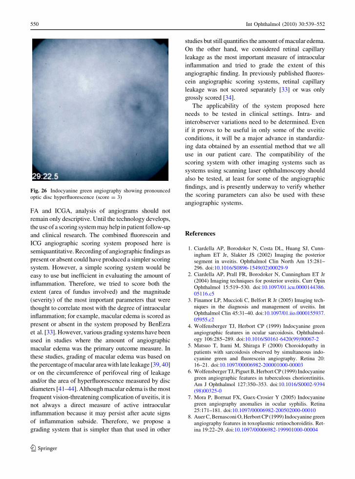
Fig. 26 Indocyanine green angiography showing pronounced
optic disc hyperfluorescence (score = 3)
550 Int Ophthalmol (2010) 30:539–552
123

9. Atmaca LS, Simsek T, Atmaca Sonmez P, Sonmez K
(2006) Fluorescein and indocyanine green angiography in
ocular toxoplasmosis. Graefes Arch Clin Exp Ophthalmol
244:1688–1691. doi:10.1007/s00417-006-0345-z
10. Auer C, Bernasconi O, Herbort CP (1997) Toxoplasmic
retinochoroiditis: new insights provided by indocyanine
green angiography. Am J Ophthalmol 123:131–133
11. Bernasconi O, Auer C, Zografos L, Herbort CP (1998)
Indocyanine green angiographic findings in sympathetic
ophthalmia. Graefes Arch Clin Exp Ophthalmol 236:635–
638. doi:10.1007/s004170050134
12. Herbort CP, Mantovanni A, Bouchenaki N (2007) Indocya-
nine gren angiography in Vogt-Koyanagi-Harada disease:
angiographic signs and utility in patient follow-up. Int Oph-
thalmol 27:173–182. doi:10.1007/s10792-007-9060-y
13. Cimino L, Auer C, Herbort CP (2000) Sensitivity of in-
docyanine green angiography for the follow-up of active
inflammatory choriocapillaropathies. Ocul Immunol In-
flamm 8:275–283. doi:10.1076/ocii.8.4.275.6462
14. Vadala M, Lodato G, Cillino S (2001) Multifocal cho-
roiditis: indocyanine green angiographic features.
Ophthalmologica 215:16–21. doi:10.1159/000050820
15. Slakter JS, Giovannini A, Yannuzzi LA, Scassellati-
Sforzolini B, Guyer DR, Sorenson JA et al (1997) Indo-
cyanine green angiography of multifocal choroiditis.
Ophthalmology 104:1813–1819
16. Di Crecchio L, Parodi MB, Saviano S, Ravalico G (2001)
Acute posterior multifocal placoid pigment epitheliopathy
and ulcerative colitis: a possible association. Acta Oph-
thalmol Scand 79:319–321. doi:10.1034/j.1600-0420.2001.
790324.x
17. Obana A, Kusumi M, Miki T (1996) Indocyanine green
angiographic aspects of multiple evanescent white dot
syndrome. Retina 16:97–104. doi:10.1097/00006982-1996
16020-00002
18. Ie D, Glaser BM, Murphy RP, Gordon LW, Sjaarda RN,
Thompson JT (1994) Indocyanine green angiography in
multiple evanescent white-dot syndrome. Am J Ophthal-
mol 117:7–12
19. Stoffelns BM (2006) Long-term follow-up and angio-
graphic findings in serpiginous choroiditis. Klin Monatsbl
Augenheilkd 223:418–421. doi:10.1055/s-2006-926575
20. Giovannini A, Mariotti C, Ripa E, Scassellati-Sforzolini B
(1996) Indocyanine green angiographic findings in ser-
piginous choroidopathy. Br J Ophthalmol 80:536–540. doi:
10.1136/bjo.80.6.536
21. Fardeau C, Herbort CP, Kullmann N, Quentel G, LeHoang
P (1999) Indocyanine green angiography in birdshot cho-
rioretinopathy. Ophthalmology 106:1928–1934. doi:
10.1016/S0161-6420(99)90403-7
22. Gharbiya M, Pecci G, Baglio V, Gargiulo A, Allievi F,
Balacco-Gabrieli C (2006) Indocyanine green angiographic
findings for patients with systemic lupus erythematosus
nephropathy. Retina 26:159–164. doi:10.1097/00006982-
200602000-00006
23. Dhingra S, Stavrou P (2004) Indocyanine green angiogra-
phy in systemic lupus erythematosus-associated uveitis.
Ocul Immunol Inflamm 12:69–73. doi:10.1076/ocii.12.1.
69.28068
24. Gharbiya M, Bozzoni-Pantaleoni F, Augello F, Balacco-Ga-
brieli C (2002) Indocyanine green angiographic findings in
systemic lupus erythematosus choroidopathy. Am J Oph-
thalmol 134:286–290. doi:10.1016/S0002-9394(02)01477-0
25. Bouchenaki N, Cimino L, Auer C, Tao Tran V, Herbort CP
(2002) Assessment and classification of choroidal vascu-
litis in posterior uveitis using indocyanine green
angiography. Klin Monatsbl Augenheilkd 219:243–249.
doi:10.1055/s-2002-30661
26. Herbort CP, LeHoang P, Guex-Crosier Y (1998) Schematic
interpretation of indocyanine gren angiography in posterior
uveitis using a standard angiographic protocol. Ophthalmol-
ogy 105:432–440. doi:10.1016/S0161-6420(98)93024-X
27. Altan-Yaycioglu R, Akova YA, Akca S, Yilmaz G (2006)
Inflammation of the posterior uvea: findings on fundus
fluorescein and indocyanine green angiography. Ocul
Immunol Inflamm 14:171–179. doi:10.1080/09273940600
660524
28. Klaeger A, Tran AT, Hiroz CA, Morisod L, Herbort CP
(2000) Indocyanine green angiography in Behcet’s uveitis.
Retina 20:309–314
29. Bozzoni-Pantaleoni F, Gharbiya M, Pirraglia MP, Accorinti
M, Pivetti-Pezzi P (2001) Indocyanine green angiographic
findings in Behcet disease. Retina 21:230–236. doi:10.1097/
00006982-200106000-00006
30. Atmaca LS, Sonmez PA (2003) Fluorescein and indocya-
nine green angiography findings in Behcet’s disease. Br J
Ophthalmol 87:1466–1468. doi:10.1136/bjo.87.12.1466
31. Gedik S, Akova YA, Yilmaz G, Bozbeyoglu S (2005) In-
docyanine green and fundus fluorescein angiographic
findings in patients with active ocular Behcet’s disease.
Ocul Immunol Inflamm 13:51–58. doi:10.1080/092739404
90518757
32. Herbort CP, Probst K, Cimino L, Tran VT (2004) Differ-
ential inflammatory involvement in retina and choroıd in
birdshot chorioretinopathy. Klin Monatsbl Augenheilkd
221:351–356. doi:10.1055/s-2004-812827
33. BenEzra D, Forrester JV, Nussenblatt RB, Tabbara K,
Timonen P (1991) Uveitis scoring system. Springer Verlag,
Berlin
34. Suhler EB, Smith JR, Wertheim MS, Lauer AK, Kurz DE,
Pickard TD et al (2005) A prospective trial of infliximab
therapy for refractory uveitis. Preliminary safety and effi-
cacy outcomes. Arch Ophthalmol 123:903–912. doi:
10.1001/archopht.123.7.903
35. Monnet D, Brezin AP, Holland GN, Yu F, Mahr A, Gordon
LK et al (2006) Longitudinal cohort study of patients with
birshot chorioretnopathy. I. Baseline clinical characteristics.
Am J Ophthalmol 141:135–142. doi:10.1016/j.ajo.2005.
08.067
36. Monnet D, Levinson RD, Holland GN, Haddad L, Yu F,
Brezin AP (2007) Longitudinal cohort study of patients with
birdshot chorioretinopathy. II. Macular imaging at baseline.
Am J Ophthalmol 144:818–828. doi:10.1016/j.ajo.2007.
08.011
37. Jabs DA, Nussenblatt RB, Rosenbaum JT, Standardization
of Uveitis Nomenclature (SUN) Working Group (2005)
Standardization of uveitis nomenclature for reporting
clinical data. Results of the first international workshop.
Am J Ophthalmol 140:509–516. doi:10.1016/j.ajo.2005.
01.035
38. Mandava N, Guyer DR, Yannuzzi LA, Nichol J, Orlock D
(1999) Principles of fluorescein angiography. In: Guyer
Int Ophthalmol (2010) 30:539–552 551
123

DR, Yannuzzi LA, Chang S, Shields JA, Green WR (eds)
Retina–vitreous–macula, vol 1. WB Saunders, Philadel-
phia, pp 29–38
39. Van Kooij B, Fijnheer R, de Boer J, Dam-Van Loon NT,
Bartelink I, Roest M et al (2006) A randomized, masked,
cross-over trial of lisinopril for inflammatory macular edema.
Am J Ophthalmol 141:451–646. doi:10.1016/j.ajo.2005.
11.056
40. Lardenoye CW, van Schooneveld MJ, Frits TW, Rothova
A (1998) Grid laser photocoagulation for macular edema in
uveitis or the Irvine-Gass syndrome. Br J Ophthalmol
82:1013–1016
41. Miyake K, Sakamura S, Miura H (1980) Long-term follow-
up study on prevention of aphakic cystoid macular oedema
by topical indomethacin. Br J Ophthalmol 64:324–328.
doi:10.1136/bjo.64.5.324
42. Spaide RF, Yannuzzi LA, Sisco LJ (1993) Chronic cystoid
macular edema and predictors of visual acuity. Ophthalmic
Surg 24:262–267
43. Antcliff RJ, Stanford MR, Chauhan DS, Graham EM,
Spalton DJ, Shilling JS, Ffytche TJ, Marshall J (2000)
Comparison between optical coherence tomography and
fundus fluorescein angiography for the detection of cystoid
macular edema in patients with uveitis. Ophthalmology
107:593–599. doi:10.1016/S0161-6420(99)00087-1
44. Whitcup SM, Csaky KG, Podgor MJ, Chew EY, Perry CH,
Nussenblatt RB (1996) A randomized, masked, cross-over
trial of acetazolamide for cystoid macular edema in
patients with uveitis. Ophthalmology 103:1054–1063
552 Int Ophthalmol (2010) 30:539–552
123


