Embed Size (px)
Citation preview
Schedule At A Glance
Thursday, March 26, 1998 5:00 pm Poster Session I (concludes at 7:00 pm) Wine and Cheese Reception sponsored by TheraTech, Inc.
8:00 am Andrology Laboratory Workshop (concludes at 5:00 pm) 7:30 pm
12:00 noon Executive Council Meeting (concludes at about 10:00 pm) Banquet Sponsored by ALZA Pharmaceuticals
Friday, March 27, 1998 8:00 am Postgraduate Course (concludes at 5:00 pm)
11E•: Annual Meeting Starts Friday Evening/ �.-:in ""' 1 Opening Reception � Sponsored by SmithKline Beecham Pharmaceuticals
7:00 pm Welcome
7:15 pm Distinguished Andrologist Award Presentation
7:30 pm Serono Award Lecture • The Molecular Biology of the Zona Pellucida
Jurrien Dean, MD
Saturday, March 28, 1998 8:00 am AUA Lecture
• Impotence and Nitric Oxide: The Future Jacob Ra�er, MD
8:55 am Distinguished Service Award Presentation Sponsored by Genetics & IVF Institute
9:05 am Buckeye State-of -the-Art Lecture • Fertilization: Ignition of the First Cell Cycle
Sally D. Perreault, PhD
10:00 am Coffee Break in the Exhibit Hall
10:30 am Oral Session I: Testis (Simultaneous session)
10:30 am Oral Session II: Sperm Function (Simultaneous session)
12:00 noon Lunch
Women In Andrology Meeting & Luncheon • Juggling Professional and Personal Obligations During
the Long-Term Illness of a Loved One ... and Afterwards '
Patricia Fail, PhD
1 :30 pm Symposium: Male Reproductive Aging • Androgen Replacement in Older Men:
Should We or Shouldn't We? J. Lisa Tenover, MD, PhD
• Male Reproductive Tract Aging Barry Zirkin, PhD
• Neuroendocrine Facets of Male Reproductive Aging Johannes Veldhuis, MD
3:30pm Refreshment Break In the Exhibit Hall
4:00 pm Oral Session Ill: Sperm Cyropreservatlon
Sunday, March 29, 1998 8:00 am ASA State-of-the-Art Lecture
• Using a Genetic Model of Male Infertility: What Good is a Sterile Mouse? Patricia Olds-Clarke, PhD
8:55 am Young Andrologist Award Presentation Sponsored by the Texas Institute for Reproductive Medicine and Endocrinology, P.A.
9:00 am Oral Session IV: Hormonal Regulation
10:00 am Coffee Break in the Exhibit Hall
10:30 am Pharmacia & Upjohn Clinical Debate • Is ICSI a Genetic Time Bomb?
- Yes. Dolores Lamb, PhD
- No; It is Safe and Effective Peter Schlegel, MD
- Ethics and Andrology in the 21st Century Glenn McGee, PhD
12:00 noon Lunch
Editorial Board Luncheon
Laboratory Science Forum Meeting & Lunch • The Role of the AndrologistlEmbryologist in a Successful
ART Program David S. Karabinus, PhD
1 :30 pm Symposium: Gene Knock-outs and Male Reproduction -Clinical Implications • Reproductive Consequences of an IGF-1 Null Mutation
Anthony R. Bellve, PhD • Genetic Defects in Mouse and Man that Affect
Gonadal Development and Function Sally Ann Camper, PhD
• Steroidogenic Factor 1 Plays Multiple Roles in Reproduction Keith L. Parker, MD, PhD
3:30 pm Refreshment Break in the Exhibit Hall
4:00 pm Awards Ceremony & Business Meeting
5:00 pm Poster Session II (concludes at 7:00 pm) Wine and Cheese Reception sponsored by TheraTech, Inc.
8:00 pm Student Colloquium & Soiree Sponsored by California Cryobank, Inc. • The XY Files: Ontogeny of an Andrologist
Stuart E. Ravnik, PhD
Twenty· Third Annual Meeting of the American Society of Andrology
President's Welcome
Welcome to the 23rd Annual Meeting of the American Society of Andrology. We are all looking forward to having a great meeting here in southern California! The Society could not hold such meetings without the willingness and hours of hard work of the Local Arrangements, Program and Post
graduate Course Committees. We should all express our thanks to Dr. Shalender Bhasin, chair of the Local Arrangements Committee, Dr. Barry Hinton, chair of the 1998 Program Committee, and Dr. Stuart Howards, chair of the Postgraduate Course Committee, for all their efforts on our behalf. The location and programs are excellent, and we will leave Long Beach not only with pleasant memories, but with updated information and new ideas about the clinical and basic aspects of Andrology.
As our Annual Meeting depends on willing volunteers within the Society, it also depends on the contributions of our industry sponsors and exhibitors. Sponsors who have generously donated to support our meeting, whether for specific events or for general support, are noted throughout the program book, and I thank them here, collectively,
Table of Contents
Abstracts .................................... 25 Andrology Laboratory Workshop ................. 10 Annual Meeting (Detailed Schedule) ............... 12 Author Index ................................ 22 CME Credit Information & Course Objectives ........ 9
Distinguished Andrologist Award .................. 6 Distinguished Service Award ...................... 7 Exhibits ..................................... 3 Future Meetings .............................. 18 Hotel Information ............................. .3
Laboratory Science Forum ....................... 3 Local Arrangements Chair's Message ................ 2 New Investigator Award ...... ................... 8 Past Presidents ................................ 4
,
�v( cnu+ Jl '8fJ-UL 'j 14 r>fr�--ef �� (
as well. Also, I urge you all to visit the exhibit area during the meeting not only to learn about the development of new products, but to express to the exhibitors your appreciation for their presence. We all need each other to make our meetings successful.
The Society's officers, members of the Executive Council, committee chairs, journal and newsletter editors, Androlog monitors and the Holland-Parlette staff, who manage our business office, all have provided excellent contributions to make this a functioning, active society. They have my sincere respect and appreciation.
Finally, I would like to thank the membership of the Society for the opportunity to serve as your president. Being president of the ASA has not only been an honor, but it has provided me the opportunity to work with a wide range of our membership and to see personally the dedication of so many. Thank you for all your efforts.
� .. :J.��
Terry T. Turner, PhD President
Poster Session I (List of Abstracts) ................ 16 Poster Session II (List of Abstracts) ................ 19 Postgraduate Course ........................... 11 President's Welcome Message ..................... 1 Program Chair's Message ......................... 4 Registration Information ......................... 3 Schedule At A Glance ................. .Inside Cover Serono Award Lectureship ....................... .5 Society Leadership ............................. 2 Sponsors ..................................... 9 Student Colloquium & Soiree ..................... 3 Travel Information ............................. 3 Women In Andrology ........................... 3 Young Andrologist Award ........................ 8
American Society of Andrology
74 New Montgomery, Suite 230 • San Francisco, CA 94105 Phone: ( 415) 764- 4823 • Fax: ( 415) 764- 4915
Email: [email protected] • URL: http://godot.urol.uic.edu/-androlog/ Executive Director: Carol Holland Parlette, MPH • Assistant Director: Sarah Morisseau Lee
Twenty· Third Annual Meeting of the American Society of Andrology 1
Message from the Local Arrangements Chair
March is a great month to hold the ASA meeting in Long Beach; the weather should be temperate, and with the Grand Prix and Easter around the corner, the city will have a festive spirit. The Hyatt Regency is a lovely property on the waterfront, a few blocks from
__.. ........ ___ _, the Queen Mary. The city has many attractions in its vicinity for spouses and children, including Disneyland, Universal Studios, Rodeo Drive, Knott's Berry Farm, Magic Mountain, and the Getty Museum. Therefore, the ASA meeting provides an excellent opportunity to combine great science with family fun.
Barry Hinton has put together an outstanding scientific
facilitate socialization and networking. The Society gratefully acknowledges the support of ALZA Corporation, SmithKline Beecham Pharmaceuticals and TheraTech, Inc. for their generous support of these social events.
Our gratitude is due to members of the Local Arrangements Committee, as well as Sarah Lee and Carol Parlette in the business office, for their efforts in making it all come together. This tradition of volunteerism is what gives ASA meetings a uniquely personal flavor. Because of the location and the excellent program, we expect terrific attendance in Long Beach, so be sure to make your reservations early! It promises to be another wonderful meeting.
program. The opening reception, wine and cheese Shalender Bhasin, MD receptions at the poster sessions and the banquet should Chair, 1998 Local Arrangements Commitee
Local Arrangements Committee: Stefan Arver, Nestor Gonzales-Cadavid, Cora Kikunaga, Ma Kun, Wael Salameh, Amiya
Sinha-Hikim, Ronald 5. Swerdloff, Wayne Taylor, Christina Wang, Shalender Bhasin (chair).
American Society of Andrology Officers
President ...................... Terry T. Turner, PhD Treasurer ....................... Terry R. Brown, PhD Vice President ............. Richard V. Clark, MD, PhD Secretary .......................... Rex A. Hess, PhD Past President ................. Arnold M. Belker, MD Executive Director . ......... Carol Holland Parlette, MPH
Members of the Executive Council Nina S. Davis, MD; Christopher]. Dejonge, PhD, HCLD; Erma Z. Drobnis, PhD;
Alvin M. Matsumoto, MD; Craig Niederberger, MD; Deborah A. O'Brien, PhD; Peter N. Schlegel, MD; Steven M. Schrader, PhD; Susan S. Suarez, MS, PhD; Jacquetta M. Trasler, MD PhD; Barry R. Zirkin, PhD
Committee Chairs Andrology Labs .... Christopher]. Dejonge, PhD, HCLD Awards ...................... Stuart E. Ravnik, PhD Bylaws ...................... Matthew P. Hardy, PhD CME Liaison ................ Hugh C. Hensleigh, PhD Finance ...................... .Joel L Marmar, MD Future Meetings ............... Bernard Robaire, PhD Industrial Relations ........... Kenneth P. Roberts, PhD International Liaison ............ Barry T. Hinton, PhD ISA 2001 ............. . . Carlos R. Morales, DVM, PhD Laboratory Science Forum .......... Carol S. Sloan, MS 1998 Local Arrangements ........ Shalender Bhasin, MD 1999 Local Arrangements ........ Arnold M. Belker, MD 2000 Local Arrangements ....... Robert A. Newton, MD
Long-Range Planning ......... Richard V. Clark, MD, PhD Membership ................... .Susan H. Benoff, PhD Newsletter Editors ............... Don E Cameron, PhD
............... Stanley]. Nazian, PhD Nominating ................. Alvin M. Matsumoto, MD Placement Service Coordinator ...... Pasquale Patrizio, MD 1998 Postgraduate Course ........ Stuart S. Howards, MD 1999 Postgraduate Course .......... Christina Wang, MD 1998 Program ................... Barry T. Hinton, PhD 1999 Program ............. William]. Bremner, MD, PhD Publications ............... Lonnie D. Russell, MS, PhD Society Liaison .................. Harris M. Nagler, MD Student Affairs .................. Don E Cameron, PhD
Journal of Andrology
2
Editorial Office: 4-144 Jackson Hall, 321 Church Street SE, Minneapolis, MN 55455 Phone: (612) 625-1488 • Fax: (612) 625-1163 • Email: [email protected]
Editors-in-Chief: David W Hamilton, PhD and Jon L Pryor, MD • Editorial Assistant: Lauren Fox Associate Editors: Shalender Bhasin, MD, Christopher De Jonge, PhD, Wayne]. Hellstrom, MD
International Associate Editors: David]. Handelsman, MD, PhD, Hector Chemes, MD, PhD, Gerhard E Weinbauer
Twenty· Third Annual Meeting of the American Society of Andrology
General Information
Registration All meeting attendees are encouraged to register in advance whenever possible. Registration forms are available by contacting the American Society of Andrology:
74 New Montgomery, Suite 230 San Francisco , CA 94105 Phone: ( 415) 764-4823 Fax: (415) 764-4915 Email : [email protected]
All registrations received after Friday, February 27, 1998 (including on-site registrations) will be assessed a $45 fee.
The meeting registration and information desk will be open at the following times for on-site registration: Thursday, March 26 , 1998 7:00 am - 6:00 pm Friday, March 27, 1998 7:00 am - 8:30 pm Saturday, March 28 , 1998 7:00 am - 5 :00 pm Sunday, March 29 , 1998 7:30 am - 5:00 pm
Travel Discounts United Airlines is offering five to ten percent off airfares to Los Angeles for attendees of the ASA Annual Meeting. To take advantage of this offer , call (800) 521-4041 and refer to Meeting ID Number 518XK - and if you make your reservations at least 60 days in advance , you'll receive an additional five percent discount. You can also receive ten percent off Alamo and Avis car rentals when you make your car reservation at the time of your plane reservation.
Airports Long Beach is served by three airports : Los Angeles International Airport (LAX), 1)3 miles_ north; john Wayne/Orange County Airport , l5 miles south; and Long Beach Airport , which is only 15 minutes from the hotel. Taxis from LAX are approximately $40 one way and air-
� p�Ht shuttles are abou,t $�/per person. From Orange County, expect to pay $45 fqr a taxi to the hotel. Taxis from the Long Beach air!Jort are about $15.
Hotel Accommodations The Hyatt Regency ip Long B�, California is the headquarters for the ms ASA Annual Meeting and Postgraduate Course. Discounted room rates are available for ASA meeting attendees: $104 single occupancy, $119 double occupancy, $134 triple occupancy, $149 quadruple occupancy Regency Club accommodations are available. These rates are guaranteed only through Fr iday, February 27, 1998, so make your reservations as soon as possible.
Reservations may be made by calling the Hyatt reservations toll-free number: (800) 233-1234 or by contacting the hotel directly at (562) 491-1234. Be sure to mention you are attending the ASA meeting to receive the reduced rates.
Poster Sessions Poster sessions will be 5:00 - 7:00 on Saturday and Sunday evenings in the Regency Ballroom with wine and cheese receptions supported by a grant from TheraTech , Inc.
Exhibits
An extensive exhibit hall featuring equipment and information for all andrologists will be open from 10:00 am to 7:00 pm on Saturday and Sunday in the Regency Ballroom. Refreshment breaks will be served in the exhibit hall and on Sunday there will also be a raffle among the exhibits.
Slide Preview Room The Regency Coatroom will be available from 6:00 am until 12:00 midnight Thursday through Sunday for speakers to preview slides and prepare their presentations.
Laboratory Science Forum The 1998 Laboratory Science Forum will be at 12:00 noon on Sunday, March 29. Luncheon tickets are available for $22. The topic this year is The Role of the Andrologist/
Embryologist in a Successful ART Program.
Women In Andrology
The Women In Andrology group will hold its annual business meeting on Saturday, March 28 at 12:00 noon in the Beacon A room of the Hyatt Regency. Following the WIA meeting , Dr. Patricia Fail will address the group on juggling Professional and Personal Obligations During the
Long-Term Illness of a Loved One . . . and Afterwards. Tickets for the luncheon are available for $25.
Student Colloquium & Soiree The 1998 ASA Student Colloquium , supported by an educational grant from California Cryobank , Inc. , will feature Dr. Stuart Ravnik. His talk The XY Files: Ontogeny of an
Andrologist will be at 8:00 pm on Sunday, March 29 in the Seaview A room of the Hyatt. The annual Student Soiree will be held immediately following the Colloquium. Both events are free of charge and everyone is welcome to attend.
ASA Home Page Visit http://godot. urol.uic.edu/-andrologl at at any time for updated information on the 1998 Annual Meeting.
Twenty· Third Annual Meeting of the American Society of Andrology 3
Message from the Program Chair
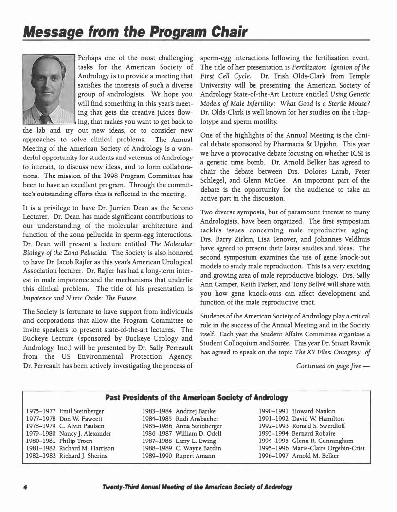
Perhaps one of the most challenging tasks for the American Society of Andrology is to provide a meeting that satisfies the interests of such a diverse group of andrologists. We hope you will find something in this year's meeting that gets the creative juices flow
....._ __ .... ing, that makes you want to get back to the lab and try out new ideas, or to consider new approaches to solve clinical problems. The Annual Meeting of the American Society of Andrology is a wonderful opportunity for students and veterans of Andrology
to interact, to discuss new ideas, and to form collaborations. The mission of the 1998 Program Committee has been to have an excellent program. Through the committee's outstanding efforts this is reflected in the meeting.
It is a privilege to have Dr. Jurrien Dean as the Serono Lecturer. Dr. Dean has made significant contributions to our understanding of the molecular architecture and function of the zona pellucida in sperm-egg interactions. Dr. Dean will present a lecture entitled The Molecular Biology of the Zona Pellucida. The Society is also honored to have Dr. Jacob Rajfer as this year's American Urological Association lecturer. Dr. Rajfer has had a long-term interest in male impotence and the mechanisms that underlie this clinical problem. The title of his presentation is Impotence and Nitric Oxide: The Future.
The Society is fortunate to have support from individuals and corporations that allow the Program Committee to invite speakers to present state-of-the-art lectures. The Buckeye Lecture (sponsored by Buckeye Urology and Andrology, Inc.) will be presented by Dr. Sally Perreault from the US Environmental Protection Agency. Dr. Perreault has been actively investigating the process of
sperm-egg interactions following the fertilization event. The title of her presentation is Fertilizaton: Ignition of the First Cell Cycle. Dr. Trish Olds-Clark from Temple University will be presenting the American Society of Andrology State-of-the-Art Lecture entitled Using Genetic Models of Male Infertility: What Good is a Sterile Mouse? Dr. Olds-Clark is well known for her studies on the t-haplotype and sperm motility.
One of the highlights of the Annual Meeting is the clinical debate sponsored by Pharmacia & Upjohn. This year we have a provocative debate focusing on whether ICSI is a genetic time bomb. Dr. Arnold Belker has agreed to chair the debate between Ors. Dolores Lamb, Peter Schlegel, and Glenn McGee. An important part of the debate is the opportunity for the audience to take an active part in the discussion.
Two diverse symposia, but of paramount interest to many Andrologists, have been organized. The first symposium tackles issues concerning male reproductive aging. Ors. Barry Zirkin, Lisa Tenover, and Johannes Veldhuis have agreed to present their latest studies and ideas. The second symposium examines the use of gene knock-out models to study male reproduction. This is a very exciting and growing area of male reproductive biology. Ors. Sally Ann Camper, Keith Parker, and Tony Bellve will share with you how gene knock-outs can affect development and function of the male reproductive tract.
Students of the American Society of Andrology play a critical role in the success of the Annual Meeting and in the Society itself. Each year the Student Affairs Committee organizes a Student Colloquium and Soiree. This year Dr. Stuart Ravnik has agreed to speak on the topic The XY �iles: Ontogeny of
Continued on page five -
Past Presidents of the American Society of Andrology
1975-1977 Emil Steinberger 1977-1978 Don W Fawcett 1978-1979 C. Alvin Paulsen 1979-1980 Nancy]. Alexander 1980-1981 Philip Troen 1981-1982 Richard M. Harrison 1982-1983 Richard]. Sherins
1983-1984 Andrzej Bartke 1984-1985 Rudi Ansbacher 1985-1986 Anna Steinberger 1986 -1987 William D. Odell 1987-1988 Larry L. Ewing 1988-1989 C. Wayne Bardin 1989-1990 Rupert Amann
1990-1991 Howard Nankin 1991-1992 David W Hamilton 1992-1993 Ronald S. Swerdloff 1993-1994 Bernard Robaire 1994-1995 Glenn R. Cunningham 1995-1996 Marie- Claire Orgebin- Crist 1996 -1997 Arnold M. Belker
4 Twenty. Third Annual Meeting of the American Society of Andrology
Serano Award Lectureship
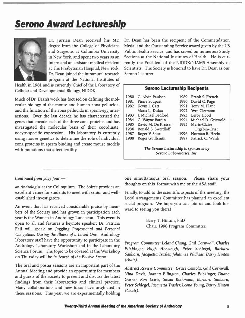
Dr. Jurrien Dean received his MD degree from the College of Physicians and Surgeons at Columbia University in New York, and spent two years as an intern and an assistant medical resident at The Presbyterian Hospital, New York. Dr. Dean joined the intramural research program at the National Institutes of
Health in 1981 and is currently Chief of the Laboratory of Cellular and Developmental Biology, NIDDK.
Much of Dr. Dean's work has focused on defining the molecular biology of the mouse and human zona pellucida, and the function of the zona pellucida in sperm-egg interactions. Over the last decade he has characterized the genes that encode each of the three zona proteins and has investigated the molecular basis of their coordinate, oocyte-specific expression. His laboratory is currently using mouse genetics to determine the role of individual zona proteins in sperm binding and create mouse models with mutations that affect fertility.
Continued from page four -
an Andrologist at the Colloquium. The Soiree provides an excellent venue for students to meet with senior and wellestablished investigators.
An event that has received considerable praise by members of the Society and has grown in participation each year is the Women in Andrology Luncheon. This event is open to all and features a keynote speaker; Dr. Patricia Fail will speak on Juggling Professional and Personal Obligations During the Illness of a Loved One. Andrology laboratory staff have the opportunity to participate in the Andrology Laboratory Workshop and in the Laboratory Science Forum. The topic to be covered at the Workshop on Thursday will be In Search of the Elusive Sperm.
The oral and poster sessions are an important part of the Annual Meeting and provide an opportunity for members and guests of the Society to present and discuss the latest findings from their laboratories and clinical practice. Many collaborations and new ideas have originated in these sessions. This year, we are experimentally holding
Dr. Dean has been the recipient of the Commendation Medal and the Outstanding Service award given by the US Public Health Service, and has served on numerous Study Sections at the National Institutes of Health. He is currently the President of the NIDDK/NIAMS Assembly of Scientists. The Society is honored to have Dr. Dean as our Serano Lecturer.
Serono Lectureship Recipents
1980 C. Alvin Paulsen 1981 Pierre Soupart 1982 Kevin]. Catt
Maria L. Dufau 1983 ]. Michael Bedford 1984 C. Wayne Bardin 1985 David M. De Kretser 1986 Ronald S. Swerdloff 1987 Roger V. Short 1988 Roger Guillemin
1989 Frank S. French 1990 David C. Page 1991 Tony M. Plant 1992 Yves Clermont 1993 Leroy Hood 1994 Michael D. Griswold 1995 Marie- Claire
Orgebin- Crist 1996 Norman B. Hecht 1997 Patrick C. Walsh
The Serono Lectureship is sponsored by Serono Laboratories, Inc.
one simultaneous oral session. Please share your thoughts on this format with me or the ASA staff.
Finally, to add to the scientific aspects of the meeting, the Local Arrangements Committee has planned an excellent social program. We hope you can join us and look forward to seeing you there!
Barry T. Hinton, PhD Chair, 1998 Program Committee
Program Committee: Leland Chung, Gail Cornwall, Charles Flickinger, Hugh Hensleigh, Peter Schlegel, Barbara Sanborn, Jacquetta Trasler, Johannes Veldhuis, Barry Hinton (chair).
Abstract Review Committee: Grace Centola, Gail Cornwall, Nina Davis, Joanna Ellington, Charles Flickinger, Duane Gamer, Ron Lewis, Susan Rothmann, Barbara Sanborn, Peter Schlegel, Jacquetta Trasler, Leona Young, Barry Hinton (Chair).
Twenty· Third Annual Meeting of the American Society of Andrology 5

1998 Distinguished Andrologist Award
Ryuzo Yanagimachi, PhD is the recipient of the Distinguished Andrologist Award for 1998. Dr. Yanagimachi began his training in reproductive biology at Hakkaido University in Japan, receiving a DSc degree in 1960. He moved to the Worcester Foundation to work with previous Distinguished
Andrologist Dr. M.C. Chang for three fruitful years and then returned to Hakkaido University as Lecturer in Embryology. Two years later, Dr. Yanagimachi began his faculty appointment in the Department of Anatomy and Reproductive Biology at the University of Hawaii, where he remains today.
Dr. Yanagimachi's contributions to the field of Reproductive Biology and, indeed, to Andrology are enormous. Few individuals in modem history have had as significant an impact on the development and direction of their field as has Dr. Yanagimachi. His research is highly significant and well-regarded, receiving numerous awards including, in 1996, the International Prize for Biology. In over 300 publications, Dr. Yanagimachi's contributions are milestones in Andrology: first demonstration of mammalian sperm capacitation in vitro, first description of the manner of sperm entry into living mammalian ova, discovery of sperm hyperactivation, first demonstration of the importance of ca++ in the sperm acrosome reaction and oocyte activation in mammal, first intracytoplasmic sperm injection in mammals, first description of the usefulness of zona-free hamster oocytes and many, many others. In addition, he has also contributed in a major way to studies on the biology of spermatozoa within the female genital tract, investigation into the stability/instability of mammalian sperm nucleus, and has demonstrated that the spermatid nucleus is potentially ready for participating in embryonic development. Dr. Yanagimachi's reviews of sperm biology are considered required reading for many Andrologists and his comparative work on multiple species (from the well known rodents to less well studied species such as bats and dolphins) should serve as a challenge to us all.
In his typical low key fashion, Dr. Yanagimachi describes his thoughts for the future. "My interest in life's begin-
ning goes back to my undergraduate days. Although artificial reproduction without germ cells may be possible in the future, reproduction via germ cells will remain the most effective means of continuing life of most animals (including our own species) on the surface of the earth. Compared with Mother Nature, our past and recent 'triumphs' in reproductive biology and technologies are trivial. We must keep learning from Mother Nature's ways over millions of years in order to better control/manage gamete formation/functions without serious errors. It is obvious that female and male contribute equally, at least genetically, to following generations. Male gametes (spermatozoa) are made for female gametes (eggs) and vice versa. Parthenogenesis, androgenesis and cloning may technically become possible during the 21st century, but would it be desirable to live in the world without the union (love) of gametes (male and female partners)?"
Dr. Yanagimachi has been a mentor to those who have worked with him directly as well as mentor to all of us through the written and oral description of his research. For unparalleled excellence in Andrology research, his many contributions to our understanding of the ability of the spermatozoon to fertilize, and his challenges to us as students of Biology, the American Society of Andrology is delighted and privileged to present Dr. Ryuzo Yanagimachi with the 1998 Distinguished Andrologist Award.
Distinguished Andrologists
1976 Roy 0. Greep M.C. Chang
1977 Robert E. Mancini 1978 Robert]. Hotchkiss 1979 Thaddeus Mann 1980 john MacLeod 1981 Alexander Albert 1982 Eugenia Rosemberg 1983 Kristen B.D. Eik- Nes 1984 Mortimer B. Lipsett 1985 Robert H. Foote 1986 Alfred D. ] ost
1987 Emil Steinberger 1988 Yves W Clermont 1989 C. Alvin Paulsen 1990 Marie- Claire
Orgebin- Crist 1991 Philip Troen 1992 C. Wayne Bardin 1993 Anna Steinberger 1994 Richard]. Sherins 1995 Rupert P. Amann 1996 ]. Michael Bedford 1997 Brian P. Setchell
The Distinguished Andrologist Award is sponsored by the American Society of Andrology
6 Twenty· Third Annual Meeting of the American Society of Andralogy
1998 Distinguished Service Award
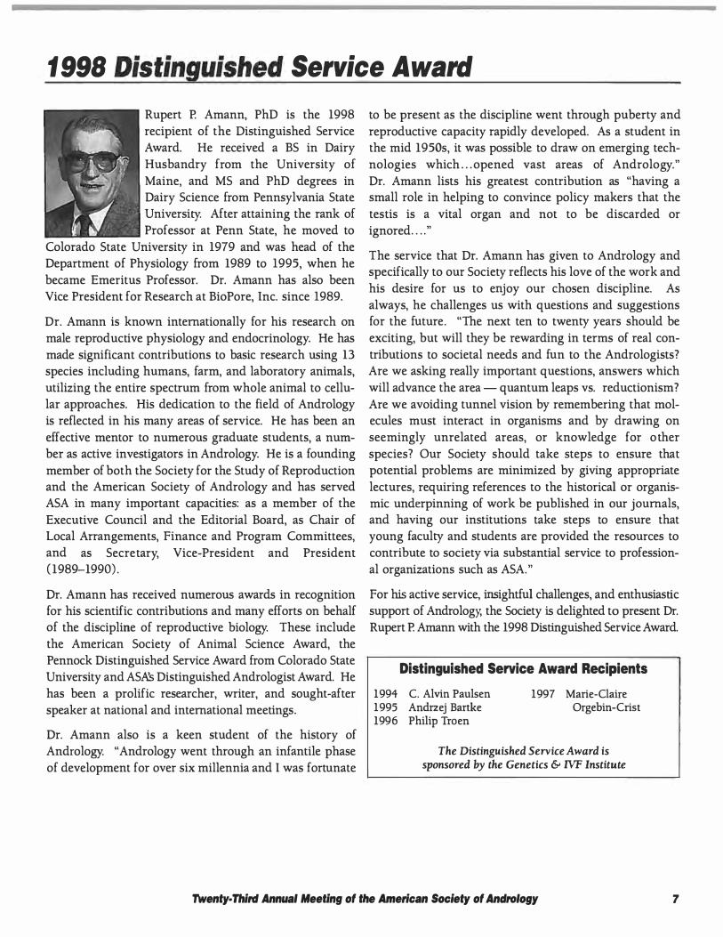
Rupert P. Amann, PhD is the 1998 recipient of the Distinguished Service Award. He received a BS in Dairy
Husbandry from the University of
Maine, and MS and PhD degrees in Dairy Science from Pennsylvania State University. After attaining the rank of Professor at Penn State, he moved to
Colorado State University in 1979 and was head of the Department of Physiology from 1989 to 1995, when he
became Emeritus Professor. Dr. Amann has also been Vice President for Research at BioPore, Inc. since 1989.
Dr. Amann is known internationally for his research on male reproductive physiology and endocrinology. He has
made significant contributions to basic research using 13 species including humans, farm, and laboratory animals, utilizing the entire spectrum from whole animal to cellular approaches. His dedication to the field of Andrology is reflected in his many areas of service. He has been an
effective mentor to numerous graduate students, a number as active investigators in Andrology. He is a founding
member of both the Society for the Study of Reproduction and the American Society of Andrology and has served ASA in many important capacities: as a member of the
Executive Council and the Editorial Board, as Chair of Local Arrangements, Finance and Program Committees, and as Secretary, Vice-President and President (1989-1990) .
Dr. Amann has received numerous awards in recognition for his scientific contributions and many efforts on behalf of the discipline of reproductive biology. These include the American Society of Animal Science Award, the Pennock Distinguished Service Award from Colorado State University and AS& Distinguished Andrologist Award. He
has been a prolific researcher, writer, and sought-after speaker at national and international meetings .
Dr. Amann also is a keen student of the history of Andrology. "Andrology went through an infantile phase of development for over six millennia and I was fortunate
to be present as the discipline went through puberty and
reproductive capacity rapidly developed. As a student in the mid 1950s, it was possible to draw on emerging tech
nologies which ... opened vast areas of Andrology."
Dr. Amann lists his greatest contribution as "having a small role in helping to convince policy makers that the
testis is a vital organ and not to be discarded or ignored . . . . "
T he service that Dr. Amann has given to Andrology and specifically to our Society reflects his love of the work and
his desire for us to enjoy our chosen discipline. As
always, he challenges us with questions and suggestions for the future. "The next ten to twenty years should be
exciting, but will they be rewarding in terms of real con
tributions to societal needs and fun to the Andrologists? Are we asking really important questions, answers which
will advance the area-quantum leaps vs. reductionism?
Are we avoiding tunnel vision by remembering that molecules must interact in organisms and by drawing on seemingly unrelated areas, or knowledge for o ther
species? Our Society should take steps to ensure that potential problems are minimized by giving appropriate
lectures, requiring references to the historical or organismic underpinning of work be published in our journals, and having our institutions take steps to ensure that young faculty and students are provided the resources to contribute to society via substantial service to professional organizations such as ASA. "
For his active service, insightful challenges, and enthusiastic support of Andrology, the Society is delighted to present Dr. Rupert P. Amann with the 1998 Distinguished Service Award.
Distinguished Service Award Recipients
1994 C. Alvin Paulsen 1995 Andrzej Bartke 1996 Philip Troen
1997 Marie- Claire Orgebin- Crist
The Distinguished Service Award is sponsored by the Genetics & IVF Institute
Twenty· Third Annual Meeting of tlle American Society of Andrology 7
1998 Young Andrologist Award
Dr. William R. Kelce is the recipient of
the 1998 Young Andrologist Award. Dr. Kelce received a double AB degree
in Biology and Psychology from Washington University in 1981 and his MS and PhD degrees in Physiology
from the University of MissouriColumbia in 1989. He was an NIEHS
NRSA fellow at johns Hopkins University working in collaboration with Larry Ewing and Barry Zirkin on endocrine toxicology until 1 992. Dr. Kelce's interests in
reproductive toxicology continued in his own research program as a principal investigator in the capacity of a Project Scientist and then Research Scientist at ManTech
Environmental in Research Triangle Park. Staying on in North Carolina, Dr. Kelce moved to the US Environmental Protection Agency's Health Effects Research Laboratory in 1995 as a Research Biologist and
Team Leader in the Reproductive Toxicology Division. In 1996, his contributions to the academic community were rewarded with an appointment as an Adjunct Associate Professor in the Department of Pediatrics and
Laboratories for Reproductive Biology at the University of North Carolina School of Medicine at Chapel Hill.
Dr. Kelce has received a number of awards, including the
First Place Young Investigator Award from the Society of Toxicology and the 1996 US EPA Gold Medal Award for Scientist of the Year. His research has been consistently recognized in terms of funding from both the US EPA and the NIEHS. Dr. Kelce describes his most significant
research accomplishment as the work in his laboratory which has identified a novel mechanism by which pesti-
New Investigator Award
The New Investigator Award is given to the ASA student member with the best abstract and research presentation
at the Annual Meeting. The award encourages student members to submit and present their best work and contribute to the scientific excellence of the Society.
The recipient of the 1998 New lnvestigator Award will be announced at the awards ceremony at 4:00 pm on Sunday, March 29, 1998.
cides and toxic substances can produce reproductive and
developmental related health effects and provides a clear
demonstration that environmental toxicants can pro
foundly alter reproductive development.
"As these molecular toxicology studies continue, we believe they have the potential to lead to a better under
standing of the molecular mechanisms responsible for
normal male sexual differentiation in addition to those responsible for inducing perturbations. " On the future of the ASA, Dr. Kelce says, "I would like the ASA to continue to embrace toxicology issues related to mechanisms of
adverse reproductive development and/or subsequent function. "
For his excellence in research and his contributions to the field of Andrology, including his recent work on environ
mental toxicants published in Nature, the American Society of Andrology is delighted to award Dr. William R. Kelce with the 1 998 Young Andrologist Award.
Young Andrologist Award Recipients
1982 LJ .D. Zaneveld 1983 William B. Neaves 1984 Lonnie D. Russell 1985 Bruce D. Schanbacher 1986 Stephen]. Winters 1987 Ilpo T. Huhtaniemi 1988 Larry Johnson 1989 Barry T. Hinton
1990 Luis Rodriguez- Rigau 1991 Patricia M. Saling 1992 Gary R. Klinefelter 1993 Robert Chapin 1994 Waynej.G. Hellstrom 1995 Christopher Dejonge 1996 Paul S. Cooke 1997 Gail A. Cornwall
The Young Andrologist Award is sponsored by the Texas Institute for Reproductive Medicine and Endocrinology, P.A.
1983 1984
1986 1987 1988 1989 1990
New Investigator Award Recipients
Thomas T. Tarter Peter S. Albertson Randall S. Zane Mark A. Hadley Peter Grosser Stuart E. Ravnik Tracy L. Rankin Donna 0. Bunch
1991 Robert Viger 1992 john Kirby 1993 Michael A. Palladino 1994 Linda R. Johnson 1995 Mehdi A. Akhondi 1996 Wei Gu
Daniel B. Rudolph 1997 Loren D. Walensky
The New Investigator Award is sponsored by the West Michigan Reproductive Institute, P.C.
B Twenty· Third Annual Meeting of the American Society of Andrology
Sponsors
The American Society of Andrology wishes to thank the following organizations for their generous support of the 1998 Annual Meeting&: Postgraduate Course.
Gold Club A minimum $10,000 contribution to an
ASA endowment fund. Buckeye Urology and Andrology, Inc.
Silver Club A minimum $5,000 contribution to an
ASA endowment fund. West Michigan Reproductive Institute, P.C.
Sustaining Sponsor A minimum $500 contribution to
ASA for five or more years. Genetics &: IVF Institute
Hamilton Thome Research Pharmacia &: Upjohn
Serono Laboratories, Inc. Texas Institute for Reproductive Medicine
and Endocrinology, P.A.
1998 Supporters A contribution of any size to support the
1998 Annual Meeting & Postgraduate Course ALZA Pharmaceuticals
American Urological Association California Cryobank, Inc.
Merck&: Co., Inc. Pfizer, Pharmaceuticals Group
Serono Laboratories, Inc. SmithKline Beecham Pharmaceuticals
TAP Pharmaceuticals, Inc. TheraTech, Inc.
CME Credit Information & Course Objectives
Annual Meeting Following this program, the participant should be able to: • Discuss the structure and function of zona pellucida in
sperm-egg interactions. • Recognize the role of nitric oxide in male impotence. • Explain the events of the cell cycle following fertilization. • Discuss new ideas and treatments of the aging male
reproductive tract. • Identify clinical and ethical issues in assisted reproduc
tive technologies. • Define new advances in the use of genetic models to
study male infertility.
This activity has been planned and implemented in accordance with the Essentials and Standards of the
Accreditation Council for Continuing Medical Education through the joint sponsorship of the University of Minnesota and the American Society of Andrology. The
University of Minnesota is accredited by the ACCME to provide continuing medical education for physicians.
The University of Minnesota designates this continuing medical education activity for up to 14 hours in category 1 credit toward the AMA Physician's Recognition Award.
Each physician should claim only those hours of credit actually spent in the educational activity.
Postgraduate Course Following the Postgraduate Course on Friday; March 27, 1998, the participant should be able to:
• Explain the developmental basic science of the male reproductive tract.
• Discuss in depth the clinical issues relating to male reproduction.
This activity has been planned and implemented in accor
dance with the Essentials and Standards of the Accreditation Council for Continuing Medical Education through the joint sponsorship of the University of Minnesota and the American Society of Andrology. The University of Minnesota is accredited by the ACCME to
provide continuing medical education for physicians.
The University of Minnesota designates this continuing
medical education activity for up to 6.5 hours in category 1 credit toward the AMA Physician's Recognition Award.
Each physician should claim only those hours of credit actually spent in the educational activity.
rwenty. Tflird Annual Meeting of the American Society of Andrology 9
Andrology Laboratory Workshop: In Search of the Elusive Sperm
Target Audience Andrologists - whether clinicians, laboratory directors, biologists, technicians, researchers or students.
Course Description & CEU Credit Information Assisted Reproductive Technologies (ARTs) are greatly advancing our ability to facilitate the reproductive process for
the subfertile couple. However, some methodologies and techniques used in the rapidly advancing ARTs can be prob
lemmatic. The 1 998 Andrology Laboratory Workshop, In Search of the Elusive Spenn, was specifically designed to provide key background information to facilitate the understanding, current use and potential applications of some of the
more advanced ART techniques. CEU Credits will be awarded pending review and acceptance by the American Board
of Bioanalysis.
Financial Support The 1998 Andrology Laboratory Workshop is supported by an educational grant from Serano Laboratories, Inc.
Thursday, March 26, 1998 Regency A Chairs: Erma Drobnis, PhD, HCLD and Pasquale Patrizio, MD
8:00 AM
8:30 AM
9:20 AM
10:10 AM
10:30 AM
11:25 AM
12:15 PM
1:30 PM
2: 15 PM
3:10 PM
3:30 PM
4:10 PM
10
Introduction Christopher Dejonge, PhD, HCLD, ASA Andrology Laboratories Committee Chair
Pathophysiology of Human Spermatogenesis and Epididymal Function Enna Drobnis, PhD, HCLD, University of Missouri-Columbia
From Erection to Ejaculation: What Can Go Wrong? Jacob Rajfer, MD, UCLA Medical Center
COFFEE BREAK
How to Search for Elusive Sperm: MESA, TESE, PESA, STW Pasquale Patrizio, MD, Hospital of the University of Pennsylvania
Laboratory Handling of Epididymal Sperm: Processing and Freezing Techniques Teri Ord, MLT, IVF Lab Consultant
LUNCH
Laboratory Handling of Testicular Sperm: Spermatids and How to Find Them jean Liu, PhD, GBMC Fertility Center
Electroejaculation and Vibrostimulation
Stephen Seager, DVM, National Rehabilitation Hospital
REFRESHMENT BREAK
Post-Mortem Sperm Collection Craig Niederberger, MD, University of Illinois at Chicago
Ethical Dilemmas in Post-Mortem Sperm Collection Glenn McGee, PhD, The University of Pennsylvania
Twenty· Third Annual Meeting of the American Society of Andrology
Postgraduate Course: Developmental and Pediatric Aspects of Male Reproduction
Course Description The 1998 Postgraduate Course is a very unique, interesting and scientifically sophisticated program on the developmental aspects of male reproduction. The course includes basic science sessions in the morning and clinical sessions
in the af temoon.
Friday, March 271 1998 Regency A
8:00 AM Introduction Stuart S. Howards, MD, Postgraduate Course Chair
8: 15 AM Prostatic Development: Role of Androgens, Cell-Cell Interactions and Growth Factors Gerald Cunha, PhD, University of California-San Francisco
9:00 AM Genetic and Developmental Aspects of Androgen Resistance Charmian Quigley, MD, Eli Lilly and Company, Indiana University School of Medicine and the Riley Hospital for Children
9:45 AM COFFEE BREAK
10: 15 AM Developmental Neurobiology of the Male Genitourinary Tract Karl-Erik Andersson, MD, PhD, Lund University, Sweden
11:00 AM Genetic Control of Sexual Development Dolores Lamb, PhD, Baylor College of Medicine
12:00 NOON LUNCH
1:15 PM The Adolescent Varicocele: Some New Thoughts on an Old Problem ]on Pryor, MD, University of Minnesota Department of Urologic Surgery
2:00 PM The Lessons of Cryptorchidism: Implications for Reproductive Biology Abraham Morgentaler, MD, Beth Israel Deaconess Medical Center I Harvard Medical School
2:45 PM REFRESHMENT BREAK
3: 15 PM Testicular Torsion: Experimental and Clinical Considerations Harris Nagler, MD, Beth Israel Medical Center
4:00 PM Recent Advances in Our Understanding of Sex Determination and Differentiation Felix A. Conte, MD, University of California-San Francisco
Twenty· Third Annual Meeting of the American Society of Andrology f1
Annual Meeting Note: Annual Meeting Begins Friday Evening!
March 17, 1998
7:00 PM
7 : 1 5 PM
7:30 PM
OPENING RECEPTION
Beacon Ballroom Sponsored by SmithKline Beecham Pharmaceuticals
WELCOME
Regency A Teny T. Turner, PhD, President Shalender Bhasin, MD, Local Arrangements
DISTINGUISHED ANDROLOGIST AWARD PRESENTATION
Regency A
SERONO AWARD LECTURE Regency A Chair: Terry T. Turner, PhD The Molecular Biology of the Zona Pellucida ]urrien Dean, MD, Laboratory of Cellular and Developmental Biology, NIDDK, NIH
Saturday, March 28, 1998 8:00 AM
8:55 AM
9:05 AM
10:00 AM
6
AMERICAN UROLOGICAL ASSOCIATION LECTURE
Regency A Chair: Richard V. Clark, MD, PhD Impotence and Nitric Oxide: The Future Jacob Rajfer, MD, UCLA Medical Center
DISTINGUISHED SERVICE AWARD PRESENTATION Regency A Sponsored by Genetics & IVF Institute
BUCKEYE STATE-OF-THE-ART LECTURE Regency A Chair: Susan S. Suarez, PhD Fertilization: Ignition of the First Cell Cycle Sally D. Perreault, PhD, US Environmental Protection Agency
COFFEE BREAK
Exhibit Hall (Regency)
ORAL SESSION I : TESTIS (SIMULTANEOUS SESSION) 1::1.. +- "3 ,")"\ ; " •
Beacon B Chairs: Rex Hess, PhD and Nina Davis, MD u. ,,, , .,, 6b C. via.)-Mo • .+. fYl«J . Ce.,.1\c..r
CD Dance of the Meiotic Cell Cycle: Menage-a-Trois or Pas-de-Deux? I S.E. Ravnik 2 Testicular Sperm Distribution in Azoospermia I S.]. Silber, H. Toumaye, A. Goossens, P. Nagy,
P. Devroey, AC. Van Steirteghem
Q) Thyroid Hormone Regulates Mullerian Inhibiting Substance (MIS) mRNA Expression in Cultured Neonatal Rat Sertoli Cells I P.S. Cooke, N.K. Arambepola, D. Bunick
4 Is Inconstant Ascending Testis a Risk Factor for Spermatogenesis? I R. Mieusset, LE. Bujan, G. Massat, E Pontonnier
Twenty· Third Annual Meeting of the American Society of Andrology
Annual Meeting
10:30 AM
The Unique Structural Diversity of the Human Testis-Specific Voltage Dependent Calcium L..esl 1.11. Channel (VDCC) I L.0. Goodwin, N.B. Leeds, A Jacob, I.R. Hurley, S. Benoff
6 Phenotype of T Cell Response in Experimental Autoimmune Orchitis I P. Turek, K. Aslam, G. Benichou
ORAL SESSION II: SPERM FUNCTION I (SIMULTANEOUS SESSION) Regency A Chairs: Joanna Ellington, DVM, PhD and Ron Lewis, MD 7 Environmental Lead (Pb2+) Exposures, The Acrosome Reaction (AR) and Human Male
Infertility I S. Benoff, A Jacob, ES. Mandel, A Hershlag, I.R. Hurley 8 The Stimulatory Effect of GnRH Upon Sperm-Human Zona Pellucida (hZP) Binding is
Mediated by a Calcium Influx I P. Morales, B. Kerr, E Ceric, ]. Scheu, C. Otero 9 Evaluation of 641 Consecutive Cycles of Artificial Insemination Using Cryopreserved Donor
Semen (AID) I M. Morshedi, C. Coddington, S. Voll, S. Oehninger 10 Stimulation of Protein Tyrosine Phosphorylation During Capacitation-Dependent
Hyperactivated Motility of Macaque Spermatozoa I M. C. Mahony, T. Gwathmey 1 1 Effects of Years of Vasectomy, Time Post-Surgery, and Fertility on Reactive Oxygen Species
Generated by Seminal Leukocytes and Sperm of Men After Vasectomy Reversal I C.H. Muller, R.H. Shapiro, G. Chen, R.E. Berger
1 2 Bull Sperm Binding to Oviductal Epithelium is Cal+ _Dependent / S.S. Suarez, I. Revah, M. Lo, S. Kolle
12:00 NOON LUNCH
:30 PM
WOMEN IN ANDROLOGY MEETING & LUNCHEON
Beacon B juggling Professional and Personal Obligations During the Long-Term Illness of a Loved One . . . and Afterwards Patricia Fail, PhD, Laboratory of Reproductive Biology, Research Triangle Institute
SYMPOSIUM I: MALE REPRODUCTIVE AGING
Regency A Chairs: Christina Wang, MD and Bernard Robaire, PhD
/ Androgen Replacement in Older Men: Should We or Shouldn't We? ]. Lisa Tenover, MD, PhD, Emory University
V Male Reproductive Tract Aging Barry Zirkin, PhD, Johns Hopkins University Neuroendocrine Facets of Male Reproductive Aging Johannes Veldhuis, MD, University of Virginia School of Medicine
REFRESHMENT BREAK Exhibit Hall (Regency)
ORAL SESSION Ill: SPERM CYROPRESERVATION Regency A Chairs: Susan Rothmann, PhD and Craig Niederberger, MD 13 Mechanisms of Cryopreservation-Induced Capacitation of Bovine Sperm I ].L. Bailey, B. Berube 14 Two Day IUI Treatment Cycles are More Successful Than One Day IUI Cycles When Using
Frozen-Thawed Donor Sperm I M. Matilsky, ]. Younis, M. Ben-Ami
Twenty· Third Annual Meeting of the American Society of Andrology 13
Annual Meeting
5:00 PM
15 Dialysis Addition of Trehalose/Glycerol Cryoprotectant Yields Post-Thaw Mouse Sperm with Adequate Fertilizing Ability I B. T. Storey, E.E. Noiles, KA Thompson
1 6 Motility of Cryopreserved Semen Specimens Declines Over Storage Time in Subpopulations of Cryobankers I R. Dolgina, G.S. Prins
==--POSTER SESSION I: MALE REPRODUCTIVE TRACT I, IMPOTENCE,
HORMONAL REGULATION I, SPERM FUNCTION II, FERTILITY AND INFERTILITY I
Regency A Wine and Cheese Reception sponsored by TheraTech, Inc. For a complete list of all abstracts in Poster Session I, see page 16.
BANQUET
Sponsored by ALZA Pharmaceuticals Regency A
Sunday, March 29, 1998 8:00 AM
8:55 AM
9:00 AM
1 0:00 AM
1 0:30 AM
14
ASA STATE-OF-THE-ART LECTURE
Regency A \Chair: Jacquetta Trasler, MD, PhD Using a Genetic Model of Male Infertility: What Good is a Sterile Mouse? Patricia Olds-Clarke, PhD, Temple University School of Medicine
YOUNG ANDROLOGIST AWARD PRESENTATION Regency A Sponsored by the Texas Institute for Reproductive Medicine and Endocrinology, P.A.
ORAL SESSION IV: HORMONAL REGULATION II Regency A Chairs: Terry R. Brown, PhD and William Bremner, MD, PhD 1 7 Differential Expression of Testosterone Biosynthetic and Metabolizing Enzymes During
Pubertal Development of Rat Leydig Cells I M.P. Hardy, R.-S. Ge 18 A Preliminary Neuroedocrine Model of Feedback in the Male Reproductive Axis I ].D. Veldhuis,
D.M. Keenan 19 Stage-Specific Hormonal Protection Against Heat-Induced Germ Cell Apoptosis in Rat I
Y.H. Lue, A.P. Sinha Hikim, A. Leung, S. Baravarian, C. Wang, R. Swerdloff 20 Marked Supression of Dihydrotestosterone by GI198745, a Novel 5-alpha Reductase Inhibitor I
R. V. Clark, D.]. Hermann
COFFEE BREAK Exhibit Hall (Regency)
PHARMACIA &: UPJOHN CLINICAL DEBATE \ Regency A Chair: Arnold M. Belker, MD Is ICSI a Genetic Time Bomb? Yes. Dolores Lamb, PhD, Baylor College of Medicine No; It is Safe and Effective Peter Schlegel, MD, The New York Hospital - Cornell Medical Center
Twenty· Third Annual Meeting of the American Society of Andrology
Annual Meeting
12:00 NOON
3:30 PM
�:00 PM
8:00 PM
Ethics and Andrology in the 21st Century Glenn McGee, PhD, The University of Pennsylvania
)..UNCH � \!prrORIAL BOARD LUNC�E?u
Seaview A LABORATORY SCIENCE FORUM MEETING /St LUNCH Regency A The Role of A Successful Andrologist/Embryologist in a Successful ART Program David S. Karabinus, PhD, University of Arizona Health Sciences Center
SYMPOSIUM II: GENE KNOCK-OUTS AND MALE REPRODUCTION - CLINICAL IMPLICATIONS
Regency A Chair: Gail Cornwall, PhD and Andrzej Bartke, PhD Reproductive Consequences of an IGF-1 Null Mutation Anthony R. Bellve, PhD, Columbia University Genetic Defects in Mouse and Man that Affect Gonadal Development and Function Sally Ann Camper, PhD, University of Michigan Medical School Steroidogenic Factor 1 Plays Multiple Roles in Reproduction Keith L. Parker, MD, PhD, University of Texas Southwestern Medical Center
REFRESHMENT BREAK
Exhibit Hall (Regency)
AWARDS CEREMONY Regency New Investigator Award Sponsored by West Michigan Reproductive Institute, P.C. Outstanding Original Research Award Sponsored by Hamilton Thorne Research Research Excellence Award for Female Student/Fellow Established by Dr. Anna Steinberger and continues to be funded by society members Student Merit Awards
_ Thomas S.K. Chang Student Travel Fund Awards
ASA BUSINESS MEETING Regency A
POSTER SESSION II: MALE !!gPRODUCTIVE TRACT II, SPERM FUNCTION Ill, � -
FERTILITY AND INFERTILITY II
Regency A Wine and Cheese Reception sponsored by TheraTech, Inc. For a complete list of all abstracts in Poster Session II, see page 19.
STUDENT COLLOQUIUM &. SOIREE Seaview A Sponsored by California Cryobank, Inc. Chair: Don F. Cameron, PhD The XY Files: Ontogeny of an Andrologist Stuart E. Ravnik, PhD, Texas Tech University Health Sciences Center
Twenty· Third Annual Meeting of the American Society of Andrology 15
Poster Session I Saturday, 5p.m. - Please have poster in place by 12:00 noon Saturday and removed by 10:00 a.m. Sunday
Male Reproductive Tract I 2 1 GPI-Anchored Proteins on the Sertoli Cell Surface I H.A. Watson, R.R. Fortna, S.E. Nyquist 22 The Role of Tyrosine Phosphorylation in the Regulation of Sertoli Cell Tight Junctions I M.]. Bellace,
S.E. Nyquist 23 Sertolin Is a Novel Sertoli Cell Product: Its cDNA Cloning, Tissue Distribution, and Regulation I D. Mruk,
M. Y. Mo, ]. Grima, B. Silvestrini, WM. Lee, C. Y. Cheng 24 Expression of the Novel CRES Gene in the Anterior Pituitary Gonadotropes I H.G. Sutton, A. Fusco,
G.A. Cornwall 25 Abstract withdrawn. 26 FAS/FAS Ligand Induction of Apoptosis: A Possible Mechanism of Hormone-Dependent Leydig Cell
Attrition I EC. Griffin, ].S. Hushen, P. Barbour, D.E Cameron 27 Do Fetal Leydig Cells Degenerate in the Postnatal Rat Testis? I H.B.S. Ariyaratne, S.M.L.C. Mendis-Handagama 28 P53-Deficiency Suppresses Germ Cell Apoptosis And Stimulates Cellular Proliferation In Vivo I Y. Lue,
AP. Sinha Hikim, T.B. Rajavashisth, C. Wang, WE. Salameh, R.S. Swerdloff 29 Differential Effects of Male Germ Cell Exposure to the Hypomethylating Drug, 5-Azacytidine in Rat and
Mouse Models I T.E. Doerkesen, J.M. Trasler esticular Microlithiasis and Infertility is Associated with Testicular Pathology I]. Ganem, K. Workman, .E Shaban
3 1 Impact of Neonatal Onset Hypothyroidism on Testicular Functions in Puberal Rats I R.R. Mani Maran, S. Subramanian, G. Rajendiran, B. Ravisankar, N. Sunil, ]. Arunakaran, P. Govindarajulu, M.M. Aruldhas
Impotence 32 Interaction Between Superoxide Dismutase (SOD) and cGMP on Nitric Oxide Mediated Penile Erection in
Rats I S.C. Sikka, G. Ruiz-Deya, M. Rajasekaran, W]. Hellstrom 33 Nitric Oxide Mediated Cytotoxicity to Human Cavernosal Smooth Muscle Cells in Culture I M. Rajasekaran,
].S. Armstrong, N.A. Baratta, W.]. Hellstrom, S.C. Sikka 34 Comparison of Erectile Responses to Galanin and Galantide in the Cat I T.]. Bivalacqua, H.C. Champion,
R. Wang, WA. Murphy, D.H. Coy, P.]. Kadowitz, W]. Hellstrom 35 Comparison of Erectile Responses to Analogs of Andrenomedullin in the Cat I T.]. Bivalacqua,
H.C. Champion, R. Wang, WA. Murphy, D.H. Coy, P.]. Kadowitz, W]. Hellstrom 36 (TYR 1 )-Nociceptin and Nociceptin Have Similar Naloxone-Insensitive Erectile Activity in the Cat I
H.C. Champion, T.]. Bivalacqua, R. Wang, P.]. Kadowitz, W.]. Hellstrom 37 Penile Erection Induced by Panax Notoginseng in Dogs I ].L. Yang, Z.Y. Xue, C. Han, Y. Sun, H.E Li, H.B. Gong,
Y.Y. Zhang
Hormonal Regulation I 38 Spontaneous Expression of Inducible Nitric Oxide Synthase (iNOS) in the Hypothalamus and Frontal
Cortex of Aging Rats I D. Vernet, ].]. Bonavera, R. Swerdloff, N.E Gonzalez-Cadavid, C. Wang 39 Do Low Dihydrotestosterone Levels Contribute to Wasting in HIV-Infected Men? I S. Arver, I. Sinha-Hikim,
R. Shen, G. Beall, M. Guerrero, S. Bhasin 40 Corticosteroid Cream Pre-Treatment Decreases Severity of Skin Irritation with Androderm® in
Hypogonadal Men I E. Rappaport, A. Haig, N. Asbel, T. Rallis 41 Differential Rates of Conversion of Testosterone to Dihydrotestosterone in Androgenetic Alopecia I
R. Arguello, M. Castro-Magana, M. Angulo, G. Prakasam, A. Canas, P. Vitollo, R. Karri 42 The Pesticide Lindane Inhibits Steroidogenesis and Steroidogenic Acute Regulatory (StAR) Protein in
Mouse MA-10 Leydig Cells I L. Walsh, D. Stocco
16 Twenty· Third Annual Meeting of the American Society of Andrology
Poster Session I
43 Cyproterone Acetate (CPA) 5 mg/day Plus Testosterone Enanthate (TE) 200 mg/week Does Not Induce a More Profound Sperm Suppression Compared to CPA 5 mg/day Plus TE 1 00 mg/week I M.C. Meriggiola, G. Di Cintio, A. Costantino, W.]. Bremner, C. Flamigni
44 Effect of Testosterone Enanthate on Structure and Nuclear DNA Content of Rhesus Monkey Prostate I T.S. Udayakumar, A. Tyagi, S.N. Das, M. Rajalakshmi
45 Effect of Testosterone Enanthate on Lipid Profile and Liver Function Tests in Monkey I A. Tyagi, M. Rajalakshmi
Sperm Function II 46 Roles of the Disintegrin Domains of the Sperm Proteins Fertilin alpha and beta in Fertilization I J.P. Evans,
R.M. Schultz, G.S. Kopf 4 7 Angiotensin II and the Acrosome Reaction I C. Mueller, U. Habenicht, D. Drescher, W.-B. Schill, EM. Koehn 48 Molecular Nature of a Sperm Acrosomal Antigen Recognized by HS-13 Monoclonal Antibody I T. Yoshiki,
C.-Y. G. Lee 49 Variation in Observer Assessment of the Acrosome Reaction in Cryopreserved Spermatozoa I S.C. Esteves,
R.K. Sharma, A. Thomas Jr., A. Agarwal 50
52 53
54
55
56
57
58
59
60
6 1
6 2
6 3
6 4
Inhibition of Acrosomal Reaction by Mlillerian Inhibiting Substance I M.E. Fallat, Y. Siow, C.L. Cook, AM. Belker Attachment of Human Sperm to Oviduct Cells In Vitro Selects for Higher Quality Sperm I]. Ellington, R. Wright, L. Jost, C. Schneider, A. ]ones, D. Evenson Semen pH and Its Relation to Sperm Function I N. Virji, S. Goldberg, H.M. Nagler Motility Enhancement of Canine Epididymal Sperm I B. Durrant, K. Russ, S. Harper, T. Diliberto, C. King, C. Rosa, M. Kubler Comparison of Sperm Separation Methods: Effect on Recovery, Motility, Motion Parameters, Hyperactivation, and Pregnancy Rates I G.M. Centola, R. Herko, E. Andolina, B. Daniels Post-Wash Percoll™ and Isolate™ Sperm Parameters are Similar But Not Equivalent I A.M. Barnes, M.R. Healy, ]. W. Ramey, V.M. Maclin, CJ. Dejonge Analysis of In Vitro Migratory Patterns of Human Spermatozoa I A.M. Hossain, B. Rizk, C. Huff, I.H. Thomeycroft Local Anesthetics Impair Human Sperm Motility I G. Weinberg, P. Studney, C. Schwartz, ]. Palmer, C. Niederberger The Effects of Reactive Oxygen Species on Human Sperm Metabolism and Motility: Implications for the Role of Intramitochondrial LDH-C 4 I ].S. Armstrong, M. Rajasekaran, W. Hellstrom, S.C. Sikka Enhancement of Sperm Motility by Nitric Oxide in Fathead Minnows, Pimephelas Promelas I M.M. Creech, R. W. Atherton, E. Arnold, S. Bohle Evaluation of the Heparin-Induced Sperm Chromatin Instability, and Its Correlates with the Sperm Penetration Assay and Sperm Motility, Concentration HOS and Morphology I B.R. Emery, C.M. Peterson, D. T. Carrell Endothelial Nitric Oxide Synthase (eNOS) on Human Spermatozoa I A. Zini, M.K. O'Bryan, C. Y. Cheng, P.N. Schlegel Semen Variables as Determinants of Sperm Separation Assay I R.]. Valenzuela, H.M. Nagler, S. Goldberg, N. Virji The Sperm Hypo-Osmotic Swelling Assay: Viability Varies with Type of Tail Coiling I N. Bachtell, ]. Conaghan, P.]. Turek Evaluation of a Multi-Laboratory, Seven Year Semen Analysis Quality Control Program I D. Cartmill, C. Thorp, D. T. Carrell
Twenty· Third Annual Meeting of the American Society of Andrology 1 7
Poster Session I
65 Effect of Lecithin on the Expression of Mannose-Ligand Receptor on Human Sperm I G. Paz, R. Gamzu, L. Yogev, H. Yavetz The c-Kit Receptor and Its Function in Human Spermatozoa I H.L. Feng, ].I. Sandlow, A. Sandra The Hamster Sperm Antigen P26h is a Glycophosphatidyl-Inositol Anchored Protein I C. Legare, R. Sullivan
68 Human Sperm Surface Progesterone (P) Receptor (R) Regulates a Voltage Gated Potassium Channel (VGK+c) I A. Jacob, I.R. Hurley, G.W Cooper; LO. Goodwin, S. Benoff
69 Cryopreservation and Recovery of Motile, Aspirated Human Sperm Within Biopsy Capsules from Hamster Eggs I P.]. Turek, M.A. Maclnnes, N. Bachtell, ]. Conaghan, R.A. Pedersen
70 Comparison of Creatine Kinase Activity Between Fresh and Cryopreserved Sperm from Normozoospermic and Subfertile Men I]. Hallak, R.K. Sharma, A.]. Thomas, Jr., A. Agarwal
71 Increased Susceptibility to Sperm Membrane Damage Due to Osmotic Stress in Teratospermic Cats I B.S. Pukazhenti, A.M. Donoghue, D.E. Wildt, E.E. Noiles, ].G. Howard
72 Correlation Between Sperm Mid-Piece Abnormalities And Creatine Kinase Content in Subfertile Men I R.K. Sharma, D. Garlak, A.]. Thomas, A. Agarwal
Fertility and Infertility I 73 Y Chromosome Deletion in Azoospermic and Severely Oligospermic Men Undergoing Testicular Sperm
Extraction (TESE) and Intracytoplasmic Sperm Injection (ICSI) I SJ. Silber; R. Algappan, L. Brown, D.C. Page 74 Microdeletions of Y Chromosome in 179 Infertile Men I L. Bujan, G. Bourrouillou, H.-L. Plaisancie, M. Daudin,
R. Mieusset, G. Massat, E Pontonnier
0) CFTR Gene Mutations in Men with Azoospermia Obstructiva I ].K. Wolski, T. Mazurczak, ]. Bal, K. Koziol, P. Lewandowski
76 The Effect of Tapered Spermatozoa on the Acrosome Reaction, In Vitro Fertilization Rates, and Embryo Cleavage During Both Standard IVF and Intracytoplasmic Sperm Injection I D. T. Carrell, KP. Jones, R.H. Hatasaka, C.M. Peterson
77 Decapitated Sperm in Raw Semen Indicates Intra-Cytoplasmic Sperm Injection (ICSI) Success I D.S. Karabinus, C. Racowsky, L.A. Rudzitis, T.]. Gelety
78 Human Sperm-Hemizona Binding Selection Against Spermatozoa with Chromosomal Numerical Aberrations I Q.E Van Dyk, M.C. Mahony, S. Lanzendotf, P. Kolm, G.D. Hodgen
79 Compromised Reproductive Efficiency in Male Black-Footed Ferrets I K.N. Wolf, D.E. Wildt, A. Vargas, P. Marinari, L. Williamson, M.A. Ottinger; ].G. Howard
Future Meetings
1999: Louisville, Kentucky Postgraduate Course April 10 Annual Meeting April 1 1-13 Laboratory Workhop April 13
2000: Boston, Massachusetts 2001: Montreal, Quebec, Canada
(ISA meeting hosted by ASA) 2002: Seattle, Washington 2003: New York, New York
For additional information on all meetings, contact: American Society of Andrology 74 New Montgomery, Suite 230 San Francisco, CA 94105 Phone: (415) 764-4823 Fax: (415) 764-49 15 Email: 105037. 1 [email protected] URL: http://godot.urol.uic.edu/-androlog/
18 Twenty· Third Annual Meeting of the American Society of Andrology
Poster Session II Sunday, 5p.m. - Please have poster in place by 12:00 noon Sunday and removed by 9:00 p.m. Sunday
Male Reproductive Tract II 80 Decreased Expression of Proliferating Cell Nuclear Antigen (PCNA) in the Testes of Aged Rats I
A. Liachenko, B.F. Hales, B. Robaire 81 Differential Expression of BCL-2 and BAX in the Rat Seminiferous Epithelium I A.P. Sinha Hikim, YH. Lue,
L. Smith, ].H. Park, C. Kung, G. Nam, C. Wang, R.S Swerdloff 82 Human RNA Binding Motif (RBM) Gene on Y Chromosome: Promoter DNA Sequence and Function in f) Germ Cell Lines I W.E. Taylor, T. Roberts, M. Mamita, I. Sinha, H. Najmabadi, S. Bhasin 83 P27 and Cyclin D i Expression in the Mouse and Human Testis Before and After X-Irradiation I
M. T.W.T. Lock, T. Buemer, H. Roepers-Gajadien, D. de Rooij 84 Molecular Cloning of a Murine Homologue of Membrane Cofactor Protein (MCP, CD46): Preferential
Expression in Testicular Germ Cells I A. Tsujimura, T. Seya, M. Yamanaka, M. Koga, K. Nishimura, K. Matsumiya, A. Okuyama
85 Cloning and Partial Characterization of a Conserved Novel Human Gene With a Unique Testicular Transcript and Homology to RNA Splicing and RNA Binding Proteins I W. Salameh, A. Premkumar, R. Guo, T. Hudson, A. Mitchell
@ Round Spermatids From Prepubertal Mouse Testis Can Develop Into Normal Offspring I I. Sasagawa, Y Adachi, T. Tateno, Y Kubota, T. Nakada
87 Characterisation of REP38: A Rabbit Epididymal Protein Present on Spermatozoa I B. Nixon, H.G. Clarke, R.C. Jones, M.K. Holland
88 Telomerase Activity in the Seminal Vesicle of Intact Adult Brown Norway Rat: Regional Distribution and Age-Dependent Changes I P.P. Banerjee, S. Banerjee, B.R. Zirkin, T.R. Brown
89 Abstract withdrawn. e Tran�ient Neonatal Hypothyroidi.sm Induced �y :�u De�reases Test�s Size, Sertoli Cell P
_opulation and
DSP m Boars I L.R. Fran,a, V.A. Stlva, Jr, H. Chtanm-Garcta, S.K. Gama, R.A. Hess, L. Debel1uk 91 Tes�is Histometric Evaluation and Sperm Production in Capybara (Hydrochaeris Hydrochaeris) I
LR. Fran,a, T.A.R. Paula, H. Chiarini-Garcia 92 Effects of Seasonality and Age on Male Reproduction in the Black Howler Monkey I R.B. Moreland,
N. Lamberski, M.E. Richardson, ].A. Long 93 NMDA Receptor Proteins are Present in the Urogenital Tract and May Participate in the Control of Its
Smooth Muscle Tone I I. Ryndin, D. Vernet, N.M. Islam, ]. Rajfer, N.F. Gonzalez-Cadavid 94 Phenotype and Genotype in 33 Azoospermic Men with Abnormal Urogenital Tract I M. Daudin, E. Bieth,
L. Bujan, R. Mieusset, C. Tallon, G. Massat, ]. Fauvel, H. Chap, F. Pontonnier 95 Testicular Cancer Treatment: Effect of Two Courses of BEP on Spermatogenesis I L. Bujan, M. Daudin, e Ch. Chevreau, M. Soulie, J.M. Bachaud, P. Plante, F. Pontonnier, R. Mieusset
Testicular Development in the Absence of the Sex Determining Region Y Gene (SRY) I R. Arguello, M. Castro Magna, M. Angulo, ].A. Canas, G. Prakasam, R. Karri, P. Vittolo
97 Predictive Factors of Successful Testicular Sperm Recovery in Non-Obstructive Azoospermic Patients I ]. Seo, ].H. Kim, YS. Lee
Sperm Function Ill 98 Benchmark Doses for Lead Induced Spermatoxicity in Rabbits I WJ. Moorman, S.R. Skaggs, E.F. Krieg,
T. W. Turner, D.A. Dankovic, S.M. Schrader 99 Features of Sperm Chromatin from Mice Transgenic for Avian Protamine I D.P. Evenson, L.K. Jost 100 Molecular Mechanisms of Heparin-Induced Bovine Sperm Capacitation I ].L. Bailey, A. Chamberland,
V. Fournier, R. Sullivan, M.-A. Sirard 101 Impact of Porcine Seminal Plasma on the Conservation of Fresh Sperm I ].L. Bailey, E Simard, G. Arsenault 102 Responses of Mammalian Sperm Exposed to Synthetic Fertplus™ Peptide I R.P. Amann, R.B. Shabanowitz
Twenty· Third Annual Meeting of the American Society of Andrology 19
Poster Session II
103 Capacitation and Acrosome Reaction of Epididymal Sperm of the Domestic Dog I K.D. Russ, B.S. Durrant, S.A. Harper
1 04 Cryopreservation of Domestic Dog Epididymal Sperm: A Model for the Preservation of Genetic Diversity I S.A. Harper, B.S. Durrant, K.D. Russ, D. Bolamba
105 Fertility Is Improved After Supplementing Wheat Seed Dehydration-Induced Proteins to Turkey Semen Stored 24 Hours In Vitro I A.M. Donoghue, M.K. Walker Simmons
1 06 Inhibition of Acrosin Activity on Human Spermatozoa by Protein C Inhibitor I M. T.WT. Lock, R]. van Kooij, ].C.M. Meijers, M.G.L.M. Elisen
107 Dose Response Effects of Gramicidin-D, EDTA and Nonoxynol-9 on Sperm Motion Parameters and Acrosome Status I G.M. Centola
108 Viability of Thawed Testicular Sperm Maintained at Room Temperature for Variable Intervals Before Freezing I ].L. Marmar, S.L. Corson, M. Gibbs, G. Huszar
109 Effects of Cryopreservation on Semen Quality in Patients With Sarcoma or Carcinoma as Compared to Donors I]. Hallak, R.K. Sharma, A]. Thomas, Jr., A. Agarwal
1 10 Evaluation of Sperm Preparation Media for Optimum Sperm Quality I R.K. Sharma, K. Seifarth, D. Garlak, A.]. Thomas, A. Agarwal
1 1 1 Origin and Developmental Changes of Hypo-Osmotic Swelling and Impact of Common Laboratory Treatments on Such Swellings of Human Sperm I A.M. Hossain, B. Rizk, C. Huff, I.H. Thomeycroft
1 12 Effects of Enzymatic Liquefaction of Semen on Sperm Motility Parameters I ].G. Alvarez, R. Strock 1 13 Comparison of Three Techniques for Sperm Capacitation I E. Gomez, B. Quesada, B. Amorocho,
M.C. Martinez, ]. Landeras, A. Ballesteros, ]. Remohf, A. Pellicer 1 14 The Effect of Preparation Methods on the Chromatin Structure of Human Sperm I D.S. Karabinus,
D.P. Evenson, L.K. Jost, L.A. Rudzitis, T.]. Gelety 1 1 5 Fluorometric Examination o f the Effect o f Glycerol on Bovine Sperm Organelles I C.A. Thomas, D.L. Gamer,
C. G. Gravance 1 16 Determination of Normal Sperm Morphology (NSM) Value Based on 25 Pre-Vasectomy ("Fertile")
Ejaculates I CJ. Babbo, ]. Boyer, B.R. Hecht, R. Jeyendran 1 17 Results of Proficiency Testing of Sperm Concentration and Sperm Viability Analysis I D.R. Kinzer, C. Caruso,
]. Quigley, S.A. Rothmann 1 18 Sperm Morphology Analysis - Problems as Demonstrated by Proficiency Testing I D.R. Kinzer, C. Caruso,
]. Quigley, S.A. Rothmann 1 19 Inter- and Intra-Analysis and Technician Variation of Computer-Assisted Bovine Sperm Head Morphometry
Analysis I C.G. Gravance, D.L. Gamer, C.A. Thomas, P.]. Casey 120 Morphometric Comparison of Human X and Y Spermatozoa Under Different Treatment Conditions I
AM. Hossain, B. Rizk, S. Barik, C. Huff, I.H. Thorneycroft 121 Computerized Structured Data Collection Supports Clinical Research and Standardization in Andrology I
EH. Pierik, AM. Van Ginneken, ]. T.M. Vreeburg, R.FA. Weber 122 Acephalic Spermatozoa: Abnormal Development of the Head-Neck Attachment. A Human Syndrome with
Possible Familial Incidence I H.E. Chemes 123 Sperm Aneuploidy Among Chinese Pesticide Factory Workers I C. Padungtod, TJ. Hassold, H.-Q. Xu, X. Xu
Fertility and Infertility II 1 24 Multiple Factors are Involved in the Spinal Cord Injury-Related Spermatogenic Regression I H.ES. Huang,
R. Anesetti, ]. Ottenweller, L.M. Pogach 125 History of Cryptorchidism, Scrotal Temperature, Spermatogenesis and Infertility I R. Mieusset, LE. Bujan,
G. Massat, E Pontonnier
Twenty· Third Annual Meeting of the American Society of Andrology
Poster Session II
1 26 Mycoplasm Detection by Electron Microscopy in Semen of Infertile Patients I G. G-A Guadalupe, R. Rositas-Martfnez, O.G. Diaz-Gutierrez
127 Viral Vectored Immunocontraception for Rabbits I M.K. Holland, R. Jackson, A. Robinson, P. Kerr 128 Intracytoplasmic Sperm Injection (ICSI) is an Effective Technique to Achieve Pregnancies Despite
Subnormal Hypoosmotic Swelling (HOS) Test Scores and Oligoasthenozoospermia I ].H. Check, ]. Choe, D. Summers, K. Swenson, A. Nazari � yelivery of a Healthy Male After Ooplasmic Inj ections of Secondary Spermatocytes (SSs) I N. Sofikitis, T. Mantzavinos, D. Loutradis, I. Miyagawa
130 Catalase Enhances Bovine Embryo Production I B.G. Brackett, A. Martins, Jr. 131 Comparison of Isolate, Percoll and Glass Wool Sperm Separation Methods on Intrauterine Insemination
Outcome I S.M. Tarchala, H.M. Hausermann, K.K. Volentine, R.G. Rawlins 132 Spontaneous Abortion Rate Not Increased in Presence of Oligoasthenospermia When Intrauterine
Insemination is Used to Achieve the Pregnancy I D. Kiefer; ].H. Check, D. Lurie 133 The Benefit of Varicocele Repair on Pregnancy Rates in Couples with Female Partner Age Greater Than 35 I
AR. McCullough, E. Megerman 134 Tail to Head Ratio (TH) of Fresh and Frozen Testicular Sperm as a Measure of Viability I ].L. Marmar;
-....,_ S.L. Corson, M. Gibbs, G. Huszar ( � Testicular and Epididymal Sperm Obtention: Still an Evolutional Procedure I S. Glina, V.B.F Brand,
N. Antunes, Jr., ].B. Soares, C.E. Czeresnia, R. Wonchockier; M.C.R. Maciel, J.B. Fragoso, EG. Martins 136 Comparative Quantification of the Membrane-Associated Urokinase Plasminogen Activator (uPA) on the
Ejaculated Spermatozoa Between Sterile Patients with Asthenospermia and Healthy Fertile Men I W.]. Xia, X.B. Huang, C.X. Xiong, D.Z. Xiao
13 7 Study on Levels of Urokinase in Semen Plasma of Infertile Men with Abnormal Liquefaction and Low Sperm Motility I C.L. Xiong, W.]. Xia, ].L. Zhou
Twenty· Third Annual Meeting at the American Society at Andrology 21
Index to Abstract Authors
A Adachi, Y. : 86 Algappan, R. : 73 Alvarez, j .G. : l l2
Amann, R.P.: 102 Amorocho, B.: l l3 Andolina, E. : S4
Anesetti, R.: 124
Angulo, M. : 4 1 , 96 Antunes, Jr. , N . : 13S
Agarwal, A.: 49, 70, 72, 109, l lO
Arambepola, N. : 3 Arguello, R. : 4 1 , 96 Ariyaratne, H.B.S . : 27 Armstrong, j.S.: 33, S8
Arnold, E . : S9 Arsenault, G . : 101 Aruldhas, M.M.: 3 1
Arunakaran, ]. : 3 1 Arver, S . : 39
Asbel, N . : 40 Aslam, K. : 6 Atherton, R.W: S9
8 Babbo, C . : l l6 Bachaud, J.M.: 9S Bachtell, N: 63, 69 Bal, ] . : 7S Bailey, j.L: 13, 100, 101 Ballesteros, A. : 1 13 Banerjee, P.P: 88 Banerjee, S .. : 88 Baratta, N.A.: 33 Baravarian, S . : 19 Barbour, P. : 26 Barik, S. : 1 20 Barnes, A. : SS Beall, G.: 39 Ben-Ami, M.: 14 Belker, A, M.: SO Bellace, M.j . . : 22 Benichou, G. : 6 Benoff, S . : S, 7, 68 Berger, R.E. : l l
Berube, B. : 13
Beumer, T. : 83
Bhasin, S . : 39, 82 Bieth, E . : 94
Bivalacqua, T.j . : 34, 3S, 36 Bohle, S . : S9
Bolamba, D.: 104
Bonavera, ].].: 38 Bourrouillou, G . : 74 Boyer, ] . : l l6
Brackett, B.G. : 130 Brand, V.B.F. : 13S Bremner, WJ.: 43
Brown, L : 73 Brown, T.R. : 88 Bujan, LE.: 4, 12S, 74,
94, 9S Bunick, D. : 3
c Cameron, D. F. : 26
Canas, A.] . : 4 1 , 96 Carrell, D.T. : 60, 64, 76 Caruso, C.: l l 7, l l8 Cartmill, D. : 64 Casey, P.J. : l l9 Castro-Magana, M. : 4 1 , 96 Centola, G . : S4, 107 Ceric, F. : 8 Chamberland, A. : 100 Champion, H.C. : 34, 3S,
36 Chap, H.: 94 Check, j.H.: 128, 132 Chemes, H.E. : 1 22 Chen, G. : l l Cheng, C.Y. : 23, 6 1 Chevreau, C. : 9S Chiarini-Garcia, H. : 90, 9 1 Choe , ] . : 128 Clarke, H.G. : 87 Clark, R.V. : 20 Coddington, C. : 9 Conaghan, ]. : 63, 69 Cook, C.L. : SO
Cooke, P.S. : 3
Cooper, G.W: 68 Cornwall, G.A.: 24
Corson, S.L : 108, 134
Costantino, A.: 43
Coy, D.H.: 34, 3S
Creech, M.M.: S9
Czeresnia, C.E. : 13S
D Daniels, B . : S4 Dankovic, D. : 98
Das, S.N. : 44
Daudin, M. : 74, 94, 9S Debeljuk, L : 90 de Rooij , D . : 83
Dejonge, C.j . : SS Devroey, P.: 2 Diaz-Gutierrez, G.O. : 126
Di Cintio, G. : 43 Di Liberto, T.: S3 Doerkesen, I.E. : 29 Dolgina, R. : 16
Donoghue, A.M. : 7 1 , lOS Drescher, D.: 47
Durrant, B.S. : S3, 103, 104
E Ellington, ] . : S l Elisen, M.G.LM.: 106 Emery, B.R. : 60 Esteves, S .C. : 49 Evans, J .P.: 46 Evenson, D. : S l , 99, l l4
F Fallat, M.E. : SO Fauvel, ] . : 94 Feng, H.L: 66 Flamigni, C. : 43 Fortna, R.R. : 21 Fournier, V. : 100 Fragoso, J .B. : 13S Franca, LR.: 90, 91 Fusco, A.: 24
G Guadalupe, G. G-A: 126 Gamzu, R. : 6S Ganem, ] . : 30
Garcia, S.K. : 90 Garlak, D. : 72, l lO Gamer, D.L. : l lS, l l9
Ge, R.-5. : 1 7 Gelety, T.J . : 77, 1 14 Gibbs, M. : 108, 134
Glina, S.: 13S
Goldberg, S. : S2, 62 Gomez, E . : l l3 Gong, H.B. : 3 7 Gonzalez-Cadavid, N.F.:
38, 93 Goodwin, LO.: S, 68 Gossens, A. : 2 Govindarajulu, P. : 3 1 Gravance, C . G . : l lS, l l9
Griffin, F. C. : 26 Grima, ]. : 23
Guerrero, M . : 39 Guo, R.: 8S Gwathmey, T.: 10
H Habenicht, U.: 47 Haig, A. : 40 Hallak, J . : 70, 109
Hales, B.F.: 80 Han, C. : 37 Hardy, M.P.: 1 7 Harper, S.A. : S3, 103, 104 Hassold, T.]. : 123 Hatasaka, H.H. : 76 Hausermann, H.M.: 131 Healy, M.R. : SS Hecht, B.R.: l l6 Hellstrom, Wj.: 32, 33, 34,
3S, 36, S8 Herko, R. : S4 Hermann, D.j . : 20 Hershlag, A. : 7
Hess, R.A. : 90 Hodgen, G.D. : 78
22 Twenty· Third Annual Meeting of the American Society of Andrology
Index to Abstract Authors
Holland, M.K. : 87, 127
Hossain, A.M.: 56, 1 1 1, 120
Howard, ] .G. : 7 1 , 79
Huang, H.ES. : 124
Huang, X.B. : 136
Hudson, T. : 85
Huff, C.: 56, 1 1 1 , 120
Hurley, LR. : 5, 7, 68
Hushen, ].S. : 26
Huszar, G . : 108, 134
I Islam, N.M.: 93
J Jackson, R. : 127
Jacob, A.: 5, 7, 68
Jeyendran, R.S . : 1 16
Jones, A. : 5 1
Jones, K P. : 76
Jones, R.C.: 87
Jost, L.K. : 5 1 , 99, 1 14
K Kadowitz, P.J . : 34, 35, 36
Karabinus, D.S. : 77, 1 14
Karri, R. : 41, 96
Keenan, D.M.: 18
Kerr, B. : 8
Kerr, P. : 12 7
Kiefer, D . : 132
Kim , ] . : 97
King, C. : 53
Kinzer, D.R. : 1 17, 1 18
Kitamura, M . : 84
Koehn, EM. : 47
Koga, M. : 84
Kolm, P. : 78
Kolle, S . : 12
Kopf, G.S. : 46
Koziol, K.: 75
Krieg, E.: 98
Kubler, M.: 53
Kubota, Y. : 86
Kung, C.: 8 1
L Lamberski, N . : 92
Landeras, ]. : 1 13
Lanzendorf, S . : 78
Lee, C.-Y. : 48
Lee, WM.: 23
Lee, Y.S . : 97
Leeds, N .B. : 5
Legare, C. : 67
Leung, A. : 19
Lewandowski, P. : 75
Li, H.E: 37
Liachenko, A. : 80
Lo, M. : 12
Lock, M.T.WT.: 83, 106
Long, ] .A. : 92
Loutradis, D.: 1 29
Lue, Y.: 19, 28, 81
Lurie, D . : 132
M Maciel, M.C.R. : 135
Macinnes, M.A. : 69
Maclin, V.M. : 55
Mahony, M.C. : 10, 78
Mamita, M.: 82
Mandel, ES. : 7
Mani Maran, R.R. : 3 1
Mantzavinos, T. : 129
Marinari, P.: 79
Marmar, J.L. : 108, 134
Martinez, M.C. : 1 13
Martins, Jr. , A. : 130
Martins, EG. : 135
Massat, G. : 4, 125, 74, 94
Matilsky, M.: 14
Matsumiya, K. : 84
Mazurczak, T.: 75
McCullough, A.R.: 133
Megerman, E.: 133
Meijers, J .C.M.: 106
Mendis-Handagama, S.M.L.C. : 27
Meriggiola, M.C. : 43
Mieusset, R.: 4, 125, 74,
94, 95
Mitchell, A. : 85
Miyagawa, I.: 1 29
Morales, P. : 8
Mo, M.Y. : 23
Moorman, WJ.: 98
Moreland, R.B. : 92
Morshedi, M. : 9
Mruk, D . : 23
Mueller, C. : 4 7
Muller, C.H. : 1 1
Murphy, WA. : 34, 35
N Nagler, H.M. : 52, 62
Nagy, P.: 2
Najmabadi, H. : 82
Nakada, T. : 86
Nam, G . : 81
Nazari, A. : 128
Niederberger, C. : 57
Nishimura, K.: 84
Nixon, B. : 87
Noiles, E.E.: 15, 71
Nyquist, S.E.: 21, 22
0 O'Bryan, M.K. : 6 1
Oehninger, S . : 9
Okuyama, A. : 84
Otero, C. : 8
Ottenweller, J .E. : 1 24
Ottinger, M.A. , 79
p
Pogach, L.M. : 124
Pontonnier, E : 4, 125, 74,
94, 95
Prakasam, G.: 4 1 , 96
Premkumar, A. : 85
Prins, G.S. : 1 6
Pukazhenti, B.S. : 71
Q Quesada, B. : 1 13
Quigley, ] . : 1 1 7, 1 18
R Racowsky, C.: 77
Rajalakshmi, M.: 44, 45
Rajasekaran, M.: 32, 33, 58
Rajavashisth, T.B. : 28
Rajendiran, G . : 3 1
Raj fer, ] . : 93
Rallis, T. : 38
Ramey, ]. : 55
Rappaport, E. : 40
Remohi, ] . : 1 1 3
Revah, I . : 1 2
Ravisankar, B. : 3 1
Ravnik, S.E.: 1 Rawlins, R.G. : 1 3 1
Richardson, M . E . : 92
Rizk, B. : 56, 1 1 1 , 120
Robaire, B . : 80 Roberts, T.: 82 Roepers-Gajadien, H. : 83
Robinson, A.: 127
Rothmann, S.A. : 1 17, 1 18
Padungtod, C. : 1 23 Rosa, C. : 53
Page, D.C. : 73 Rositas-Martinez, R.E. : 126
Palmer, ] . : 57 Rudzitis, L.A. : 77, 1 14
Park, j.H.: 81 Ruiz-Deya, G. : 32
Paula, T.A.R. : 9 1 Russ, K.D. : 53, 1 03, 104
Paz, G . : 65 Ryndin, I . : 93
Pedersen, R.A. : 69
Pellicer, A. : 1 13 r-- S P�te:son, C.M: 60, 76'__ *°Salameh, WE. : 2� P1enkr, EH. : 121 Sandlow, j .I . : 66
Plante, P. : 95 Sandra, A. : 66
Plaisancie, H.-L. : 74 Sasagawa, I. : 86
Twenty· Third Annual Meeting of the American Society of Andrology 23
Index to Abstract Authors
Scheu, ]. : 8
Schill, WB. : 47
Schlegel, P.N. : 6 1 Schneider, C. : 5 1
Schrader, S.M.: 98 Schultz, R.M. : 46 Schwartz, C. : 57
Seifarth, K.: 1 10
Seo, ] . : 97 Seya, T. : 84 Shaban, S.F.: 30 Shabanowitz, R.B. : 1 02 Shapiro, R.H. : 1 1 Sharma, R.K.: 49, 70 72
109, 1 10 , ,
Shen, R. : 39 Sikka, S .C. : 32, 33, 58 Silber, SJ.: 2, 73 Silva jr. , V.A. : 90
Silvestrini, B.: 23 Simard, F.: 101 Sinha, I . : 82 Sinha Hikim, A.P.: 19, 28,
39, 8 1 Siow, Y. : SO Sirard, M.-A. : 100 Skaggs, S.R. : 98 Smith, L. : 81 Soares, J.B.: 135 Sofikitis, N . : 1 29 Soulie, M. : 95 Stocco, D.M . : 42 Storey, B.T. : 15 Strock, R. : 1 12
Suarez, S.S. : 12 Subramanian, S . : 3 1 Sullivan, R. : 6 7 , 100 Summers, D. : 1 28 Sun, Y. : 37 Sunil, N.: 31 Sutton, H.C.: 24 Studney, P. : 57 Swenson, K. : 128 Swerdloff, R.S. : 19, 28,
38, 81
T Tarchala, S.M. : 131
Tateno, T. : 86 Taylor, WE.: 82
Thomas, A.].: 72, 1 10 Thomas, Jr. , A.]. : 49,
70, 109
Thomas, C.A. : US, 1 19 Thompson, K.A. : 1 5 Thorneycroft, I.H. : 56 ,
1 1 1 , 1 20 Thorp, C. : 64
Toll on, C. : 94
Tournaye, H. : 2 Trasler, J.M. : 29 Tsujimura, A. : 84
Turek, P.j . : 6, 63, 69 Turner, T.W : 98 Tyagi, A.: 44, 45
u Udayakumar, T.S. : 44
v Valenzuela, R.j . : 62 Van Dyk, Q.F.: 78 Van Ginnekenr, A.M.: 1 2 1 Van Kooij , R.j . : 106 Van Steirteghem, A.C. : 2 Vargas, A. : 79 Veldhuis, j . D . : 18 Vernet, D. : 38, 93
Virji, N.: 52, 62 Vittolo, P.: 41, 96 Volentine, K.K. : 131 Voll, S. : 9 Vreeburgr, ].T.M.: 121
w Walker Simmons, M.: lOS Walsh, LP.: 42
Wang, C. : 19, 28, 38, 81 Wang, R. : 34, 35, 36
Watson, H.A.: 21 Weber, RF.A. : 121
Weinberg, G. : 57
Wildt, D.E. : 71
Williamson, L. : 79
Wolf, K.N. : 79
Wolski, j .K. : 75
Wonchockier, R.: 1 35 Workman, K. : 30 Wright, R. : 5 1
x Xia, W]. : 136, 137 Xiao, D.Z.: 136 Xiong, C.L. : 136, 137 Xu, H.-Q. : 123 Xu, X.: 123
Xue, Z.Y. : 37
y Yamanaka, M.: 84 Yang, j.L. : 37 Yavetz, H. : 65 Yogev, L.: 65 Yoshiki, T.: 48 Younis, ] . : 14
z Zhang, Y.Y. : 37 Zhou, j .L. : 137 Zini, A. : 61
Zirkin, B.R.: 88
Twenty· Third Annual Meeting of the American Society of Andrology
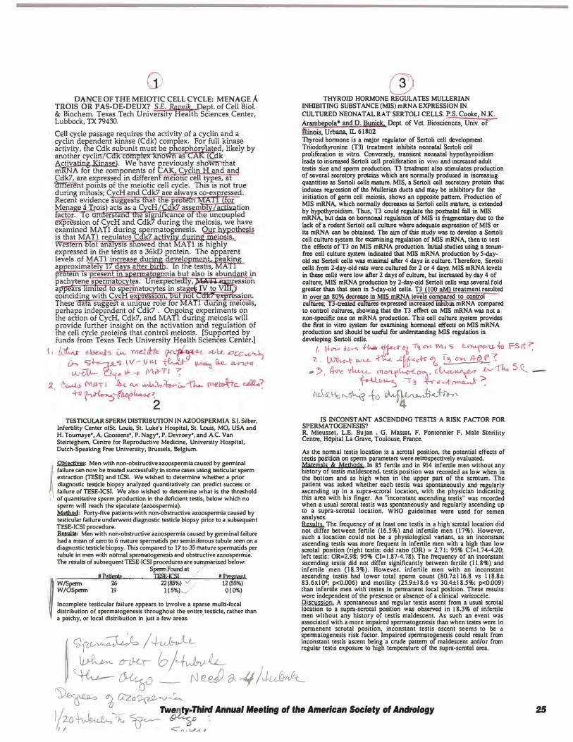
DANCE OF THE MEIOTIC CELL CYCLE: MENAGE A TROIS OR PAS-DE-DEUX? S.E. Ravuik Dept. of Cell Biol. & Biochem. Texas Tech University Health Sciences Center, Lubbock, TX 79430.
TESTICULAR SPERM DISTRIBUTION IN AZOOSPERMIA S.J. Silber, Infertility Center olSl �uis, St. Luke's Hospital, St. Louis, MO, USA and H. Tournaye•, A. Goossens•, P. Nagy", P. Devroey•, and A.C. Van Steirteghem, Centre for Reproductive Medicine, University Hospital, Dutch-Speaking Free University, Brussels, Belgium. � �: Men with non-obstructive azoospermia caused by germinal failure can now be ireated successfully in some eases using testicular sperm extraction (TESE) and JCS!. We wished to determine whether a prior diagnostic ll!stlcle biopsy analyzed quantitatively can predict success or failure of TESE-ICSI. We also wished to determine what is the threshold
1 of quantitative sperm production in the deficient testis, below which no sperm will reach the ejaculate (azoospermia). �: Forty-five patients with non-obstructive azoospermia caused by testicular failure underwent diagnostic testicle biopsy prior to a subsequent TESE-ICSI procedure. Bulllll: Men with non-obstructive azoospermla caused by germinal failure had a mean of zero to 6 mature spermatids per seminiferous tubule seen on a diagnostic testicle biopsy. This compared to 17 to 35 mature sperrnatids per tubule in men with normal spermatogenesis and obstructive azoospermia. The results of subsequent TESE-ICSJ procedures are surrunarized below:
Sperm Found at / • #Patients TESE-!CSI # Pregnant f W/Sperm 26 22(85%) v' 12 (55%)
W/OSperm 19 1 ( 5%)._, 0( 0%) Incomplete testicular failure appears to involve a sparse multi focal distribution of spermatogenesis throughout the entire. testicle, rather than a patchy, or local distribution in just a few areas.
@) TiiYROID HORMONE REGULATES MULLERIAN
INHIBITING SUBSTANCE (MIS) mRNA EXPRESSION IN CULTURED NEONATAL RAT SERTOLI CELLS. r.s. Cooke, N.K. Ararnbepola* and D. Bunic!c. Dept. of Vet. Biosciences, Univ. of iilil\ois, Urbana, IL 6 1 802 Thyroid honnone is a major regulator of Sertoli cell development. Triiodothyronine (TI) treatment inhibits neonatal Sertoli cell proliferation in vitro. Conversely, transient neonatal hypothyroidism leads to increased Scrtoli cell proliferation in vivo and increased adult testis size and sperm production. T3 treatment also stimulates production of several secretory proteins which are normally produced in increasing quantities as Sertoli cells mature. MIS, a Sertoli cell secretory protein that induces regression of the Mullerian ducts and may be inhibitory for the initiation of genn cell meiosis, shows an opposite pattern. Production of MIS mRNA, which normally d=eases as Scrtoli cells mature, is extended by hypothyroidism. Thus, T3 could regulate the postnatal fall in MIS mRNA, but data on honnonal regulation of MIS is fragmentary due to the lack of a rodent Sertoli cell culture where adequate expression of MIS or its mRNA can be obtained. The aim of this study was to develop a Sertoli cell culture system for examining regulation of MIS mRNA, then to test the effects of T3 on MIS mRNA production. Initial studies using a serumfrcc cell culture system indicated that MIS mRNA production by 5-ilayokl rat Scrtoli cells was minimal after 4 days in culture. Therefore, Sertoli cells from 2-<lay-old rats were cultured for 2 or 4 days. MIS mRNA levels in these cells were low after 2 days of culture, but increased by day 4 of culture; MIS mRNA production by 2-ilay-old Sertoli cells was several fold greater than that seen in 5-<lay-old cells. T3 (100 nM) treatment resulted in ,aver an 80% decrease in MIS mRNA levels comJiared to control cultures; T3-treaid cultures expressed increased 1 1610 mRNA compared to control cultures, showing that the T3 effect oo MIS mRNA was not a non-specific one on mRNA production: This cell culture system provides the first in vitro system for examining honnonal effects on MIS mRN A production and should be useful for Wlderstanding MIS regulation in developing Sertoli cells.
(. � Jo•s � �.t-°ti T� cvi Wl• S Ci..1�-lti FSct"; 'Z • Wk...t- � � ..'2.Wc«- � 12, c...... .4-8 �-:
- ? . (j.,,-� 'tku.... tll<Ytf�°"'A· l�IU'<f':7 .Lo---- � s <(_ � \ 3 -¥-.-e c-{'� � iu-\e<..\-'rol'-S� -fo �.,t�J-i�
IS INCONSTANT ASCENDING TESTIS A RISK FACTOR FOR SPERMATOGENESIS? R. Mieusset, L.E. Bujan , G. Massat, F. Pomonnier F. Male Sterility Centre, HOpital La Grave, Toulouse, France.
As the normal testis location is a scrotal position, the potential effects of testis �sition on spenn parameters were retrospectively evaluated. Matena]s & Methods In 85 fertile and in 914 infertile men without any history of testis maldescend, testis position was recorded as low when in the bottom and as high when in the upper part of the scrotum. The patient was asked whether each testis was spontaneously and regularly ascending up in a supra-scrotal location, with the physician indicating this area with his finger. An "inconstant ascending testis" was recorded when a usual scrotal testis was spontaneously and regularly ascending up to a supra-scrotal location. WHO guidelines were used for semen analyses. � The frequency of at least one testis in a high scrotal location did not differ between fertile (16.5%) and infertile men (17%). However, such a location could not be a physiological �ariant, as an inconstant ascending testis was more frequent in infertile men with a high than low scrotal position (right testis: odd ratio (OR) = 2.7 1; 95% CI=l.74-4.20; left testis: OR=2.98; 95% Cl= l .87-4.78). The frequency of an inconstant ascending testis did not differ significantly between fertile (1 1.8%) and infertile men (1 8.3%). However. infertile men with an inconstant ascendinf. testis had lower total spenn count (80.7±1 16.8 vs 1 1 8.8± 83.6xl 0 ; p<0.006) and motility (25.9±1 8.6 vs 30.4±1 8.5%; p<0.009) than infertile men with testes in pennanent local position. These results were independent of the presence or absence of a clinical varicocele. Discussion A spontaneous and regular testis ascent from a usual scrotal location to a supra-scrotal position was observed in 1 8.3% of infertile men without any history of testis maldescent. As such an event was associated with a more impaired spennatogenesis than when testes were in permenent scrotal position, inconstant testis ascent seems to be a spermatogenesis risk factor. Impaired spennatogenesis could result from inconstant testis ascent being a crude pattern of maldescent and/or from regular testis exposure to high temperature of the supra-scrotal area. I ��Jo /-+t-�lt_
� �, fol� � � _ N e erY � --If J Ju.,@-ttflc Y�� � G:zo� II 1 ,, � _
Tw�IJ�· Third Annual Meeting of the American Society of Andrology 2-o � >u � � : I / <::":,., ' VA ,
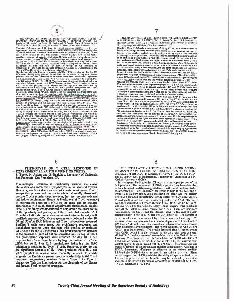
THE UNIQUE STRUCTURAL DIVERSITY OF THE HUMAN TESTISSPECIFIC VOLTAGE DEPENDENT CALCIUM CHANNEL �VDCC) 'LO Goodwin, 'NB Leeds*, 1A Jacob, 1IR Hurley and 15 Benoff. Depts of Research and 'OB/GYN, North Shore University Hospital-NYU School of Medicine, Manha5'el, NY. �: Calcium channel blockers, i.e. dihydropyridinp CDHPs), prescribed for hypencnsion, produce reversible infertility in normospcnnic men in an IVF sening due, in pan, to inhibition of calcium influx required for the acrosomc reaction (AR)_ In the current study we have determined the complete primary struclurc of the human tcstisspccific pore forming al subunit. which binds DHPs, to establish ff a correlation exists between changes in sperm VDCC al subunit structure and response to AR agonists. Iha.i.&n;. Analyzing testis-specific al structure by DNA/cDNA sequencing and Western blot (WB) analysis with specific antibodies (Ab), as compared to somatic tissue. Examination of inter-fertile male variabilily to undergo an AR stimulated by progesterone (P) and model zona ligands containing mannose (Ml. Comparison of the effects of P and M on al protein phosphorylation (Phos). Mgrcria!s Bnd Mcrhods: Human testis pooled poly(A)+ mRNA served as template for RTPCR based cloning using primers derived from the rat cardiac al sequence. Human genomic DNA was used as template to determine intron/exon boundaries. Capacitated motile sperm from fertile donon and JVF-fertile men were challenged wilh 1 ugfmL P or with 100 ug/mL M-BSA + 7S mM M. The percentages or agonist-stimulated AR were determined following staining with RITC-fiiwn agglutinin. The distribution on human sperm of al and phosphotyrosine (P-tyr) antigenic epitopes was determined by immunofluorescence microscopy. W8 of total sperm proteins were probed with rabbil skeletal muscle VDCC al and P-tyr Abs, Statistical comparisons used t-tests. �The entire human VDCC a 1 coding region was sequenced. The 5' end of the testis at subunit is tnmcated, shares no homolo with the cardiac muscle isoform in this region. is tissue restricte enera emauve s a e re
BS s. 1est1s c annel expresses an alternative exon enc mg region important or vo tage gating and the kinelics of activation/inactivation, and a
potential DHP binding sile) not previously described in cardiac cDNA. Potential Phos sites flank 1S6. At least 16 alternatively spliced al transcripts, including lranscripts with both alternative exons in DHP binding regions, 111S2 and IVS3, are detected by RTPCR in lhe human mRNA. Similarly, considerable inter-male variation is detected in the ability to undergo a P or M·stimulated AR (n=6, 7'll>-32'l&; n=l6, l l 'l&-46%). Human VOCC al epitopes are found on proteins of 160-200kDa co-migrating with muscle a l subunits. and o n a -75kDa species, not observed in muscle. Proteins o f similar size are Phos following sperm exposure to model zona ligands but not P (n=l6, P<0.0001 ; n=4, P<0.7, NS). Genestein or DHPs blocks Phos, (n=8, P<0.03 ;n=4, P<O.OS). Conclysjons· Our data suggests that: 1 1 1 the biophysical propcnies, kinetics, and pharmacological sensitivities of the testis-s cific VDCC al subunit differ fro cardiac muscl · a mallc ussues, and 31
es may be blocked by nup hjodjog This inhibition of Phos may propose a hRcha111sm by which DHPs affect VOCC activation. VDCC al structural d1venity may mirror the broad spectrum of response observed in the M-stimulatcd AR between ferlilc men. (Support: ASRMIOrganon grant to L.0.G and NIH Grant No. ES 06100 to S.B.)
6 PHENOTYPE OF T CELL RESPONSE IN
- EXPERIMENT AL AUTOIMMUNE ORCWTIS. P. Turek, K. Aslam and G. Benichou, University of California San Francisco, San Francisco, CA.
Immunological tolerance is traditionally ensured via clonal elimination of autoreactive T lymphocytes in the neonatal thymus. However, ample evidence exists that certain autoimmune T cells escape this process and remain in adults. Normally, these selfreactive T cells remain silent; however, they may become activated and induce autoimmune disease. A breakdown of T cell tolerance' to antigens on genn cells (GC) in the testis can be induced experimentally in mice, tenned experimental autoimmune orchitis f (EAO). This study was undertaken to help define the exact nature of the specificity and phenotype of the T cells that mediate EAO. To induce EAO, AJJ mice were immunized intraperitoneally with purified syngeneic GCs. Mouse spleens were collected at day 10, 20 and SO after EAO induction and T cell suspensions prepared. Purified T cells were tested for proliferative responses and lymphokine patterns upon challenge with purified or sonicated GC. At day 10 and 20, vigorous T cell proliferation was detected in the presence of purified but not sonicated GC. By day SO, no T cell proliferative responses were recorded. At day 10, T cell response to GC was characterized by large production of IL-2 and ylFN; but no IL-4 or IL-5 lymphokines, indicating that EAO llrutiation is mediated by Type I T cells. However, at day 20 and SO, significant amounts of IL-4 were produced while IL-2 and ylFN lymphokine release were markedly reduc;ed. This data
• suggests that EAO is a dynamic process in which the initial T cell 'response progressively evolves from a Type I to Type II phenotype. This has implications for the diagnosis of the disease and for anti-T cell treatment strategies.
j 7
ENVIRONMENTAL LEAD (Pb2+) EXPOSURES, THE ACROSOME REACTION (AR) AND HUMAN MALE INFERTILITY. 'S. Benoff, 1A. Jacob, 'F.S. Mandel*, 'A. H"?hlag• and 'l.R. Hurley. Depts of tobstetrics & Gynecology and 'Research, North Shore Umversity Hospital·NYU School of Medicine, Manhasset, NY. � Blood Pb2+1evels in the range of 4()..70 ug/100 mL have adverse effects on human male fertility, including decreased sperm counts, increased abnonnal morphology. low�red �perm motility, testicular atrophy and prostatic hyperplasia. Dose-response relahonsh1ps between sperm Pb2+ exposure and abnormal sperm function. however. remain to be defined. In murine models, in Yim Pb2+ exposure results in premature AR and decreased penetration/fertilization of ova. Io.mm exposure of human fenile donor spcnn to Pb2+ (S.18-S l 8 ug/100 mL) results in a dose-dependent inhibition of the AR induced by model zona ligands containing mannose. As the association between abnormalities in AR and human male infertility is well recognized, the goal of the current study was to monitor the primary effects of environmental exposure in .ri!g to Pb2+ on the human spenn AR. � A prospective, double blinded study of the mannose lectin (ML) and non-nuclear progesterone receptor (NNPR) properties of motile spennatozoa and of Pb2+ levels in blood pl�ma <B.P) �d seminal plasma (SP) from husbands of 95 consecutive couples undergoing their fintml.llm fertd1zation cycle and who were not occupationally exposed to Pb2+. Mlllcrial1 llll!I M'1hl!lb; Motile spenn were tested for their ability to bind FITC-labeled BSA conjugated to mannose or cell-impermeant progesterone (P). AR was simultaneously evaluated with TRITC·!abeled f. ati= agglutinin .. BP and SP Pb2+ levels were determined by atomic absorption spectroscopy. The relationship be1wecn Pb2+ levels, the percentages of spenn expressing ML and NNPR, and the rate of fertilization of metaphase II oocytes was examined using corrclational and analysis of variance models. ft.Gllllll: More than 40% (42195) of the males studied exhibited BP Pb2+ values above that permissible in men occupationally exposed to Pb2+ (e.g., ;,40 ug/100 mL, the OSHA action limit). BP and SP Pb2+ levels were highly correlated (r= 0.SS4, P<0.0001) and exhibited an inverse relationship with fertilization rates (r= -0.SS9, P<0.0001). SP Pb2+ levels had no signi�cant effec� on ML expression. ML and NNPR are co-expressed on a subpopulation of capacotated mottle spenn. Cross talk between ML and NNPR produces the physiological AR and effects fertilization (r= 0.619, P<0.001). In cases with nonnal ML expression, reduced or failed fenilization could be attributed to defective NNPR expression/function. Importantly, a strong inverse relationship was detected between SP Pb2+, the percentages of spenn expressing NNPR, and ligand-stimulated NNPR aggregation (respectively, r= -0.471 , P<0.04 and r= -0.523, P<0.001), accounting for >80% of the fertilization rate variance. � Pb2+ inhibits the zona-induced AR indirectly at the level of the NNPR. Measurements of NNPR expression/function provide a sensitive endpoint for Pb2+-induced �productive to�icity: "!'e conclude lhat environmental Pb2+ exposures play a greater role on human male onfertdtty than currently appreciated. (Supported by NIEHS/NIH Grant No. ES 06100 to SB with a supplemental Minority Postdoctoral Fellowship to AJ.)
8 THE STIMULATORY EFFECT OF GnRH UPON SPERM
HUMAN ZONA PELLUCIDA (hZP) BINDING IS MEDIATED BY A CALCIUM INFLUX. P. Morales, B. Kerr*, F. Ceric*, J. Scheu* and C. Otero*, Dpt. of Biomedicine, University of Antofagasta and P. Catholic University of Chile.
In vivo, spenn binding to the hZP occurs in the upper portion of the fallopian tube. The presence of GnRH-like peptides has been described in both the female and the male genital tract. In this work we have studied the effect of GnRH on: a) ability of the spenn to bind to the hZP, and b) intracellular calcium levels, using the hemizona assay and the calcium indicator Fura-2AM, respectively. Motile spenn were obtained using a Percoll gradient and the concentration adjusted to l x l 0
7/ml. The cells
were then incubated in Tyrode's medium (2.6% BSA) for 4.5 hr, 37 "C and 5% CO,. For the hemizona assay, spenn aliquots were incubated with 20 nM GnRH or saline (control) for 5 min. Then, one hemizona was added to the GnRH and the identical halve to the control spenn suspension for 1 5 min at 37 °C and 5% co,. under oil. The number of zona bound spenn was counted by phase contrast microscopy. To measure intracellular calcium levels, spenn aliquots were treated with 3 µM Fura-2AM for 30 min. The intracellular calcium level was measured using a spectrofluorophotometer. The spenn were treated with 20 nM GnRH or saline (control). The results indicated that: I ) spenn treated with GnRH bound in higher number to the zona than control spenn (P<0.005); 2) in the absence of extracellular calcium the GnRH did not have any effect; 3) spenn treated before addition of GnRH with 1 00 nM nifedipine or diltiazem did not bind to the ZP in higher numbers than control spenn; 4) spenn treated with 20 nM GnRH showed a rapid and transient increase in the intracellular calcium concentration. Addition of EGT A, Lanthanum, nifedipine or diltiazem to the culture medium inhibited the GnRH-induced increase in intracellular calcium. These results suggest that GnRH modulates the ability of spenn to bind to the human zona pellucida and that this effect may be mediated by a transient increase in the intracellular calcium concentration. (CONRAD MFG-96-17 and FONDECYT 1 97 1 243).
26 Twenty. Third Annual Meeting of the American Society of Andrology
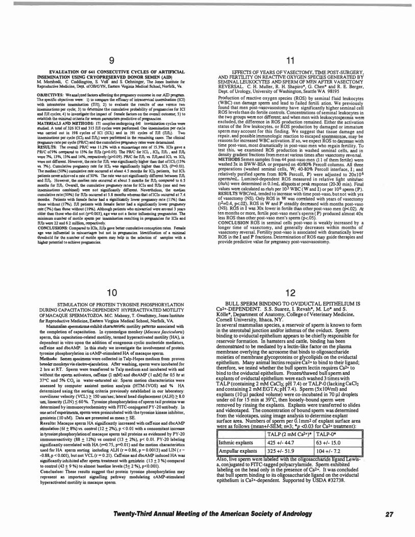
9 EVALUATION OF 641 CONSECUTIVE CYCLES OF ARTIFICIAL
INSEMINATION USING CRYOPRESERVED DONOR SEMEN (AID) M Morshcdi, C, Coddington, S. Voll' and S. Ochningcr, The Jones Institute for Reproductive Medicine, Dept ofOB/OYN, Eastern Virginia Medical School, Norfolk, Va,
OBJECTIVES: We analyud factors affecting the pregnancy oul<DIDC in our AID program. The specific objectives wen:: I) to compare the efficacy of intnu:ervical insemination (!Cl) with intrauterine insemination (nJI); 2) to evaluate the results of one vemis two inseminations per cycle; 3) to detennine the cumulative probability of prqnancies for IC! and M cycles; 4) to investigate the impact of female factors on the ovcnill outcome; 5) to establish the minimal criteria for semen pmmneten predictive of pregnancies. MATERIALJi AND MlmODS: 171 couples undergoing 641 insemination cycles wen: studied. A total of 326 !Cl and 315 M cycles wen: performed. One insemination per cycle was carried out in 198 cycles of !Cl (ICl1) and in 191 cycles of M (M1). Two inseminations per cyde (IC!, and M2) were perfonned in the r=aining cues. The clinical pregnancy rate per cycle (PR/CJ and the cumulative pregnancy rates wen: determined. RESULTS: The ovc:rall PR/C was 1 1.2% with a misauriage rate of 15.3%. !Cls gave a PR/C of9% comparc:d to 13% for IUis (p>0.05). The PR/C for ICl1 , ICli, M1 , and M2 '"""' 7%, 13"o, 13% and 14%, respectively (p>0.09). PR/C for M1 vs. M2and ICI1 vs. !CI, '"""' not different However, the rate for M1 was significanUy bisher than that of!Cl1 (13% vs. 7%). Cumulative prqnancy rate for !CJ, was significanUy bisher than ICl1 (P-0.01). The median (50%) cumulative rate oa:wrc:d at about 4.5 months for IC!, patients, but !Cli patients never achieved a rate of 50%. The rate wu not significantly different between M1 and IUJ,. However, the median rate occwrcd at about 5 months for IUJ, comparc:d to 6.S months for M1. Ovcnil� the cumulative pregnancy rates for !Cls and IUis (one and two inseminations combined) wen: not significanUy different Nevertheless, the median cumulative rate (50%) for IUis oa:wrc:d at 5.8 montha ,Wcreas that of!Cls occwrcd at 7.4 months. Patients with female factor had a significanUy lower prqnancy rate (1 1%) than those without (17%). M patients with female factor had a signilicanUy lower pregnancy rate (7%) than those without (19%). Although patients ..no miscarried were around 3 years older than those who did not (p=0.003 ), age was not a factor influencing pregnancies. The minimum number of motile sperm per insemination resulting in pregnancies for ICls and IUis were 22 and 6 2 million, respectively. CONCLUSIONS: Compared to ICls, 1U1s gave better cumulative conception rates. Female age was inlluential in misauriages but not in prqp111D<ies. Identification of a minimal thn:sbold for the number of motile sperm may help in the sel«tion of samples with a hisher potential to achieve prqnancies.
1 0 STIMULATION OF PROTEIN TYROSINE PHOSPHORYLATION
DURING CAPACITATION-DEPENDENT HYPERACTIVATED MOTil.ITY OF MACAQUE SPERMATOZOA. MC. Mahony, T. Gwathmey, Jones Institute for Reproductive Medicine, Eastern Virginia Medical School, Norfolk, VA.
Mammalian spermatozoa exhibit characteristic motility patterns associated with the completion of capacitation. In cynomolgus monkey (Macaca fasclcu/arts) sperm, this capacitation-related motility, termed byperactivated motility (HA), is dependent in vitro upon the addition of exogenous cyclic nucleotide mediators, caffeine and dbcAMP. In this study we investigate the involvement of protein tyrosine phosphorylation in cAMP-stimulated HA of macaque sperm Methods: Semen specimens were collected in Talp-Hepes medium from proven brocder monkeys via electro-ejaculation. After washing, sperm were incubated for 2 hrs at RT. Sperm were transferred to Talp medium and incubated with and without the sperm 1<:tivators, caffeine (I mM) and dbcAMP (I mM) for 0.5 hr at 37'C and 5% C02 in water-saturated air. Sperm motion characteristics were assessed by computer assisted motion analysis (HTM-IVOS) and % HA determined using the sorting criteria previously established in our laboratory: curvilinear velocity (VCL) � 150 um/sec; lateral bead displacement (ALH) � 8.0 um; linearity (LIN) � 60 %. Tyrosine phosphorylation of sperm tail proteins was determined by immunocytocbemistry with FITC-conjugated PY-20 antibody. In one set of experiments, sperm were preincubated with the tyrosine kinase inhibitor, genistein ( I 0 uM). Data are presented as mean ± SE. Ruulta: Macaque sperm HA significanUy increased with caffeine and dbcAMP stimulation (61 ± 8%) vs. control (12 ± 2%), p < 0.01 with a concomitant increase in tyrosine phosphorylation of macaque sperm tail proteins as evidenced by PY-20 immunoreactivity (88 ± 1 2%) vs control (13 ± 2%), p< 0.01. PY-20 labeling significanUy correlated with HA (t=0.75, p=0.01) and the motion characteristics used for HA sperm sorting including ALH (r = 0.86, p = 0.0013) and LIN ( r • -0.88, p < 0.001), but not VCL (r = 0.21). Caffeine and dbcAMP induced HA was significantly inhibited after sperm treatment with genistein ( 1 3 ± 3 %) compared to control (43 ± 9 %) to almost baseline levels (5± 2 %), p<0.001). Conclusion: These results suggest that protein tyrosine phosphorylation may represent an important signalling pathway modulating cAMP-stimulated hyperactivated motility in macaque sperm.
1 1 EFFECTS OF YEARS OF VASECTOMY, TIME POST-SURGERY
AND FERTll.ITY ON REACTIVE OXYGEN SPECIES GENERA IBO BY SEMINAL LEUKOCYI'ES AND SPERM OF MEN AFTER VASECTOMY REVERSAL. C. H. Muller, R. H. Shapiro*, G. Chen* and R. E. Berger Dept. of Urology, University of Washington, Seattle WA 98195 '
Production of reactive oxygen species (ROS) by seminal fluid leukocytes (WBC) can damage sperm and lead to failed fertilization. We previously found that men post-vasovasostomy have significantly higher seminal cell ROS levels than do fertile controls. Concentrations of seminal leukocytes in the two groups ':"ere not d.ifferent; and when men with leukocytospcnnia were excluded, the difference ID ROS production remained. Either the activation status of the few leukocytes, or ROS production by damaged or immature sperm may account for this finding. We suggest that tissue damage and repair, and possible immunologic reaction to escaped spermatozoa may be reasons for increased WBC activation. If so, we expect ROS to dec�e with time �st-vase, most dramatically in post-vase men who regain fertility. To test this, we examined ROS production in washed seminal cells and in density gradient fractions from men at various times after vasectomy �versa!. METIIODS Semen samples from 44 post-vaso men (I I of them fertile) were washed 3x in BWW-BSA or prepared on 40/80% Percell columns. All three preparations (washed seminal cells, W; 40-80% Percell interface I · and relatively purifie� sperm from 80% Percell, P) were adjusted to '2oxt06 spennlmL. Lummol-dependent ROS measured in relative light units/sec (rlu/s) were determined in 0.1 mL aliquots at peak response (20-30 min). Final values were calculated as rlu/s per to3 WBC (W and I) or per to6 sperm (P). RESULTS WBC/mL tended to increase with time post-vase, but not with yrs of vasectomy (NS). Only ROS in W was correlated with years of vasectomy (r2=0.4, p<.02). ROS in W and P steadily decreased with months post-vaso (NS). ROS in I was 30x lower in fertile than other post-vase men (p<.02). Al ten months or more, fertile post-vase men's spenn ( P) produced almost 40x less ROS than other post-vase men's sperm (p<.05). CONCL�SION ROS in seminal cells post-vase is weakly increased by a longer ttme of vasectomy, and generally decreases within months of vasec!omy reversal. Fe�ility post-v�o i� associated with dramatically lower ROS ID the I and P fmcuons. DetennmatJon of ROS may guide therapies and provide predictive value for pregnancy post-vasovasostomy.
1 2 BULL SPERM BINDING TO OVIDUCT AL EPITHELIUM IS
Ca2+-DEPENDENT. S.S. Suarez, I. Revah*, M. Lo* and S. K!llle*, Department of Anatomy, College of Veterinary Medicine, Cornell University, Ithaca, NY. In several mammalian species, a reservoir of sperm is known to form in the uterotubal junction and/or isthmus of the oviduct Sperm binding to oviductal epithelium appears to be chiefly responsible for reservoir formation. In hamsters and cattle, binding has been demonstrated to be mediated by a lectin-like factor on the plasma membrane overlying the acrosome that binds to oligosaccharide moieties of membrane glycoproteins or glycolipids on the oviductal epithelium. Many animal lectins require Ca2+ to bind to their ligand; therefore, we tested whether the bull sperm lectin requires Ca2+ to bind to the oviductal epithelium. Frozen/thawed bull sperm and explants of oviductal epithelium were each washed 3 times with TALP (containing 2 mM CaCJi; pH 7.4) or TALP-0 (lacking CaCh and containing 2 mM EGTA; pH 7.4). Sperm (5xl Q6/ml) and explants (10 µI packed volume) were co-incubated in 70 µI droplets under oil for 1 5 min at 39'C, then loosely-bound sperm were removed by rinsing the explants. Explants were transferred to slides and videotaped. The concentration of bound sperm was determined from the videotapes, using image analysis to determine explant surface area_ Numbers of sperm per 0. 1 mm2 of explant surface area were as follows (mean+/-SEM; n=3; • <0.03 for Ca2+ treatment):
Isthmic explants
Ampullar explants
T ALP (2 mM Ca2+)* T ALP-0*
425 +/- 44.7 63 +/- 15.0
325 +/- 5 1 .9 104 +/- 7.2
Also, .1ve sperm were a wt e o 1gosacc an 1gan wts-a, conjugated to ffi'C-tagged polyacrylamide. Sperm exhibited labeling on the head only in the presence of Cal+, It was concluded that bull sperm binding to its oligosaccharide ligand on the oviductal epithelium is Ca2+-dependent. Supported by USDA #32738.
Twenty· Third Annual Meeting of the American Society of Andrology 27
1 3 MECHANISMS OF CRYOPRESERVATION-INDUCED
CAPACITATION OF BOVINE SPERM. J.L. Bailey and B. Bero�·. Centre de Recherche en Biologic de la Reproduction, mpt des sciences animales, Universit� Laval, "Qu&ec, "QC u IX 7P4.
The fertility of bovine spenn following cryopreservation is dramatically reduced and is not fully explained by poor post-thaw motility and morpholojlY· We have demonstrated th�t immediat�ly aftertfiawing, a pro�rtton of spenn are already capac1tated (Conni er et al., 1997). We suggest that the sub-fertility of cryopreserved spenn is partly due to premature capacitation. To test the hypothesis that the mechanisms of capacitation induced by cryopreservation or by heparin (physiological capacitation) are different, this study compared the intracellular Ca2+ dynamics and protein profiles of fresh and cryopreserved spenn. To monitor [Ca2+]i, the fluorescence of fora-loaded spenn was measured at 0, 2.5 and Sh of capacitation ± 10 µg/ml heparin, prior to and after the addition. Br-A23 187 (n > .20 spz from 4 ejaculates). To assess spenn protem profiles, protems were extracted from cryopreserved cells during capacitation (± heparin), subjected to silver nitrate staining-SOS-PAGE and western blotting using an antiphosphosphotyrosine antibody (n = 3). Internal [Ca2+] seemed not to differ in fresh or cryopreserved sperm, either ± heparin before ionophore treatment (P>O.�S). At 2.S and S h of capacitation, cryopreserved spenn + hepann underwe_nt a greater ionophore-induced increase in [Ca2+] than spenn - hep!'"':! (P:::O.OS). This difference was only observed after S h of capac1tation m fresh spenn. At O h, a series of tyrosine phosphorylated sperm proteins were observed + heparin. At 2.5 and 5 h, tyrosine-phosphorylated proteins were observed only from sperm + heparin. These data demonstrate that immediately after thawing there is a population of precapacitated sperm and another more cryo-resistant population t!iat undergoes capacitation in a manner similar to that of fresh cells (1.e. heparin and time-dependent). (Supporud by the CBRC and NSERC. Thanks to the Centre d'l�..Unation Artilicielle du Qul!bec for semen and technical assistance )
1 4 TWO DAY IUI TREATMENT CYCLES ARE MORE SUCCESSFUL THAN
ONE DAY IUI CYCLES WHEN USING FROZEN-THAWED DONOR SPERM M. Matilsky , J. Younis and M. Ben-Ami, Reproductive Medicine Uni� Poriya Government Hospital, Tiberias, Israel
Relatively few publications have addressed the best ways of performing donor insemination. The difference in pregnancy rates following IUI for one vs. two days in the periovulatory period has been reported as either inconsequential or favoring the use of two consecutive inseminations, 24 hours apart. Some studies, related to donor insemination, presented either small numbers of patients or Jess than JOO cycles per treatment group, making conclusions difficult to interpret. Two large studies (Brook eta!, 1994, Deary eta!, 1 997) , presented unbalanced study groups and one (Deary eta!), used !CI of unwashed frozen-thawed sperm and timed the inseminations with home use urinary LH kits. Our study compared the monthly fecundity and cumulative probability of pregnancy in a large group of women (N�I23) undergoing controlled ovarian hyperstimulation and one or two day inseminations with donor sperm prepared from frozen-thawed samples. The choice of single or double insemination was decided by the day of the week each patient received hCG for ovulation induction. Approximately 80% of all the patients underwent both single and double insemination treatments during the 2.S year study period. 93 patients received single inseminations in 180 cycles, while l OJ patients received double inseminations in 222 cycles. Nine clinical pregnancies were achieved in the one day group (So/Jcycle, 9.7%/patient), while 39 pregnancies occurred in the two day group ( 17.9"/Jcycle, 29.8%/patient).Two and five spontaneous abortions occurred in the one and two day groups, yielding take home baby rates of 3.9%1 cycle and IS.J'Y.tcycle, respectively. The cumulative probability of conception over I S cycles of treatment was consistently twice as high or higher for the two day group. In addition, the data revealed that a significant number of pregnancies occurred between the 7" and 12" treatment cycles(Jl9-one day; 7139-two day). The results of this study suppon the use of two day JUI treatment eye Jes when using frozen-thawed donor sperm. A large, multicentered, prospective study would be useful in substantiating these results. Brook eta! (1994) Fenil Steril 6 1 :308-3 13. Deary eta! ( 1997) Hum Reprod 12:1494-1496.
1 5 DIALYSIS ADDmON OF TREHAWSFJGLYCEROL
CRYOPROTECTANT YIELDS POST-THAW MOUSE SPERM WITH ADF.QUATE FERTILIZING ABILITY. B.T. Storey, E.E. Noiles and K.A. Thompson*, Center for Research on Reproduction and Women's Health, University of Pennsylvania. Philadelphia. PA.
Cryopreserved sperm offer a low cost and space-saving means for maintenance of mutant, transgenic and knockout strains of mice. Mouse sperm are prone to membrane damage from osmotic stress during cryopreservation from osmotic imbalance on addition prefree:zc and removal post-thaw of cryoprotectant and from imbalance due to dessication/rehydration during freezing/thawing. We showed (J. Androl. 1 8 (Suppl.): P-45, 19117) that glycerol (G) plus trehalose (T) provided a useful cryprotectant for mouse spenn in terms of plasma membrane integrity. Stress from osmotic imbalance on addition and removal of cryoprotcctant is reduced by serial addition pre-free:zc (SA) and serial dilution post-thaw (SD) (Gilmore, Liu, Gao and Critser, Hum. Reprod. 1 2: 1 12-1 18, 19117); in this study, we compared fertilizing ability (FA) of CDl mouse spenn recovered post-thaw after cryopreservation with cryoprotectant 6% Gn.5% T using SA/SD, addition/removal by dialysis (DL), and DUdircct addition. FA was assayed by insemination of 10 CD! mouse eggs in 0.5 mL drops with 2.5 x 1 OS spenn and scoring symmetrical twa-«11 embryos. Under these conditions, untreated fresh control sperm gave 73::!:9% eggs fertilized; DlJDL gave lfu:7%; SA/SD gave 23::1:1 1 %; DUdirect addition gave 45::!:9% (mean::l:SD, n = 8). Additon of cryoprotectant by DL prefree:zc and direct addition of sperm without removal of cryoprotectant post-thaw gave significandy superior FA (P < 0.01) compared to DUDL and SA/SD. Normalized to conrols, DUdirect addition gave 62%. DUdirect addition using the chemically defined Off cryoprotectant offers post-thaw FA adequate for eventual transgenc transmission. Supported by NICHD through grant HD-31757
1 6 MOTILITY OF CRYOPRESERVED SEMEN SPECIMENS DECLINES OVER STORAGE TIME IN SUBPOPULATIONS OF CRYOBANKERS. R. Dolgina• and GS Prins. Dept of Urology, University of 1Ilinois, Chicago, IL.
Long-term freezing of burnan sperm is frequently desired by men facing the possibility of sterilization, reduction of fertility potential or genetic damage. The success of the freezing-storage-Oiawing procedure is dependent on lllBDY factors one of those being the initial semen quality. In many cryobanks, post-thaw motility estimates are made on a frozen aliquot soon after cryopreservation to provide diagnostic infonnation for future use. This study retrospectively determines factors affecting the prognosis of post-thaw sperm motility after various periods of cryostorage. We analyzed SI specimen designated to be destroyed either because of patient request or due to death. Specimens were cryopreserved for 1-9 years (3.44 ± 2.21 yrs; mean ± SEM ). The clinical records, including reason for sperm banking, initial semen parameters, 24 hr post-thaw analysis and post-thaw parametelS after variable periods of cryostorage were studied In general, we observed that postthaw motility estimates made at 24 hr after cryopreservalion (P1) reliably predicted post-thaw motility years later after long-term cryostorage (P:i): P1 IP1 • 1 .22 ± 0.09, n=S I . However, subpopulations existed which significantly affected the predictive capacity of the 24-br post-thaw evaluation. Firs4 initial sperm concentration was found to affect the post-thaw motility estimates over time. For patients with an initial sperm concentration between 20-60 x I<l'lml, the P1 IP1 = 0.93 ± 0.06 (n- 17) indicating that motility remained constant over time in cryostorage. In contras4 patients with oligozoospermia (< 20 x lO'lml) had a P, IP1 = 1.77 ± 0.43 (n-9) while patients with high sperm concentrations (>60 x IO"lml) had a P, IP, = 2.60 ± 1 .39 (n=2S), indicating that in both situations there is a marked motility loss over time in storage perhaps due to uneven distribution of sperm cells in cryobuffer. Secondly, patient diagnosis prior lo cryostorage affected the post-thew motility over time. Men with testicular cancer who froze specimens prior lo chemotherapy possessed a P1 IP1 = 1 .47 ± 0.27 (n-16 ) while all remaining patients (including other cancers, pre-vasectomy or other surgeries) had a P, IP, ratio of 1.09 ± O.OS (n=JS). In conclusion, the post-thaw motility 24 hr after cryopreservation is a useful prognostic test; however, the reliability of this parameter will depend on the initial sperm concentration as well as the presence of testicular cancer in the banking population.
as Twenty· Third Annual Meeting of the American Society of Andrology
1 7 DIFFERENTIAL EXPRESSION OF TESTOSTERONE BIOSYNTHETIC AND
METABOLIZING ENZVMES DURING PUBERTAL DEVELOPMENT OF RAT LEYDIG CELLS M.P. Hardy and R.·S. Ge•, Tha Population Council and Rockefeller University, 1230 York Avenue, New York, NY
The amount of testosterone (T) secreted by the Leydlg cell Is determined by a balance between T blosynthetlc and metabolizing enzyme acUvHles. U has been established that Sa-androstan-3a.17�-dlol (3a-DIOL) Is the predomlnanl Immature androgen In rals during days 20 to 40 pcstpart\Jm, whereas T Is the major androgen by day 56. However, the underlying changes In T blosynthetlc and metabollzlng enzymes during Leydlg cell development, and their magnitudes, have remained unclear. The aim of the present study was to define the developmental trends for T blosynlhetlc and metabolizing enzymes In Leydlg cells at three distinct slsges of pubertal differentiation: mesenchymal-llke progenitors on day 21, Immature Leydlg cells on day 35 and adult Leydlg cells on day 90. Four major androgens, androstenedlone (DIONE), androsterone (AO), T and 3a-DIOL were measured In progenitor, lmmetunt and aduh Leydlg cells In spent medl\Jm alter 3 h In vitro. Sleady-stata mRNA levels and enzyme activities of blosyntheUc and metabolizing enzymes were detennlned. LH-stlmulated levels of total androgens (DIONE + AO + T + 3a·DIOL) were slgnlflcantiy lower In progenitor, compared to Immature and adult Leydlg cells (P <0.05) with 84.33 ± 8.74 ng/101 calls/3 h In progenitors (mean ± SE) versus 330.13 ± 44.19 In Immature, end 523.23 ± 67 29 In adult. Progenitor, Immature and adult Leydlg cells produced different androgens, wtlh AO being released by progenitor cells (72.08 ± 9.02% of total androgens), 3a·DIOL by Immature (73.33 ± 1 4.52%) and T by adult Leydlg cells (74.38 ± 14.73%). Further examination showed lhat this difference resulted from dlfferentlal expression of T biosyntheUc and metabolizing enzymes. Low levels of type Ill 17�-hydroxysterold dehydrogenase (17�·HSD) mRNA and 1 7-ketosterold reductase activity were present In progenitor cells compared to Immature and adult Leydlg cells. In contrast, type I Sa· reductase (5a·R) and 3a-hydroxysterold dehydrogenase (3a·HSD) mRNAs and enzyme activities dramatically decreased In adult Leydlg cells, compared to progenitor and Immature cells. Most of the T blosynthettc enzymes attained equivalent levels In Immature and adult Leydlg cells, but T was rapidly metabolized to 3a·DIOL by high 5a·R and 3a·HSD reductive activities In the former and not the latter. The dramatic decline In Sa-R and 3a-HSD activities resulted In maximal T production In adult Leydlg cells. The results Indicate that steroldogenlc enzyme gene expression Is not Induced simultaneously, and that sequential changes In T blosynthetlc and metabolizing enzyme activities result In different androgen end products being secreted by Leydlg cells during pubertal development. Supported In part by the NIH grant HD-35288 (M.P.H.) and Iha Population Council (R.·S.G.).
1 8 A PRELIMINARY NEUROENDOCRINE MODEL OF FEED
BACK IN THE MALE REPRODUCTIVE AXIS. JD Veldhuls and OM Keenan•, Mathematics and Medicine, Univ. Virginia, Chartottesvllle, VA 22908 A hallmark of neuroendocrine axes Is feedback control exerted by the hypothalamus, pituitary gland, and target tissues (e.g. gonad). The regulation of such networks Is complex, because non-linear time-delayed dose-response mechanisms operate at each locus. The mechanisms that modulate such pulsatile (episodic) and circadian (rhythmic) output are virtually unknown. To formalize these neuroendocrine relatlonships, we have implemented a feedback and feedforward blomathematical model of the male hypothalamic (GnRH)-pitultary (LH)-gonadal (testosterone) axis. Our stochastic differential equation formulation consists of a nonstationary stochastic point process responsible for generating pulses of GnRH, the frequency and amplitude of which are modulated by short-loop (GnRH) and long-loop (testosterone) negative feedback. Pulsatlle GnRH release In tum drives bursts of LH secretion via an agonlstlc dose-response curve, which is partlally damped by testosterone's negative feedback. Available LH pulses stimulate (feed-forward) testosterone synthesis and release, which In tum acts via negative feedback on both GnRH and LH. Five experiments conducted with this model document physiologically expected feedback behavior for the GnRH-LH-testosterone axis. Six other experiments test distinct within-model coupling mechanisms between a circadian Input and the pulsatile axis that would explicate the known circadian variations in testosterone > LH. We conclude that a nonlinear feedback I feedforward biomathematical control system with combined pulsatlle and circadian features closely emulates the behavior of the male hypothalamo-pitultary-testis axis.
1 9 STAGE-SPECIFIC HORMONAL PROTECTION AGAINST
HEAT-INDUCED GERM CELL APOPTOSIS IN RAT Y.H. Lue, A.P. Sinha Hikim, A Leung*, S. Baravarian•, C. Wang, and R.S. Swerdloff, Department of Medicine, Harbor-UCLA Medical Center, Torrance, CA.
We have shown that local testicular heating (43 • C for IS min) n:sulted in a marked acceleration of genn cell apoptosis by day 2 at early (I-IV) and late (XII-XIV) stages with minimum effects on the hormone dependant stages (VII-VIII) of spermatogenesis. We hypothesized that intratesticular testosterone (T) plays an important role in protecting germ cells against heatinduced apoptosis at stages VII-VIII. To test this hypothesis, adult male SD rats were assigned into 4 groups to receive: I ) a daily sc injections of vehicle for 4 days; 2) a daily sc injection ofGnRH antagonist (GnRH-A) at a dosage of 1 .25 mg/kg BW fur 4 days; 3) a single exposure to local testicular heating (43 • C for 1 5 min) applied on day 2; and 4) GnRH-A plus heating (applied on day 2). All animals were sacrificed by day 4. Serum levels of FSH and T as well as the total content of testicular T were markedly reduced in both GnRH-A and GnRH-A + heat-treated groups when compared to either control or heat-treated group alone. Incidence of apoptosis (expressed as numbers/Sertoli cell) was low (0.0 1-0.10) in both control and GnRH-A treated groups. As expected, hyperthermia within 2 days resulted in stagespecific activation of germ cell apoptosis with lowest level of apoptosis ( 1.1) detected at stages VII-VIII and highest levels (3. 1-5.3) at stages I-IV, V-VI, IX-XI, and XII-XIV. While the incidence ofapoptosis at stages other than VII-VIII remained unchanged between heat and GnRH-A + heat treated groups, GnRH-A addition to heat resulted in further acceleration (by 3.2 -fold) ofapoptosis at stages VII-VIIl over the values measured in heat alone and was comparable to those measured at other stages. These results clearly demonstrate the preventive effect ofT on heat-induced germ cell apoptosis at stages VII-VIII.
20 MARKED SUPPRESSION OF DIHYDROTESTOSTERONE BY
GI1 98745, A NOVEL 5-a REDUCTASE INHIBITOR. RV Clark, DJ Hermann•, Glaxo Wellcome, Research Triangle Park, NC
Testosterone (T) is converted to the more potent androgen dihydrote�tosterone (D� by t�e enzyme S-12 reductase (SAR). While DHT ts cntical for mascuhmzallon of the genitalia in the fetus it seems to have limited importance in adult men except for its effects in prostatic hyperpl�sia.- We compared the honnonal effects ofGI198745 (GG745) to finastende m BPH patients. GG745 is a potent inhibitor of type I wid 2 SAR, whereas finasteride only inhibits type 2 SAR.
Fifty-three BPH patients (International Prostate Syptom Score ?.8 and enlarged prostate on DRE) received daily oral doses of GG745, placebo, or finastende (Smg) for 28 days m this randomized, blinded, parallel group tnal. GG745 doses of 0.1 , O.S, 2.5, Smg, and 2.5 with a 40mg loading dose were studied. DHT and T measures were taken before and after 28 days of study drug administration. GG745 groups were compared to placebo and finasteride using a general linear model with pairwise comparisons.
The results after 28 da s of treatment arc as follows mean±SD : roup n 0 O
% Chan e
'p<0.05 Vs placebo , #p<0.05 Va ftnaslsrtde .w� concluded that dual . inhi�ition of SAR with GG74S produced
s1gmficantly greater reducl!ons m serum DHT compared to finastcride whil� � remained within th� normal range. GG74S is clearly capable of proV1dmg >95% red.uct1on m serum DHT with little variability. Larger, longer duration studtes arc needed to determine the added clinical benefit ofa potent dual SAK inhibitor over a type 2 inhibitor such as finasteride.
twenty· Third Annual Meeting of the American Society of Andrology 29
30
21 GPI-ANCHORED PROTEINS ON THE SERTOLI CELL SURF ACE.
H.A. Wat.son•, R.R. Fonna• and S.E. Nyquist, Department of Biology, Bucknell University. Lewisburg, PA 17837
Glycosylphosphatidylinositiol (GPI) • anchored proteins constitute a class of membrane proteins that has recently gained attention in the scientific community. These proteins are sorted to the apical-lateral domain of epithelial cells, perhaps via specialized lipid rafts Conned in the trans Golgi network. At the apical cell swface they reside in association with cholesterol and glycosphingolipid rich domains related to caveolae. The study reported here is an investigation of the GPI-anchored proteins found on the Sertoli cell membrane. Four-<lay old Sertoli cell cultures were established from 20-<lay rat pups on both the standard plastic culture flask and on the porous bicameral insert. After four days of culture the cell surface proteins were biotinylated using a cell impenncant biotinylating reagent, sulfosuccinimidyl-6-(biotinamido) hexanoate (Pierce Chem. Co.). Cells were mechanically scraped off of the plate, washed extensively and subjected to digestion by phosphatidylinositol specific phospholipasc C (Boehringer Mannheim) to release GPI-anchored proteins. Following centrifugation the supernatant was collected and precipitated via acetone/methanol. The resultant protein pellet was collected and prepared for SOS-PAGE. The resultant gels were western blotted and visualized using ECL with an avidin-horse radish peroxidase kit (Amersham). The Sertoli cell cultures grown both on plastic and on the porous bicameral insert showed a major band at approximately 132 kD plus several less prominent protein bands of lower molecular weight Biotinylation in the bicameral well from the basal surface produced no identifiable bands. Studies are presently underway to determine whether this GPI -anchored protein is the 130 kD cell adhesion molecule N-<:adherin of rat testis.
22 1HE ROLE OF TYROSINE PHOSPHORYLATION IN 1HE REGULATION OF SERTOLI CELL TIGHT JUNCTIONS, M.J. Bellace• and S.E. Nyquist, Department of Biology, Bucknell University, Lewisburg, PA. In epithelial cells, tight junctions (TJ) create a regulated barrier to the passage of solutes, water and migrating cells. Research on mammalian cells has identified multiple TJ-associated proteins, of which Z0-1 (214 kD), Z0-2 (160 kD), and 7H6 (175 kD) are reported to be tyrosine phosphorylated. Despite progress with other tissues, little is known about the molecular components of the Scrtoli cell TJ or their phosphorylation status. The Scrtoli·Scrtoli TJ constitutes the barrier separating the basal and the adluminal compartments; it is this barrier that must be traversed by the early spermatocytc. The pwpose of this study was to examine tyrosine phosphorylation patterns on putative tight junction proteins. To accomplish this, three experimental systems were used: a) staged seminiferous tubule segments, b) prepubertal testes from rat pups and c) Scrtoli cell cultures. Using adult rats, and transillumination, seminiferous tubules were cut into four segments (pale, stages IX-XII; weak spot, stages XIIl-1; strong spot, stages 11-VI; and dark zone, stages Vil-VIII). All samples were incubated with or without 100 µM pcrvanadate (a membrane penneant tyrosine phosphatase inhibitor) for 15 min. @ 34.S'C to preserve/enhance phosphorylation patterns. Whole testis homogenates were made from 12· to 24-day old rat pups and incubated with or without pcrvanadate. It is during this period of development that the TJ first forms. The third approach was Scrtoli cell cultures grown both on plastic and pcnneable inserts in bicameral chambers. Protein samples were run on SDS· PAGE gels and immunoblotted using anti-phosphotyrosine monoclonal antibody (Upstate Biotech). Blots were stripped and restaincd using anti-actin antibody (gift of J.L. Lessard) to control for load. A comparison of the four tubule segments showed differences in multiple phosphoprotein bands, including 215, 175, and 130 kD bands. The developmental study showed strong, changing phosphotyrosine staining in several bands, including the 175 kD band. The Sertoli cell cultures also had similar bands including a strong band at 215 kD and a weak 175 kD band in both the standard and bicameral chambers. These studies suggest that the phosphorylation status of cellular proteins including putative tight junction proteins varies with the stage of the seminiferous cycle, the development of the tight junction, and Scrtoli cell culture conditions.
23 SERTOUN IS A NOVEL SERTOLI CELL PRODUCT: ITS cDNA CLONING, TISSUE
DISTRIBUTION, AND REGULATION. D. Mruk•), M.Y. Ml", J. Grima•, B. Silvestrini", WM. Lee'•), and C.Y. Cheng•. •Population CouncH, Center for Biomedical Research, 1230 York Avenue, New York, NY 10021; 21Jepartmenl of Zoology, University ol Hong Kong, Hong Kong; m �nslitute of Phannacology and Phannacognosy, University of Rome, llaly.
Throughout spennatogenesls, there are extensive Interactions belween Sertoll fSC) ard genn cells (GC) in the semln/ferousepilhelli.m lafllely due to the mlgratton ol developing genn cellslrom the basal lamilato Iha adkmilal compartment. To Identify molecules that are illely to be involved in SC-GC Interactions, RNA differential display technique was utilized usi1g RNAs Isolated from primary cultures of SC, GC, andSC-GC. Briefly, an 0Hgo (dT)11CA was used as the primer to reverse transcribe RNAs Into cDNAs. Thereafter, PCR was performed using a random sense primer of 5'-ACCATGCAGGGC-3'and oligo ( dT)11CA In the presence ol f:fiSJ-dATP to amplify genes expressed In the corraspondlng prlmaiy cell cultures. PCR products were rasolved by gel eleclrophoresls on a sequencing gel unit and visualized by autoradlography. A cDNA that was lound in SC whose abundancyappeared lo be elevated i1 SC-GC co-cultures but was absent In GC was selected, gel purified, re-amplified by PCR, and subcloned Into pGem T™ vector (Promega) lor nucleotide sequencing. This cDNA has a size of 675 bp with anopenreading lrame (ORF) lrom the 5' endbelweennucleolides 3-437 coding lor an145·amlno acid peptide. Comparison of this ORF with existing dalabasesat BLAST and GENBANK yiekled no significant homologies revealing this Is Indeed an 111Q,Je molecule and designated sertolln. Using a specific sertolln primer pair of 5'ACCATGCAGGGCTGCCCCCCCGAGGAGCTG-3' (sense, nuclaotidas 1-30) ard 5'GGTGTACCGGCTGTCCAA-3' (antisense, nucleotides 1 99-216) lor RT-PCR, sertolin mRNA was detected in the adult rat brain, klrtiey, spleen, liver, lung, ovary, epididymis, testis, andimmature SC, but not il GC, heart, thyroid, thymus, or uterus. Furlhennora, the steadyslate mRNA level of sertolin increased by as much as 15-fold from 20 lo 60 days of age during tesllcular maturation suggeS1klg Its likely involvement in spennalogenesls. Moreover, sertolin mRNA expression was shown to be signillcanlly stimulated by interleukin-la (IL· 1., 10 unlls/ml) but not by basic flbroblasl growlh faclor (150 ng/ml) or inlerferon.., (100 units/ml). Since IL-la is known to be a SC product and possibly released by GC, ii is very likely that sertolin expt"esslon may in I act be regulated by GC. Hen:e, work Is now in progress to examilethe elfects ol GC on SC sertolln expression in vitro . In addition, we are in the process of clonlngthe full-length sertolln cDNA lrom aSC expresslonlibrary conslructad i1 an Uni-ZAP™ unidirectional vector (Slralagene). In Sllllrnaiy,we havaldentilied a novel SC gene designated sertolin. Results of this study show th al sertolin is likely to be a novel marker in S1udying SC-GC interactions inthe testis. This work was supported In part by grants lrom NIH (HD 13541), the Conrad Program (CIG 96·05), the Rockefeller FoundaUon,the Noopolis FoundaUon, and the Hong Kong Research Grant Council (HKU 7235/97M).
24 EXPRESSION OF 1HE NOVEL CRES GENE IN 1HE
ANTERIOR PITUITARY GONADOTROPES. H.G. Sutton*, A. Fusco*, G.A. Cornwall. Texas Tech University Health Sciences Center, Lubbock, TX
Our lab is interested in examining the role of the novel CRES (cystatin-related epididymal and spermatogenic) gene in male reproduction. We have previously shown that CRES expression is highly restricted to the proximal caput epididymidis and to the round and elongating spermatids in the testis. More recently, utilizing RTPCR we identified CRES expression in individual mouse pituitary glands. To identify the cell population expressing the CRES gene we examined available pituitary cell lines. Northern blot analysis showed CRES expression in the P cell line, a differentiated gonadotrope, but not in the ct cell line, a less differentiated gonadotrope. CRES was also not expressed in the somatotrope/ lactotrope GH, cell line. Double immunofluorescence analysis of mouse pituitary tissue showed that CRES protein co-localized with LHp, confirming that CRES is expressed in anterior pituitary gonadotropes. Western blot analysis of p gonadotrope cell lysate and conditioned media showed that CRES protein was secreted by the p cells. Interestingly, in contrast to the 1 9 and 14 kDa CRES proteins in the testis and epididymis, 1 7 and 12 kDa CRES proteins were present in the P cells. N-glycanase treatment of testis and epididymal lysates and p cell conditioned media showed that the higher molecular weight proteins (1 9 and I 7 kDa) are the result of N-linked glycosylation. The highly restricted expression of the CRES gene to the testis, epididymis, and anterior pituitary gonadotrophs suggests that CRES may be involved in several aspects of male reproduction.
Twenty. Third Annual Meeting of the American Society of Andrology
25 Abstract withdrawn.
26 FAS/FAS LJ:GAND J:HDUCTJ:OH OF APOPTOSJ:S : A
POSSJ:BLB MECllAHJ:SM OF HORMONB-DEPEHDENT LEYDJ:G CBLL ATTllJ:TJ:OH F . C . Griffin, J . S . Hushen , P . Barbour and D . F . Cameron , University o f South Florida College of Medicine, Tampa , FL 3 3 6 12 Fas and FasL proteins (TNF family) are involved in CTL-mediated cytotoxicity and down-regul ation of the immune response by inducing suicidal apoptosis of activated lymphocytes . Although Fas and FasL have been detected in rat testes , their cel lular localization is not wel l defined. Death of Leydig cells by hormone {LH) depletion was studied in vivo {hypophysectomized rats) and in yitro (primary cell cultures with and without hCG) . Apoptosis was determined by TUNEL analysis ; Fas and FasL was detected by Western blot analysis and immunohistochemistry . FasL appeared to be constitutively expressed on Leydig cells in normal testes and in fresh isolates . Fas , however , was not apparent in either freshly isolated cells or in Leydig cells of normal testes . When cultured for 48 h in non-supportive medium without hormone , western blot analysis showed de novo expression of the Fas protein .
Results suggest that attrition of Leydig cel l s following LH depletion is by suicidal apoptos i s , similar to that observed in activated T-lymphocytes , and involves the up regulation of Fas receptor and its subsequent binding by the already present FasL.
27 DO FETAL LEYDIG CELLS DEGENERATE IN THE POSTNATAL RAT TESTIS? H.B.S. Ariyurutne und S.M.L.C. Mendis-Hundogomo. Department of Animal Science, College of Veterinary Medicine, 111e University of Tennessee, Knoxville, TN.
Fetal Leydig cells (FLC) ore present ot birth but their destiny in the postnntal testis is still unknown. 111erefore, we designed this study to understand the fote of the FLC in the postnutnl rat testis. Sprague Dawley rots of 1 ,7, 14 ,21 ,28,40,60 and 90 days were used. 111eir testes were either prepared for stercology (fixed in 2 5% glutnraldehyde in cacodylate and embedded in Epon-Araldite, 11=4) or i11111111noc)10chemistry (Bouin's fixed ond embedded in paraplnst, n=3). Tiie presence ofFLC ond udult Leydig cells (ALC ) in rots of 28-90 days of nge wos detennined by 1 1 �HSD I inununocytochemistry(FLC are I l �HSD 1 -ve, ALC ore I l �HSDl+ve); udditionolly, the ratios of FLC:ALC numbers were obtnined. FLC ond ALC numbers in neonntnl rot testes (I ·2 1 days) were quantified by stereology; they were dilferentiully identified on their morphology. FLC had euchromotic nuclei ond lipid rich C)1oplasm .. ALC (obsen·ed from doy 14 onwards) in 14 und 2 1 doy testes had liMle or no C)1oplasmic lipid in them. To quanti�· FLC and ALC in testes of 28-90 dny old rats, first the total Leydig cell number per testis was detennined by stereology ond separated this valne into FLC ond ALC by using the number ratios for FLC:ALC obtained from the 1 1 �HSDI imm11noC)1ochemicol studies (inunature ALC in the prepubertal-pubertnl testes are also rich in C)1oplnsmic lipid, ond therefore, this feature ca1mot be used to dilferentiolly identify these cells ofier day 21 ). 1 1 �HSD 1-ve Leydig cells, i.e. FLC were continued to be present fro111 birth to 90 days. Moreover, their nwnber per testis did not change from birth to 90 days of oge, although the ALC number per testis concomitantly increased " ith age 111is study convincingly demonstrated that FLC in rots do not degenerate in the postnatal testis, but continue to be present ot least up to 90 days of age. (Supported by NSFIBN94092R8,COE ond Minkel Grants from UT)
28 PS3·DEFICIENCY SUPPRESSES GERM CELL APOPTOSIS
AND STIMULATES CELLULAR PROLIFERATION IN VIVO Y. Lue, A.P. SinhaHikim, T.B Rajavashisth*, C. Wang, W.E. Salameh and R.S. Swerdloff, Department of Medicine, Harbor-UCLA Medical Center, Torrance, CA.
The protein pS3 is a key tumor suppressor, as evidenced by its frequent inactivation in human cancers. This study, using pS3 ../-, mice, addresses the functional role of pS3 on genn cell homeostasis. Mice deficient in pS3 exhibited a significant increase in the numbers of both Brd U-labeled as well as total preleptotene spennatocytes, but no apparent change in spermatogonial apoptosis, suggesting increased spennatogonial proliferation. This implies a lack of surveillance of various cell cycle checkpoints involving vigorously cycling mitotic lineage (spennatogonia) in pS3 -/- mice. Apoptosis was most commonly found in spennatocytes, including the dividing spennatocytes, less frequently in spennatogonia and seldom in spennatids of adult mice. The lowest level of apoptosis (expressed as numbers/JOO Sertoli cells) was detected at stages VII-VIll and IX-XII, and highest levels at stages I-III, IV-VI and XII. The levels of spennatocyte apoptosis at stages I-III (S.4 ± 0.9) and XIl (19.3 ± 3 . 1) in pS3 -/- mice were significantly lower than those of the pS3 +I+ mice {14.2 ± 2.4 and 33.6 ± 4.7, respectively), suggesting that defective genn cells that would normally have been deleted by apoptosis instead survived. Increased spermatogonial prolifi:ration and decreased apoptosis of spennatocytes were further reflected by a higher testicular spenn count in pS3 ./. mice {27.6 ± O.S x 106/testis) compared to nonnal mice (22.8 ± 1 .4 x 106/testis). pS3 deficiency also had stimulatory effects on testicular steroidogenesis, including Leydig cell hypertrophy (1265 ± 37 µml vs. 1068 ± 26 µml). Leydig cell numbers per testis remained unchanged. These results clearly establish the importance of pS3 in regulating genn cell homeostasis during nonnal spennatogenesis and further illustrates the complexity of the pS3 pathway in the testis.
Twenty. Third Annual Meeting of the American Society of Andrology 31
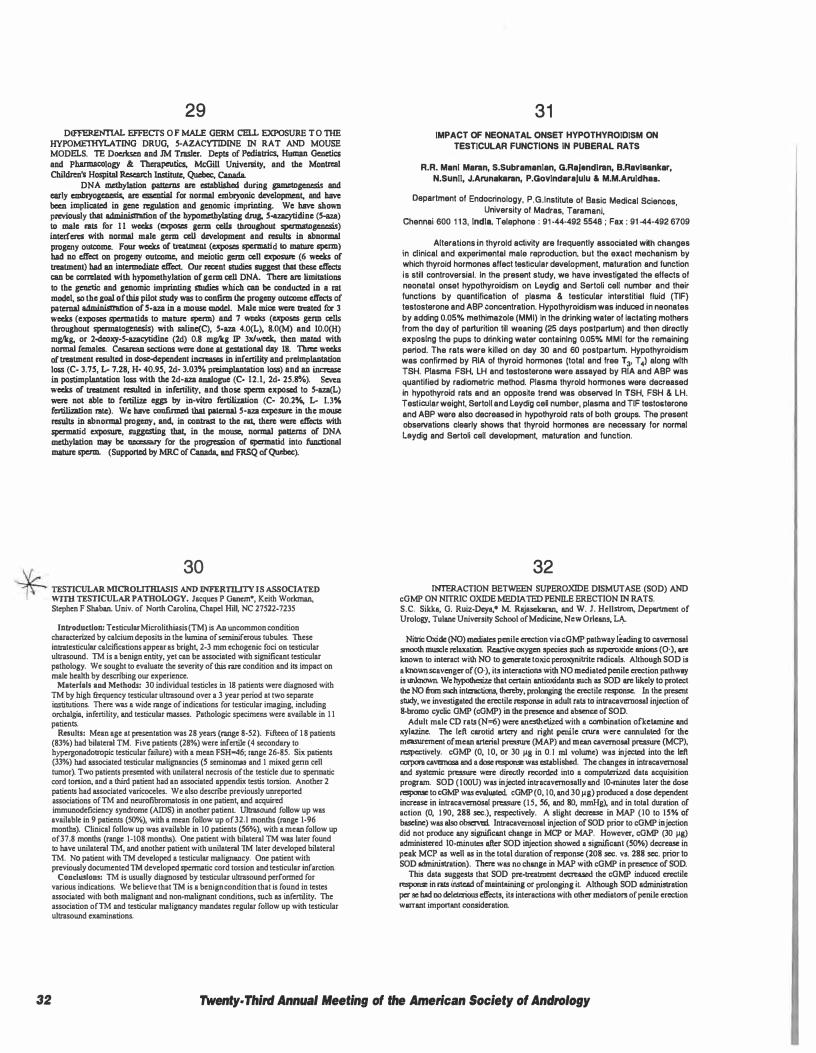
29 DIFFERENTIAL EFFECTS OF MALE GERM CELL EXPOSURE TO 1HE
HYPOME'IHYLATING DRUG, 5-AZACYTIDINE IN RAT AND MOUSE MODELS. TE DOCiksen and JM Traslcr. Dcpts of Pediatrics, Human Genetics and Pbannacology & Therapeutics, McGill University, and the Montreal Cbildien's Hospital Rcscan:h Institule, Quebc:c, Canada.
DNA mdhylation patterns arc eslablished during gamctogenesis and early cmbiyogcncsis, arc essential for normal embiyonic development, and have been implicated in gene regulation and genomic imprinting. We have shown previously that administration of the hypomdhylating drug. 5-azacytidine (5-a1JI) to male rats for 1 1 weeks (exposes germ cells throughout spermatogenesis) interferes with normal male germ cell development and results in abnormal progeny ouu:ome. Four weeks of tn:atment (exposes spcrmatid to mature sperm) bad no effect on progeny outcome, and meiotic germ cell exposure (6 weeks of batmen!) bad an intermediate effect. Our recent studies suggest that these effects can be correlated with hypomethylation of germ cell DNA. There arc limitations to the genetic and genomic imprinting studies which can be cond� in a rat model, so the goal of this pilot study was to confirm the progeny ouu:ome effects of patemal administration of S-aza in a mouse model. Male mice were treated for 3 weeks (exposes spermatids to mature sperm) and 7 weeks (exposes germ cells throughout spermatogenesis) with saline(C), 5-aza 4.0(L), 8.0(M) and 10.0(H) mg/kg. or 2-deoxy-5-azacytidine (2d) 0.8 mg/kg IP 3x/week, then � with normal females. Cesarean sections were done at gestational day 18. Three weeks of batmen! n:sulted in dose-dependent increases in infertility and preimplantation Joss (C- 3.75, L- 7.28, H- 40.95, 2d- 3.03% preimplantation loss) and an increase in postimplantation Joss with the 2d-aza analogue (C· 12.1, 2d· 25.8%). � weeks of batmen! n:sulted in infertility, and those sperm exposed to 5-aza(L) were not able to fertilize eggs by in-vitro fcrtilization (C· 20.2%, L- 1.3% fertilization rate). We have confirmed that patcmal 5-aza exposure in the mouse results in abnormal progeny, and, in contrast to the rat. there were effects with spermatid exposure. suggesting that, in the mouse, normal patterns of DNA methylation may be necessacy for the progression of spermatid into functional mature sperm. (Supported by MRC of Canada, and FRSQ of Quebec).
30 TESTICULAR MICROLITillASIS AND INFERTll.ITY IS ASSOCIATED WITH TESTICULAR PATHOLOGY. Jacques P Ganem•, Keith Workman, Stephen F Shaban. Univ. of North Carolina, Chapel Hill, NC 27522-7235
Introduction: Testicular Microlithiasis (TM) is An uncommon condition characterized by calcium deposits in the lumina of seminiferous rubules. These in1Illtesticular calcifications appear as brigh� 2-3 mm echogenic foci on testicular ul11asound. TM is a benign entity, yet can be associated with significant testicular pathology. We sought to evaluate the severity of this 111re condition and its impact on male health by descnbing our experience.
Materials and Methods: 30 individual testicles in 18 patients were diagnosed with TM by high mquency testicular ul1Iasound over a 3 year period at two separate instirutions. There was a wide range of indications for testicular imaging, including on:halg;a, infertility, and testicular masses. Pathologic specimens were available in 1 1 patients.
Results: Mean age at presentation was 28 years (range 8·52). Fifteen of 1 8 patients (83%) had bilateral TM. Five patients (28%) were infertile (4 secondary to hypergonadotropic testicular failure) with a mean FSH=46; range 26-85. Six patients (33%) had associated testicular malignancies (5 scminomas and 1 mixed genn cell tumor). Two patients presented with unilateral necrosis of the testicle due to spennatic cord torsion, and a third patient had an associated appendix testis torsion. Another 2 patients had associated varicoceles. We also descnbe previously unreported associations of TM and neurofibromatosis in one patien� and acquired immunodeficiency syndrome (AIDS) in another patient Ul1Iasound follow up was available in 9 patients (50%), with a mean follow up of32.l months (range 1-96 months). Clinical follow up was available in 10 patients (56%), with a mean follow up of37.8 months (range 1-108 months). One patient with bilateral TM was later found to have unilateral TM, and another patient with unilateral TM later developed bilateral TM. No patient with TM developed a testicular malignancy. One patient with previously documented TM developed spennatic cord torsion and testicular infarction.
CondWJlom: TM is usually diagnosed by testicular ul1Iasound performed for various indications. We believe that TM is a benign condition that is found in testes associated with both malignant and non-malignant conditions, such as infertility. The association ofTM and testicular malignancy mandates regular follow up with testicular ul1Iasound examinations.
31 IMPACT OF NEONATAL ONSET HYPOTHYROIDISM ON
TESTICULAR FUNCTIONS IN PUBERAL RATS
R.R. Mani Maran, S.Subramanlan, G.RaJandlran, B.Ravlaankar, N.Sunll, J.Anmakaran, P.GovlndaraJulu & M.M.Anlldhaa.
Department of Endocrinology, P.G.lnstitute of Basic Medical Sciences University of Madras, Taramani,
'
Chennai 600 1 1 3, India, Telephone : 91 -44-492 5548 ; Fax : 91 -44-492 6709
Alterations in thyroid activity are frequently associated wtth changes in �linical a�d experimental male
_reproduction, but the exact mechanism by
which thyroid hormones affect testicular development, maturation and function is still controversial. In the present study, we have investigated the effects of neonatal onset hypothyroidism on Leydig and Sertoli cell number and their functions by quantification of plasma & testicular interstitial fluid (TIF) testosterone and ABP concentration. Hypothyroidism was induced in neonates by adding 0.05% methimazole (MMI) In the drinking water of lactating mothers from the day of parturition till weaning (25 days postpartum) and then directly exposing the pups to drinking water containing 0.05% MMI for the remaining period. The rats were killed on day 30 and 60 postpartum. Hypothyroidism was confirmed by RIA of thyroid hormones (total and free T 3, T 4) along with TSH. Plasma FSH, LH and testosterone were assayed by RIA and ABP was quantified by radiometric method. Plasma thyroid hormones were decreased in hypothyroid rats and an opposite trend was observed In TSH, FSH & LH. Testicular weight, Sertoll and Leydig ceO number, plasma and TIF testosterone and ABP were also decreased in hypothyroid rats of bolh groups. The present observations clearly shows that thyroid hormones are necessaJY for normal Laydig and Sertoli cell development, maturation and function.
32 INTERACTION BETWEEN SUPEROXIDE DISMUTASE (SOD) AND
cGMP ON NITRIC OXIDE MEDIATED PENILE ERECTION IN RA TS. S.C. Sikka, G. Ruiz-Deya,• M Rajaselcaran, and W. J. Hellstrom, Department of Urology, Tulane University School of Medicine, New Orleans, �
Nitric Oxide (NO) mediates penile erection via cGMP pathway leading to cavcrnosal smooth muscle relaxatioo. Reactive oxygen species such as supcroxide anions (0-), are known to interact with NO to generate toxic pcroxynitritc nidicals. Although SOD is a known scavenger of (O· ), its interactions with NO mediated penile erection pathway is unlcnown. We hypothesize that certain antioxidants such as SOD are likely to protect the NO from such interactions, thereby, prolooging the erectile response. In the present study, we investigated the erectile response in adult rats to intracavcmosal injection of 8-bromo cyclic GMP ( cGMP) in the presence and ab!!erlce of SOD.
Adult male CD rats (N=6) were anesthetized with a ccrnbination ofkctarnine and xylazinc. The left carotid artery and right penile CIUf& were cannulated for the measurement of mean arterial pressure (MAP) and mean cavcrnosal pressure (MCP), respectively. cGMP (0, I 0, or 30 µ8 in 0. 1 ml volume) was injected into the left corpora cavomosa and a dose response was es!Jlblished. The changes in intracavcrnosal and systemic pressure wcno directly recorded into a computerized data acquisition program. SOD (IOOU) was injected intracavcrnosally and IO-minutes later the dose response to cGMP was evaluated. cGMP (0, I 0, and 30 µg) produced a dose dependent increase in intracavcmosal pressure (15, 56, and 80, mmHg), and in total duration of action (0, 1 90, 288 sec.), respectively. A slight decrease in MAP (JO to 15% of baseline) was also observed. Intracavcrnosal injection of SOD prior to cGMP injection did not produce any significant change in MCP or MAP. However, cGMP (30 µg) administered JO-minutes after SOD injection showed a significant (50"/o) decrease in peak MCP as well as in the total duration of response (208 sec. vs. 288 sec. prior to SOD administration). There was no change in MAP with cGMP in presence of SOD.
This data suggests that SOD pre-treatment decreased the cGMP induced erectile response in rats instead of maintaining or prolonging it Although SOD administration per sc had no deleterious effects, its interactions with other mediators of penile erection warrant important consideration.
32 Twenty• Third Annual Meeting of the American Society of Andrology
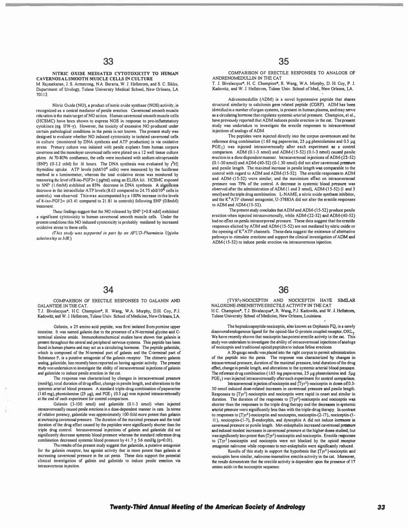
33 NITRIC OXIDE MEDIATED CYTOTOXICITY TO HUMAN
CAVERNOSALSMOOTH MUSCLE CELLS IN CULTURE M Rojosekoran, J. S. Armstrong, N.A. Baratta, W. J. Hellstrom, and S. C. Sikka, Department of Urology, Tulane University Medical School, New Orleans, LA 701 1 2
Nitric Oxide (NO), a product ofnilric oxide synthase (NOS) activity, is recognized as a central mediator of penile erection. Cavernosal smooth muscle rcla,olion is the main target of NO action. Human cavemosal smooth muscle cells (HCSMC) have been shown to express NOS in response lo pro-inflammatory cylokines (eg. IFN-y). However, the toxicity of excessive NO produced under certain palhological conditions in the penis is not known. The present study was designed to evaluate whether NO induced cytotoxicity in isolated cavemosal cells in culture (monitored by DNA synthesis and ATP production) is via oxidative stress. Primary cullure wos inilioted with penile explanl• from human corpora cnvemosn nnd the monoloyer cnvernosnl cells were plated on a 1 2 well tissue culture plates. At 70-80% confluence, the cells were incubated with sodium nitroprusside (SNP) (0-3.2 mM) for 16 hours. The DNA synthesis was evaluated by 13H] thymidine uptake. ATP levels (nM/J 04 cells) were measured by the Juciferase method in n Juminometer, whereas the tolal oxidative stress was monitored by mensuring lhc level of8-iso-PGF2� ( pg/ml) using an ELISA kit. HCSMC exposed to SNP ( J .6mM) exhibited an g5% decrease in DNA synthesis. A significant decreuse in the intracellular ATP levels (8.63 compared to 24.75 nM/J04 cells in controls) was observed. This was accompanied by a I 00% increase in the levels of 8-iso-PGF2� (43.41 compared to 2 1 .8 1 in controls) following SNP (0.8mM) treatment.
These findings suggest !hat the NO released by SNP [>O.g mM] exhibited a signilicont C)�Oloxicily to human covemosal smooth muscle cells. Under the present conditions this NO induced cytotoxicity is probably mediated by increased oxidntive stress to these cells.
(This s111dy was supponed i11 pan by 011 AFUD-Pharmacia ·Upjahn
scholarship 10 MR.)
34 COMPARISON OF ERECTILE RESPONSES TO GALANIN AND
GALANTIDE IN THE CAT. T.J. Bivalocqua', H.C. Champion•, R. Weng, W.A. Murphy, D.H. Coy, P.J. Kedowitz. ond W. J. Hellstrom, Tulane Univ. School of Medicine, New Orleans, LA
Golonin, a 29 amino acid peptide, was first isolated from porcine upper intestine. It was named galenin due to the presence of a N-terminel glycine and Cterminal alanine amide. Immunohistochemical studies have shown that gelanin is present throughout !he central and peripheral nervous systems. This peptide hes been found in human plosmo and moy net as a circulating hormone. The peptide gelantide, which is composed of the N-terminol part of galanin and the C-terminel pert of Substance P, is a putative antagonist of the galanin receptor. The chimeric gelanin analog. galuntide, has recently been reported us having agonist activity. The present study wn.• undertaken to investigate the ability of inlracavernosal injections of galanin end golnnlide to induce penile erection in the cat.
The response was characterized by changes in intrecevernosal pressure (mmHg), lolal durolion of drug effect, change in penile length, and alterations to the systemic arterial blood pressure. A stondord triple-drug combination ofpapeverine (1 .65 mg), phentolomine (25 µg), and PGE 1 (0.5 µg) wos injected intrecavemosally at the end of each experiment for control comparison.
Gulanin (3-100 nmol) and galantide (0. 1-3 nmol) when injected introcavemosally caused penile erections in a dose-dependent manner in cats. In terms of relative potency, galantide was approximately JOO-fold more potent than galanin et incrcnsing cavcmosal pressure. The duration of the maximal pressure and the total duration of the drug eflect caused by the peptides were significantly shorter than the triple drug control. lntrocavemosal injections of galonin end galantide did not significantly decrease systemic blood pressure whereas the standard reference drug combinntion decreased systemic blood pressure by 4 1 .7 ± 5.6 mmHg (p<0.01).
The results oftl1e present study suggest !hat gelantide, a putative antagonist for the galanin receptor, has agonist activity that is more potent than gelanin et increasing cavemosol pressure in the cot penis. These dato support the potential clinicol investigntion of gnlinin ond golintide to induce penile erection vie introcovcmous injection
35 COMPARISON OF ERECTILE RESPONSES TO ANALOGS OF
ANDRENOMEDULLIN IN THE CAT T. J. Bivolacquu•, H. C. Champion•, R. Wong, W.A. Murphy, D. H. Coy, P. J. Kadowitz, end W. J. Hellstrom, Tulane Univ. School of Med., New Orleans, LA.
Adrenomcdullin (ADM) is o novel hypotensive peptide that shares structural similarity to calcitonin gene related peptide (CORP). ADM has been identified in a nurnher of organ systems, is present in human plasma, and may serve as a circulating hormone !hot regulates systemic urtcrial pressure. Champion, et el., have prcvioLL<ly reported thut ADM induces penile erection in the eel The present study wes undertaken to investigate the erectile responses to intrecavernosal injections of analogs of ADM.
The peptides were injected directly inlo the corpus cavernosum and the reference drug combination ( 1 .65 mg popaverinc, 25 µg phentolamine and 0.5 µg PGE I ) was injected intracavemosolly ofter each experiment as a control comparison. ADM (0. 1 -3 nmol) and ADM-( 1 5-52) (0.1-3 nmol) caused penile erection in a dose dependent manner. lntracavemosal injections of ADM-(22-52) (O. J -30 nmol) and ADM-( 40-52) (O· I .30 nmol) did not alter cavernosal pressure end penile length. The maximal increase in penile length was comparable to the control with regard 10 ADM and ADM·( 1 5-52). The erectile responses to ADM and ADM-(1 5-52) were similar; and the maximium effect on intrecevernosal pressure was 75% of the control. A decrease in systemic blood pressure was obscivcd ofter the administration of ADM ( I and 3 nmol), ADM-(1 5-52) (I and 3 nmol) and the triple drug combination. L-NAME, a nitric oxide synthase inhibitor, and the K+ A Tl' channel antagonist, U-37883A did not alter the erectile responses to ADM and ADM-( 1 5-52).
lbe present study concludes !hot ADM and ADM-(1 5-52) produce penile erection when injected intracovemosully, while ADM-(22-52) and ADM-(40-52) had no clfcct on penile intracorporeel pressure. These dota suggest that the erectile responses elicited by ADM and ADM-( 15-52) ore not mediated by nitric oxide or the opening of K+ ATP channels. These data suggest the existence of alternative pothwoys to stimulate erections and support the clinical investigation of ADM and ADM·( 1 5-52) to induce penile erection via intracavemous injection.
36 [TYR'J-NOCICEPTIN AND NOCICEPTIN HAVE SIMILAR
NALOXONE-INSENSITIVE ERECTILE ACTIVITY IN THE CAT H.C. Champion•, T.J. Bivelacqua•, R. Weng, P.J. Kadowitz, and W. J. Hellstrom. Tulane University School of Medicine, New Orleans, Louisiana.
The heptodecapcptide nociceptin, also known as Orphanin FQ, is a newly discovered endogenous ligand for the opioid-like G-protein coupled receptor, ORL1. We hove recently shown that nociceptin has potent erectile activity in the cat This study wn.• undettaken to investigate the ability of intracavemosal injections of analogs of nociceptin and truditional opioid peptides to induce feline erections.
A 30-gaugc needle was placed into the right corpus to permit administration of the peptide into the penis. The response was characterized by changes in intracavernosol pressure, duration of the maximal pressure, total duration of the drug effcc� change in penile length, end alterations to the systemic arterial blood pressure. The reference drug combination ( 1.65 mg pepoverine, 25 µg phentolemine and .5µg PGE1) was injected intrecnvcmosally afier eoch experiment for control comparison.
lntracavernosal injection ofnociceptin end rryr'J-nociccptin in doses of0.3-30 nmol induced dose-related increases in cavernosal pressure and penile length. Responses to [Tyrl] ·nociceptin and nociceptin were rapid in onset and similar in duration. The duration of the responses to [Tyrl ]-nociceptin and nociceptin was shorter than the responses to the triple drug therapy and the decreases in systemic arterial pressure were significantly less then with the triple-drug therapy. In contrast to responses to [Tyrl ]·nociceptin and nociccptin, nociceptin-(2- 17), nociceptin-(1-1 1 ), nociceptin-( 1 -7), �-endorphin, and dynorphin A did not induce increases in cavernosal pressure or penile length. Met-enkepholin increased cavernosel pressure and induced modest increases in cavemosal pres.'lllre at the higher doses studied, but was significantly Jess potent lhan (Tyr1]-nociceptin and nociceptin. Erectile responses to [Tyrl ]-nociceptin and nociceptin were not blocked by the opioid receptor an!Jlgonist neloxone while responses to met-enkepholin were significantly reduced.
Results of this study in support the hypothesis !hot [Tyrt ]-nociceptin and nocict!plin hove similor, naloxone-insensitivc erectile activity in the cat. Moreover, the results dC111onstrate that the erectile activity is dependent upon the presence of 17 amino acids in the noc1ceptin sequence.
Twenty· Third Annual Meeting of the American Society of Andrology 33
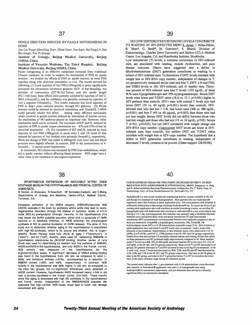
34
37 PENILE ERECTION INDUCED BY PANAX NOTOGINSENG IN DOGS Jinn Lin Yang•, Zhao Ying Xue• , Chide Hnn•, Yan Sw1•, Hai Feng Li•, Hai Bin Gong•, You Yi Zhang•.
Institute of U rology, Beijing Medical University, Beijing 100034, China. Institute of Vascular Medicine, The Third Hospital, Beijing Medical Unh·ersity, Beijing 100083, China. Pnnnx notoginseng is nu effective component extracted from traditional Chinese medicine. In order to explore the mechanism of PNS on penile erection , we studied the effects of PNS on penile erection by local PNS injection nloug with electrical stimulation in vivo. ll1e results showed the following: ( I ) Local injection of lml PNS ( IOOmg/ml) in penis significantly increased the intracorpus cavemous pressure (ICP: 21 .8±1 .SnunHg), tl1e duration or tumescence (DT: l8.5±2.7mins) and the penile lengtl1 (PL: l .4±0.2cm), these effects were partially inhibited by injection of lml LNNA ( I Onunol/L), and the inhibition was partially reversed by injection of lml L-arginine ( IOuunol/L). The results indicated that local injection of PNS in dog's pcms induced erection through NO pathway. (2) Penile erection could be mduced by electrical stimulation and IOnunol/L L-NNA could partially inhibit this effect . Among many different mechanism which involved in penile erection induced by stimulation of erectile nerves, the mechanism of NO pathway played an important role. However, other mechanism could not be excluded. ll1e increment of!CP and PL induced by injection of PNS in penile corpus cavemosum are 41 % and 47% of that by electrical stimulation. (3) ll1e increment of ICP and PL induced by local injection of lml PNS ( IOOmg/ml) in penis were 2 and 2.8 times of tlmt induced by injection of I ml Sodium nitre pmsside (3nunol/L), respectively. When the dosage of PNS was raised to 400mg/ml, only heart rate w1d blood pressure were slightly affected. In contrast, SNP at the concentration of 4-6nunol/L, it caused severe hypotension.
In conclusion, NO release was increased by PNS from endothelium, which led to penile erection without affecting blood pressure. PNS might have a clinic value in the treatment of non-organic impotence.
38 SPONTANEOUS EXPRESSION OF INDUCIBLE NITRIC OXIDE
SYNTHASE (NOS) IN THE HYPOTHALAMUS AND FRONTAL CORTEX OF AGING RATS. D Vernet, JJ Bonavera, R Swerdloff, NF Gonzalez-Cadavid, and C Wang. Departments of Urology and Medicine, Harbor-UCLA Medical Center, Torrance, CA.
Excessive activation of the NMDA receptor (NMDAR)/neuronal 111:6 (nNOS) cascade In the brain by excitatory amino acids may lead to neurodegeneratlve disorders through the release of cytotoxic levels of nitric oxide (NO) by postsynaptic nltrerglc neurons. In the hypothalamus this may Impair the GnRH pulsatlle secretion, either due to apoptosls of Gift{ neurons or to feedback Inhibition of nNOS affecting the physiological synthesis of NO (a positive modulator of GnRH release). The alms of this study are to determine whether aging in the hypothalamus is associated with high NO synthesis, which Is Its source and whether this Is organspecllic. Brown Norway male rats (N=5) at ages 1 ("immature'). 3 ('adult'), and 24 ('old') months, were used for measuring NMDARs in hypothalamic membranes by (3H)CGP binding. Another series of rats (N=9) was used for determining by western blot the contents of NMDAR, nNOS and INOS In the hypothalamus, and only INOS In the frontal cortex. NOS activity was measured In the hypothalamus by the arglnlne/cltrulllne assay. A significant decrease of NMDA analog binding was found In the hypothalamus from old rats as compared to adult ( -66%) and Immature animals (-57%), accompanied by a reduction In NMDAR content (-36% and -46o/o, respectively). In contrast, 111:6 activity In the hypothalamus was 68% higher In old rats as compared to the other two groups, but no significant differences were observed in nNOS content. However, hypothalamic iNOS Increased nearly 4-fold In old rats, a process paralleled In the frontal cortex (3.6-fold). These results show that aging Is associated with high NO synthesis In the hypothalamus and frontal cortex, Independent of the NMDAR/nNOS cascade. We speculate that high cortical NOS levels could lead to brain cell damage associated with aging.
39 DO LOW DIHYDROTESTOSTERONE LEVELS CONTRIBUTE
TO WASTING IN HIV-INFECTED MEN? S. Arver. I. Sinha-Hikim, R. Shen*, G. Beall*, M. Guerrero*, S. Bhasin. Division of Endocrinology, Charles Drew University and Harbor-UCLA Medical Center, Los Angeles, CA; and Karolinska Institute, Stockholm. Low testosterone Cn levels, a common occurrence in HIV-infected men, are associated with wasting, muscle dysfunction, and poor disease outcome. Others have suggested that a defect in dihydrotestosterone (DHn generation contributes to wasting in a subset of HIV-infected men. To detennine ifDHT levels correlate with weight loss or HIV-RNA copy number, independent of changes in T, we prospectively measured serum total and free T, DHT, LH and FSH, and SHBG levels in 150, HIV-infected, and 31 healthy men. Thirtyone percent of HIV-infected men had T levels <275 ng/dL; of these 81 % were hypogonadotropic and 19% hypergonadotropic. Serum DHT levels were lower and T/DHT ratios (18.6 vs. 12.1, p<0.001) higher in HIV-patients than controls. HIV+ men with nonnal T levels also had lower DHT (25 vs. 40 ng/dL p<0.001) levels than controls. HIVinfected men who had lost > 5 lb. had lower total (308 vs. 480 ng/dL, p<0.001) and free T (49 vs. 60 pg/mL, p<0.003) than those who had not lost weight. Serum DHT levels did not differ between those who had lost weight and those who had not ( 19 vs. 24 ng/dL, p=NS). Serum T (r=0.2, p<0.025), but not DHT correlated with weight change and HIV-RNA copy number. Conclusions: DHT levels are lower in HIVinfected men than controls, but neither DHT nor T:DHT ratios correlate with weight loss or IIlV-copy number. The hypothesis that a defect in DHT generation contributes to wasting, independent of decreased T levels, remains to be proven. (Grant support: DK49296)
40 CORTICOSTEROID CREAM PRE-TREATMENT DECREASES SEVERITY OF SKIN IRRITATION wml ANDRODERM® IN HYPOGONADAL MEN E. Roppapon, A. Haig and N. Asbct SmithKline Beechum Phumaccuticals, Collcgcviltc, PA; :r. Rallis, Dcpl of Dcnnatology, Univ. of Utah, Health Sciences Ctr., Sall Lake City, UT
Androdenn® is a non-scrotal testosterone transdcnnal delivery system available in the US and Europe for treatment of ma.Jc hypogonadism. Most patients who use Androdcnn® experience some skin irritation at patch application sites. For some patients, skin irritation is sufficiently bothersome to discourage continued Androdenn® use. To assess the effect of pre treating patch application sites with emollient or steroid containing creams. we enrolled 30 hypogonadal men with bothersome skin reactions to Androdenn® in a two week clinical trial During a 3 to 7 day screening period, skin irritation was assessed using a Modified Marzulli Maibach score nnd patient diary cards and scrum testosterone m and bioavailable testosterone (BT) concentrations were measured. Patients were randomly assigned to one of three prc-trclllmcnts: Cctnphit®, a hypoallergenic moisturizer (M); Cortaid®, I% hydrocortisone cream (HC): or 0.1 % triamcinolone acctonide cream (TA). Skin irritation at patch application sites and serum T 1111d BT levels were re·asscssed 1 and 2 weeks after initiation of pre·treabnenl Improvements in skin irritation scores were observed in 4 of 10 (40%). 6 of 9 (67%). and 8 of II (73%) patients in the M, HC, and TA groups respectively. Patient diary data showed that TA mmkcdly reduced rednc55 and itching; M bad little effect on these parameters 1111d HC produced slight improvements compared to M. Mean b11Seline scrum T levels were 609, 554, & 660 ng/dL and mean baseline BT levels were 312, 274, & 320 ng/dL in the M, HC, and TA groups respectively. Mean scrum T and BT decreased in all groups; the greatest decreases in T and BT occurred in men receiving M prc·treatment. At the 2 week visi� scrum T levels had decreased by 36%, 22%, and 8% and BT had decreased by 3 1 %, 24%, and I I% in the M, HC, and TA groups respectively. One man in the M group tw• men in the HC group, and none in the TA group had serum T or BT levels below the lower limit of the assay reference range during the treatment period.
The current study indicates that 1.) pre-treatment with 0.1 % triamcinolone cream decreases skin initation at Androderm@ application sites and 2.) in hypogonadal men using AndrodcnnCBI for testosterone replacement, such pre-treatment does not have a clinically significant effect on testosterone absorption.
Twenty· Third Annual Meeting of the American Society of Andrology
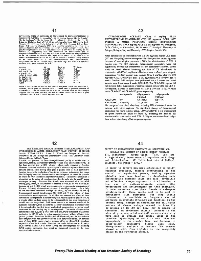
41 DI FFERENTIAL RATES D F CONVERSION O F TESTOSTERONE T O D I HYDROTESTOSTERONE IN ANDROGENETlC ALOPECJA. R . Argue l l o*, MD; M. Castro-Magana, MD; M. Angulo*, MD; G. Prakasam*, MD; A. Canas MD; P. Vftollo* , APA.C; A. Karri * , MD. Endocrine Division, Dept of Ped. Winthrop Universi ty Hosp. 120 M i neola Blvd. Mineola, NY 1 1501 and State University of NY, Hlth Sciences Cntr. , Stony Brook. Androgenetfc Alopeci a (AA) is a geneti c condit ion inherited in a dominant fash ion, but its c l i nical expression is under androgen control . Male pseudohermephrodftes with 5-a reductl!ISe type 11 (Sa-R I I ) def i c i ency do not exhibi t AA, suggesting that the conversion of testosterone (T) to dibydrotestosterone (OHT> is a key mechanism in the promotion and progression of AA. We studied 49 healthy men, ages 17-38 years, 24 of them whh AA (Hami lton clnss t f f cation J t t to V I ) and 25 controls, wi thout family h i story of AA. Ser1.n levels of T, OHT, Androstenedfone (A) , Androstanedfol glucuronfde (Adiol G), Estrone CE1 ) , Estradiol CE2> end Prostetic spec i f i c antigen (PSA) were measured by R I A .
Hormone AA T (ng/dl tSO) 602t153 DHT (ng/dl tSD) 62t14 A (ng/dl tSD) 161t32 Adlol G (ng/dl tSD) 1405t237 E1 (pg/ml •SD> 58•34
��A (
����l ·��) 54•26 1 . 0.0.4
T/DHT 9.6•0.9
Controls 649• 1 1 7
38t8 136t31 854•187
45•12 36•9
o. 7t0.3 17.7•3.3
11p11 Value <0.2 <0.0001 <0.01 <0.0001 <0.05 <D.02 <0.02 <0.0001
Serun T was sf mi lar f n both AA and control subjects. Sen1n DHT and Adiol G, however were h i gher f n subjects with AA. These results provide evidence of d t ffere:,tf a l rates of conversion of T to DHT in males whh AA and strongly support the view that systemic DHT and peri feral conversion to A.dial G play iq>artant role in the cl inical expressi on of AA.
42 THE PESTICIDE LINDANE INHIBITS STEROIDOGENESIS AND
STEROIDOGENIC ACUTE REGULATORY (SIAR) PROTEIN IN MOUSE MA-JO LEYDIG CELLS. Lance P. Walsh* and Douglas M. Stocco•. Department of Cell Biology and Biochemistry, Texas Tech University Health Sciences Cenler, Lubbock, Texas. Llndane, the y-isomer of hexachlorocyclohexanc (HCH) is widely used in agriculture, forestry and veterimuy practice because of its insecticidal activity. It has been reported that y-HCH adversely affects male reproductive function. However, the actual mechanism by which y-HCH influences reproductive function remains unclear. Since Leydig cells play a pivotal role in male reproductive function through the production of the sleroid honnone, testoslerone, the mouse MA-10 Leydig lumor cell line was used as a model system to assess the polential effects of the HCH isomers on reproductive function. Tesloslerone production is stimulated by the action of gonadolropin on Leydig cells via the cAMP second messenger pathway. Therefore, MA-10 cells were stimulated with the cAMP analog, (Bu),cAMP, in the presence or absence of y-HCH or two other HCHisomers, a and li-HCH which are contaminants in commercial preparations of Llndane. Following stimulation we measured I) sleroid production, 2) the activity of the cholesterol side-chain cleavage (P450scc), 3) the activity of the 3� hydroxysleroid sleroid dehydrogcnase (J�HSD) and 4) the effects of these compounds on prolein synthesis. In addition, we measured the effects of these loxicants on the expression of the Sleroidogenic Acule Regulatory protein (SIAR), a protein which has been shown to be indispensable in the acute regulation of sleroid honnone biosynthesis. SIAR action results in an incrcased lranSfer of the subslrale, choleslerol, from the ouler to the inner mitochondrial membrane when: it is mclabolized to the first sleroid synthesized, pregnenolone, an action which constitutes the rule limiting slep in sleroidogenesis. The results of these studies demonstrated that a, &, aqd y-HCH inhibited (Bu),cAMP stimulated proges1erone production in MA-10 cells in a dose dependent manner without affecting Iola! protein synthesis. In addition, P450scc and J�HSD activity and the expression of these enzymes were not affectal by any of these compounds tested. In contrast, each of these HCH isomers had a profound effect on (Bu),cAMP stimulaled expression of the SIAR prolein. Therefore, our results indicate that a, &, and yHCH directly inhibil MA-10 mouse Leydig cell sleroidogenesis by inhibiting SIAR protein expression, thus impairing cholesierol transfer to the inner milochondrial membrane.
43 CYPROTERONE ACETATE (CPA) S mg/day PLUS
TESTOSTERONE ENANTBATE (fE) 200 mg/week DOES NOT
INDUCE A MORE PROFOUND SPERM SUPPRESSION COMPARED TO CPA S mglday PLUS TE 100 mg/week MC Meriggiola, G Di Cintio•, A Costantino*, WJ Brenmer C Flamigni* University of Bologna, Bologna, Italy and University of Washington, Seattle, WA.
When administered in combination with TE 100 mw'week, higher CPA doses ( 100 and 50 mw'day) induced azoospennia in all subjects but caused a greater decrease of hematological parameters. With the administration of CPA 5 mw'day plus TE 100 mw'week, hematological parameters were not significantly affected but azoospennia was not consistently achieved. In this study we tested whether increasing the dose of TE administered in combination with CPA 5 mw'day would cause a more profound spennatogenic suppression. Thirteen nonnal men received CPA 5 mw'day plus TE 200 mw'week (CPA-5-200 n=7) or plus TE 100 mw'week (CPA-5- 100 n=5) for 16 weeks. Seminal fluid analyses were perfonned every 2 weeks and blood samples were drawn every 4 weeks. RESULTS: The CPA-5-200 regimen did not induce a better suppression of spennatogenesis compared to the CPA-5-100 regimen. In week 16, spenn cowrt was 0.14 ± 0.09 and 1 .57±0.79 Mimi in the CPA-5-100 and CPA-5-200 group respectively.
azoospermia oligospermia oligospermia (<3M/ml) �JM/ml)
CPA-S-200 on 6n (86%) 1n (14%) CPA-5-100 2/5 (40%) 3/5 (60%) 0/5 No change of any blood chemistry, including HDL-cholesterol, could be detected with either regimen. No significant change of hematological parameters was found in either group. CONCLUSIONS: I. No improvement of spenn suppression could be found by increasing the dose of TE administered in combination with CPA. 2. Higher testosterone levels might have a direct stimulatory effect on spennatogenesis.
44 EFFECT OF TESTOSTERONE ENANTllATE ON STRUCTURE AND
NUCLEAR DNA CONTENT OF RHESUS MOJCEY PROSTATE T . S . Ud ayakumar, Alpena Tyagi , S . N . Das and M . R\j alakshmi , Departments o f Reprod uctive Biology and Biotechnology, All Ind ia Institute of Med ical Sciences , New Delhi - 1 1002 9
In ord er to involve men more extensively in family planning practices , thereby contributing to the control o f population growth, fund ing agencies globally have been involved in d eveloping male contraceptive regimens which are safe , reversi bl e and effective. A major approach i n this d irection i s the u s e o f antispermatogenic agents like progestogens and antiandrogens and GnRH analogues . In ord e r to maintain peripheral levels of and rogens physiologically, these agents need to be used in combination with androgens . But very 11 ttle information is available on long-term use o f and rogens on pros tate structure and function. I n the present s tudy, changes in morphology and cell cycle pattern o f rhesus monkeys exposed to bimonthly inj ections o f TE ( 50 mg ; i .m . ) for 30 months i s
d escribed . In TE-treated animal s , increas e in the size o f prostate, acini and cell s ecretory activity were s een in cranial and caud a l lobes of the prostate. Hypertrophy o f cells in both lobes and hyperplasilJ! in the crania l lobe , and increase in inter-seiner connective ti ssue were s een. Flowcytometric analysis of nuclear DNA content showed a shift from d i ploidy to the aneuploidy status in the TE-treated animals .
Twenty· Third Annual Meeting of the American Society of Andrology 35
36
45 EFFECT OF TESTOSTERONE ENANnIATE ON LIPID PROFILE
LIVER FUNCTION TES'ISIN HOl'KEY Alpana Tyagi and H. Rajalakshmi , Department Reproductive Biology , All Ind i a Institute of Med Sciences, New Delhi - 1 1 0029
One o f the promising a pproaches to the d evelopmen a saf e , effective and reversible male horm• contraceptive i s the u s e of exogenous s ex s teroid suppress gonadotropin release thereby suppres spermatogenesis . But , very l i ttle information available of the aid e effects of long-term UBE and rogens on metabolic and systemic functions. s tudy evaluates the effect of long-administration of the commonly-used and r testos terone enantha te ( TE ) on lipid profile liver function parameters i n rhesus monkeys correlates these effects with serum levels testosterone. Ad ult rhesus monkeys were inj ected 50 mg of TE ( f .m . ) b imonthly for two years w controls received only the vehicle. No s i gnifi change was observed in total cholesterol , LDL t riglycerid es l evels in both groups. But HDL-C le decreased i n treated monkeys . Changes were observed i n plasma l evels o f alkaline phosphatase significant change s were obs erved i n SGOT and levels hut within the normel renee: It ! ! eonc1 that long-term administration o f TE d ecreased H levels in monkeys . The impl ication of such a d ect of the risk for coronary artery d isease is d iscuss
46 ROLES OF THE DISINTEGRIN DOMAINS OF THE
SPERM PROTEINS FERTILIN a AND p IN FERTILIZATION J.P. Evans!, R.M. Schultzl,2, and G.S. Kopf!; !Center for Research on Reproduction and Women's Health, Dept. of Ob/Gyn and 2Dept. of Biology, Univ. of Pennsylvania, Philadelphia, PA
Fertilin is a mammalian sperm protein that is heterodimer of a and p subunits, both of which are members of a family of proteins known as ADAMs (A ]Jisintegrin and A Metalloprotease domain) or MDCs (Metalloprotease-Qisintegrin-,Cysteine-rich). We have previously demonstrated that recombinant forms of the itive extracellular domains of fertilin a and fertilin p bind to muuse eggs and inhibit sperm-egg membrane binding (Evans et al., 1997 Dev. Biol. ill. 79-93, 94-106). In this study, we made recombinant forms of fertilin a and fertilin p that include the disintegrin domains (aDCE and pDCE) or that are truncated so that they lack the disintegrin domains (aCE and pCE). Fertilin pDCE was able to inhibit sperm-egg binding, but fertilin pCE was relatively ineffective, indicating that the disintegrin domain of fertilin p is required for proper protein folding and/or for interactions with egg binding sites. Fertilin aDCE and aCE both inhibited sperm-egg binding, although fertilin aDCE was more effective. This indicates that the other domains of fertilin a extracellular region (cysteine-rich and EGF-like repeat) have the ability to block sperm binding, but that the disintegrin domain enhances this ability, either by improving the efficiency of protein folding or by interacting with egg binding sites. Finally, since an anti-ll(; antibody inhibits sperm binding (Almeida et al., 1995 Cell .Bl. 1095-1 104) but not fertilin PDCE binding (Evans et al., 1997 Dev. Biol. ill. 79-93), fertilin aDCE or aCE might function as an ll6 integrin ligand. However, an anti-ll(; antibody did not significantly inhibit the binding of either fertilin aDCE and aCE, suggesting that a different sperm ligand binds to an ll6 integrin on the egg surface.
47 ANGIOTENSIN 11 AND 1llE ACROSOME REACTION
C Mueller•. U Habenicht•, D.Drescher•, W.-B.Schill• and F.M.Koehn•, Center of Dermatology and Andrology, Justus Liebig University Giessen, Germany.
Introduction: The renin-angiotensin-system is widely distributed in mammalian tissues. Until now the physiological functions on the male reproductive tract have not been clarified. Angiotensin II (All) was found in Leydigcell homogenates and within the epididymis. Therefore, All plays a role in steroidogenesis and is important for sperm maturation. In earlier experiments we showed that All induces the acrosome reaction (AR) significantly. The purpose of the present experiments was to investigate the second messenger pathways involved into the induction of AR by All. Materials and Methods: After glass wool filtration and two washing steps in HTFM (1% HSA), spermatozoa were treated with IOOnM All for 4 h at 37°C Since HSA may be contaminated with peptidases and proteinases, which cause degradation of All, spermatozoa were washed in HTFM (without HSA) after capacitation (3h, 37°C) and incubated with JOO nM All for 15 - 60 min. The effect of All on different second messangcr systems was studied by preincubation of spermatozoa with an unspecific proteinase inhibitor H7 (IOµM) or pertussis toxin (I OOng/ml). The percentages of living acrosome reacted spermatozoa were detemiined by triple staining and compared to the corresponding values obtained after incubation with HTFM-{HSA) (negative control) and with IO µM ionophore A23 I 87 for 60 min after 3h capacitation (positive control). Results: Compared to the negative control the percentages of acrosome reacted spermatozoa were higher after treatment with I OOnM All in HTFM (without HSA) for 45 min after 3h-capacitation ( IS 4 +/- 5 .4% vs 6.4 +/- 2.8%; n""'S). 60 min-incubation did not cause funher increase of AR (15.0 +/- 5.4%, n•S) Treatment of spermatozoa with IOOnM All in the presence of HSA resulted lower rates of AR (9,8 +/- 2,5% vs 15.4 +/- 5,4%, n=S). Pretreatment with H7 showed nearly comparable values obtained to those of the negative control. Penussis toxin did not influence the effect of All on the AR Conclusion: The induction of AR by All is protein kinase- but not G-protein dependent. Peptidases and proteinases that are present in the capacitation medium may inactivate All.
48 MOLECULAR NATURE OF A SPERM ACROSOMAL
ANTIGEN RECOGNIZED BY HS-13 MONOCLONAL ANTIBODY T. Yoshi.ki and C.·Y. G. Lee*. Andrology Laboratory, Department of Obstetrics and Gynecology, The University of British Columbia, Vancouver, Canada
Among the numerous monoclonal antibodies generated against
human sperm antigens, HS-13 monoclonal antibody was shown to react with the antigen on the acrosome of spermatozoa from human, mouse and rat. In this study, HS-13 was used as the affinity ligand for the purification of the cognate antigen from human sperm by immunoaffinity chromatography. The purified cognate antigen from human sperm designated as HSAg· 13 was found to be a protein with molecular weight of approximately 80 kDa on SDS-PAG E in the presence of reducing reagents. This monoclonal antibody was used as the probe to study the tissue distributions and developmental expression of the cognate antigen from human, mouse and rat by immunohistochemistry. It was concluded that the antigen recognized by HS-13 antibody is highly sperm specific and found only in sperm and mature testis, but not in any other somatic tissues examined in human and mouse. The antigen was shown to be expressed at the postmeiotic stages of spermatogenesis in mouse. By using indirect immunofluorescent staining method, HS-13 was shown to react only with the methanol-fixed acrosome·intact sperm but not with the live sperm. Following calcium ionophore A23187 treatment, acrosome· reacted sperm showed a negative staining, suggesting the intraacrosomal location of HSAg-13. The induced acrosome reaction resulted in a statistically significant decrease of antibody-stained human sperm (p<0.05). Therefore, this antigen can be a useful sperm-specific marker for monitoring sperm capacitation and acrosome reaction.
Twenty· Third Annual Meeting of the American Society of Andrology
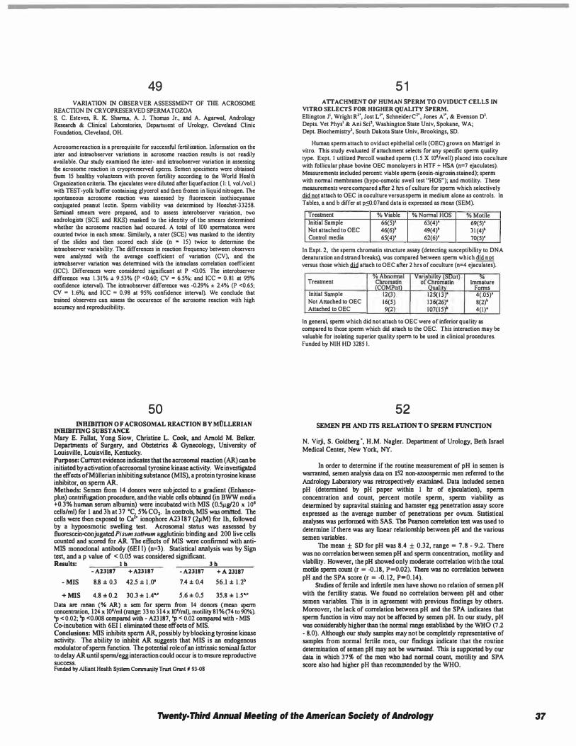
49 VARIATION IN OBSERVER ASSESSMENT OF TIIE ACROSOME
REACTION IN CRYOPRESERVED SPERMATOZOA S. C. Esteves, R. K. Shanna, A. J. Thomas Jr., and A. Agarwal, Androiogy Research & Clinical Laboratories, Department of Urology, Cleveland Clinic Foundation, Cleveland, OH.
Acrosome reaction is a prerequisite for successful fertilization. Wormation on the inter and intra.observer variations in acrosome reaction results is not readily available. Our study examined the inter- and intraobserver variation in assessing the acrosome reaction in cryoprerserved sperm. Semen specimens were obtained from IS healthy volun� with proven fertility according to the World Health Organization criteria. The ejaculates were diluted after liquefaction ( I : I, volJvol.) with TEST-yolk buffer containing glycerol and then frozen in liquid nitrogen. The spontaneous acrosome reaction was assessed by fluorescein isothiocyana<e conjugated peanut lectin. Sperm viability was determined by Hoechst-332S8. Seminal smean were prepared, and to assess interobserver variation, two andrologists (SCE and RKS) masked to the identity of the smears determined whether the acrosome reaction had occured. A total of 100 spermatozoa were counted twice in each smear. Similarly, a rater (SCE) was masked to the identity of the slides and then scored each slide (n • IS) twice to determine the intraobserver variability, The differences in reaction frequency between observers were analyzed with the average coefficient of variation (CV), and the intraohserver variation was determined with the intraclass correlation coefficient (ICC). Differences were considered significant at P <O.OS. The interobserver difference was 1.31% ± 9.S3% (P <0.60; CV = 6.S%; and ICC = 0.81 at 9S% confidence interval), The intraobserver difference was -0.29% ± 2.4% (P <0.6S; CV = 1 .6%; and ICC = 0.98 at 9S% confidence interval). We conclude that trained observers can assess the occurence of the acrosome reaction with high accuracy and reproducibility.
50 INBIBmON OF ACROSOMAL REACTION BY MOLLERIAN
INHIBITING SUBSTANCE Mary E. Fallat, Yong Siow, Christine L. Cook, and Arnold M. Belker. Departments of Surgery, and Obstetrics & Gynecology, University of Louisville, Louisville, Kentucky. Purpose: Current evidence indicates that the acrosomal reaction (AR) can be initiated by activation ofacrosomal tyrosine kinase activity. We investigated the effects ofMiillerian inhibiting substance (MIS), a protein tyrosine kinase inhibitor, on sperm AR. Methods: Semen from 14 donors were subjected to a gradient (Enhanceplus) centrifugation procedure, and the viable cells obtained (in BWW media +o.3% human serum albumin) were incubated with MIS (O.Sµg/20 x 106 cells/ml) for 1 and 3h at 37 "C, 5% CO,. In controls, MIS was omitted. The cells were then exposed to Ca2• ionophore A23 187 (2µM) for lh, followed by a hypoosmotic swelling test. Acrosomal status was assessed by fluorescein-eonjugated Pisum sattvum agglutinin binding and 200 live cells counted and scored for AR. The effects of MIS were confirmed with antiMIS monoclonal antibody (6E I I) (n=3). Statistical analysis was by Sign test, and a p value of < 0.05 was considered significant. Results: 1 h 3 h
- Al3187 + Al3187 - A23187 + A 23187
- MIS 8.8 ± 0.3 42.5 ± 1 .0' 7.4 ± 0.4 56.1 ± 1.2•
+ MIS 4.8 ± 0.2 30.3 ± 1 .4" S .6 ± 0.S 35.8 ± 1.5"
Data arc mean (% AR) ± sem for sperm from 14 donors (mean sperm concentration, 124 x IO'/ml (range: 33 to 314x 10'/ml), motility 81% (74 to 90%). 'p < 0.02; 'p <0,008 compared with - A23187, 'p < O.Q2 compared with - MIS Co-incubation with 6EI 1 eliminated these effects of MIS. ·
Conclusions: MIS inhibits spenn AR, possibly by blocking tyrosine kinase activity. The ability to inhibit AR suggests that MIS is an endogenous modulator of sperm function. The potential role of an intrinsic seminal factor to delay AR until sperm/egg interaction could occur is to ensure reproductive success. Funded by Alliant Health System Community Trust Grant # 93-08
5 1 A TI ACHMENT OF HUMAN SPERM TO OVIDUCT CELLS IN
VITRO SELECTS FOR HIGHER QUALITY SPERM. Ellington J', Wright R", Jost L'". SchneiderC", Jones A'", & Evenson D'. Depts. Vet Phys' & Ani Sci', Washington State Univ, Spokane, WA; Dept. Biochemistry', South Dakota State Univ, Brookings, SD.
Human sperm attach to oviduct epithelial cells (OEC) grown on Matrigel in vitro. This study evaluated if attachment selects for any specific sperm quality type. Expt. I utilized Percoll washed sperm (1.5 X 10⁶/well) placed into coculture with follicular phase bovine OEC monolayers in HTF + HSA (n=7 ejaculates). Measurements included percent: viable sperm (eosin-nigrosin stained}; sperm with normal membranes (hypo-osmotic swell test "HOS"); and motility. These measurements were compared after 2 hrs of culture for sperm which selectively did not attach to OEC in coculture versus sperm in medium alone as controls. In Tables, a and b differ at p:::0.07and data is expressed as mean (SEM).
Treabnent % Viable % Normal HOS % Motile Initial Sample 66(5)' 63(4)' 69(S)' Not attached to OEC 46(6)' 49(4)' 31 (4)' Control media 65(4)' 62(6)' 70(S)'
In Expt. 2, the sperm chromatin structure assay (detecting susceptibility to DNA denaturation and strand breaks), was compared between sperm which did not versus those which did attach to OEC after 2 hrs of coculture (n=4 ejaculates).
Treabnent
Initial Sample Not Attached to OEC Attached to OEC
. Immature
Forms 4(.0S)' 8(2)' 4(1)'
In general, sperm which did not attach to OEC were of inferior quality as compared to those sperm which did attach to the OEC. This interaction may be valuable for isolating superior quality sperm to be used in clinical procedures. funded by NIH HD 328S I .
52 SEMEN PH AND ITS RELATION TO SPERM FUNCTION
N. Virji, S. Goldberg ·, H.M. Nagler. Department of Urology, Beth Israel Medical Center, New York, NY.
In order to determine if the routine measurement of pH in semen is warranted, semen analysis data on 152 non-87.00spermic men referred to the Andrology Laboratory was retrospectively examined. Data included semen pH (determined by pH paper within 1 hr of ejaculation), sperm concentration and count, percent motile sperm, sperm viability as determined by supravital staining and hamster egg penetration assay score expressed as the average number of penetrations per ovum. Statistical analyses was performed with SAS. The Pearson correlation test was used to determine if there was any linear relationship between pH and the various semen variables.
Tue mean ± SD for pH was 8.4 ± 0.32, range = 7.8 - 9.2. There was no correlation between semen pH and sperm concentration, motility and viability. However, the pH showed only moderate correlation with the total motile sperm count (r = -0.18, P=0.02). There was no correlation between pH and the SPA score (r = -0. 12, P=0. 14).
Studies of fenile and infertile men have shown no relation of semen pH with the fenility status. We found no correlation between pH and other semen variables. This is in agreement with previous findings by others. Moreover, the lack of correlation between pH and the SPA indicates that spenn function in vitro may not be affected by semen pH. lo our study, pH was considerably higher than the normal range established by the WHO (7 .2 - 8.0). Although our study samples may not be completely representative of samples from normal fenile men, our findings indicate that the routine determination of semen pH may not be warranted. This is supported by our data in which 37% of the men wbo bad normal count, motility and SPA score also had higher pH than reco1DD1Cnded by the WHO.
Twenty· Third Annual Meeting of the American Society of Andrology 37
53 MOTILITY ENHANCEMENT OF CANINE EPIDIDYMAL SPERM
Barbara Durrant, Kara Russ', Sally Harper', Thomas Diliberto', Cathy King', Cheryl Rosa• and Michael KObler'. Center for Reproduction of Endangered Species, Zoological Society of San Diego, CA 921 12
Epididymal sperm represents a reservoir of genetic material which can be opportunistically salvaged at castration or necropsy. Although this resource has been exploited for the preservation of endangered species in 'frozen zoos', poor post-thaw motillties limlts its usefulness. There have been no reports describing the empirical derivation of appropriate cryopreservation techniques for epldidymal sperm of any carnivore except the domestic cat. In vitro capacltation and rno111ity enhancement techniques have not been developed. In this series of experiments the domestic dog served as a model for exotic carnivores. Sperm was ex1racted from minced cauda epldidymides following rouUne neutering for population control. Following suspension in � medium with .1% BSA, sperm was assessed for motility, speed of progression (SOP) and % live (%L, by eosin/nigrosin staining). A motility score (MS) was calculated as motility x SOP2. Sperm was extended In TEST-yolk buffer with a final concentration ol 3.2% glycerol and frozen over liquid nitrogen vapor for 15 minutes (7.3°cnnln to ·100"C) before storage in liquid nitrogen for a minimum of 1 o days. Aliquots were thawed for 1 min in a 37"C water bath prior to incuba1ion at 37°C with one of three motility enhancing agents; caffeine (CA), IBMX, or deoxyadenosine (DA). Untreated controls were Incubated In BWW medium with BSA. Motility and %L analyses were repeated at 30 min (T1) and 60 min (T2) ol incubation. To correct for differences between dogs, all resulls were expressed as % Initial MS (IMS) and % initial L (IL). Experiment 1 compared the effects of o, 5, 10, 20 and 40 mM CA on sperm from three donors. At T1 and T2 only 20 and 40 mM treatment groups had significantly higher %IMS than controls (p<.05). The 40 mM group was more motile than 20 mM at T1, bu1 not at T2. Although not signillcantly different from the control group, there was a sllgh1 reduction in %IL at the two highest CA concentrations at T1 and T2. Experiment 2 compared CA (20 mM) with IBMX (250 µM) and DA (20 mM). Both CA and DA significantly reduced %IL at T1 compared to untreated controls, but the effect was not significant by T2. All three enhancers signifJCanUy increased %IMS at T1 and T2, with CA and DA having a greater effect than IBMX. These results indicate that CA and DA, each at 20 mM, can maintain increased motifrty In thawed epidldymal canine sperm for al least one hour without signifJCant loss of viability. In both experiments, increased MS was due primarily 10 enhanced SOP. These data suggest that motility enhancement of cryopreserved epididymal sperm can be a useful adjunct to assisted reproduction in exotic carnivores.
54 COMPARISON OF SPERM SEPARATION METHODS: EFFECT ON
RECOVERY, MOTILITY, MOTION PARAMETERS, HYPERACTIVA TION, AND PREGNANCY RA TES. G.M. Centola, R. Herko, E. Andolina, B. Daniels Andrology Lab, Dept. Ob/Gyn, Univ of Rochester Medical Center, Rochester, NY. Percoll ™ (PVP· coated beads) had been the most widely used sperm separation product, until it was removed from the clinical commercial market in late 1996. Subsequently, silica stabilized with covalently bound hydrophilic silane, has become available for sperm gradient separation. The purpose of this study was to compare Enhance "'(ENH)(Pcrcoll ™) and Pure Sperm "' (PS) with respect to recovery (%; of motile sperm), motility (%), path (Vcl) and progressive velocity (V sl) (um/sec) and hypcractivation (%). Pregnancy rates (PR) were also retrospectively examined for the period of January, 1997-March, 1997 and April, 1997- July, 1997 during which time the ENH and PS methods, respectively, were being used. Semen specimens (n = 25) obtained within 30 minutes of ejaculation were assessed by CASA (HTM-2030; Hamilton-Thom Research). The initial mean sperm count was 63.9 ± 8.1 (range 1.5 to 145). One milliliter of semen was placed onto an Enhance gradient (l .5ml each; 90%145%), and I ml onto a PureSperm gradient ( l .5ml each; 80%/45%), and centrifuged at 300 X g for 20 min. The pellet was resuspended in synthetic human tubal nuid (HTF) (0.5% HSA), centrifuged at 300 X g for 10 min, with the final pellet resuspended in 1 .0 ml of fresh HTF prior to CASA analysis. The data was analyzed using paired I-test, and two sample Hes! with significance set at p < 0.05 . The data (mean ± SEM) arc as follows:
% Recovery % Motility Vcl Vsl 'lo HA Fresh 79.7(33.4) 39.2(1.7) 30.8(1.7) 3.9(.7) Enhance 56(.5) 64.6(4.4) 44.3(2.0) 38. 1(2.0) 5.2(.84) PureSperm 64(.06) 62.8(4.7) 45.9(2.4) 38.4(2.4) 7.5(1.8) There was no difference in the % motility, and molile count between ENH, PS and the fresh specimen. Significant differences were found (fresh vs test) in the Vcl and Vsl, and in HA (PS only), and no differences were found between the ENH and PS groups. For ENH, I 5 of 80 patients conceived for a PR of 1 8.8%, and a monthly f of 13.2%. For PS, 6 of 48 palients conceived for a PR of 12.5% and an f of 7.6%. The motile counl, motility and Vsl were nol different in pregnant and not pregnant groups. All but one pregnant patient were on ovulation medication. PureSperm "' appears to be as effective as Percoll"' (Enhance) for recovery of good progressively motile sperm for IUI or ART, but PRs appear to be lower with PS 1han ENH.
55 POST-WASH PERCOLL'" AND ISOLATE'" SPERM
PARAMETERS ARE SIMILAR BUT NOT EQUIVALENT. A.M. Barnes, M.R. Healy•, J.W. Ramey•, V.M. Maclin* and C.J. DeJonge, Center for Reproductive Medicine, University of Nebraska Medical Center, Omaha, NE. Most spcnn preparation techniques produce highly motile spenn populations which are preferred for IUI and !VF. One method involves density gradient centrifugation using Percoll, a silica sol consisting of polyvinylpyrrolidone-coated beads. Percoll has been removed from the market in part to avoid the possibility for iatrogenic effects resulting from spenn transporting silica particles. New gradient materials are deemed to be more safe and one of these is !So late. ISolate is similar to Percoll but it consists of silica particles stabilized with covalently bound hydrophilic silane. The purpose of this prospective study was to compare post-wash spenn parameters after using Percoll and !Solate. Semen specimens from 1 1 donors were used. One mL of semen was placed over a 45%/90% Percoll gradient and one mL over the "upper/lower" layers of an I So late gradient and centrifuged according to optimized protocols. Post-wash total motile count, % motility, % forward progression and % recovery were compared. Both Percoll and !Solate yielded significantly (P<0.05) enriched motile spenn populations relative to the neat specimen. No differences (P>0.05) were detected in total motile count, % motility or % recovery between Percoll and !Solate. A significantly (p=0.003) higher forward progression was detected in the post-Percoll specimen (x=90%) versus !Solate (x=82%). Thus, !Solate appears to be an acceptable substitute for Percoll. Pregnancy data, e.g., JUI, are required to detennine ifthe diminished forward progression is clinically significant.
56 ANALYSIS OF JN VITRO MIGRATORY PATIERNS OF HUMAN
SPERMATOZOA. A.M Hossain, B. Rizk•, C. Huft" and l.H. Thorneycroft'. Dept of OB/OYN, University of South Alabama, Mobile, AL.
The spermatozoa are required to travel a long distance in vivo to meet the ooc:ytc at lhe fertilization site. One of lhe many explanations of low success of natural pregnancy is Iha! some sperm populations may be less capable lhan othcr.J of reaching the oocytc because oftbcir unique inherent or acquired migratory potential. However, the sperm motility evaluation in the existing semen analyses does nol perform any assessment on spcnn migration pattern. In lhc present study we have utilized a hcrizDOtal ooham technique to study lhe in vitro sperm migration pattern to explore the benefits of adding sperm migration analysis in male fertility evaluation. A column, 35 cm long and 3 mm in diameter, was designed to be accommodated in a 100 mm petri dish. Approximately 35 µI ofHTF meditDD, containing 10% synthetic serum substitute, was required to make such a column. A I 0 µI sperm sample, of lcnown ntDDber and motility, was added to one end of the column and covered with mineral oil. The migration characteristics of the sperm in the column were monitored under an inverted microscope. The sperm distribution in the column was quantilated by counting the number of spcnn per unit microscopic field along the column. Twenty three opc:rm populations of different seminal and treabnent conditions were analyzed in 68 columns. The sperm that travdcd 5, I 0, 15, 20, 25, 30 and 35 cm distance in the column were 16, 9, 4, 2, 0.6, 0.1, O.o7 and 0.02 %, respectively, of the total. The sperm distribution paltcm in the colWIUl at 12 hr was different from that of24 and 48 hr. Considerable contrast in sperm migration patterns was doctDDcnted among the semen samples, between fresh and frozen semen, washed and unwashed sperm, I hr and 24 hr post washed sperm. Propagalioo along the column edge, tendency of exiting from the colWIUl and hiding in the blind pouches were some of the important charactaistic feolUre:s cxlubil<d by the migralmy sperm. The in vitro migration pattern seen in our study clascly mimics the known in vivo migratory pattern. The significant variations in the migratory pattClll3 of sperm populations of different etiologic backgrounds and treatment conditions leads to speculation that the in vitro sperm migration analysis will be informative in predicting their in vivo potential of reaching the fertilization site.
38 Twenty· Third Annual Meeting of the American Society of Andrology
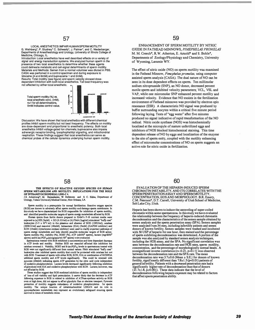
57 LOCAL ANESTHETICS IMPAIR HUMAN SPERM MOTILITY
G. Weinberg*, P. Studney* C. Schwartz•, J. Palmer*, and C. Niederberger, Deparments of Anesthesiology and Urology, University of Illinois College of Medicine, Chicago, IL Introduction: Local anesthetics have well described effects on a variety of signal and energy transduction systems. We analyzed human sperm in the presence of two local anesthetics to determine whether these agents could delineate metabolic and cell-signal determinants of sperm motility. Materials and Methods: Semen from a normal volunteer was diluted in PBS. CASA was perfomed in a control specimen and during exposure to lidocalne (4 and 8mM) and bupivacaine 1 and 2mM). Results: Total motility (see figure) and sperm velocity showed dosedependent Inhibition with both local anesthetics. Tall beat frequency was not affected by either local anesthetic. •
Total sperm motility (%) vs. local anesthetic cone. (mM). n=1 for all determinations, OmM Indicates control value.
I · J . •
Discussion: We have shown that local anethetlcs with different chemical profiles Inhibit sperm motility but not beat frequency. The effects on motility are dose-dependent and proportional to anesthetic potency. Both local anesthetlcs Inhibit voltage-gated Ion channels; bupivacaine also impairs adrenerglc receptor binding, lysophophatldyl signaling, and mitochondrial respiration. These findings suggest that local anesthetics can serve as chemical probes of the cellular dynamics underlying human sperm motility.
58 THE EFFECTS OF REACTIVE OXYGEN SPECIES ON HUMAN
SPERM METABOLISM AND MOTILITY: IMPLICATIONS FOR 1111! ROLE OF JNTRAMITOCHONDRIAL LDH-C 4. J.S. Armstrong•, M. Rajaselwan, W. Hellstrom and S. C. Siklca, Department of Urology, Tulane Univemty Medical Center, Now Orleans, LA.
Sperm motility is a prerequisite for normal fertilization. Reactive oxygen species (ROS) are known to adversely affect sperm motility and damage sperm membranes. In this study we have characterized the ROS responsible for inhibition of sperm motility, and identified possible molecular targets of sperm energy metabolism affected by ROS.
Human sperm from fertile donors prepared in HAM's F-10 nutrient media were exposed to (a) HX (SmM) + XO (O. IU/ml) or (b) PMA (lµg/ml) stimulated "butry coal" leukocyles (3-S x tO'i ml) in the presence and absence of SOD (IOOU/ml), CB!alase (200KU/ml) and mannitol (40mM). lodoacctate (JOA) (SmM) (GAPDH inhibitor) and KCN (IOmM) (cytochrome oxidase inhibitor) were used to clarify important pathways of sperm energy metabolism and help identify possible molecular targets of ROS action Sperm motility (%), viability (%), HOST (%), ATP (nM/101 sperm), lactate (rng/dVIO' sperm) and B-iso-PGF,.(picognuns/ml /lo' sperm) were evaluated.
Spermatozoa treated with ROS exhibited a conccntnttion and time dependent decrease: in ATP levels and motility. Neither SOD nor mannitol affected this inhibition but catala.se reversed il Viability, HOST and B-iso-PGF,. levels in spermatozoa treated with ROS were not significantly different from conlrol values. PMA stimulated "buffy coal" leukocyles also inhibited sperm motility that could be prol<:cted with catalase but not with SOD. Treatment of sperm with either ROS, KCN, IOA or combination of KCN/IOA inhibited sperm motility and ATP levels significantly. This could be reversed with pyruvale (20mM) indicating sperm ATP genenition by the eilric acid cycle independent of oxidative phosphorylation. Lactate levels decreased significantly following inhibition of sJycolysis with JOA and oxidative phosphorylation with KCN. Lactate; however, was not affected by ROS.
These studies suggest that ROS mediated inhibition of sperm motility is independent of to .. of cell viability and lipid peroxidation. II seems likely that the decrease in ATP following exposure to ROS is related to inhibition of ATPase-synlhase activity as ROS even at high dose, did not appear lo affect glytolytic flux or electron lransporl. Pyruvate protection of motility suggests rodWldancy of oxidative phosphorylation for sperm motility. The unique location of inlrarnitochondrial LDH.C4 and its role in pyruvate/lactate metabolism may repruent an evolutionary safeguard ensuring sperm survival in times of metabolic stress.
59 ENHANCEMENT OF SPERM MOTILITY BY NITRIC
OXIDE IN FATHEAD MINNOWS, PIMEPHELAS PROMELAS M. M. Creech•, R W. Atherton, E. Arnold• and S. BohJe•,
Departments of Zoology/Physiology and Chemistry, University of Wyoming, Laramie WY.
The effect of nitric oxide (NO) on sperm motility was examined in the Fathead Minnow, Pimephelas promelas, using computer assisted sperm analysis (CASA). The dual nature of NO can be seen in its dose dependent effects on sperm. Ten millimolar sodium nitroprusside (SNP). an NO donor, decreased percent motile sperm and inhibited velocity parameters; VCL, VSL and V AP, while one micromolar SNP enhanced percent motility and increased velocity. Evidence that NO exsists in the fertilization environment of Fathead minnows was provided by electron spin resonance (ESR). A characteristic NO signal was produced by buffer sWTounding oocytes within a critical five minute period
following laying. Tests of"egg water" after five minutes produced no signal indicative ofrapid transformation of the NO radical. Nitric oxide synthase (NOS) was histochemically localized at the micropyle of mature unfertilized eggs and inhibitors of NOS blocked histochemical staining. This time dependent release ofNO by.eggs and localization of the enzyme to the site of sperm entry, coupled with the motility enhancing effect ofmicromolar concentrations of NO on sperm suggests an active role for nitric oxide in fertilization.
60 EVALUATION OF THE HEPARIN-INDUCED SPERM
CHROMA TIN INSTABILITY, AND ITS CORRELATES WITH THE SPERM PENEfRA TION ASSAY AND SPERM MITTILITY, CONCENfRATION, HOS AND MORPHOLOGY. B.R. Emery, C.M. Peterson*, D.T. Carrell, University of Utah School of Medicine, Salt Lake City, Utah.
Heparin has been shown to induce the unraveling of super-coiled chromatin within some spennatozoa. In this study we have evaluated the relationship between the frequency of heparin-induced chromatin decondensation and the characteristics of the semen sample obtained by semen analysis and the sperm penetration assay (SPA). Semen samples were analyzed from 34 men, including infertility patients and semen donors of known fertility. Semen samples were washed and incubated with .SO USP of heparin for one hour, then stained and the percentage of sperm exhibiting decondensation was determined. A portion of the sample was also analyzed by s!Bndard semen analysis techniques, including the HOS assay, and the SPA. No significant correlation was seen between the decondensation rate and HOS rates, sperm motility, concentration, and the percentage of morphologically normal heads. A nonsignificant inverse correlation (r=-0.21, P---0.1 I) was observed between the decondensation rate and the SPA rate. The mean decondensation rate was 3.7±0.6 (Mean ± S.E.) for donors of known fertility, significantly different than 7.8± 1 .5 (p<0.01) patients of general infertility. Patients with a decreased penetration rate had a significantly higher rate of decondensation than that of donors (21 .7±1.8, p<0.001). These data indicate that the level of decondensation following heparin exposure may be related to factors that affect spenn penetration ability.
Twenty· Third Annual Meeting of the American Society of Andrology 39
61 ENDOTHELIAL NITRIC OXIDE SYNTHASE (eNOS)
ON HUMAN SPERMATOZOA. A. Zini, M.K. O'Bryan•, C.Y. Cheng• and P.N. Schlegel. The James Buchanan Brady Foundation, Department of Urology, The New York Hospital-Cornell Medical Center and The Population Council, Center for Biomedical Research, New York, NY.
It has been reported that nitric oxide (NO), a ubiquitous free radical gas produced by the nitric oxide synthases (NOS), may be involved in normal and abnormal sperm function. The objectives of this study were to determine if human spermatozoa express eNOS and whether the expression of eNOS on spermatozoa correlates with sperm motility. Semen samples (n=l2) were obtained from nonazoospermic men presenting for infertility evaluation at our institution. Samples were fractionated into high, intermediate and low density subpopulations on discontinuous Percoll gradients in order to examine the correlation between eNOS staining on spermatozoa and sperm motility. eNOS protein was detected using a previously characterized eNOS monoclonal antibody (Zini et al, Biol Reprod 55: 935-94 1 , 1 996). Control sections were incubated with preabsorbed antibody or mouse lgG. eNOS was primarily detected on the head and midpiece regions of abnormally shaped spermatozoa. A significant negative correlation was observed between the percentage of sperm with eNOS immunostaining and the percentage of motile sperm (r =-0.46, p<0.05). The preferential localization of eNOS on abnormally shaped spermatozoa suggests that sperm-bound eNOS is unlikely to be involved in normal sperm function. Alternatively, sperm eNOS may be responsible for the generation of cytotoxic oxidants.
62 SEMEN VARIABLES AS DETERMINAJ'iTS OF SPER.\l SEPARATION
ASSAY. R.J. VaknzueJa•. H.!1.1. Nagler. S. Goldberg. �. Virji. Depamnent of Urology, Beth l.sr:lel !\.lodical Center, New York, NY.
Objectives: Assisted Reproductive Technology relios on sperm preparation by separation techniques. Utilizing different preparation me:hods for sperm separation, we have observed a difference in the quality of sperm reco\'ered. In order to predict the optimum method of choice for sperm preparation fur an individual fur intrauterine insemination. we studied the pre and post separation "•riables of semen from infertile men utilizing swim-up and Permll gradient separation :nethods. Deslp: Routine semen anal��is was perfurrned on unwashed semen from infertile men. Specimens were divided into 2 groups group I and 1 based on initial morphology. Motile spcnn were separated using swim-up and Percoll gradient scparation assays. Semen variables were compared between the groups "' identify the optimal separation assay Material and :'1-letbods: Unwashed semen from 17 infertile men was evaluated by routine semen anal��is fur motility, viability , and morphology wing Kruger strict criteria. Based on initial morphology, the specimens were divided into 2 group, group I and 2. Group I consisted of8 specimens with normal morphology of<l4 % by Kruger strict criteria and group 2 had 9 specimens with normal morphology of<: 14 %. The specimens were divided two equal aliquots and the spcnnatoma were separated by swim-up using Ham's F-10 and on Pcrcoll gradients rcspoaively. Motility and viability of the post separation specimens were analyzed after further incubation fur 2 and 24 hours in order to determine the survival of spermatozoa o•·er time Results: Compared to the unwashed specimens, all thr<c variables improved after separation by both swim-up and Pcrcoll. In group I, Sp<:m morphology after swim-up was significantly improved compared to that obtained by Percoll gradient separation. (p<0.045). In group 2 no significant difference was obsmed (p<0.087). Similarly, motility and viability over time were significantly bener in specimens separated by swim·up compared to Pcrcoll in group! (p<O.OOS,<ll.023 and <0.036 at 0,2 and 24 hours rcspccrively fur modlity; p<0.010,<ll.024 and <ll.012 at 0,2 and 24 hours rcspccrivcly fur viability). No difference was obscr\ed fur these parameters in group 2 between the two separation methods. Canclasloas: Separation of motile sperm by swim-up from samples with abnormal morphology provides a higher yield of normal sperm:11ozca ,,;th better motility and viability over time than Pcrcoll gradient separation. Based on these findings it is possible to improve conception rates in tcratomospcnnic patients wing the swim-up method as the separation assay.
· ·
63 THE SPERM HYPO-OSMOTIC SWELLING ASSAY:
VIABILITY VARIES WITH TYPE OF TAIL COILING N. Bachtell*, J. Conaghan• and P.J.Turek, Departments of Urology and Ob-Gyn and Reproductive Sciences, University of California, San Francisco, CA. Objectives: The HOS Assay is an adjunct procedure to select viable, nonmotile sperm for ICSI. We compared sperm viabilities associated with several different types of tail coiling to refine the utility of the HOS test in the selection of fresh and frozen-thawed sperm for ICSI. Design: Prospective study of fresh and frozen-thawed sperm from infertile men. Methods: Ejaculated sperm from 8 infertile men was subject to viability staining before and after cryopreservation. Viability tests included motility, propidium iodide (20 mg/ml, 0.03 mM) vital stain, and HOS (75 mM fructose, 25mM citrate). Sperm tail coiling after HOS exposure was classified as Distal (WHO coiling types b and c), Proximal (WHO types d and e) and Other (WHO types f and g, all others). Fresh sperm was washed and the pellet resuspended in 0.5 ml HOS solution and simultaneously stained with 50 µI of propidium iodide. After I 0 minutes, the type of sperm tail coiling and vital stain exclusion were assessed in 200 spenn/ejaculate. Viability differences among different types of tail coiled sperm were assessed by t-test. Results: Fresh, Distally coiled sperm had an uncorrected mean viability of 97%, significantly higher than Proximally coiled sperm (mean 85%, P=0.002) and all Other coiling types (mean 70%, P<0.00 I ). Distally coiled sperm maintained a significantly higher mean viability after freeze-thaw (91 %) compared to Proximal (76%, P=0.004) and Other types (72%, P<0.001 ). After freeze-thaw, the fraction of sperm with Distal tail coiling decreased significantly from a mean of 64% to 33% (P<0.001). Conclusions: With the HOS Assay, the most viable fraction of HOS-exposed, ejaculated sperm are those that exhibit Distal tail coiling. However, the use of Proximal tail coiled and Other tail-coiling types may increase the therapeutic versatility of the HOS test.
64 EVALUATION OFA MULTI-lABORATORY, SEVEN YEAR,
SEMEN ANALYSIS QUALITY CONTROL PROGRAM. D. Cartmill, C. Thorp•, D.T. Carrell, University of Utah School of Medicine, Salt Lake City, UT. The semen analysis is the main diagnostic test reported by an Andrology laboratory. It is therefore important that the petformance of each technician be closely and continuously monitored. Our Andrology laboratory has developed a semen analysis quality control program, which has been perf onned in conjunction with four other Andrology laboratories for the past seven years. Complete semen analyses are perfonned monthly on cyropreserved semen lots. Additionally, specific parameters of the semen analysis are studied in multiple lots on a quarterly basis. Performance is evaluated for progressive sperm motility, viability, hypoosmolarity, quantitative morphology and spenn count. Results are analyzed, standard deviations calculated and subsequently monitored for individuals performing outside of acceptable limits. Competence is detennined, and corrective action or retraining is given based on the QC results. Analysis of the quality control data indicates that these monitoring and retraining techniques have allowed the acceptable range of perfonnance to be gradually changed from two standard deviations to one standard deviation over time, without increasing the number of technicians out of the maximum allowable range. The lowest coefficients of variance are nonnal sperm head morphology (0.04-0.27) and spenn tail morphology (0.05-0. 18). The widest range of variance occurs with specific classes of abnormal spenn head morphology such as immature and amorphous shapes. Trends have been noticed with individual technicians and continuing education has been given to prevent divergence from standards. This is an invaluable part of the quality control program. These data suggest that a carefully monitored quality control program is useful in maintaining and evaluation of laboratory technician proficiency.
40 Twenty· Third Annual Meeting of the American Society of Andrology
65 EFFECT OF LECITHIN ON THE EXPRESSION OF
MANNOSE-LIGAND RECEPTOR ON HUMAN SPERM G.Paz,R.Gamzu*,L.Yogev*,H.Yavetz*,lnstitute for the Study of Fertility, Us maternity Hospital, Tel-Aviv Sourasky Medical Center,Sackler School of Medicine, Tel-Aviv University, Israel. Introduction: Mannosa-llgand racaplors have bean shown lo appear on spenn plasma membrane In a pallam relaled to !hair malurlly, membrnne lipid composlllon and lerllllzlng capaclly. However, !hair role In sperm-zona pclluclda binding Is sllll unresolved. The aim of Iha prasenl sludy was lo cvnlunlo Iha change In !he expression of lhese receplors and lls possible nssoclnllon wllh sperm binding capaclly lollowlng lncuballon of sperm cells wllh lacllhln llposomes. Materials and Methods: FIRaen sperm samples were oblalned from 10 lcrllla donors and 5 man wllh mlld leralozoospermla. Spenn conconlrallon, percenl of mollla spenn and normal forms ware 82 ± 8.3, 51 ± 1 .3 and 1 4 ± 0.7, rcspecllvely (mean ± SE). After washing, sperm samples were lncubaled lor 2 hours wllh ellher conlrol-madlum (Ham's F-10+1% HSA) or punned locllhln (1mg/mQ, washed again and prepared as suspensions by swim up. An aliquot of each sample was token for sperm binding test (by Iha hemlzona assay) and the rest was ullllzed lo slain lhe mannosa-llgand receptor by mannosylated-BSA-FITC. Tho expression or mannosa-llgand receptors on spann plasma membrane was dalermlned nccon:flng lo Iha degree and pallem or nuorescenca: l: pre-capacltallon, II: capncllaled sperm, and Ill: acrosoma-reacted sperm. Results: Tha pereent of pallam I, II and Ill spermatozoa lhal were lrcnled wllh control-medium were 86%, 1 1 % and 3%, respecllvely. Following lecllhln lrcnlment the percent of pallem l , II and II were 71%, 22% and 7%, respecllvely (slgnmcanlly dlllarenl from the control group, p<0.01). The number or spenn bound lo hamlzonae following treetmenl wllh control-medium or lecllhln was 116 ± 29.6 nnd 178 :1: 32.4, respectively (p<0.01). Slgnincanl correlallons were observed between the shin In pallem I, II and Ill and Iha enhancement or spenn binding following lacllhln lrealmanl. Discussion: lncuballon of sperm cells wllh lecllhln shills !he expression of mannose-llgand receplors to !ho capacllaled and acrosome-rcncled pnllcrns. The correlallon observed ba!waan lhls ellect and Iha enhnncemenl or sperm binding capaclly hlnls lo a possible Inler-dependenca belween monnosa-llgnnd receptor expression and sperm binding. lo zone pelluclda.
66 TIIE C-KIT RECEPTOR AND ITS FUNCTION IN HUMAN
SPERMATOZOA. H.L. Feng•', J.I. Sandlow 1, and A. Sandra•', Departments of Urology' and Anatomy' The University of Iowa, Iowa City, IA
The c-kit proto-oncogene encodes a transmembrane tyrosine kinase receptor. It has been demonstrated to be important in the migration and maturation of germ cells, and in sperm function in the mouse. Loss or alteration of the expression of this factor leads to anemia, albinism, and sterility. However, it is unknown whether the c-kit protooncogene receptor has any physiologic role in human sperm function. The presence and role of the c-kit protein were investigated in normal human sperm utilizing immunochemistry, electron microscopy, and Western blotting techniques. A polyclonal antibody against the c-kit peptide was utilized. Immunostaining demonstrated that the c-kit antibody reacted specifically with the acrosomal region of normal human sperm. No acrosomal staining was noted in acrosome-reacted (AR) sperm. Electron microscopy studies demonstrated immunogold labeling of the plasma membrane of the acrosome and the vesicular membrane following the acrosome reaction. Western blotting demonstrated a I 50 kDa protein in acrosome-intact (Al) sperm. The percentage of AR sperm (as determined by staining with an antibody to the acrosome) following incubation for 6 hrs at 37°C changed significantly, from 7.61% at 0 hr to 20.25% at 6 hrs. The c-kit antibody significantly inhibited the acrosome reaction (control 37 .24% AR vs. antibody 20.3 I% AR), and increased sperm head-to-head agglutination (control 2.55% vs. antibody 10.43%) after 3 hrs. Negative controls utilized the c-kit peptide and normal rabbit serum. These results suggest that c-kit related proteins are present in the normal mature human sperm and may play a role in capacitation and/or the acrosome reaction.
67 THE HAMSTER SPERM ANTIGEN P26h IS A GLYCO
PHOSPHA TIDYL-INOSITOL ANCHORED PROTEIN C. Ugare, R. Sullivan. Centre de Recherche en Biologie de la Reproduction and Department of Obstettics and Gynecology, Faculty of Medicine, Universite Laval, Ste-Foy, Qc, Canada We have previously identified a hamster sperm protein, P26b, proposed to be involved in the cascade of events governing the interaction between spermatozoa and the egg's wna pellucida. In this study we investigated the mechanism of P26h accumulation on hamster spermatozoa during epididymal maturation. Immunocytochemical studies using an anti-P26h antiserum showed an accumulation, from the caput to the cauda epididymal segment, of this protein on spermatozoa. P26h was exclusively localized on the acrosomal cap of the maturing spermatozoa. In order to document the anchoring mechanism of P26h, cauda epididymal spermatozoa were exposed to different treatments. Expositions to high-salt buffered solutions were unable to remove P26h from the surface of intactcauda epididymal spermatozoa. P26h was efficiently released when live spermatozoa were treated with a phospholipase C specific to phosphatidylinositol. The amount of P26h released from the sperm surface was proportional to the concentration of phospholipase C used. In contrast, all the P26h remained associated to the sperm surface following treatment with trypsin. Prostasomes, present in the epididymal fluid have been proposed to be involved in the transfer of GP! anchored sperm protein. These organelles were prepared by ultracenttifugation of fluid oollected from different epididymal regions Western blots. Greater amount of P26h was associated with prostasomes prepared from the caput and corpus regions than the one originating from the cauda epididymal segment These results suggest a cell to cell transfer of a phosphaditylinositol-anchored protein which would be part of the epididymal maturation process of the hamster spermatozoa. Supported by MRC Canada
68 HUMAN SPERM SURFACE PROGES'IERONE (P) RECEPfOR (R) RF.GULA'ICS .
VOLTAGE GA1ED POTASSIUM OIANNEI.. (VGK+C). A. Jacob', 1.R. Hurley', G.\\ Cooper•1, L.O.Goodwin1 and S. Benoff'. Division of Human Reproduction' &. Dept o Research '. Nonh Shore University Hospital, Manhasset, NY Co-expression of mannosc (Man) and PR on the human spenn plasma membrane define a subpopulation of spenn capable of undergoing an agonist stimulated (stim) acrosom reaction (AR) (AJRI 1995;34: 100-15). A recent prospective study by our laboralor confinned that the fertilization rate of metaphasc JI oocytes following conventiom IVF insemination is highly correlated with Man- and P-stim AR. In addition, a stron correlation between Man- and P-stim AR was observed. The interaction of Man and P t produce the human spenn AR is the focus of our present research. Zona ligand (e.g Man) binding to human spenn activates a voltage-dependent calcium channel (VDCC Calcium flux through VDCC effects spenn head actin depolymerization, thus removin barriers to membrane fusion and AR. Man induces AR via a signal transduction cascad involving G-proteins and tyrosine kinases. Repons by others suggested that P an zonae sequentially activate two cation channels during fenilization, and that P activate a non-selective cation channel. In jelly fish, ram and bull spenn, K+ efflux depolarize sperm membranes which opens VDCCs. In our inri.1.al studies fertile men can be divide into two groups, based on the ability of human spenn to respond to P: group (responders) and group II (non-responders). Group I show an average of I n spontaneous AR and 28% 1'-stim AR. Addition o f 8 0 m M KCl to the incubation mixtur increases the P-stim AR to >4K. Sperm from these men treated with K+ C inhibitor, mM TEA, showed a 60% inhibition of the spontaneous AR and 40% inhibition of the F sum AR. In the presence of 50 nM charybdotoxin, sperm from these men showed a 859 inhibition of spontaneous AR and 60% inhibition of P-stim AR. Group II showed n inhibition of either Spontaneous or P-stim AR with the inhibitors. These dat demonsttate thal a K..C is n:gulaled by the human sperm surface PR. Preliminar observations using in.J.i.UI reverse transcriptase PCR suggest that VGK+C transcripts ar present in post meiotic elongated spermatids in rat testis. Using specific primers fror ral neural conex VGK..C cDNA (RCK3), a PCR product of 307 bp was detecled in cDN1 derived from rat testis RNA. This sequence is 99% homologous to rat VGK..C cDN1 sequences, RATRGKS, RNRCK3 and RATKV3. The same primers generated two PC! products of 315 bp and 165 bp in human testis cDNA. The 315 bp sequence showed 989 homology to human VGK..C sequences, KCNA3, HGKS and HLK3. The 165 bp produc does not show significant homology to any known sequence. In conclusion, a VGK+< ttanscript is present in both ra1 and human testis RNA and rat spermatids. The inhibito studies strongly suggest the presence of a K+C in human spenn. Taken together thcs data suggest thal the C associaled with human sperm PR is a VGK..C. Considerabl inter-male variation is detected in P-stim AR (n=20, J.5%-32%). We hypothesize varations in VGK+C structure can be correlated with variations in AR in response to P CNIEHS/NIH Grant No. ES 06100 to S.BJ supplemental Minority Fellowship to AJ).
Twenty· Third Annual Meeting of the American Society of Andrology 41
69 CRYOPRESERVATION AND RECOVERY OF MOTILE,
ASPIRATED HUMAN SPERM WITHIN BIOPSY CAPSULES FROM HAMSTER EGGS. P.J. Turek, M.A.Maclnnes 1 •, N. Bachtell*, J. Conaghan* and R.A. Pedersen•. University of California San Francisco, San Francisco CA and 1Los Alamos National Laboratory, Los Alamos NM.
Problems encountered with the use of testicular or epididymal aspirated sperm for !VF/JCS! include: a) limited numbers of sperm retrieved and b) limited recovery of viable sperm after cryopreservation because of low motility and tissue contamination. To address this second limitation, we evaluated a novel cryo-preservation approach in which 'ICSI-ready' motile sperm are placed and frozen within biopsy capsules from emptied hamster zona pellucidae (Cohen, J. et al., Hum. Reprod. In press)
With !RB consent, ejaculated (n,.3 patients), epididymal (n,.3 patients) and testicular (n=3 patients) sperm were assessed for viability by motility and vital stain (carboxyfluorescein and propidium iodide), and each sample aliquoted and cryopreserved by two methods: I ) sperm were frozen and thawed i n TEST-yolk-glycerol buffer using conventional methods and 2) in parallel, 35-50 motile sperm from each sample were microinjected into hamster zona capsules. Zonaencapsulated sperm were frozen by microdrop transfer into medium with PBS-8% glycerol/3% synthetic serum substitute. The freezing protocol for both groups was identical.
After thaw, sperm viability was reassessed by motility and vital stain. The mean fractions of post-thaw sperm with motility and viability in the zona-encapsulated group were as follows: ejaculated sperm-5 1 % motile, 75% viable; epididymal sperm-49% motile, 67% viable; testicular sperm-61 % motile, 69% viable. Recovery with conventionally cryopreserved sperm: ejaculated-7% motile, 29% viable; epididymal-6% motile, 28% viable; testicular-0.3% motile, 53% viable. It appears that zona-encapsulated, aspirated sperm exhibit improved survival after cryopreservation compared to the conventional method. (Supported by a UC-Los Alamos National Laboratory Directed Research and Development Grant to MAM).
70 COMPARISON OF CREATINE KINASE AcnvITY BETWEEN FRESH
AND CRYOPRESERVED SPERM FROM NORMOZOOSPERMIC AND SUBFERTILE MEN J. Hallak, RK.Shanna, A.J. Thomas Jr., and A. Agarwal; Andrology Research & Clinical Laboratories, Department of Urology, Cleveland Clinic Foundation, Cleveland, OH.
Pregnancy rates are poor with cryopreserved spermatozoa compared with fresh spenn, regardless of male fertility status. Sublethal damage to the plasma membrane occurs during the freeze-thaw process, resulting in poor spcnn motion and functional characteristics. In fresh spermatozoa, elevated creatine kinase (CK) activity is related to an abnormal cytoplasm level and morphological defects in sperm. This study determined whether sperm cell damage and changes in functional characteristics that occur during the freeze-thaw process are related to an inhibition or reduction in spenn cell metabolism as indicated by CK level. Semen specimens were obtained by masturbation from normal healthy volunteers (n = IO) and subfertile men (n = 19) after 2 to 3 days of sexual abstinence. Sperm concentration and motility were determined by the World Health Organization method after liquefaction. Each ejaculate was then divided into two aliquots and one aliquot of fresh semen was used to measure CK activity using a kit (Sigma Diagnostics). Results are expressed as median and interquartile range. The second aliquot was cryopreserved with TEST-yolk buffer. After 24 hours, samples were thawed for 20 minutes at 37°C in an incubator. After the cryoprotective medium was removed, percenlllge motility and CK were assessed in thawed specimens and compared with prefreeze values. CK levels did not significantly differ between fresh and cryopreserved specimens in donors: 0.01 units/I01 spenn (0.01 to 0.05) vs. 0.01 units1I 01 sperm (O.QJ to 0.04) (P � 0 .49). In subfertile men, CK levels decreased significantly in cryopreserved specunens compared to fresh specimens: 0.06 units/I01 sperm (0.04 to 0.24) vs. 0.06 units/101 spenn (0,03 to 0.15) (P = 0.01). Spermatozoa from subfertile men may be more susceptible to cryopreservation-induced damage as indicated by a reduced CK level. This may be due to the loss of cytoplasm during cryoprotectant removal before processing the specimen for CK activity.
71 INCREASED SUSCEPTIBILITY TO SPERM MEMBRANE DAMAGE DUE TO OSMOTIC STRESS IN
TERATOSPERMIC CATS. B.S. Pukazhenthl, A.M. Donoghue§, D.E. Wildt', E.E. Noiles§ and J.G. Howard. National Zoological Park and Conservation & Research Center, Smithsonian Institution, Washington, DC 20008; and §Germplasm and Gamete Physiology Laboratory, AAS, USDA, Beltsville, MD 20705.
We recently reported that sperm from teratospermlc (T; >60% morphologically abnormal sperm/ejaculate) domestic cats are more susceptible to both cooling and freeze/thaw Induced sperm membrane damage than normospermlc (N; >60% normal sperm/ejaculate) cats (J. Andrei. Suppl., 1 997). To develop an optimal sperm cryopreservation protocol for cats, we have begun assessing factors affecting sperm plasma membrane Integrity. In this study, we assessed the effects of osmotic stress (hypo· and hyperosmolality) on sperm from both N and T cats by evaluating the proportion of Intact plasma membranes (IPM). Electroejaculates (2 males/group; 2 ejaculates/male) were diluted 1 :1 in Ham's F1 O + 25 mM HEPES + 5% FCS (300 mOsm), washed by centrifugation, and the resulting pellets were resuspended and maintained at 25'C. Hypoosmotic solutions (0, 35,75 and 1 50 mOsm) were prepared by diluting Ham's F1 0 with reagent grade water, while hyperosmotic solutions (600 and 1200 mOsm) were prepared by diluting a 10X solution of Ham's F10. Ten µI of sperm suspension were added to 500 µI of the respective test solutions, equilibrated (1 O min, 25'C) and evaluated for IPM following staining with SYBR-14 (intact) and propidlum iodide (non-intact). Sperm from N males exhibited a precipitous decline In IPM following exposure to hypoosmotic solutions (300 mOsm, 94%; 1 50 mOsm, 94%; 75 mOsm, 75%; 35 mOsm, 1 4%; O mOsm, 0%). However, sperm from T males exhibited a more dramatic decline (300 mOsm, 1 00%; 150 mOsm, 53%; 75 mOsm, 20%; 35 mOsm, 4%; O mOsm, 0%). In contrast, sperm from both populations were resistant (p>0.05) to hyperosmotic stresses and maintained high proportions of IPM at 600 mOsm (75·97%) and 1200 mOsm (79·94%). Critical tonlclty (osmolality at which 50% of the ce!ls swell and lyse) was 58 mOsm for N cats and 142 mOsm for T cats. These results indicate that sperm from T males are more susceptible to osmotic stress than their N counterparts, which In tum, may arise from differences In membrane composition or flexibility. (Smithsonian Institution Scholarly Studies Program and Women's Committee).
72 CORRELATION BETWEEN SPERM MID-PIECE ABNORMALITIES
AND CREA TINE KINASE CON1ENT IN SUBFERTILE MEN R.K. Shanna, D. Garlak,' A.J. Thomas and A. Agarwal, Andrology Research & Clinical Laboratories, Department of Urology, Cleveland Clinic Foundation, Cleveland OH.
The spermatozoa! creatine kinase (CK) level indicates sperm maturity and correlates with spermatozoa fertilizing potential. Excessive cytoplasmic CK near the mid-piece region is linked to sperm inunaturity. Our study assessed the association between specific sperm morphological abnormalities and the sperm CK activity in subfertile men. Semen specimens were collected from 66 men who were subfertile according to the World Health Organization (WHO) criteria. Characteristics of liquefied semen were analyzed using a computer-assisted semen analyzer. Seminal smears were stained with a Giemsa stain (Diff-Quick) and morphological features were scored according to the WHO method. Creatine kinase activity was determined with a CK kit (Sigma Diagnostics) by suspending a semen aliquot in imidazole buffer at pH 7 .0 and measuring the change in absorbance ofNADH at 340 nm. Results were expressed as units/I01 sperm. and the relationship between CK activity and sperm morphological abnormalities was analyzed. The average CK activity was 0.46 ± 0.08 units/I 08 sperm in the 66 samples. The CK level was significantly lower when the percentage of normal sperm fonns was high (P <0.001). A high CK level significantly correlated with an increase in mid-piece swelling (P <0.0009). This strong correlation between sperm mid-piece swelling and CK suggests that these abnormalities may be related to defects in cytoplasmic extrusion or in mitochondrial function, which may impair spermatogenesis.
42 Twenty· Third Annual Meeting of the American Society of Andrology
73 Y CHROMOSOME DELETIONS IN AZOOSPERMIC AND
SEVERELY OLIGOSPMERIC MEN UNDERGOING TESTICULAR SPERM EXTRACTION (TESE) AND INTRACYTOPLASMIC SPERM INJECTION (ICSJ) S.J. Silber, Infertility Center of St Louis, St Luke's Hospital, St. Louis, MO, USA and R. Alagappan•, L. Brown•, and D.C. Page' Howard Hughes Medical Institute, Whitehead Institute, and Massachusetts Institute of Technology, Cambridge, MA, USA.
Introduction: The DAZ gene cluster on the Y chromosome is deleted in 13% of azoospermic men. We, therefore, wished to determine what impact these deletions might have on ICSJ patients.
Materials and Methods: 160 infertile men with azoospermia or severe oligospermia underwent Y chromosomal mapping. 125 were azoospermic. 51 of these azoospermic men chose to have testicular sperm extraction with ICSI performed, and 28 of 35 men with severe oligospermia ( < 1 million/ cc) underwent ICSI with ejaculated sperm.
Results &c Conclusions: Sixteen of the 125 azoospermic men (13%), and two of the 28 severely oligospermic men (5.7%) were found to have deletions of the DAZ ("deleted in azoospermia"') gene cluster on the Y chromosome. None of the 100 controls had Y deletions, and none of the parents tested had these Y deletions.
Ten of the 51 azoospermic men (18%) who underwent TESE-ICSI were deleted for DAZ. Of those ten, five (50%) had sperm retrievable from the testis, and two (20%) became pregnant. Of the 41 azoospermic men who were not DAZ deleted, 22 (54%) had sperm retrievable from the testis, and twelve (29%) became pregnant. The embryo implantation rate was not significantly different for Y-deleted or Y intact men.
In those azoospermic men who were Y-deleted, deletions limited to the DAZ region were associated with the presence of sufficient testicular sperm for JCSI. Larger Y deletions extending beyond the DAZ region were associated with a total absence of testicular sperm. This suggests that there are several genes on the non-recombining portion of the Y chromosome (NRY) other than DAZ that affect spermatogenesis.
74 MICRODELETIONS OF Y CHROMOSOME IN 179
INFERTILE MEN. Louis Bujan. Georges Bourrouillou, Henri-Louis Plaisancie, Myriam
Daudin, Roger Mieusset, Gerard Massat, Fran�is Pontonnier. CECOS Midi-Pyrenees - Centre de Sterilite Masculine - Toulouse, France.
Introduction: Jn 1976, after identification of deletions on the long arm of Y Tiepolo and Zuffardi postulated the existence of the Azoospermia Factor (AZF). More recently microdeletions of the Y chromosome were found in azoospermic or oligospermic men. We investigate infertile men for microdeletions of Y chromosome in relation with clinical and semen parameters. Methods : 179 infertile men classified as follow : 52 azoospermic, 54 severe oligospermia, 73 moderate oligospermia or normospermia. The detection of Yq microdeletions was done using a sequence-tagged site (STS) - mapping strategy in a PCR procedure. Moreover the cytogenetic study was performed on blood lymphocyte on 67 patients. Jn Yq microdeleted patients a study of SRY (sbort arm of Y) was also done. Spermograms and clinical investigations including testicular volume were analysed in all patients. 10 fertile men were also analysed. Results : Using 12 STS we found a microdeletion of Yq in five secretory azoospermic patients ( 19 ,2 % in this group) and in 3 severe oligospermic patients (5,5 %) with respectively 0.01, 0.02, 0.4 x 10⁶ spermatozoa per ml. The microdeletion were large in 4 �ases and only on the DAZ gene in 4 cases. Jn 3 azoospermic cases with Yq microdeletion a 46 XY/45XO mosaicism was found. None of the normospermic men or fertile men bave microdeletion. The results of testicular volume, spermogram and cytogenetic studies were reported. Conclusions : Results from this study sbow a Yq microdeletions in 19.2 % of the azoospermic men and in 5.5 % of the severe oligospermic men in agreement with other studies. Jn time of JCS! method our results emphasise the interest of the genetic studies as the question of transmission of infertility-linked genetic defects may have not only medical consequences but also ethical and psychological.
<iIJ CfTR GENE MUTATIONS IN MEN WITH AZOOSPEltMtA
OBSTRUCTIVA
J.K. Wolskr(1.2), T. Mazurczak*(3), J. Bal*(3), K. Koziol'(!) and P. Lewandowski•(1) : (1)Private Polyclinic .Novum·, Warsaw, (2)Cliflic of Urology, Tile Children's Memorlal Health Institute, Wllfll8W and (3) llepaflment of Genetic, Institute of Mother and Child, Warsaw, POlAND INTRODUCTION: One of tile C8U!l8 of azoospermia obslructiva (a.o.) is tile congenital bilateral absence of vas defefens (CBAVD). This defoonatlon is connected with cystic fibrosis (CF) and Its rellSOllS are mutations in the gene of cystic fibrosis transmembrane oonductaooe regulator (CFTR). Due to Hterature, 65% of infertile men with a.o. and CBA VD have tlle98 genetic abnoonality. MATERIAi. & METHODS: The group of 39 Infertile men with az009fl8rmia, from sterile mantages, without any clinical signs of CF, were investigated before tile qualification for micromanipulation procedures : ICSl-PESA or ICSI-TESE. In this group, the test of 10 CFTR gene mutations (4F!I08, G542X, N1 303, 171 7(·1), W1282,
GSS10, RS53X, 1507, R117H and IVS8-5T) using polymerase chain reaction technique (PCR) were dona and In 28 patients (from 39) needle testicular biopsies were performed as a sieges of diagnostic procedures of azoospermia. RESULTS: Among 39 investigated patients, In 6 cases (15%) mutation in CFTR gene - 4F508 - was detected. Among 28 men after biopsy, in 15 cases (53,5%) hlstopathologlcat findings were positive (i.e. good spennatogenesis) and these men were qualified for ICSl-PESA or ICSlTESE. In group with positive biopsy, 3 patients (20%) had CFTR gene mutation and their wives were e11amlned as well with negative results. CONCLUSIONS: The risk of mutallons in CFTR gene In men with aioospermla obstructive due to CBAVO. with positive testlculef biopsy, 115 lllgll. So, the ellBminatlon of CFTR gene mutations is absolutely necessary in these group of patients before the ICSl.f'ESA or ICSlTESE procedures.
76 THE EFFECT OF TAPERED SPERMATOZOA ON THE
ACROSOME REACTION, IN VITRO FERTILIZATION RATES, AND EMBRYO CLEAVAGE DURING BOTH STANDARD !VF AND INrRACYTOPLASMIC SPERM INJECTION. D.T.Carrell, K.P. Jones•, H.H. Hatasaka*, C.M. Peterson•, University of Utah School of Medicine, Salt Lake City, lff. Tapered spermatozoa are observed in the ejaculates of most men, but are often seen with increased frequency in men with varicoceles. Because of the commonness of tapered sperm, our laboratorr. has been involved in ongoing studies to evaluate the functional capability of tapered spenn compared to nonna!, oval-shaped sperm. In this study we have compared the ability of tapered sperm to undergo spontaneous and ionophoreinduced acrosome reactions during in vitro culture of sperm from 288 samples from patients undergoing !VF. In addition, the effecl of tapered sperm on fertilization rates and embryo quality were evaluated and compared for 204 undergoing standard !VF. Eighty-four of the patients underwent intracytoplasmic sperm injection (ICSI). In those patienls the morphology of the injecled sperm was delermincd prior to injection, then resulting fertilization rares and embryo quality scores were compared for tapered and normal sperm. The mean percent of spontaneously acrosome reaclcd sperm following 18 hours of culture was lower as the percentage of tapered sperm increased, however, no significanl difference was observed in the level of ionophore-induced reactions. The mean fertilizalion rate for samples with <30% tapered spenn was 67.4 :1: 4.2 compared lo 65.4 :1: 5.9 for samples with > 50% tapered sperm. The mean embryo score was significantly (p < 0.01) lower for samples with > 50% tapered sperm ( 4.5 :1: 0.8) lhan samples with < 30 % tapered (5.8 :1: 0.4). Interestingly, no difference was seen in either the fertilization rate or resulting embryo quality of oocytes injected with tapered sperm, compared lo those injected wilh nonna! sperm. These data indicate that tapered sperm may have a decreased acrosome reaction capability, but do nol have a decreased fertilization ability with the protocols used for !VF. The data do indicate an effect on resulting embryo quality.
Twenty· Third Annual Meeting at the American Society at Andrology 43
44
77 DECAPITATED SPERM IN RAW SEMEN INDICATES INTRA
CYTOPLASMIC SPERM INJECTION (ICSI) SUCCESS. D.S. Karabinus, C. Racowsky•, L.A. Rudzitis•, and T.J. Gelety•, OB/GYN Department, The University of Arimna, Tucson, AZ.
Although sperm morphology reportedly has little effect on ICSI success, sperm neck-region abnormalities are tied lo impaired embryo development after ICSI. Since decapitated sperm msy be considered the most severe of sperm neck abnormalities the present study was conducted lo detennine whether the incidence of decapitated sperm in raw ejaculated semen (n=55 specimens; 36 patients) affected !CS! outcome. Inclusion criteria were severe oligomospermia, severe asthenomospermia, and/or previous failure lo fertiliz.e in conventional IVF. Only the most normal-appearing of available motile sperm were selected for injecting Mii oocyles (n=588). Sperm concentration and motility were determined by CASA. Specific sperm morphology traits were recorded for those sperm judged lo be abnormal by strict criteria. Raw semen averaged (±SEM) 40.0±5.2xl0' sperm/ml, 32.6±3.0% motility, and 7.9±1.1% normal forms. Predominant abnormslities were large oval heads (29.8± 1.9%), pyriform heads (13.0± 1 . 1%), asymmetrical heads (12.1 ± 1. 1%), decapitated (9.7±0.9%), amorphous heads (8.9±0.8%), and tapered heads (7.8±0.9%). Data were stratified by incidence of decapitated sperm in raw semen (:$;5%, n=l7; 6-10%, n= 18; > 10%, n=20). Sperm concentration and motility decreased with increased level of decapitated sperm. Fertili:l.lllion rate was greater for the :$;5% stratum than for other strata (71.9± 1.9% vs. 65.8± 1.9% and 66.3±1.9%, P< .05). Pregnancy rate tended lo decrease in the higher-<>rder strata (:$;5%: 35.2± 1 1.9%, 6-10%: 27.8±10.9%, and > 10%: 25.0±9.9%). Strata did nol differ for patient age, number of oocytes retrieved or injected, embryo cleavage rate, or incidence of the other predominant abnormalities. When the other predominant abnormalities were similarly stratified, there wer;e no significant among-strata differences. Results provide further evidence that abnormalities of the sperm neck adversely affect !CS! succesa. The incidence of decapitsted sperm in the ejaculated population msy infer a degree of reduced competence of the intact, normal-appearing sperm in that population lo support normal ferlilimtion and the establishment of pregnancy.
78 HUMAN SPERM-HEMIZONA BINDING SELECTION AGAINST
SPERMATOZOA WITH CHROMOSOMAL NUMERICAL ABERRATIONS. Q.F. Van Dyk*, M. C. Mahony, S. L8117.endorf". P. Kolm*, G.D. Hodgen•. The Jones Institute for Reproductive Medicine. Department of Obstetrics and Gynecology and Office of Biostatistics, Eastern Virginia Medical School, Norfolk VA
Spermaloma-mna pellucida binding selects for human spermatoma with progressive motility, normal morphology and functional competency. We hypothesized that this gamete interaction would also act to select against spermaloma with chromosomal numerical aberrations. Methoda: Ejaculates from 45 patients undergoing ICSI were processed by standard swim-up technique. After processing, a Hemizona Assay was performed with the motile fraction obtained for !CS!. The aneuploidy incidences for chromosomes X, Y and 18 in the swim-up (SU) and pellet (pellet) fractions and sperm bound to the hemimna (HZ-sperm) were compared using fluorescent in situ hybridization with commercially available centromeric probes and DAPI II counterstained (Vysis) Dsta were analyzed by Chi-square analysis. The relationships between aneuploidy incidence and other semen parameters, fertilization rate, and pregnancy outcome were also evaluated and analyzed using Spearman's rank order correlation. Results: Chromosomal numerical aberrations were significantly decreased in HZspenn (0.7 %) compared to SU (22 %) and the pellet (2.8 %) fractions, p < 0.0001 The aneuploidy incidence for the sex chromosomes and chromosome 18 ofHZsperm (0.61% and 0.10%, respectively) was significantly lower than those of the SU (1.41 % and 0.60 %. respectively) or pellet (1.69 % and 0.72 %, respectively) fractions, p < 0.0001. The incidence of diploidy of HZ-sperm (0.07 %) was significantly lower than pellet (0.35 %) but not SU (0.26 %) fractions, p < 0.01. Fertilization rate was negatively correlated with incidence of 18,}(Y (r = -0.45,p = 0.003) and 46,XY (r= - 0.41, p = 0.007). Pregnancy outcome was negatively correlated with 46,YY (r= - 0.38, p = 0.013) and diploidy (r= - 0.41, p=0.008). Conclusions: These data support our hypothesis that the human spermatoma- mna pellucida interaction serves as a barrier against aneuploid spermaloma and indicates the clinical use of Hemizona binding as a filtering device lo exclude abnormal sperm.
79 COMPROMISED REPRODUCTIVE EFFICIENCY IN MALE BLACK-FOOTED FERRETS. 1K.N. Wolf, 1D.E. Wildt*
2A. Vargas•, 2P. Marinari', 1L. Williamson', 3M.A. Ottinger* and 1J.G. Howard'. 1National Zoological Park and Conservation & Research Center, Smtthsonlan Institution, Washington, DC 20008; 2united States Fish and Wildlife Service, National Black-Footed Ferret Conservation Center, Laramie, WY 82070, 3University of Maryland, College Park, MD 20742.
The black-footed ferret (Mustela nlgrlpes), once considered extinct, has benefitted from intensive captive breeding and reintroduction efforts. Desptte the overall success of the program, many prime breeding age males (1-3 yr old) do not sire offspring within a given breeding season, resulting in the loss of valuable genetic material. Our objectives were to: 1) determine the % of 1-3 yr old males not siring offspring within a given year despite having the chance to breed; 2) determine the % of males (born In 1 994) not siring offspring for 3 consecutive years (1 995-1 997); 3) compare ejaculate tratts in males which have sired offspring (proven breeders; P) with males which have not sired (non-proven; NP); and 4) use laparoscopic Intrauterine artificial insemination (Al) to enhance reproduction In NP males. To obtain breeding histories, Individual animal records for 1 995-1997 were reviewed. For semen evaluations, electro· ejaculates from 29 P males and 21 NP males were assessed for motile sperm/ejaculate (MSE) and % normal sperm (NS). For Al, 1 7 females In natural estrus were treated with 90 IU hCG to Induce ovulation and inseminated with semen diluted in an egg-yolk diluent from 9 NP males. A high proportion of males failed to sire offspring in 1 995 (54.8%), 1 996 (55.1 %) and 1 997 (58.3%). Further, 21.1 % of males born in 1 994 failed to sire young in all three breeding seasons. Mean MSE and NS were similar (P>0.05) for P males (6.3x1os, 53.5 %, respectively) and NP males (8.1x1os, 55.8 %, respectively). Compromised reproductive success was due to behavioral incompatibility, poor breeding position, poor testes development or copulation with no resulting pregnancy. Thirteen of the 17 (76.5 %) females Inseminated became pregnant and produced 35 kits (mean litter size, 2. 7). Results demonstrate thal: 1 ) captive black-footed ferret males exhibit a high incidence of reproductive failure within and across breeding seasons; 2) etiology of reproductive failure Is not due to semen quality; and 3) Al Is effective for enhancing reproduction In NP males. (U.S. Fish & Wildlife Service, Phoenix Zoo, M. & C. Kelley)
80 DECREASED EXPRESSION OF PROLIFERATING CELL
NUCLEAR ANTIGEN (PCNA) IN THE TESTES OF AGED RATS. A. Llachenko, B.F. Hales and B. Robalre, Department of Pharmacology and Therapeutics, McGiii University, Montreal. Canada
While aging Is associated with decreased cell proliferation, the reason for age-related ollgospermla remains unclear. The objective of the present study Is to determine whether this ollgospermia is due to the diminished ability of spermatogonla to undergo mitotic and/or meiotic divisions. Testes of 3 (young), 1 8 (aging) and 24 (aged) month old Brown Norway rats were examined lmmunohlstochemlcally for the presence of PCNA, an essential component of DNA replication and repair machinery, at different stages of spermatogenesls. In all rats, Intense labelling Is detected In the nuclei of spermatogonla and pre-pachytene spermatocytes at all stages. The labelling gradually decreases In pachytene spermatocytes between stages VI-VIII and becomes completely undetectable by stage IX. No labelling Is detected In late spermatocytes or spermatlds. The Incidence of labelling Is affected by the aging process In a stage-specific manner. At stages IV-V (preceding mitosis 1 ), the percentage of labelled spermatogonia A 1 decreases significantly from 70% In the young to 57% In the aging and 47% In the aged rats. At stages XI-XII (preceding mitosis 2), the percentage of labelled spermatogonla A2 Is 23% In the young rats and does not change with age. This suggests that, with age, fewer spermatogonla are capable of undergoing the first mitotic division; however, those that retain this ability undergo the second mitotic division. During S-phase of meiosis, the Incidence of labelled preleptotene spermatocytes (stage VII) Is 79% In young rats, decreases lo 63% In aging and to 50% In aged rats. Thus, with age, fewer cells are able to replicate DNA when entering meiosis. Further, the pattern of labelling differs in the three groups. In the young rats, labelling Is uniform around entire tubules; in the aging and aged rats some tubules contain cohorts of unlabelled cells. Thus a decrease In labelling precedes the regression of the tubules. In conclusion, the ability of spermatogenlc cells to replicate and/or repair their DNA before the first mitotic and the first meiotic divisions declines with age. Age-related ollgospermla seems to result from a depression of DNA synthesis and repair. Supported by NIHAG08321 and by FCAR (Quebec).
Twenty· Third Annual Meeting of the American Society of Andrology
81 DIFFERENI1AL EXPRESSION OF BCL-2 AND BAX IN TIIE
RAT SEMINIFEROUS EPITHELIUM A.P. Sinha Hilcim, Y.H. Luc, L. Smith*, J.H. Park*, C. Kung•, G. Nam•, C. Wang, and R.S. Swerdloff, Department of Medicine, Harbor-UCLA Medical Center, Torrance, CA.
Programmed gcnn cell death (apoptosis), in general, is regulated by the Bcl-2 family of proteins which contains both anti-apoptotic (Bcl-2) and proapoptotic family members (Bax). In this study, we examined the in vivo expression of Bcl-2 and Bax during normal spermatogcnesis of adult rat. lmmunostaining was performed on Bou in' s fixed, paraffin embedded testicular sections using affinity purified rabbit polyclonal Bcl-2 or Bax antibody. Negative Controls were run in every assay. In some cases, the primary antibody was incubated with a 10-fold excess of antigen overnight (according to the manufacturer's instructions), thus providing additional control of immunospecificity. Both these proteins were localized in the cytoplasm in a granular or punctate pattern suggestive of association with intracellular 01J!311Clles in various germ cells and somatic cells. Among germ cells, Bax immunostaining was most striking in both round and elongated spermatids, whereas little or no Bcl-2 immunoreactivity was seen in these cells. Interestingly, intense Bcl-2 immunostaining was observed in mid (VIIVIII) and late (IX-XII) pachytenes, diplotcne (XIII) and secondary spermatocytcs (XIV). Unlike Bcl-2, only moderate Bax immunoreactivity was noted in these cells. Also, moderate Bax immunostaining was seen in the spermatogonia as well as in the early primary (prelcptotene/leptotene/ zygotene) spermatocytcs, in contrast to Bcl-2 which was essentially absent in these cells. Scitoli and the Leydig cells contained high levels of both Bcl-2 and Bax. These data show that Bcl-2 and Bax proteins arc differentially expressed in the germ cells and suggest that the ratio of these two proteins may be a critical determinant of cell fate during normal spermatogcnesis and during spermatogenic regression.
82 HUMAN RNA Bit-ONG MOTIF (ABM) GENE ON Y CHROMOSOME:
PROMOTER ONA SEQUENCE ANO RJNCTION � GB™ CELL LINES. WE Taylor', T. Roberts•, M Mamlla", I Sinha", H Najmabadt", and S Bhasin, Endocrinology Division, Char1es R. Draw Unlvershy ol Medicine and Science, Los Angeles, CA 90059 The RNA binding motif (ABM) gene Is expresseq speciflcaly In germ cells of the manmlllan testis, and plays an Integral role In male fertHity. Multiple copies (25 to 30) of the human RBM gene 118 located on the long arm of the Y chromosome In Interval 6 which Is deleted In some Infertile men who lack germ ceDs (sertoll cell only.) We do not know the RSM gene product's function, nor how gene expression Is regulated. To determine the critical region ol the promoter needed for gene expression, we cioned and characterized the 5' upstream regulatory region of an ABM gene. The human ABM promoter sequence on a 1 .8 kb DNA fragment was fused upstream from the luciferase reporter gene on vector pXP1. The site of fusion was 60 bases downstream from the putative transcription start site for the RSM gene. A series of deletions of the promoter were fused to lucllerese, with lengths of 1.8 kb, 1.5 kb, 1.4 kb, 0.97 kb, 0.7 kb, 0.43 kb, 0.2 kb, and 0.18 kb. We utDlzed a mouse spermatagonla cell One GC2 to examine the promoter's overaD activity, because this cell llne has been shown to express androgen receptors. The series of ONA plasmids were transfected Into GC2 cell nuclell using llposomes. A luciferase assay was used to measure the activity of the promoter fragments. Segments from 1 .B to 1.4 and the 0.97 to 0.16 portion contained activator sequences, and the region from 1 .4 to 0.97 had represaor activity. Deletion of an SP1 she on the 0.2 kb region Inactivated the basal promoter. Transfectlon experiments followed by cell growth In media with various ligands Indicated that the promoter Is not regulated by ligands testosterone and TPA, although forskoHn (cAMP) showed some sllmulatlon (80%). We conclude that the promoter region necessary for activating ABM gene expression Is the 430 base pair region proximal to the start of transaipllon, which contains several putative DNA elements for transcription factors SP1, AP2, and AP1. However, the upstream region may be required to regulate tissue specificity of ABM expression In germ cells. (Supported by grant # GM08140·22 from NIGMS)
83 P27 AND CYCLIN D1 EXPRESSION IN THE MOUSE AND
HUMAN TESTIS BEFORE AND AFTER X-IRRADIATION. Tycho Lock'. Tim Beumer'', Hermien Roepers-Gajadien1
0, Dirk de Rooij' 1 Department of Cell Biology, Utrecht University, Utrecht, the Netherlands. ' Department of Urology, University Hospital, Utrecht, the Netherlands.
During normal human and mouse spermatogenesis, the proliferative activity of spermatogenic cells is tightly regulated. First, undiffenmtiated spenn:itogonia are in G/G0 arrest prior to their differentiation into A1 spermatogorua. Second, during the prophase of the meiotic division, pachytene spermatocytes are possibly in cell cycle arrest. The cyclin dependent kinase inhibitor (CDKI) p2lwas found not to be involved in this process. To elucidate whether the CDKI p27, which is able to bind to cyclin D and cyclin E, is involved in the regulation of the G1/S transition in spermatogenic cells, the expression of p27 and cyclin 01 in both mouse and human testis was determined.
lmmunohistochemistry wos performed on formalin fixed humon testis biopsies and adult FvB Nico mouse testis using monoclonal p27 and cyclin D antibodies. Mouse p27 knock out testis was used as a negative control for p27 staining. For Western analysis 50 µg total testis lysate was electrophoresed on a 13'1& SOS-PAGE gel and blotted onto a PVDF membrane.
In both mouse and humon testis, immunohistochemical staining ror p27 was not prosent in germ cells. However, Sertoli cells and some Leydig cells were stoined for p27. Cyclin 01 staining wos present in spermatogonia. After a dose of 4 Gy of X-rays, whereafter a G/S or G,IM block can be expected, cyclin D staining was not detectable from 1.5 hours post irradiation onwards. Westen: analysis showed that p27 is upregulated after irradiation while cyclin 01 was downregulated.
Concluding, the downregulation of cyclin 01 after irradiation i n spermatogonia suggest that cyclin 01 may b e a n important regulator for the G1/S phase transition of the cell cycle. The expression of the CDKI p27 in Sertoli cells but not in germ cells suggest that germ cells are not under direct control of p27.
84 MOLECULAR CLONING OFAMURINE HOMOLOGUE OF MEMBRANE COFACTOR PROTEIN (MCP, CD46): PREFERENTIAL EXPRESSION IN TESTICULAR GERM CELLS Akira Tsujimura1•2, Tsukasa Seya2, Masaki Yamanaka1, Minoru Koga1, Kenji Nishimura1, Masaya Kitamura1*, Kiyomi Matsumiya1* and Akihiko Okuyama1* 1Department of Urology, Osaka University Medical School and 2Department of Immunology, Osaka Medical Center for Cancer and Cardiovascular Diseases, Osaka, Japan
Human membrane cofactor protein (MCP, CD46) has been considered to be a fertilization-associated protein in addition to its primary functions, complement regulator and measles virus receptor. Here, we first cloned cDNA of murine homologue of MCP. It was 45% identity in protein sequence and 67% identity in nucleotide sequence with human MCP. Its ectodomains were 4 SCRs and a Serffhr-rich domains, and appeared to be a type 1 membrane protein with a 23 amino acid transmembrane and a short cytoplasmic tail. It served as a cofactor for factor I-mediated inactivation of murine C3b but virtually not human C3b. Recent studies on MCP suggested that in guinea-pigs, MCP may be expressed predominantly in the testis whereas in pigs. monkeys and humans, MCP is ubiquitously expressed. The major message of murine MCP was 1.5 kbp expressed predominantly in the testis. It was not detected in mice defective in spennatogenesis and having immature genn cells (until 23 days old). Here, we first demonstrated that murinc MCP is a protein developmentally expressed in the testis paralleling formation of spennatids. Based on the present findings of murinc MCP, our hypothesis is that the primary role of murine MCP is also somewhat associated with fertilization.
1'\venfy· Third Annual Meeting of the American Society of Andrology 45
46
85 CLONING AND PARTIAL CHARACTERIZATION OF A
CONSERVED NOVEL HUMAN GENE WITH A UNIQUE TESTICULAR TRANSCRIPT AND HOMOLOGY TO RNA SPLICING AND RNA BINDING PROTEINS. Salameh, A, Premlcumar, R, Guo, T•, Hudson, A• Mitchell, A•. Department of Medicine, Harbor-UCLA medical center, Torrance, CA. Fels Institute for Cancer Research and Molecular Biology, Philadelphia, PA. Department of Immunology and Microbiology, Wayne State Univeraity, Detroit, Ml. Institute for Cancer Research College of Physici1111.S & Surgeons of Columbia Univeraity, New York, NY.
Using a yeast meiosis complementation assay we isolated several human testicular cDNAs that appear to overcome an RMEl (Regulator of Meiosis 1) inhibition of meiotic recombination. This finding was not consistently reproducible. The size of one of the cloned cDNAs was 743 bp. Usina 3' RACE the sequence was extended to a total of l l98 bp containing a 215 amino acid (aa) open readin& frame. Using Blast and Bestfit sequence analysis algorithms, conservation was demonstraled in the firat 80 aa with RNA splicing and RNA binding proteins. The homology in this portion of the protein was 46 II' identity and 67 .5 \II\ similarity with human Spliceosomc Associated Protein (SAP-49) and 36 \II\ identity and 57 .5 \II\ similarity with AZF (YRRM2/spennatogenesis factor), and a similar homology with the yeast RNA binding protein PUB 1. This portion of the protein contains the RNP (Ribonucleoprotein) consensus domain. Zoo blots probed with the 743bp fragment showed hybridiution with all species tested except chicken. This indicates conservation among yeast and all of the tested mammalian species. Hybridiution of two multiple tissue human northern blots containing 16 different human tissues showed two transcripts (3. 7 and 2. 1 kb) present in all tested tissues, and a unique I. I kb transcript uniquely and abundantly expressed in the testes albeit a very faint expression was observed in polymorphonuclear cells. 5' RACE is being done to characterize the alternate transcripts and in-situ Hybridiution will be done to reveal the cell specific expression of this cDNA.
86 ROUND SPEMATIDS FROM PREPUBERTAL MOUSE TESTIS CAN DEVELOP INTO NORMAL OFFSPRING I . Sas agawa , Y . Adach i * , T . Tateno* , Y . Kubota* and T . Nakada* , Department of Urology , Yamagata University School of Medicine , Yamagata 9 9 02 3 , Japan.
The male gamete that has just complete the second meiotic divis ion in the testis is the round spermatid and therefore the nucleus of the round spermatid contains a complete haploid set o f chromosome s . I t was demonstrated that the round spermatids microsurgically inj ected into mature oocytes participate in syngamy with normal embryonic development therafter. We inve stigated fert i l i zation and embryo development after intracytoplasmic inj ection o f round spermatids collected from mice before puberty. Spermatids were collected from 1 6 , 1 8 , 2 0 and 2 2 days old mice ( B 6D2F l ) . Each necleus was inj ected into mature oocyte ( B 6D2Fl , 8 - 1 2 weeks o l d ) after electro-stimulation . A s control , 7 0 - day-old mature males were u s e d . The incidence of normal fertilization and embryo development was about the s ame when the oocytes were inj ected with round spermatids from mature or prepubertal males . The rates of development of 2-cell embryos to term were also about the same, regardless o f the origin o f round spermatids inj ected . It is suggested that round spermatids from the prepubertal testis already have the ability for normal ferti l i z ation and embryo development .
87 CHARACTERISATION OF REP38: A RABBIT EPIDIDYMAL PROTEIN
PRESENT ON SPERMATOZOA B. Nixon•l .2. H.G. Clarke•2. R.C. Jones•! and M.K. Holland2. IDepanment of Biological Sciences, University of Newcastle and 2Veterbrate Biocontrol Cooperative Research Centre. Canberra, Australia.
Exposure of spenn to the epididymal microcnvironment is a prerequisite for lhe development of fenilizing capacity in most mammalian species. A number of proteins have been implicated in this process of post-testicular spenn maturation. In this study we have isolated and panially characterised a rabbit cpididymal protein, REP38, whi�h interacts with spenn and which appears to be involved in gamete interaction at the level of the oocyte plasma membrane. REP38 was identified by SOS-PAGE as a band of Mr 38kDa present in rabbit cpididymal fluid and also present in protein extnlcts from the plasma membrane of mature sperm. The protein was partially purified by preparative SOS-PAGE and elcctroelution and used to generate a polyclonal antiserum in mice. Using this antisera and indirect immunofluoresence, intense staining was detCCled within the supranuclcar region of principal cells and on sperm from the distal caput and corpus cpididymidis. In the cauda epididymidis fluorescence was restricted to the luminal border of principal cells. Auorescence was observed on the head and mid-piece of methanol fixed caudal spcnn and this labelling pattern remained unafTCCled by both ejaculation and capacitation. No staining was observed within the cells of the testis or on testicular sperm. REP38 antisera had a significant, dose dependent, inhibitory effect on in vitro fenilization (Table I). Numbers of sperm bound to the zona pellucida (ZP) were not affected by REP38 antisera, nor were the number of spcnn in the perivitelline (PV) space. This suggests anti REP38 acts to prevent gamete fusion.
Table I· Results of !VF nial Anhscra laG ND. N'· •u• M"" N'· Mean NO. � Mean
eoncallration '"' wilb Sjltlm IJ>'ml - spenn in rtnilisation - IO ZP PV_ll!acc Pro- 40iii to to !li.2 + 2.9 o.6 + o.� 71
Immune ol©IJ t4 t2 3.6 + 2.3 0.1 + O.!li 71
REP38 '°"' ll lt 3.l + 2.) O.!li + 0.4 lB 400ua 21 17 2.5 + 2.1 0.3 + 0.2 4
88 TELOMERASE ACTIVITY IN THE SEMINAL VESICLE
OF INTACT ADULT BROWN NORWAY RAT: REGIONAL DISTRIBUTION AND AGE-DEPENDENT CHANGES. P.P.Banetjee•, S.Banerjee+, B .R.Zirkin and T.R.Brown, Division of Reproductive Biology, Johns Hopkins University. Baltimore, MD.
Telomernse activity is essential for protection of cells against the telomere erosion tlml occurs with each round of cell replication, and thus appears to play a role in the indelinite replication potential of some, but not all, eukaryotic cells. In this regard, some tissues contain stem cells that have a long proliferative life-span and are capable of regenerating or renewing the somatic epithelial cell population within the tissue. Because the adult seminal vesicle (SV) exhibits the ability to regenerate during androgen-replacement after castration, we hypothesized that a pool of cells with regenerating potential and which express telomerase activity is present in the adult SV. In this study, using a PCR-based telomerase (TRAP) assay. we show that telomerase activity is, indeed, present in the normal adult rat SV, but only when SV fluid is extrnded. Using lung extract as a positive control, we observed a dose-dependent decrease of telomerase activity when increasing amounts of SV fluid protein were added to the assay mixture; I µg of SV fluid caused an 8591 reduction in telomerase activity. The inhibitory factor(s) present in SV lluid remain to be identified. Telomerase activity in the SV was found to change with age. Compared to activity present in the lung tissue extract ( 1 00%), telomerase activity in the SV of young (4 mo) and old (24 mo) rats was 48% and 1 30%, respectively. Telomerase activity was highest in the distal > intermediate > proximal segment of SV in both young and old rats. These results suggest that similar to the rat prostate, telomerase positive cells are present within the adult rat SV, perhaps accounting for the fuel that this organ retains the potential to regenerate throughout life in response to androgen replacement following castration-induced apoptotic cell death. (Supported by NIH grant AG083 2 1 )
Twenty· Third Annual Meeting of the American Society of Andrology
89 Abstract withdrawn.
90 TRANSIENT NEONATAL HYPOTIIYROIDISM INDUCED BY PTU
DECREASES TESTIS SIZE, SERTOIJ CELL POPULATION AND DSP IN BOARS. L.R. Fran� V.A Silva Jr., H. Chiarini-Garcia•, S.K. Garcia•, 'R.A. Hess and 'L. De!Jeljuk•, Dept. of Morphology, Federal University of Mi.nas .Gcrais, . Be.lo Horizonte • MG • Brazil, 1Dept. of Veterinary B10SC1enccs, Uruvcmty of lllinolS, Utbana, n and 'Dept. of Physiology, Southern Illinois University, Cmbondale, 11.
Studies with the goitrogen 6-propyl-2-thiouracil (PI'lJ) have shown that transient hypothyroidism leads to an increase in Scrtoli c:ell (SC) population �d testis size in some rodents and also in domestic fowl. However, no data ate llVllllable to date for farm animals. Jn the present study 9 treated and 7 control Piau pigs were utilized. Treated animals rcc:eived P1U orally in gel capsule containing 8mg/kgBW from the first week of age to approximately 90 days of age, while cootrol pigs received only vehicle. Blood samples to measure scrum levels of T ,, FSH and testosterone (1), and body weight were taken wcckly from the beginning to the end of the experimental period. All animals were castrated at approximately 17 !"°nths. of age and one testis was perfused with either 4'Yo glutaraldebydc or Bouin s fixauvc. Al the end or the treatment period P'IU-trcated animals had BW about 25% lower than controls showing treatment cfl'ectiveness. Despite similar BW at 17 months of age animals' treated with P'IlJ showed testis weight 30"/o lower compared with controls (161 ± 25g x 227 ± 16g, p < 0.05). Paradoxically, trcatcd.�mals had higher � FSH levels during all experimental period. The cbaractensttc peak or T observed m pigs during puberty was not evident in treated animals and was delaycd for about two months. Histomctric analyzes showed that Leydig c:ell compartment volume, tubular diameter, SC number per gram of testis, spermatids.'SC ratios and �P per gram of testis were similar in treated and control pigs. However, senunifcrous tubule compartment volume (90 ± 16ml x 160 ± I !ml), total tubular lenght (1,599 ± 296m x 2,700 ± JS4m), total SC population (2.3 ± 0.6xl0' x 3.8 ± 0.4x10'>. and DSP per testis (2.5 ± 0.6xio" x 4.9 ± 0.5xl09) were significantly lower in P'IU· treated animals compared to controls, respectively; p < 0.05. These results show that, unexpectedly, neonatal transient hypothyroidism in pigs significantly decrcascs testis size, Scrtoli cell population and DSP per testis. Thus, in contra5t to other animals, it appears that Scrtoli c:ell proliferation in the boars is not enhanced by thyroid hormone depletion postnatally. SllllOOrted bv F APEMIG/MG/BRAZIL, FUNDEP/UFMG/BRAZIL and PRPq/UFMG,
9 1 TESTIS BISTOMETRIC EVALUATION AND SPERM PRODUCTION
IN CAPYBARA (Hydrochaerb hydrochaeri1). L.R. Fran� T.A.R. Paula• and H. Chiarini-Garcia•, Laboratory of Cellular Biology, Dept. of Morphology, Fcdcral University of Minas Gerais, Belo Horizonte MG · Brazil. 31270-901.
Capybara is the largest rodent in the world and is largely distributed from Panama to Uruguay, being appreciated for the good flavor of its meat and the good quality of its fur. This semi-aquatic grazing rodent lives in familiar group ranging from 5-14 adults in which a dominant male may secure most of the copulations and one female bas about 4 pups rwic:e a year. Some data in the literature suggest that the observed male dominance in this species is more associated with androgendepcndent sc:ent glands than with spenn production. Nevertheless, the literature concerning the rcprodw:tive aspects in this species, mainly surveying well fixed testis, is still scarc:e. In this study, a total of 22 adult dominant males characterized by the prcsenc:e ofa large snout gland (37.9 ± 2.9ml) and weighing 54.7 ± l .6kg, were investigated. Testis samples from capybaras sacrificed at a slaughterhouse were taken at each three months. After sacrifice one of their testes was immediately perfused through testicular artery with Glut. 4o/o, the contralateral testis was frozen to estimate spenn production. Compared with most rodents capybara testis weight (34.7 ± J.Og) and epididymis weight (8.1 : 0.5g) ate not high, leading to a very low gonadosomatic index (0. 13 ± 0.005%). Histometric analyzes showed that the volume density (%) of seminiferous tubule, Leydig cc1I compartment and lymphatic space were 48.8 ± 2.8; 36.3 ± 2.6 and 3.7 ± 0.5, rcspectivcly. The mean tubular diameter was 216 ± 3µm, while the total tubule lenght per testis was 372 ± 25m. By homogenization of the testis parenchyma it was found that spenn production per testis and per testis gllllll were 1.5 ± 0.2xlO' and 48 ± 6xl06 , rcspcctively. No seasonal difl'ercru:es were observed for the parameters investigated. Histological evaluation of capybara testis by electron and light microscopy showed that the intcrtubular space in this species bas a peculiar organization which docs not follow the patterns already established for other mammals. In conclusion, our results show that, compared with most rodents studied, capybara bas low sperm production and spermatogenic efliciency. Further studies should be done to better characterize physiological aspects of capybara testis. Supported by FAPEMIG/MG/BRAZIL, CNPq/BRAZIL and PRPq/UFMG/BRAZIL
92 EFFECTS OF SEASONALITY AND AGE ON MALE REPRODUCTION
IN mE BLACK HOWLER MONKEY (Alouatta caraya). RB Moreland' , N Lamberski'•, ME Richardson'* and JA Long1 . Conservation Rescan:h Program, Riverbanks Zoo and Garden, Columbia, SC1; Clemson University, Clemson, Sc'.
Of the 6 species of howler monkeys, only the Southern black howler reproduces successfully in captivity. Because of this brccding succ:ess, the black howler monkey is an ideal model for other howler species that ate threatened in the wild. Objectives here were to evaluate the effect of: I) time of year on semen production; 2) age on semen quality; and 3) seminal plasma on spcnn longevity. Electroejaculates were collected from 3 adult (age, 6-7 yr) and 3 subadult (age, 2-3 yr) howler monkeys at quarterly intervals for I year. Raw semen was assessed for spenn motility, sperm conc:entration, spcnn morphology, pH and osmolality. Semen was diluted 1: I (Spenn Washing Medium + 5 mg/ml human serum albumin) and split inlo 2 aliquots. Following c:entrifugation (300 x g; 10 min), seminal plasma was removed from one aliquot and replaced with an equal volume of medium. Aliquots with (+SP) and without (-SP) seminal plasma were incubated (37'C; 5% CO,) for up to 6 h and assessed hourly for perc:ent motile spenn. Overall, seminal characteristics were similar (P > 0.05) between adult and subadult males, regardless of the time of year. Average ejaculate volume, spenn conc:entration, perc:ent motile sperm, pH and osmolality were 0.09 ± 0.02 ml, 129.9x10' ± 67.8 sperm/ml, 70.3 ± 2. 7o/o, 8.6 ± 0.2 and 345.6 ± 4.6 mOsm, respectively. Although males produced semen year-round, average ejaculate volumes tended to increase steadily from Feb to Aug and to decline in Oct. For individual males, sperm concentrations were higher during Apr and Jun. Within age groups, testicular volumes did not vary (P > 0.05) during the year; however average testicular volume was lower (P < 0.05) for subadult (9.3 ± 2.2) compared to adult (16. 1 ± 3.2) males. For adult males, removal of seminal plasma enhanced (P < 0.05) spcnn longevity in vitro (+SP, 2.3 ± 0.3 h; -SP, 6 ± 0.0 h); whereas In vitro spenn longevity for subadult males was poor, regardless of SP removal (1.4 ± 0.2 h). Finally, ejaculales from adult and subadult males contained high numbers of structurally normal spenn (56.S ± 2.4%). In conclusion, the male black bowler monkey does not exhibit reproductive seasonality in captivity. Although the ejaculate quality of subadull and adult males is similar, the reduced viability of spcnn from the fonner may be why younger males arc less capable of siring offspring. (Funding: John Boll Zoo Conservation Fund)
Twenty· Third Annual Meeting of the American Society of Andrology 47
93 NMDA RECEPTOR PROTEINS ARE PRESENT IN iHE
UROGENITAL TRACT AND MAY PARTICIPATE IN THE CCNTRQ.. OF ITS SMOOTH MUSCLE TONE I Ryndln, 0 Vernet, NM Islam, J Rajfer, and NF Gonzalez-Cadavid. Department of Surgery (Urology), Harbor-UCLA Medical Center, Torrance, CA.
The NMDA receptors (NMDARs) act In neurotransmlsslon through the opening of calcium channels In postsynaptlc neurons participating In the central control of mlcturltlon and penile erection. Binding of excitatory amino acids to NMDAR triggers several biochemical cascades, Including the activation of nitric oxide synthase (NOS), which In tha penis catalyzes the synthesis of nitric oxide (NO), the mediator of corpora cavernosa relaxation. NO may also act In the control of prostatlc and bladder tone. The alms of this study are to determine whether NMDAR Is present In these organs and elicits a NO-dependent smooth muscle relaxation. The corpora cavernosa, bfildder body and prostate were excised from adult male rats and homologous human tissue was also obtained. NMOARs were detected by western blots In membrane and soluble fractions from all specimens using an antibody against subunit 2B, and confirmed In bladder and prostate by binding assays with NMDA analog (3H-CGP). Blockade of NMDAR with antagonists Inhibited the l.n lLLlLI! contraction of tissue strips by 0.2 mM noreplnephrlne (prostate), 0.3 mM betanechol (bladder), or 0.01 mM phenyleplnephrine (corpora cavernosa), or by EFS. The tone Inhibitors Included agents against the following NMDAR sites: transmitter (D·AP-7, pentamldlne); polyamlne (ifenprodil); Ion channnel, low affinity -(memantidine, amantidine, dextromethorphan, ketamlne) and high affinity (dlzocilpine). The Inhibition was not mediated by NO, and was not counteracted by excitatory amino acids. It Is concluded that NMDARs or related receptors are present in the urogenital tract and that they may reinforce Its smooth muscle tone by a mechanism different from NMDAR effects In the central nervous system.
94 PHENOTYPE AND GENOTYPE IN 33 AZOOSPERMIC MEN WITH
ABNORMAL UROGENITAL TRACT M. Daudin, E. Bieth, L. Bujan, R. Mieusset, C. Tollon, G. Massat, J. Fauvel, H. Chap, F. Pontonnier. Service d'Urologie-Andrologie, Laboratoire de Genctique MCdicale, Service de Biocbimie, Toulouse, France
Congenital bilateral absence of the vas deferens (CBA VD) is found in 97 % of cystic fibrosis (CF) male and in infertile male with CBA VD a higher frequency of CFTR mutation is reported to indicate a molecular screening in these patients. Tue aim of our srudy is to describe the precise phenotype of these patients in relation to the genotype.
Patient!! & Methods : Retrospective srudy of 33 obstructive azoospcrmic men
submitted to CF mutation analysis because of clinical and biological suspected
CBA VD diagnosis. Intron 8/ST allele analysis was also performed. Phenotypes
were established from the following : history, clinical examination, semen and
hormones analysis, seminal biochemical markers (n=28) of epididymis (L
carnitine, glycerophosphocholine, alpha-glucosidase), prostate (zinc, cilrate),
seminal vesicles (fructose, choline) and vas deferens (acyl-camitine), as well as
renal (n=9) and lrBDSrectal (n=25) ecbography and scrotal surgical exploration with
MESA (n=20). Ballll&.: A CFTR gene mutation was found in 17/33 patients. Out of the 33 patients, there were 26 CBA VD, I Congenital Unilatcral A VD (conlralateral epididymal obstruction), 6 suspected CBA VD (no surgical exploration). Seminal vesicles abnormalities were observed in 23/25 (6 bilateral aplasia, 9 uoilateral aplasia with conlralateral seminal vesicle atrophy or hypotropby, 8 bilateral atrophy or hypotrophy). Uoilatcral renal agenesia was found in 5/9. Degree of epididymal development was variable : caput-caput n=6, caput·( corpuslcauda) n=4, cauda-cauda n=2, (corpuslcauda)-(corpuslcauda) n=8. pH was � 7 in 10 patients. Epididymal and seminal vesicles biochemical markers were very low in 27 f28 but with a normal fructose value in the last patient (CBA VD, &1'508/+). Copclusjons : The genotypic and phenotypic polymorphism in CBA VD patients emphasizes the value of a complet and precise evaluation of such patients so as to allow both a genetic advice and a therapeutic approach.
95 TESTICULAR CANCER TREATMENT: EFFECT OF TWO
COURSES OF BEP ON SPERMATOGENESIS L Bujan, M. Daudin, Cb. Cbevreau, M. Soulie, J.M Bachaud, P. Plante, F. Pontonnier, R. Mieussct. CECOS Midi-Pyrenees-Centre de Stenlite Masculine, Service d'Urologie Centre Hospitalier Universitaire and Service d'oncologie medicale CCR Toulouse, France.
Introduction: In patients with high risk stage I non-scminomatous germ cell testicular tumors (NSGCTI) adjuvant chemotherapy using two courses of BEP (Cisplatin, Etoposide, bleomycin) appears an interesting treatment to prevent relapse. Althoug the long term toxicity was good very few data have been reported about sperm recovery. The aim of our study is to analyse the recovery of the spermatogenesis after two BEP courses. Methods : 1 6 patients treated by two BEP courses for stage I NSGCTI were included. They provided one to three sperm samples during sperm banking program before chemotherapy and at least one specimen during the follow-up period (mean: 653.4 days, range: 170- 1 4 1 8). Sperm characteristics were analysed according previously described methods. Results : No mean sperm values statistically differ between before and after chemotherapy. However considering the individuals values: in oligospermic group before chemotherapy : 6 patients are always oligopermic after, while 3 have higher sperm count with in two cases an important improvment. In normopsermic group before treatment 4 out of the 7 patients have a reduction on their sperm count whith only one presenting a moderate oligospermia 456 days after the chemotherapy. Conclusions : Its appears that spermatogenesis is not drastically affected by chemotherapy consisting in two courses of BEP. The sperm values obtained during the follow-up are mainly correlated with the sperm characteristics existing before chemotherapy. Only two patients whith severe oligospermia have a high improvment of their sperm count suggering a testicular tumour effects on spermatogenesis before treatment. Adjuvant chemotherapy with two BEP courses was an interesting alternative in treatment of stage I NSGCTI without important spermatogenesis toxicity.
96 TESTICULJ..R DEVELOPMENT I N THE ABSE!IC!:: O F THE: S!'X
DETER.\!INING REGION Y GENE ( SR'L) R. Ar-guellc MD* ; M . Castro Magna HD; M . Angulo MO• ; J' . A . Canas MD: G. �rakasam HD* ; R . Karri MO• and P. Vietolo RPA . C . Endocrine Division, Oepart:nent ot' Pediatrics, Winthroc-t:niversit•1 Hoscital . �����l:
r:�
k l��O i
1�9�:a'Ce University ct N'l; Health Scie;ca c&nter:
T�e heni.aphr:d itis::i i s defined by t..'le presence of both cvarian and tes'Cicular t!.ssue in an individua l . The m.echanis::i res:::cnsible tor testicular d i t :'e :'entiation i s not well unde=stocd. We studied a 4 6 , XX ne•Jbor:i with al:lbiguous qenita l i a . At:.e:- conqenital Adrenal Hyperp lasia was excluded by the nor:nal levels ot 17 -t-.vdroxyprog�sterone (77 ng/ d l ) and 11-deoxycortisol (27 nq/dl) : a human c;·•orionic qonadctropin {hCG) tes:. was pe:-tor::ied (hCG 500 IU IM/daJ - o = .. J ... days ) . Baseline seruQ est:-adiol { ! 2 ) { 10 1 pq/:al ) ar.d tes .. os .. erone (T) ( J J ng/dl) levels were inc:-eased by the ad::r.inist::-ation c t hCG ( E2 : 165 pq/?31 ; T : 118 ng/dl ) , ind!cating the presence ot testicular and ovarian tissue 'and thereto:-a suqgestinq t!':.e diagnosis c t 4 6 , XX true he?"::laphroditisj';!,, Genitoqra::i and pelvic ultrasounC revealed urogenital sinus as well as no�al vagina and uterus. S�:gical exploration shc"Jed the presence of nooal left ovary and r-qht side ovotest!.s a:id conti::-:ied t�e prese:-.ce o f ncr:raa · uterus a_nd hypoplastic ri;ht tallopian t:.il: a . On his-:elogicai ex�:iinatl.on the testicula:- tissue cor.sis-:.ed of se::iinife:-=us tubules l ined by Sertoli cells without sper::iatcgenes i s . Clus-:e:-s of Levdig cells ::-�re net.ad bet·•een the tubules . The ovarian por':.ion of· e::.e ovotes .. l.s was nor::ial , containing several s::ia:.l qraa!ian follicles . C'/tc9enetic analy!lis per�o r::::ied on peripheral bleed, lvttphccytes , tastic'.llar and ovarian tissue revealed a ncr::ial 4 6 , :<:< C:hrooosocal ccmpli:nent. Molecular analysis by souther:'\ blot for '{ soeci!• c chro::iosomal OU>. using '{ p probes (pO? 3 4 , pOP 6 1 , pDP 9 ; , pbP 105 , pOF 13 2 , PY 4 J 4 � 1 H!.nf;; and RU-6.iSCg J . 5 ) including the Srt'! ge:-:e was pe:'tor::ie� in periphera::. ly::iphocy-:es , tes':icular and ovar!.an tissue ol:::ta�r.e-:! f::-:::n:i the ovotest i s , revealing a!:::se:-:.ce of '{ chr:;I:1osor.1�l
ittat�r!.al. Th!.s tir.d!.ng su;�es':s tha':. tes':icula:dif .. erent ... a':-on in our patient could be t!le result c t a :iuta-:ion in autosc::i.al or X ch:'oi:ioscmal qenes of the "gain-of f-.:nc':.!.cn" t'.';:e i:"t a put:ative repressor gene result!.nq in ac:iva:ion o f the testis d i f ferentiating pathway in the absence o f SR'l.
48 Twenty· Third Annual Meeting of the American Society of Andrology
97 PREDICTIVE FACTORS OF SUCCESSFUL TESTICULAR
SPERM RECOVERY IN NON-OBSTRUCTIVE AZOOSPERMIC PATIBNTS Jutae Seo, J.H. Kim• and Y.S. J...eeo, Department of Urology, College
of Medicine, Sungkyunkwan University, Samsung Cheil Hospital,
Seoul, Korea.
Testicular spermatozoa from non-obstructive azoospermic patients
plus intracytoplasmic sperm injection(ICSI) is a successful treatment
in non-obstructive azoospermic patients. The aim of this study was
to see whether there are any predictive factors for successful
testicular sperm recovery.
The authors reviewed 78 testicular sperm recovery procedures in
non-obstructive azoospermic patients, and analysed testicular volume,
serum follicular stimulating hormone(FSH) and testicular
histopatholgy.
Spermatozoa was successfully recovered in 39 out of 78 (50%),
complete germ cell aplasia (8 out of 36, 22.2%), maturation arrest (4
out of 9, 44.4%) and severe hypospermagenesis ('%/ out of 33, 81.8%).
Spermatozoa recovery has no correlation with testicular size or
serum level of FSH. In patients with high serum FSH( > 45 IU/I) or
extremely small testicular volume( < 3 ml), spermatozoa was
successfully recovered. The testicular volume and serum FSH were
not good predictive factors.
No strong predictors for successful testicular sperm recovery are available but testicular histopathology was most available predictor.
98 BENCHMARK DOSES FOR LEAD INDUCED
SPERMA TOXICITY IN RABBITS William J. Moorman•, Stephen R. Skaggs•, Edward F. Krieg•,
Terry W. Turner•, David A. Dankovic• and Steven M. Schrader National Institute for Occupational Safety & Health, Cincinnati OH
The dose-response relationships for elevated blood lead and semen quality have been developed in the rabbit model. The Benchplllrk Dose (BMD) has been proposed to replace the no-observed-adverseeffect-level (NOAEL) and to overcome the limitations recognized in establishing a Reference Dose (RID). Benchmark doses that were associated with a 5% and I Oo/o response (BMDL,,..1� for the principal endpoints of semen analysis were determined. Eight treatments were used producing target blood lead levels from 0 to 1 10 µg/dl. A 5 week pre-exposure period was followed by a IS week exposure testing period.
Semen analysis revealed that increased blood lead levels resulted in adverse changes in sperm count, volume, motility, and morphology. This report provides a basis for determining the blood lead levels that result in a 5% or I 0% impairment in the principal variables of semen analysis. For the 5% response, an average (of sperm count, volume, motion, & morphology) BMDL,,. is 19.9 µg/dl and for a 1 0% response an average BMDL '°" is 44.S µg/dl. RIDs were calculated for elevated blood lead effects on semen quality. A 5% adverse response divided by a uncertainty factor of 1 0 results in a RID for blood lead o f 1.9 µg/dl (1 9.9/10=1.9); likewise a 1 0% adverse response would result in a RID of 4.45µg/dl (44.5/10=4.45).
99 FEATURES OF SPERM CHROMATIN FROM MICE
TRANSGENIC FOR A VIAN PROT AMINE D.P. Evenson and L.K. Jost•, Department of Chemistry and Biochemistry, South Dakota State University, Brookings, SD.
Mice transgenic for avian protarnine [TgN (Prml gal) 223Bri] produce sperm all containing avian protamine but with heterogenous amounts between lines and within the same line. These mice are fertile but at a reduced level due to either altered chromatin and/or altered morphology and motility parameters (Biol Reprod 52:20; 58:in press). Since avian protamines do not contain cysteine residues with -SH groups and consequential intra- and intermolecular disulfide bonding as in mouse chromatin, we hypothesized that the transgenic sperm would show a high susceptibility to DNA denaturation in situ, as has been suggested by other authors (J Androl 13 :342). Transgenic sperm showed heterogeneity to susceptibility to DNA denaturation, ranging from near normal to moderate levels; thus, -SH status appears not to be highly correlated with suscepboility to DNA denaturation as determined by the acridine orange based sperm chromatin structure assay (SCSA). Sperm from six mice {2 wild, 4 transgenic) were Feulgen stained and measured by image analysis. The SCSA derived variation of alpha t ( "t = red/red+green fluorescence) was correlated (p<.01) with the variation of sperm image sphericity, eccentricity and length. The mean of "t was correlated (p<.01) with the variation of chromatin texture (heterogeneity, condensity and dark and light blobs) as well as the degree of transgenic expression. DNA denaturation susceptibility is thus related to organization of chromatin structure but appears not to be highly dependent on disulfide bonding.
1 00 MOLECULAR MECHANISMS OF HEPARIN-INDUCED
BOVINE SPERM CAPACITATION. J.L. Bailey, A. Chamberland•, V. Foumie�. R. Sullivan• and M.-A. Sirard*. Centre de Recherche en Biologic de la Reproduction, D6partement des sciences animales Universite Laval, Quebec, QC GlK 7P4.
'
Sperm capacitation occurs in vivo in the female reproductive tract and may occur in vitro. Capacitation is necessary to enable the sperm to undergo the acrosome reaction, which is the fusion of the sperm membranes and subsequent exocytosis of the acrosome. These events allow the sperm to fertilize the oocyte, however, the molecular basis of capacitation is poorly understood. In the bovine, heparin is considered to be necessary to permit capacitation in vitro. The objectives of this study were to determine the effects of heparin on capacitation and on the motility of fresh bovine sperm. Capacitation was determined by the ability of the sperm to acquire c�ortetracycline (CTC) "pattern B, • as detected by fluorescence microscopy (BV filter). To study the molecular mechanisms of capacitation, sperm were electroporated 3X (200V, 13 c , 25 µF) with ATPy32p every hour for 5 h (n = 6). Sperm proteins were extracted, subjected to SDS-PAGE (5%-12%) and autoradiography. Over time, heparin induced bovine sperm capacitation according to the appearance ofCTC pattern B (P<0.001; n = 8). Autoradiography of the 32P-labeled gels revealed a group of sperm proteins ( 45-105 kDa) that were phosphorylated during incubation with heparin. Motility parameters of capacitating sperm were analyzed by computer-assisted motion analysis (n = 4). Motility decreased with or without heparin in a time-dependent fashion (P<0.05). However, the flagellar beat frequency and the amplitude of lateral head displacement significantly increased with heparin in a time-dependent fashion (P<0.001) and may be indicative of hyperactivation. In conclusion, the phosphorylation of a specific population of sperm protei�s and motility changes seem to be associated with capacitation of bovine sperm. (Supported by NSERC or Canada. Special thanks 10 the Centre d�m6nination artific:ielle du Qu�bec for semen and technical assislallCe.)
Twenty· Third Annual Meeting of the American Society of Andrology 49
1 01 IMPACT OF PORCINE SEMINAL PLASMA ON THE
CONSERVATION OF FRESH SPERM. J.L. Bailey, F. Simard* and G. Arsenault•. Centre de Recherche en Biologie de la Reproduction, Departement des sciences animales, Universite Laval, Quebec, QC GIK 7P4.
Artificial insemination (Al) of fresh semen in pigs is increasing dramatically throughout North America. Typically, the sperm-rich fraction of the ejaculate is collected and diluted -100-fold, reducing the quantity of seminal plasma (SP) to 5-6%. Recent reports suggest that SP improves fertility since it appears to have positive effects on the female reproductive system, such as immunological conditioning of the uterus, improving tract mobility, and accelerating the luteinizing hormone peak. It is, therefore, of interest to assess the effects of additional SP on sperm conservation. This study was performed to test the hypothesis that prolonged exposure to SP does not affect sperm quality. Sperm-rich ejaculates (n=7) were diluted with BTS into AI doses containing increasing concentrations of SP derived from the pooled whole ejaculates from 4 different boars. Controls contained BTS + sperm, without additional SP. Treatments were as control but received 2-, 5- and IO-fold the volumes of SP. Diluted semen was stored for 5 days. For each treatment, % motile sperm and viability were evaluated daily. On day 5, the ability of the sperm to undergo the A23 187-induced acrosome reaction (AR) was assessed. Motility decreased over the 5 days only in the presence of a IO-fold volume of SP (P::0.0001). On days 2-3, when AI is usually carried out, motility was lower from semen containing a 5-fold volume of SP as compared to control (P<0.03). Motility was poorer from days 2-5 for sperm diluted with IO-fold the normal volume of SP (P<0.0007). Neither sperm viability nor rates of spontaneous and induced AR on day 5 were affected by SP (P><l.05). These results demonstrate that the addition of moderate quantities of SP to AI doses has no detrimental effects on sperm quality and conservation offresh porcine semen. (Supponcd by CORPAQ.)
1 02 RESPONSES OF MAMMALIAN SPERM EXPOSED TO
SYNTHETIC FERTPLUS™ PEPTIDE R. P. Amann and Robert B Shabanowitz, BioPore Inc., State College, PA, and Geisinger Medical Center, Danville, PA.
L��t year w� described: (1) a novel sperm-binding assay (SBA) ut11izlng a m1crowell assay plate coated with an extract of perivitelline membrane from hen's eggs (J Andrology, 1 7(Suppl):P43, 1997), and (2) that in vitro binding of sperm from many, but not all, bulls or men in this SBA was increased by prior exposure to a synthetic peptide (J Andrology, 1 7(Suppl):P-29, P45, 1 997). Observations have been extended. Preparation of samples and conduct of the SBA were as previously described. Briefly, aliquots of each sperm suspension (200x1 0'/ml in mHTF) were exposed to rat sequence FertPlus™ p�ptide at 0, 20, 40, 80, 1 60, 320 or 640 pM for 10 min at 37C, diluted to 5x101/ml, and assayed as 0.5x10' sperm/well (incubation for SBA was 60 min at 37C). Individual samples from 25 men were evaluated after routine preparation (2X wash in mHTF). For 9 of these samples, an aliquot also was evaluated after Percoll processing (Human Reprod 5:987, 1 990). For washed human sperm, % sperm bound was increased by each dose of peptide (20-640 pM) relative to untreated control aliquots. For 1 1 of 25 samples exposed to 640 pM, binding was � 1 .4X the control (0 pM) value. Processing through Percell did increase quality (80 vs 65% motile), and for 5 of 9 samples gave a substantial increase (� 1 .5X) in % sperm bound; for 1 sample % bound sperm was decreased. ANOVA revealed that both preparation method and peptide exposure (0 vs 640 pM) were significant. Hence, brief exposure of washed or Percoll-processed sperm to FertPlus ™ peptide increased % sperm bound in the SBA The critical question is whether fertilizing potential of sperm exposed in vitro to peptide is enhanced. For poultry, the o/o of eggs yielding a chick or poult is increased. Studies with cattle are ongoing. M Hazen and DL McElrath provided technical assistance.
1 03 CAPACITATION AND ACROSOME REACTION OF EPIDIDYMAL SPERM
OF THE DOMESTIC DOG Kara D. Russ•, Barbara S. Durrant and Sally A. Harpe,... Center for Reproduction of
Endangered Species, Zoological Society of San Diego, CA 92112
Canine epldldymal sperm serves as a model for the routine post-mortem sperm collection and IVF In endangered carnivores.To maximize the success rate of IVF, the time and temperature requirements for Jn vitro capacltatlon must � understood. It has been reported that capacltatlon of ejaculated dog sperm occurs In 4-7 hours, however the time required for capacitatlon of epldldymal sperm Is not known. In addition, temperature regulation of capacltatlon rate may Improve coordination of IVF procedures. For these experiments, capacltatlon was confirmed by lonophore Induced acrosome reaction. S�rm were stained with Coomassle blue and acrosomes were classified as Intact (I) or responding (R, Including reacted sperm and those In the process of reactln9). In Experiment 1, capacltatlon times of ejaculated and epldldymal sperm were compared. Ejaculated semen was extended In room temperature BWW medium. Epldldymal sperm was collected by mincing cauda epldldymldes of castrated do9s Into BWW. All samples were evaluated within 2 hours for Initial motility, and assessed for vitality usln9 an eosln-nl9rosln stain (%live). Samples with acceptable motility and vitality were Incubated at 37°C for o (To) and 4 hours (T1). In Experiment 2, the effect of temperature on capacltatlon of epldldymal sperm was examined. Sperm was processed as In Experiment 1. For each of three replicates sperm from 2-s dogs was pooled and equal volumes were Incubated at 4•<. 24•C or 37'C for o {To), 4 (T1), and 20 (T2) hours. In both experiments, at each time Interval, an aliquot of sperm was exposed to calcium lonophore A23187 (Ion) for 1s minutes at 37'C. Simultaneously, untreated Incubated sperm served as a control (Con) to assess the rate of spontaneous acrosome reaction. Following Incubation, sperm was evaluated for motility, %live, and acrosome status. In Experiment 1, at To slgnlflcantly more <x1. p< o.oos) ejaculated sperm (78.65%) than epldldymal sperm (51.3%) responded to ionophore. Although the difference remained significant (X2, p•o.01) at T1, the response to lonophore by epldldymal sperm (72.3%) was approaching that of ejaculated sperm (82.4%). I nterestingly, at both time Intervals, the two Con groups exhibited similar rates of spontaneous acrosome reaction. In Experiment 2, low levels of spontaneous acrosome rt�actlon and high levels of lonophore induced reactions were observed at each temperature at Ti. By Tl, spontaneous acrosome reaction had Increased overT1 In all groups, with the 37'C group Increasing significantly more than 4'C group (p•o.0237). At T1, 86-98% of the Induced reaction potential observed at T2 had been realized, Indicating that capacltatlon was nearly complete after + hours of Incubation. The addition of lonophore did not significantly decrease the %live and only sli9htly decreased motility. These results Indicate that the requirements for In vitro capacltatlon of epldldymal and ejaculated sperm are slmllar and that epldldymal sperm can capacitate over a wide temperature range.
1 04 CRYOPRESERVATION OF DOMESTIC DOG EPIDIDYMAL SPERM: A MODEL
FOR THE PRESERVATION OF GENETIC DIVERSITY. S.A. Harpe,.., B.S. Durrant, K.D. Russ', and D. Bolamba'. Center for Reproduction of Endangered Species, Zoological Society of San Diego, CA Epldidymal sperm Is a valuable resource for the preservation of genetic diversity. Sperm can be harvested post·mortem from endangered or valuable domestic animals. The common practice of neutering domestic dogs provides an economical means of studying epldldymal sperm thus providing a model for storing Important germplasm. Cauda epldldymldes from castrated dogs were removed, minced Into PBS, filtered, and evaluated for Initial motility, speed of progression (SOP), and percent live (IL, using eosln-nlgrosln stain). An Initial motility score (IMS) was calculated as motlllty x SOP2. Sperm were extended with TEST-yolk buffer (TEST) and cooled by one of 4 methods: No cool; 30 min (from room temperature to 4'C over 30 min); 2.s hr fast (same as 30 min cool followed by a 2 hr equlllbratlon at 4'C); and 2.5 hr slow (from room temperature to 4'C over 2.5 hr). Glycerolated TEST (final concentration 3.2%) was added either before (B) or after (A) cooling, resultln9 In 7 pre-freeze treatments. In Experiment 1, sperm collected within 4 hrs of castration were frozen In liquid nitrogen vapor for is minutes at 7.3a(/mln, and stored in liquid nitrogen for a minimum of io days. In Experiment 2, testes were stored overnight at 4ac prior to sperm extraction. Two pre-freeze treatments (30 min B and 30 min A) and two freeze rates (7.3'CJmln to -1oo•c and 12.8"C/mln to -180'<) were compared. Sperm from both experiments were thawed for imln at 37ac and divided Into 3 post-thaw treatments: untreated control; 2:i dilution with BWW; and centrlfllgatlon at 5oog for 10 min followed by resuspension In BWW medium (Cent). Each sample was Incubated at 37'C and evaluated for %IMS and %IL at 60 minutes post-thaw (T 6o). The effects of pre-freeze treatment, glycerol addition time, freeze rate, post-thaw treatment and testes storage on % IMS and %IL were analyzed by ANOVA. In Experiment 1 %IMS and % I L were highest (p<.OS) when sperm were not cooled or cooled for 30 minutes. A nearly significant (p•.0691) Interaction between pre-freeze treatment and glycerol addition time on %IL Indicated that adding glycerol after a 30 min cool may enhance sperm cryosurvlval. In Experiment 2 the faster freeze rate resulted In the highest %IMS (p•.0019) and %IL (p•.0001). When glycerol was added after cooling sperm, %IMS (p• .0796) and %IL (p=.0253) were Increased compared to glycerolizatlon before coolln9. There was no significant Interaction between freeze rate and glycerol addition time. Based on these results, glycerol should be added to extended sperm after cooling for 30 min followed by a 12.8'</mln freeze to -180'<. In both experiments %IMS was greatest In the Cent treatment group, although %IL was greatest when the sperm were not treated post-thaw. These data suggest that glycerol removal post-thaw enhances motility, although centrifugation and/or tfllutlon appear to reduce overall survival. Olfferences In testes storage between experiments did not effect IMS, IL, or %IMS and %IL at 60 min post-thaw. Therefore It Is possible to postpone epldldymal sperm harvest without compromising sperm quality If freezing facilities are not available at the testes collection site.
50 Twenty· Third Annual Meeting of the American Society of Andrology
1 05 FERTILITY IS IMPROVED AFTER SUPPLEMENTING
WHEAT SEED DEHYDRATION-INDUCED PROTEINS TO TURKEY SEMEN STORED 24 HOURS IN VITRO A. M. Donoghue· Gennplasm and Gamete Physiology Lab., ARS, USDA, Beltsville, MD 2070S and M.K. Walker Simmons· Wheat Genetics, Quality, Physiology and Disease Research, ARS, USDA, Pullman, WA 99164
Dehydration acclimation is a common method used by plants and animals to tolerate changes in environment. Many dehydration-induced proteins are hydrophilic and protect plants during dehydration stress. The object of this study was to detennine if proteins from desiccationtolerant wheat seeds could protect turkey spenn and improve survival and function after liquid storage. Proteins were isolated from wheat embryos (MW cut off 12,000- 14,000 K Dal) and added to semen diluent. Hens were inseminated with either fresh or semen stored 24 h at SC and fertility and hatchability data collected. Studies compared the supplementation of dialyzed and nondialyzed protein at S , 10 and 203 and the addition of BSA. Semen was evaluated before and after 24 h storage for membrane integrity, hypoosmotic membrane integrity, motility and ATP content. The addition of 103 dialyzed wheat protein to semen stored 24 h at SC improved fertili and hatchabili of e'ggsoverSe- n s ored in 1 uent alone or supplemen wt A (P < .� motiliiylind A IP content after in vitro storage was also higher (P < .05) for the 10% wheat protein treatment compared to the control. Proteins isolated from wheat embryos are capable of protecting turkey spenn during in vitro storage and could potentially improve long tenn storage of spenn from other species.
1 06 INIDBITION OF ACROSIN ACTIVITY ON HUMAN SPERMATOZOA BY PROTEIN C INIDBITOR. M.T.W.T. Lock' , R.J. van Kooij2•, J.C.M. Meijers•·, and M.G.L.M. Elisen•· Departments of Haematology', Obstetrics and Gyneacology', and Urology', University Hospital Utrecht, the Netherlands Introduction and Objecdves: Protein C inhibitor (PCI) is a heparin binding plasma serine protease inhibitor, which was originally identified as an inhibitor of activated protein C. PCI has a broad protease specility, inhibiting several proteases in haemostasis and fibrinolysis by acting as a suicide substrate. Recently it has been described that proteases of the reproductive system such as acrosin, prostate specific antigen and tissue kallikrein can also be effectively inhibited by PCI. However, a direct relation between PC! and physiological events during fertilization has not yet been established. An attempt was made to monitor and localize the inhibition of the sperm-protease acrosin by PC!. Results: Localization experiments of PCI on epididymal spermatozoa showed that PCI is present on the acrosomal cap of buman spermatozoa, which demonstrates the early presence of PCI in the male reproductive traet. Induction of the acrosome reaction in ejaculated human spermatozoa results in the disappearance of PC! from the plasma membrane overlaying the acrosomal head and the appearance of a strict distribution at the equatorial segment of human spermatozoa. The activity of acrosin in sperm extraets could be effectively inhibited by PCI. Zonabinding assays showed that active PCI is able to block sperm-egg binding in a concentration dependent manner. Conclusions: Combination of the potent inhibition of acrosin and the sperm-egg binding by PC! and the localization studies suggested that PC! may protect spermatozoa against premature acrosome reaction and degradation, thereby modulating the acrosin activity so that it can coincide with binding to the oocyte. We hypothesize that PC! looses its activity after ejaculation, thereby avoiding interference with fertilization.
1 07 DOSE RESPONSE EFFECTS OF GRAMICIDIN-D, EDTA AND
NONOXYNOL-9 ON SPERM MOTION PARAMETERS AND ACROSOME STATUS. G.M. Centola, Andrology Lab, Dept of Ob/Gyn, University of Rochester Medical Center, Rochester, NY. Previous reports showed that gramicidin-D (G-D), a polypeptide with antiviral and antimicrobial properties, nonoxynol-9 (N9) a common spermicidal detergent, and EDTA, a Ca-Mg chelating agen� inhibited sperm motility and cervical mucus penetration (Bourinbaiar and Lee, Contraception 54: 367-372, 1996). The purpose of this study was to determine the dose response effects ofG-D, N9, EDTA and G-D + EDTA on sperm motion parameters and acrosome status. Semen specimens from known fertile donors (n=6) were subjected to CASA analysis (HTM-2030; Hamilton-Thom Research) of motility (%), path velocity (Vcl, um/sec), progressive velocity (Vsl, um/sec) and hyperactivation (%). An aliquot of each was incubated at RT for 2 min in G-D (.004, .01 , .04, .4, 4 ug/ml) in PBS, and in Ca-Mg free PBS; EDTA (25, 50, 500, 2500, 5000 ug/ml); G-D (.Olug) in 1% EDTA; and N9 (10, 20, IOO, 1000, 2000 ug/ml), then assessed by CASA. Each specimen was also prepared for acrosome status using RITC conjugated pisum sativum agglutinin (PSA) (Benoff et al. Fertil Steril 59:854-862, l 993). There was a significant decrease in motility by G-D in Ca-Mg-free PBS, EDTA, G-D + EDTA and N9 at all doses as compared to the fresh specimen. G-D + EDTA resulted in a significant decrease in motility as compared to G-D alone. N9 completely immobilized all sperm at each dose, except at IOug/ml in one specimen. Vsl decreased in a dose response manner, and was significant only with EDTA (5000ug/ml) and N9. Vcl decreased in a dose response manner, and was significant for G-D in Ca-Mg free PBS (4ug/ml), EDTA, G-D + EDTA and N9. HA also significantly decreased in all groups. The majority of sperm remained acrosome intact (59-72% RITC+) following exposure to all doses tested. Exposure to N9 resulted in complete breakdown/release of the acrosomal contents (73-83% RITC-), presumably due to its Jytic activity. This study confirms previous reports that G-D, EDTA and N9 significantly impairs sperm motility and motion parameters. The effective 100% inhibitory concentration was seen only with N9, whereas G-D, EDT A and G + EDTA generally resulted in incomplete impairment of sperm motion parameters, except in certain specimens where complete immobilization was seen. At the concentrations used, N9 demonstrated potent spermostatic activity. Gramicidin-D and EDTA should be further studied for their potential contraceptive spermostatic activity.
1 08 VIABILITY OF THAWED TESTICULAR SPERM MAINTAINED AT
ROOM TEMPERATURE FOR VARIABLE INTERVALS BEFORE FREEZING J.L.Marmar, S.L.Corson, M.Gibbs, G.Huszar, Division of Urology, Ro bl Wood Johnson Medical School at Camden, NJ, Fertility Testing Laboratory, Phlla, PA, Dept. of OB-GYN, Yale University, New Haven, CT
Cryopreserved testicular sperm have been used successfully for ICSI, but freezing equipment is not always available to all urologists. Therefore, several clinicians have questioned whether testicular biopsy specimens may be shipped to a sperm bank. In this report, we examined the optimal time for storage at room temperature before freezing. Approximately 250 mgs of testicular biopsy material was obtained from each of 5 men undergoing vasectomy reversal surgery. About 50 mgs of tissue was shredded and placed in a screw top vial with 0.5 ml of Test Yolk Buffer (TYB). One specimen was examined fresh and the others were maintained at room temperature for 1-4 days prior to staged freezing. The fresh and thawed specimens were squashed with a wooden applicator stick and the TYB was centrifuged and resuspended in 0.1 ml. A droplet was stained with SYBR-14 and propidium iodide. The viable sperm were green and the non-viable sperm were red. The total number of sperm were recorded from 20 fields on each specimen. The total viable sperm were 1 1 9, 108, 87, 62 and 20 for days 0,1 ,2,3, and 4 respectively. The total non-viable sperm were 54, 55, 81, 99 and 91 for days 0, 1,2,3 and 4 respectively. The total number of viable sperm was 195 for days 1 and 2 compared to 82 for days 3 and 4. The differences between these totals were significant (chi square = 1 3.4, P=0.001 ). In 1 case, there were no viable sperm after 4 days of prefreeze at room temperature. These data suggest that testicular tissue may be shipped to a sperm bank in lYB so long as there is no more than 48 hours between acquisition and freezing. Shipment by overnight mail may be possible.
Twenty· Third Annual Meeting of the American Society of Andrology 51
1 09 EFFECTS OF CRYOPRESERVATION ON SEMEN QUALITY IN
PATIENTS Wl1H SARCOMA OR CARCINOMA AS COMPARED TO DONORS J. Hallak., R.K. Sharma, A.J. Thomas Jr., and A. Agarwal; Andrology Research & Clinical Laboratories, Department of Urology, Cleveland Clinic Foundation, Cleveland, OH.
Treatments, such as chemotherapy, for carcinoma or sarcoma in men increase survival but adversely affect reproductive potential. How cryoprcscrvation affects semen quality in these cancer patients as compared to normal donors has yet to be established; thus, we examined the effects of cryoprescrvation in patients with different types of carcinoma or sarcoma as compared to donors. Semen specimens were obtained by masturbation from patients with carcinoma (n ="21) or sarcoma (n = 13) before treatment, and normal donors (n = 50) after 48 to 72 h of sexual abstinence. Liquificd specimens were cryoprcscrvcd with a standard freezing procedure using TEST-yolk buffer. Prefrceze and postthaw motion characteristics were assessed with a computer-assisted semen analyzer and results verified manually. Patients were similar to donors in age, ejaculate volwne, and duration of sexual abstinence. Prefrcczc total motile sperm count (median and interquartile range) was significantly lower in �atients with carcinoma (46.9 X 10⁶; 12.7 to 84.6 XIO') or sarcoma (58.0 X IO ; 26.7 to 72.9 X I06) as compared to donors (129.6 X I06; 61.5 to 240 XI06; P = 0.001); postthaw TMS count was significantly lower in the carcinoma group (17.3 X 10⁶; 5.2 to 33.9 X 10⁶) compared to donors (59.1 XI06; 23 to 90.8 XI06; P = 0.002). Similarly, postthaw percent motility and linearity were significantly lower in patients with carcinoma as compared to donors (P <0.05). Postthaw, these characteristics did not differ between patients with sarcoma and donors. In conclusion, patients with carcinoma have poorer prefrccze semen quality than donors. Cryoprescrvation similarly affects sperm quality in patients with carcinoma or sarcoma compared to normal donors. Overall, postthaw semen quality is better in patients with sarcoma than carcinoma. Semen should be cryoprescrved before treatment for sarcoma or carcinoma. Patients with sarcoma can possibly achieve pregnancy with simpler assisted reproductive technique (cg, intrauterine insemination) because they have better postthaw semen quality.
1 1 0 EVALUATION OF SPERM PREPARATION MEDIA FOR OPTIMUM
SPERM QUALITY R.K. Sharma, K. Seifarth•, D. Garlak•, A.J. Thomas and A. Agarwal, Andrology Research & Clinical Laboratories, Department of Urology, Cleveland Clinic Foundation, Cleveland, OH.
In assisted reproduction, spermatozoa must be effectively separated from seminal plasma because separation permits capacitation, a prerequisite for successful fertilization. Pcrcoll was recently withdrawn from the US market because of safety concerns. This study evaluated the quality of sperm separated by two d1ffercn1 media and compared it to quality after separation on a Percoll gradient. Semen characteristics from IO normozoospermic men were compared after sperm separation on three media: a Pcrcoll-typc mediwn (Pcrwash, Conception Technologies, La Jolla, CA), Isolate (Irvine Scientific, Santa Ana, CA), and SpermFcrtil (Embryotech Laboratories, Inc., Wilmington, MA). Semen characteristics examined were: sperm coun" percentage motility, curvilinear velocity, lateral head displaccmcn" percentage recovery of motile sperm, viability, hypo-osmotic swelling, and penetration in bovine cervical mucus. Sperm morphology was scored using WHO and Kruger's strict criteria. Sperm motility was examined at 0 minutes to 180 minutes after sperm separation on three media to assess which method retained motility for the longest period. Total motile sperm coun" motility, velocity, percentage of normal morphological forms as determined by the WHO method decreased significantly in specimens prepared by SpermFcrtil compared to Isolate or Perwash separation (P<0.05). Tail abnormalities were significantly higher in specimens prepared by SpcrmFcrtil as compared to Isolate and Perwasb (P<0.001). Percentage recovery of motile sperm was significantly higher in Isolate and Perwash compared to SpcrmFcrtil (P<0.05). Semen characteristics were similar in specimens prepared with Isolate or Pcrwash. Sperm motility was higher in Isolate and Perwash specimens at all time intervals compared to SpcrmF ertil (P<0.001). In conclusion, SpcrmFertil separation produced poorer quality sperm compared to Isolate or Pcrwasb separation. This finding and the similarity in the sperm preparation procedures suggests that Isolate is a good alternative to Pcrcoll type media to prepare sperm for assisted reproduction.
1 1 1 ORIGIN AND DEVELOPMENTAL CHANGES OF HYPO-OSMOTIC
SWEILING AND IMPACT OF COMMON LABORATORY TREATMENTS ON SUCH SWEUJNGS OF HUMAN SPERM A.M Hossain, B. Rizk" , C. Huff' and I.H. Thomeycroft". Dept ofOB/GYN, University of South Alabama, Mobile, AL
Lilcc other spcnn function tests, the hypo-osmotic swelling test (HOS test) in its J;RSCllt fam docs not provide unequivocal information regarding the fertilizing ability of the sperm. The test usually takes into consideration the total HOS response value with no emphasis oo thevalucofthercspoose sub-types. Secondly the test ignores the pOSS!"ble impact of laboratory handling in scoring the test results. In reality, the ejaculate undergoes multiple laboraloty treatments for recovering the spcnn from semen. This study investigated the origin ond development of different HOS responses ond the impact of coounon labora!Cly treatments on them. The morphological changes in the spcnn tail were mcnitiml by incubating the spcnn in the hypo-osmotic solution for 16 different time periods. Also, the semen samples underwent 5 laboratory treatmenlspriortothestandardHOS test The HOS reactive spcnnatozoa and the type of HOS reaction (swelling sub-types) of the samples subjected to different cxpcrimcntal amditioos were idcnlified UDdcr a phase contrast microscope. The HOS response pattern of the aliquot that underwent heat shock, freeze/thaw and Pcrcoll wash were significantly (p < 0.05) different from that of the corresponding control. The prolonged postejaculation time did not diminish the HOS potential of the spermatozoa. The response subtypes a and g were more affected compared to other responses (b, c, d, e and f) by all the treatments tested. None of the treatments could influence the d and e type of swellings. The I 00% HOS response in the first minute of incubation was type b response. Owing the first 5 minutes, the HOS responses were confined to a, b and c types. From I 0 minutes onward, the type d, e, f and g responses were additionally observed. More than 90"/o of the reacting spermatozoa showed swellings within 25 minutes and incubation beyond that time did not cause significant variation in the relative abundance of the swelling sub-types. These obscrvatioos provide evidcnoc of time coW"Se morphological changes in hwnan sperm tails. The ultrasbuctllre based analysis of the HOS responses indicate that the response sub-types are the apparent reflection of the differences in the cytoskelctal assembly of the spcnn tail. The evaluation of the HOS response sub-types may, therefore, help in dclccting these physiological variants within 1 spcnn population of a semen sample.
1 1 2 EFFECTS OF ENZYMATIC LIQUEFACTION OF SEMEN
ON SPERM MOTILITY PARAMETERS. JG Alvarez and R. Strock• . Dept. of Ob/Gyn, Beth Israel Deaconess Medical Center, Harvard Medical School, Bo•ton, MA.
Sperm motility parameters in semen arc routinely determined by microscopic or computer-assisted sperm analyses following semen liquefaction. However, •emen may still retain some degree of viscosity after liquefaction that may influence sperm motility parameters. The objccti vc of thi• study was to investigate the effects of enzymatic liquefaction of ••men on sperm motility parameters. A total of 21 semen sample• were obtained from 21 males presenting for infertility •creening. Sample• were allowed to liquefy at mom temperature for 2 hours and divided in two-lmL aliquots. One aliquot was used as control and the other was treated with 2.5 mg of o<:hymotrypsin and incubated for op to 3 hours at room temperature. Sperm motility parameters, including percent motility and curvilinear velocity (VCL), were scored at 5 and 180 minutes using an HTM-IVOS instrument (Hamilton-Thome Research, Beverly, MA). Aliquots of IOOpL of control and cbymotrypsin-treated samples were al•o centrifuged at 800g for 8 min and the viscosity of the seminal plasma determined by capillary flow. In a separate set of experiments, IOOµL aliquots of seminal plasma of varying viscosity were also added to sperm from 95% Percoll pellets. The means and standard deviations for measurements obtained in control and experimental samples were calculated and compared by I-test analysis. At 5 minutes, mean semen viscosity, percent motility. and VCL in control samples were 22±9scc, 36±6%, and 45±6µm/sec, respectively and in cbymotrypsio-treatcd samples, 9±3sec, 46±4%, and 90±9pmlscc, respectively. At 3 hours these values were 20±4 sec, 28±4%, and 39±6µm/•ec, in control samples and 8±2scc, 42±4% and 81±7 }lJD/scc in cbymotrypsio-trcated samples. Differences in control and chymotrypsin-treated values were statistically significant (P< 0.005). Percent sperm motility and VCL of sperm from 95% Pcn:oll pellets decreased significantly after addition of seminal plasma with viscosities greater than 14 sec (P<0.01). In conclusion, the results of this study indicate that (i) enzymatic liquefaction of semen results in increased percent motility and VCL, (ii) that this effect is maintained for at lcast 3 hours, and (iii) that this effect is due to a dc=ase in semen viscosity. Based on the above, enzymatic liquefaction of semen could potentially be used to normali:r.c semen viscosity and to improve the accuracy and turnaround time of semen analysis.
52 Twenty· Third Annual Meeting of the American Society of Andrology
1 1 3 COMPARISON OF THREE TECNIQUES FOR SPERM CAPACITATION
E GOmez', B Quesada', B Amorocho1, MC Martinez', J Landeras1, A Ballest<ros1, J Rcmohl2, A Pellicer2. M-MURCIA. MURCIA (SPAIN) 1• Instituto Valenciana de Intertilidad. Valencia (Spain) 2
The Percoll discontinuous density gradient centrifugation technique has been used � one of tho most usofuI methods in connection with assisted reproduction techmques (ART). However, the manufiu:turer, Pharmacia, has withdrawn Percoll media for the use in human ART. Consequently, alternatived products haven been developed.
Th� objetive of this study is to evaluate different sperm capacitation techniques, SWIDl-up, PureSpenn (Scandinavian, Sweden), lxaPrep (Medicult, Dennmark), B?d to �par� the results obtained in AR (lntaruterine Insemination (Ill), In Vitro Fertilization (!VF), lntracytoplasmatic Sperm Injection (ICSI)) with these three capacitation media.
Sixty nine inseminations cycles, fuurteen !VF cycles and fifty-eight JCS! cycles were analyzed (motile sperm recovery, fertilization rate, pregnancy rate, implantation rate, etc) in order to compare the effect of the capacitation techniques in the outcome ofhuman ART.
No. differences were observed in the average recovery of motile sperm between Swun-up, PureSperm and lxaPrep, such as no differences were detected in the fertilization rate in !VF and ICSI. The pregnancy rate was similar in all treabnents (intrauterin insemination, !VF and JCS!) with the three capacitation techniques.
In conclusions, the new capacitation media, Ixa-Prep and PureSpenn, do not produce better resulsts than the Swim-up technique in connection with assisted reproduction techniques (ART).
1 1 4 TIIE EFFECT OF PREPARATION METIIODS ON TIIE CHROMATIN
STRUCTURE OF HUMAN SPERM. D.S. Karabinus1, D.P. Evenson', LK. Jost'"', L.A. Rudzitis°' and T.I. Gelety*1, 108/GYN Department, The University of Arimna, Tucson, AZ and 'Chemistry Department, South Dakota State University, Brookings, SD.
Two common semen preparation methods, wash/swim-<>ut ond density gradient centrifugation, are reported to yield sperm populations of biah motility and improved morphology; however, tho effects of those methods on sperm chromatin are not well defined. Sperm chromatin structure was evaluated in semen before and after prepanotion for IUI. The wash/swim-up method (W /SU; n-29) or, for low quality semen, discontinuous Percoll gradient (PG; n=28) were employed. Sperm motility was evaluated by CASA. Sperm morphology was evaluated using strict criteria. The resistance of DNA in sperm nuclear chromatin to in filly denaturation was measured by flow cytometry after acridino orange staining. The results of the sperm chromatin structure assay (SCSA) are expressed by the variables Mean a,, SDa,, and Cells Outside the Main Population (COMPa,), where a,=redl(red+green) fluorescence. Increased values indicate reduced resistance to denaturation. The percentage of sperm exhibiting high green fluorescence (%HG; reflects incomplete chromatin coodensation) was also determined. Both preparation methods yielded a greater percentage of motile sperm and a reduced percentage of morphologically normal sperm in prepared specimens. Values for SCSA variables were significantly greater in raw semen for PG vs W /SU specimens. Most values for SCSA variables did not differ between raw and prepared PG specimens. For W/SU specimens, Meana,. SDa,. and % COMP a, were greater in raw vs prepared semen (259.0±2.4 vs 214.3±2.4, 172.3 ± 1.9 vs 114.4±1.9, and 16.9±0.5% vs 7.8±0.5 %, respectively, P< .001). %HG for both PG and W/SU was areater in raw vs prepared specimens (1 1.4±0.3% vs 7.5±0.3% and 7.3±0.3% vs 1.9±0.3%, respectively, P < .001). Results show that neither prepanotion method adversely affected sperm chromatin. The wash/swim-up method was superior to Porcoll for recovering a sperm population exhibiting improved chromatin quality. Both methods reduced lhe incidence of sperm exhibiting incomplete chromatin condensation.
1 1 5 FLUOROMETRIC EXAMINATION OF THE EFFECT OF
GLYCEROL ON BOVINE SPERM ORGANELLES C.A. Thomas•, O.L Gamer and C.G. Gravance•, School of Veterinary Medicine, University of Nevada, Reno, NV. Ejaculates from eight dairy bulls were split In half to evaluate the effects of glycerol addition (G+) on sperm organelle function, versus the effects in extended semen lacklng glycerol (G-). Although glycerol Is the protective agent that helps stablllze sperm membranes during cryopreservatlon, prellmlnary data suggested 1hat glycerol had detrimental effects on sperm compartments to varying degrees. To assess compartmental effects, three organelle-specific ftuorophores were used to analyze G+ and G- sperm stored 24 hr at 5°C. The mitochondria! probe, 5,5',6,6'-tetrachloro-1 , 1 ',3,3'-tetraethyl benzimldazolylcarbocyanine Iodide (JC-1) provided a more rigorous estimate of metabolic function than rhodamlne because It discriminates between relatlvely high and low membrane potentials. In this experiment, sperm mitochondria were stained with JC-1 and displayed red-orange aggregates or green monomers, Identifying high or low membrane potentlals, respectively. Intact plasmalemmae were Identified by SYBR-14/propldium Iodide staining. The ftuoresceln-labeled lectin trom arachls hypogaea, PNA-FITC, Identified sperm that were acrosomereacted. Spllt-plot analysis of variance revealed that that the addition of glycerol modified the proportions of sperm that stained with organellespeciftc ftuorophores. in 24-hr stored samples, the populations of JC-1 aggregates decreased with glycerol addition (p < 0.001) whlle the proportions of JC-1 monomers Increased (p = 0.02). Therefore, total JC-1 -labeled mitochondria were slmllar In G- and G+ samples (p = 0.90). The proportions of SYBR-14-stained sperm were also similar (p = 0.1 1). The proportions of acrosome-reacted sperm, however, were greater in the G- samples than in the G+ samples as Indicated by PNA-FITC (p = 0.03). These findings suggest that the plasmalemma, the acrosome, and the mitochondria of unfrozen sperm varied as to their functional status In response 10 the addition of glycerol.
1 1 6 DETERMINATION OF NORMAL SPERM MORPHOLOGY INSM) VALUE BASED ON 25 PRE-VASECTOMY ("FERTILE") EJACUUTES �i�lfi?;ffir�ilfifo��A��{T��.&r�?611P,F ANDROLOGY LABORATORY SERVJ.,ES, ClilCAOO, IL
NSM, though associated with the fertility potcnlial of s�tozoa, is a very subjective assessment. The WHO Laboratory Manual far the Examination of Human Semen and Semen Cervical Mucus Interaction, 3rd edition, ausgcst5 an "empirical value" of 30'" or moR far NSM as acceptable for routine semen analysis. The objeotivc of our study thmfoR WIS to establish a NSM value for a local population of fertile men.
Subjeots consisted of25 men with proven fertility rocruitcd from a private UrokJsy pnctice rcqucstins vasectomy. Demosraphic information, reproductive histories and infonncd consent were obtained from each individual. One ejaculate WIS produced by masturbation and evaluated for semen volume, sperm density, total and prosrcssivc motility, and spcnn morpholoSY. Multiple cytological slides WCR made simultaneously from each ejaculate, and stained using the Papanicolaou technique modified for spermatozoa. Four alides per subject were analyzed by a aingle technician (CIB). The morpholoSY data obtained were analyzed for rcpcatability.
Subjeots had an avcrase of two childRn. The average length oftime from their youngest child's birth to aubmission of the study semen aample wss five years (nnse: unborn to 1 1 years). Across subjeots, the mean ±SD. for sperm concentration was 103 *65.9 million per ml and far ovcnll motility WIS 59 ±12.5%. The mean pcn:cntage ofNSM WIS 30 ±9.8%. For 17 of the 25 ejaculates studied, the within subject repeatability of the morphokJsy asse1Smcnt WIS within I S.D. of the mean.
Althougll the mean values obtained for spcnn concentration and motility for this study ("fcrtile'1 population are above the nonnal values, NSM will now be rcporlcd as >20'", boscd on the rcpcalAbility of the morpholoSY assessment (I SD. below the mean). "Equivocal" will be defined u 10-20% nonnal fonns, representing those values that fall between one and two standard deviations below the mean. • Abnonnal" will be less than or equal to I 0% nonnal fonns, representing gnoater than two lllandsrd deviations below the mean of this study population. The cut off values for NSM Rllcct the statistical meaning of nonnality, thougll may not accurately distinguish clinically fertile from subfertile men.
Twenty· Third Annual Meeting of the American Society of Andrology 53
1 1 7 RESULTS OF PROFICIENCY TESTING OF SPERM CONCENTRATION
AND SPERM VIABILITY ANALYSIS D. R. Khmr, C. Caruso, J. Quigley•, S. A. Ratb111UJ1, Fertility Solatloas Inc, a,,..bnd, Olllo.
Objective: To analy>IC proficiency testing (P'I) results for sperm concentration and viability analysis and to compare them to reference lab values. Dalp: Retrospective analysis of n:sults of S PT events sent to 72S participants between July of 199S and July of 1997. Materials and Methocb: Labs wen: sent 2 <XlllCClllralions ofstabilizm sperm (I low, I high) for sperm concentration analysis, and 2 eosin-nigrosin stained slides for sperm viability analysis. They wen: instructed to retwn their results along with the methods used for analysis. Remits: The ml\jority of iabonllories participating in the proficiency tests wen: general hospital pathology laboratories that perfunn body fluid analysis. Sperm concentration n:sults wen: stratified according to cotmting chamber used and also by method (manual or CASA). Most observers used a hemacytometer chamber (64%), with the n:mainder using the Makler (21%), Humagen (8%), Miao-Ceil (S%) and CeilVu (2%) chambers. 9S% of the participants perfunned manual counts and S% used CASA. For low sperm concentrations (<IS milUml) then: was no difference between the means obtained using different chambers, indicating a similar degn:e of accuracy. For concentrations >IS milUml, there wen: slight differences in means from different chambers that increased with higher concentrations. The precision for the low and high concentrations as reOected by standard deviation (SD) was variable fur all chambers used. In contrast to published single lab studies of chamber pn:cision, no chamber was consistently more precise among this large group of observers. The accuracy and precision for manual vs CASA n:sults were similar for both high and low concentrations. When reference lab values were compared to the PT values for high and law concentrations, the means usually wen: not different but the precision was better in the reference values as expected. Means of n:sults for % viable fur all PT events were the same as refOreru:e lab values, although variability was consistently about two times higher in the PT group. Dlscaaloa: High variability exists in the determination of sperm concentration and viability among labs that participated in our PT. In spite of the large variation in precisillll generated by the n:sults, S% of concentration and 3% of viability observations consistently exceeded acceptable limits. These n:sults support the need for better quality control and standards fur semen analysis.
1 1 8 SPERM MORPHOLOGY ANALYSIS - PROBLEMS AS
DEMONSTRATED BY PROFICIENCY TESTING D. R. Khmr, C. Caruso, J. Qulgle,.-, S. A. Rodmwua, Fertility Solatloas Inc, a,,..bad, Olllo.
Objective: To llDBiy>IC proficiency testing (P'I) n:sults fi>r sperm morphology and to compare them to reference lab values. Deslp: Retrospective analysis of n:sults of S PT events sent to various labs between July of 199S and July of 1997. Materials 1ad Methocb: Labs were sent 2 tmStained semen smears fur sperm morphology analysis. They were instructed to retwn their n:sults of% normal sperm for each slide along with the classification system and staining method that was used fi>r analysis. Resalts: Sperm morphology n:sults were stratified according to the classification system used. Most observers n:ported using an ASCP classification system (S4%), while the n:mainder used Strict (16%), WH0-3rd ed. (13%), WH0-2nd ed. (9%), and other or unknown systems (8%). In general, the means generated by the n:sults for each classification system used for analysis wen: variable from event to event Means for observations using the Strict system wen: lowest as expected for 3 of the PT events, but wen: tmcxpecledly similar to the other classifications in the 2 most n:cent PT events. The pn:cision of all methods was very low as reflected by consistently large standard deviations in each event which often encompassed most of the relevant analytic range (i.e. 0-100% normal). The reference lab means using the WH0-3rd ed. and Strict methods were generally the same as the PT values for similar methods with the exception of the Strict n:sults from the last 2 PTs noted earlier, which were about 2-3 times higher than reference values. The reference lab values were always more precise than those of the PT group. Dlscmsioa: These n:sults show that high variability exists in the analysis of sperm morphology among the labs that participated in our PT. In spite of extremely large SD's generated by the results, about S% of observations exceeded acceptable limits. Many participants appeared to be confused about the classification method used in their labs. The finding of 2-3 times higher than expected means for labs clliming to use Strict criteria supports this conclusion. The n:sults confirm previous findings of poor standardization among methods of sperm morphology analysis and demonstrated a need fur the development of new well-<lefined standards and training methods. Until participants can establish clear standards for normal sperm morphology, proficiency testing n:sults are not likely to improve and the predictive value of sperm morphology will remain problematic.
1 1 9 INTER- AND INTRA - ANALYSIS AND TECHNICIAN VARIATION OF
COMPUTER-ASSISTED BOVINE SPERM HEAD MORPHOMETRY ANALYSIS CG Gravance•1.2, DL Gamer1, CA Thomas•t and PJ Casey"2. 'University of Nevada, Reno NV, 2Unlverslty of Auckland, New Zealand.
Sperm morphology Is an Important component of bull semen evaluation. hi Increase In abnormal sperm is associated wllh reduced fertility. Manual melhods of analysis are subjective and highly variable wllhln and between technicians. To reduce !he subjectivity and variability of sperm morphology assessmen� computer automated sperm head rnorphometry analysis (ASMA) has been developed. The objective of !his study was to determine !he Inter- and Intra-analysis and technician variation assoclaled wllh ASMA of bull sperm heads. Semen from ten bulls was diluted to 200x1()6 sperm/ml, slide smears prepared and stained using hemaloxylin and rose bengal (MGZIN; Gravance et al., 1996 Therio 46:1205). Each of 2 technicians analyzed 200 sperm from each slide, 3 times, using ASMA The morphometric dimensions of area (A), perimeler (P), lenglh (L), wldlh (W) and wn.. for Individual sperm of each analysis were assessed by GLM-ANOVA for effects of bulls, repetitions, technicians and lnleractions. The coefficients of variation (CV) were recorded for each analysis and determined across repetitions. The mean CVs wllhin and between analysis were compared between technicians by GLMANOVA. No differences (E>0.10) between technicians were found between or among bulls for A (29.63µrn2 vs 29.262µm), P (23.73µm vs 23.BSµm), L (B.73µm vs 8.71 µm), W (4.47µm vs 4.46µm), or Wn.. (0.51 vs 0.51). No differences (E>0.10) between replicates on sperm head dimensions were defected wllhln or among bulls for eilher technician. No intra- or Inter-analysis differences (E>0.10) between technicians on CVs were observed. The mean intra-analysis CVs for all bulls for bolh technicians were A=S.9%, P=4.9%, L=4.5%, W=5.6% and WA.=6.5%. The mean inler-analysls CVs for bolh technicians were A=3.0%, P=2.4%, L=2.0%, W=2.0%, and Wn..=1 .7%. The results Indicate !hat ASMA Is a repeatable and objective melhod of assessing bull sperm head morphometry wllhln and between technicians. No differences between replications were detected, hence replicate analyses are not necessary to acquire accurate morphometric data.
1 20 MORPHOMETRIC COMPARISON OF HUMAN X AND V
SPERMATOZOA UNDER DIFFERENT TREATMENT CONDITIONS. A.M Hossain' , B. Rizk'', S. Barile: 2' , C. Huff'' , and I.H. Thomeycro!Y' . Dept of OB/OYN' end Dept ofBiochcm & Mol. Biol.2 , University of South Alabama, Mobile, AL. ldcntilicalioo oftbc X and Y chromosome specific spermatozoa in their intact fonn is done by fluorescence in situ hybridization (FISH) technique. Since sperm chromosomes arc highly condcnscd, the FISH procedure requires appropriate prchybridization trcatmcnt(s) to obtain optimwn hybridization signal. In this study we have analyzed the motphologic changes that occur in X end Y spcnn due to some commonly used prchybridization treabnents as en attempt to open en avenue for studying the functional morphology of these two haploid cell types. Sperm SIIIClllS were prepared with the Percoll washed samples (n=4). The air dried slides were either fixed with Carnoy's fixative or one of the following treatments: lithiwn diiodosalicyiate (LIS), repeated freezing at -20" or - 1 96' C followed by thawing before double probe (X & Y) FISH. The FISH procedure was identical for all slides. In each category al least I 00 hybridization positive spermatozoa were scored for tail length (TL), head length (HI.) end head width (HW). The area of the head (HA) was approximated by multiplying HL end HW. As a control, Wlbybridizcd immotile livc and dead sperm scaled under a coverslip were measured. The relative location of the X end Y signals in the head was dctennincd. The hybridization rate was> 95% in US group while< 50% in others. The hybridization rate did not affect the X:V ratio. The X end Y chromosome specific signals were 4.35 and 4.06 µm away from the head-neck junction, respectively. All 3 head parameters (HI., HW and HA) ofhybridizal spermatozoa in all treabnent groups were significantly (p =;: 0.002) larger compared to that of control. There was a significant FISH treabnent induced shrinkage in the tail (p =;: 0.006) in unfixed spermatozoa. Tue LIS trealment caused the most prominent morphological alteration in both head and tail. No statistical significant difference (p ?: 0.397) was found between X and V sperm neither in head nor in tail in any of the trcabnent groups. The occurrence of disomy I dipioidy was also identical in X and Y population (0.04 and 0.03%, respectively). Our findings indicate that the response of X end Y sperm to FISH end FISH related treabnents arc identical but the impact of these treabnents on sperm head and tail is contrasting.
54 Twenty· Third Annual Meeting of the American Society of Andrology
1 21 COMPUTERIZED STRUCTURED DATA COLLECTION SUPPORTS
CLINICAL RESEARCH AND STANDARDIZATION IN ANDROLOGY F.H. Piertk'1�·'. A.M. Van Glnneken"', J.T.M. Vreeburg•,.. and R.F.A. Weber,.., Departments of Andrology1, Endocrinology and Reproductlon2, and Medical lnfonnallcs'. Erasmus Medical Center Rotterdam, PO Box 1738, 3000 DR, Rotterdam, The Nethertands.
Standardization of the work-up of andrology patients and clinical research are Indispensable tools for the Improvement of the quality of patient cara. Clinical research requires the collection of reliable, complete, and unambiguous data. Routinely collected patient data can be a potent source for clinical research, provided that an Infrastructure Is present that supports and stimulates collection of structured and complete data. We have started a standardized electronic data collection In 1988. Analysis of the data was, however, hampered by the scattering of data over several sources. By the development of ARIS (Andrology Research lnfonnatlon System), data was Integrated for clinical research. The frequency distributions of semen parameters, honnone assays, diagnoses, and Items of history taklng, physical examination, scrotal ultrasonography and varlcocele treatment, were studied for a group of 1549 andrology patients. The data structure In ARIS facilitated the assignment of diagnoses according to WHO diagnostic classification rules. We found positive correlations between testicular volume, Johnson score, and spenn count, and negative correlations between testicular volume, FSH, and spenn counts (p<0.01). lnhlbln B serum concentrations showed a stronger correlation with testicular volume, Johnson score, and spenn parameters, than serum FSH. The results Imply that lnhlbin B Is a sensitive endocrine marker for spennatogenesls. Doppler ultrasonography detected subcllnical varlcoceles, which were treated just as effective as clinical varlcoceles In tenns of post-surgery Improvement of spenn count and spenn motility. Moreover, ultrasonography detected additional scrotal pathology. Testicular tumors, not detected by palpation, were found In 0.5% of the patients. The routine entry of structured patient data Into electronic repositories results In a valuable data source for clinical research, and in standardization of the patient approach. The evaluation of the data gives physicians Important Insight In their population, and Is useful for planning, quality assessment and patient care.
1 22 ACEPHALIC SPERMATOZOA: ABNORMAL DEVELOPMENT OF THE
HEAD-NECK ATTACHMENT . A HUMAN SYNDROME WITH POSSIBLI!: FAMILIAL INCIDENCE . H . E . Chemes, Laboratory Of Testicular Pathology. Buenos Aires Children ' s Hospital. Argentina.
"Microcephalic" spermatozoa have been occasionally reported as the main seminal abnormality in sterile men . We present a series of 9 patients with this rare anomaly. All of them were young adult males with primary sterility. Motility ranged from normal to severe asthenozoospermia. 75-lOO\ spermatozoa from all 9 patients showed very minute cephalic ends ( "pin heads" ) and 0-25\ abnormal head-middle piece attachments . Loose heads amounted to 0-30 each 100 spermatozoa. Normal forms were exceedingly rare. Two patients were brothers ( not twins ) . Semen characteristics remained stable for long periods in numeroua spermiograma . On ultrastructural examination the head was absent from the cephalic end. The anterior aspect of the middle piece ended in a neck region directly covered by the plasma membrane . When present, heads implanted at abnormal angles on the aides or the anterior end of the middle piece. The few loose heads in semen did not show the implantation fossa that normally accommodates the flagellum. In one patient a testicular biopsy was processed for electron microscopy . Spermatogenesis evidenced normal spermatogonia and spermatocytes, but severe alterations in spermatids . Nuclear condensation and acrosome formation advanced normally, but the implantation fossa was never formed . The flagellar anlage developed independently from the nucleus and never migrated to its caudal pole, resulting in an abnormal head-middle piece connection. sertoli cell phagocytoeis was intense. In one patient with l\ normal spermatozoa, azoospermia was induced with parenteral testosterone to attempt an increase of the normal sperm clone during the rebound phenomenon, but all newly formed spermatozoa were acephalic. The findings indicate that acephalic spermatozoa or abnormal head-middle piece implantations are of testicular origin and arise as the result of an abnormal neck development during testicular spermiogenes i s . The familial incidence and the typical phenotype found in all spermatozoa from different patients strongly suggest a genetic origin of the syndrome.
1 23 SPERM ANEUPLOIDY AMONG CHINESE PESTICIDE FACTORY ;no�. a.- Padun8'<Jd1', Tmy J. Hassotd2", Hmg-Qiu Xu'', Xiping x..1•• Occup:Ucmal Health Program. Harvard School of Public Health, Booton, MA 02115 USA 3DqJ� ?fClm<tks. Case W� Rmerve University, Cleveland, OH 44106: USA Anqmg Public Hcallb Bwaau, Anhu� Oiina.
A cross-sectional study was conducted to investigate the prevalence of sperm aneuploidy among pesticide factory worker.I in Anhui, China. We recruited 60 m� 30 from a large pesticide manufacturing plant and 30 from a nearby textile factory. Each subject met the following criteria; between ages 2040 years, ':"Mking in
.the plant continuously for 3 months prior to the study, no
�emtal anomalies of the extemal genitalia and no histoty of recent febrile illness or mwnps. Semen evaluation and fresh semen smear slides were made within one hour after its collection from each subject Slides were subseq�tly sent to Case Western Reserve University for aneuploidy evaluation. Exposure assessment revealed that the workers in pesticide plant � � to ethyl parathion or methamidophos, 2 potent organophosphate pestic1des,
. at the mean level of 0.02 mg!m3 (8-hour time weighted average
(TWA) usmg personal pump) and mean end-of-shift urinary p-nitrophenol of 0.13 mg while the workers in control plant had no such occupational exposure. After assessing quality of slides, 29 slides were randomly chosen for anC1;1ploidy scoring using 3-ailor fluorescence in situ hybridiution (FISH), employmg probes to centromeric alpha satellite repeats of chromosome 18 and the X and Y chromosomes. Scorers were blinded to the exposure status of the worker.I. The 13 exposed (and 16 non-iiosed) men had mean semen parame� as followed: abstinence period 5 days (7 days), 145.4• 10• (186.8•10 ) sperm count, 49% (58%) percentage of normal motility and 59% (60%) percentage of normal mO!Jlbology. The abnormalities of interest consisted of disomy for chromosome 18, and the three different types of sex chromosome disomy; i.e. XX, XY, and YY sperm. The crude rate of all aneuploidy combined was 0.30% and 0. 19% for the exposed and the non�sed �lively . . Poisson regression with overdispersion adjusbnent yielded S1gnificantly different rates of aneuploidy; 3.03 per 1000 sperms (exposed) and 1.94 per 1000 sperms (non-exposed) giving a rate ratio of 1.56 (95% confidence limit 1.06 - 2.31). The regression coefficients remained wichanged after adjusting for age. Therefore, we conclude that occupational exposure to organophospbate pesticide moderately increases the prevalence of sperm aneuploidy in Chinese workers.
1 24 MULTIPLE FACTORS ARE INVOLVED IN mE SPINAL
CORD INJURY-RELATED SPERMATOGENIC REGRESSION H.F.S. Huang, R. Anesetti, J.E. Ottmweller and L.M. Pogach, V. A. Medical Center, East Orange, N. J. and UMD-New Jersey Medical School, Newarl<, N. J. Spennatogenesis becomes impaired after spinal cord injwy (SCI), and is characterized by initial presence of non-specific lesions, decrease in spemiatogonial proliferation and ewntuaI regression of the seminiferous epithelium. These changes are preceded by a transient but acute suppression of the pituitary-testis hormone axis. We therefure examined (1) the efficacy of FSH and/or testosterone replacemmt in the presel'Vlltion of spennatogenesis in SCI rats and (2) whether testicular denervation is responsible for the spennatogenic effects of SCI. Results: Administration of FSH (0. I unit twice daily), testosterone capsule (TC, 2 x 5 an) or both for 4 weeb did not maintain normal number of A 1 and B spennatogonia and preleptotene spermatocytes during acute phase, nor regression of spennatogenesis 3 months after the injwy. In subsequent experimmts, under a surgical microscope, the superior or infimor spermatic nerves, or both, of the right testis ftom each rat were isolated and sewred. The left testis of each rat was sham operated. Histology revealed the persistmce of normal spennatogeoesis in testes subjected to interior spermatic denervation. On the other hand, the testes in which the superior spermatic nerw was severed showed various spennatogenic lesions I month after the surgery, but spemiatogonial prolimratim was quantitatively normal. By 3 months, spennatogenesis was totally regressed, characterized by the presence of apparent reduced number of proliferating spennatogonia, abnormal morphology of Sertoli cells and putative hypetplasia ofthe interstitial cells. Concllllion: Neither honnone deprivation nor testicular dellel'Vlltion alone is suflicimt to cause the regression of spennatogenesis seen in the testes of SCI-rats (Supponed by V.A. Rehab R & D # B85-863A).
Twenty· Third Annual Meeting of the American Society of Andrology 55
1 25 HISTORY OF CRYPTORCHIDISM, SCROTAL TEMPERATURE.
SPERMATOGENESIS AND FERTILITY. R. Mieusset, L.E. Bujan , G. Massat, F. Pontonnier F. Male Sterility Centre, HOpital La Grave, Toulouse, France.
The effects of a history of cryptorchidsm on spermatogenesis and fertility repeatedly produce divergent opinions. The aim of this retrospective study was to bring clinical and biological information. Materials &: Methods Testis position was recorded in 85 fertile and in 95 men with a history of cryptorchidism among I 014 consecutive infertile men. Testis position was recorded as low when in the bottom and high when in the upper part of the scrotum. The patient was asked whether each testis was spontaneously and regularly ascending up in a suprascrotal location, with the physician indicating this area with his finger; a positive answer was recorded as an "inconstant ascending testis". Testes volumes, scrotal temperatures, serum testosterone, FSH and LH were measured. WHO guidelines were used for semen analyses. � The frequency of a history of cryptorchidism was significantly higher in infertile (9.4%) than in fertile (2.4%) men (p<0.01). The volume of a former cryptorchid testis was significantly smaller than the conualateral normally descended testis. The mean sperm count, motility and normal form were significantly lower in cryptorchid than in fertile men, but mean FSH levels were higher ( 10.6±6.8 vs 6. 1±3.5 IU/L; p<0.001). The frequency of at least one testis in a high scrotal location did not differ between fertile (16.5%) and cryptorchid men (27.2%). However, an inconstant ascending testis was significantly more frequent in cryptorchid (30.4%) than in fertile men (1 1 .8%). In cryptorchid men, an inconstant ascending testis was significantly more frequent on the cryptorchid side, and after hormonal than surgical treatment. Among the cryptorchid men, 45% had an abnormally high scrotal temperature which was associated with significantly lower total sperm count (79.5±107 vs 28.7±64x 106/ml; p<0,009) and higher rate of primary infertility (12/5 I vs 2/4 1; OR = 6.00; 95% CI = 1 . 19-57.73). Djscussjon In former cryptorchid men, inconstant ascending testis and abnormally elevated scrotal temperature appear to be risk factors for both spermatogenesis and fertility.
1 26 MYCOPLASM DETECTION BY ELECTRON MICROSCOPY IN SEMEN OF INFERTILE PATIENTS.. GallegG81'vll• Oulldalupe, MC; Roalt.-M•rtlnez RH.I E".,QCB., Dlaz-OuU6mtz Oclavto 0., QCB; Lab. of Andrology and Dept of Microbiology, School of Medicine U.AN.L Mty. N.L Mex. INTRODUCTION: Untap/llsma u181yticum has been recognized as the aetiology of ganltour1nary infections In humans wich In tum relate to lnfartillty fOund on genltourtnary tract are M. hom/nfs and U. urHlytJcum. Those species can be distinguished from each other by differences on colonial morphology, metabolic features and antibiotic susceptiblllty. Mycoplasma like structures have been reporhld on spermatozoa from Infertile males with culture proven U. urHlyticum infacllon, yet there Is not enough avidence to ascertain Iha exact reletlonshlp .,_n mycoplasma inf8ctlon and human spermatozoa. Pleomorphlsm of this germes and spermatozoa autolisls have hamporad structural dascrtptlons of anomalies on 1permatozoa. The goal of this work Is to find mycoplasma Ilka partlcles on &emen samples from lnfartlle males and descrtbe Its ultrastruclural features. METHODS: 22 mycoplasma Infected volunleenl were selected from 74 Infertile patients. A sample of semen was obtalnd by masturbation, and procesad by Electron Microscopy (EM) A!J a control a u. uraalyticum positive cuttura broth was processed fOr almllar morphology studies. RESULTS: EM observation of cenlrtfugated sediment of seminal ftuld revealed 150-280 nm diameter particles sphertcel to elyptlcel In shape regular edges and membrane de llmlted. Their contents are homogenous, moderately eleclnH:lense and fOund both free In the seminal fluid as well as adhered to cells on the ejaculate. A fixed to spermatozoa tails, mainly to middle piece. Particles ware also observed Within spermatozoa cytoplasms. Abundant nautrophlls were observed often whlck cerrted mycoplasma-llke particles Within phagocltlc vacuoles. Mycoplasma morphology was confirmed on ultrastructural analysis of cenlrtfuge pellets from u. uraalyticum grown In specific broth. DISCUSION: Identification and description of Mycoplasma sp. on biological ftulds such a semen Is fraquantiy hamperad due to heavy autollsls, lnduood by this microorganism whick waaks the presence of Mycoplasma. On the other hand, pleomorphl•m fOund by other authors on this bacteria may cause dlflcul!Jes to make a dlagn0$ll on ultnlllructura studies of the ejaculate. Our data show that pleomorphlsm aflacts the size rather than the shape on appearance of mycoplasma. Thus we suggest that 1811rch fOr ureaplasma uraalyticum by ultrastructural techniques be considered a• a liable and lmDOrtant tool fOr dlaanosls.
1 27 VIRAL VECTORED IMMUNOCONTRACEPTION FOR RABBITS
M. K. Holland, R. Jackson•, A. Robinson• and P Kerr*, Vertebrate Biocontrol Co-operative Research Center, PO Box 84, Lyneham, ACT 2602, Australia.
Fertility control using genetically engineered, species-specific, infective viruses containing species-specific antigens is an option for controlling animal numbers in species which arc geographically widely dispersed.
The zona pellucida antigen ZPB is essential to fertilization in the rabbit and thus provides a target for immunocontraception. We produced large quantities ofrecombinant ZPB which possesses many of the biochemical characteristics of the native molecule, using the vaccinia T7 expression system. This was used to immunise (ZPB IOOµg in Freund's Complete Adjuvant followed 30 and 45 days later with boosts in Incomplete Adjuvant) both male and female New Zealand white rabbits. Male rabbits, in which ZPB is a foreign antigen, responded with a rapid rise in serum lgG after the priming injection. These titres increased by several logs with subsequent boosting. In contrast female rabbits did not respond with detectable serum lgG until after the initial boost indicating a degree of immunological tolerance. Final levels achieved in males and females were comparable. At day 60, 75% of treated females were infertile. Interestingly, when the gene encoding rabbit ZPB was inserted into an intergenic site in the myxoma viras and the recombinant virus used to infect male and female rabbits both sexes responded with a similar rapid rise in serum lgG levels which reached maximum approximately 15 days post infection and thereafter declined. Clearly the antigen presented in the virus overcomes tolerance. At 30 day5 post infection 25% of these females were infertile. None of the females infected with the parental strain were infertile. However, if animals were boosted by subcutaneous administration of recombinant ZPB protein (lOOµg) in Freund's incomplete adjuvant at days 30 and 45 post viral infection the antibody response increased several logs in both sexes and at day 60 80% of the females were infertile.
1 28 INTRACYTOPLASMIC SPERM INJECTION (ICSI) IS AN EFFECTIVE
TEOINIQUE TO ACHIEVE PREGNANCIES DESPITE SUBNORMAL HYPOOSMOTIC SWELLING (HOS) TEST SCORES AND OLIGOASTHENOZOOSPERMIA J.H. Check, J. Choe•. D. summers•, K. S\WllSOD and A. Nazari, UMDNJ, Rd>ert Wood Johnson Med. School at Camden, Dept. OBIGYN, Div. Repro. Endo. &: Infertility, Camden, NJ
It has been shown that although the results of the HOS test arc not correlated with fertilization, they arc associated with reduced fecundity following !VF-ET. One e"Jllanation that could account for the reduced pregnancy rates (PRs) is that exposure of the :zona pellucida to large numbers of abnormal sperm may result in damage to the developing embryo and interference with implantation. We hypothesized that one way to circumvent this problem would be to use !CS! to inseminate oocytes when the male partner has a low HOS score (<50%). We previously reported the attainment of 2 viable pregnancies using ICSI in a small pilot series of 4 patients. The objective of this study is to present the results of a larger case series. 49 couples with the male partner having subnormal sperm count and/or motility and an HOS score <50% underwent IVF with insemination by ICSI of all oftheiroocytcsbetwccn January I, 1995 and August 31 , 1997. The results of the first oocyte retrieval and ET at our center was analyzed for each couple. If all embiyos were cryopreserved, the results of the first frozen ET was used. The median number of mature oocytes retrieved was 13. Following ICSI, 424 (55. 1%) of the 769 oocytes inseminated fertilized. 45 of 49 patients underwent ET, only 2 patients had no fertilization. The median number of embryos transferred was 3. 18 clinical pregnancies resulted from these 45 ETs; the clinical PR/transfer was 40.0% and the implantation rate was 24.3% (33 sacs from the 136 embiyos transferred). Stratifying the couples by the presence or absence of antisperm antJDodies, the PRs/tmnsfcr were: 41 .2% (14/34) for negative antibodies versus 36.4% (4/11) for positive antibodies. The last 14 cases in this series occumd following a change in our embiyo transfer methodology. For this subgroup, the clinical PR was 64.WJtransfer with an implantation rate of 39.6%. These results from a larger series of patients demonstrate that ICSI is an efl'ective technique to achieve prcgpancies when the male partner has subnormal HOS tests even in the presence of oligoasthenozoospermia and/or antisperm antibodies.
56 Twenty· Third Annual Meeting of the American Society of Andrology
1 29 DELIVERY OF A llEALTIIY llALE AFTER OOPLASllIC INJECTIONS OF
SECONDARY SPERllATOCYTES (SSs). N. Sofiki tis 1 , T. llantzavinos' ' , D. Loutradis • 2 , I. Miyagawa' 1 1 Dept. of Urology, Tottori University School of ledicine, Yonago, Japan ; 2 Dept. of Ob/Gyn, A thens University School of lledicine, Athens, Greece
le applied ooplas•ic injections of SSs for the treatment of patients without sper11Btozoa and spenatids in their testicular biopsy speci•en. Participants of the present study were 41 non obstructed azoospermic men in wholl previous diagnostic testicular biopsies and assisted reproduction trials had shown absence of both spermatozoa and spenatids. An additional therapeutic testicular biopsy was performed. No spenatozoa were found. Round spermatids were observed in 8 •en. Four en were negative for both spefl!Btozoa and speretids, whereas, they were positive for SSs. Thirty oocytes frOll the latter four couples were injected with SSs. A double ooplasmic penetration technique was appl ied. The first penetration of an oocyte fol lowed by a •inor ooplas•ic stimulation was performed to inject the SS into the oocyte. One hour later, the second penetration of the oocyte fol lowed by vigorous ooplasmic sti1111Jation was applied to activate the oocyte. Twelve oocytes were ferti l ized. Al I of them exposed a ma le (pseudo) polar body. Fluorescent in situ hybridization(FISH)technique showed that the male polar body had haploid (Y) n-DNA or haploid(X) -n-DNA content. Furthermore, FISH techniques showed that the eel Is we considered as SSs were chrollosomally haploid but had 2n-DNA content. Twe Ive embryos were transferred to the wives of three participants. One pregnancy was achieved. A heal thy 11Ble was del ivered on July 1997. Injections of SSs may serve as an experimental treatment for en with neither spermatozoa nor sper11Btids in their testicular biopsy specimen. The aale second meiotic division (llSllD) can be cc-.pleted within the ooplasa. In this case, one haploid- nDNA-product of the llSllD (i. e. , male polar body) is extruded into the perivitel l ine space.
1 30 CATALASE ENHANCES BOVINE EMBRYO PRODUCTION
B.G. Brackett and A. Martins, Jr.•, Dept. of Physiology & Pharmacology, College of Veterinary Medicine, The University of Georgia, Athens, GA
To further investigate a putative embryotropic effect after catalase treatment of mouse sperm (Kuribayashi & Gagnon, Fertil. Steril. 66:1012-1017, 1996) catalase was added to fertilization medium and bovine embryonic development was assessed. Immature oocytes recovered from 2-6 mm diameter ovarian follicles at slaughter were selected (127 to 132 per group) for in vitro maturation (IVM), fertilization (!VF) and culture (IVC), or IVMFC, and washed in Medium 199 (M-3769, Sigma Chemical Co., St. Louis, MO) with bicarbonate, pyruvate, gentamicin and PV A. IVM was in the same medium without PVA but with 25 ng FSH (NIDDK-oFSH-17) per ml and r-hIGF-1 (JOO ng/ml; Promega Corp. , Madison, WI). !VF and IVC were similar to Keskintepe & Brackett, Biol. Reprod. 55:333-339, 1996, but sperm capacitation was with 20 µ.g heparin per ml and 106 sperm/ml were used for a S h !VF insemination interval. For IVC, 6 to 12 presumptive zygotes were cultured (3 µ.I per ovum) in IVC medium modified by addition of glutamine (0.5 mM), no glucose and citrate, for the first 67 h. Pen-strep replaced gentamicin throughout. At 72 h and 144 h post-insemination (pi) embryos were transferred to fresh c-SOF + NEA. Results (% of oocyres) reaching 4-cells by 72 h, morulae by 144 h, blastocysts by 192 h and expanded blastocysts by 216 h pi corresponding to IVF with additions of 0 and 80 µ.g catalase (C-6665, Sigma) per ml were 66.7, 29.5, 18.6, 8.5, and 83.3, 42.4, 36.4 and 20.4, respectively. Intermediate values were obtained for catalase concentrations of 10, 20, and 40 µ.g/ml. Analyses were by ANOV A and Bonferroni t-rest after arcsin transformation. Catalase (80 µ.g/ml) addition for IVF enabled significantly higher (p < 0.05) proportions of development at every stage. Results suggest peroxidation damage leading to compromised bovine IVMFC results in absence of catalase. (Supported by FAPESP, Sao Paulo, Brazil, Genex Cooperative, Inc. , Ithaca, NY, Shapiro Packing Co., Augusta, GA, and NHPP, NIDDK, NICHD, and USDA).
1 31 COMPARISON OF ISOLATE, PERCOLL AND GLASS WOOL SPERM
SEPARATION METllODS ON INTRAUTERINE INSEMINATION OUTCOME. S. M. Tan:bala, H. M. Hausermann•, K.K. Volentine• and R.G. Rawlins, Dept. Ob/Gyn, Rusi Medical Center, Chicago, n..
Objective: The removal of Percoll from the commercial market bas created the need for a comparable method of sperm separation for use in assissted reproductive technologies. !So late (Irvine Scientific) is a colloidal suspension of silica particles stabilized with covalently bound hydrophilic silane. !Solate bas nocently been approved by the FDA for human use. The purpose of this study was to compare the pregnancy rates (PR) in JUI cycles with sperm processed by !Solate(I), Percoll(P) or Gla55 Wool(GW). Design: A reuospective analysis of292 IUI cycles performed over a 6 month period. Materials and Methods: Fresh ejaculates obtained ftom patients reporting to our clinic for intrauterine insemination were analyzed according to WHO guidelines for sperm coun� motility, fmwanl progression (FP) and total motile count The ejaculates were then processed by one of three methods: !Solate (n-139). Percoll (n-99) or Glass Wool (n-S4) separation, according to establiJbed protocols. Postwash sperm quality was assessed. Ovarian stimulation included clomiphene citrate (CC), CC + hMG, hMG alone or natural cycle. All patients had patent fallopian tubes. Insemination occurred 24 - 36 hrs. post LH surge or hCG injection. ResullJ: No significant differences in PR were seen among the three methods of sperm separation. In all cases, per cycle PR were: I (10.8%). P ( 1 1 %) and GW(7.4%). In normospermic samples only, ISolate exhibited a greater PR (IJ'Yo) than did the other methods ( P: 1 1 .4%, GW: 9%). although this was not statistically significant. In male factor cases, no significant differences were observed in the PR between I (7%) vs. P (10.3%) or I vs. GW (O'Yo). However, the PR for male factor sperm processed by P separation (I0.3%) was significantly different (p<O.OS) from that ofGW (0%). No significant differences were observed among the three methods of separation for the recovery of total motile sperm (mean ± S.D., 1(46.l'Y o± 1 9.S) P(41l.6o/o ± 18.4) and GW (43.6% ± 16.9)] or postwash motility (!: 91.1% ± 13.3, P: 91.S % ± 1 1 .g and GW: 92.7% ± IO.SJ However, the postwash FP was significantly different (p<0.01) for sperm separated by the I method (mean± S.D: 93.3% ± 10.9) compared to that of P (96.2% ± 3.2) or GW (96.8%± 2.0). Contusion: ISolate is as effective as other methods of sperm processing in achieving pregnancy in intrauterine insemination cycles for both normospermic and male factor cases.
1 32 SPONTANEOUS ABORTION RATE NOT INCREASED IN
PRESENCE OF OLIGOASlHENOSPERMIA WHEN INTRAUTERINE INSEMINATION IS USED TO ACHIEVE TIIE PREGNANCY D. Kiefer*, J.H. Check and D. Lurie, UMDNJ, Robert Wood Johnson Medical School at Camden, Cooper Hosp./Univ. Med. Cntr., Dept. OB/GYN, Div. Repro. Endo. & Infertility, Camden, NJ.
Previous data have demonstrated an increased spontaneous abortion (SAB) rate following conventional insemination of oocytes during in vitro fertilimtion (IVF) in the presence of oligoasthenosperrnia. Since some preliminary data suggested that the use of intracytoplasmic sperm injection (ICSI) for subnormal specimens was not associated with pregnancy loss, the etiology of SAB may not be genetic but rather may be related to the extra sperm attaching to the zona pellucida. Implantation defects possibly related to these bound sperm have been found when specimens have demonstrated either a decreased hypo-osmotic swelling (HOS) test or subnormal stress test. The study presented herein evaluated whether pregnancies achieved by intrauterine insemination (IUI) with sperm from oligoasthenozoosperrnic males versus those with normal motile densities would also be associated with higher rates of SAB. A retrospective study was conducted; patients using donor sperm or exhibiting antisperm antibodies were excluded. A total of 52 pregnant patients who conceived following lUl were included in the study. The SAB rate in the normal group (n=34) was 29.4% and in the low motile density group (n= 18) was 2 7. 8 %. Thus, these data fail to corroborate conclusions by IVF pregnancies that oligoasthenosperrnia may be a cause of SAB. However, the two conclusions need not necessarily be interpreted as contradictory since there is a large difference in the numbers of sperm reaching the zona pellucida following conventional oocyte insemination with IVF than with IUI. An alternate hypothesis is that the implantation defect does not become manifested until a certain number of defective sperm attach to the zona pellucida.
Twenty· Third Annual Meeting ot the American Society ot Andrology 57
1 33 THE BENEFIT OF VARICOCB.E REPAIR ON PREGNANCY RATES I N
COUPLES WITH FEMALE PARTNER AGE GREATER THAN 3 5 Andrew R. McCullough an d Eliana Megerman•NYU Medical Center, New York, NY Currendy varlcocele Induced ollgozoospermla Is frequendy treated IVF or JCS!. Many of these couples, are faced with an advanced maternal age (>35). The pregnancy rates with IVF/ICSI are, optimally 5096, and decrease slgnlficandy with maternal age. The female parmer age and subsequent assisted reproductive technologies (ART) are never addressed In varicocelectomy outcome reports. We undertook a retrospecdve study of 59 men with varicoceles presenting to a male lnferdllty clinic. Padents with varicoceles and normal seminal parameters were not offered surgical repair. We evaluated the difference In pregnancy outcome and seminal parameters after varicocele surgery compared to the patients who did not have surgery. Approximately 5096 of the patients underwent surgery and 5796 from both groups underwent IVF. The mean post operadve seminal parameters Improved slgnlflcandy and reached normal levels. The overall pregnancy rates (6096) for the n.o groups compared favorably to post varicocelectomy published series. Statisdcal significance In pregnancy rates was reached (p<.02) when the wm:IYf groups with partners greater than 35 years of age -re considered. With IVF/ICSI ensuring egg penetration by the sperm, ...e nonetheless saw an improvement In egg fertilization rates and pregnancy rates In the whole surgical group. When the surgical and nonsurgical groups with female parmers greater than 35 were compared, the pregnancy rate differential approached statistical significance. Conclusion: We have shown that In the couple with male factor due to varicocele
and with a partner greater than age 35 there appears to be a benefit In pregnancy rates after varicocelectomy, even when those couples undergo IVF.
1 34 TAIL TO HEAD RATIO (TH) OF FRESH AND FROZEN
TESTICULAR SPERM AS A MEASURE OF VIABILITY J.L.Marmar, S.L.Corson, M.Gibbs, G.Huszar, Division of Urology, Robt. Wood Johnson Medical School at Camden, NJ, Fertility Testing Laboratory, Phila, PA, Dept. of OB-GYN, Yale University, New Haven, CT
When testicular sperm are extracted from biopsy material for JCSJ, they are often nonmotile. The question that follows is whether these sperm are viable. Although vital stains may determine viability, they are undesirable for intracytoplasmic transfer. The hypoosmotic swelling test may determine viability, but this test may be difficult for cases with few available sperm. Recently, measurement of the TH ratio was used to determine sperm maturity based upon extrusion, of the cytoplasmic droplet and CK values. Those sperm with long tails or at least a 1O:1 ratio were most mature. In this report, we applied this simple measurement to fresh and frozen testicular sperm prestained for viability to determine whether TH could predict live sperm without further processing. Approximately 250 mgs of testicular tissue was obtained by open biopsy from 5 men undergoing vasectomy reversal. Each specimen was placed in a screw top vial with 0.5 ml Test Yolk Buffer (TYB). The fresh and post thaw specimen was compressed with a wooden applicator stick to remove the visible tissue and the remainder of the specimen was centrifuged and resuspended in 0.5 ml. A droplet was stained with SYBR-14 and propidium iodide. The viable sperm were green and the nonviable sperm were red. TH measurements were determined by a calibrated eye piece. A total of 1 16 sperm were counted, 51 viable and 65 nonviable. 82.3% viable sperm had TH>10, 24.6% nonviable sperm had TH>10. These differences were significant (chi square = 33, P>0.001). With TH ratio > 1 0 and viability, sensitivity was 82.3%, specificity was 75.3%, positive predictive value was 72.4% and likelihood ratio was 3.32. Thus, TH of fresh and frozen testicular sperm seems predictive of viability even among nonmotile sperm.
1 35 TESTICULAR AND EPIDIDYMAL SPERM OBTENTION: STILL AN EYOLUTIONAL PROCEDURE. S.Olina,V.BF Brmd', N Anilines.Jr', JB Soaro:I', CEC""""1ia', R Wmdiocticr', MCR M..:id,' JB Fnll"O'· FO Martins', Unidade de Rq>� Hwmna do H01pital bndita Albert Eiruacin, Slo Paulo, Bnzil. Ohjectlva: lnlncitoplumic op<m1 injedian (ICSI) bu provided "fcJtilty" to men widt wzy low spamatic caicadnlim. Tmtiadar and cpididymal ipCIDl arc widely UICd, altbou§i there is not a llandanliDtim of thme procalurc. This pop<r moiow the udmiqucs used by us to sit lalic:ular 1nd cpidid)mol spcnn &om "uompcnnic" mm md the !CS! OUIODlllCO\•ith them. Mateliol .... -'bodl: During l996, 24 ICS!cyd'"wcra dalo in 2 1 mupksin whomthc spcnnwu
obtained &om the tCltis (open hiDJHY D< pcrailanCIJlll nocdlc upintim) md cpididymul (paallanCIJlll nocdlc upintim). Open blopzy or tuticular $perm mroction (fESE): This proocduR: wu done in 8 cycles in 8 couples md 1111D •Yenll' •II' wu ll yan. l\.luqJI ordUtis, bilol<nl <riptordiidy, matjoallllion, obesity, ing&maJ vucdomy mid failed \'Uedomy reversal were the etiology of infertility. Avenge level of FSH wu :Z:Z,8 ( 1 to38, aonnal lewl < 10 mlJllml). Tf1lftl-la'Cltal iacisimal biopsy under local mathcaia was dclle in all cuca. Sperm wu found in S cua md the fcrtilizalion nte after ICSI wu 56,6%. An avenge of3 embryos was tranlfaal to t:Verf womm. but I which there wu no clc:avaaic of the only egg fertilized. Two of them arc prepanl now (27 and 30 weeks). In the other 3 oouples in vitro fertiliatim wu dcme wilb sanm dmor. Two of the three bad a previoua tellicular bicpsy md no spam found Their FSH I� were 9,:Z, :Z:Z,8 and :Zl,9. Two bad a normal tcstimlar volume, dm wu ralucal in me. P11rcutanemu needl4 tuticular qJerm tuplrarJon (7ESA.): After june 96 it were done 6 cycles in 6 couples and man avenge ap was 39 yc:aB. One had had me previous cycle with TESE. Billleral c:ript«dlldy, """" IDlllUraim mat, fililcd """"""11y ....,..l md aioejoadotim waethe etiology of spcnn abscocc in the llClllClt. Awrqe lcvcl ofFSH WU 17,8 ( 4 to 21� Spam "l'inlim WU dooc IDlda' local mc:Rbesia with a :ZOCIC syringe and a l:ZO na:dlc. In cvay patimt. a parmc:bymal hgmmt wu obtoincd. Spam WU found in l ..... md the fatiliDlion .... Iller ICSI WU l0%. An ·- of 4 anbryos were tranlfual to 4 womm. In 1 cue there was no fertilizltim of S oocit.es iajcdcd. One of than is pregnant (16 weeks). Palimt ia whom no sperm wu found had had a poaitive previous biopsy 3 ycm bcf .... His FSH level WU 8,9. PercutanttOUJ r1ndi. tpldldymal $f1ertn tUpiration (PESA.): h were done 10 cycles in 9 couples wbCRI ma avenge ap wu 43 ycm. Six patiadl bad a vuec:tomy, 1 bad a epididymal obllruction aflU' C!pididimitis, 1 had a bilatcnl atrcsia of \'II defenm and 1 had a cxagenital absence of vu defsms (CAVD� Spam wu got widt pcraitan...,. upinibm of tho cpidid)mus widt l oc l)'rinF ond 270 needle. under local aneslhcsia. Sperm wu found in every men, but 1 with a ingr.llnal vuc:domy and apam WU olllined with 'Ir:SE. In man wilb CA VD the procedure WU dcne twice and sperm WU always fiound In :Z cues more than 5 milions of lpel'mlmJ wu ottained 111d part of the UJqde wu &oan. ICSI WU dcme in 8 cycles (there WU no OOC)te in 1 cue and m the adur the only ooc:yte WU fiap!llllal <bing ICSl)widt a fcrtilizotion """ of 44,11%. Two womm ue PfC8IOl1l (37 ond 21 woob� There were no con.,licllims in nc:ne of3 proaxlura.
Coacl-: 1. IBSE, TESA ond PESA ore usdW ond ..re proocdun:s. 2. FSH lcvcls 111d testiadar volume arc nat prcdidive dl1a for finding mpcnn it testimlar tiaue of amospamic mm. 3.Prcvioua testicular biopsy or aspiration could prcvml unnecessary owlatory inductfon in ccqJles which men have no spam in the testis.
1 36 COMPARATIVE QUANTIFICATION OF THE MEMBRANE
ASSOCIATED UROKINASE PLASMINOGEN ACTIVATUR (uPAJ
ON THE EJACULATED SPERMATOZOA BETWEEN STERILE
PATIENTS WITH ASTIIENOSPERMIA AND HEALTHY FERTILE MEN
11'. J. Xia, * X. 8. Huang, * C. X. Xiong, * U. Z. Xiao•
C:P1t l 1•r or ff<'llf.fHlur l iv1• Mr•d i c i rlt'. 'lollJCjj Mr•d ir:.nl Un i vers i l y 11'11hnn, 1:mu:l0 I'. R. Chinn
Seaens wrre sel Pc.trd fro• 20 rnsrs or st rri l r pnl i r•n t s w i l h nsl 11 1•1111 ... 111'1 ·• I n uud llf'n 1 1 I i y l'r•rl 1 1 i• ml'l1 1 1· .. 1wr I I v1• I y m·rot 11 I nK to thr stnndurds of 1110. S1wr•nt o1011 WC!l'P Wn!ihPd mul l ys1:d w i t h O . I M Tris llC I buffer con ta in inR l'Ji Tri t on-IOU. To qunn t i f'y uPA in thP. sn•p l C!s, pol yr.lonnl ttn t i horJ i t•s ngu i nsl hu•nn urok innsr w1•1·1· 1•111, l oyi·d hy 11c•n11 !i or n .... nnclw irli EL I SA. I n l y.!-oul t•.!-o ,,, :-.111·1m11 tozoa. s i gn i ficantly lower levels of uPA were found in patients
w i th ast hnosp•mi a 129, H9+7. 35mu/ 106 cel ls) as co•pnrod to healthy
f<•rt i l c• •1•11 c�xh i h i l i uK 1101 ·mo!-i1w1'8 i 11 (:l9. mJ •Y. 10mu/tofi 1 1· 1 1 -.) . A
lJUSi Li vu rorrc l u l i on •us ohsr.. l'vud hul•cc•n ul'A 11un11 l i ty 011 !-iJwraa
tozoa and spefll viauility (r<U. 64, P<0. 0001) , as wel l as w i th spen1
•oti l i ty (r•0. 48, P< U. 005 ) . 'Jhus, it is i11J'er1 ·!'d ! hat •••ahrnn� associ o t r.d uPA on !ipc•r•l ozoa mny 1,,. rr l u t r.d l o !-.rwr• 1101 i l i t y nnd fcrli I i Ly.
58 Twenty· Third Annual Meeting of the American Society of Andrology
1 37 STUDY ON LEVELS OF UROKINASE IN SEMEN PLASMA OF INFERTILE MEN WITH ABNORMAL LIQUEFACTION AND LOW SPERM MOTILITY
C.L. Xiong•, W.J. Xia., J.L Zhou., Cenler of Reproduclive Medicine, Tongji Medical Universily, Wuhan, 430030 P.R. Chinu.
The fact thal urokinasc c.�ists in the human semen has been reported in many arliclcs. Urokinasc is a prolein enzyme which is lhoughl lo play a role in semen liquefaction. Urokinasc in semen plasma of 72 inferlile men was examined by a sandwich ELISA 72 semen samples were divided inlo three groups, normal sperm parameler group(30 cases), abnormal liquefaction group(211 cases) and aslhcnospcrmia group (22 cases). The rcsulls stiowcd lhul lcvcls or urokinasc were 4600:tl9001U/L in normal sperm paramcler group, 2720±133 llU/L in abnormal liquefaction group, 2447t9331U/L in aslhcnospcrmia group (P>O 01), !here was no signilicanl difference bclwccn abnormal liqucruclion group and aslhenozoospermia group (P>U.OS). This suggcslS lhal changes or level of urokinasc in semen plasma may influence sperm motility and semen liquefaction.
OUR NEWEST TEACHING TOOL IS AVAILABLE FOR. IMMEDIATE SHIPMENT!
SPERM CONRRM™ Atlas of Semen Cytology and Sperm Morphology
88 Full Color Images classified using 5 systems Available in photoslide or photoprint versions
And don't forget
Our semen analysis routine quality assessment
products and proficiency tests help your laboratory • . .
• Meet CUA Regulations • Test Personnel Competency • Assess Procedural Quality • Train New Staff
For more information or orders, call . . . . . . Fertility Solutions Inc.
Your Partner in Laboratory Excellence 1 -800-959-7656 x 200
Twenty· Third Annual Meeting of the American Society of Andrology 59

ALLEN PREss Online Buyer 's Guide
Access important information on
Andrology in addition to 300+ other scientific and scholarly publications
over the Internet when you log into the Allen Press Online Buyer's Guide
Each journal page includes:
t/ Journal description
t/ 1998 prices
t/ Back issue information
t/ Intended readership
t/ Abstracting and indexing information
You'll also reach other society, association and publisher home pages, as well as the Allen Press Journal Publishing Newsletter series. It's information at your fingertips on the Allen Press Home Page.
For questions regarding the Allen Press Online Buyer's Guide please call or write:
S. Rachel Wright, Managing Editor Buyer's Guide • 810 E. 10th Street • Lawrence, Kansas 66044 • USA
Phone: 800-627-0932 or 785-843-1235 Fax: 785-843-1274 E-mail: [email protected]
60


