Embed Size (px)
Citation preview
ARTICLE
Concomitant control of mechanical properties anddegradation in resorbable elastomer-like materialsusing stereochemistry and stoichiometry for softtissue engineeringMary Beth Wandel1,7, Craig A. Bell 2,3,4,7, Jiayi Yu1,7, Maria C. Arno 5, Nathan Z. Dreger 1, Yen-Hao Hsu1,
Anaïs Pitto-Barry 2, Joshua C. Worch 5, Andrew P. Dove 5✉ & Matthew L. Becker 6✉
Complex biological tissues are highly viscoelastic and dynamic. Efforts to repair or replace
cartilage, tendon, muscle, and vasculature using materials that facilitate repair and regen-
eration have been ongoing for decades. However, materials that possess the mechanical,
chemical, and resorption characteristics necessary to recapitulate these tissues have been
difficult to mimic using synthetic resorbable biomaterials. Herein, we report a series of
resorbable elastomer-like materials that are compositionally identical and possess varying
ratios of cis:trans double bonds in the backbone. These features afford concomitant control
over the mechanical and surface eroding degradation properties of these materials. We show
the materials can be functionalized post-polymerization with bioactive species and enhance
cell adhesion. Furthermore, an in vivo rat model demonstrates that degradation and
resorption are dependent on succinate stoichiometry in the elastomers and the results show
limited inflammation highlighting their potential for use in soft tissue regeneration and drug
delivery.
https://doi.org/10.1038/s41467-020-20610-5 OPEN
1 Department of Polymer Science, The University of Akron, Akron, OH 44325, USA. 2Department of Chemistry, The University of Warwick, Coventry CV47AL, UK. 3 Centre for Advanced Imaging, The University of Queensland, St Lucia, QLD 4072, Australia. 4 Australian Institute for Bioengineering andNanotechnology, The University of Queensland, St Lucia, QLD 4072, Australia. 5 School of Chemistry, The University of Birmingham, Edgbaston, BirminghamB15 2TT, UK. 6Department of Chemistry, Mechanical Engineering and Materials Science, Orthopaedic Surgery, Duke University, Durham, NC 20899, USA.7These authors contributed equally: Mary Beth Wandel, Craig A. Bell, Jiayi Yu. ✉email: [email protected]; [email protected]
NATURE COMMUNICATIONS | (2021) 12:446 | https://doi.org/10.1038/s41467-020-20610-5 | www.nature.com/naturecommunications 1
1234
5678
90():,;
B iological tissues are highly viscoelastic and dynamic1,2.These qualities are lacking in synthetic degradable materialsthat are routinely available and applied to regenerative
medicine3. Many of the biomaterials that have been used widelyfor regenerative medicine, such as poly(L-lactic acid) (PLLA) andpoly(ε-caprolactone) (PCL), are semi-crystalline and do notreplicate the elastic properties of native tissues. These materialsalso exhibit anisotropic degradation as a consequence of thepresence of both amorphous and crystalline domains which leadsto limited control over the resorption timelines2–4. Attempts toengineer elastomeric materials with mechanical properties similarto native tissues have been focused on non-degradable systems5,6.While these have been directed towards obtaining materialsthat possess the elastic properties of natural rubber, they havenot followed its design principles, namely the incorporation ofcis-1,4 alkene segments, to control the mechanical properties.While synthetic surrogates such as cis-1,4-polyisoprene, cis-1,4-polybutadiene and analogs are available, each of these materialslack degradable units that facilitate resorption and lack the phy-sical chemical or topological properties necessary to recapitulate awide variety of tissues2,7. In addition, anionic or metallocene-based polymerization synthesis methodologies are functionalgroup intolerant making the incorporation of bioactive groupspre- or post-polymerization that facilitate specific cellular inter-actions challenging.
Significant efforts have been expended to investigate degradablethermoplastic elastomers for biomaterials applications8–23. How-ever, nearly all elastomer-like materials developed for tissueengineering to date require crosslinking or blending to achievedesirable mechanical and degradation properties2. Polyurethanescan be modified to control degradation by altering the hard seg-ments, soft segments, and chain extenders to include variedamounts of hydrolytically degradable esters, orthoesters, amides,anhydrides, or enzymatically degradable units such as elastase-sensitive amino-acid chains13. The materials are known to degradeheterogeneously on account of anisotropic degradation within thesoft block-forming component that leaves non-degraded hardblock (typically urethane-based) components and results inexponential decreases in mechanical properties24. Beyond this, theresultant degradation byproducts are acidic and often elicit astrong inflammatory response2,13,24,25. To overcome the lack ofhard block degradation, poly(ester urethane) ureas (PEUUs),which contain biodegradable urea linkages, have also beeninvestigated12. These materials however largely retain bulk erosionprofiles and like polyurethanes, the hard-soft block ratio dictatesboth the mechanical and degradative properties in a manner thatcannot be decoupled10. Chemically crosslinked polymers like poly(glycerol sebacate) (PGS) and similar derivatives are capable ofachieving elastic properties that mimic several soft tissues, and canachieve varied degradation rates by altering the crosslink densityduring preparation, but these materials are difficult to synthesizereproducibly, cannot be thermally processed after crosslinking,and are known to degrade too rapidly for long-term regenerationstrategies (around 6 weeks in vivo)13,17,26–28.
The need to change the chemical structure to vary themechanical properties presents the central dogma in thesematerials that has made it difficult to decouple the effects ofchemistry and mechanical properties on degradability and tissueregeneration. Until now, no synthetic resorbable elastomer orelastomer-like polymer system have afforded independent controlof mechanical properties and degradation de novo2. We recentlyreported the first metal-free, stereocontrolled step-growth poly-merization via a nucleophilic thiol-yne addition which yieldeda series of thermally-processable elastomers in which themechanical properties were controlled by the double bondstereochemistry5,29. The double bond stereochemistry (% cis) in
each thiol-yne step growth polymer was tuned based on solventpolarity and organic base which is able to preferentially direct thethiol addition to the cis stereochemistry. Truong et al. have shownthat low and high % cis can be achieved by changing the basefrom Et3N (pKa = 10.75) to DBU (pKa = 13.5) while maintainingthe solvent (CDCl3). However, moderately high % cis subunitscan be achieved with Et3N base when a more polar solvent suchas DMSO is used. All high % cis polymers were formed usingDBU/CHCl3 but lower % cis contents were formed by using Et3Nand varying compositions of DMF and CHCl3 (17:3, 7:3, and100% DMF). However, in this initial report, the materials werenon-degradable and display no significant mass loss over one yearin 5M KOH(aq) solution, most likely a result of resistance to esterhydrolysis due to conjugation.
In order to translate these elastomer-like systems into regen-erative medicine applications, a new series of polymers have beendeveloped that incorporates degradable succinate-based mono-mer units (Fig. 1A). By altering the stoichiometry of succinateincorporation, the degradation rate of the material can be tunedprecisely while retaining control over the mechanical propertiesby maintaining the cis/trans stereochemistry of the double bond(Fig. 1B). This structural control enables the independent tuningof mechanical and degradative properties and thus overcomes amajor hurdle in biomaterials. Furthermore, as a consequence ofthe highly hydrophobic nature of the material, they likely exhibitsurface erosion behavior. In turn, these materials display excellentin vitro cell viability and have been implanted in vivo to assessdegradation and the inflammatory response over 4 months in asubcutaneous rat model.
Succinic acid is found naturally within the body and can bemetabolized by the Krebs cycle30. As such, it provided the idealbuilding block from which to introduce non-conjugated estersinto the elastomer structure with which to influence biode-gradation rates. Creation of a series of materials using a nucleo-philic thiol-yne polymerization methodology was undertakento target high cis- content at comparable molar mass (Mw=100–150 kDa) using propane-1,3-diyl dipropiolate (C3A, 1) incombination with equimolar dithiols composed from mixtures of1,6-hexanedithiol (C6S) and the succinate-derived dithiol mono-mer bis(3-mercaptopropyl) succinate (2) (Table 1).
The ability to significantly influence mechanical properties bysimply altering the cis:trans ratio by judicious choice of poly-merization catalyst and solvent enables the manipulation of thematerials’ mechanical properties without changing the funda-mental composition of the copolymer and thus affecting itsdegradation behavior. In order to demonstrate this, a series ofmaterials were synthesized at constant ratio of C6s and succinate-based monomer, 2, (9%) while varying the cis:trans ratio between62 and 80% which represents more than an order of magnitudechange in elastic modulus of the material. The stoichiometricratio of 2:3 and the %cis is determined easily from the splitting ofthe vinyl proton doublets at δ= 5.7 and 7.7 ppm (trans, 15 Hz)and δ= 5.8 and 7.1 ppm (cis, 9 Hz), respectively, in the 1H NMRspectra of the polymers in solution (Fig. 1B).
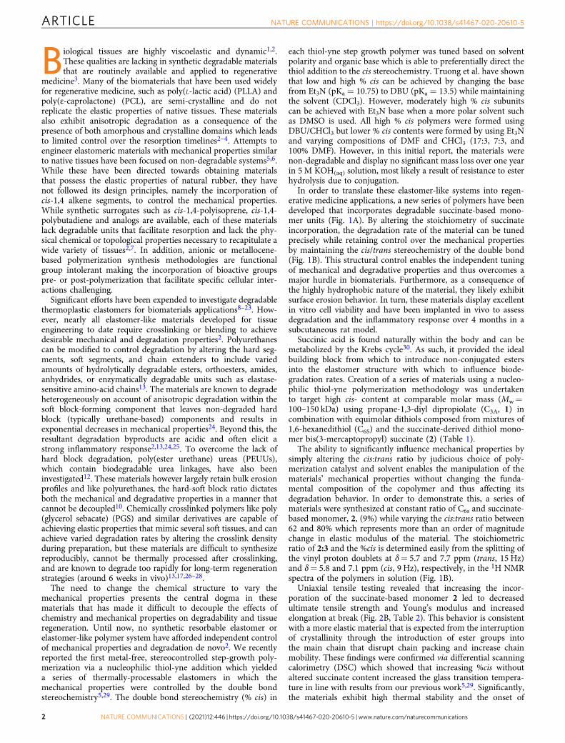
Uniaxial tensile testing revealed that increasing the incor-poration of the succinate-based monomer 2 led to decreasedultimate tensile strength and Young’s modulus and increasedelongation at break (Fig. 2B, Table 2). This behavior is consistentwith a more elastic material that is expected from the interruptionof crystallinity through the introduction of ester groups intothe main chain that disrupt chain packing and increase chainmobility. These findings were confirmed via differential scanningcalorimetry (DSC) which showed that increasing %cis withoutaltered succinate content increased the glass transition tempera-ture in line with results from our previous work5,29. Significantly,the materials exhibit high thermal stability and the onset of
ARTICLE NATURE COMMUNICATIONS | https://doi.org/10.1038/s41467-020-20610-5
2 NATURE COMMUNICATIONS | (2021) 12:446 | https://doi.org/10.1038/s41467-020-20610-5 | www.nature.com/naturecommunications

degradation temperatures exceeds 350 °C. These traits are criticalfor thermal processing and fabrication.
An in vitro investigation of the hydrolytic swelling anddegradation behavior showed the polymers to be chemicallystable with no visible degradation in PBS at ambient tempera-ture over a 1-month period (Fig. 3). In order to accelerate thehydrolytic degradation process, the samples were incubated in5 M KOH (aq) solution at ambient temperature. The data showthat the materials with increased succinate-based monomer, 2,yielded faster rates of degradation. Importantly, the mass lossprofiles are nearly linear in nature and show no evidence ofaccelerated degradation as a result of acidosis and swelling viabulk erosion. The dimensions of the materials were noted todecrease concomitantly with time which is highly indicative of a
surface erosion mechanism. SEM analysis of test substrates(Fig. 3B) exposed to accelerated degradation conditions indi-cates uniform degradation and pitting that confirms surfaceerosion as the most prevalent degradation process. Takentogether, these observations demonstrate that, unlike any otherdegradable biomaterials, the mechanical and degradationproperties of these elastomer-like polymers can be controlledindependently. This is a distinct difference from known polye-sters. To demonstrate the potential, by careful control overdouble bond stereochemistry and succinate monomer (2) con-tent, we prepared materials that displayed comparable degra-dation rates but markedly different mechanical properties andvice versa (comparable mechanical properties with markedlydifferent degradation rates – (Fig. 2A, B). The control over each
Fig. 1 Stereocontrolled synthesis and characterization of the resorbable elastomers. A A one-pot thiol-yne step-growth polymerization of propane-1,3-diyl dipropiolate (C3A, 1) with bis(3-mercaptopropyl) succinate (2) and with 1,6-hexane dithiol (C6S) forms a copolymer that shows tunable degradationrates depending on the % of amount of repeat unit x (1+ 2) that is incorporated. B The stereochemistry is easily determined by the vinyl proton doublets atδ= 5.7 and 7.7 ppm (trans, 15 Hz) and δ= 5.8 and 7.1 ppm (cis, 9 Hz), respectively. The extent of succinate incorporation will determine both the rate andextent of degradation.
Table 1 Polymerization conditions and characterization of all thiol-yne step growth polymers.
Entry % feed of 2 % incorporation Monomerratioa
Solvent Base Time(h)
% cisb Mn
(kDa)Mw
(kDa)ÐM
1 0 0 0.9965 CHCl3 DBU 1 80 26.4 147.5 5.602 9.0 9.0 0.9920 CHCl3 DBU 1 80 29.7 111.2 3.743 19.9 18.7 0.9922 CHCl3 DBU 1 79 35.2 110.8 3.154 50.5 49.4 0.9895 CHCl3 DBU 1 79 52.5 123.7 2.365 100 100 0.9870 CHCl3 DBU 1 79 35.9 112.2 3.126 19.8 19.0 0.9920 DMF Et3N 16 72 43.0 127.3 2.967 0 0 0.9920 CHCl3/DMF (7:3) Et3N 16 62 37.0 110.8 3.008 11.2 8.7 0.9959 CHCl3/DMF (7:3) Et3N 16 62 34.2 117.1 3.429 19.6 18.3 0.9920 CHCl3/DMF (7:3) Et3N 16 61 35.3 107.8 3.0510c 0 0 0.9950 CHCl3 DBU 1 80 24.7 35.4 1.46
aAn excess of the dialkyne monomer was used to reduce any disulfide coupling and UV crosslinking side reactions.bDetermined by comparison of 1H NMR integration of cis peaks at δ= 7.10 ppm and trans peaks at δ= 7.70 ppm.cSynthesis conditions for poly(bis(4-(propioloyloxy)but-2-yn-1-yl) 3,3′-(hexane-1,6-diylbis(sulfanediyl))).
NATURE COMMUNICATIONS | https://doi.org/10.1038/s41467-020-20610-5 ARTICLE
NATURE COMMUNICATIONS | (2021) 12:446 | https://doi.org/10.1038/s41467-020-20610-5 | www.nature.com/naturecommunications 3
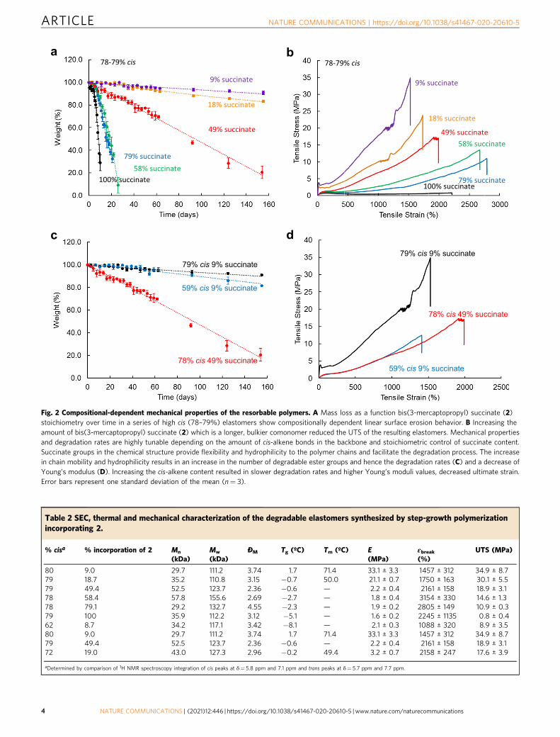
Fig. 2 Compositional-dependent mechanical properties of the resorbable polymers. A Mass loss as a function bis(3-mercaptopropyl) succinate (2)stoichiometry over time in a series of high cis (78–79%) elastomers show compositionally dependent linear surface erosion behavior. B Increasing theamount of bis(3-mercaptopropyl) succinate (2) which is a longer, bulkier comonomer reduced the UTS of the resulting elastomers. Mechanical propertiesand degradation rates are highly tunable depending on the amount of cis-alkene bonds in the backbone and stoichiometric control of succinate content.Succinate groups in the chemical structure provide flexibility and hydrophilicity to the polymer chains and facilitate the degradation process. The increasein chain mobility and hydrophilicity results in an increase in the number of degradable ester groups and hence the degradation rates (C) and a decrease ofYoung’s modulus (D). Increasing the cis-alkene content resulted in slower degradation rates and higher Young’s moduli values, decreased ultimate strain.Error bars represent one standard deviation of the mean (n= 3).
Table 2 SEC, thermal and mechanical characterization of the degradable elastomers synthesized by step-growth polymerizationincorporating 2.
% cisa % incorporation of 2 Mn
(kDa)Mw
(kDa)ÐM Tg (ºC) Tm (ºC) E
(MPa)εbreak(%)
UTS (MPa)
80 9.0 29.7 111.2 3.74 1.7 71.4 33.1 ± 3.3 1457 ± 312 34.9 ± 8.779 18.7 35.2 110.8 3.15 −0.7 50.0 21.1 ± 0.7 1750 ± 163 30.1 ± 5.579 49.4 52.5 123.7 2.36 −0.6 — 2.2 ± 0.4 2161 ± 158 18.9 ± 3.178 58.4 57.8 155.6 2.69 −2.7 — 1.8 ± 0.4 3154 ± 330 14.6 ± 1.378 79.1 29.2 132.7 4.55 −2.3 — 1.9 ± 0.2 2805 ± 149 10.9 ± 0.379 100 35.9 112.2 3.12 −5.1 — 1.6 ± 0.2 2245 ± 1135 0.8 ± 0.462 8.7 34.2 117.1 3.42 −8.1 — 2.1 ± 0.3 1088 ± 320 8.9 ± 3.580 9.0 29.7 111.2 3.74 1.7 71.4 33.1 ± 3.3 1457 ± 312 34.9 ± 8.779 49.4 52.5 123.7 2.36 −0.6 — 2.2 ± 0.4 2161 ± 158 18.9 ± 3.172 19.0 43.0 127.3 2.96 −0.2 49.4 3.2 ± 0.7 2158 ± 247 17.6 ± 3.9
aDetermined by comparison of 1H NMR spectroscopy integration of cis peaks at δ= 5.8 ppm and 7.1 ppm and trans peaks at δ= 5.7 ppm and 7.7 ppm.
ARTICLE NATURE COMMUNICATIONS | https://doi.org/10.1038/s41467-020-20610-5
4 NATURE COMMUNICATIONS | (2021) 12:446 | https://doi.org/10.1038/s41467-020-20610-5 | www.nature.com/naturecommunications
of these properties will be critical to future applications wheredesigners will need to engineer subtle changes without returningto new synthetic methods.
To investigate the potential for use in biomaterial applications,cell viability, spreading, and proliferation assays were used as aninitial method to determine the cellular responses to theelastomer-like polymers. Human mesenchymal stem cells(hMSCs) and MC3T3 cells were cultured on glass slides spin-coated with each material variant or on the control polymer, poly(L-lactide), PLLA. Cell viability was found to be higher than 95%on all samples using a Live/Dead® assay. Cell adhesion andspreading were assessed by staining F-actin, vinculin-labeled focaladhesion contacts, and cell nuclei and revealed that the hMSCsadopted an elongated and spindle-like shape on all samples(Fig. 4C). Cell proliferation was measured with a PrestoBlue®metabolic assay, after 24 h, 3 days, and 7 days of incubation. After7 days the population of cells on each sample increasedto approximately 5 times the original concentration (Fig. 4D).
One of the key aspects of a translationally relevant material is theability to control the placement and concentration of functionalspecies (drug, peptide, protein) on the surface of a material wherethe group is bioavailable to the surrounding cells and tissues31.While many methods are available for peptide polymer conjuga-tion, we designed a dialkyne monomer, but-2-yne-1,4-diyl dipro-piolate (3) that possesses an internal triple bond. As a consequenceof the increased distance from the electron withdrawing groups,the reactivity of internal alkynes is distinctly different than terminalalkynes. The internal alkynes were found to be stable during thenucleophilic thiol-yne addition polymerization process with C3A
(1) and C6S (1,6-hexanedithiol) as comonomers which left itavailable for selective post-polymerization functionalization of theresulting materials (Fig. 4E). Following the polymerization and afilm casting process, a Megastokes®-673-azide dye surrogate(Fig. 4F) was covalently tethered to the internal alkyne functio-nalized poly(bis(4-(propioloyloxy)but-2-yn-1-yl)-3,3’-(hexane-1,6-
diylbis(sulfanediyl))) using Cp*RuCl(COD) as a catalyst32. Afterwashing, the film remained fluorescent thus evidencing the con-jugation of the dye to the polymer. To extend the concept andshow utility from a biomaterials viewpoint, we also used thismethodology to attach an azide-functionalized GRGDS peptide tothe films. While only an initial demonstration, the presence of theadhesion peptide had a distinct influence on the cell adhesion andspreading properties (Figs. 4G and 4H). Following peptide con-jugation, increased cell adhesion, spreading and integrin-associatedactin fiber formation was evident in the RGD derivatized filmsrelative to the unfunctionalized films. Future studies are developingthis technique to apply other bioactive groups designed to influ-ence specific cellular activities.
The lack of cytotoxicity and enhanced cellular activity confirmedthat the thiol-yne stereoelastomer materials could be implantedin vivo for tissue compatibility studies. Elastomeric discs possessingvarious cis content and succinate stoichiometry were implantedsubcutaneously for 4 months to observe the degradation behaviorand tissue inflammatory responses in vivo. Significantly, no grossinflammation, which would appear as a dense calcified capsule, wasevident from macroscopic images of the samples taken at eachtimepoint (Fig. 5B). Sections stained with hematoxylin and eosin(H&E) were analyzed for inflammatory responses in the form offibrous capsule formation. Sections stained with H&E were alsoassessed for inflammatory cell infiltration. Fibrous encapsulationoccurred as expected, and the granuloma grew thicker over4 months of incubation with no significant difference compared toPLLA (Fig. 5). The granuloma was less than 200 μm thick for allsamples, which has previously been reported as acceptable in termsof tissue compatibility for long-term implants4,33,34. This is indi-cative of the tunable degradation profiles from varied succinatecontent as increasing implant degradation rates correlates withgreater cellular remodeling processes34.
H&E slides were quantitatively analyzed for neutrophils, lym-phocytes, plasma cells, single macrophages, multinucleated giant
Fig. 3 Surface Erosion and Swelling. A Discs (4mm diameter, 0.5 mm thick) were cut from vacuum film compression samples and placed in 1× PBS in theincubator (37 °C, 5% CO2 humidified atmosphere) for up to 32 days. The data were plotted in three different ways: dry mass change compared to theoriginal mass (degradation), wet mass change compared to the original mass (traditional swelling if no degradation), and wet mass/dry mass at each timepoint (swelling if degradation). The swelling behavior of the polymers was determined by tracking the wet and dry mass of the disc samples at each timepoint. Error bars represent one standard deviation of the mean (n= 4). B Analysis of SEM micrographs of the respective test coupons exposed toaccelerated degradation conditions indicates uniform degradation and pitting indicative of surface erosion processes. Scale bars= 10 μm.
NATURE COMMUNICATIONS | https://doi.org/10.1038/s41467-020-20610-5 ARTICLE
NATURE COMMUNICATIONS | (2021) 12:446 | https://doi.org/10.1038/s41467-020-20610-5 | www.nature.com/naturecommunications 5
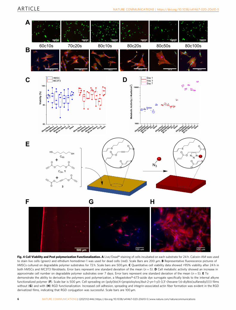
Fig. 4 Cell Viability and Post polymerization Functionalization. A Live/Dead® staining of cells incubated on each substrate for 24 h. Calcein-AM was usedto stain live cells (green) and ethidium homodimer-1 was used for dead cells (red). Scale Bars are 200 μm. B Representative fluorescence pictures ofhMSCs cultured on degradable polymer substrates for 72 h. Scale bars are 500 μm. C Quantitative cell viability data showed >95% viability after 24 h inboth hMSCs and MC3T3 fibroblasts. Error bars represent one standard deviation of the mean (n= 5). D Cell metabolic activity showed an increase inapproximate cell number on degradable polymer substrates over 7 days. Error bars represent one standard deviation of the mean (n= 5). E Todemonstrate the ability to derivatize the polymers post polymerization, a Megastokes®-673-azide dye surrogate specifically binds to the internal alkynefunctionalized polymer (F). Scale bar is 500 μm. Cell spreading on (poly(bis(4-(propioloyloxy)but-2-yn-1-yl)-3,3’-(hexane-1,6-diylbis(sulfanediyl)))) filmswithout (G) and with (H) RGD functionalization. Increased cell adhesion, spreading and integrin-associated actin fiber formation was evident in the RGDderivatized films, indicating that RGD conjugation was successful. Scale bars are 100 μm.
ARTICLE NATURE COMMUNICATIONS | https://doi.org/10.1038/s41467-020-20610-5
6 NATURE COMMUNICATIONS | (2021) 12:446 | https://doi.org/10.1038/s41467-020-20610-5 | www.nature.com/naturecommunications
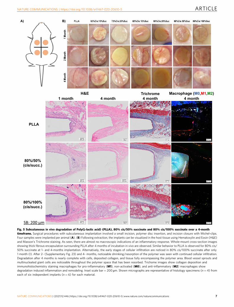
Fig. 5 Subcutaneous in vivo degradation of Poly(L-lactic acid) (PLLA), 80% cis/50% succinate and 80% cis/100% succinate over a 4-monthtimeframe. Surgical procedures with subcutaneous implantation involved a small incision, polymer disc insertion, and incision closure with Michel-clips.Four samples were implanted per animal (A). (B) Following extraction, the implants can be visualized in the host tissue using Hematoxylin and Eosin (H&E)and Masson’s Trichrome staining. As seen, there are almost no macroscopic indications of an inflammatory response. Whole-mount cross-section imagesshowing thick fibrous encapsulation surrounding PLLA after 4 months of incubation in vivo are observed. Similar behavior to PLLA is observed for 80% cis/50% succinate at 1- and 4-months implantation. Alternatively, the early stages of cellular infiltration are noticed in 80% cis/100% succinate after only1 month (I). After 2- (Supplementary Fig. 23) and 4- months, noticeable shrinking/resorption of the polymer was seen with continued cellular infiltration.Degradation after 4 months is nearly complete with cells, deposited collagen, and tissue fully encompassing the polymer area. Blood vessel sprouts andmultinucleated giant cells are noticeable throughout the polymer space that has been resorbed. Trichome images show collagen deposition andimmunohistochemistry staining macrophages for pro-inflammatory (M1), non-activated (M0), and anti-inflammatory (M2) macrophages showdegradation induced inflammation and remodeling. Inset scale bar= 200 μm. Shown micrographs are representative of histology specimens (n= 4) fromeach of six independent implants (n= 6) for each material.
NATURE COMMUNICATIONS | https://doi.org/10.1038/s41467-020-20610-5 ARTICLE
NATURE COMMUNICATIONS | (2021) 12:446 | https://doi.org/10.1038/s41467-020-20610-5 | www.nature.com/naturecommunications 7
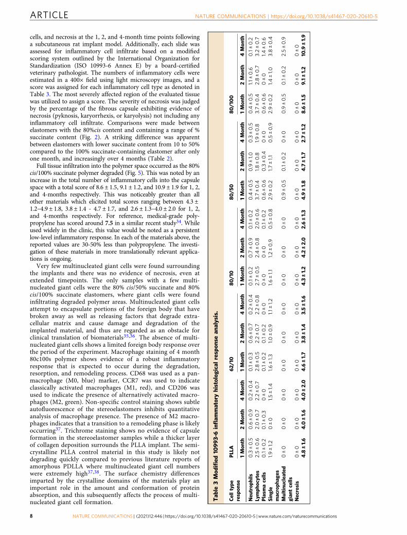
cells, and necrosis at the 1, 2, and 4-month time points followinga subcutaneous rat implant model. Additionally, each slide wasassessed for inflammatory cell infiltrate based on a modifiedscoring system outlined by the International Organization forStandardization (ISO 10993-6 Annex E) by a board-certifiedveterinary pathologist. The numbers of inflammatory cells wereestimated in a 400× field using light microscopy images, and ascore was assigned for each inflammatory cell type as denoted inTable 3. The most severely affected region of the evaluated tissuewas utilized to assign a score. The severity of necrosis was judgedby the percentage of the fibrous capsule exhibiting evidence ofnecrosis (pyknosis, karyorrhexis, or karyolysis) not including anyinflammatory cell infiltrate. Comparisons were made betweenelastomers with the 80%cis content and containing a range of %succinate content (Fig. 2). A striking difference was apparentbetween elastomers with lower succinate content from 10 to 50%compared to the 100% succinate-containing elastomer after onlyone month, and increasingly over 4 months (Table 2).
Full tissue infiltration into the polymer space occurred as the 80%cis/100% succinate polymer degraded (Fig. 5). This was noted by anincrease in the total number of inflammatory cells into the capsulespace with a total score of 8.6 ± 1.5, 9.1 ± 1.2, and 10.9 ± 1.9 for 1, 2,and 4-months respectively. This was noticeably greater than allother materials which elicited total scores ranging between 4.3 ±1.2–4.9 ± 1.8, 3.8 ± 1.4 - 4.7 ± 1.7, and 2.6 ± 1.3–4.0 ± 2.0 for 1, 2,and 4-months respectively. For reference, medical-grade poly-propylene has scored around 7.5 in a similar recent study34. Whileused widely in the clinic, this value would be noted as a persistentlow-level inflammatory response. In each of the materials above, thereported values are 30-50% less than polypropylene. The investi-gation of these materials in more translationally relevant applica-tions is ongoing.
Very few multinucleated giant cells were found surroundingthe implants and there was no evidence of necrosis, even atextended timepoints. The only samples with a few multi-nucleated giant cells were the 80% cis/50% succinate and 80%cis/100% succinate elastomers, where giant cells were foundinfiltrating degraded polymer areas. Multinucleated giant cellsattempt to encapsulate portions of the foreign body that havebroken away as well as releasing factors that degrade extra-cellular matrix and cause damage and degradation of theimplanted material, and thus are regarded as an obstacle forclinical translation of biomaterials35,36. The absence of multi-nucleated giant cells shows a limited foreign body response overthe period of the experiment. Macrophage staining of 4 month80c100s polymer shows evidence of a robust inflammatoryresponse that is expected to occur during the degradation,resorption, and remodeling process. CD68 was used as a pan-macrophage (M0, blue) marker, CCR7 was used to indicateclassically activated macrophages (M1, red), and CD206 wasused to indicate the presence of alternatively activated macro-phages (M2, green). Non-specific control staining shows subtleautofluorescence of the stereoelastomers inhibits quantitativeanalysis of macrophage presence. The presence of M2 macro-phages indicates that a transition to a remodeling phase is likelyoccurring37. Trichrome staining shows no evidence of capsuleformation in the stereoelastomer samples while a thicker layerof collagen deposition surrounds the PLLA implant. The semi-crystalline PLLA control material in this study is likely notdegrading quickly compared to previous literature reports ofamorphous PDLLA where multinucleated giant cell numberswere extremely high37,38. The surface chemistry differencesimparted by the crystalline domains of the materials play animportant role in the amount and conformation of proteinabsorption, and this subsequently affects the process of multi-nucleated giant cell formation. T
able
3Mod
ified
10993-6inflam
matoryhistolog
ical
respon
sean
alysis.
Celltype
respon
sePLLA
62/
1080/10
80/5
080/100
1Mon
th2Mon
th4Mon
th1Mon
th2Mon
th4Mon
th1Mon
th2Mon
th4Mon
th1Mon
th2Mon
th4Mon
th1Mon
th2Mon
th4Mon
th
Neu
trop
hils
0.3±0.5
0.6±0.9
0.2±0.4
0.1±0.3
0.6±0.7
0.2±0.4
0.1±0.2
0.7±0.9
0.1±0.2
0.4±0.5
0.9±1.0
0.3±0.5
0.4±0.5
1.1±0.6
0.1±0.2
Lymph
ocytes
2.5±0.6
2.0±0.7
2.2±0.7
2.8±0.5
2.2±0.7
2.2±0.8
2.7±0.5
2.4±0.8
2.0±0.6
3.7±0.4
1.8±0.8
1.9±0.8
3.7±0.4
2.8±0.7
3.2±0.7
Plasm
acells
0.1±0.2
0.1±0.3
0±0
0.1±0.2
0.1±0.2
0±0
0±0
0±0
0.1±0.2
0.6±0.6
0.3±0.4
0±0
0.6±0.6
0±0
1.4±0.6
Single
macroph
ages
1.9±1.2
0±0
1.5±1.4
1.6±1.3
1.0±0.9
1.1±
1.2
1.6±1.1
1.2±0.9
0.5±0.8
2.9±0.2
1.7±1.1
0.5±0.9
2.9±0.2
1.4±1.0
3.8±0.4
Multinu
clea
ted
gian
tcells
0±0
0±0
0±0
0±0
0±0
0±0
0±0
0±0
0±0
0.9±0.5
0.1±0.2
0±0
0.9±0.5
0.1±0.2
2.5±0.9
Necrosis
0±0
0±0
0±0
0±0
0±0
0±0
0±0
0±0
0±0
0±0
0±0
0±0
0±0
0±0
0±0
4.8
±1.6
4.0
±1.6
4.0
±2.0
4.6
±1.7
3.8±1.4
3.5±1.6
4.3±1.2
4.2±2.0
2.6±1.3
4.9
±1.8
4.7±1.7
2.7±1.2
8.6
±1.5
9.1±1.2
10.9
±1.9
ARTICLE NATURE COMMUNICATIONS | https://doi.org/10.1038/s41467-020-20610-5
8 NATURE COMMUNICATIONS | (2021) 12:446 | https://doi.org/10.1038/s41467-020-20610-5 | www.nature.com/naturecommunications
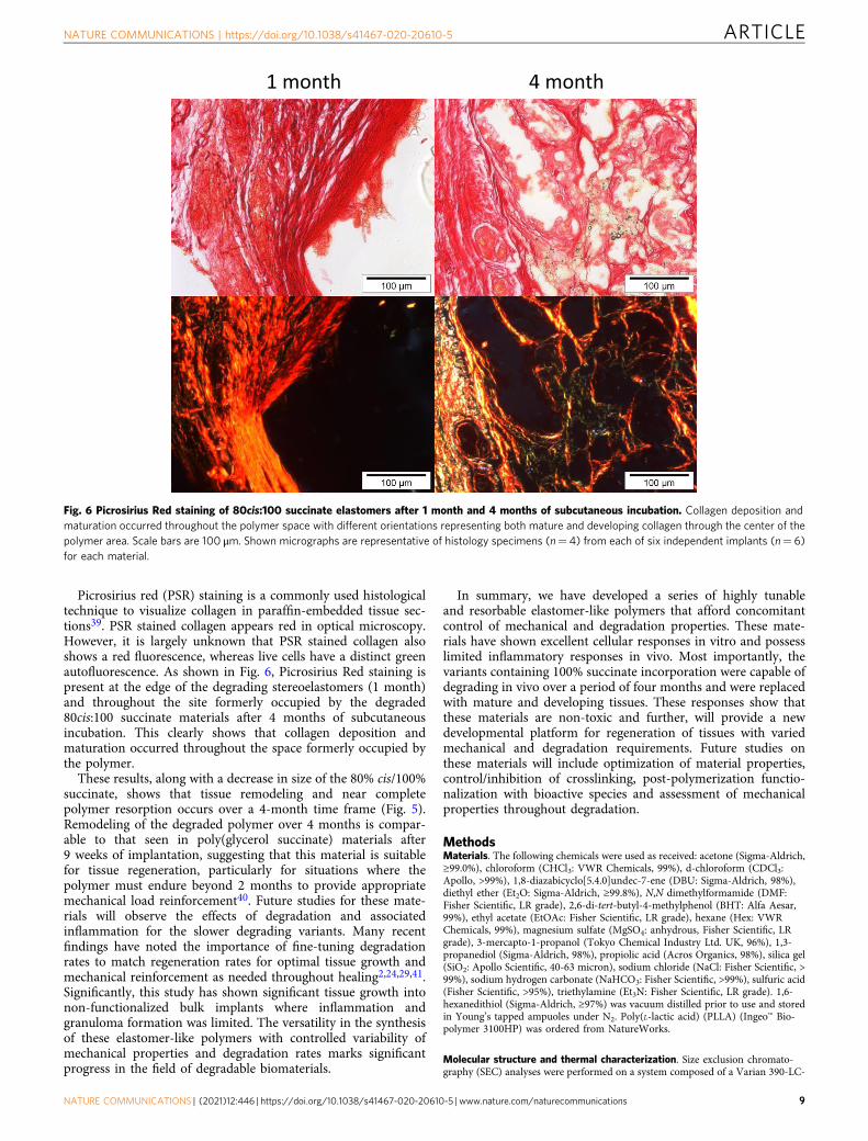
Picrosirius red (PSR) staining is a commonly used histologicaltechnique to visualize collagen in paraffin-embedded tissue sec-tions39. PSR stained collagen appears red in optical microscopy.However, it is largely unknown that PSR stained collagen alsoshows a red fluorescence, whereas live cells have a distinct greenautofluorescence. As shown in Fig. 6, Picrosirius Red staining ispresent at the edge of the degrading stereoelastomers (1 month)and throughout the site formerly occupied by the degraded80cis:100 succinate materials after 4 months of subcutaneousincubation. This clearly shows that collagen deposition andmaturation occurred throughout the space formerly occupied bythe polymer.
These results, along with a decrease in size of the 80% cis/100%succinate, shows that tissue remodeling and near completepolymer resorption occurs over a 4-month time frame (Fig. 5).Remodeling of the degraded polymer over 4 months is compar-able to that seen in poly(glycerol succinate) materials after9 weeks of implantation, suggesting that this material is suitablefor tissue regeneration, particularly for situations where thepolymer must endure beyond 2 months to provide appropriatemechanical load reinforcement40. Future studies for these mate-rials will observe the effects of degradation and associatedinflammation for the slower degrading variants. Many recentfindings have noted the importance of fine-tuning degradationrates to match regeneration rates for optimal tissue growth andmechanical reinforcement as needed throughout healing2,24,29,41.Significantly, this study has shown significant tissue growth intonon-functionalized bulk implants where inflammation andgranuloma formation was limited. The versatility in the synthesisof these elastomer-like polymers with controlled variability ofmechanical properties and degradation rates marks significantprogress in the field of degradable biomaterials.
In summary, we have developed a series of highly tunableand resorbable elastomer-like polymers that afford concomitantcontrol of mechanical and degradation properties. These mate-rials have shown excellent cellular responses in vitro and possesslimited inflammatory responses in vivo. Most importantly, thevariants containing 100% succinate incorporation were capable ofdegrading in vivo over a period of four months and were replacedwith mature and developing tissues. These responses show thatthese materials are non-toxic and further, will provide a newdevelopmental platform for regeneration of tissues with variedmechanical and degradation requirements. Future studies onthese materials will include optimization of material properties,control/inhibition of crosslinking, post-polymerization functio-nalization with bioactive species and assessment of mechanicalproperties throughout degradation.
MethodsMaterials. The following chemicals were used as received: acetone (Sigma-Aldrich,≥99.0%), chloroform (CHCl3: VWR Chemicals, 99%), d-chloroform (CDCl3:Apollo, >99%), 1,8-diazabicyclo[5.4.0]undec-7-ene (DBU: Sigma-Aldrich, 98%),diethyl ether (Et2O: Sigma-Aldrich, ≥99.8%), N,N dimethylformamide (DMF:Fisher Scientific, LR grade), 2,6-di-tert-butyl-4-methylphenol (BHT: Alfa Aesar,99%), ethyl acetate (EtOAc: Fisher Scientific, LR grade), hexane (Hex: VWRChemicals, 99%), magnesium sulfate (MgSO4: anhydrous, Fisher Scientific, LRgrade), 3-mercapto-1-propanol (Tokyo Chemical Industry Ltd. UK, 96%), 1,3-propanediol (Sigma-Aldrich, 98%), propiolic acid (Acros Organics, 98%), silica gel(SiO2: Apollo Scientific, 40-63 micron), sodium chloride (NaCl: Fisher Scientific, >99%), sodium hydrogen carbonate (NaHCO3: Fisher Scientific, >99%), sulfuric acid(Fisher Scientific, >95%), triethylamine (Et3N: Fisher Scientific, LR grade). 1,6-hexanedithiol (Sigma-Aldrich, ≥97%) was vacuum distilled prior to use and storedin Young’s tapped ampuoles under N2. Poly(L-lactic acid) (PLLA) (Ingeo™ Bio-polymer 3100HP) was ordered from NatureWorks.
Molecular structure and thermal characterization. Size exclusion chromato-graphy (SEC) analyses were performed on a system composed of a Varian 390-LC-
Fig. 6 Picrosirius Red staining of 80cis:100 succinate elastomers after 1 month and 4 months of subcutaneous incubation. Collagen deposition andmaturation occurred throughout the polymer space with different orientations representing both mature and developing collagen through the center of thepolymer area. Scale bars are 100 μm. Shown micrographs are representative of histology specimens (n= 4) from each of six independent implants (n= 6)for each material.
NATURE COMMUNICATIONS | https://doi.org/10.1038/s41467-020-20610-5 ARTICLE
NATURE COMMUNICATIONS | (2021) 12:446 | https://doi.org/10.1038/s41467-020-20610-5 | www.nature.com/naturecommunications 9
Multi detector using a Varian Polymer Laboratories guard column (PLGel 5 μM,50 × 7.5 mm), two mixed D Varian Polymer Laboratories columns (PLGel 5 μM,300 × 7.5 mm) and a PLAST RT autosampler. Detection was conducted using adifferential refractive index (RI) detector. The analyses were performed in CHCl3 at40 °C and containing 0.5% w/w Et3N at a flow rate of 1.0 mL/min. Linear poly-styrene (PS) (162–2.4 × 10⁵ g mol−1) standards were used to calibrate the system.EcoSEC HLC-8320 GPC (Tosoh Bioscience LLC, King of Prussia, PA) equippedwith a TSKgel GMHHR-M mixed bed column and refractive index (RI) detectorwas performed to analyze poly(bis(4-(propioloyloxy)but-2-yn-1-yl)-3,3′-(hexane-1,6-diylbis(sulfanediyl))) (10). Molecular mass was calculated using a calibrationcurve determined from polystyrene standards (PStQuick MP-M standards, TosohBioscience, LLC) with DMF with 0.1 M LiBr as eluent flowing at 1.0 mLmin−1 at323 K, and a sample concentration of 3 mgmL−1.
Nuclear magnetic resonance (1H, 13C) spectra were recorded in CDCl3 on a BrukerDPX-400 spectrometer at 298 K. Chemical shifts are reported as δ in parts per million(ppm) and referenced to the chemical shift of the residual solvent resonances (CDCl31H: δ= 7.26 ppm, 13C: δ= 77.16 ppm). The resonance multiplicities are described as s(singlet), d (doublet), t (triplet), q (quartet) or m (multiplet).
Thermogravimetric analysis (TGA) (Q500, TA Instruments, New Castle, DE)was performed over a temperature range from 0 to 600 °C at a heating rate of10 °C/min. A 5% loss in mass was used to determine the onset temperature ofdegradation (Td).
Differential scanning calorimetry (DSC) (Q2000, TA Instruments, New Castle,DE) was used with a temperature range from −20 to 200 °C and a scanning rate of10 °C/min in a heating/cooling/heating mode to determine glass transitiontemperatures (Tg) of polymers obtained during the second heating cycle.
Monomer synthesisPropane-1,3-diyl dipropiolate (C3A, 1). 1,3-propanediol (20.00 g, 0.263 mol) wasadded to a 1 L single neck round bottom flask. To this was added toluene (100 mL)and benzene (100 mL). Two drops of H2SO4 were added and the solution wasallowed to stir at room temperature for 5 min before adding propiolic acid (50.00 g,0.714 mol). A Dean-Stark apparatus with condenser was fitted and the reaction wasthen refluxed for 16 h at 120 °C or until the required amount of water was collected.The solution was then cooled to room temperature and solvent-extracted withsaturated NaHCO3 solution (2 × 200 mL) to remove any residual acids. The organicphase was then collected, dried over MgSO4, filtered, and reduced in volume todryness. The product was then purified on silica gel isocratically using 4:1 hexane/EtOAc and collecting the 1st fraction. After removal of the solvent, the final pro-duct was further purified by distillation under high vacuum at 160 °C to yieldcolorless oil that slightly crystallized on sitting (24.63 g, 52% yield). Rf (3:2 Hex/EtOAc)= 0.43; Melting point: 25 °C; 1H NMR (500MHz, CDCl3) δ 4.30 (t, 3JHH=6.2 Hz, 4H), 2.88 (s, 2H), 2.19–1.96 (m, 2H); 13C NMR (125MHz, CDCl3) δ 152.6,75.3, 74.5, 62.6, 27.5; ESI-MS Calcd for C9H8O4Na (M+Na): 203.0, Found: 203.0;Anal Calcd for C9H8O4: C 60.00; H 4.48%. Found: C 59.70; H 4.41%.
Bis(3-mercaptopropyl) succinate (2). 3-mercaptopropanol (7.30 g, 0.079 mol) wasadded to a 250 mL single neck round bottom flask. To this was added toluene (60mL) and benzene (60 mL). Two drops of H2SO4 were added and the solution wasallowed to stir at room temperature for 5 min before adding succinic acid (4.40 g,0.037 mol). A Dean-Stark apparatus with condenser was fitted and the reaction wasthen refluxed for 16 h at 120 °C or until the required amount of water was collected.The solution was then cooled to room temperature and solvent removed byvacuum transfer. The product was resolubilized in CHCl3 (100 mL) and extractedwith saturated NaHCO3 solution (2 × 200 mL) to remove any residual acids. Theorganic phase was then collected, dried over MgSO4, filtered, and reduced involume to dryness. The product was then purified on silica gel isocratically using3:2 hexane/EtOAc and collecting the 1st fraction. After removal of the solvent, thefinal product was further purified by distillation under high vacuum (0.15 Torr) at220 °C to yield colorless oil (7.8 g, 79% yield). Rf (3:2 Hex/EtOAc) = 0.4; 1H NMR(400MHz, CDCl3) δ 4.21 (t, 3JHH= 6.2 Hz, 4H), 2.62 (s, 4H), 2.58 (q, 3JHH=6.6 Hz, 4H), 1.40 (t, 3JHH= 8.1 Hz, 2H); 13C NMR (100MHz, CDCl3) δ 172.3, 62.9,32.9, 29.2, 21.2; ESI-MS Calcd for C10H18O4S2Na+ (M+Na+): 289.1, Found:289.0; Anal Calcd for C10H18O4S2: C 45.09; H 6.81%. Found: C 59.70; H 4.41%.
Sodium propiolate. Sodium propiolate was synthesized according to the proceduredescribed by Bonnesen et al. 1, Sodium hydroxide (0.645 g, 0.016 mol) was dis-solved in methanol (50 mL) in a 250 mL round-bottom flask and protected fromlight. The solution was cooled to 0 °C for 10 min. Then propionic acid (1.00 mL,0.016 mol) was added with stirring. The solution was allowed to warm to ambienttemperature and stirred for additional 2 h. The solvent was then removed by rotaryevaporation. A white solid product was formed and dried under high vacuum toyield 3 (1.44 g, 97%). The product should be stored in the dark due to light sen-sitivities. 1H NMR (300 MHz, CD3OD) δ 2.95 (s, 1H). 13C NMR (75MHz,CD3OD) δ 160.64, 81.83, 69.12.
But-2-yne-1,4-diyl bis(4-methylbenzenesulfonate). But-2-yne-1,4-diyl bis(4-methyl-benzene sulfonate) was synthesized according to the procedure describedby Maisonial et al.2 Briefly, p-toluenesulfonyl chloride (24.00 g, 0.126 mol) and
1,4-butynediol (4.00 g, 0.046 mol) were dissolved in Et2O (300 mL). The mixturewas cooled to −15 °C for 15 min before potassium hydroxide (16.00 g, 0.285 mol)was added slowly. The resulting solution was stirred at 0 °C for 3 h and poured intoice water (300 mL). When the solution reached ambient temperature, the solutionwas extracted with DCM (200 mL × 3) and the organic layer was collected, driedwith anhydrous Na2SO4, filtered, and concentrated. The solid was washed withEt2O (100 mL × 3) and dried under vacuum 24 h to yield 4 (14.66 g, 80%) as awhite solid. 1H NMR (300MHz, CDCl3) δ 7.77 (d, J= 8.8 Hz, 4H), 7.34 (d, J= 8.8Hz, 4H), 4.58 (s, 4H), 2.46 (s, 6H). 13C NMR (75MHz, CDCl3) δ 145.54, 132.80,130.01 (×2), 128.16 (×2), 81.04, 57.21, 21.76.
But-2-yne-1,4-diyl dipropiolate (3). Under reduced light conditions, sodium pro-piolate (7.600 g, 0.083 mol) and But-2-yne-1,4-diyl bis(4-methylbenzene sulfonate)(12.00 g, 0.030 mol) were dissolved in DMF (120 mL), and the mixture was heatedto 50 °C, and allowed to stir for 24 h. After the reaction was cooled down toambient temperature, a saturated solution of NH4Cl (200 mL) was added to themixture and the reaction was stirred for 10 min. The mixture was extracted withDCM (150 mL × 3) and the organic extracts were combined, extracted with satu-rated solution of NaHCO3 (150 mL × 3). The organic layer was combined and driedover anhydrous Na2SO4, filtered, and concentrated. The residue was purified byflash column chromatography on silica gel (EtOAc/hexanes 1:3; Rf= 0.30). Afterremoval of the solvent, the final product was further purified by distillation underhigh vacuum at 110 °C to yield a colorless oil (3.76 g, 65%). 1H NMR (300MHz,CDCl3) δ 4.81 (s, 4H), 2.97 (s, 2H). 13C NMR (75MHz, CDCl3) δ 151.78, 80.60,76.34, 73.82, 53.28. ESI-MS for C10H6O4Na, m/z theoretical: [M+Na]+ = 213.02Da, observed: [M+Na]+ = 213.0 Da.
General procedure for thiol-yne step growth polymerization. An example of thethiol-yne step growth polymerization is as follows: 1,6-hexanedithiol (0.73 g, 4.9 ×10⁻³ mol) and 2 (0.32 g, 1.2 × 10⁻³ mol) were added to a 20 mL scintillation vial.Propane-1,3-diyl dipropiolate (1.10 g, 6.1 × 10⁻³ mol) was added to the solution byquantitative transfer with CHCl3 (12 mL). The solution was then cooled to −15 °Cwith stirring for 15 min before DBU (9 µL, 6.0 × 10⁻⁵ mol) was added. Theaddition of DBU produced an exotherm causing the solvent to bubble. After 2 minof stirring, the reaction was then allowed to warm to room temperature andcontinued to stir, during which time the solution became very viscous. After 1 h,the solution was diluted with CHCl3 (8 mL). The polymer solution was thenprecipitated into 1:1 diethyl ether/acetone (200 mL) and collected by decanting thesupernatant. The polymer was then redissolved in CHCl3 (20 mL) and reprecipi-tated into 1:1 diethyl ether/acetone (200 mL). The polymer was again redissolved inCHCl3 (20 mL) and 100 mg BHT (5 %w/w) was added. The final solution was thenprecipitated into n-hexane (200 mL), collected by decanting the supernatant, anddried in vacuo at room temperature for 24 h. SEC (CHCl3+ 0.5% Et3N) Mn=35.2 kDa, Mw= 110.8 kDa, Mp= 106.9 kDa, ÐM= 3.15. 1H NMR (CDCl3,400 MHz) % incorporation of 2= 18.7%; % cis= 79%.
Variation of molecular mass. The molecular mass of the thiol-yne stepgrowth polymers was varied by changing the amount of dithiol in relation to thedialkyne such that the dialkyne was always in excess. Monomer ratios weredetermined using the extended Carothers equation for one monomer in excess(assuming p→ 100%)1.
Procedure of thiol-yne step-growth polymerization for but-2-yne-1,4-diyldipropiolate. 1,6-Hexanedithiol (4.300 g, 0.028 mol) was added into 500 mL roundbottom flask and but-2-yne-1,4-diyl dipropiolate (5) (5.500 g, 0.029 mol) wasadded to a 500 mL round bottom flask with 200 mL CHCl3. The solution was thencooled to −15 °C with stirring for 20 min before DBU (44 μL, 29 mmol) was addedin one portion. Notably, the addition of DBU caused the solvent to bubble due toan exothermic reaction. After stirring for 10 min, the reaction was allowed to warmto room temperature and continued to stir. After 1 h, a couple of drops of but-2-yne-1,4-diyl dipropiolate 5 in CHCl3 (5 mL) was added into the reaction solution.After stirring for an additional 30 min, the solution was diluted with CHCl3(50 mL) and BHT (0.48 g, 0.002 mol) before the precipitation steps. The polymersolution was then precipitated into diethyl ether (1.5 L) and collected by decantingthe supernatant. The polymer was then redissolved in CHCl3 (150 mL) andreprecipitated into diethyl ether (1.5 L), collected by decanting the supernatant,and dried by high vacuum system at room temperature for 24 h to give paleyellow polymer poly(bis(4-(propioloyloxy)but-2-yn-1-yl)-3,3’-(hexane-1,6-diylbis(sulfanediyl))) (10) (8.3 g, 85%). SEC (DMF+ 0.1 M LiBr, based on PS standards)Mn= 24.7 kDa, Mw= 35.4 kDa, ÐM= 1.46. 1H NMR (CDCl3, 300MHz) % cis: %trans= 78 %: 22 %. DSC: Tg= 22 °C. TGA: Td= 287 °C.
Post-polymerization functionalization with GRGDS peptide. The end-cappedpolymer poly(bis(4-(propioloyloxy)but-2-yn-1-yl) 3,3’-(hexane-1,6-diylbis(sulfa-nediyl))) (10) (300 mg; Mn= 24.7 kDa, Mw= 35.4 kDa, ÐM= 1.46.) and Cp*RuCl(COD) (1 mg, 0.003 mmol) were added to a 100 mL two neck round bottom flaskand the round bottom flask was evacuated and purged with N2 three times beforedried DMF (40 mL) was added. Then, 3 wt% N3-GRGDS peptide (FW= 629.63 g/mol; 9 mg) was dissolved in dried DMF (5 mL) and added into the reaction
ARTICLE NATURE COMMUNICATIONS | https://doi.org/10.1038/s41467-020-20610-5
10 NATURE COMMUNICATIONS | (2021) 12:446 | https://doi.org/10.1038/s41467-020-20610-5 | www.nature.com/naturecommunications
solution by syringe and allowed to stir for 12 h. The solution of GRGDS peptidefunctionalized polymer was precipitated into ethanol (500 mL), collected and driedunder vacuum for 24 h to afford GRGDS peptide functionalized polymer poly(bis(4-(propioloyloxy)but-2-yn-1-yl)-3,3′-(hexane-1,6-diylbis(sulfanediyl)))-GRGDS.
Mechanical testing. Destructive tensile tests were performed to determine theeffects of altered cis-alkene and succinate incorporation on Young’s Modulus (E)and ultimate strain (εU). Samples (n= 3) were prepared using vacuum film com-pression (Technical Machine Products, Cleveland, OH) to press films measuring50 mm × 50mm × 0.5 mm. Polymer was preheated at 120 °C for 15 min, and thencompressed at 10,000 lbs of pressure for 4 min before cooling rapidly undervacuum. Tensile bars were cut using a custom-made dog bone-shaped die cutterand were pulled at several rates to determine the rate at which equilibrium modulusof all samples could be obtained. Rates tested were 0.1, 1, 5, 10, and 20 mm/min.A rate of 10 mm/min was determined to be appropriate. Samples were tested in anInstron® (5567) equipped with a 100 N load cell. The results were recorded usingBluehill® 3 software (Instron®, Norwood, MA). The modulus values quoted arefrom the tangent of the initial yield point at low strain where it exists or over the2–10% strain regime. The results are the average values of 5 (n= 5) individualmeasurements for each material.
Accelerated in vitro degradation studies. A film in 0.5 mm thickness of eachelastomer was prepared from vacuum film compression using the same method asstated above. Discs with 4 mm in diameter were cut from the film and placed in 5M NaOH solution in the incubator (37 °C, 5% CO2 humidified atmosphere) for upto 200 days. The films absorbed, swell, degraded and the 5M NaOH solution waschanged every week to ensure the degradation process. At specified intervals, thesamples were removed, dried, and weighed. The results of mass changes are theaverage values of four (n= 4) individual samples for each material at eachtime point.
Biological reagents. Human mesenchymal stem cells (hMSCs) were ordered fromLonza and used at passage 4 following manufacturer protocols. Standard MC3T3fibroblasts were obtained from Riken and used at passage 6 following manufacturerprotocols. The following reagents were used as received for cell culture andassessment of cellular activity: α-MEM, penicillin (10,000 U/mL)/streptomycin(10,000 μg/mL) (pen/strep), fetal bovine serum (FBS), trypan blue, and the Live/Dead® assay kit were ordered from Life Technologies; trypsin-ethylene diaminetetraacetice acid (EDTA), Dulbecco’s phosphate-buffered saline (PBS), 1,4-piperazinediethanesulfonic acid (PIPES), polyethylene glycol (PEG, 8000 kDa),paraformaldehyde, Triton™ X-100, sodium borohydride, donkey serum, and sec-ondary donkey anti-mouse IgG-488 antibody were ordered from Fisher Scientific;ethylene glycol-bis(2-aminoethylether)-N,N,N′,N′-tetraacetic acid (EGTA), andmouse monoclonal primary anti-vinculin antibody were purchased from SigmaAldrich; Rhodamine phalloidin was ordered from VWR; 4’,6-diamidino-2-pheny-lindole (DAPI) nuclear stain and the PrestoBlue® metabolic assay were orderedfrom Life Technologies Invitrogen™.
Ketamine HCl (KetaVed®, 100 mg/mL), Xylazine (AnaSed®, 20 mg/mL),Acepromazine Maleate (PromAce®, 10 mg/mL), Buprenorphine (Buprenex,0.3 mg/mL), sodium pentobarbital (Beuthanasia®-D); Povidone-iodine solution(Vetadine); Modified Masson’s Trichrome staining kit was ordered from ScytekLaboratories, Inc.; goat polyclonal primary anti-CD206 (C-20) antibody wasordered from SantaCruz Biotechnology; Mayer’s Hematoxylin, Eosin Y, mousemonoclonal primary anti-CD68/SR-D1 (KP1) antibody, rabbit monoclonalprimary anti-CCR7 (Y59) antibody, Sodium Citrate Dihydrate, DPX Mountant,Trizma® base (Tris(hydroxymethyl)aminomethane) (Acros Organics, 99.85%),Sodium Chloride (NaCl, Acros Organics), and Tween® 20 (Polyethylene glycolsorbitan monolaurate, Acros Organics) were ordered from Fisher Scientific;VECTASHIELD HardSet Mounting Medium was ordered from VectorLaboratories; donkey anti-mouse Alexa Fluor® IgG-350 secondary antibody(polyclonal, 2 mg/mL), donkey anti-goat Alexa Fluor® IgG-488 secondary antibody(polyclonal, 2 mg/mL), donkey anti-rabbit Alexa Fluor® IgG-546 secondaryantibody (polyclonal, 2 mg/mL) and TRIS-HCl were ordered from LifeTechnologies Invitrogen™.
In vitro characterization of cellular responses to degradable elastomersSample preparation and cell culture. Samples for cell culture studies (n= 5) wereprepared by spin-coating a solution of 0.4wt% polymer in CHCl3 on a glass coverslip(1min at 1000 rpm). Films were spin-coated onto silicon wafers and glass coverslipsto determine thickness by ellipsometry on a variable angle spectroscopic ellipsometer(VASE, M-2000 UV− visible−NIR [240−1700 nm] J. A. Woollam Co., Inc.). Anglesused were 55–70 degrees in 5-degree increments, and the Cauchy Layer model wasused to determine sample thickness, with all samples measuring ca. 60 nm. Spin-coated glass coverslips were then placed into 12 well plates for ethylene oxide (EtO)sterilization, using 0.5 cc/L EtO at room temperature and 35% humidity for 12 h withan Anprolene benchtop sterilizer (Anderson Products, Inc., Haw River, NC), fol-lowed by a 48 h purge.
Human Mesenchymal Stem Cells (hMSCs) and MC3T3 fibroblasts wereexpanded according to the manufacturer’s protocol and cultured in α-MEM
supplemented with 10% FBS and 1% pen/strep in incubators maintained at 37 °Cwith 5% CO2. Media was changed every day for the duration of culture.
Cell viability. Cell viability was assessed using a Live/Dead® assay kit. hMSCs (passage4) and MC3T3 cells (passage 7) were seeded on spin coated coverslips (n= 5) at 4000cells/cm2. After 24 h the medium was removed and cells were stained using a Live/Dead® assay kit with 4 μM calcein AM (acetoxymethyl ester) and 2 μM ethidiumhomodimer-1 in PBS, and incubated in the dark at room temperature for 15minbefore imaging using an Olympus IX81 inverted fluorescent microscopeequipped with a Hamamatsu Orca R2 fluorescent camera and Olympus CellSens®Dimension imaging software under TRITC (wavelength = 556/563 nm excitation/emission) and FITC (wavelength = 490/525 nm excitation/emission) channels toobtain 10 images at 10× magnification of each specimen. Live and dead cells werecounted using NIH ImageJ. The number of live cells was divided by the total numberof cells on each specimen to obtain a percentage of cell viability.
Cell proliferation. Cell proliferation of hMSCs seeded on spin-coated glass slides(n= 4, 1000 cells/cm2) was evaluated by metabolic activity using a PrestoBlue®Assay following the supplier’s protocol. Metabolic activity was analyzed at 24 h,3 days, and 7 days of culture. A standard curve was prepared by seeding cellsuspensions at known concentrations into 12 well plates at least 6 h before theexperiment to allow full attachment. Ten descending concentrations of cellsobtained by serial dilution and one blank were included in the standard curve.After removing the medium, 1.5 mL of PrestoBlue® solution (10% in cell culturemedium) was added to each well, followed by incubation at 37 °C for 2–4 h. Samplefluorescence was read when the fluorescence from the standard curve gave a linearfit. 100 μL of solution was taken from each well and placed in triplicate into a 96-well plate. The fluorescence intensity (FI) was detected in a BioTek® Synergy™ MXMicroplate Reader at wavelengths of 570 nm for excitation and 615 nm for emis-sion. The standard curve was fit with a linear relationship by plotting FI vs cellnumber, with a coefficient of determination (R2) above 0.99. The cell number wasapproximated using the obtained equation.
Cell seeding onto GRGDS functionalized polymer thin films. Mouse calvarial stemcells (MC3T3-E1, Passage 10) were cultured using MEM α (Gibco, Life Technologies)supplied with 10 vol % FBS, 100 units/mL penicillin, and 100 μg/mL streptomycin at37 °C and 5% CO2. Cells were subcultured every 3 days with a 0.25% (w/v) trypsinand 0.5% (w/v) EDTA solution. Polymer films were sterilized by UV irradiation for15min, washed 3 times with PBS, and soaked with MEM α for 2 h prior to cellseeding. Cells were seeded onto polymer thin films at 25,000 cells/cm2 (n= 4).
In vivo characterization of tissue responses to degradable elastomers. Elas-tomer samples were prepared using vacuum film compression (Technical MachineProducts Corporation) to press 0.5 mm thick films at 110 °C under 2,000 lbs ofpressure for one hour. PLLA samples were pressed at 200 °C for 10 min under10,000 lbs of pressure, ten minutes under 15,000 lbs of pressure, and 10 min under20,000 lbs of pressure. Elastomer samples all swelled after compression to beapproximately 1 mm thick. PLLA films maintained 0.5 mm thickness. After cool-ing, films were cut into 8 mm diameter discs and placed into 12 well plates forethylene oxide (EtO) sterilization. Two weeks prior to surgery, samples weresterilized with a dose of approximately 0.5 cc/L EtO at room temperature and 35%humidity for 12 h using an Anprolene benchtop sterilizer (Anderson Products,Inc.) followed by a 48 h purge.
Animals were handled and cared for in accordance with protocols that wereapproved by the University of Akron Institutional Animal Care and Use Committee.Female Sprague-Dawley rats (Harlan Laboratories) aged 60-80 days and weighingapproximately 200-224 g were given one week to acclimate to the facility beforeperforming surgeries. General anesthesia was induced using a cocktail of ketamine(29.6mg/kg), xylazine (5.95mg/kg) and acepromazine (0.53mg/kg). Prior to surgeryrats were also given 0.02mg/kg Buprenorphine, which was administered again every12 h as needed. The back was shaved and disinfected using several washes withpovidone-iodine solution and sterile alcohol wipes. Four 1 cm incisions were made,each 1 cm away from the spine with at least 2 cm separating each incision to avoidsample crossover. A subcutaneous pocket was created using curved hemostats totunnel into the fascia space anterior to the incision. Each polymer implant was placedinto a pocket, and the incision was closed using Michel clips. An n= 6 for eachpolymer sample type was implanted per time point, with an n= 12 for the PLLAcontrol in order to have one control sample per rat as direct comparison. The sampleswere randomized so that analyses could be performed to check for interactionsbetween samples implanted within the same animal. Samples were implanted for1 month, 2 months and 4 months.
Animals were euthanized using a fatal dose of sodium pentobarbital (0.5 cc perrat) at their respective time points. A midline incision was made along the spine of therat and between each sample. Each sample was isolated, exposed, and photographedto observe macroscopic inflammation before being removed and fixed in 4%paraformaldehyde overnight. After fixation samples were rinsed for 15min in distilledwater three times, followed by three 15-min rinses in 70% ethanol. Samples were thenprocessed into wax overnight using a tissue processor (Leica ASP300 S, LeicaBiosystems) before embedding in paraffin wax for sectioning.
NATURE COMMUNICATIONS | https://doi.org/10.1038/s41467-020-20610-5 ARTICLE
NATURE COMMUNICATIONS | (2021) 12:446 | https://doi.org/10.1038/s41467-020-20610-5 | www.nature.com/naturecommunications 11
Histology staining and imaging. Sections (8 to 14 μm thick) were stained forbrightfield imaging with hematoxylin and eosin (H&E), and modified Masson’strichrome. All images were taken with a VS120-S6-W automated microscopeequipped with both a CCD color camera and a fluorescence Hamamatsu Orca-Flash4.0 fluorescence camera using DAPI (ex/em = 350/470 nm), FITC (ex/em =490/525 nm), and TRITC (ex/em = 556/563 nm) filters. Brightfield images wereanalyzed for inflammatory markers including granuloma thickness, and qualitativelyassessed for general inflammation compared to PLLA control samples. H&E slideswere also analyzed under 400× light microscopy for a number of inflammatory cellsby a board-certified veterinary pathologist utilizing a modified scoring systemdesigned by the International Organization for Standardization (ISO 10993-6 AnnexE). Scoring was based on a scale from 0 to 4 (0 = none; 1 = Rare, 1-5 Minimal; 2=5–10, Mild; 3 = Heavy Infiltrate, Moderate; 4 = Packed, Severe). The Macrophageanalysis from immunofluorescent images included qualitative assessment of cellslocated within the inflammatory region surrounding the implants where CD68 was apositive indicator of a macrophage, CCR7 indicated primarily M1 expression, andCD206 indicated M2 expression. Samples that showed tissue infiltration into thepolymer space were stained with picrosirius red to observe collagen deposition andorientation. Images were taken using an Olympus IX70 microscope equipped with acamera at 40× magnification under brightfield and polarized light conditions usingOlympus MicroSuite™ imaging software.
Swelling tests. A film (0.5mm thickness) of each elastomer was prepared fromvacuum film compression using the same method as stated above. Discs (4mm) werecut from the film and placed in 1× PBS in the incubator (37 °C, 5% CO2 humidifiedatmosphere) for up to 32 days. The swelling behavior of the elastomers were deter-mined by tracking the wet and dry mass of the disc samples (n= 3) at each time point.
Statistics. Results are reported as mean ± standard deviation. One-way analysis ofvariance (ANOVA) with Tukey’s post-hoc was performed with a 95% confidence.
Data availabilityAll raw spectroscopic and histology data will be made available upon request.
Received: 30 September 2018; Accepted: 7 December 2020;
References1. Chaudhuri, O. et al. Hydrogels with tunable stress relaxation regulate stem cell
fate and activity. Nat. Mater. 15, 326–334 (2016).2. Chen, Q., Liang, S. & Thouas, G. A. Elastic biomaterials for tissue engineering.
Prog. Polym. Sci. 38, 584–671 (2013).3. Nair, L. S. & Laurencin, C. T. Biodegradable polymers as biomaterials. Prog.
Polym. Sci. 32, 762–798 (2007).4. Mainil-Varlet, P., Gogolewski, S. & Neiuwenhuis, P. Long-term soft tissue
reaction to various polylactides and their in vivo degradation. J. Mater. Sci.Mater. Med. 7, 713–721 (1996).
5. Worch, J. C. et al. Elastomeric polyamide biomaterials with stereochemicallytuneable mechanical properties and shape memory. Nat. Commun. 11, 3250(2020).
6. Worch, J. C. et al. Stereochemical enhancement of polymer properties. Nat.Rev. Chem. 3, 514–535 (2019).
7. Ribeiro, S. et al. Electrospun styrene–butadiene–styrene elastomer copolymersfor tissue engineering applications: effect of butadiene/styrene ratio, blockstructure, hydrogenation and carbon nanotube loading on physical propertiesand cytotoxicity. Compos. Part B: Eng. 67, 30–38 (2014).
8. Cohn, D., Lando, G., Sosnik, A., Garty, S. & Levi, A. PEO–PPO–PEO-basedpoly(ether ester urethane)s as degradable reverse thermo-responsivemultiblock copolymers. Biomaterials 27, 1718–1727 (2006).
9. Dempsey, D. K. et al. Characterization of a resorbable poly(ester urethane) withbiodegradable hard segments. J. Biomat. Sci. Polym. Ed. 25, 535–554 (2014).
10. Guan, J., Sacks, M. S., Beckman, E. J. & Wagner, W. R. Biodegradable poly(ether ester urethane)urea elastomers based on poly(ether ester) triblockcopolymers and putrescine: synthesis, characterization and cytocompatibility.Biomaterials 25, 85–96 (2004).
11. Guan, J. & Wagner, W. R. Synthesis, characterization and cytocompatibility ofpolyurethaneurea elastomers with designed elastase sensitivity.Biomacromolecules 6, 2833–2842 (2005).
12. Hong, Y. et al. Tailoring the degradation kinetics of poly(ester-carbonateurethane)urea thermoplastic elastomers for tissue engineering scaffolds.Biomaterials 31, 4249–4258 (2010).
13. Liu, Q., Jiang, L., Shi, R. & Zhang, L. Synthesis, preparation, in vitrodegradation, and application of novel degradable bioelastomers—A review.Prog. Polym. Sci. 37, 715–765 (2012).
14. Montgomery, M. et al. Flexible shape-memory scaffold for minimally invasivedelivery of functional tissues. Nat. Mater. 16, 1038–1046 (2017).
15. Stankus, J. J., Guan, J. & Wagner, W. R. Fabrication of biodegradableelastomeric scaffolds with sub-micron morphologies. J. Biomat. Mater. Res. A70, 603–614 (2004).
16. Tatai, L. et al. Thermoplastic biodegradable polyurethanes: the effect of chainextender structure on properties and in-vitro degradation. Biomaterials 28,5407–5417 (2007).
17. Wang, Y., Ameer, G. A., Sheppard, B. J. & Langer, R. A tough biodegradableelastomer. Nat. Biotechnologies 20, 602–606 (2002).
18. Widjaja, L. K. et al. Triblock copolymers of ε-caprolactone, L-lactide, andtrimethylene carbonate: biodegradability and elastomeric behavior. J. Biomed.Mater. Res. A. 99, 38–46 (2011).
19. Wu, W., Allen, R. A. & Wang, Y. Fast degrading elastomer enables rapidremodeling of a cell-free synthetic graft into a neo-artery. Nat. Med. 18,1148–1153 (2012).
20. Yildirimer, L. et al. Controllable degradation kinetics of POSS nanoparticle-integrated poly(ε-caprolactone urea)urethane elastomers for tissueengineering applications. Sci. Rep. 5, 15040 (2015).
21. Zhang, B. et al. Biodegradable scaffold with built-in vasculature for organ-on-a-chip engineering and direct surgical anastomosis. Nat. Mater. 15, 669–678(2016).
22. Zhang, S. et al. pH-responsive supramolecular polymer gel as an entericelastomer for use in gastric devices. Nat. Mater. 14, 1065–1071 (2015).
23. Zhang, Z., Grijpima, D. W. & Feijen, J. Triblock copolymers based on 1,3-trimethylene carbonate and lactide as biodegradable thermoplastic elastomers.Macromol. Chem. Phys. 205, 867–875 (2004).
24. Santerre, J. P., Woodhouse, K., Laroche, G. & Labow, R. S. Understanding thebiodegradation of polyurethanes: From classical implants to tissue engineeringmaterials. Biomaterials 26, 7457–7470 (2005).
25. Mathur, A. B. et al. In vivo biocompatibility and biostability of modifiedpolyurethanes. J. Biomed. Mater. Res. 36, 246–257 (1997).
26. Li, X., Hong, A. T., Naskar, N. & Chung, H. J. Criteria for quick and consistentsynthesis of poly(glycerol sebacate) for tailored mechanical properties.Biomacromolecules 16, 1525–1533 (2015).
27. Liu, Q., Tan, T., Weng, J. & Zhang, L. Study on the control of thecompositions and properties of a biodegradable polyester elastomer. Biomed.Mater. 4, 025015 (2009).
28. Wang, Y., Kim, Y. M. & Langer, R. In vivo degradation characteristics of poly(glycerol sebacate). J. Biomed. Mater. Res. A. 66A, 192–197 (2003).
29. Bell, C. A. et al. Independent control of elastomer properties throughstereocontrolled synthesis. Angew. Chem. Int Ed. Engl. 55, 13076–13080(2016).
30. Benit, P. et al. Unsuspected task for an old team: Succinate, fumarate andother Krebs cycle acids in metabolic remodeling. Biochimica et. BiophysicaActa - Bioenerg. 1837, 1330–1337 (2014).
31. Tang, W. & Becker, M. L. “Click” reactions: a versatile toolbox for thesynthesis of peptide conjugates. Chem. Soc. Rev. 43, 7013–7039 (2014).
32. Boren, B. C. et al. Ruthenium-catalyzed azide−alkyne cycloaddition: scopeand mechanism. J. Am. Chem. Soc. 130, 8923–8930 (2008).
33. Dreger, N. Z. et al. Amino acid-based Poly(ester urea) copolymer films forhernia-repair applications. Biomaterials 182, 44–57 (2018).
34. Dreger, N. Z. et al. Preclinical in vitro and in vivo assessment of linear andbranched l-valine-based poly(ester urea)s for soft tissue applications. ACSBiomater. Sci. Eng. 4, 1346–1356 (2018).
35. Rhee, H. J., Hillebrands, W. & Daems, W. T. Are Langhans giant cellsprecursors of foreign-body giant cells? Arch. Dermatol. Res. 263, 13–21 (1978).
36. Sheikh, Z., Brooks, P. J., Barzilay, O., Fine, N. & Glogauer, M. Macrophages,foreign body giant cells and their response to implantable biomaterials.Materials 8, 5671–5701 (2015).
37. Witherel, C. E., Abebayehu, D., Barker, T. H. & Spiller, K. L. Macrophage andfibroblast interactions in biomaterial-mediated fibrosis. Adv. Healthc. Mater.8, e1801451 (2019).
38. Maluf-Meiken, L. C. V., Silva, D. R. & Duek, E. A. R. Morphometrical analysisof multinucleated giant cells in subdermal implants of poly-lactic acid in rats.J. Mater. Sci. Mater. Med. 17, 481–485 (2006).
39. Vogel, B., Siebert, H., Hofmann, U. & Frantz, S. Determination of collagencontent within picrosirius red stained paraffin-embedded tissue sections usingfluorescence microscopy. MethodsX 2, 124–134 (2015).
40. Pomerantseva, I. et al. Degradation behavior of poly(glycerol sebacate).J. Biomat. Mater. Res. A 91A, 1038–1047 (2009).
41. O’Brian, F. J. Biomaterials & scaffolds for tissue engineering. Mater. Today 14,88–95 (2011).
AcknowledgementsWe are grateful for financial support from the Biomaterials Division of the NationalScience Foundation (DMR-1507420), the W. Gerald Austen Endowed Chair in Polymer
ARTICLE NATURE COMMUNICATIONS | https://doi.org/10.1038/s41467-020-20610-5
12 NATURE COMMUNICATIONS | (2021) 12:446 | https://doi.org/10.1038/s41467-020-20610-5 | www.nature.com/naturecommunications
Science and Polymer Engineering via the John S. and James L. Knight Foundation(MLB), ERC Grant (Number 681559) (APD), and the National Health and MedicalResearch Council (NHMRC) of Australia (APP 1054569) (CAB). The authors would liketo thank Gina M. Policastro for monomer synthesis, James A. Wilson for polymerprecursor synthesis, Derek Luong for help with in vitro assessments, Dr. ChristopherKlonk and Dr. Christopher Premanandan for assistance with histological assessments.
Author contributionsA.P.D. and M.L.B. conceived the project idea. C.A.B., A.P.D., and M.L.B. designed thematerials and synthetic routes while C.A.B., A.P.B., and Y-H.H. synthesized and char-acterized the materials. C.B. and J.Y. performed thermal, mechanical analyses andin vitro degradation studies. M.B.W. and M.L.B designed the in vitro and in vivoexperiments. M.C.A., N.Z.D., Y-H. H. and M.B.W. prepared samples and performedin vitro analyses. M.B.W., M.L.B., and N.Z.D. prepared samples, performed in vivoanalyses, and performed the histology. A.P.D. and M.L.B. wrote the manuscript, allauthors edited and commented on the manuscript.
Competing interestsA patent application was submitted in 2018 by M.L.B. and A.P.D. covering some aspectsof this work.
Additional informationSupplementary information is available for this paper at https://doi.org/10.1038/s41467-020-20610-5.
Correspondence and requests for materials should be addressed to A.P.D. or M.L.B.
Peer review information Nature Communications thanks the anonymous reviewers fortheir contribution to the peer review of this work. Peer reviewer reports are available.
Reprints and permission information is available at http://www.nature.com/reprints
Publisher’s note Springer Nature remains neutral with regard to jurisdictional claims inpublished maps and institutional affiliations.
Open Access This article is licensed under a Creative CommonsAttribution 4.0 International License, which permits use, sharing,
adaptation, distribution and reproduction in any medium or format, as long as you giveappropriate credit to the original author(s) and the source, provide a link to the CreativeCommons license, and indicate if changes were made. The images or other third partymaterial in this article are included in the article’s Creative Commons license, unlessindicated otherwise in a credit line to the material. If material is not included in thearticle’s Creative Commons license and your intended use is not permitted by statutoryregulation or exceeds the permitted use, you will need to obtain permission directly fromthe copyright holder. To view a copy of this license, visit http://creativecommons.org/licenses/by/4.0/.
© The Author(s) 2021
NATURE COMMUNICATIONS | https://doi.org/10.1038/s41467-020-20610-5 ARTICLE
NATURE COMMUNICATIONS | (2021) 12:446 | https://doi.org/10.1038/s41467-020-20610-5 | www.nature.com/naturecommunications 13


![[MI 020-463] I/A Series Temperature Transmitters Model](https://img.dokumen.tips/doc/110x75/631f30265ff22fc745070afb/mi-020-463-ia-series-temperature-transmitters-model-.jpg)


