Embed Size (px)
Citation preview
Journal of Catalysis 242 (2006) 71–81
www.elsevier.com/locate/jcat
Role of gold cations in the oxidation of carbon monoxide catalyzedby iron oxide-supported gold
Graham J. Hutchings a,∗, Matthew S. Hall a, Albert F. Carley a, Philip Landon a,Benjamin E. Solsona a, Christopher J. Kiely b, Andrew Herzing b, Michiel Makkee c,
Jacob A. Moulijn c, Arian Overweg d, Juan Carlos Fierro-Gonzalez e, Javier Guzman e,Bruce C. Gates e
a School of Chemistry, Cardiff University, Main Building, Park Place, Cardiff, CF10 3AT, UKb Center for Advanced Materials and Nanotechnology, Lehigh University, 5 East Packer Avenue, Bethlehem, PA 18015, USA
c DelftChemTech, Reactor & Catalysis Engineering, Faculty of Applied Sciences, Delft University of Technology, Julianalaan 136, 2628 BL Delft, The Netherlandsd Department of Radiation, Radionuclides & Reactors, Faculty of Applied Sciences, Delft University of Technology, Mekelweg 15, 2629 JB Delft, The Netherlands
e Department of Chemical Engineering and Materials Science, University of California, Davis, CA 95616, USA
Received 7 February 2006; revised 1 June 2006; accepted 2 June 2006
Available online 30 June 2006
Abstract
Recent intense interest in catalysis by supported gold has focused mainly on the oxidation of CO at low temperatures, which offers the potentialfor many important applications, including the purification of hydrogen for automotive fuel cells. Numerous, contradictory proposals have beenmade concerning the nature of the active sites in supported gold catalysts. We now present evidence from a set of complementary experimentalmethods characterizing Au/α-Fe2O3 catalysts, demonstrating that cationic gold plays a crucial role in catalyzing CO oxidation at 298 K, as wellas in the hydrogenation of crotonaldehyde. A series of catalysts based on a co-precipitated 5-wt% Au/Fe2O3 precursor heat treated in differentways were compared for CO oxidation at ambient temperature. The catalysts were structurally and chemically analyzed by HREM and STEM-XEDS. A combination of EXAFS, in situ XANES, XPS, and Mössbauer effect spectroscopy demonstrates the important role of cationic gold inthe activity of these iron oxide-supported gold catalysts.© 2006 Elsevier Inc. All rights reserved.
Keywords: Gold; Oxidation state; Catalysis; XANES; STEM-XEDS, Mössbauer spectroscopy, XPS; CO oxidation
1. Introduction
Gold highly dispersed on metal oxide supports has unex-pectedly been found to be highly active for a number of cat-alytic reactions, including vinyl chloride synthesis from thehydrochlorination of acetylene, [1–4] propene epoxidation us-ing hydrogen as a sacrificial reductant, [5–8] hydrogenation ofunsaturated aldehydes, [9,10] and CO oxidation [11–20]. Re-cently, gold-containing catalysts have been found to be highlyefficient for the direct oxidation of hydrogen to produce hy-drogen peroxide [21–27], as well as the oxidation of alcohols
* Corresponding author. Fax: +44 (0) 29 2087 4030.E-mail address: [email protected] (G.J. Hutchings).
0021-9517/$ – see front matter © 2006 Elsevier Inc. All rights reserved.doi:10.1016/j.jcat.2006.06.001
[28–33]. Most recently, alkene epoxidation in the absence ofsacrificial hydrogen has been achieved [34]. However, althoughthere is considerable interest in investigating new gold cata-lysts and gold-catalyzed reactions, most research interest hasbeen focused on understanding the role of gold in the acti-vation and oxidation of CO. These studies have involved twotypes of experimental investigation, namely those concernedwith finely dispersed gold nanoparticles [15] on high-area sup-ports and those based on theoretical and model investigationsusing single-crystal oxide surfaces with highly controlled goldstructures [16]. Comparing data acquired using these two ap-proaches clearly demonstrates that the electronic nature of thegold species in active catalysts has not been fully elucidated,and highly active catalysts can be prepared using TiO2 [18],CeO2 [19], and Fe2O3 [20] as supports.

72 G.J. Hutchings et al. / Journal of Catalysis 242 (2006) 71–81
A key issue that remains unresolved concerns the role ofcationic gold in the oxidation of CO in these high-activity cat-alysts. Some studies have suggested a role for gold cations[19,20,37,38]; however, it is unclear whether cationic goldalone or in combination with metallic gold in these catalystsacts as the active species, or whether small residues of metal-lic gold compose the active centers. A number of model studies[39–41] led mainly to the conclusion that just the metallic formof gold is active. The species that catalyze CO oxidation inmodel catalysts dispersed on planar hydroxyl-free surfaces havebeen variously identified as gold nanoclusters [39] or layers[40] on single-crystal metal oxide supports with size-dependentelectronic and chemical properties [41,42], gold anions occu-pying defect sites on supports [43], and low-coordinated goldatoms in nanoclusters [44]. However, the structures of themodel catalysts differ significantly from those of catalysts usedin large-scale commercial applications. Furthermore, in no in-vestigation with model catalysts was there any reported attemptto test for the presence of cationic gold. On the other hand,gold cations alone on a zeolite [35] and on La2O3 [36] havebeen identified as catalytic sites for CO oxidation catalysis, andgold cations in the presence of zerovalent gold nanoclusters onpartially hydroxylated MgO have been found to increase thecatalytic activity [37]. Cationic gold has also been deemed im-portant in lanthanide-supported gold catalysts [19,37,38,45].
The current debate on the oxidation state of gold in activesupported gold catalysts arises in part from the fact that inter-preting the results of individual spectroscopic techniques thatprovide information about the oxidation state of supported met-als is not straightforward. Here we attempt to provide someinsight into the interpretation of CO oxidation catalysis by sup-ported gold, reporting evidence of the structure and oxidationstates of gold in Fe2O3-supported catalysts determined by a setof independent yet complementary physical methods, includ-ing characterization of the catalysts in the functioning state.The data clearly demonstrate the involvement of gold cationsin the catalysis of CO oxidation with our “real” high-activitynanocrystalline catalyst dispersed on a hydroxylated iron oxidesupport. Moreover, this highly detailed study considerably ex-tends our previous investigations of CO oxidation on Au/Fe2O3
[20,46].
2. Experimental
2.1. Preparation of iron oxide-supported catalysts
Au/Fe2O3 catalysts were prepared by co-precipitation. Di-lute aqueous solutions of HAuCl4·3H2O and Fe(NO3)3·9H2Ocontaining the calculated amounts of Au and Fe required togive the desired loading on Fe2O3 were mixed together understirring at 353 K. Na2CO3 (0.25 mol l−1) was added dropwiseuntil a pH 8.2 was attained. The resulting precipitate was thenrecovered by filtration and washed with hot deionized water(353 K, 1 l). The material was then treated in three differentways: (i) A sample was dried at 393 K in flowing air; (ii) asample was dried at 393 K in static air; and (iii) a sample was
Table 1Summary of samples and various heat treatment (HT) conditions
Sample ID* Heat treatment HT atmosphere HT time
A Dried, 393 K Flowing air 8 hB Calcined, 693 K Flowing air 3 hC Dried, 393 K Flowing air 8 hD Dried, 393 K Static air 8 hE Dried, 393 K Flowing air 8 hF Calcined, 693 K Flowing air 4 h
* All samples contained 5-wt% gold.
calcined at 673 K in flowing air. Table 1 summarizes the sam-ples prepared and studied in this investigation.
2.2. Catalyst testing: CO oxidation
Catalyst samples (50 mg) were evaluated for CO oxida-tion using a fixed-bed reactor (i.d. = 3 mm). The standard testconditions were as follows: CO (flow rate, 0.5 ml min−1), He(4.5 ml min−1), and O2 (50 ml min−1), with total gas spacevelocity of 66,000 ml of gas (g of catalyst)−1 h−1 fed to thereactor (held at 293 K) using mass flow controllers. The prod-uct gases were analyzed by on-line gas chromatography, andthe conversion of CO and formation of CO2 were both quanti-fied. Catalysts were evaluated for crotyl alcohol formation byhydrogenation of crotonaldehyde, as described previously [10].After being used in the reactor, the catalysts were stored at roomtemperature in sealed containers before spectroscopic analysis(Section 2.3). Under such conditions, the state of the catalystsurface will be unaffected; in particular, the oxidation state ofthe gold particles will remain unchanged.
2.3. Catalyst characterization
2.3.1. X-ray photoelectron spectroscopyXPS spectra were recorded with three spectrometers: a Sci-
enta ESCA 300 instrument at the National Centre for ElectronSpectroscopy and Surface Analysis, Daresbury, Cheshire, UK,and a custom-made spectrometer (VG Scientific) and a VG ES-CALAB 220 spectrometer, both at Cardiff. A monochromaticAlKα source was used with the Scienta instrument, and a stan-dard AlKα source was used with the ESCALAB instrument.
2.3.2. X-ray absorption spectroscopyEXAFS and XANES spectra characterizing the iron oxide-
supported Au catalysts were recorded at X-ray beamline X-18Bat the National Synchrotron Light Source, Brookhaven Na-tional Laboratory. The storage ring energy was 2.8 GeV, andthe ring current varied within the range of 110–250 mA. XASspectra were recorded at the Au LIII edge (11,919 eV) in fluo-rescence mode. In some experiments, the samples were loadedinto a XAS cell [47] aligned in the X-ray beam, and the datawere recorded at room temperature and atmospheric pressure.In other experiments, the samples were loaded into an in situXAS cell [48], and data were collected during CO oxidationcatalysis. The Si(111) double-crystal monochromator was de-tuned by 20% at the Au LIII edge to suppress higher harmonics
G.J. Hutchings et al. / Journal of Catalysis 242 (2006) 71–81 73
in the X-ray beam. EXAFS data were analyzed by a difference-file technique embedded in the XDAP program [49], and thefeatures in the XANES spectra were compared with those ofreference materials containing gold in known oxidation states.Iterative fitting was carried out until excellent agreement wasattained between the calculated k0-, k1-, and k2-weighted data(where k is the photoelectron wave vector) and the postulatedmodel. The number of parameters used in fitting the data to eachmodel was justified statistically by the Nyquist theorem.
2.3.3. Mössbauer effect spectroscopyThe experiments were performed with Mössbauer sources
created by irradiating platinum powder, enriched in 196Pt (97%)with thermal neutrons. The resultant 197Pt decays to the Möss-bauer isotope 197Au. The 77.3-keV gamma Mössbauer line wasused; because of this line’s high energy, the recoil-free fraction(i.e., the fraction of gamma emissions and absorptions that oc-cur recoil-free, which can be used for the Mössbauer effect)was negligible at temperatures above 30 K. Therefore, both thesource and the absorber were cooled to 4.2 K in a special heliumbath-type cryostat. The spectrum was calibrated by recording aspectrum of sodium nitroprusside using a 57Co:Rh source; ab-solute velocities are reported.
2.3.4. Electron microscopy measurementsSamples of each catalyst were prepared for examination by
TEM by dispersing the catalyst powder in high-purity ethanol.A drop of the suspension was then allowed to evaporate ona holey-carbon film supported by a 300-mesh copper TEMgrid. Bright field imaging experiments were carried out on aJEOL 2000FX TEM instrument operating at 200 kV with aLaB6 source. High-resolution lattice imaging was performedin a JEOL 2010 FEG-TEM operating at 200 kV. In addition,high-angle annular dark-field (HAADF) imaging was carriedout on a VG HB603 dedicated scanning transmission electronmicroscope operating at 300 kV, which was also equipped withan Oxford Instruments Inca software package for X-ray energydispersive spectroscopic (XEDS) mapping.
3. Results and discussion
This investigation focused on gold supported on α-Fe2O3prepared by co-precipitation, which has been found to be oneof the most active and stable catalysts yet reported for CO ox-idation [20,46]. We have previously shown [20] that the heattreatment to which the precipitate is subjected can control thecatalytic activity for CO oxidation—in particular, as the calci-nation temperature is increased, the activity decreases. Here wehave extended this earlier work, which was based on only a fewcatalyst samples, and have analyzed a larger selection of ironoxide-supported gold catalysts prepared using co-precipitationand subjected to various subsequent heat treatments. We againconfirmed that the thermal history of the samples indeed has aprofound influence on the activities of the catalysts for this re-action. In particular, we found that high activity was generallyassociated with samples dried at 393 K, whereas low activitywas associated with catalysts heat-treated at 673 K.
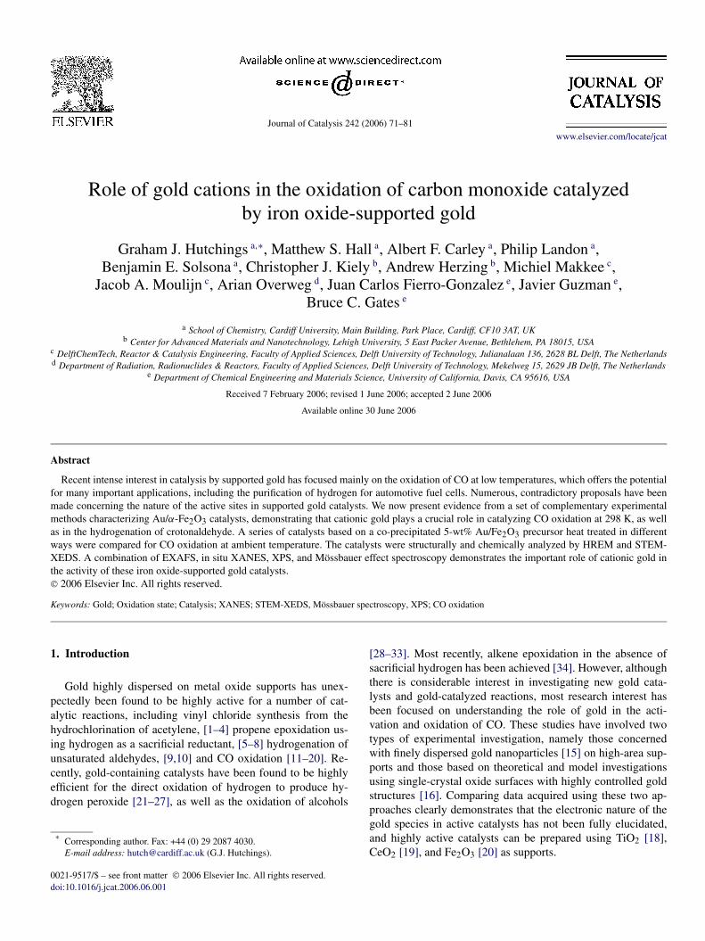
Fig. 1. Correlation between activity for CO conversion and Au(4d5/2) bindingenergy. The data for a series of catalysts (5-wt% Au supported on Fe2O3) areshown; the cluster of points labeled A corresponds to catalysts dried at 393 K,and the cluster B to those calcined at 673 K.
XPS data characterizing catalysts prepared using these twoheat treatment regimes showed a broad correlation between COconversion and Au(4d5/2) core-level binding energy (Fig. 1).These data clearly clustered into two groups. One group ofcatalyst samples dried at 393 K (labeled A) corresponded tohigh conversion (65–100%) and was generally associated witha higher Au(4d5/2) binding energy than the less active group(B), calcined at 673 K.
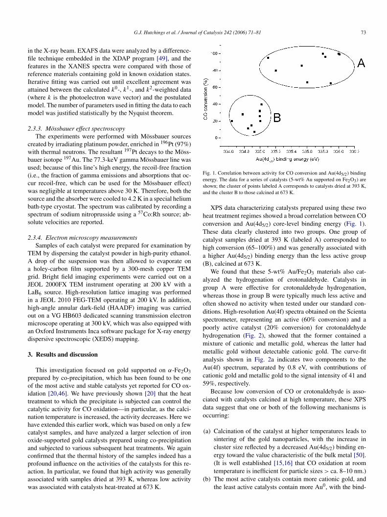
We found that these 5-wt% Au/Fe2O3 materials also cat-alyzed the hydrogenation of crotonaldehyde. Catalysts ingroup A were effective for crotonaldehyde hydrogenation,whereas those in group B were typically much less active andoften showed no activity when tested under our standard con-ditions. High-resolution Au(4f) spectra obtained on the Scientaspectrometer, representing an active (60% conversion) and apoorly active catalyst (20% conversion) for crotonaldehydehydrogenation (Fig. 2), showed that the former contained amixture of cationic and metallic gold, whereas the latter hadmetallic gold without detectable cationic gold. The curve-fitanalysis shown in Fig. 2a indicates two components to theAu(4f) spectrum, separated by 0.8 eV, with contributions ofcationic gold and metallic gold to the signal intensity of 41 and59%, respectively.
Because low conversion of CO or crotonaldehyde is asso-ciated with catalysts calcined at high temperature, these XPSdata suggest that one or both of the following mechanisms isoccurring:
(a) Calcination of the catalyst at higher temperatures leads tosintering of the gold nanoparticles, with the increase incluster size reflected by a decreased Au(4d5/2) binding en-ergy toward the value characteristic of the bulk metal [50].(It is well established [15,16] that CO oxidation at roomtemperature is inefficient for particle sizes > ca. 8–10 nm.)
(b) The most active catalysts contain more cationic gold, andthe least active catalysts contain more Au0, with the bind-
74 G.J. Hutchings et al. / Journal of Catalysis 242 (2006) 71–81
Fig. 2. High-resolution Au(4f) spectra obtained for a 5-wt% Au/Fe2O3 catalyst:(a) with a relatively high conversion (60%) for the hydrogenation of croton-aldehyde to crotyl alcohol, and (b) after calcination at 673 K, which reducedthe conversion to 20%.
ing energy difference observed arising from the differencein charges on the gold species.
But this issue cannot be resolved using just a single tech-nique, such as XPS. This limitation is particularly true forXPS, because final state effects associated with particle sizecan dominate the spectra [51–54]. We note that in principal,the initial state effects, reflecting the gold’s electronic nature,can be checked by measuring the Auger parameter, but that thismeasurement is difficult for gold because of the high kineticenergies (2016 and 2102 eV) of the relevant Auger feature. Weattempted to measure this Auger parameter for the catalysts inthis study, but this proved impossible for our sample array be-cause of the low Au concentration and consequent poor signalintensity. In an attempt to resolve this dilemma, we carefullychose a subset of samples and analyzed each using a set of com-plementary techniques; in all cases, only fresh samples wereevaluated.
Initially, we chose two representative samples of 5-wt%Au/α-Fe2O3, one dried at 393 K in flowing air for 8 h (de-noted as type A) and the other calcined in flowing air at 673 K(type B), corresponding to the high-activity and low-activity re-gions identified in Fig. 1. We evaluated these samples using awide range of techniques to determine the nature of the activegold species. We characterized the gold in the catalysts with ex-tended X-ray absorption fine structure spectroscopy (EXAFS),high-resolution transmission electron microscopy (HRTEM),and scanning transmission electron microscopy (STEM) withXEDS mapping to determine gold cluster sizes. We appliedthese techniques in concert with X-ray absorption near-edgestructure (XANES) and XPS, which provide information aboutthe gold oxidation states.
The STEM (Fig. 3) and EXAFS (Fig. 4; Table 2) data pro-vide clear evidence of particles of gold in the catalysts. TheEXAFS data (Fig. 4; Table 2) provide only average structuralinformation about these gold nanoparticles, showing a differ-
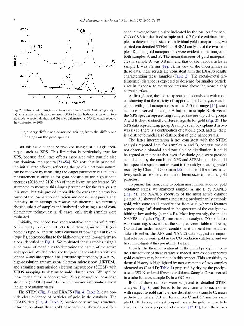
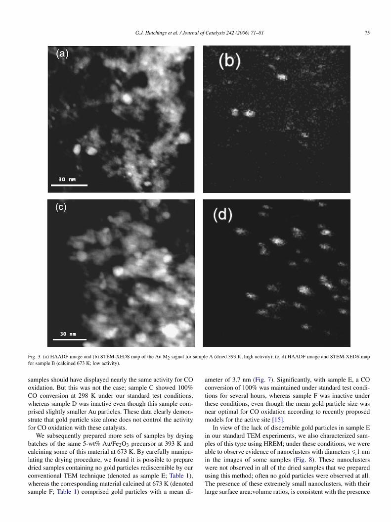
ence in average particle size indicated by the Au–Au first-shellCNs of 8.3 for the dried sample and 10.7 for the calcined sam-ple. To determine the sizes of individual gold nanoparticles, wecarried out detailed STEM and HREM analyses of the two sam-ples. Distinct gold nanoparticles were evident in the images ofboth samples A and B. The mean diameter of gold nanoparti-cles in sample A was 3.8 nm, and that of the nanoparticles insample B was 8.2 nm (Fig. 3). In view of the uncertainties inthese data, these results are consistent with the EXAFS resultscharacterizing these samples (Table 2). The metal–metal (in-teratomic) distance is expected to decrease for smaller particlesizes in response to the vapor pressure above the more highlycurved surface.
At first glance, these data appear to be consistent with mod-els showing that the activity of supported gold catalysts is asso-ciated with gold nanoparticles in the 2–5 nm range [15], suchas those observed in sample A but not in sample B. However,the XPS spectra representing samples that are typical of groupsA and B show distinctly different signals for gold (Fig. 2). TheXPS data representing group A samples can be explained in twoways: (1) There is a contribution of cationic gold, and (2) thereis a distinct bimodal size distribution of gold nanocrystals.
The latter interpretation is not consistent with the STEManalysis reported here for samples A and B, because we didnot observe a bimodal gold particle size distribution. It couldbe argued at this point that even if cationic gold were present,as indicated by the combined XPS and STEM data, this couldbe a spectator species not relevant to the catalysis, as suggestedrecently by Chen and Goodman [55], and the differences in ac-tivity could arise solely from the different sizes of metallic goldparticles.
To pursue this issue, and to obtain more information on goldoxidation states, we analyzed samples A and B by XANES(Fig. 5). The XANES spectrum of the most active catalyst(sample A) showed features indicating predominantly cationicgold, with some small contribution from Au0, whereas featuresrepresenting Au0 dominated in the spectrum of the catalyst ex-hibiting low activity (sample B). Most importantly, the in situXANES analysis (Fig. 5), measured as catalytic CO oxidationwas occurring, showed that the samples were stable in flowingCO and air under reaction conditions at ambient temperature.Taken together, the XPS and XANES data suggest an impor-tant role for cationic gold in the CO oxidation catalysis, and wehave investigated this possibility further.
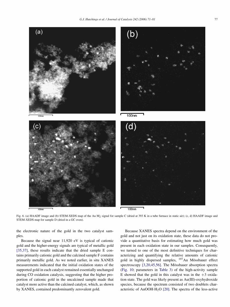
Clearly, the thermal treatment of the initial precipitate con-trols the activity of these catalysts; indeed, iron oxide-supportedgold catalysts may be unique in this respect. This sensitivity tothermal history is highlighted by measurements of two samples(denoted as C and D; Table 1) prepared by drying the precipi-tate at 393 K under different conditions. Sample C was treatedin a tube furnace; sample D, in a GC oven.
Both of these samples were subjected to detailed STEManalysis (Fig. 6) and found to be very similar to each otherwith respect to gold particle size distributions (sample C, meanparticle diameters, 7.0 nm for sample C and 5.4 nm for sam-ple D). If the key catalyst property were the gold nanoparticlesize, as has been proposed elsewhere [12,15], then these two
G.J. Hutchings et al. / Journal of Catalysis 242 (2006) 71–81 75
Fig. 3. (a) HAADF image and (b) STEM-XEDS map of the Au M2 signal for sample A (dried 393 K; high activity); (c, d) HAADF image and STEM-XEDS mapfor sample B (calcined 673 K; low activity).
samples should have displayed nearly the same activity for COoxidation. But this was not the case; sample C showed 100%CO conversion at 298 K under our standard test conditions,whereas sample D was inactive even though this sample com-prised slightly smaller Au particles. These data clearly demon-strate that gold particle size alone does not control the activityfor CO oxidation with these catalysts.
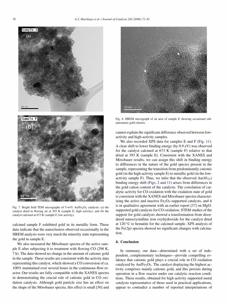
We subsequently prepared more sets of samples by dryingbatches of the same 5-wt% Au/Fe2O3 precursor at 393 K andcalcining some of this material at 673 K. By carefully manipu-lating the drying procedure, we found it is possible to preparedried samples containing no gold particles rediscernible by ourconventional TEM technique (denoted as sample E; Table 1),whereas the corresponding material calcined at 673 K (denotedsample F; Table 1) comprised gold particles with a mean di-
ameter of 3.7 nm (Fig. 7). Significantly, with sample E, a COconversion of 100% was maintained under standard test condi-tions for several hours, whereas sample F was inactive underthese conditions, even though the mean gold particle size wasnear optimal for CO oxidation according to recently proposedmodels for the active site [15].
In view of the lack of discernible gold particles in sample Ein our standard TEM experiments, we also characterized sam-ples of this type using HREM; under these conditions, we wereable to observe evidence of nanoclusters with diameters �1 nmin the images of some samples (Fig. 8). These nanoclusterswere not observed in all of the dried samples that we preparedusing this method; often no gold particles were observed at all.The presence of these extremely small nanoclusters, with theirlarge surface area:volume ratios, is consistent with the presence
76 G.J. Hutchings et al. / Journal of Catalysis 242 (2006) 71–81
(a) (b)
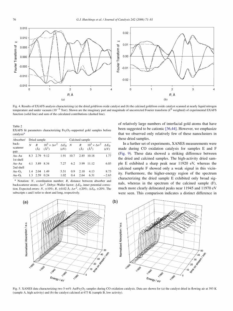
Fig. 4. Results of EXAFS analysis characterizing (a) the dried gold/iron oxide catalyst and (b) the calcined gold/iron oxide catalyst scanned at nearly liquid nitrogentemperature and under vacuum (10−5 Torr). Shown are the imaginary part and magnitude of uncorrected Fourier transform (k0 weighted) of experimental EXAFSfunction (solid line) and sum of the calculated contributions (dashed line).
Table 2EXAFS fit parameters characterizing Fe2O3-supported gold samples beforecatalysisa
Absorber/back-scattererpair
Dried sample Calcined sample
N R
(Å)103 ×�σ 2
(Å2)�E0(eV)
N R
(Å)103 ×�σ 2
(Å2)�E0(eV)
Au–Au1st shell
8.3 2.79 9.12 1.91 10.7 2.85 10.18 1.77
Au–Au2nd shell
4.1 3.89 8.34 7.27 6.2 3.99 11.12 6.03
Au–Os 1.4 2.04 1.49 5.51 0.9 2.10 4.13 8.73Au–Ol 1.3 2.59 0.24 1.02 0.4 2.64 6.31 −2.63
a Notation: N , coordination number; R, distance between absorber andbackscatterer atoms; �σ 2, Debye–Waller factor; �E0, inner potential correc-tion. Expected errors: N , ±10%; R, ±0.02 Å; �σ 2, ±20%; �E0, ±20%. Thesubscripts s and l refer to short and long, respectively.
of relatively large numbers of interfacial gold atoms that havebeen suggested to be cationic [36,44]. However, we emphasizethat we observed only relatively few of these nanoclusters inthese dried samples.
In a further set of experiments, XANES measurements weremade during CO oxidation catalysis for samples E and F(Fig. 9). These data showed a striking difference betweenthe dried and calcined samples. The high-activity dried sam-ple E exhibited a sharp peak near 11920 eV, whereas thecalcined sample F showed only a weak signal in this vicin-ity. Furthermore, the higher-energy region of the spectrumcharacterizing the dried sample E exhibited only broad sig-nals, whereas in the spectrum of the calcined sample (F),much more clearly delineated peaks near 11945 and 11970 eVwere seen. This comparison indicates a distinct difference in
Fig. 5. XANES data characterizing two 5-wt% Au/Fe2O3 samples during CO oxidation catalysis. Data are shown for (a) the catalyst dried in flowing air at 393 K(sample A, high activity) and (b) the catalyst calcined at 673 K (sample B, low activity).
G.J. Hutchings et al. / Journal of Catalysis 242 (2006) 71–81 77
Fig. 6. (a) HAADF image and (b) STEM-XEDS map of the Au M2 signal for sample C (dried at 393 K in a tube furnace in static air); (c, d) HAADF image andSTEM-XEDS map for sample D (dried in a GC oven).
the electronic nature of the gold in the two catalyst sam-ples.
Because the signal near 11,920 eV is typical of cationicgold and the higher-energy signals are typical of metallic gold[35,37], these results indicate that the dried sample E con-tains primarily cationic gold and the calcined sample F containsprimarily metallic gold. As we noted earlier, in situ XANESmeasurements indicated that the initial oxidation states of thesupported gold in each catalyst remained essentially unchangedduring CO oxidation catalysis, suggesting that the higher pro-portion of cationic gold in the uncalcined sample made thatcatalyst more active than the calcined catalyst, which, as shownby XANES, contained predominantly zerovalent gold.
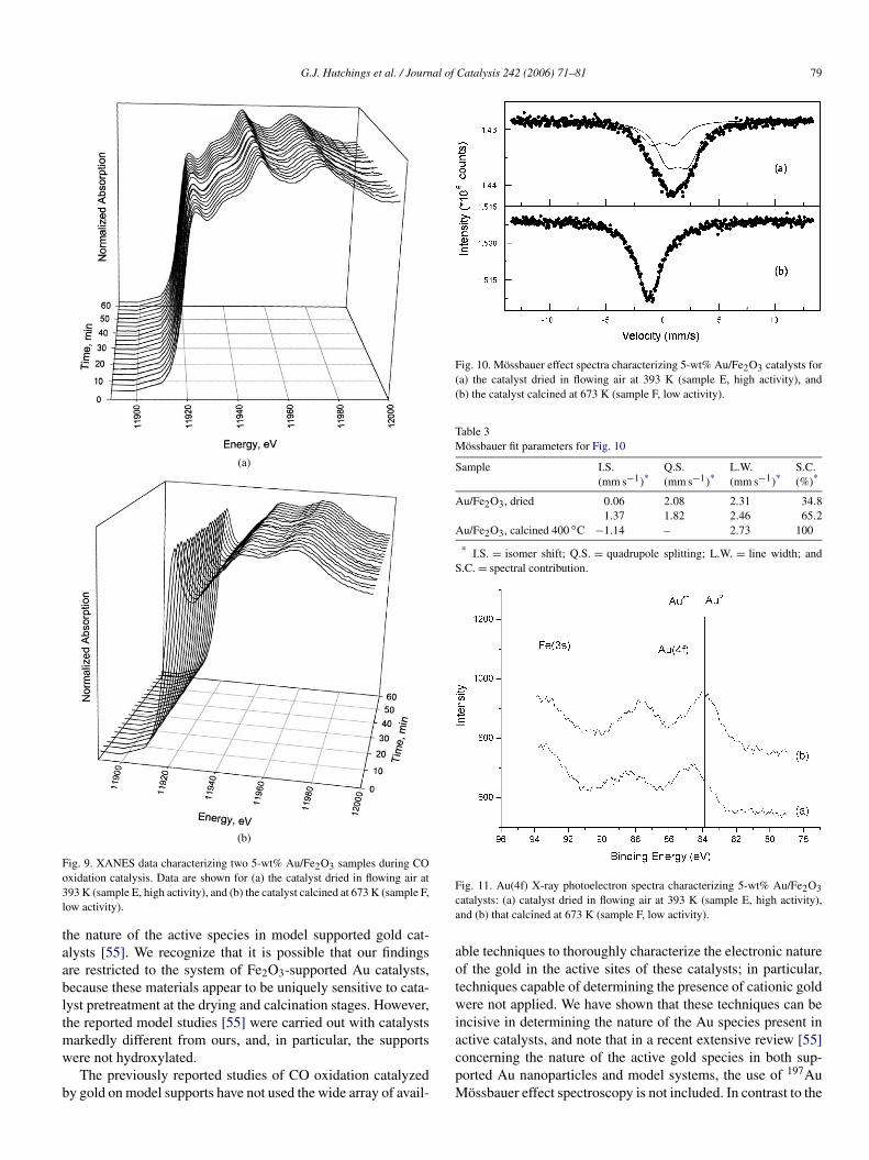
Because XANES spectra depend on the environment of thegold and not just on its oxidation state, these data do not pro-vide a quantitative basis for estimating how much gold waspresent in each oxidation state in our samples. Consequently,we turned to one of the most definitive techniques for char-acterizing and quantifying the relative amounts of cationicgold in highly dispersed samples, 197Au Mössbauer effectspectroscopy [3,20,45,56]. The Mössbauer absorption spectra(Fig. 10; parameters in Table 3) of the high-activity sampleE showed that the gold in this catalyst was in the +3 oxida-tion state. The gold was likely present as Au(III)-oxyhydroxidespecies, because the spectrum consisted of two doublets char-acteristic of AuOOH·H2O [20]. The spectra of the less-active
78 G.J. Hutchings et al. / Journal of Catalysis 242 (2006) 71–81
Fig. 7. Bright field TEM micrographs of 5-wt% Au/Fe2O3 catalysts: (a) thecatalyst dried in flowing air at 393 K (sample E, high activity), and (b) thecatalyst calcined at 673 K (sample F, low activity).
calcined sample F exhibited gold in its metallic form. Thesedata indicate that the nanoclusters observed occasionally in theHREM analysis were very much the minority state representingthe gold in sample E.
We also measured the Mössbauer spectra of the active sam-ple E after subjecting it to treatment with flowing CO (298 K,7 h). The data showed no change in the amount of cationic goldin the sample. These results are consistent with the activity datarepresenting this catalyst, which showed a CO conversion of ca.100% maintained over several hours in the continuous-flow re-actor. Our results are fully compatible with the XANES spectrain demonstrating the crucial role of cationic gold in CO oxi-dation catalysis. Although gold particle size has an effect onthe shape of the Mössbauer spectra, this effect is small [56] and
Fig. 8. HREM micrograph of an area of sample E showing occasional sub-nanometer gold clusters.
cannot explain the significant difference observed between low-activity and high-activity samples.
We also recorded XPS data for samples E and F (Fig. 11).A clear shift to lower binding energy (by 0.9 eV) was observedfor the catalyst calcined at 673 K (sample F) relative to thatdried at 393 K (sample E). Consistent with the XANES andMössbauer results, we can assign this shift in binding energyto differences in the nature of the gold species present in thesample, representing the transition from predominantly cationicgold (in the high-activity sample E) to metallic gold (in the low-activity sample F). Thus, we infer that the observed Au(4f7/2)binding energy shift (Figs. 2 and 11) arises from differences inthe gold cation content of the catalysts. The correlation of cat-alytic activity for CO oxidation with the oxidation state of goldis consistent with the XANES and Mössbauer spectra character-izing the active and inactive Fe2O3-supported catalysts, and itis in qualitative agreement with an earlier report [37] on MgO-supported gold catalysts for CO oxidation. STEM studies of thesupport for gold catalysts showed a transformation from disor-dered nanocrystalline iron oxyhydroxide for the catalyst driedat 120 ◦C to hematite for the calcined sample. XPS analysis ofthe Fe(2p) spectra showed no significant changes with calcina-tion.
4. Conclusion
In summary, our data—determined with a set of inde-pendent, complementary techniques—provide compelling ev-idence that cationic gold plays a crucial role in CO oxidationcatalyzed by Au/Fe2O3. The catalyst displaying the highest ac-tivity comprises mainly cationic gold, and this persists duringoperation in a flow reactor under our catalytic reaction condi-tions. These results, obtained for high-activity supported metalcatalysts representative of those used in practical applications,appear to contradict a number of reported interpretations of
G.J. Hutchings et al. / Journal of Catalysis 242 (2006) 71–81 79
(a)
(b)
Fig. 9. XANES data characterizing two 5-wt% Au/Fe2O3 samples during COoxidation catalysis. Data are shown for (a) the catalyst dried in flowing air at393 K (sample E, high activity), and (b) the catalyst calcined at 673 K (sample F,low activity).
the nature of the active species in model supported gold cat-alysts [55]. We recognize that it is possible that our findingsare restricted to the system of Fe2O3-supported Au catalysts,because these materials appear to be uniquely sensitive to cata-lyst pretreatment at the drying and calcination stages. However,the reported model studies [55] were carried out with catalystsmarkedly different from ours, and, in particular, the supportswere not hydroxylated.
The previously reported studies of CO oxidation catalyzedby gold on model supports have not used the wide array of avail-
Fig. 10. Mössbauer effect spectra characterizing 5-wt% Au/Fe2O3 catalysts for(a) the catalyst dried in flowing air at 393 K (sample E, high activity), and(b) the catalyst calcined at 673 K (sample F, low activity).
Table 3Mössbauer fit parameters for Fig. 10
Sample I.S.(mm s−1)*
Q.S.(mm s−1)*
L.W.(mm s−1)*
S.C.(%)*
Au/Fe2O3, dried 0.06 2.08 2.31 34.81.37 1.82 2.46 65.2
Au/Fe2O3, calcined 400 ◦C −1.14 – 2.73 100
* I.S. = isomer shift; Q.S. = quadrupole splitting; L.W. = line width; andS.C. = spectral contribution.
Fig. 11. Au(4f) X-ray photoelectron spectra characterizing 5-wt% Au/Fe2O3catalysts: (a) catalyst dried in flowing air at 393 K (sample E, high activity),and (b) that calcined at 673 K (sample F, low activity).
able techniques to thoroughly characterize the electronic natureof the gold in the active sites of these catalysts; in particular,techniques capable of determining the presence of cationic goldwere not applied. We have shown that these techniques can beincisive in determining the nature of the Au species present inactive catalysts, and note that in a recent extensive review [55]concerning the nature of the active gold species in both sup-ported Au nanoparticles and model systems, the use of 197AuMössbauer effect spectroscopy is not included. In contrast to the
80 G.J. Hutchings et al. / Journal of Catalysis 242 (2006) 71–81
investigations of model systems, we examined catalysts withhigh activities using a wide range of available techniques, andour data clearly show that gold is present in both metallic andcationic forms in the Au/Fe2O3 samples, and that the presenceof a significant fraction of cationic gold is essential for high COoxidation activity. Moreover, our results demonstrate the com-plexity of a realistic high-area gold catalyst compared with thestructurally less-complex model catalysts. Although investiga-tions of such model systems can provide valuable data, theymay fall short of permitting a thorough exploration of the sub-tlety of high-activity supported catalysts of the kind that findtechnological application. Indeed, in this investigation we haveclearly demonstrated the complexity of Au/Fe2O3 catalysts;many different structures can be prepared, and only by using arange of spectroscopic and other techniques can this complexitycan be unraveled. Our key observation is that the high activityobserved by many researchers for CO oxidation at 298 K withAu/Fe2O3 catalysts is predicated on the presence of cationicgold that persists under these reaction conditions for extendedperiods.
Acknowledgments
The authors acknowledge the National Synchrotron LightSource, Brookhaven National Laboratory, which is supportedby the US Department of Energy, Division of Materials Sci-ences and Division of Chemical Sciences, under contract DE-AC02-98CH10886, and the staff of Beamline X-18B. The re-search at the University of California was supported by theUS Department of Energy (grant FG02-04ER15513) and theNational Science Foundation (grant CTS-0121619). The re-search at Cardiff University was supported by the Science andEngineering Research Council and Johnson-Matthey (projectATHENA), the Science and Engineering Research Council(Ph.D. studentship), and the European Union (project AURI-CAT; contract HPRN-CT-2002-00174). The authors thank thestaff at the National Centre for Electron Spectroscopy and Sur-face Analysis, Daresbury, UK, as well as the generous supportof the National Science Foundation Materials Research Scienceand Engineering Center at Lehigh University (grant NSF DMR-0079996).
References
[1] G.J. Hutchings, J. Catal. 96 (1985) 292.[2] B. Nkosi, N.J. Coville, G.J. Hutchings, Appl. Catal. 43 (1988) 33.[3] B. Nkosi, M.D. Adams, N.J. Coville, G.J. Hutchings, J. Catal. 128 (1991)
378.[4] B. Nkosi, N.J. Coville, G.J. Hutchings, M.D. Adams, J. Friedl, F. Wagner,
J. Catal. 128 (1991) 366.[5] T. Hayashi, K. Tanaka, M. Haruta, J. Catal. 178 (1998) 566.[6] T.A. Nijhuis, H. Huizinga, M. Makkee, J.A. Moulijn, Ind. Eng. Chem.
Res. 38 (1999) 884.[7] E.E. Stangland, K.B. Stevens, R.P. Andres, W.N. Delgass, J. Catal. 191
(2000) 332.[8] G. Mul, A. Zwijnenburg, B. Linden, M. van der Makkee, J.A. Moulijn,
J. Catal. 201 (2001) 128.[9] M. Haruta, Catal. Today 36 (1997) 123.
[10] J.E. Bailie, G.J. Hutchings, Chem. Commun. (1999) 2151.
[11] G.C. Bond, D.T. Thompson, Catal. Rev.-Sci. Eng. 41 (1999)319.
[12] G.C. Bond, D.T. Thompson, Gold Bull. 33 (2000) 41.[13] M. Haruta, CATTECH 6 (2002) 102.[14] M. Haruta, M. Date, Appl. Catal. A 222 (2001) 427.[15] M. Haruta, Gold Bull. 37 (2004) 27.[16] R. Meyer, C. Lemaire, Sh.K. Shaikutdinov, H.-J. Freund, Gold Bull. 37
(2004) 72.[17] A.S.K. Hashmi, Gold Bull. 37 (2004) 51.[18] M. Date, M. Okumura, S. Tsubota, M. Haruta, Angew. Chem. Int. Ed. 43
(2004) 2129.[19] S. Carrettin, P. Concepcion, A. Corma, J.M. Lopez Nieto, V.F. Puntes,
Angew. Chem. Int. Ed. 43 (2004) 2538.[20] R.M. Finch, N.A. Hodge, G.J. Hutchings, A. Meagher, Q.A. Pankhurst,
M.R.H. Siddiqui, F.E. Wagner, R. Whyman, Phys. Chem. Chem. Phys. 1(1999) 485.
[21] D.T. Thompson, Appl. Catal. A 243 (2003) 201.[22] G.J. Hutchings, Gold Bull. 29 (1996) 123.[23] P. Landon, P.J. Collier, A.J. Papworth, C.J. Kiely, G.J. Hutchings, Chem.
Commun. (2002) 2058.[24] P. Landon, P.J. Collier, A.F. Carley, D. Chadwick, A.J. Papworth, A. Bur-
rows, C.J. Kiely, G.J. Hutchings, Phys. Chem. Chem. Phys. 5 (2003) 1917.[25] J.K. Edwards, B. Solsona, P. Landon, A.F. Carley, A. Herzing, M. Watan-
abe, C.J. Kiely, G.J. Hutchings, J. Mater. Chem. 15 (2005) 4595.[26] J.K. Edwards, B.E. Solsona, P. Landon, A.F. Carley, A. Herzing, C.J.
Kiely, G.J. Hutchings, J. Catal. 236 (2005) 69.[27] T. Ishihara, Y. Ohura, S. Yoshida, Y. Hata, H. Nishiguchi, Y. Takita, Appl.
Catal. A 291 (2005) 215.[28] L. Prati, M. Rossi, J. Catal. 176 (1998) 552.[29] F. Porta, L. Prati, M. Rossi, S. Colluccia, G. Martra, Catal. Today 61
(2000) 165.[30] C. Bianchi, F. Porta, L. Prati, M. Rossi, Top. Catal. 13 (2000) 231.[31] L. Prati, Gold Bull. 32 (1999) 96.[32] S. Carrettin, P. McMorn, P. Johnston, K. Griffin, G.J. Hutchings, Chem.
Commun. (2002) 696.[33] S. Carrettin, P. McMorn, P. Johnston, K. Griffin, C.J. Kiely, G.J. Hutch-
ings, Phys. Chem. Chem. Phys. 5 (2003) 1329.[34] M.D. Hughes, Y.-J. Xu, P. Jenkins, P. McMorn, P. Landon, A.F. Carley,
G.A. Attard, G.J. Hutchings, F. King, E.H. Stitt, P. Johnston, K. Griffin,C.J. Kiely, Nature 437 (2005) 1132.
[35] J.C. Fierro-Gonzalez, B.C. Gates, J. Phys. Chem. B 108 (2004) 16999.[36] J.C. Fierro-Gonzalez, V.A. Bhirud, B.C. Gates, Chem. Commun. (2005)
5275.[37] J. Guzman, B.C. Gates, J. Am. Chem. Soc. 126 (2004) 2672.[38] Q. Fu, H. Saltsburg, M. Flytzani-Stephanopoulos, Science 301 (2003) 935.[39] M. Valden, X. Lai, D.W. Goodman, Science 281 (1998) 1647.[40] M.S. Chen, D.W. Goodman, Science 306 (2004) 252.[41] D.C. Maeir, D.W. Goodman, J. Am. Chem. Soc. 126 (2004) 1892.[42] V.A. Bondzie, S.C. Parker, C.T. Campbell, Catal. Lett. 63 (1999) 143.[43] A. Sanchez, S. Abbet, U. Heiz, W.-D. Schneider, H. Häkkinen, R.N. Bar-
nett, U. Landman, J. Phys. Chem. A 103 (1999) 9573.[44] N. Lopez, J.K. Nørskov, J. Am. Chem. Soc. 124 (2002) 11262.[45] J. Guzman, S. Carrettin, J.C. Fierro-Gonzalez, Y. Hao, B.C. Gates, A. Cor-
ma, Angew. Chem. Int. Ed. 44 (2005) 4778.[46] N.A. Hodge, C.J. Kiely, R. Whyman, M.R.H. Siddiqui, G.J. Hutchings,
Q.A. Pankhurst, F.E. Wagner, R.R. Rajaram, S.E. Golunski, Catal. To-day 72 (2002) 133.
[47] R.E. Jentoft, S.E. Deutsch, B.C. Gates, Rev. Sci. Instrum. 67 (1996) 2111.[48] J.F. Odzak, A.M. Argo, F.S. Lai, B.C. Gates, K. Pandya, L. Feraria, Rev.
Sci. Instrum. 72 (2001) 3943.[49] M. Vaarkamp, J.C. Linders, D.C. Koningsberger, Physica B 209 (1995)
159.[50] A.F. Carley, M.K. Rajumon, M.W. Roberts, J. Solid State Chem. 106
(1993) 156.[51] M.G. Mason, Phys. Rev. B 27 (1983) 748.[52] G.K. Wertheim, S.B. Di Cenzo, S.E. Youngquist, Phys. Rev. Lett. 51
(1983) 2310.

G.J. Hutchings et al. / Journal of Catalysis 242 (2006) 71–81 81
[53] S.B. Di Cenzo, G.K. Wertheim, Comments Solid State Phys. 11 (1985)203.
[54] C.C. Chusuei, X. Lai, K.I. Luo, D.W. Goodman, Top. Catal. 14 (2001) 71.
[55] M.S. Chen, D.W. Goodman, Catal. Today 111 (2006) 22.[56] L. Stievano, S. Santucci, L. Lozzi, S. Calogero, F.E. Wagner, J. Non-Cryst.
Solids 232–234 (1998) 644.


