Embed Size (px)
Citation preview
Role of Bacterial Exopolysaccharides (EPS) in the Fate ofthe Oil Released during the Deepwater Horizon Oil SpillTony Gutierrez*,1,2, David Berry3, Tingting Yang4, Sara Mishamandani1, Luke McKay4, Andreas Teske4,Michael D. Aitken1
1 Department of Environmental Sciences and Engineering, Gillings School of Global Public Health, University of North Carolina, Chapel Hill, North Carolina,United States of America, 2 School of Life Sciences, Heriot-Watt University, Edinburgh, United Kingdom, 3 Department of Microbial Ecology, Vienna EcologyCentre, Faculty of Sciences, University of Vienna, Vienna, Austria, 4 Department of Marine Sciences, University of North Carolina, Chapel Hill, North Carolina,United States of America
Abstract
Halomonas species are recognized for producing exopolysaccharides (EPS) exhibiting amphiphilic properties thatallow these macromolecules to interface with hydrophobic substrates, such as hydrocarbons. There remains apaucity of knowledge, however, on the potential of Halomonas EPS to influence the biodegradation of hydrocarbons.In this study, the well-characterized amphiphilic EPS produced by Halomonas species strain TG39 was shown toeffectively increase the solubilization of aromatic hydrocarbons and enhance their biodegradation by an indigenousmicrobial community from oil-contaminated surface waters collected during the active phase of the DeepwaterHorizon oil spill. Three Halomonas strains were isolated from the Deepwater Horizon site, all of which produced EPSwith excellent emulsifying qualities and shared high (97-100%) 16S rRNA sequence identity with strain TG39 andother EPS-producing Halomonas strains. Analysis of pyrosequence data from surface water samples collected duringthe spill revealed several distinct Halomonas phylotypes, of which some shared a high sequence identity (≥97%) tostrain TG39 and the Gulf spill isolates. Other bacterial groups comprising members with well-characterized EPS-producing qualities, such as Alteromonas, Colwellia and Pseudoalteromonas, were also found enriched in surfacewaters, suggesting that the total pool of EPS in the Gulf during the spill may have been supplemented by theseorganisms. Roller bottle incubations with one of the Halomonas isolates from the Deepwater Horizon spill sitedemonstrated its ability to effectively produce oil aggregates and emulsify the oil. The enrichment of EPS-producingbacteria during the spill coupled with their capacity to produce amphiphilic EPS is likely to have contributed to theultimate removal of the oil and to the formation of oil aggregates, which were a dominant feature observed incontaminated surface waters.
Citation: Gutierrez T, Berry D, Yang T, Mishamandani S, McKay L, et al. (2013) Role of Bacterial Exopolysaccharides (EPS) in the Fate of the OilReleased during the Deepwater Horizon Oil Spill. PLoS ONE 8(6): e67717. doi:10.1371/journal.pone.0067717
Editor: Wei-Chun Chin, University of California, Merced, United States of America
Received February 1, 2013; Accepted May 21, 2013; Published June 27, 2013
Copyright: © 2013 Gutierrez et al. This is an open-access article distributed under the terms of the Creative Commons Attribution License, which permitsunrestricted use, distribution, and reproduction in any medium, provided the original author and source are credited.
Funding: This work was supported by a Marie Curie International Outgoing Fellowship (PIOF-GA-2008-220129) within the 7th European CommunityFramework Programme. Partial support was also provided through the U.S. National Institute of Environmental Health Sciences grant 5 P42ES005948,and through the BP/Gulf of Mexico Research Initiative to support consortium research entitled “Ecosystem Impacts of Oil and Gas Inputs to the Gulf(ECOGIG)” administered by the University of Mississippi. Andreas Teske, Tony Gutierrez and Luke McKay were also supported by NSF (RAPIDResponse: The microbial response to the Deepwater Horizon Oil Spill; NSF-OCE 1045115). The funders had no role in study design, data collection andanalysis, decision to publish, or preparation of the manuscript.
Competing interests: The authors have declared that no competing interests exist.
* E-mail: [email protected]
Introduction
Dissolved organic matter (DOM) in the ocean is the largestand possibly least understood pool of carbon - ca. 6.9 x 10¹⁷ gC, which is comparable in mass to the carbon in atmosphericCO2 [1]. Much of this DOM exists as biopolymers (ca. 10-25%of total oceanic DOM) that undergo reversible transitionbetween colloidal and dissolved phases [2,3]. A major fractionof marine DOM derives from the synthesis and release ofextracellular polymeric substances or exopolysaccharides(EPS) by bacteria and eukaryotic phytoplankton [4,5]. These
are high molecular weight polymers composed mainly ofmonosaccharides, some of which may contain non-carbohydrate substituents. Several studies have reported largequantities of these bacterial polymers in Antarctic marineenvironments and around hydrothermal vents, where they arethought to complex with various metal ions and contribute totheir mobility and entry into the food web [6 and referencestherein]. The negative charge associated with carboxyl groupsof uronic acids of EPS has been implicated in the capacity ofthese macromolecules to complex with transition metals [7,8].EPS from marine bacteria, in particular, are recognized for
PLOS ONE | www.plosone.org 1 June 2013 | Volume 8 | Issue 6 | e67717
having much higher levels of uronic acids [9] compared to thatfound in EPS produced by marine eukaryotic phytoplankton [7]and non-marine bacteria [10]. There is a growing body ofevidence to implicate uronic acids of EPS in conferring thesemacromolecules with an ability to interface with hydrophobicorganic chemicals, such as hydrocarbons [11]–[13]. Aminoacids and peptides are also often found associated with marinebacterial EPS, which can confer amphiphilic characteristics tothese polymers [4,11,14]. The potential significance of marinebacterial EPS to influencing the fate and ultimate degradationof hydrocarbon pollutants in the ocean, particularly during oilspills, remains largely unknown.
The explosion and sinking of the Deepwater Horizon oil rigon April 20, 2010, led to the release of massive quantities ofcrude oil into the northern Gulf of Mexico, resulting in what hasbeen deemed the worst accidental marine oil spill in the historyof the oil and gas industry. The response and complex shiftsobserved in the microbial communities within and adjacent towaters impacted by the spill have been reported in severalpublications. Notably, the enrichment of a specific cluster ofbacteria within the Order Oceanospirillales – organismsassociated with aliphatic hydrocarbon degradation – was foundinitially (late May 2010) dominating an oil plume [15] that hadbeen identified ca. 1000 and 1300 m depth [16]. Dominance ofthe Oceanospirillales in the plume was succeeded by membersaffiliated to Colwellia and Cycloclasticus by early June 2010[17]. This succession from aliphatic hydrocarbon-degradingbacteria (Oceanospirillales) to obligate degraders of aromatichydrocarbons (Cycloclasticus) and psychrophilic hydrocarbon-degrading generalists (Colwellia) closely resembles themicrobial dynamics observed in seawater following enrichmentwith crude oil [18]–[23]. Several other bacterial groupscomprising members with EPS-producing qualities, notablyHalomonas, Alteromonas and Pseudoalteromonas, were alsoenriched within the water column [15,17]. However, onedistinctive feature that has received little attention and remainspoorly understood was the observed formation of oilaggregates that were abundant on the surface and within deepwater oil plumes [24]. Oil aggregates can be broadly defined asfloating gelatinous or “fluffy” material, which, by nature of their“stickiness”, can contain embedded droplets of oil. Oilaggregates representing those formed under conditionssimulating the Gulf spill were shown to have a glycoproteincomposition – i.e. made up largely of carbohydrate and protein[25]. Bacteria can contribute large quantities of EPS to the totalDOM pool in the ocean [26], a large fraction of which can be ofglycoprotein composition [3,27]. We therefore hypothesizedthat EPS-producing bacteria enriched during the spill hadcontributed to the formation of oil aggregates, as well as to thefate of the oil by influencing the dissolution, bioavailability andultimate degradation of hydrocarbons by indigenous oil-degrading communities. Whilst oil-aggregate formation atDeepwater Horizon has been shown to be associated with theactivities of indigenous microbial communities [25,28], theidentity of key species involved in potentially triggering thisprocess remains unknown.
In addition to the enrichment of halomonads in the deepwater oil plume at the Gulf spill site [15], these organisms have
also been shown to become enriched in laboratory enrichmentsand in the field after exposure to hydrocarbons [29,32].Halomonas species are slight to moderately halophilic andoligotrophic organisms that are ubiquitous in marine andhypersaline environments [33], many of which are recognizedfor producing moderate to large quantities of EPS that canexhibit amphiphilic, or biosurfactant-like, properties. Severalreports have shown that these polymers effectively emulsifyhydrocarbons, crude oils or refined petroleum products[11,31,34]–[39]. In the marine environment, where the fate ofspilled oil is both poorly understood and of ecological concern,there is a paucity of knowledge regarding the capacity of EPSproduced by these and other EPS-producing bacteria toinfluence the degradation of hydrocarbons. In particular,polycyclic aromatic hydrocarbons (PAHs) are poorly solubleand generally less amenable to biodegradation compared totheir aliphatic counterparts. To circumvent limitations inhydrocarbon bioavailability, some microorganisms producebiosurfactants or bioemulsifiers (e.g. amphiphilic EPS) as amechanism to increase the bioavailability of these compoundsfor biodegradation.
In this study, we report on the isolation of three EPS-producing strains of Halomonas from oil-contaminated surfacewaters during the active phase of the Deepwater Horizon oilspill. Two of these isolates, designated strains TGOS-10 andGOS-2, shared 100% 16S rRNA sequence identity to twopreviously described EPS-producing halomonads –respectively Halomonas sp. strain TG39 and TG67 [37]. Thechemical composition and associated functional properties ofthe EPS produced by strain TG39 has been extensively studied[11,37], and therefore served as a model to infer the potentialinfluence of in situ bacterial-produced EPS on the fate of the oilreleased by the Deepwater Horizon blowout. We present newdata on the capacity of Halomonas EPS to influence thedissolution of aromatic hydrocarbons and enhance thebioavailability of these compounds and rate of biodegradationby the in situ sea surface microbial community in the Gulf ofMexico. Using a roller-bottle experimental design, we alsoprovide empirical evidence that implicates indigenous EPS-producing halomonads from Gulf of Mexico waters to havingplayed a role in the formation of oil aggregates during theDeepwater Horizon oil spill.
Materials and Methods
Microorganisms and field samplesHalomonas sp. strain TG39 was previously isolated from a
laboratory culture of an unclassified marine Chrysophyte(CCAP958/1) based on its growth on n-hexadecane as a solecarbon and energy source [37]. The strain was routinely grownon a marine broth (ZM/10) composed of ¾-strength naturallyaged seawater, peptone (0.05%), yeast extract (0.01%), andsupplemented after autoclaving with filter-sterile (0.2 µm) traceelements and vitamins to final concentrations as previouslydescribed [40]. A synthetic seawater medium, ONR7a [41], wasused to assess the strain’s ability to grow on or mineralizevarious hydrocarbons.
Role of Bacterial EPS in Oil-Contaminated Seawater
PLOS ONE | www.plosone.org 2 June 2013 | Volume 8 | Issue 6 | e67717
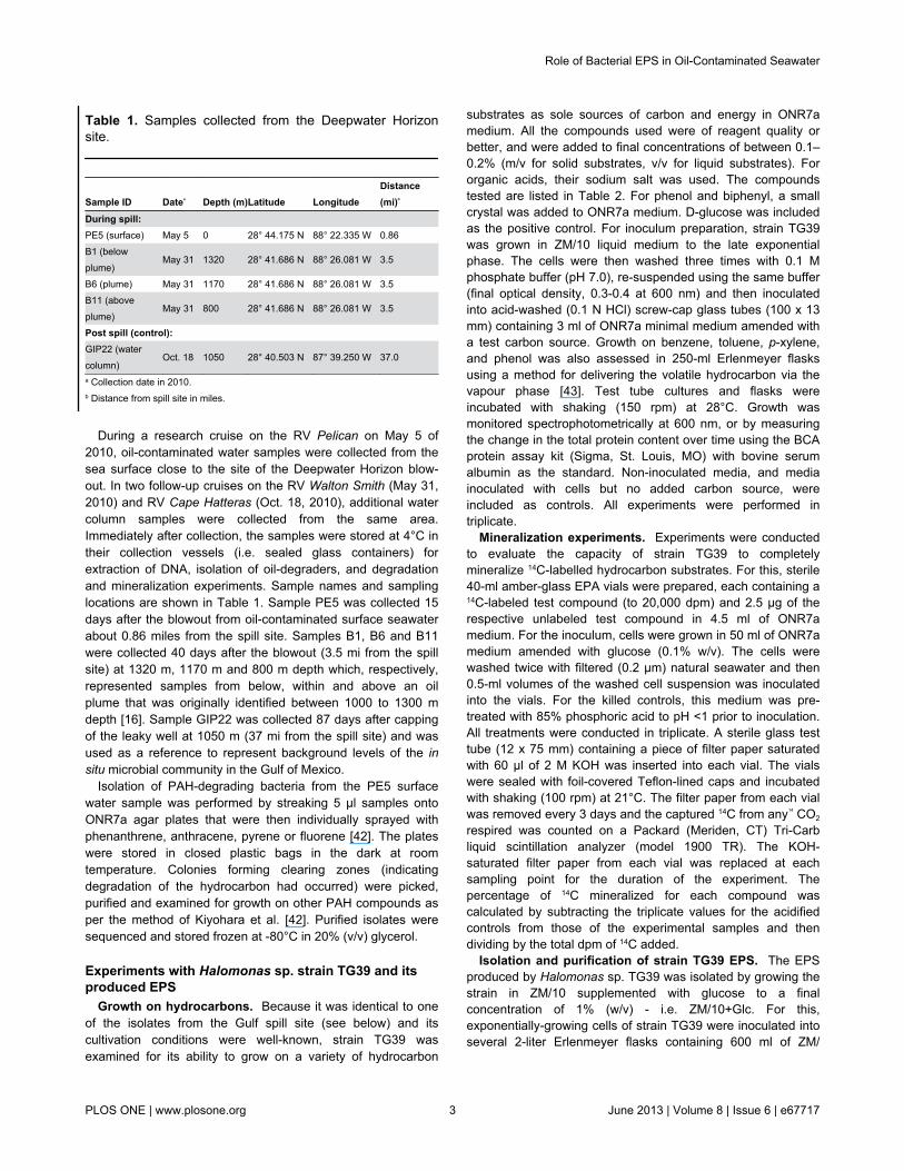
During a research cruise on the RV Pelican on May 5 of2010, oil-contaminated water samples were collected from thesea surface close to the site of the Deepwater Horizon blow-out. In two follow-up cruises on the RV Walton Smith (May 31,2010) and RV Cape Hatteras (Oct. 18, 2010), additional watercolumn samples were collected from the same area.Immediately after collection, the samples were stored at 4°C intheir collection vessels (i.e. sealed glass containers) forextraction of DNA, isolation of oil-degraders, and degradationand mineralization experiments. Sample names and samplinglocations are shown in Table 1. Sample PE5 was collected 15days after the blowout from oil-contaminated surface seawaterabout 0.86 miles from the spill site. Samples B1, B6 and B11were collected 40 days after the blowout (3.5 mi from the spillsite) at 1320 m, 1170 m and 800 m depth which, respectively,represented samples from below, within and above an oilplume that was originally identified between 1000 to 1300 mdepth [16]. Sample GIP22 was collected 87 days after cappingof the leaky well at 1050 m (37 mi from the spill site) and wasused as a reference to represent background levels of the insitu microbial community in the Gulf of Mexico.
Isolation of PAH-degrading bacteria from the PE5 surfacewater sample was performed by streaking 5 µl samples ontoONR7a agar plates that were then individually sprayed withphenanthrene, anthracene, pyrene or fluorene [42]. The plateswere stored in closed plastic bags in the dark at roomtemperature. Colonies forming clearing zones (indicatingdegradation of the hydrocarbon had occurred) were picked,purified and examined for growth on other PAH compounds asper the method of Kiyohara et al. [42]. Purified isolates weresequenced and stored frozen at -80°C in 20% (v/v) glycerol.
Experiments with Halomonas sp. strain TG39 and itsproduced EPS
Growth on hydrocarbons. Because it was identical to oneof the isolates from the Gulf spill site (see below) and itscultivation conditions were well-known, strain TG39 wasexamined for its ability to grow on a variety of hydrocarbon
substrates as sole sources of carbon and energy in ONR7amedium. All the compounds used were of reagent quality orbetter, and were added to final concentrations of between 0.1–0.2% (m/v for solid substrates, v/v for liquid substrates). Fororganic acids, their sodium salt was used. The compoundstested are listed in Table 2. For phenol and biphenyl, a smallcrystal was added to ONR7a medium. D-glucose was includedas the positive control. For inoculum preparation, strain TG39was grown in ZM/10 liquid medium to the late exponentialphase. The cells were then washed three times with 0.1 Mphosphate buffer (pH 7.0), re-suspended using the same buffer(final optical density, 0.3-0.4 at 600 nm) and then inoculatedinto acid-washed (0.1 N HCl) screw-cap glass tubes (100 x 13mm) containing 3 ml of ONR7a minimal medium amended witha test carbon source. Growth on benzene, toluene, p-xylene,and phenol was also assessed in 250-ml Erlenmeyer flasksusing a method for delivering the volatile hydrocarbon via thevapour phase [43]. Test tube cultures and flasks wereincubated with shaking (150 rpm) at 28°C. Growth wasmonitored spectrophotometrically at 600 nm, or by measuringthe change in the total protein content over time using the BCAprotein assay kit (Sigma, St. Louis, MO) with bovine serumalbumin as the standard. Non-inoculated media, and mediainoculated with cells but no added carbon source, wereincluded as controls. All experiments were performed intriplicate.
Mineralization experiments. Experiments were conductedto evaluate the capacity of strain TG39 to completelymineralize 14C-labelled hydrocarbon substrates. For this, sterile40-ml amber-glass EPA vials were prepared, each containing a14C-labeled test compound (to 20,000 dpm) and 2.5 µg of therespective unlabeled test compound in 4.5 ml of ONR7amedium. For the inoculum, cells were grown in 50 ml of ONR7amedium amended with glucose (0.1% w/v). The cells werewashed twice with filtered (0.2 µm) natural seawater and then0.5-ml volumes of the washed cell suspension was inoculatedinto the vials. For the killed controls, this medium was pre-treated with 85% phosphoric acid to pH <1 prior to inoculation.All treatments were conducted in triplicate. A sterile glass testtube (12 x 75 mm) containing a piece of filter paper saturatedwith 60 µl of 2 M KOH was inserted into each vial. The vialswere sealed with foil-covered Teflon-lined caps and incubatedwith shaking (100 rpm) at 21°C. The filter paper from each vialwas removed every 3 days and the captured 14C from any14 CO2
respired was counted on a Packard (Meriden, CT) Tri-Carbliquid scintillation analyzer (model 1900 TR). The KOH-saturated filter paper from each vial was replaced at eachsampling point for the duration of the experiment. Thepercentage of 14C mineralized for each compound wascalculated by subtracting the triplicate values for the acidifiedcontrols from those of the experimental samples and thendividing by the total dpm of 14C added.
Isolation and purification of strain TG39 EPS. The EPSproduced by Halomonas sp. TG39 was isolated by growing thestrain in ZM/10 supplemented with glucose to a finalconcentration of 1% (w/v) - i.e. ZM/10+Glc. For this,exponentially-growing cells of strain TG39 were inoculated intoseveral 2-liter Erlenmeyer flasks containing 600 ml of ZM/
Table 1. Samples collected from the Deepwater Horizonsite.
Sample ID Datea Depth (m)Latitude LongitudeDistance(mi)b
During spill:PE5 (surface) May 5 0 28° 44.175 N 88° 22.335 W 0.86B1 (belowplume)
May 31 1320 28° 41.686 N 88° 26.081 W 3.5
B6 (plume) May 31 1170 28° 41.686 N 88° 26.081 W 3.5B11 (aboveplume)
May 31 800 28° 41.686 N 88° 26.081 W 3.5
Post spill (control):GIP22 (watercolumn)
Oct. 18 1050 28° 40.503 N 87° 39.250 W 37.0
a Collection date in 2010.b Distance from spill site in miles.
Role of Bacterial EPS in Oil-Contaminated Seawater
PLOS ONE | www.plosone.org 3 June 2013 | Volume 8 | Issue 6 | e67717

10+Glc medium. The flasks were incubated with shaking (150rpm) at 28°C in the dark. After 3 days, the biomass wasremoved (10,000 x g; 30 min) and the supernatant filtered (0.2µm) to remove residual cells. Isolation of purified EPS wasperformed as previously described [37]. Briefly, one volume ofdistilled water was added to the cell-free supernatant to lowerits ionic strength as this has been shown to increase the EPSyield [11]. After extensive dialysis, the supernatant wasconcentrated (Centriplus centrifugal concentrators, 100 kDamolecular-weight cut-off Centricon membranes, Millipore) andthe EPS precipitated overnight at 4°C after the addition of KCl(to 7% w/v) and two volumes of cold ethanol. The precipitatedEPS was recovered by centrifugation (5000 x g; 10 min),extensively dialysed against distilled water and freeze-dried.The resultant EPS was highly purified, the composition ofwhich has been previously characterized and shown to becomposed of carbohydrate and protein [11,37].
Solubilization assays. The potential of Halomonas EPS toincrease the solubility of phenanthrene, fluorene, pyrene andbiphenyl was evaluated with the EPS from strain TG39 underconditions of high (natural filtered seawater (FSW) at ca. 0.6 M)and low (diluted FSW to ca. 0.1 M) ionic strength. Bothsolvents were adjusted to pH 8.0 before use. Theseexperiments were adapted from the method of Barkay et al.
[44]. Stock solutions of the PAHs, at 2000 mg/l in hexane, wereprepared and then distributed into acid-washed (0.1 M HCl)glass test tubes (100 x 13 mm) to yield 60 µg of PAH per tube.The hexane was allowed to evaporate before 3-ml volumes ofdiluent (0.1 M or 0.6 M FSW) containing increasingconcentrations of EPS were added to each tube. Theconcentration range of polymer tested in the 0.1 M FSWtreatments was 0.0 to 1.0 mg/ml. Lower concentrations (0.0 to0.4 mg/ml) were used in the 0.6 M FSW treatments becausethe addition of more polymer produced turbid solutions. It wastherefore apparent that the polymer’s maximal solubility limit inundiluted FSW was equal to or slightly above 0.4 mg/ml. Thetubes were capped and gently vortexed prior to incubating at aslight angle overnight in the dark with shaking (150 rpm; 21°C).All experiments were performed in triplicate. The solutionswere then filtered (0.2 µm) to remove non-dissolved PAHcrystals. Fractions (2 ml each) of the filtrates were taken intoclean glass test tubes containing 2 ml hexane and extracted byvortexing for 2 min. A few drops of a saturated solution of NaClwere added to extractions that formed emulsions. Fractions ofthe hexane phases were taken into quartz cuvettes andmeasured spectrophotometrically at 251, 273, 247, and 261 nmfor phenanthrene, pyrene, biphenyl, and fluorene, respectively.The A251, A273, A247 and A261 values were converted toconcentrations of phenanthrene, pyrene, biphenyl and fluorene,respectively, using calibration curves that were constructed foreach PAH in hexane, as previously described [44].
Effect of Halomonas EPS on the biodegradation ofphenanthrene. Phenanthrene was selected as a model PAHto investigate the influence of the EPS produced by Halomonasstrain TG39 on the bioavailability and biodegradation of thiscompound. This was tested using the PE5 water samplecollected from the Deepwater Horizon site. For theseexperiments, sterile 40-ml screw-cap EPA glass vials wereprepared, each containing increasing concentrations of EPS-0.0, 0.1, 0.2, 0.4 and 0.8 mg/ml – in 5 ml of ONR7a medium.Each vial was inoculated with 200 µl of cell suspension. Inorder to maintain a consistent mass transfer of phenanthrene ineach vial, a two-phase system employing heptamethylnonane(HMN) was used within which phenanthrene (PHE) wasdissolved. HMN is generally considered recalcitrant tobiodegradation [45], although some studies have reported itspartial breakdown by microorganisms [46,47]. Preliminaryexperiments in our laboratory with the Gulf water sampleyielded no growth on HMN as a sole carbon and energysource. Hence, it was applied in this study as a delivery systemfor transferring phenanthrene into the aqueous phase. For this,2 ml of the HMN-PHE solution was carefully dispensed abovethe aqueous phase in each vial in order to maintain the surfacearea of the oil–water interface constant and standardizedbetween treatments. All the vials were incubated with gentlerotary shaking (100 rpm) at 21°C for a period of 12 days. Eachtreatment was performed in triplicate. Sampling was performedby taking 2.5-µl volumes from the HMN-PHE phase in each vialat the time of inoculation and thereafter as indicated. Thesamples were immediately dissolved in an appropriate volumeof ethyl acetate and their absorbance measuredspectrophotometrically at 251 nm. The A251 values were
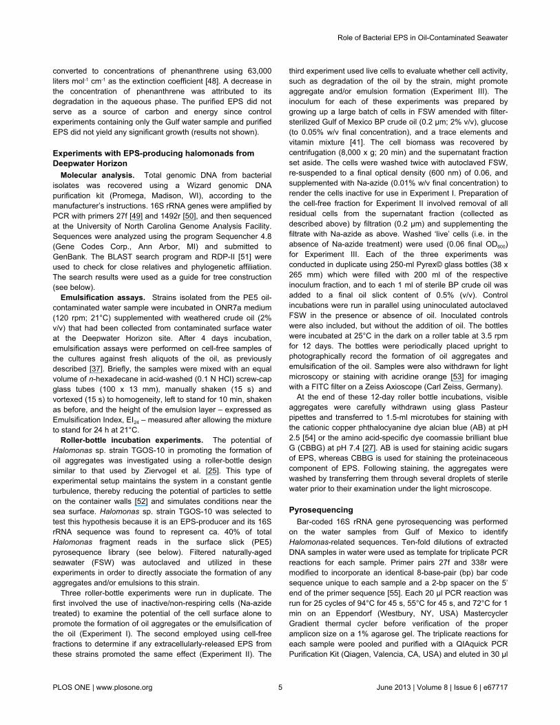
Table 2. Growth on and mineralization of some aliphaticand aromatic hydrocarbons by Halomonas sp. strain TG39.
Substrate Growtha Mineralizationb
Phthalate ND +Salicylate + +Catechol + NDPhenol +c NDBenzoate + NDp-Cresol ND +o-Cresol ND +Benzene – NDToluene – NDp-Xylene – NDNaphthalene + +Biphenyl – NDFluorene – NDPhenanthrene – –Anthracene – –Fluoranthene w +Pyrene w –Decane – –Chrysene w –Benzo[a]pyrene – –Hexadecane + –a Determination of growth was assessed spectrophotometrically (OD600) or bymeasuring the total protein concentration in the medium over time, as described inthe Materials & Methods. +, positive; –, negative; w, weakly positive; ND, notdetermined.b Mineralization was assessed as described in the Materials & Methods sectionusing 14C-labelled compounds.c Growth occurred only when phenol was supplied via the vapor phase.
Role of Bacterial EPS in Oil-Contaminated Seawater
PLOS ONE | www.plosone.org 4 June 2013 | Volume 8 | Issue 6 | e67717
converted to concentrations of phenanthrene using 63,000liters mol-1 cm-1 as the extinction coefficient [48]. A decrease inthe concentration of phenanthrene was attributed to itsdegradation in the aqueous phase. The purified EPS did notserve as a source of carbon and energy since controlexperiments containing only the Gulf water sample and purifiedEPS did not yield any significant growth (results not shown).
Experiments with EPS-producing halomonads fromDeepwater Horizon
Molecular analysis. Total genomic DNA from bacterialisolates was recovered using a Wizard genomic DNApurification kit (Promega, Madison, WI), according to themanufacturer’s instructions. 16S rRNA genes were amplified byPCR with primers 27f [49] and 1492r [50], and then sequencedat the University of North Carolina Genome Analysis Facility.Sequences were analyzed using the program Sequencher 4.8(Gene Codes Corp., Ann Arbor, MI) and submitted toGenBank. The BLAST search program and RDP-II [51] wereused to check for close relatives and phylogenetic affiliation.The search results were used as a guide for tree construction(see below).
Emulsification assays. Strains isolated from the PE5 oil-contaminated water sample were incubated in ONR7a medium(120 rpm; 21°C) supplemented with weathered crude oil (2%v/v) that had been collected from contaminated surface waterat the Deepwater Horizon site. After 4 days incubation,emulsification assays were performed on cell-free samples ofthe cultures against fresh aliquots of the oil, as previouslydescribed [37]. Briefly, the samples were mixed with an equalvolume of n-hexadecane in acid-washed (0.1 N HCl) screw-capglass tubes (100 x 13 mm), manually shaken (15 s) andvortexed (15 s) to homogeneity, left to stand for 10 min, shakenas before, and the height of the emulsion layer – expressed asEmulsification Index, EI24 – measured after allowing the mixtureto stand for 24 h at 21°C.
Roller-bottle incubation experiments. The potential ofHalomonas sp. strain TGOS-10 in promoting the formation ofoil aggregates was investigated using a roller-bottle designsimilar to that used by Ziervogel et al. [25]. This type ofexperimental setup maintains the system in a constant gentleturbulence, thereby reducing the potential of particles to settleon the container walls [52] and simulates conditions near thesea surface. Halomonas sp. strain TGOS-10 was selected totest this hypothesis because it is an EPS-producer and its 16SrRNA sequence was found to represent ca. 40% of totalHalomonas fragment reads in the surface slick (PE5)pyrosequence library (see below). Filtered naturally-agedseawater (FSW) was autoclaved and utilized in theseexperiments in order to directly associate the formation of anyaggregates and/or emulsions to this strain.
Three roller-bottle experiments were run in duplicate. Thefirst involved the use of inactive/non-respiring cells (Na-azidetreated) to examine the potential of the cell surface alone topromote the formation of oil aggregates or the emulsification ofthe oil (Experiment I). The second employed using cell-freefractions to determine if any extracellularly-released EPS fromthese strains promoted the same effect (Experiment II). The
third experiment used live cells to evaluate whether cell activity,such as degradation of the oil by the strain, might promoteaggregate and/or emulsion formation (Experiment III). Theinoculum for each of these experiments was prepared bygrowing up a large batch of cells in FSW amended with filter-sterilized Gulf of Mexico BP crude oil (0.2 µm; 2% v/v), glucose(to 0.05% w/v final concentration), and a trace elements andvitamin mixture [41]. The cell biomass was recovered bycentrifugation (8,000 x g; 20 min) and the supernatant fractionset aside. The cells were washed twice with autoclaved FSW,re-suspended to a final optical density (600 nm) of 0.06, andsupplemented with Na-azide (0.01% w/v final concentration) torender the cells inactive for use in Experiment I. Preparation ofthe cell-free fraction for Experiment II involved removal of allresidual cells from the supernatant fraction (collected asdescribed above) by filtration (0.2 µm) and supplementing thefiltrate with Na-azide as above. Washed ‘live’ cells (i.e. in theabsence of Na-azide treatment) were used (0.06 final OD600)for Experiment III. Each of the three experiments wasconducted in duplicate using 250-ml Pyrex© glass bottles (38 x265 mm) which were filled with 200 ml of the respectiveinoculum fraction, and to each 1 ml of sterile BP crude oil wasadded to a final oil slick content of 0.5% (v/v). Controlincubations were run in parallel using uninoculated autoclavedFSW in the presence or absence of oil. Inoculated controlswere also included, but without the addition of oil. The bottleswere incubated at 25°C in the dark on a roller table at 3.5 rpmfor 12 days. The bottles were periodically placed upright tophotographically record the formation of oil aggregates andemulsification of the oil. Samples were also withdrawn for lightmicroscopy or staining with acridine orange [53] for imagingwith a FITC filter on a Zeiss Axioscope (Carl Zeiss, Germany).
At the end of these 12-day roller bottle incubations, visibleaggregates were carefully withdrawn using glass Pasteurpipettes and transferred to 1.5-ml microtubes for staining withthe cationic copper phthalocyanine dye alcian blue (AB) at pH2.5 [54] or the amino acid-specific dye coomassie brilliant blueG (CBBG) at pH 7.4 [27]. AB is used for staining acidic sugarsof EPS, whereas CBBG is used for staining the proteinaceouscomponent of EPS. Following staining, the aggregates werewashed by transferring them through several droplets of sterilewater prior to their examination under the light microscope.
PyrosequencingBar-coded 16S rRNA gene pyrosequencing was performed
on the water samples from Gulf of Mexico to identifyHalomonas-related sequences. Ten-fold dilutions of extractedDNA samples in water were used as template for triplicate PCRreactions for each sample. Primer pairs 27f and 338r weremodified to incorporate an identical 8-base-pair (bp) bar codesequence unique to each sample and a 2-bp spacer on the 5’end of the primer sequence [55]. Each 20 µl PCR reaction wasrun for 25 cycles of 94°C for 45 s, 55°C for 45 s, and 72°C for 1min on an Eppendorf (Westbury, NY, USA) MastercyclerGradient thermal cycler before verification of the properamplicon size on a 1% agarose gel. The triplicate reactions foreach sample were pooled and purified with a QIAquick PCRPurification Kit (Qiagen, Valencia, CA, USA) and eluted in 30 µl
Role of Bacterial EPS in Oil-Contaminated Seawater
PLOS ONE | www.plosone.org 5 June 2013 | Volume 8 | Issue 6 | e67717
of 10 mM Tris-Cl (pH 8.5) buffer. The DNA concentration ofpooled amplicons was then measured using a NanoDropND-3300 Fluorospectrometer (Thermo, Waltham, MA, USA)and Quant-iT Picogreen dsDNA Kit (Invitrogen, Carlsbad, CA,USA) prior to combining into a single sample at a concentrationsuitable for pyrosequencing. The sample was submitted to theHigh-Throughput Sequencing Facility at the University of NorthCarolina-Chapel Hill for sequencing using the 454 LifeSciences Titanium platform (Roche Diagnostics, Branford, CT,USA).
Pyrosequencing reads were trimmed and filtered using theLUCY program with a minimum PHRED score of 27.5 andminimum length of 200 nt to remove low-quality regions andshort reads [56]. Reads were de-multiplexed based on the 8 bpbar code sequence, and the primer and bar code regions wereremoved using QIIME [57]. To form operational taxonomic unitsthe reads were clustered at 97% sequence identity withUCLUST [58] and the most abundant unique read within eachcluster was used as its representative sequence. Initialphylogenetic identification was made using BLAST [59] andchimeras were detected with Chimera Slayer [60]. Sequencedata were submitted to the European Nucleotide ArchiveSequence Read Archive under the study accession numberERP002443.
Halomonas sequences were identified by manually aligningthe representative sequences in ARB [61] to sequences ofcultivated and well-characterized Halomonas species. Abootstrapped neighbour-joining phylogenetic tree (with 1000bootstraps) of near-full-length 16S rRNA sequences wasproduced using the Jukes-Cantor substitution model and thepyrosequencing reads were then added to this guide tree usingthe parsimony quick-add function in ARB.
Statistical analysisA Student’s t test was performed to test for significant
differences (P < 0.05) in the degradation and solubilization ofhydrocarbons between the different treatments.
Nucleotide sequence accession numbersThe 16S rRNA gene sequence of strains GOS-2, GOS-3a
and TGOS-10 were deposited with GenBank under accessionnumbers JQ246430, JQ246431 and JQ246432, respectively.
Results
Hydrocarbon degradation by Halomonas sp. strainTG39 and amphiphilic properties of its produced EPS
Utilization of hydrocarbons. Table 2 shows the varioussubstrates that were tested for growth or mineralization by therepresentative strain TG39. Growth was observed onsalicylate, catechol, phenol, benzoate, naphthalene andhexadecane. Weak growth was recorded on fluoranthene,pyrene and chrysene, and no growth was measured ondecane, benzene, toluene, p-xylene, biphenyl, fluorene,phenanthrene, anthracene and benzo[a]pyrene. The liberationof14 CO2 from 14C-labeled compounds was also assessed. After9 days of incubation, significant levels (P < 0.05) of salicylate
(73.9 ± 1.4%), o-cresol (47.2 ± 14.3%), p-cresol (11.8 ± 0.9%),naphthalene (6.1 ± 0.2%), fluoranthene (3.9 ± 0.6%) andphthalate (1.2 ± 0.2%) were mineralized of total added 14C-labeled compound when compared to their respective acid-killed controls. Strain TG39 did not significantly mineralizephenanthrene, anthracene, pyrene, benzo[a]pyrene, chrysene,decane and hexadecane compared to their respective acidifiedcontrols. The strain’s ability to mineralize p-cresol, o-cresol,naphthalene and fluoranthene, and produce weak growth onpyrene, is reflected in its ability to assimilate phthalate,salicylate, catechol and benzoate which are commonintermediates in PAH degradation.
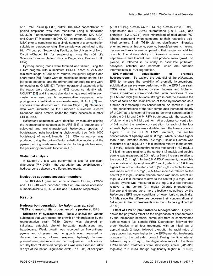
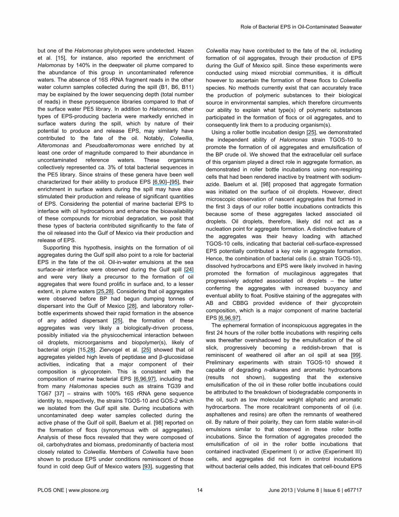
EPS-mediated solubilization of aromatichydrocarbons. To explore the potential of the HalomonasEPS to increase the solubility of aromatic hydrocarbons,solubilization assays were performed with the EPS from strainTG39 using phenanthrene, pyrene, fluorene and biphenyl.These experiments were conducted under conditions of low(0.1 M) and high (0.6 M) ionic strength in order to evaluate theeffect of salts on the solubilization of these hydrocarbons as afunction of increasing EPS concentration. As shown in Figure1, the concentrations of the four compounds increased linearly(r2 ≥ 0.94) as a function of increasing polymer concentration inboth the 0.1 M and 0.6 M FSW treatments, with the exceptionof biphenyl in the 0.1 M treatment. At a polymer concentrationof 0.4 mg/ml, the soluble concentration for each of the fouraromatic hydrocarbons was calculated from the linear curves inFigure 1. In the 0.1 M FSW treatment, the solubleconcentration of biphenyl was 36.8 mg/L, which is 5-fold higherthan in the untreated control (7.2 mg/L); soluble fluorene wasmeasured at 8.5 mg/L, a 4.7-fold increase relative to the control(1.8 mg/L); soluble phenanthrene was measured at 4.9 mg/L, a3.8-fold increase relative to the control (1.3 mg/L); and solublepyrene was measured at 0.3 mg/L, a 3-fold increase relative tothe control (0.1 mg/L). In the 0.6 M FSW treatment, the solubleconcentration of biphenyl was 42.5 mg/L, which is 11.8 timeshigher than in the untreated control (3.6 mg/L); soluble fluorenewas measured at 6.5 mg/L, a 5.4-fold increase relative to thecontrol (1.2 mg/L); soluble phenanthrene was measured at 3.3mg/L, a 2.4-fold increase relative to the control (1.4 mg/L); andsoluble pyrene was measured at 0.2 mg/L, a 2-fold increaserelative to the control (0.1 mg/L). Overall, phenanthrene,fluorene and pyrene were more effectively solubilised by theHalomonas EPS under conditions of lower ionic strength (i.e.0.1 M), since the differences between their concentrations at0.4 mg/ml in the two treatments was found to be significant (P< 0.05).
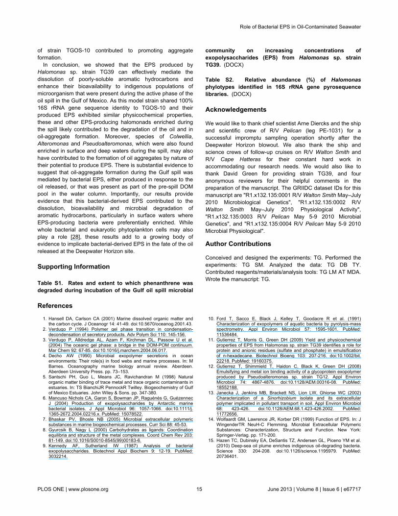
Effect of EPS on phenanthrene biodegradation. Figure 2shows the polymer’s effect on the degradation of phenanthreneby the indigenous microbial community from oil-contaminatedsurface waters (i.e. sample PE5). Degradation followed firstorder kinetics in all four treatments with an initial lag ofapproximately 2 days, followed thereafter by rapid rates ofdegradation that were higher for the EPS-amended treatmentscompared to the untreated control. During the time intervalbetween day 2 to day 5, the degradation rates for the threeEPS-amended treatments were statistically similar (281–310mg/l/day; P > 0.05), though significantly higher (P < 0.05)
Role of Bacterial EPS in Oil-Contaminated Seawater
PLOS ONE | www.plosone.org 6 June 2013 | Volume 8 | Issue 6 | e67717
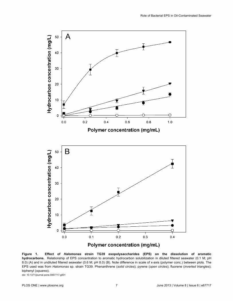
Figure 1. Effect of Halomonas strain TG39 exopolysaccharides (EPS) on the dissolution of aromatichydrocarbons. Relationship of EPS concentration to aromatic hydrocarbon solubilization in diluted filtered seawater (0.1 M; pH8.0) (A) and in undiluted filtered seawater (0.6 M; pH 8.0) (B). Note difference in scale of x-axis (polymer conc.) between plots. TheEPS used was from Halomonas sp. strain TG39. Phenanthrene (solid circles); pyrene (open circles); fluorene (inverted triangles);biphenyl (squares).doi: 10.1371/journal.pone.0067717.g001
Role of Bacterial EPS in Oil-Contaminated Seawater
PLOS ONE | www.plosone.org 7 June 2013 | Volume 8 | Issue 6 | e67717
compared to that of the untreated control (211 mg/l/day)(Supplementary table S1). During the interval between day 5 today 8, the degradation rate of the untreated control was slightlyless (476 mg/l/day) though not significantly different (P > 0.05)compared to that of the EPS-amended treatments (526–537mg/l/day). Complete removal of the phenanthrene in the EPS-amended treatments occurred sooner (i.e. by day 8) than in theuntreated control (i.e. 11-12 days). Incubations that were run inparallel showed that the microbial community in the PE5sample was capable of producing measurable, albeit veryweak, growth on the polymer as a carbon source (results notshown).
EPS-producing bacteria, their abundance at DeepwaterHorizon and role in oil-aggregate formation
Molecular analysis and characteristics of Halomonasisolates. Several colonies surrounded by clearing zones grewout on agar plates that had been streaked with the PE5 watersample and sprayed with phenanthrene. Subsequentpurification and sequencing identified three distinct Halomonasstrains – strains GOS-2, GOS-3a and TGOS-10. Coloniesforming clearing zones on fluorene-sprayed plates were alsoobserved, but attempts to purify and maintain these organisms
proved difficult. No clearing zones were observed on platessprayed with anthracene or pyrene. Versatility for degradingvarious PAH compounds was observed with strain TGOS-10as it was able to grow on different PAHs.
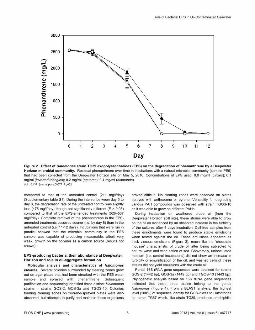
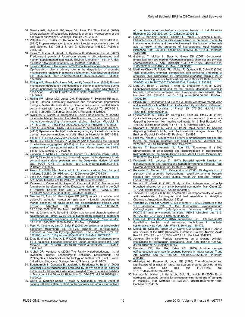
During incubation on weathered crude oil (from theDeepwater Horizon spill site), these strains were able to growon the oil as evidenced by an observed increase in the turbidityof the cultures after 4 days incubation. Cell-free samples fromthese enrichments were found to produce stable emulsionswhen tested against the oil. These emulsions appeared asthick viscous emulsions (Figure 3), much like the ‘chocolatemousse’ characteristic of crude oil after being subjected tonatural wave and wind action at sea. Conversely, uninoculatedmedium (i.e. control incubations) did not show an increase inturbidity or emulsification of the oil, and washed cells of thesestrains did not yield emulsions with the crude oil.
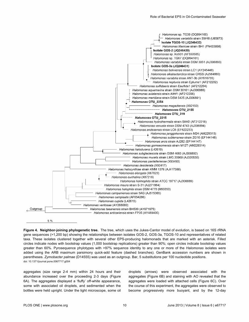
Partial 16S rRNA gene sequences were obtained for strainsGOS-2 (1442 bp), GOS-3a (1449 bp) and TGOS-10 (1443 bp).Phylogenetic analysis based on 16S rRNA gene sequencesindicated that these three strains belong to the genusHalomonas (Figure 4). From a BLAST analysis, the highestlevel (100%) of sequence identity for GOS-2 was to Halomonassp. strain TG67 which, like strain TG39, produces amphiphilic
Figure 2. Effect of Halomonas strain TG39 exopolysaccharides (EPS) on the degradation of phenanthrene by a DeepwaterHorizon microbial community. Residual phenanthrene over time in incubations with a natural microbial community (sample PE5)that had been collected from the Deepwater Horizon site on May 5, 2010. Concentrations of EPS used: 0.0 mg/ml (circles); 0.1mg/ml (inverted triangles); 0.2 mg/ml (squares); 0.4 mg/ml (diamonds).doi: 10.1371/journal.pone.0067717.g002
Role of Bacterial EPS in Oil-Contaminated Seawater
PLOS ONE | www.plosone.org 8 June 2013 | Volume 8 | Issue 6 | e67717
EPS that can effectively emulsify various types of oils [37].Strain GOS-3a was most closely related to Halomonasalkaliantarctica CRSS [62] and Halomonas variabilis ANT-3b[31] (99.8% sequence identity), both of which are known EPS-producers, the latter of which has been reported to interfacewith hydrocarbons [31]. The next closest cultivated relatives toGOS-3a are Halomonas boliviensis LC1 [63] and Halomonasneptuniae Eplume1 [64] (99.7% sequence identity). StrainTGOS-10 shared 100% sequence identity to Halomonas sp.strain TG39, and its next closest characterized relative was theEPS-producing organism Halomonas titanicae BH1 [65] with99.1% sequence identity.
Relative abundance during the spill. Bar-coded 16S rRNAgene pyrosequencing was used to analyze the bacterialcommunities present in the 5 water samples from the Gulf ofMexico collected during (PE5, B1, B6, B11) and after (GIP22)the spill. A complete presentation of the community structurerepresented by these libraries is reported elsewhere [66], [T.Yang, L. M. Nigro, T. Gutierrez, L. D’Ambrosio, S. M. Joye, R.Highsmith, A. Teske, unpublished data]. A total of 23999, 2186,133, 412 and 6498 high quality partial gene sequences wereobtained for the PE5, B1, B6, B11 and GIP22 libraries,respectively, from which five Halomonas phylotypes wereidentified across these libraries and their relative abundancesshown in Supplementary table S2. All five phylotypes were
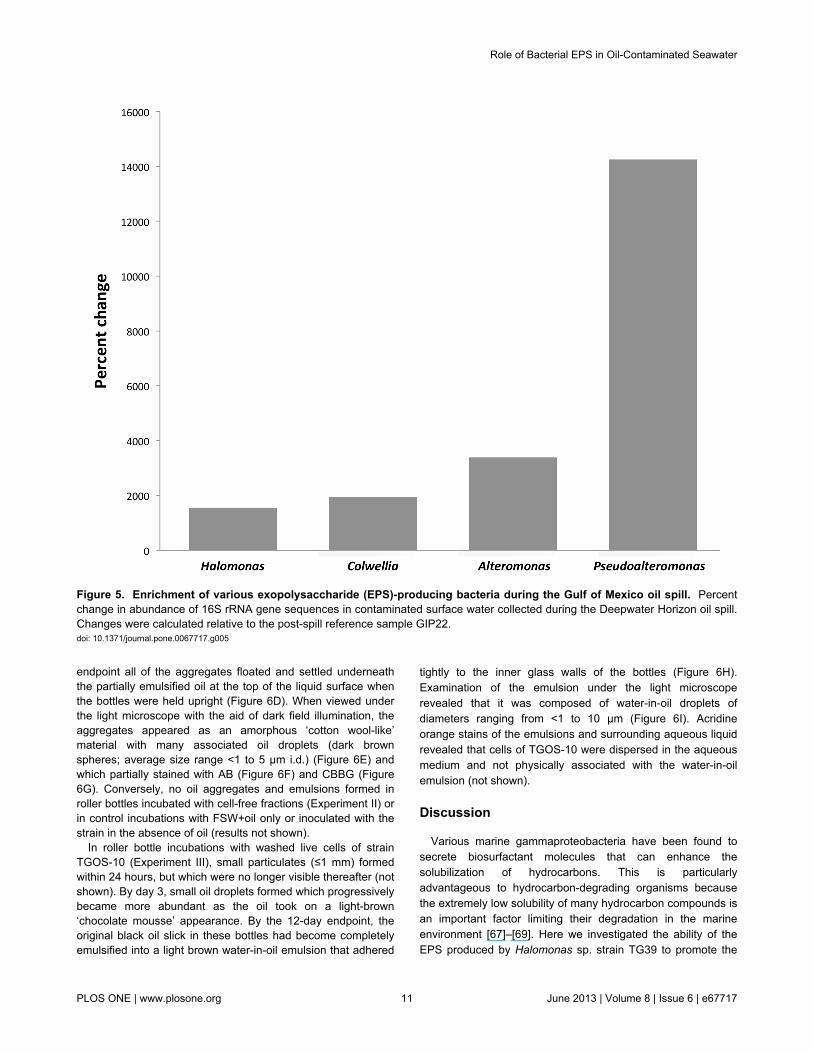
identified in the PE5 library. Of these, highest sequencesimilarity (≥97%) to isolate GOS-2 occurred with OTU_319 andOTU_2155; to isolate GOS-3a, with OTU_2155, OTU_2215and OTU_2254; and to isolate TGOS-10, with OTU_319 andOTU_2215. Only one of these Halomonas phylotypes,OTU_2155, was identified in the GIP22 library. These andother related sequences were used to construct the neighbour-joining tree displayed in Figure 4. Our analysis did not revealany Halomonas-related sequences in the B1, B6 and B11pyrosequence libraries. The change in abundance ofHalomonas 16S rRNA gene sequences, and of other bacterialgenera recognized for their EPS-producing abilities, incontaminated surface water (PE5 sample) during the spill areshown in Figure 5, expressed relative to their abundance in theuncontaminated reference sample (GIP22). Halomonas,Colwellia, Alteromonas and Pseudoalteromonas were enrichedby 1550%, 1950%, 3390 and 14250%, respectively. Theseorganisms were not detected in the other water columnsamples, with the exception of Colwellia which was foundenriched in plume water (B6 sample) by 1450% relative to thereference sample.
Formation of oil aggregates in roller bottles. Roller bottleincubations using FSW amended with filter-sterilized Gulf ofMexico BP crude oil and with inactivated cells of HalomonasTGOS-10 (Experiment I) showed a rapid formation of oil
Figure 3. Photos of water-in-oil emulsions formed by cell-free fractions of strains GOS-2, GOS-3a and TGOS-10. Emulsionswere conducted of the cell-free fractions after incubation of each of the strains for 4 days in minimal seawater medium (ONR7a)amended with weathered crude oil 2% (w/v) from the Deepwater Horizon spill. Cell-free culture samples were derived after removalof the cells by centrifugation (13,000 x g; 10 min) and then filtration (0.2 µm).doi: 10.1371/journal.pone.0067717.g003
Role of Bacterial EPS in Oil-Contaminated Seawater
PLOS ONE | www.plosone.org 9 June 2013 | Volume 8 | Issue 6 | e67717
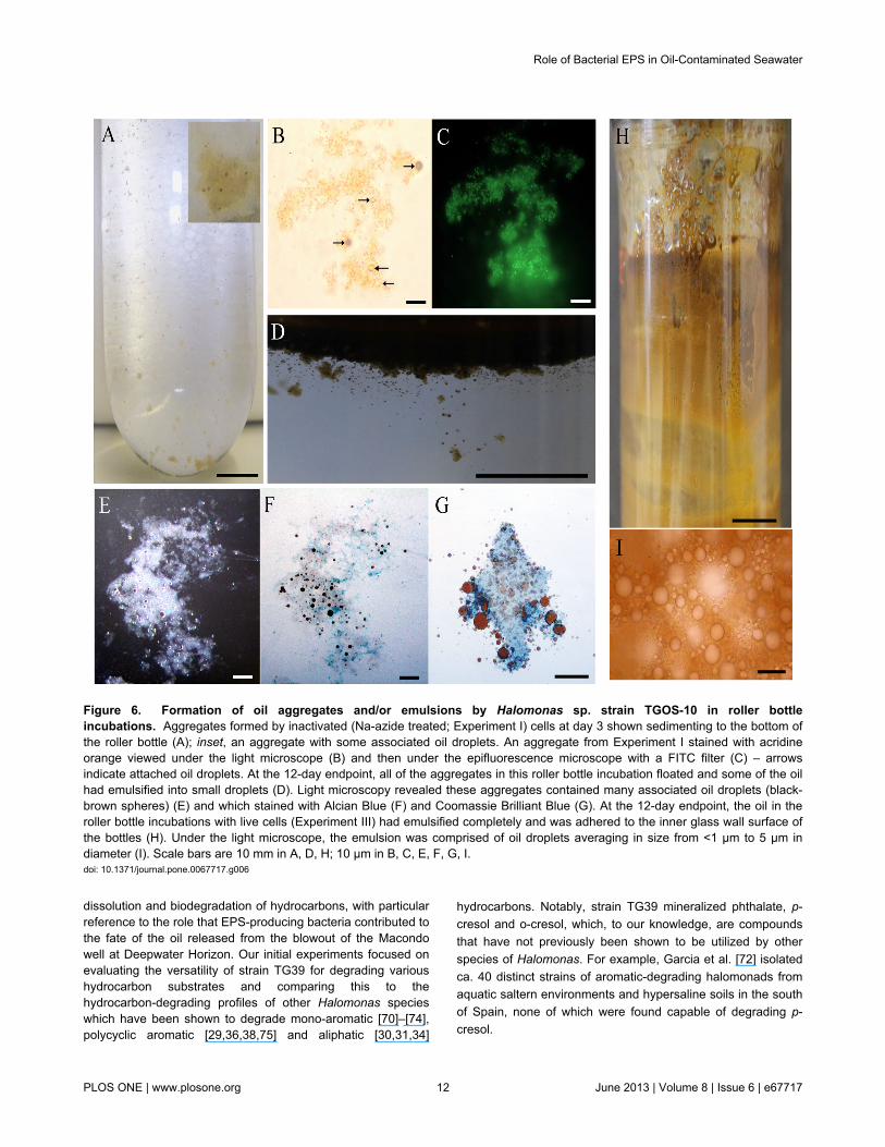
aggregates (size range 2-4 mm) within 24 hours and theirabundance increased over the proceeding 2-3 days (Figure6A). The aggregates displayed a ‘fluffy’ off-white appearance,some with associated oil droplets, and sedimented when thebottles were held upright. Under the light microscope, some oil
droplets (arrows) were observed associated with theaggregates (Figure 6B) and staining with AO revealed that theaggregates were loaded with attached cells (Figure 6C). Overthe course of this experiment, the aggregates were observed tobecome progressively more buoyant, and by the 12-day
Figure 4. Neighbor-joining phylogenetic tree. The tree, which uses the Jukes-Cantor model of evolution, is based on 16S rRNAgene sequences (>1,200 bp) showing the relationships between isolates GOS-2, GOS-3a, TGOS-10 and representatives of relatedtaxa. These isolates clustered together with several other EPS-producing halomonads that are marked with an asterisk. Filledcircles indicate nodes with bootstrap values (1,000 bootstrap replications) greater than 90%; open circles indicate bootstrap valuesgreater than 60%. Pyrosequence phylotypes with >97% sequence identity to any one or more of the Halomonas isolates wereadded using the ARB maximum parsimony quick-add feature (dashed branches). GenBank accession numbers are shown inparentheses. Zymobacter palmae (D14555) was used as an outgroup. Bar, 5 substitutions per 100 nucleotide positions.doi: 10.1371/journal.pone.0067717.g004
Role of Bacterial EPS in Oil-Contaminated Seawater
PLOS ONE | www.plosone.org 10 June 2013 | Volume 8 | Issue 6 | e67717
endpoint all of the aggregates floated and settled underneaththe partially emulsified oil at the top of the liquid surface whenthe bottles were held upright (Figure 6D). When viewed underthe light microscope with the aid of dark field illumination, theaggregates appeared as an amorphous ‘cotton wool-like’material with many associated oil droplets (dark brownspheres; average size range <1 to 5 µm i.d.) (Figure 6E) andwhich partially stained with AB (Figure 6F) and CBBG (Figure6G). Conversely, no oil aggregates and emulsions formed inroller bottles incubated with cell-free fractions (Experiment II) orin control incubations with FSW+oil only or inoculated with thestrain in the absence of oil (results not shown).
In roller bottle incubations with washed live cells of strainTGOS-10 (Experiment III), small particulates (≤1 mm) formedwithin 24 hours, but which were no longer visible thereafter (notshown). By day 3, small oil droplets formed which progressivelybecame more abundant as the oil took on a light-brown‘chocolate mousse’ appearance. By the 12-day endpoint, theoriginal black oil slick in these bottles had become completelyemulsified into a light brown water-in-oil emulsion that adhered
tightly to the inner glass walls of the bottles (Figure 6H).Examination of the emulsion under the light microscoperevealed that it was composed of water-in-oil droplets ofdiameters ranging from <1 to 10 µm (Figure 6I). Acridineorange stains of the emulsions and surrounding aqueous liquidrevealed that cells of TGOS-10 were dispersed in the aqueousmedium and not physically associated with the water-in-oilemulsion (not shown).
Discussion
Various marine gammaproteobacteria have been found tosecrete biosurfactant molecules that can enhance thesolubilization of hydrocarbons. This is particularlyadvantageous to hydrocarbon-degrading organisms becausethe extremely low solubility of many hydrocarbon compounds isan important factor limiting their degradation in the marineenvironment [67]–[69]. Here we investigated the ability of theEPS produced by Halomonas sp. strain TG39 to promote the
Figure 5. Enrichment of various exopolysaccharide (EPS)-producing bacteria during the Gulf of Mexico oil spill. Percentchange in abundance of 16S rRNA gene sequences in contaminated surface water collected during the Deepwater Horizon oil spill.Changes were calculated relative to the post-spill reference sample GIP22.doi: 10.1371/journal.pone.0067717.g005
Role of Bacterial EPS in Oil-Contaminated Seawater
PLOS ONE | www.plosone.org 11 June 2013 | Volume 8 | Issue 6 | e67717
dissolution and biodegradation of hydrocarbons, with particularreference to the role that EPS-producing bacteria contributed tothe fate of the oil released from the blowout of the Macondowell at Deepwater Horizon. Our initial experiments focused onevaluating the versatility of strain TG39 for degrading varioushydrocarbon substrates and comparing this to thehydrocarbon-degrading profiles of other Halomonas specieswhich have been shown to degrade mono-aromatic [70]–[74],polycyclic aromatic [29,36,38,75] and aliphatic [30,31,34]
hydrocarbons. Notably, strain TG39 mineralized phthalate, p-cresol and o-cresol, which, to our knowledge, are compoundsthat have not previously been shown to be utilized by otherspecies of Halomonas. For example, Garcia et al. [72] isolatedca. 40 distinct strains of aromatic-degrading halomonads fromaquatic saltern environments and hypersaline soils in the southof Spain, none of which were found capable of degrading p-cresol.
Figure 6. Formation of oil aggregates and/or emulsions by Halomonas sp. strain TGOS-10 in roller bottleincubations. Aggregates formed by inactivated (Na-azide treated; Experiment I) cells at day 3 shown sedimenting to the bottom ofthe roller bottle (A); inset, an aggregate with some associated oil droplets. An aggregate from Experiment I stained with acridineorange viewed under the light microscope (B) and then under the epifluorescence microscope with a FITC filter (C) – arrowsindicate attached oil droplets. At the 12-day endpoint, all of the aggregates in this roller bottle incubation floated and some of the oilhad emulsified into small droplets (D). Light microscopy revealed these aggregates contained many associated oil droplets (black-brown spheres) (E) and which stained with Alcian Blue (F) and Coomassie Brilliant Blue (G). At the 12-day endpoint, the oil in theroller bottle incubations with live cells (Experiment III) had emulsified completely and was adhered to the inner glass wall surface ofthe bottles (H). Under the light microscope, the emulsion was comprised of oil droplets averaging in size from <1 µm to 5 µm indiameter (I). Scale bars are 10 mm in A, D, H; 10 µm in B, C, E, F, G, I.doi: 10.1371/journal.pone.0067717.g006
Role of Bacterial EPS in Oil-Contaminated Seawater
PLOS ONE | www.plosone.org 12 June 2013 | Volume 8 | Issue 6 | e67717
Some species of Halomonas have been found to expressEPS on their cell surface [31,34,76], which in some cases hasbeen shown to enable the cells to make direct contact with oildroplets [31], possibly as a mechanism to access the substrateas a food source. For example, Halomonas sp. ANT-3bproduces an emulsifying glycolipid that allows the cells tocolonize and disrupt the oil–water interface of n-hexadecanedroplets [31]. Microscopic examination of strain TG39 cellsgrowing on n-hexadecane showed no evidence of theirattachment to oil droplets, even after cell suspensions hadbeen subjected to vigorous shaking under our emulsificationassay (results not shown). This suggests that the cell surfaceof strain TG39 lacks the amphiphilic quality that ischaracteristic of its produced EPS. This corroborates earlierobservations that showed washed whole-cell suspensions ofstrain TG39 to completely lack the capacity to emulsify oils,and that emulsifying activities were solely associated with cell-free extracts which contained EPS produced by the strain [37].The Halomonas strains GOS-2, GOS-3a and TGOS-10isolated from Deepwater Horizon exhibited similarcharacteristics, including the capacity to produce EPS thateffected the emulsification of oil.
Previous work with the EPS of strain TG39 showed that iteffectively emulsified n-hexadecane and several food-gradeoils [37]. In preliminary experiments, emulsification assaysperformed with several other hydrocarbons, such ascycloparaffins, and mono- and alkyl-aromatics, showed thisEPS to exhibit exceptional versatility in its capacity to emulsifya wide spectrum of hydrocarbon substrates (results notshown). Chemical characterization of strain TG39’s EPSrevealed that its ability to associate with and emulsifyhydrocarbons may be conferred by its high uronic acids(30.8%) and rhamnose content (31.7%) [37] – moieties thatcan render EPS quite lipophilic and mediate the adsorption ofpolysaccharides to oil droplets [77]–[80]. Additionally, theproteinaceous component of this EPS could also mediate itsadsorption to oil droplets by nature of its hydrophobicity,penetrating into the oil where they become solvated and act asanchoring points. The observed emulsification activityprompted us to evaluate whether this polymer might enhancethe solubilisation of hydrocarbons and, if so, whether this couldalso influence their availability for microbial degradation. Themodel aromatic pollutants phenanthrene, pyrene, fluorene andbiphenyl were selected since these compounds have extremelylow aqueous solubilities [81], making them ideal substrates forthis investigation. The aromatic hydrocarbons tested covered arange of partition coefficient (logP) values -4.0 for biphenyl; 4.2for fluorene; 4.5 for phenanthrene; 5.2 for pyrene [82,83].Irrespective of ionic-strength, the effect of EPS on the aqueoussolubility of the hydrocarbons correlated inversely with theinherent logP of these compounds. In other words,hydrocarbons with lower logP values were more effectivelysolubilised by the EPS, possibly due to physico-chemicalinteractions between the hydrocarbons and hydrophobicregions of the amphiphilic polymers [44,84]. The EPS-mediatedenhancement in the solubilisation of hydrocarbons with lowerlogP values may be related to the polarity of these compounds,potentially allowing them to associate, to some degree, with
negatively-charged residues (e.g. uronic acids) associated withthe EPS [11,77]. It is noteworthy that lower solubleconcentrations were measured for each hydrocarbon in the 0.6M FSW treatment (ca. 35% salinity) compared to that in the 0.1M treatment (ca. 0.8% salinity) at each EPS concentrationused, possibly because an inverse relationship exists betweensalinity and hydrocarbon solubility [85]. As hydrocarbons areless soluble in seawater, the production of amphiphilic EPS byhydrocarbon-degrading bacteria in marine environments wouldbe considered advantageous to increasing the solubility ofthese substrates, and thus also their bioavailability fordegradation.
Interestingly, the effect of increasing concentrations of theEPS on the dissolution of biphenyl in the 0.1 M (low ionicstrength) treatment did not follow the linear trend observed withthe other hydrocarbons. This EPS has been shown to have apolydispersity index of 1.8, which is relatively high and denotesconsiderable heterogeneity in the molecular-size range ofpolysaccharide molecules [11]. It has also been shown thatunder conditions of low ionic strength, this heterogeneity issubstantially reduced as polysaccharide molecules of differentsizes come together to form larger molecular-weight species,or aggregates [11]. This would be expected to significantlyreduce the multivalency and exposure of hydrophobic regionsof the EPS that would otherwise be available to interface withhydrocarbons. The formation of these aggregates apparentlyoccurred at EPS concentrations above 0.25 mg/ml, whichwould have reduced the number of biphenyl-binding sites onthe EPS macromolecular structure and thereby explain theincapacity of the EPS to solubilize higher concentrations of thebiphenyl. Since the rate of dissolution of a compound is acritical measure of its bioavailability [86], it can have asignificant influence on the degradation rate of the compound.This is particularly important with hydrocarbons that exhibitextremely low aqueous solubilities [87,88]. Since we did notobserve the attachment of strain TG39 cells to hydrocarbondroplets (results not shown), this assumes that its growth onhydrocarbons would occur at the expense of dissolvedsubstrate [89]. Whereas the strain was unable to utilizebiphenyl, fluorene and phenanthrene as sole sources of carbonand energy, its produced EPS however was found to effectivelyincrease the solubility of these hydrocarbons. In theseinstances, organisms such as strain TG39 may be expected toprovide an advantage to other marine bacteria that wouldotherwise be limited in their capacity to gain access to thesecompounds, as was demonstrated in our phenanthreneexperiments with a natural community of indigenous microbiotafrom the Deepwater Horizon site. These results provide the firstline of evidence that implicate Halomonas EPS in enhancingthe degradation of oil hydrocarbons in the marine environmentby increasing the dissolution and bioavailability of thesecompounds to indigenous oil-degrading microorganisms, suchas during the Deepwater Horizon oil spill.
Though the relative abundance of Halomonas in oil-contaminated surface waters at Deepwater Horizon was quitelow, they were by no means inactive since several Halomonasphylotypes were enriched (by 1550% collectively) as a result ofthe spill compared to the reference sample (GIP22) where all
Role of Bacterial EPS in Oil-Contaminated Seawater
PLOS ONE | www.plosone.org 13 June 2013 | Volume 8 | Issue 6 | e67717
but one of the Halomonas phylotypes were undetected. Hazenet al. [15], for instance, also reported the enrichment ofHalomonas by 140% in the deepwater oil plume compared tothe abundance of this group in uncontaminated referencewaters. The absence of 16S rRNA fragment reads in the otherwater column samples collected during the spill (B1, B6, B11)may be explained by the lower sequencing depth (total numberof reads) in these pyrosequence libraries compared to that ofthe surface water PE5 library. In addition to Halomonas, othertypes of EPS-producing bacteria were markedly enriched insurface waters during the spill, which by nature of theirpotential to produce and release EPS, may similarly havecontributed to the fate of the oil. Notably, Colwellia,Alteromonas and Pseudoalteromonas were enriched by atleast one order of magnitude compared to their abundance inuncontaminated reference waters. These organismscollectively represented ca. 3% of total bacterial sequences inthe PE5 library. Since strains of these genera have been wellcharacterized for their ability to produce EPS [6,90]–[95], theirenrichment in surface waters during the spill may have alsostimulated their production and release of significant quantitiesof EPS. Considering the potential of marine bacterial EPS tointerface with oil hydrocarbons and enhance the bioavailabilityof these compounds for microbial degradation, we posit thatthese types of bacteria contributed significantly to the fate ofthe oil released into the Gulf of Mexico via their production andrelease of EPS.
Supporting this hypothesis, insights on the formation of oilaggregates during the Gulf spill also point to a role for bacterialEPS in the fate of the oil. Oil-in-water emulsions at the seasurface-air interface were observed during the Gulf spill [24]and were very likely a precursor to the formation of oilaggregates that were found prolific in surface and, to a lesserextent, in plume waters [25,28]. Considering that oil aggregateswere observed before BP had begun dumping tonnes ofdispersant into the Gulf of Mexico [28], and laboratory roller-bottle experiments showed their rapid formation in the absenceof any added dispersant [25], the formation of theseaggregates was very likely a biologically-driven process,possibly initiated via the physicochemical interaction betweenoil droplets, microorganisms and biopolymer(s), likely ofbacterial origin [15,28]. Ziervogel et al. [25] showed that oilaggregates yielded high levels of peptidase and β-glucosidaseactivities, indicating that a major component of theircomposition is glycoprotein. This is consistent with thecomposition of marine bacterial EPS [6,96,97], including thatfrom many Halomonas species such as strains TG39 andTG67 [37] – strains with 100% 16S rRNA gene sequenceidentity to, respectively, the strains TGOS-10 and GOS-2 whichwe isolated from the Gulf spill site. During incubations withuncontaminated deep water samples collected during theactive phase of the Gulf oil spill, Baelum et al. [98] reported onthe formation of flocs (synonymous with oil aggregates).Analysis of these flocs revealed that they were composed ofoil, carbohydrates and biomass, predominantly of bacteria mostclosely related to Colwellia. Members of Colwellia have beenshown to produce EPS under conditions reminiscent of thosefound in cold deep Gulf of Mexico waters [93], suggesting that
Colwellia may have contributed to the fate of the oil, includingformation of oil aggregates, through their production of EPSduring the Gulf of Mexico spill. Since these experiments wereconducted using mixed microbial communities, it is difficulthowever to ascertain the formation of these flocs to Colwelliaspecies. No methods currently exist that can accurately tracethe production of polymeric substances to their biologicalsource in environmental samples, which therefore circumventsour ability to explain what type(s) of polymeric substancesparticipated in the formation of flocs or oil aggregates, and toconsequently link them to a producing organism(s).
Using a roller bottle incubation design [25], we demonstratedthe independent ability of Halomonas strain TGOS-10 topromote the formation of oil aggregates and emulsification ofthe BP crude oil. We showed that the extracellular cell surfaceof this organism played a direct role in aggregate formation, asdemonstrated in roller bottle incubations using non-respiringcells that had been rendered inactive by treatment with sodium-azide. Baelum et al. [98] proposed that aggregate formationwas initiated on the surface of oil droplets. However, directmicroscopic observation of nascent aggregates that formed inthe first 3 days of our roller bottle incubations contradicts thisbecause some of these aggregates lacked associated oildroplets. Oil droplets, therefore, likely did not act as anucleation point for aggregate formation. A distinctive feature ofthe aggregates was their heavy loading with attachedTGOS-10 cells, indicating that bacterial cell-surface-expressedEPS potentially contributed a key role in aggregate formation.Hence, the combination of bacterial cells (i.e. strain TGOS-10),dissolved hydrocarbons and EPS were likely involved in havingpromoted the formation of mucilaginous aggregates thatprogressively adopted associated oil droplets – the latterconferring the aggregates with increased buoyancy andeventual ability to float. Positive staining of the aggregates withAB and CBBG provided evidence of their glycoproteincomposition, which is a major component of marine bacterialEPS [6,96,97].
The ephemeral formation of inconspicuous aggregates in thefirst 24 hours of the roller bottle incubations with respiring cellswas thereafter overshadowed by the emulsification of the oilslick, progressively becoming a reddish-brown that isreminiscent of weathered oil after an oil spill at sea [99].Preliminary experiments with strain TGOS-10 showed itcapable of degrading n-alkanes and aromatic hydrocarbons(results not shown), suggesting that the extensiveemulsification of the oil in these roller bottle incubations couldbe attributed to the breakdown of biodegradable components inthe oil, such as low molecular weight aliphatic and aromatichydrocarbons. The more recalcitrant components of oil (i.e.asphaltenes and resins) are often the remnants of weatheredoil. By nature of their polarity, they can form stable water-in-oilemulsions similar to that observed in these roller bottleincubations. Since the formation of aggregates preceded theemulsification of oil in the roller bottle incubations thatcontained inactivated (Experiment I) or active (Experiment III)cells, and aggregates did not form in control incubationswithout bacterial cells added, this indicates that cell-bound EPS
Role of Bacterial EPS in Oil-Contaminated Seawater
PLOS ONE | www.plosone.org 14 June 2013 | Volume 8 | Issue 6 | e67717
of strain TGOS-10 contributed to promoting aggregateformation.
In conclusion, we showed that the EPS produced byHalomonas sp. strain TG39 can effectively mediate thedissolution of poorly-soluble aromatic hydrocarbons andenhance their bioavailability to indigenous populations ofmicroorganism that were present during the active phase of theoil spill in the Gulf of Mexico. As this model strain shared 100%16S rRNA gene sequence identity to TGOS-10 and theirproduced EPS exhibited similar physicochemical properties,these and other EPS-producing halomonads enriched duringthe spill likely contributed to the degradation of the oil and inoil-aggregate formation. Moreover, species of Colwellia,Alteromonas and Pseudoalteromonas, which were also foundenriched in surface and deep waters during the spill, may alsohave contributed to the formation of oil aggregates by nature oftheir potential to produce EPS. There is substantial evidence tosuggest that oil-aggregate formation during the Gulf spill wasmediated by bacterial EPS, either produced in response to theoil released, or that was present as part of the pre-spill DOMpool in the water column. Importantly, our results provideevidence that this bacterial-derived EPS contributed to thedissolution, bioavailability and microbial degradation ofaromatic hydrocarbons, particularly in surface waters whereEPS-producing bacteria were preferentially enriched. Whilewhole bacterial and eukaryotic phytoplankton cells may alsoplay a role [28], these results add to a growing body ofevidence to implicate bacterial-derived EPS in the fate of the oilreleased at the Deepwater Horizon site.
Supporting Information
Table S1. Rates and extent to which phenanthrene wasdegraded during incubation of the Gulf oil spill microbial
community on increasing concentrations ofexopolysaccharides (EPS) from Halomonas sp. strainTG39. (DOCX)
Table S2. Relative abundance (%) of Halomonasphylotypes identified in 16S rRNA gene pyrosequencelibraries. (DOCX)
Acknowledgements
We would like to thank chief scientist Arne Diercks and the shipand scientific crew of R/V Pelican (leg PE-1031) for asuccessful impromptu sampling operation shortly after theDeepwater Horizon blowout. We also thank the ship andscience crews of follow-up cruises on R/V Walton Smith andR/V Cape Hatteras for their constant hard work inaccommodating our research needs. We would also like tothank David Green for providing strain TG39, and fouranonymous reviewers for their helpful comments in thepreparation of the manuscript. The GRIIDC dataset IDs for thismanuscript are "R1.x132.135:0001 R/V Walton Smith May–July2010 Microbiological Genetics", "R1.x132.135:0002 R/VWalton Smith May–July 2010 Physiological Activity","R1.x132.135:0003 R/V Pelican May 5-9 2010 MicrobialGenetics", and "R1.x132.135:0004 R/V Pelican May 5-9 2010Microbial Physiological".
Author Contributions
Conceived and designed the experiments: TG. Performed theexperiments: TG SM. Analyzed the data: TG DB TY.Contributed reagents/materials/analysis tools: TG LM AT MDA.Wrote the manuscript: TG.
References
1. Hansell DA, Carlson CA (2001) Marine dissolved organic matter andthe carbon cycle. J Oceanogr 14: 41-49. doi:10.5670/oceanog.2001.43.
2. Verdugo P (1994) Polymer gel phase transition in condensation-decondensation of secretory products. Adv Polym Sci 110: 145-156.
3. Verdugo P, Alldredge AL, Azam F, Kirchman DL, Passow U et al.(2004) The oceanic gel phase: a bridge in the DOM-POM continuum.Mar Chem 92: 67-85. doi:10.1016/j.marchem.2004.06.017.
4. Decho AW (1990) Microbial exopolymer secretions in oceanenvironments: Their role(s) in food webs and marine processes. In: MBarnes. Oceanography marine biology annual review. Aberdeen.Aberdeen University Press. pp. 73–153.
5. Santschi PH, Guo L, Means JC, Ravichandran M (1998) Naturalorganic matter binding of trace metal and trace organic contaminants inestuaries. In: TS BianchiJR PennockR Twilley. Biogeochemistry of Gulfof Mexico Estuaries. John Wiley & Sons. pp. 347-380.
6. Mancuso Nichols CA, Garon S, Bowman JP, Raguénès G, GuézennecJ (2004) Production of exopolysaccharides by Antarctic marinebacterial isolates. J Appl Microbiol 96: 1057-1066. doi:10.1111/j.1365-2672.2004.02216.x. PubMed: 15078522.
7. Bhaskar PV, Bhosle NB (2005) Microbial extracellular polymericsubstances in marine biogeochemical processes. Curr Sci 88: 45-53.
8. Gyurcsik B, Nagy L (2000) Carbohydrates as ligands: Coordinationequilibria and structure of the metal complexes. Coord Chem Rev 203:81-149. doi:10.1016/S0010-8545(99)00183-6.
9. Kennedy AF, Sutherland IW (1987) Analysis of bacterialexopolysaccharides. Biotechnol Appl Biochem 9: 12-19. PubMed:3032214.
10. Ford T, Sacco E, Black J, Kelley T, Goodacre R et al. (1991)Characterization of exopolymers of aquatic bacteria by pyrolysis-massspectrometry. Appl Environ Microbiol 57: 1595-1601. PubMed:11536484.
11. Gutierrez T, Morris G, Green DH (2009) Yield and physicochemicalproperties of EPS from Halomonas sp. strain TG39 identifies a role forprotein and anionic residues (sulfate and phosphate) in emulsificationof n-hexadecane. Biotechnol Bioeng 103: 207-216. doi:10.1002/bit.22218. PubMed: 19160375.
12. Gutierrez T, Shimmield T, Haidon C, Black K, Green DH (2008)Emulsifying and metal ion binding activity of a glycoprotein exopolymerproduced by Pseudoalteromonas sp. strain TG12. Appl EnvironMicrobiol 74: 4867-4876. doi:10.1128/AEM.00316-08. PubMed:18552188.
13. Janecka J, Jenkins MB, Brackett NS, Lion LW, Ghiorse WC (2002)Characterization of a Sinorhizobium isolate and its extracellularpolymer implicated in pollutant transport in soil. Appl Environ Microbiol68: 423-426. doi:10.1128/AEM.68.1.423-426.2002. PubMed:11772656.
14. Wolfaardt GM, Lawrence JR, Korber DR (1999) Function of EPS. In: JWingenderTR NeuH-C Flemming. Microbial Extracellular PolymericSubstances: Characterization, Structure and Function. New York:Springer-Verlag. pp. 171-200.
15. Hazen TC, Dubinsky EA, DeSantis TZ, Andersen GL, Piceno YM et al.(2010) Deep-sea oil plume enriches indigenous oil-degrading bacteria.Science 330: 204-208. doi:10.1126/science.1195979. PubMed:20736401.
Role of Bacterial EPS in Oil-Contaminated Seawater
PLOS ONE | www.plosone.org 15 June 2013 | Volume 8 | Issue 6 | e67717
16. Diercks A-R, Highsmith RC, Asper VL, Joung DJ, Zhou Z et al. (2010)Characterization of subsurface polycyclic aromatic hydrocarbons at thedeepwater horizon site. Geophys Res Lett 37: L20602.
17. Valentine DL, Kessler JD, Redmond MC, Mendes SD, Heintz MB et al.(2010) Propane respiration jump-starts microbial response to a deep oilspill. Science 330: 208-211. doi:10.1126/science.1196830. PubMed:20847236.
18. Kasai Y, Kishira H, Sasaki T, Syutsubo K, Watanabe K et al. (2002)Predominant growth of Alcanivorax strains in oil-contaminated andnutrient-supplemented sea water. Environ Microbiol 4: 141-147. doi:10.1046/j.1462-2920.2002.00275.x. PubMed: 12000314.
19. Kasai Y, Kishira H, Harayama S (2002) Bacteria belonging to the genusCycloclasticus play a primary role in the degradation of aromatichydrocarbons released in a marine environment. Appl Environ Microbiol68: 5625-5633. doi:10.1128/AEM.68.11.5625-5633.2002. PubMed:12406758.
20. Röling WF, Milner MG, Jones DM, Lee K, Daniel F et al. (2002) Robusthydrocarbon degradation and dynamics of bacterial communities duringnutrient-enhanced oil spill bioremediation. Appl Environ Microbiol 68:5537-5548. doi:10.1128/AEM.68.11.5537-5548.2002. PubMed:12406747.
21. Röling WF, Milner MG, Jones DM, Fratepietro F, Swannell RP et al.(2004) Bacterial community dynamics and hydrocarbon degradationduring a field-scale evaluation of bioremediation on a mudflat beachcontaminated with buried oil. Appl Environ Microbiol 70: 2603-2613.doi:10.1128/AEM.70.5.2603-2613.2004. PubMed: 15128509.
22. Syutsubo K, Kishira H, Harayama S (2001) Development of specificoligonucleotide probes for the identification and in situ detection ofhydrocarbon-degrading Alcanivorax strains. Environ Microbiol 3:371-379. doi:10.1046/j.1462-2920.2001.00204.x. PubMed: 11472502.
23. Teira E, Lekunberri I, Gasol JM, Nieto-Cid M, Alvarez-Salgado XA et al.(2007) Dynamics of the hydrocarbon-degrading Cycloclasticus bacteriaduring mesocosm-simulated oil spills. Environ Microbiol 9: 2551-2562.doi:10.1111/j.1462-2920.2007.01373.x. PubMed: 17803779.
24. Niu H, Li Z, Lee K, Kepkay P, Mullin JV (2011) Modelling the transportof oil-mineral-aggregates (OMAs) in the marine environment andassessment of their potential risks. Environ Model Assess 16: 61-75.doi:10.1007/s10666-010-9228-0.
25. Ziervogel K, McKay L, Rhodes B, Osburn CL, Dickson-Brown J et al.(2012) Microbial activities and dissolved organic matter dynamics in oil-contaminated surface seawater from the Deepwater Horizon oil spillsite. PLOS ONE 7: e34816. doi:10.1371/journal.pone.0034816.PubMed: 22509359.
26. Azam F (1998) Microbial control of oceanic carbon flux: the plotthickens. Sci 280: 694-696. doi:10.1126/science.280.5364.694.
27. Long RA, Azam F (1996) Abundant protein-containing particles in thesea. Aquat Microb Ecol 10: 213-221. doi:10.3354/ame010213.
28. Passow U, Ziervogel K, Asper V, Diercks A (2012) Marine snowformation in the aftermath of the Deepwater Horizon oil spill in the Gulfof Mexico. Environ Res Lett 7: ([MedlinePgn:]) (035301, doi:10.1088/1748-9326/7/3/035301). PubMed: 23284587
29. Melcher RJ, Apitz SE, Hemmingsen BB (2002) Impact of irradiation andpolycyclic aromatic hydrocarbon spiking on microbial populations inmarine sediment for future aging and biodegradability studies. ApplEnviron Microbiol 68: 2858-2868. doi:10.1128/AEM.68.6.2858-2868.2002. PubMed: 12039743.
30. Mnif S, Chamkha M, Sayadi S (2009) Isolation and characterization ofHalomonas sp. strain C2SS100, a hydrocarbon-degrading bacteriumunder hypersaline conditions. J Appl Microbiol 107: 785-794. doi:10.1111/j.1365-2672.2009.04251.x. PubMed: 19320948.
31. Pepi M, Cesàro A, Liut G, Baldi F (2005) An antarctic psychrotrophicbacterium Halomonas sp. ANT-3b, growing on n-hexadecane,produces a new emulsyfying glycolipid. FEMS Microbiol Ecol 53:157-166. doi:10.1016/j.femsec.2004.09.013. PubMed: 16329937.
32. Zhao B, Wang H, Mao X, Li R (2009) Biodegradation of phenanthreneby a halophilic bacterial consortium under aerobic conditions. CurrMicrobiol 58: 205-210. doi:10.1007/s00284-008-9309-3. PubMed:19011941.
33. Arahal DR, Ventosa A (2006) The Family Halomonadaceae. In: MDworkinS FalkowE EosenbergK-H SchleiferE Stackebrandt. TheProkaryotes: a handbook on the biology of bacteria, vol 6, vol 6, vol 6.3rd edition. Singapore: Springer Verlag Science. pp. 811-835.
34. Bouchotroch S, Quesada E, Izquierdo I, Rodriguez M, Bejar V (2000)Bacterial exopolysaccharides produced by newly discovered bacteriabelonging to the genus Halomonas, isolated from hypersaline habitatsin Morocco. J Ind Microbiol Biotechnol 24: 374-378. doi:10.1038/sj.jim.7000002.
35. Calvo C, Martinez-Checa F, Mota A, Quesada E (1998) Effect ofcations, pH and sulfate content on the viscosity and emulsifying activity
of the Halomonas eurihalina exopolysaccharide. J Ind MicrobiolBiotechnol 20: 205-209. doi:10.1038/sj.jim.2900513.
36. Calvo C, Martínez-Checa F, Toledo FL, Porcel J, Quesada E (2002)Characteristics of bioemulsifiers synthesised in crude oil media byHalomonas eurihalina and their effectiveness in the isolation of bacteriaable to grow in the presence of hydrocarbons. Appl MicrobiolBiotechnol 60: 347-351. doi:10.1007/s00253-002-1115-4. PubMed:12436318.
37. Gutiérrez T, Mulloy B, Black K, Green DH (2007) Glycoproteinemulsifiers from two marine Halomonas species: chemical and physicalcharacterization. J Appl Microbiol 103: 1716-1727. doi:10.1111/j.1365-2672.2007.03407.x. PubMed: 17953582.
38. Martínez-Checa F, Toledo FL, Vilchez R, Quesada E, Calvo C (2002)Yield production, chemical composition, and functional properties ofemulsifier H28 synthesized by Halomonas eurihalina strain H-28 inmedia containing various hydrocarbons. Appl Microbiol Biotechnol 58:358-363. doi:10.1007/s00253-001-0903-6. PubMed: 11935188.
39. Mata JA, Béjar V, Llamas I, Arias S, Bressollier P et al. (2006)Exopolysaccharides produced by the recently described halophilicbacteria Halomonas ventosae and Halomonas anticariensis. ResMicrobiol 157: 827-835. doi:10.1016/j.resmic.2006.06.004. PubMed:17005380.
40. Blackburn SI, Hallegraeff GM, Bolch CJ (1989) Vegetative reproductionand sexual life cycle of the toxic dinoflagellate Gymnodinium catenatumfrom Tasmania, Australia. J Phycol 25: 577-590. doi:10.1111/j.1529-8817.1989.tb00264.x.
41. Dyksterhouse SE, Gray JP, Herwig RP, Lara JC, Staley JT (1995)Cycloclasticus pugetii gen. nov., sp. nov., an aromatic hydrocarbon-degrading bacterium from marine sediments. Int J Syst Bacteriol 45:116-123. doi:10.1099/00207713-45-1-116. PubMed: 7857792.
42. Kiyohara H, Nagao K, Yana K (1982) Rapid screen for bacteriadegrading water-insoluble, solid hydrocarbons on agar plates. ApplEnviron Microbiol 43: 454-457. PubMed: 16345951.
43. Paje ML, Neilan B, Couperwhite I (1997) A Rhodococcus species thatthrives on medium saturated with liquid benzene. Microbiol 143:2975-2981. doi:10.1099/00221287-143-9-2975.
44. Barkay T, Navon-Venezia S, Ron EZ, Rosenberg E (1999)Enhancement of solubilization and biodegradation of polyaromatichydrocarbons by the bioemulsifier alasan. Appl Environ Microbiol 65:2697-2702. PubMed: 10347063.
45. Wodzinski RS, Larocca D (1977) Bacterial growth kinetics ondiphenylmethane and naphthalene-heptamethylnonane mixtures. ApplEnviron Microbiol 33: 660-665. PubMed: 16345223.
46. Katsivela E, Moore ERB, Kalogerakis N (2003) Biodegradation ofaliphatic and aromatic hydrocarbons: specificity among bacteriaisolated from refinery waste sludge. Water, Air, and Soil Pollution.Focus 3: 103-115.
47. Rontani JF, Giusti G (1986) Study of the biodegradation of poly-branched alkanes by a marine bacterial community. Mar Chem 20:197-205. doi:10.1016/0304-4203(86)90039-3.
48. Thomas O, Burgess C (2007) UV-Visible Spectrophotometry of Waterand Wastewater: Techniques and Instrumentation in AnalyticalChemistry. Amsterdam: Elsevier. 357pp.
49. Wilmotte A, Van der Auwera G, De Wachter R (1993) Structure of the16S ribosomal RNA of the thermophilic cyanobacteriumChlorogloeopsis HTF (‘Mastigocladus laminosus HTF’) strainPCC7518, and phylogenetic analysis. FEMS Microbiol Lett 317:96-100. doi:10.1016/0014-5793(93)81499-P.
50. Lane DJ (1991) 16S/23S rRNA sequencing. In: E StackebrandtMGoodfellow. Nucleic acid sequencing techniques in bacterialsystematics. New York: John Wiley & Sons. pp. 115-175.
51. Maidak BL, Cole JR, Parker CT Jr, Garrity GM, Larsen N et al. (1999) Anew version of the RDP (Ribosomal Database Project). Nucleic AcidsRes 27: 171-173. doi:10.1093/nar/27.1.171. PubMed: 9847171.
52. Jackson GA (1994) Particle trajectories on a rotating cylinder:implications for aggregation incubations. Deep Sea Res 41: 429-437.doi:10.1016/0967-0637(94)90089-2.
53. Francisco DE, Mah RA, Rabin AC (1973) Acridine orange-epifluorescence technique for counting bacteria in natural waters. TransAm Microsc Soc 92: 416-421. doi:10.2307/3225245. PubMed:4581469.
54. Alldredge AL, Passow U, Logan BE (1993) The abundance andsignificance of a class of large, transparent organic particles in theocean. Deep Sea Res 40: 1131-1140. doi:10.1016/0967-0637(93)90129-Q.
55. Hamady M, Walker JJ, Harris JK, Gold NJ, Knight R (2008) Error-correcting barcoded primers for pyrosequencing hundreds of samplesin multiplex. Nat Methods 5: 235-237. doi:10.1038/nmeth.1184.PubMed: 18264105.
Role of Bacterial EPS in Oil-Contaminated Seawater
PLOS ONE | www.plosone.org 16 June 2013 | Volume 8 | Issue 6 | e67717
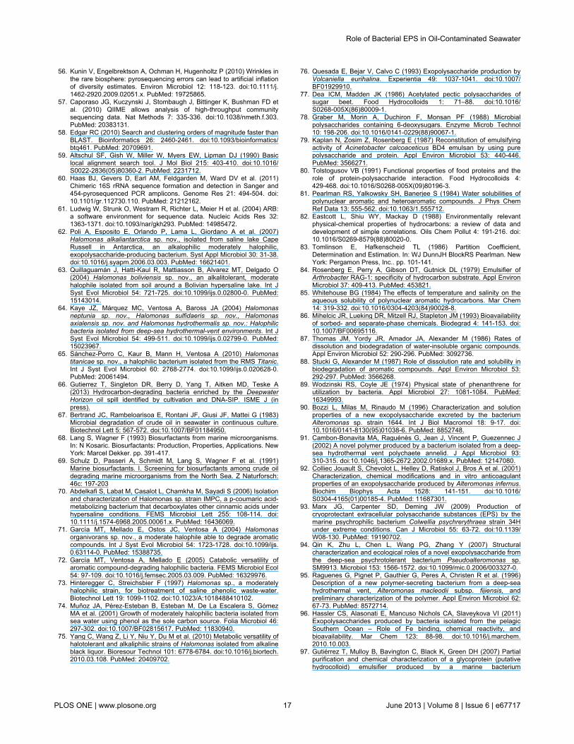
56. Kunin V, Engelbrektson A, Ochman H, Hugenholtz P (2010) Wrinkles inthe rare biosphere: pyrosequencing errors can lead to artificial inflationof diversity estimates. Environ Microbiol 12: 118-123. doi:10.1111/j.1462-2920.2009.02051.x. PubMed: 19725865.
57. Caporaso JG, Kuczynski J, Stombaugh J, Bittinger K, Bushman FD etal. (2010) QIIME allows analysis of high-throughput communitysequencing data. Nat Methods 7: 335-336. doi:10.1038/nmeth.f.303.PubMed: 20383131.
58. Edgar RC (2010) Search and clustering orders of magnitude faster thanBLAST. Bioinformatics 26: 2460-2461. doi:10.1093/bioinformatics/btq461. PubMed: 20709691.
59. Altschul SF, Gish W, Miller W, Myers EW, Lipman DJ (1990) Basiclocal alignment search tool. J Mol Biol 215: 403-410. doi:10.1016/S0022-2836(05)80360-2. PubMed: 2231712.
60. Haas BJ, Gevers D, Earl AM, Feldgarden M, Ward DV et al. (2011)Chimeric 16S rRNA sequence formation and detection in Sanger and454-pyrosequenced PCR amplicons. Genome Res 21: 494-504. doi:10.1101/gr.112730.110. PubMed: 21212162.
61. Ludwig W, Strunk O, Westram R, Richter L, Meier H et al. (2004) ARB:a software environment for sequence data. Nucleic Acids Res 32:1363-1371. doi:10.1093/nar/gkh293. PubMed: 14985472.
62. Poli A, Esposito E, Orlando P, Lama L, Giordano A et al. (2007)Halomonas alkaliantarctica sp. nov., isolated from saline lake CapeRussell in Antarctica, an alkalophilic moderately halophilic,exopolysaccharide-producing bacterium. Syst Appl Microbiol 30: 31-38.doi:10.1016/j.syapm.2006.03.003. PubMed: 16621401.
63. Quillaguamán J, Hatti-Kaul R, Mattiasson B, Alvarez MT, Delgado O(2004) Halomonas boliviensis sp. nov., an alkalitolerant, moderatehalophile isolated from soil around a Bolivian hypersaline lake. Int JSyst Evol Microbiol 54: 721-725. doi:10.1099/ijs.0.02800-0. PubMed:15143014.
64. Kaye JZ, Márquez MC, Ventosa A, Baross JA (2004) Halomonasneptunia sp. nov., Halomonas sulfidaeris sp. nov., Halomonasaxialensis sp. nov. and Halomonas hydrothermalis sp. nov.: Halophilicbacteria isolated from deep-sea hydrothermal-vent environments. Int JSyst Evol Microbiol 54: 499-511. doi:10.1099/ijs.0.02799-0. PubMed:15023967.
65. Sánchez-Porro C, Kaur B, Mann H, Ventosa A (2010) Halomonastitanicae sp. nov., a halophilic bacterium isolated from the RMS Titanic.Int J Syst Evol Microbiol 60: 2768-2774. doi:10.1099/ijs.0.020628-0.PubMed: 20061494.
66. Gutierrez T, Singleton DR, Berry D, Yang T, Aitken MD, Teske A(2013) Hydrocarbon-degrading bacteria enriched by the DeepwaterHorizon oil spill identified by cultivation and DNA-SIP. ISME J (inpress).
67. Bertrand JC, Rambeloarisoa E, Rontani JF, Giusi JF, Mattei G (1983)Microbial degradation of crude oil in seawater in continuous culture.Biotechnol Lett 5: 567-572. doi:10.1007/BF01184950.
68. Lang S, Wagner F (1993) Biosurfactants from marine microorganisms.In: N Kosaric. Biosurfactants: Production, Properties, Applications. NewYork: Marcel Dekker. pp. 391-417.
69. Schulz D, Passeri A, Schmidt M, Lang S, Wagner F et al. (1991)Marine biosurfactants. I. Screening for biosurfactants among crude oildegrading marine microorganisms from the North Sea. Z Naturforsch:46c: 197-203
70. Abdelkafi S, Labat M, Casalot L, Chamkha M, Sayadi S (2006) Isolationand characterization of Halomonas sp. strain IMPC, a p-coumaric acid-metabolizing bacterium that decarboxylates other cinnamic acids underhypersaline conditions. FEMS Microbiol Lett 255: 108-114. doi:10.1111/j.1574-6968.2005.00061.x. PubMed: 16436069.
71. García MT, Mellado E, Ostos JC, Ventosa A (2004) Halomonasorganivorans sp. nov., a moderate halophile able to degrade aromaticcompounds. Int J Syst Evol Microbiol 54: 1723-1728. doi:10.1099/ijs.0.63114-0. PubMed: 15388735.
72. García MT, Ventosa A, Mellado E (2005) Catabolic versatility ofaromatic compound-degrading halophilic bacteria. FEMS Microbiol Ecol54: 97-109. doi:10.1016/j.femsec.2005.03.009. PubMed: 16329976.
73. Hinteregger C, Streichsbier F (1997) Halomonas sp., a moderatelyhalophilic strain, for biotreatment of saline phenolic waste-water.Biotechnol Lett 19: 1099-1102. doi:10.1023/A:1018488410102.
74. Muñoz JA, Pérez-Esteban B, Esteban M, De La Escalera S, GómezMA et al. (2001) Growth of moderately halophilic bacteria isolated fromsea water using phenol as the sole carbon source. Folia Microbiol 46:297-302. doi:10.1007/BF02815617. PubMed: 11830940.
75. Yang C, Wang Z, Li Y, Niu Y, Du M et al. (2010) Metabolic versatility ofhalotolerant and alkaliphilic strains of Halomonas isolated from alkalineblack liquor. Bioresour Technol 101: 6778-6784. doi:10.1016/j.biortech.2010.03.108. PubMed: 20409702.
76. Quesada E, Bejar V, Calvo C (1993) Exopolysaccharide production byVolcaniella eurihalina. Experientia 49: 1037-1041. doi:10.1007/BF01929910.
77. Dea ICM, Madden JK (1986) Acetylated pectic polysaccharides ofsugar beet. Food Hydrocolloids 1: 71–88. doi:10.1016/S0268-005X(86)80009-1.
78. Graber M, Morin A, Duchiron F, Monsan PF (1988) Microbialpolysaccharides containing 6-deoxysugars. Enzyme Microb Technol10: 198-206. doi:10.1016/0141-0229(88)90067-1.
79. Kaplan N, Zosim Z, Rosenberg E (1987) Reconstitution of emulsifyingactivity of Acinetobacter calcoaceticus BD4 emulsan by using purepolysaccharide and protein. Appl Environ Microbiol 53: 440-446.PubMed: 3566271.
80. Tolstogusov VB (1991) Functional properties of food proteins and therole of protein-polysaccharide interaction. Food Hydrocolloids 4:429-468. doi:10.1016/S0268-005X(09)80196-3.
81. Pearlman RS, Yalkowsky SH, Banerjee S (1984) Water solubilities ofpolynuclear aromatic and heteroaromatic compounds. J Phys ChemRef Data 13: 555-562. doi:10.1063/1.555712.
82. Eastcott L, Shiu WY, Mackay D (1988) Environmentally relevantphysical-chemical properties of hydrocarbons: a review of data anddevelopment of simple correlations. Oils Chem Pollut 4: 191-216. doi:10.1016/S0269-8579(88)80020-0.
83. Tomlinson E, Hafkenscheid TL (1986) Partition Coefficient,Determination and Estimation. In: WJ DunnJH BlockRS Pearlman. NewYork: Pergamon Press, Inc.. pp. 101-141.
84. Rosenberg E, Perry A, Gibson DT, Gutnick DL (1979) Emulsifier ofArthrobacter RAG-1: specificity of hydrocarbon substrate. Appl EnvironMicrobiol 37: 409-413. PubMed: 453821.
85. Whitehouse BG (1984) The effects of temperature and salinity on theaqueous solubility of polynuclear aromatic hydrocarbons. Mar Chem14: 319-332. doi:10.1016/0304-4203(84)90028-8.
86. Mihelcic JR, Lueking DR, Mitzell RJ, Stapleton JM (1993) Bioavailabilityof sorbed- and separate-phase chemicals. Biodegrad 4: 141-153. doi:10.1007/BF00695116.
87. Thomas JM, Yordy JR, Amador JA, Alexander M (1986) Rates ofdissolution and biodegradation of water-insoluble organic compounds.Appl Environ Microbiol 52: 290-296. PubMed: 3092736.
88. Stucki G, Alexander M (1987) Role of dissolution rate and solubility inbiodegradation of aromatic compounds. Appl Environ Microbiol 53:292-297. PubMed: 3566268.
89. Wodzinski RS, Coyle JE (1974) Physical state of phenanthrene forutilization by bacteria. Appl Microbiol 27: 1081-1084. PubMed:16349993.
90. Bozzi L, Milas M, Rinaudo M (1996) Characterization and solutionproperties of a new exopolysaccharide excreted by the bacteriumAlteromonas sp. strain 1644. Int J Biol Macromol 18: 9-17. doi:10.1016/0141-8130(95)01038-6. PubMed: 8852748.
91. Cambon-Bonavita MA, Raguénès G, Jean J, Vincent P, Guezennec J(2002) A novel polymer produced by a bacterium isolated from a deep-sea hydrothermal vent polychaete annelid. J Appl Microbiol 93:310-315. doi:10.1046/j.1365-2672.2002.01689.x. PubMed: 12147080.
92. Colliec Jouault S, Chevolot L, Helley D, Ratiskol J, Bros A et al. (2001)Characterization, chemical modifications and in vitro anticoagulantproperties of an exopolysaccharide produced by Alteromonas infernus.Biochim Biophys Acta 1528: 141-151. doi:10.1016/S0304-4165(01)00185-4. PubMed: 11687301.
93. Marx JG, Carpenter SD, Deming JW (2009) Production ofcryoprotectant extracellular polysaccharide substances (EPS) by themarine psychrophilic bacterium Colwellia psychrerythraea strain 34Hunder extreme conditions. Can J Microbiol 55: 63-72. doi:10.1139/W08-130. PubMed: 19190702.
94. Qin K, Zhu L, Chen L, Wang PG, Zhang Y (2007) Structuralcharacterization and ecological roles of a novel exopolysaccharide fromthe deep-sea psychrotolerant bacterium Pseudoalteromonas sp.SM9913. Microbiol 153: 1566-1572. doi:10.1099/mic.0.2006/003327-0.
95. Raguenes G, Pignet P, Gauthier G, Peres A, Christen R et al. (1996)Description of a new polymer-secreting bacterium from a deep-seahydrothermal vent, Alteromonas macleodii subsp. fijiensis, andpreliminary characterization of the polymer. Appl Environ Microbiol 62:67-73. PubMed: 8572714.
96. Hassler CS, Alasonati E, Mancuso Nichols CA, Slaveykova VI (2011)Exopolysaccharides produced by bacteria isolated from the pelagicSouthern Ocean – Role of Fe binding, chemical reactivity, andbioavailability. Mar Chem 123: 88-98. doi:10.1016/j.marchem.2010.10.003.
97. Gutiérrez T, Mulloy B, Bavington C, Black K, Green DH (2007) Partialpurification and chemical characterization of a glycoprotein (putativehydrocolloid) emulsifier produced by a marine bacterium
Role of Bacterial EPS in Oil-Contaminated Seawater
PLOS ONE | www.plosone.org 17 June 2013 | Volume 8 | Issue 6 | e67717
Antarctobacter. Appl Microbiol Biotechnol 76: 1017-1026. doi:10.1007/s00253-007-1091-9. PubMed: 17641887.
98. Baelum J, Borglin S, Chakraborty R, Fortney JL, Lamendella R et al.(2012) Deep-sea bacteria enriched by oil and dispersant from the
Deepwater Horizon spill. Environ Microbiol 14: 2405-2416. doi:10.1111/j.1462-2920.2012.02780.x. PubMed: 22616650.
99. Fingas M, Fieldhouse B (2003) Studies of the formation process ofwater-in-oil emulsions. Mar Pollut Bull 47: 369-396. doi:10.1016/S0025-326X(03)00212-1. PubMed: 12899884.
Role of Bacterial EPS in Oil-Contaminated Seawater
PLOS ONE | www.plosone.org 18 June 2013 | Volume 8 | Issue 6 | e67717


