Embed Size (px)
Citation preview

3154–3163 Nucleic Acids Research, 2001, Vol. 29, No. 15 © 2001 Oxford University Press
Reverse transcriptase incorporation of1,5-anhydrohexitol nucleotidesKaren Vastmans, Matheus Froeyen, Luc Kerremans, Sylvie Pochet1 and Piet Herdewijn*
Laboratory of Medicinal Chemistry, Rega Institute for Medical Research, Katholieke Universiteit Leuven, Leuven,Belgium and 1Unité de Chimie Organique (ERS 558), Institut Pasteur, 25 Rue du Docteur Roux, 75015 Paris, France
Received May 4, 2001; Revised and Accepted June 14, 2001
ABSTRACT
Several reverse transcriptases were studied for theirability to accept anhydrohexitol triphosphates,having a conformationally restricted six-memberedring, as substrate for template-directed synthesis ofHNA. It was found that AMV, M-MLV, M-MLV (H–),RAV2 and HIV-1 reverse transcriptases were able torecognise the anhydrohexitol triphosphate assubstrate and to efficiently catalyse the incorpora-tion of one non-natural anhydrohexitol nucleotideopposite a natural complementary nucleotide.However, only the dimeric enzymes, the RAV2 andHIV-1 reverse transcriptases, seemed to be able tofurther extend the primer with another anhydrohexitolbuilding block. Subsequently, several HIV-1 mutants(4×AZT, 4×AZT/L100I, L74V, M184V and K65A) werelikewise analysed, resulting in selection of K65A and,in particular, M184V as the most succesful mutantHIV-1 reverse transcriptases capable of elongating aDNA primer with several 1,5-anhydrohexitoladenines in an efficient way. Results of kinetic exper-iments in the presence of this enzyme revealed thatincorporation of one anhydrohexitol nucleotide ofadenine or thymine gave an increased (for 1,5-anhydro-hexitol-ATP) and a slightly decreased (for 1,5-anhydro-hexitol-TTP) Km value in comparison to that of theirnatural counterparts. However, no more than fouranalogues could be inserted under the experimentalconditions required for selective incorporation.Investigation of incorporation of the altritol anhydro-hexitol nucleotide of adenine in the presence ofM184V and Vent (exo–) DNA polymerase proved thatan adjacent hydroxyl group on C3 of 1,5-anhydro-hexitol-ATP has a detrimental effect on the substrateactivity of the six-ring analogue. These results couldbe rationalised based on the X-ray structure of HIV-1reverse transcriptase.
INTRODUCTION
Reverse transcriptases (RT) play a defining role in the retro-virus life cycle. The enzyme is responsible for the synthesis of
a double-stranded DNA copy from the single-stranded RNAgenome, which is inserted into the host genome to establish theintegrated provirus (1). In an effort to obtain detailed structuralinformation about these enzymes and to gain considerableunderstanding of their biochemical mechanism, a sizeablenumber of crystal structures of RTs have been determined (2–6).Although these X-ray structures have provided useful infor-mation regarding the tertiary folding pattern and general loca-tion of the various amino acids, a detailed mapping of thesubstrate-binding site is still missing. With the exception of thethree acidic residues (Asp110, Asp185 and Asp186), which arepresumed to interact with the dNTP substrate by binding to thedivalent cation required for the polymerase reaction, no otherresidue has been shown experimentally to participate in thesubstrate binding function of human immunodeficiency virustype 1 (HIV-1) RT. To expand our knowledge in this field, a lotof mutational studies have been performed (7–15) to gain aninsight into the influence of altered amino acid residues onfidelity of DNA synthesis. In particular, residues belonging tothe dNTP-binding pocket (amino acids at positions 65, 72, 113,115, 151, 183, 184 and 219) have been investigated exten-sively. Since it is, however, not easy to translate structural datainto functional properties, an interesting strategy is to focus onthe difference in substrate specificity of analogues of naturalnucleoside triphosphates between several RTs. In this regard,triphosphate building blocks modified in their base part (16–20),sugar part (21–25) or triphosphate moiety (26) have beenstudied in primers, templates (18–20,27) or as triphosphatebuilding blocks (17–27) as substrates for HIV-1 (17–25),AMV (16,22,26,27) and M-MLV RTs (16,22). Concerningtriphosphate building blocks with an altered base moiety,several researchers (18–20,27,28) established that for mostRTs the size and shape complementarity of the base part of adNTP is responsible for the high fidelity during DNA replica-tion, rather than the ability of dNTPs to hydrogen bond to thetemplate nucleotide. Concerning the sugar part, it is proven, onthe one hand, that elongation cannot be accomplished in theabsence of a 3′-OH group (22). On the other hand, it has beenshown that the presence of a 3′-OH group does not automati-cally lead to extension. Also, some conformational flexibilityis proven to be indispensable for polymerisation (21–25).
Studies of this kind, investigating interactions between thepolymerase-binding site and triphoshates, are extremely usefulto ascertain the mechanism of substrate binding, providing abetter insight into the mechanism of fidelity during informationtransfer in replication and in disease states. This information
*To whom correspondence should be addressed. Tel: +32 16 337387; Fax: +32 16 337340; Email: [email protected]
Nucleic Acids Research, 2001, Vol. 29, No. 15 3155
takes on practical importance as well, since polymerases (suchas HIV-1 RT) are targets for a number of useful therapeuticagents. Moreover, identification of the parts of the dNTP moleculethat specifically bind to the active center of the enzyme areuseful for the design of new and more selective anti-retroviralagents.
The present study investigates the sugar part of dNTPs. Inparticular, the influence of the ring size of the sugar residue ofa triphosphate building block on its binding capacity to theactive site of RT is investigated, as well as its ability to beinserted into a DNA hybrid. Hereto, the anhydrohexitoltriphosphate analogues of adenine, guanine, cytosine andthymine have been synthesised. These analogues are similar tonatural dNTPs having the same base part and flexible triphos-phate moiety. The sugar residue, however, consists of a six-membered anhydrohexitol ring conformationally similar to the3′-endo (north) conformation found in rNTPs, in contrast to the2′-endo (south) conformation mainly present in dNTPs. Theanhydrohexitol triphosphate building blocks are also stericallylarger as well as conformationally locked and can therefore beused to study the influence of conformation and bulkiness ofthe sugar moiety on the replication process. This informationmay be used for the design of new anti-HIV agents having asix-membered carbohydrate moiety as the glycon part.
Recently it has been reported that these anhydrohexitoltriphosphates can be used as substrates for several DNApolymerases (29). Enzymes belonging to the B family seemedto be more efficient in this capacity. However, no more thantwo consecutive 1,5-anhydrohexitol nucleotides could beinserted into a DNA primer–template complex in a selectivemanner. In this study we have investigated whether RTs aremore able to accept six-membered nucleoside triphosphates assubstrates in DNA-templated HNA synthesis. In addition, therole of a 2′-hydroxyl group in the sugar part of a triphosphatehas been investigated by studying the substrate capacity of1,5-anhydro-2-deoxy-D-altritol nucleoside triphosphates(aNTPs) for RT. In contrast to 1,5-anhydrohexitol nucleosidetriphosphates (hNTPs), aNTPs possess an adjacent hydroxylgroup analogous to the hydroxyl group at the 2′-position of thesugar residue in rNTPs.
MATERIALS AND METHODS
Chemicals and DNA
The highly purified 2′-(deoxy)ribonucleoside triphosphatesused in the reverse transcriptase and DNA polymerase reac-tions and the Mono Q 5/5 and 10/10 columns were obtainedfrom Pharmacia. DEAE–Sephadex A-25 was purchased fromSigma.
Synthesis of hNTP and aATP
Anhydrohexitol nucleosides (hN) were prepared as previouslydescribed (30,31). Their respective monophosphates wereobtained by means of the method of Yoshikawa et al. (32,33).The triphosphate residues (hNTP) were synthesised asdescribed by Moffat and Khorana (34). Convertion to theirsodium salts, HPLC purification and NMR identification wereperformed according to previously described procedures (29).
The 1,5-anhydro-2-deoxy-D-altritol nucleoside of adenine(aA) was prepared as described (35,36). The triphosphate
residue was synthesised from its respective nucleoside in atwo-step procedure. First, the monophosphate (aAMP) wasformed according to the method of Yoshikawa et al. (32,33).Treatment of the nucleotide morpholidate with tributylammoniumpyrophosphate yielded the triphosphate (aATP) (34). Thetriphosphate was than dissolved in H2O and applied to aSephadex column (Sephadex A-25 in the HCO3
– form). Alinear gradient of TEAB (0–0.5 M TEAB) was used to elute theproduct. Following collection of the major triphosphate peak,it was converted to its sodium salt by addition of a 1 M solutionof NaI in acetone. The precipitate was washed and dried over-night in vacuo over phosphorus pentoxide at room tempera-ture. The identity of the product was confirmed by NMR andHPLC analysis performed as described below. To removehydrolysed triphosphate products which could be formedduring storage of the material, aATP was freshly purifiedbefore use on a mono Q 10/10 column (Pharmacia) by prepar-ative ion exchange HPLC. A linear gradient of TEAB (pH 7.5)from 0.2 to 0.65 M over 45 min was used, with a retention timeof ∼35 min. The overall yield for the isolated aATP was ∼40%.31P NMR (aATP) δ (p.p.m.) (D2O) –9.70 (d, γ-P), –10.0 (d, α-P),–22.15 (t, β-P). The extinction coefficient ε (λ = 260 nM) ofaATP (15000 M–1 cm–1) was used to determine the respectiveconcentration.
HPLC
Ion exchange HPLC was performed using an L-6200 A Merck-Hitachi pump with UV monitoring. For hNTP, samples werepurified and analysed as described. For aATP, analysis of thesamples was achieved on a Mono Q 5/5 column using a lineargradient of TEAB (pH 7.5) from 0.1 to 0.6 M over 40 min at aflow rate of 1.0 ml/min. For preparative purposes, a semi-preparative Mono Q 10/10 column was used with a flow rate of2 ml/min and a gradient of 0.2 to 0.65 M TEAB over 45 min.
Spectroscopy
The 31P NMR spectra were recorded with a Varian Unity 500spectrometer with 85% H3PO4 in H2O as internal standard.Samples for NMR spectroscopy were prepared in a non-buff-ered solution of 99.96% D2O.
Oligodeoxyribonucleotides
Oligodeoxyribonucleotides were obtained from Eurogentec.The lyophilised oligonucleotides were dissolved in DEPC-treated water and stored at –20°C. Labelling of the primeroligonucleotides at their 5′-end was achieved followingstandard procedures using T4 polynucleotide kinase (LifeTechnologies) and [γ-32P]ATP (4500 Ci/mmol, 10 mCi/ml)(ICN).
Reverse transcriptase reactions
Hybridisation was performed by combining 5′-end-labelledprimers with their template in a molar ratio of 1:2.5. Hybridswere denaturated by heating at 70°C for 10 min and annealedby slow cooling to room temperature over 2.5 h. Reactionswere initiated by adding 3 µl of enzyme dilution to a reactionmixture of 6 µl consisting of hybrid, reaction buffer (suppliedwith the RTs) and triphosphate building block.
The first series of incorporation experiments were set up toinvestigate several commercially available RTs for theircapacity to insert one, two or three hATP into the hybrids P1/T1,

3156 Nucleic Acids Research, 2001, Vol. 29, No. 15
P1/T2 and P1/T3 (Fig. 1), respectively. The enzymes consid-ered were the RTs of avian myeloblastis virus (AMV), Moloneymurine leukaemia virus [both the wild-type (M-MLV) and theRNase H– point mutant (M-MLV (H–)] (all provided byPromega) and the RTs of Rous-associated virus 2 (RAV2) andHIV-1 (both from Amersham). A second series of insertionexperiments focused on mutant HIV-1 RTs and their ability toincorporate three consecutive 1,5-anhydrohexitol adenine (hA)into hybrid P1/T3. Hereto, a series of reactions was performed inthe presence of each of five enzymes: 4×AZT, 4×AZT/L100I,L74V, K65A and M184V. The final reaction mixturescontained 50 nM hybrid, 100 µM triphosphate building block(dATP, hATP, dGTP, dGTP + dATP or dGTP + hATP) and0.5 U/µl commercially available RT for the first series ofincorporation experiments. For the second series of insertionassays 0.005 U/µl (mutant) HIV-1 RT was used and 100 µMdATP or hATP. The reaction mixtures were incubated at 37°Cand aliquots were quenched after 120 min.
Selectivity of incorporation was investigated by annealingprimers P1, P2, P3 and P4 to template T4 (Fig. 1) as describedabove. A series of 15 µl reactions was performed in the presenceof M184V HIV-1 RT. The final mixture contained 50 nMprimer–template complex, 10 µM each triphosphate buildingblock (for hybrid P1/T4, dT, hT, dG, hG, dT + dG, hT + hG, dT+ dG + dA, hT + hG + hA, dT + dG + dA + dC or hT + hG +hA + hC; for hybrid P2/T4, dG, hG, dA, hA, dG + dA, hG +hA, dG + dA + dC, hG + hA + hC, dG + dA + dC + dT or hG+ hA + hC + hT; for hybrid P3/T4, dA, hA, dC, hC, dA + dC,hA + hC, dA + dC + dT or hA + hC + hT; for hybrid P4/T4, dC,hC, dT, hT, dC + dT or hC + hT) and 0.001 U/µl M184V. Themixture was incubated at 37°C and the reaction was quenchedafter 60 min.
Incorporation of multiple hN and aN nucleotides
Two series of insertion experiments were performed in thepresence of either M184V or Vent (exo–) DNA polymerase anda hybrid consisting of primer P1 annealed to template T5. Thefinal reaction mixture consisted of 50 nM hybrid, 10 or 1000 µMtriphosphate building block (dATP, rATP, hATP or aATP) and0.005 U/µl Vent (exo–) or M184V. Reaction temperature was55°C for Vent (exo–) DNA polymerase and 37°C for M184V.Reaction times were 10 and 60 min.
Kinetic experiments
Kinetic parameters were derived from a steady-state kineticassay (37) in the presence of dATP, hATP, dTTP and hTTP.The incorporation experiment was started by mixing M184Vdilution with a mixture containing P3-T4 complex (Fig. 1) forkinetic characterisation of dATP or hATP, and P1/T4 complex(Fig. 1) for dTTP and hTTP. The final mixture was composedof 250 nM primer–template complex, 0.0001 U/µl M184V,reaction buffer and various concentrations of triphosphatebuilding block. A concentration range of 0.125–25 µM wasused for all four triphosphate building blocks. Reactions wereincubated at 37°C. Reaction times were between 1 and 12 min.
Electrophoresis
During enzymatic incorporation assays, samples wereremoved and reactions were terminated by adding a doublevolume of loading buffer (95% formamide, 0.05%bromophenol blue, 0.05% xylene cyanol and 50 mM EDTA).
The products were heat denaturated at 70°C for 5 min andseparated on a 0.4 mm 20% denaturating polyacrylamide gel,in the presence of 89 mM Tris–borate and 2 M EDTA buffer,pH 8.3, at 2000 V for ∼2 h. Visualisation of the polymerisedproducts was performed with a phosphorimager. The amountof radioactivity in the bands representing the respective poly-merised products of enzymatic reactions was determined withOptiquant image analysis software (Packard).
RESULTS
Incorporation of hATP into a DNA hybrid and extensionby RTs
In a first series of incorporation experiments, five commer-cially available RTs were investigated for their capacity toaccept hNTPs as substrates and incorporate them into a DNA–DNA hybrid. Incorporation assays were performed by deter-mining the insertion of one, two or three hATPs in the presenceof each of five RTs and primed (P1) templates T1, T2 and T3,respectively (Fig. 1). Likewise, further elongation with thenatural guanosine triphosphate building block (dGTP)following insertion of the anhydrohexitol nucleotide ofadenine was investigated. Table 1 gives quantitative data onthe incorporation experiments in the presence of hybrid P1/T1and P1/T2. The percentages represent the amount of elongationproduct in comparison to the total amount of primer originallypresent in the sample. These results demonstrate that all RTsconsidered can efficiently incorporate one anhydrohexitolnucleotide with an adenine base moiety. However, the RAV2and HIV-1 RTs are the only RTs that succeeded in incorpo-rating two (RAV2 RT) or more (HIV-1 RT) hA opposite theirnatural counterparts in the template. HIV-1 RT seems to be themost error prone, since it succeeded in insertion of three andfour consecutive hA, using P1/T1 and P1/T2 as hybrids,respectively, as a result of misincorporation opposite thenatural dC building block in the template. Further elongationwith a natural nucleotide after this insertion seemed possiblefor all the considered RTs, except M-MLV (H–) RT using P1/T1as hybrid. In the presence of P1/T2 further polymerisation wasonly possible for HIV-1 RT.
Figure 2 shows the incorporation patterns of the RTs consideredin the presence of M-MLV (Fig. 2A), M-MLV (H–) (Fig. 2B),
Figure 1. Sequences of primers (P) and templates (T) used in the incorporationexperiments.

Nucleic Acids Research, 2001, Vol. 29, No. 15 3157
AMV (Fig. 2C), RAV2 (Fig. 2D) and HIV-1 (Fig. 2E) RTs incombination with several triphosphate building blocks,including dATP (lane 1), hATP (lane 2), dGTP (lane 3), dGTP+ dATP (lane 4) or dGTP + hATP (lane 5). It is shown thatM-MLV, M-MLV (H–) and AMV RTs, although efficientlyincorporating dA (Fig. 2A–C, lanes 1), only succeed inincorporation of one hA (Fig. 2A–C, lanes 2). The RT ofM-MLV proved to have high fidelity since, under the consideredreaction conditions, dG is not misincorporated opposite dT inthe template (Fig. 2A and B, lanes 3). In the presence of AMVRT this misinsertion is unavoidable (Fig. 2C, lane 3). ConsideringRAV2 and HIV-1 RTs, an insertion of three hA is seen(Fig. 2D and E, lanes 2) (although very weak for RAV2 RT)relative to incorporation of four building blocks in the presenceof dA (Fig. 2D and E, lanes 1) (the last incorporation resultingfrom misinsertion of dA opposite dC in the template). Arelatively small amount of dG is incorporated in the presenceof RAV2 RT (Fig. 2D, lane 3), in contrast to full-lengthextension using HIV-1 RT (Fig. 2E, lane 3). Extension with dGafter inserting hA in the DNA–DNA hybrid only seemedpossible in the presence of HIV-1 RT (Fig. 2E, lane 5), with aclear pause at positions p + 2 and p + 3. The promising resultsachieved with HIV-1 RT in the incorporation assay stimulatedus to investigate whether HIV-1 RT mutants could be selectedthat insert three hA consecutively into a DNA hybrid. In thissecond series of insertion assays, five HIV-1 mutants were
considered, investigating incorporation of three triphosphatebuilding block analogues of adenine into hybrid P1/T3. InFigure 7, the different amino acids, representing the HIV-1 RTmutants, are indicated in their respective positions 67, 70, 215and 219 (4×AZT) and 100 (4×AZT/L100I), 74 (L74V), 65(K65A) and 184 (M184V). Results of the incorporation exper-iments are given in Figure 3. Wild-type HIV-1 RT (wt HIV-1RT), under the considered reaction conditions (enzymeconcentration 100 times lower than in the first series ofincorporation assays), is able to insert one hA into hybrid P1/T3with an efficiency that equals that of insertion of a natural dA.RTs 4×AZT, 4×AZT/L100I and L74V succeed in incorpo-rating a second hA, although with an efficiency that is muchbelow that of a natural dA. RTs K65A and M184V are capableof elongating the primer with three hA. The first two buildingblocks are incorporated as efficiently as dA. Extension with athird hA is <50% compared to primer extension in the presenceof dATP for K65A, in contrast to M184V (primer extension>50% of primer extension in the presence of dATP).
Selectivity tests
To investigate whether hNTPs could be incorporated selec-tively into a DNA hybrid opposite their natural counterparts, aseries of insertion experiments was performed in the presenceof each of four hybrids, P1/T4, P2/T4, P3/T4 and P4/T4. Eachhybrid was used in four series of incorporation assays in the
Table 1. Percentage enzymatic incorporation of hATP and further elongation in the presence of hybrids P1/T1 and P1/T2 by different reverse transcriptases
The given percentages represent the total amount of extension products in comparison to the original total amount of primer. By how many building blocks theprimer was elongated is also indicated.
Hybrid P1/T1 Hybrid P1/T2
hATP incorporation hATP + dGTP hATP incorporation hATP + dGTP
M-MLV RT 89% p + 1 101.1% p + 5 82.8% p + 1 68.1% p + 1
M-MLV (RNase H–) RT 86.7% p + 1 84.35 p + 1 83.0% p + 1 79.4% p + 1
AMV RT 94.9% p + 1 101.0% p + 5 94.2% p + 1 98.3% p + 1
RAV2 RT 97.4% p + 1 98.0% p + 5 94.1% p + 2 94.4% p + 2
HIV-1 RT 103.2% p + 3 101.0% p + 7 102.7% p + 4 97.3% p + 6
Figure 2. Phosphorimages of the enzymatic incorporation of three hA into 50 nM hybrid P1/T3 in the presence of 0.5 U/µl M-MLV RT (A), M-MLV (H–) RT(B), AMV RT (C), RAV2 RT (D) and HIV-1 RT (E) and 100 µM NTP: lane 1, dATP; lane 2, hATP; lane 3, dGTP; lane 4, dGTP + dATP; lane 5, dGTP + hATP.Bl, blank reaction in the absence of NTP. The reaction time was 120 min.

3158 Nucleic Acids Research, 2001, Vol. 29, No. 15
presence of different triphosphate building blocks. In this way,insertion of the hN building blocks was investigated incomparison to their natural analogues. The reaction conditionsused (as described in Materials and Methods) were a compro-mise between an acceptable degree of incorporation and thelowest degree of misincorporation for the anhydrohexitolanalogues. Figure 4 shows the resulting phosphorimages in thepresence of hybrid P1/T4 (Fig. 4A), hybrid P2/T4 (Fig. 4B),hybrid P3/T4 (Fig. 4C) and hybrid P4/T4 (Fig. 4D). Whentaking a closer look at these pictures, it is seen that, using onetriphosphate building block of the anhydrohexitol analogue assubstrate, the desired incorporation pattern is seen. There isinsertion of the analogue complementary to the natural base inthe template (lane 3), however, no incorporation is seen usingthe wrong substrate (lanes 5, 13 and 15). In contrast, addingdNTP as substrate in some cases resulted in misinsertion(Fig. 4B, lane 2, and Fig. 4D, lanes 2 and 4). When using morethan one triphosphate building block in the reaction mixture(lanes 6–11), the desired insertion patterns for hNTPs areobserved, however, it seems that the primer could not beelongated by more than four building block analogues. UsingdNTPs in the reaction mixture, full-length products could beobtained. However, more misinsertions were seen in compar-ison to their six-ring analogues (Fig. 4A and C, lanes 6, andFig. 4A and B, lanes 8). From the picture it is also clear that,despite the fact that M184V is 3′→5′ exonuclease free, degra-dation of the primer is seen using the wrong substrate in thereaction mixture (Fig. 4A–D, lanes 4–5 and lanes 12–13).
Incorporation of multiple hN and aN nucleotides
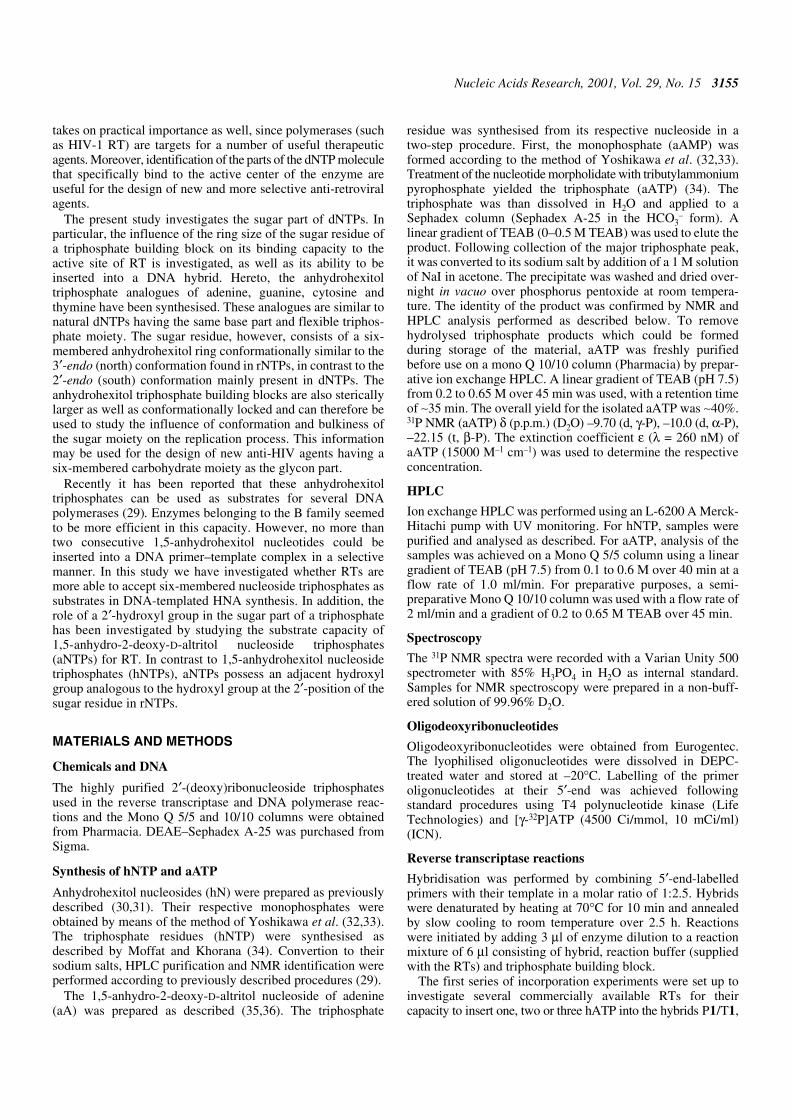
We investigated whether elongation of a DNA hybrid withmore than three anhydrohexitol triphosphate building blockscould be achieved. In addition, the substrate activity of aATP(Fig. 5) for M184V and Vent (exo–) was investigated. Thus,incorporation was performed in the presence of hybrid P1/T5(Fig. 6) consisting of a template with a seven base overhang ofnatural thymine building blocks. Figure 6A represents theincorporation pattern of the triphosphate building blocks of
adenine in the presence of M184V (lanes 1–4) and Vent (exo–)DNA polymerase (lanes 6–9) under conditions allowing selec-tive incorporation of the anhydrohexitol analogues. Theseconditions were derived in the above mentioned selectivityexperiment for M184V and in previously described experi-ments for Vent (exo–) DNA polymerase (29). The picturemakes clear that under selective reaction conditions (Fig. 6A),a similar insertion profile is seen for both M184V and Vent(exo–). However, under more drastic reaction conditions(Fig. 6B) the incorporation pattern is quite different for the twoenzymes. For Vent (exo–), incorporation of rA (lane 7) isfavoured over hA (lane 8), in contrast to M184V, which prefershATP (lane 3) as substrate over rATP (lane 2). Neither enzymeseemed to accept the altritol analogue as a good substrate(lanes 4 and 9).
Steady-state kinetic analysis of hATP and hTTP insertionin a DNA hybrid
To gain an idea of the efficiency of incorporation by M184V ofone triphosphate building block of the anhydrohexitolanalogues of adenine and thymine in comparison to the respec-tive natural nucleotides, the kinetic parameters Km and Vmaxwere determined. This experiment was performed according tothe standing start assay described by Boosalis et al. (37). Thetarget site was placed at the first position downstream of the5′-end label of the primer. Quantification of radioactive spotsrepresenting polymerised products and remaining primersallowed calculation of the initial velocity of the reaction.Table 2 lists the kinetic parameters (averages of three experi-ments) for the four triphosphates considered. These data showa decrease in Vmax values for both hATP and hTTP in comparisonto the natural nucleotides. This decrease was more pronouncedwhen thymine was the base. Km values, on the other hand,increased for hATP and slightly decreased for hTTP ascompared to dATP and dTTP, respectively.
DISCUSSION
Results from insertion studies investigating incorporation ofhA into hybrids P1/T1, P1/T2 (Table 1) and P1/T3 (Figs 2 and3) reveal that all RT considered are able to elongate a DNAprimer with one anhydrohexitol building block analogue oppositeits natural counterpart in the template. A comparison betweenthe incorporation capacity of different RTs gives valuableinformation about the fidelity of the RT considered. Based onour results, HIV-1 RT seems to be the most error prone relativeto the other RT, since it can incorporate additional hA oppositedC in templates T1 and T2 after insertion of one hA. AMV,M-MLV and M-MLV (H–) RTs, under the reaction conditionsconsidered, cannot further elongate the DNA primer. Thisobservation supports the generalisation that RTs of lentiviruses(EIAV, HIV-1 and HIV-2 RTs) are usually more error prone inDNA synthesis than other retroviral RTs (7,8), as was alsoillustrated by Morales and Kool (16). Moreover, it suggeststhat all RTs, although structurally related, have different charac-teristics that might explain their divergent capacity to acceptanalogues as substrates. Consequently, to support thishypothesis each polymerase must be described individually.However, the incorporation experiment in the presence ofDNA primer P1 annealed to T2 (Table 1), investigating theability of RTs to elongate the primer with more than one hA
Figure 3. Representation of results of the enzymatic incorporation of three hAinto 50 nM hybrid P1/T3 in the presence of 0.005 U/µl wild-type HIV RT andits mutants 4×AZT, 4×AZT/L100I, L74V, M184V and K65A (indicated at thebottom of each bar) and 100 µM NTP: dATP and hATP. The reaction time was120 min. Each bar is divided into three parts representing the insertion of one,two and three hA, respectively. The filling gives an idea of the relative amountof hA that is incorporated relative to insertion of a natural dA.

Nucleic Acids Research, 2001, Vol. 29, No. 15 3159
opposite their natural counterparts, allows us to divide the RTsinto two distinct groups. On the one hand, the dimeric enzymes,the RTs of RAV2 and HIV-1, can insert two consecutive hAopposite dT in the template. On the other hand, the monomericenzymes, the RTs of AMV, M-MLV and M-MLV (H–), canextend the primer by only one building block analogue. One ofthe dimeric enzymes, HIV-1 RT, is the only enzyme that
inserted three consecutive hA into a DNA hybrid in an efficientway (Fig. 2). Due to the discovery of HIV RT as a causativeagent of AIDS in the last decade, an enormous amount ofresearch has been performed to obtain a detailed map of thevarious amino acids involved in HIV-1 RT fidelity duringDNA synthesis (7–15). In particular, much attention has beenpaid to the dNTP-binding site. To further extend our knowledgeabout which amino acids in HIV-1 RT are involved in sugarrecognition of an incoming dNTP during DNA synthesis, fiveHIV-1 RT mutants were investigated for their ability to accepthA as a substrate. These investigations might be useful in thedesign of new nucleoside analogues active against mutatedviruses. The M184V mutant of HIV-1 RT, in which Met184 isreplaced by valine, resulted in a higher efficiency of incorpora-tion of hA into a DNA hybrid (Fig. 3). Figure 7 represents thepolymerase active site of HIV-1 RT complexed with a DNAtemplate–primer duplex and an incoming dTTP (yellow) or
Figure 4. Phosphorimages of selectivity tests in the presence of 0.005 U/µl M184V and 50 nM hybrid P1/T4 (A), hybrid P2/T4 (B), hybrid P3/T4 (C) or hybridP4/T4 (D). The concentration of building blocks is 10 µM. On the bottom of the resulting phosphorimages are indicated the triphosphate building blocks added assubstrates. The left lane of the two columns represents the natural triphosphate building block, the right lane its anhydrohexitol analogue. (A) Lane 2, dTTP; lane3, hTTP; lane 4, dGTP; lane 5, hGTP; lane 6, dTTP + dGTP; lane 7, hTTP + hGTP; lane 8, dTTP + dGTP + dATP; lane 9, hTTP + hGTP + hATP; lane 10, dTTP+ dGTP + dATP + dCTP; lane 11, hTTP + hGTP + hATP + hCTP; lane 12, dATP; lane 13, hATP; lane 14, dCTP; lane 15, hCTP. (B) Lane 2, dGTP; lane 3, hGTP;lane 4, dATP; lane 5, hATP; lane 6, dGTP + dATP; lane 7, hGTP + hATP; lane 8, dGTP + dATP + dCTP; lane 9, hGTP + hATP + hCTP; lane 10, dGTP + dATP +dCTP + dTTP; lane 11, hGTP + hATP + hCTP + hTTP; lane 12, dCTP; lane 13, hCTP; lane 14, dTTP; lane 15, hTTP. (C) Lane 2, dATP; lane 3, hATP; lane 4,dCTP; lane 5, hCTP; lane 6, dATP + dCTP; lane 7, hATP + hCTP; lane 8, dATP + dCTP + dTTP; lane 9, hATP + hCTP + hTTP; lane 12, dGTP; lane 13, hGTP;lane 14, dATP; lane 15, hATP. (D) Lane 2, dCTP; lane 3, hCTP; lane 4, dTTP; lane 5, hTTP; lane 6, dCTP + dTTP; lane 7, hCTP + hGTP; lane 12, dGTP; lane 13,hGTP; lane 14, dATP; lane 15, hATP. Lane 1 represents the blank reaction in the absence of NTP and enzyme. The reaction time was 60 min.
Figure 5. Structures of the anhydrohexitol nucleoside triphosphates (R = H)[base = adenine (hATP), guanine (hGTP), cytosine (hCTP) or thymine (hTTP)]and altritol nucleoside triphosphate (R = OH) of adenine [base = adenine(aATP)].
3160 Nucleic Acids Research, 2001, Vol. 29, No. 15
hTTP (grey). In the figure, the different amino acids repre-senting the HIV-1 RT mutants are indicated in the respectivepositions 67, 70, 215 and 219 (4×AZT) and 100 (4×AZT/L100I), 74 (L74V), 65 (K65A) and 184 (M184V). As alreadypreviously suggested (38,39), this figure further confirms thatthere is a direct interaction of the amino acid 184 side chainwith the sugar moiety of the dNTP substrate. Met184 indeedlies in the vicinity of the sugar residue of an incoming anhydro-hexitol. Amino acid 65 is also in the proximity of the sugarpart. It is therefore not surprising that a different incorporationpattern of hATP is seen for M184V and K65A in comparisonto wild-type HIV RT and the other mutants considered.
As indicated in our kinetic experiments in the presence ofM184V, there is a difference in Km and Vmax values for hATPand hTTP in comparison to dATP and dTTP, respectively. TheKm values increased (adenine) or decreased slightly (thymine).The major difference between dNTPs and hNTPs is the presenceof a rigid six-membered ring for hNTPs with a conformationsimilar to the 3′-endo conformation of an RNA building block.Since in nature RTs use dNTPs, having a 2′-endo conforma-tion, as substrate, it is not surprising that a lower affinity ofM184V for hATP is observed.
DNA polymerases are thought to catalyse DNA polymerisationby sequential conformational changes in the enzyme structure(40,41). Likewise, HIV-1 RT is considered to undergo aconformational change from the open to the closed state,
positioning the nucleotide for phosphodiester bond formation(42). Since hNTPs, however, have a rigid sugar moiety, thechange in conformation, necessary for elongation, is hindered,resulting in chain termination. This phenomenon is illustratedin our selectivity experiments, where no further elongation canbe achieved after incorporation of four building blockanalogues. It confirms the observations from other researchersthat a certain flexibility in conformation is indispensable forelongation (21,22). Previous selectivity experiments in thepresence of Vent (exo–) DNA polymerase and hNTPs showedthat chain termination was achieved after incorporation of onlytwo building block analogues opposite their natural counter-parts in the template under experimental conditions allowingfor selective incorporation (29). This observation is inagreement with the common view that the active site of HIV-1RT, capable of inserting two more hN under similar reactionconditions, is more flexible than those of other polymerases.That there is indeed a difference between RTs and DNApolymerases in their ability to accept triphosphate analoguesfor DNA replication is also demonstrated by the difference inincorporation pattern obtained in the experiment comparinginsertion in a DNA hybrid of dATP, rATP, hATP and aATP inthe presence of Vent (exo–) DNA polymerase and M184V(Fig. 6). The results for incorporation of aATP are interestingin this regard. The structure and conformation of aATP issimilar to that of hATP (35,36). However, the hexitol ring inaATP has an extra hydroxyl group analogous to the OH groupat the 2′ position of the sugar residue in rNTPs. No or onlyweak incorporation could be observed for both enzymes, incontrast to a more pronounced insertion of dATP and hATPunder the same reaction conditions. These results suggest thatboth polymerases are hindered in their polymerisation functionby the presence of the adjacent hydroxyl group. A few yearsago Astatke et al. (43) and Gao et al. (44) and very recentlyMarquez et al. (24) mentioned that a single amino acid residueat the active site of the polymerases is considered to be responsiblefor discrimination against the 2′ residue of an incoming ribo-nucleotide. For DNA polymerases, this residue has been
Table 2. Kinetic parameters of M184V using a steady-state kinetic analysis ofhATP and hTTP in a DNA hybrid
Substrate Vmax (% min–1) Km (µM)
dATP 2.80 ± 0.10 0.23 ± 0.04
hATP 2.17 ± 0.12 2.26 ± 0.50
dTTP 2.30 ± 0.13 0.98 ± 0.26
hTTP 1.27 ± 0.07 0.60 ± 0.16
Figure 6. Phosphorimage of the enzymatic incorporation of seven A into 50 nM P1/T5. Both (A) and (B) represent incorporation patterns in the presence of 0.005 U/µlM184V (lanes 1–4) or Vent (exo–) DNA polymerase (lanes 6–9). To clarify the picture, the enzymes considered are indicated at the top and the triphosphate build-ing blocks used as substrates at the bottom of the resulting phosphorimages. (A) A concentration of 10 µM NTP was used and the reaction time was 10 min.(B) Insertion in the presence of 1000 µM NTP at a reaction time of 60 min. The NTP was dATP (lanes 1 and 6), rATP (lanes 2 and 7), hATP (lanes 3 and 8) oraATP (lanes 4 and 9). Bl, blank reaction in the absence of NTP and enzyme.

Nucleic Acids Research, 2001, Vol. 29, No. 15 3161
identified in the Klenow fragment as Glu710 (43). For RTs,Phe155 in M-MLV RT (similar to Tyr115 in HIV-1 RT) isassumed to be responsible for this phenomenon. This is illus-trated in Figure 7, showing Tyr115 lying in the vicinity of thehydroxyl group on position C2 (dNTP) or C3 (hNTP).
That the 2′-OH group, however, is not the only factorresponsible for the decrease in efficiency is illustrated by thefact that rATP, in contrast to aATP, can be inserted by M184V(Fig. 6B, lane 2) and Vent (exo–) DNA polymerase (Fig. 6B,lane 7) into the DNA hybrid under extreme reaction condi-tions. Since aATP and rATP have comparable conformationalstates it is evident that the decrease in incorporation is due tothe locked form of the more bulky six-membered ring, incontrast to the flexible furanose ring in dNTP and rNTP.
CONCLUSION
It has been shown that triphosphate building blocks having anunnatural 1,5-anhydrohexitol ring can be accepted assubstrates by all RTs. Km values, however, indicate a loweraffinity of the six-membered ring of the anhydrohexitolanalogue for the RT active center when adenine is the base.Moreover, the presence of an adjacent hydroxyl group at thesix-membered ring analogous to the hydroxyl group at the 2′position of the sugar residue in rNTPs proves to be detrimentalfor substrate activity of the building block. The selectivityassay suggests that the locked form of the more bulky six-membered ring is most probably the reason for chain termination
after incorporation of several hexitol nucleotides. Since previ-ously published data in the presence of Vent (exo–) DNApolymerase revealed insertion of only two anhydrohexitoltriphosphates into a DNA–DNA hybrid, the hypothesis thatRTs possess a ‘looser’ active site in comparison to DNApolymerases is confirmed. The observation of M184V as themost succesful HIV-1 RT mutant for consecutive incorpora-tion of the anhydrohexitol analogues suggests a role of Met184in HIV-1 RT in the interaction with the sugar part of anincoming triphosphate building block. The present investiga-tions are useful for the design of new anti-HIV agents based onthe anhydrohexitol scaffold.
ACKNOWLEDGEMENTS
We are grateful to R. Busson for performance and interpreta-tion of the NMR spectra. We also thank Arthur Van Aerschotfor helpful discussions. We are deeply thankful to Prof. J.Balzarini for providing the mutants of HIV-1 RT. Financialsupport for this work was provided by a fellowship of the IWTfor K.V. and by the ‘Geconcerteerde OnderzoeksActie’ of theKatholieke Universiteit Leuven (GAO 97/11).
REFERENCES
1. Gao,G. and Goff,S.P. (1998) Replication defect of Moloney murineleukemia virus with a mutant reverse transcriptase that can incorporateribonucleotides and deoxyribonucleotides. J. Virol., 72, 5905–5911.
Figure 7. A model for the RT–DNA–hATP complex. The model has been created starting from the RT–DNA–dNTP structure (pdb code 1 RTD) (6). The dNTP:dAbase pair (shown in yellow) was replaced by a hA:dT (CPK colours), after which the complex was energy minimised using the Amber software (45). The RTsurface is rendered as blue (fingers domain), flesh (thumb domain), cream (palm domain) and white (rest). Amino acids Y115 (grey) and M184 (red), which mayinteract with the sugar of the entering nucleotide, are highlighted. The positions of other amino acids involved in resistance to some non-nucleoside RT inhibitorsare labelled: 4×AZT (yellow), 4×AZT/L100I (red), K65A (white) and L74V (pink). The picture was generated using Bobscript and Raster3D (46–48).

3162 Nucleic Acids Research, 2001, Vol. 29, No. 15
2. Najmudin,S., Coté,M.L., Sun,D., Yohannan,S., Montano,S.P., Gu,J. andGeorgiadis,M.M. (2000) Crystal structures of an N-terminal fragmentfrom moloney murine leukemia virus reverse transcriptase complexedwith nucleic acid: functional implications for template-primer binding tothe fingers domain. J. Mol. Biol., 296, 613–632.
3. Rodgers,D.W., Gamblin,S.J., Harris,B.A., Ray,S., Culp,J.S., Hellmig,B.,Woolf,D.J., Debouck,C. and Harrison,S.C. (1995) The structure ofunliganded reverse transcriptase from the human immunodeficiency virustype 1. Proc. Natl Acad. Sci. USA, 92, 1222–1226.
4. Kohlsteadt,L.A., Wang,J., Friedman,J.M., Rice,P.A. and Steitz,T.A.(1992) Crystal structure at 3.5 Å resolution of HIV-1 reverse transcriptasecomplexed with an inhibitor. Science, 256, 1783–1790.
5. Jacobo-Molina,A., Ding,J., Nanni,R.G., Clark,A.D., Lu,X., Tantillo,C.,Williams,R.L., Kamer,G., Ferris,A.L., Clark,P. et al. (1993) Crystalstructure of human immunodeficiency virus type 1 reverse transcriptasecomplexed with double-stranded DNA at 3.0 Å resolution shows bentDNA. Proc. Natl Acad. Sci. USA, 90, 6320–6324.
6. Huang,H., Chopra,R., Verdine,G.L. and Harrison,S.C. (1998) Structure ofa covalently trapped catalytic complex of HIV-1 reverse transcriptase:implications for drug resistance. Science, 282, 1669–1675.
7. Bakhanashvili,M., Avidan,O. and Hizi,A. (1996) Mutational studies ofhuman immunodificiency virus type 1 reverse transcriptase: theinvolvement of residues 183 and 184 in the fidelity of DNA synthesis.FEBS Lett., 391, 257–262.
8. Rubinek,T., Bakhanashvili,M., Taube,R., Avidan,O. and Hizi,A. (1997)The fidelity of 3′ misinsertion and mispair extension during DNAsynthesis exhibited by two drug-resistant mutants of the reversetranscriptase of human immunodeficiency virus type 1 with Leu74→Valand Glu89→Gly. Eur. J. Biochem., 247, 238–247.
9. Boyer,P.L., Ferris,A.L. and Hughes,S.H. (1992) Mutational analysis ofthe fingers domain of human immunodeficiency virus type 1 reversetranscriptase. J. Virol., 66, 7533–7537.
10. Kaushik,N., Rege,N., Yadav,P.N.S., Sarafianos,S.G., Modak,M.J. andPandey,V.N. (1996) Biochemical analysis of catalytically crucialaspartate mutants of human immunodeficiency virus type 1 reversetranscriptase. Biochemistry, 35, 11536–11546.
11. Sluis-Cremer,N., Arion,D., Kaushik,N., Lim,H. and Parniak,M.A. (2000)Mutational analysis of Lys65 of HIV-1 reverse transcriptase. Biochem. J.,348, 77–82.
12. Boyer,P.L., Sarafianos,S.G., Arnold,E. and Hughes,S.H. (2000) Analysisof mutations at positions 115 and 116 in the dNTP binding site of HIV-1reverse transcriptase. Proc. Natl Acad. Sci. USA, 97, 3056–3061.
13. Klarmann,G.J., Smith,R.A., Schinazi,R.F., North,T.W. and Preston,B.D.(2000) Site specific incorporation of nucleoside analogs by HIV-1 reversetranscriptases and the template grip mutant P157S. J. Biol. Chem., 275,359–366.
14. Kim,B., Ayran,J.C., Sagar,S.G., Adman,E.T., Fuller,S.M., Tran,N.G. andHorrigan,J. (1999) New human immunodeficiency virus type 1 reversetranscriptase (HIV-1 RT) mutants with increased fidelity of DNAsynthesis. J. Biol. Chem., 274, 27666–27673.
15. Boyer,P.L. and Hughes,S.H. (1995) Analysis of mutations at position 184in reverse transcriptase of human immundeficiency virus. Antimicrob.Agents Chemother., 39, 1624–1628.
16. Morales,J.C. and Kool,E.T. (2000) Varied molecular interactions at theactive sites of several DNA polymerases: nonpolar nucleoside isosteres asprobes. J. Am. Chem. Soc., 122, 1001–1007.
17. Bebenek,K., Boyer,J.C. and Kunkel,T.A. (1999) The base substitutionfidelity of HIV-1 reverse transcriptase on DNA and RNA templatesprobed with 8-oxo-deoxyguanosine triphosphate. Mutat. Res., 429,149–158.
18. Horlacher,J., Hottiger,M., Podust,V.N., Hübscher,U. and Benner,S.A.(1995) Recognition by viral and cellular DNA polymerases of nucleosidesbearing bases with nonstandard hydrogen bonding patterns. Proc. NatlAcad. Sci. USA, 92, 6329–6333.
19. Lutz,M.J., Held,H.A., Hottiger,M., Hübscher,U. and Benner,S.A. (1996)Differential discrimination of DNA polymerases for variants of the non-standard nucleobase pair between xanthosine and 2,4-diaminopyrimidine,two components of an expanded genetic alphabet. Nucleic Acids Res., 24,1308–1313.
20. Lutz,M.J., Horlacher,J. and Benner,S.A. (1998), Recognition of2′-deoxyisoguanosine triphosphate by HIV-1 reverse transcriptase andmammalian cellular DNA polymerases. Bioorg. Med. Chem. Lett., 8, 499–504.
21. Marx,A., MacWilliams,M.P., Bickle,T.A., Schwitter,U. and Giese,B. (1997)4′-Acylated thymidines: a new class of DNA chain terminators andphotocleavable DNA building blocks. J. Am. Chem. Soc., 119, 1131–1132.
22. Marx,A., Amacker,M., Stucki,M. Hübscher,U., Bickle,T.A. and Giese,B.(1998) 4′-Acylated thymidine 5′-triphosphates: a tool to increaseselectivity towards HIV-1 reverse transcriptase. Nucleic Acids Res., 26,4063–4067.
23. Savoshkina,L.P., Skrypina,N.A., Bibilashvili,R.Sh., Pupeiko,N.E.,Zaitseva,G.V., Kalinichenko,E.N. and Mikhailopulo,I.A. (1996) Sugar-modified nucleotide analogs in DNA synthesis in vitro. Mol. Biol., 30,605–609.
24. Marquez,V.E., Ezzitouni,A., Russ,P., Siddiqui,M.A., Ford,H.,Feldman,R.J., Mitsuya,H., George,C. and Barchi,J.J. (1998) HIV-1reverse transcriptase can discriminate between two conformationallylocked carbocyclic AZT triphosphate analogues. J. Am. Chem. Soc., 120,2780–2789.
25. Mu,L., Sarafianos,S.G., Nicklaus,M.C., Russ,P., Siddiqui,M.A., Ford,H.,Mitsuya,H., Le,R., Kodama,E., Meier,C., Knispel,T., Anderson,L.,Barchi,J.J. and Marquez,V.E. (2000) Interactions of conformationallybiased north and south 2′-fluoro-2′,3′-dideoxynucleoside 5′-triphsopahteswith the active site of HIV-1 reverse transcriptase. Biochemistry, 39,11205–11215.
26. Alexandrova,L.A., Skoblov,A.Y., Jasko,M.V., Victorova,L.S. andKrayevsky,A.A. (1998) 2′-Deoxynucleoside 5′-triphosphates modified atα-, β- and γ-phosphates as substrates for DNA polyemrases. Nucleic AcidsRes., 26, 778–786.
27. Switzer,C.Y., Moroney,S.E. and Benner,S.A. (1993) Enzymaticrecognition of the base pair between isocytidine and isoguanosine.Biochemistry, 32, 10489–10496.
28. Moran,S., Ren,X.F.R., Rumney,S. and Kool,E.T. (1997) Difluorotoluene,a nonpolar isostere for thymine, codes specifically and efficiently adeninein DNA replication. J. Am. Chem. Soc., 119, 2056–2057.
29. Vastmans,K., Pochet,S., Peys,A., Kerremans,L., Van Aerschot,A.,Hendrix,C., Marlière,P. and Herdewijn,P. (2000) Enzymaticincorporation in DNA of 1,5-anhydrohexitol nucleotides. Biochemistry,39, 12757–12765.
30. Verheggen,I., Van Aerschot,A., Van Meervelt,L., Rozenski,J., Wiebe,L.,Snoeck,R. andrei,G., Balzarini,F., Claes,P., De Clercq,E. andHerdewijn,P. (1995) Synthesis, biological evaluation and structureanalysis of a series of new 1,5-anhydrohexitol nucleosides. J. Med. Chem.,38, 826–835.
31. De Bouvere,B., Kerremans,L., Rozenski,J., Janssen,G., Van Aerschot,A.,Claes,P., Busson,R. and Herdewijn,P. (1997) Improved synthesis ofanhydrohexitol building blocks for oligonucleotide synthesis. LiebigsAnn., 1453–1461.
32. Yoshikawa,M., Kato,T. and Takenishi,T. (1969) Studies ofphosphorylation. III. Selective phosphorylation of unprotectednucleosides. Bull. Chem. Soc. Jpn, 42, 3505–3508.
33. Yoshikawa,M., Kato,T. and Takenishi, T (1967) A novel method forphosphorylation of nucleosides to 5′-nucleotides. Tetrahedron Lett., 50,5065–5068.
34. Moffat,J.G. and Khorana,H.G. (1961) A general synthesis of nucleoside-5′triphosphates. J. Am. Chem. Soc., 93, 649–658.
35. Allart,B., Van Aerschot,A. and Herdewijn,P. (1998) 1,5-Anhydro-2-deoxy-D-altritol oligonucleotides as conformationally restrictedanalogues of RNA. Nucl. Nucl., 17, 1523–1526.
36. Allart,B., Busson,B., Rozenski,J., Van Aerschot,A. and Herdewijn,P.(1999) Synthesis of protected D-altritol nucleosides as building blocks foroligonucleotide synthesis. Tetrahedron, 55, 6527–6546.
37. Boosalis,M. Petruska,J. and Goodman,M.F. (1987) DNA polymeraseinsertion fidelity. Gel assay for site-specific kinetics. J. Biol. Chem., 262,14689–14696.
38. Harris,D., Kaushik,N., Pandey,P.K., Yadav,N.S. and Pandey,V.N. (1998)Functional analysis of amino acid residues constituting the dNTP bindingpocket of HIV-1 reverse transcriptase. J. Biol. Chem., 273, 33624–33634.
39. Pandey,V.N., Kaushik,N., Rege,N., Sarafianos,S.G., Yadav,P.N.S. andModak,M.J. (1996) Role of methionine 184 of human immunodeficiencyvirus type-1 reverse transcriptase in the polymerase function and fidelityof DNA synthesis. Biochemistry, 35, 2168–2179.
40. Joyce,C.M. and Steitz,T.A. (1994) Function and structure relationship inDNA polymerases. Annu. Rev. Biochem., 63, 777–822.
41. Patel,P.H., Jacobo-Molina,A., Ding,J., Tantillo,C., Clark,A.D., Raag,R.,Nanni,R.G., Hughes,S.G. and Arnold,E. (1995) Insights into DNA

Nucleic Acids Research, 2001, Vol. 29, No. 15 3163
polymerization mechanisms from structure and function analysis ofHIV-1 reverse transcriptase. Biochemistry, 34, 5351–5363.
42. Richetti,M. and Buc,H. (1990) Reverse transcriptases and genomicvariability: the accuracy of DNA replication is enzyme specific andsequence dependent. EMBO J., 9, 1583–1593.
43. Astatke,M., Nigel,K.V., Grindley,N.D.F. and Joyce,C.M. (1997) A singleside chain prevents Escherichia coli DNA polymerase I (Klenowfragment) from incorporating ribonucleotides. Proc. Natl Acad. Sci. USA,95, 3402–3407.
44. Gao,G., Orlova,M., Georgiadis,M.M., Hendrickson,W.A. and Goff,S.P.(1997) Conferring RNA polymerase activity to a DNA polymerase: asingle residue in reverse transcriptase controls substrate selection. Proc.Natl Acad. Sci. USA, 94, 407–411.
45. Cornell,W., Cieplak,P., Boyly,C., Gould,K., Merz,K., Ferguson,D.,Spellmeyer,D., Fox,T., Caldwell,J. and Kollman,P.A. (1995) A secondgeneration force field for the stimulation of proteins, nucleic acids andorganic molecules. J. Am. Chem. Soc., 117, 5179–5197.
46. Kraulis,P. (1991) Molscript: a program to produce both detailed andschematic plots of protein structures. J. Appl. Crystallogr., 24, 946–950.
47. Merrit,E. and Bacon,D., (1997) Raster3D: photorealistic moleculargraphics. Methods Enzymol., 277, 505–524.
48. Esnouf,R. (1999) Further addition to Molscript 1.4, including reading andcontouring of electron-density maps. Acta Crystallogr., 55D, 938–940.


![1,5-Benzodiazepines XIV. Synthesis of new substituted 9H-bis-[1,2,4]triazolo[4,3-a:3′,4′-d] [1,5]benzodiazepines and relate compounds endowed with in vitro cytotoxic properties](https://img.dokumen.tips/doc/110x75/635a19250f31679e830c60c3/15-benzodiazepines-xiv-synthesis-of-new-substituted-9h-bis-124triazolo43-a34-d.jpg)


![Microwave assisted synthesis of 2-(2-(tetrazolo[1,5-a]quinolin](https://img.dokumen.tips/doc/110x75/633ca5d38293ddaa5f0e878a/microwave-assisted-synthesis-of-2-2-tetrazolo15-aquinolin-.jpg)

![6 H ,12 H -5,11-Ethanodibenzo[ b , f ][1,5]diazocine](https://img.dokumen.tips/doc/110x75/63505d15275cc44f4c091073/6-h-12-h-511-ethanodibenzo-b-f-15diazocine.jpg)


