Embed Size (px)
Citation preview
JOURNAL OF VIROLOGY,0022-538X/00/$04.0010
Jan. 2000, p. 74–82 Vol. 74, No. 1
Copyright © 2000, American Society for Microbiology. All Rights Reserved.
Respiratory Syncytial Virus That Lacks Open Reading Frame 2of the M2 Gene (M2-2) Has Altered Growth Characteristics
and Is Attenuated in RodentsHONG JIN,* XING CHENG, HELEN Z. Y. ZHOU, SHENGQIANG LI, AND ADAM SEDDIQUI
Aviron, Mountain View, California 94043
Received 28 May 1999/Accepted 20 September 1999
The M2 gene of respiratory syncytial virus (RSV) encodes two putative proteins: M2-1 and M2-2; both arebelieved to be involved in the RNA transcription or replication process. To understand the function of the M2-2protein in virus replication, we deleted the majority of the M2-2 open reading frame from an infectious cDNAclone derived from the human RSV A2 strain. Transfection of HEp-2 cells with the cDNA clone containing theM2-2 deletion, together with plasmids that encoded the RSV N, P, and L proteins, produced a recombinantRSV that lacked the M2-2 protein (rA2DM2-2). Recombinant virus rA2DM2-2 was recovered and character-ized. The levels of viral mRNA expression for 10 RSV genes examined were unchanged in cells infected withrA2DM2-2, except that a shorter M2 mRNA was detected. However, the ratio of viral genomic or antigenomicRNA to mRNA was reduced in rA2DM2-2-infected cells. By use of an antibody directed against the bacteriallyexpressed M2-2 protein, the putative M2-2 protein was detected in cells infected with wild-type RSV but not incells infected with rA2DM2-2. rA2DM2-2 displayed a small-plaque morphology and grew much more slowlythan wild-type RSV in HEp-2 cells. In infected Vero cells, rA2DM2-2 exhibited very large syncytium formationcompared to that of wild-type recombinant RSV. rA2DM2-2 appeared to be a host range mutant, since itreplicated poorly in HEp-2, HeLa, and MRC5 cells but replicated efficiently in Vero and LLC-MK2 cells.Replication of rA2DM2-2 in the upper and lower respiratory tracts of mice and cotton rats was highlyrestricted. Despite its attenuated replication in rodents, rA2DM2-2 was able to provide protection againstchallenge with wild-type RSV A2. The genotype and phenotype of the M2-2 deletion mutant were stablymaintained after extensive in vitro passages. The attenuated phenotype of rA2DM2-2 suggested that rA2DM2-2may be a potential candidate for use as a live attenuated vaccine.
Human respiratory syncytial virus (RSV) has been recog-nized as a major infectious etiologic agent of pediatric respi-ratory tract diseases worldwide. RSV is the prototype memberof the Pneumovirus genus of the Paramyxoviridae family (23).The RSV genome is a single-stranded negative-sense RNA of15,222 nucleotides (nt) and encodes 11 proteins: NS1, NS2, N,P, M, SH, G, F, M2-1, M2-2, and L. The nucleoprotein (Nprotein), the phosphoprotein (P protein), and the major poly-merase protein (L protein) are associated with the viral RNAgenome in the form of nucleocapsids. The N, P, and L proteinsform the viral RNA-dependent RNA polymerase complex fortranscription and replication of the RSV genome (13, 33). TheG and F proteins are the major integral surface glycoproteinsinvolved in virus entry into cells. The matrix protein (M pro-tein) is a peripheral membrane protein located between viralnucleocapsids and the viral envelope. The small hydrophobicprotein (SH protein) is also membrane associated and hascounterparts only in the rubulaviruses SV5 (16, 18) and mumpsvirus (11). Recombinant RSV lacking the SH protein genereplicates very well in tissue cultures, demonstrating that theSH protein is a nonessential protein (3). The NS1, NS2, M2-1,and M2-2 proteins lack known counterparts in otherparamyxoviruses. The NS1 and NS2 proteins are nonstructuralproteins, and the NS1 protein has been shown to be a potentviral RNA transcription and replication inhibitor (1). Recentwork has shown that the NS2 gene is also dispensable for RSVreplication in vitro, but small-plaque morphology and reduced
replication were observed for the virus lacking the NS2 gene(2, 28).
The RSV M2 gene is located between the genes encodingthe F and L proteins and encodes two putative proteins: M2-1and M2-2. The 22-kDa M2-1 protein is encoded by the 59-proximal open reading frame of the M2 mRNA, and its openreading frame partially overlaps the second, M2-2, open read-ing frame by a sequence encoding 10 amino acids (10). TheM2-1 protein has been shown to be a transcriptional proces-sivity factor that is involved in RNA transcription elongation(9). The M2-1 protein also decreases RNA transcription ter-mination and facilitates read-through of RNA transcription ateach gene junction (14, 15). The predicted M2-2 polypeptidecontains 90 amino acids, but the M2-2 protein has not yet beenidentified intracellularly (10). The M2-2 protein down-regu-lates RSV RNA transcription and replication in a minigenomemodel system (9). The significance of this negative effect onRSV RNA transcription and replication in the viral replicationcycle is not known.
To examine the function of the M2-2 protein, we generateda recombinant RSV that no longer expresses the M2-2 proteinby using a recently developed reverse-genetics system (8, 19).Virus recovery was obtained by cotransfecting the RSV anti-genomic cDNA that had the M2-2 open reading frame largelydeleted, together with plasmids encoding the N, P, and Lproteins, into cells that were infected concomitantly with arecombinant vaccinia virus expressing the T7 RNA polymer-ase. Viable RSV that lacked M2-2 protein expression wasobtained, but it displayed altered growth phenotypes in tissueculture cells and was attenuated in rodent hosts. Our datasuggested that the M2-2 protein, although dispensable for virus
* Corresponding author. Mailing address: Aviron, 297 NorthBernardo Ave., Mountain View, CA 94043. Phone: (650) 919-6587.Fax: (650) 919-6611. E-mail: [email protected].
74
on January 5, 2015 by guesthttp://jvi.asm
.org/D
ownloaded from
replication, plays an important role in virus infection andpathogenesis in vivo.
MATERIALS AND METHODS
Cells and viruses. Monolayer cultures of HEp-2, HeLa, MDBK, LLC-MK2,and Vero cells (obtained from the American Type Culture Collection [ATCC])were maintained in minimal essential medium containing 10% fetal bovineserum (FBS). MRC5 cells (obtained from the ATCC) were maintained in Dul-becco’s modified Eagle medium containing 10% FBS. HEp-2 cells were obtainedat passage level 362 and were not used beyond passage level 375. All the othercell lines were used within 20 in vitro passages. Modified vaccinia virus Ankara(MVA-T7) expressing bacteriophage T7 RNA polymerase (26, 32) was providedby Bernard Moss and grown in CEK cells.
Production of polyclonal antibody against the M2-2 protein. To produceantiserum against the M2-2 protein of RSV, a cDNA fragment encoding theM2-2 open reading frame from nt 8155 to nt 8430 was amplified by PCR andcloned into the pRSETA vector (Invitrogen, Carlsbad, Calif.). The resultingconstruct, pRSETA/M2-2, was transformed into BL21-Gold(DE3)plysS cells(Strategene, La Jolla, Calif.), and the expression of the His-tagged M2-2 proteinwas induced by isopropyl-b-D-thiogalactopyranoside (IPTG). The M2-2 fusionprotein was purified through HiTrap affinity columns (Amersham PharmaciaBiotech, Piscataway, N.J.) and was used to immunize rabbits. Two weeks after abooster immunization, rabbits were bled and the serum was collected.
Construction of an M2-2 deletion cDNA. To generate an RSV antigenomiccDNA with an M2-2 deletion (pA2DM2-2), a cDNA fragment of 234 nt thatcontained the majority of the C-terminal part of the M2-2 open reading framewas removed from an antigenomic cDNA clone. The sequence encoding theN-terminal 12 amino acids of the M2-2 open reading frame that mostly overlapsthe M2-1 open reading frame was maintained. A two-step cloning procedure wasperformed to delete the M2-2 open reading frame. Two HindIII restrictionenzyme sites were introduced at RSV nt 8197 and nt 8431 in a cDNA subclone(pET-S/B) that contained the RSV SacI (nt 4477)-BamHI (nt 8499) cDNA
fragment by use of a Quickchange mutagenesis kit (Strategene). Digestion of thiscDNA subclone with the HindIII restriction enzyme removed the 234-nt HindIIIcDNA fragment that contained the majority of the M2-2 open reading frame,and the remaining SacI-BamHI fragment with the M2-2 deletion was then clonedinto an RSV antigenomic cDNA clone that contained a C to G change at thefourth position of the leader sequence, pRSVC4G (19). The resulting plasmidwas designated pA2DM2-2 (Fig. 1).
Recovery of rA2DM2-2. Recombinant RSV was recovered from cDNA asdescribed by Jin et al. (19). Briefly, HEp-2 cells at 80% confluence in a six-wellplate were infected with MVA-T7 at a multiplicity of infection (MOI) of 5PFU/cell for 1 h and then were transfected with plasmids encoding the RSV N,P, and L proteins and pA2DM2-2 by use of LipofecTACE (Life Technologies,Gaithersburg, Md.). After 5 h of incubation of the transfected HEp-2 cells at35°C, the medium was replaced with minimal essential medium containing 2%FBS, and the cells were further incubated at 35°C for 3 days. The rescued virus(rA2DM2-2 [recombinant RSV that lacked the M2-2 open reading frame]) re-covered from the transfected cells was plaque purified three times and amplifiedin Vero cells. The virus titer was determined by a plaque assay, and plaques werevisualized by immunostaining with polyclonal anti-RSV A2 serum (Biogenesis,Sandown, N.H.).
Growth analysis of recombinant RSV in tissue cultures. To compare theplaque morphology of rA2DM2-2 with that of recombinant RSV A2 (rA2),HEp-2 or Vero cells were infected with each virus and overlaid with semisolidmedium composed of 1% methylcellulose and L15 medium (JRH Biosciences,Lenexa, Kans.) with 2% FBS. Five days after infection, infected cells wereimmunostained with antisera against the RSV A2 strain. Plaque size was deter-mined by measuring plaques from photographed microscopic images. A growthcycle analysis of rA2DM2-2 in comparison with rA2 was performed with bothHEp-2 and Vero cells. Cells grown in 6-cm dishes were infected with rA2 orrA2DM2-2 at an MOI of 0.5. After 1 h of adsorption at room temperature,infected cells were washed three times with phosphate-buffered saline, the me-dium was replaced with 4 ml of OptiMEM (Life Technologies), and the culturewas incubated at 35°C in an incubator containing 5% CO2. At various times
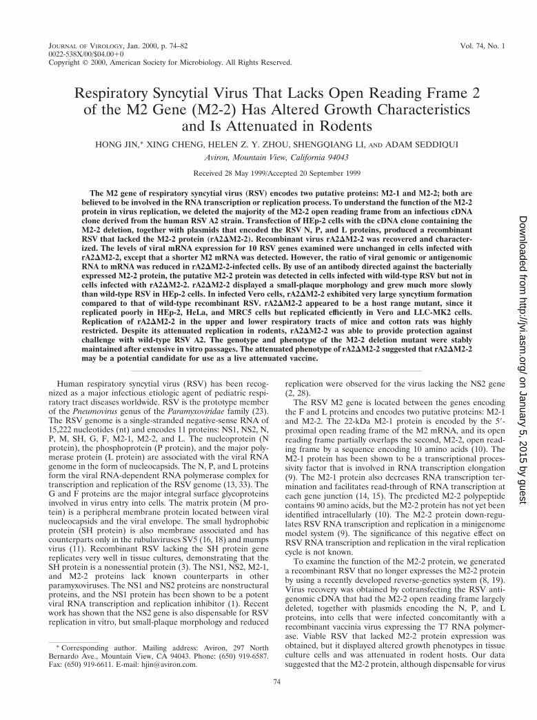
FIG. 1. Structure of the rA2DM2-2 genome and recovery of rA2DM2-2. (A) Sequences of the M2 gene in which the M2-1 and M2-2 open reading frames overlap.A total of 234 nt encoding the C-terminal 78 amino acids of the M2-2 protein were deleted through the introduced HindIII sites (underlined). The N-terminal 12 aminoacid residues encoded by the M2-2 open reading frame were maintained at the region of overlap with the M2-1 open reading frame. (B) RT-PCR products of rA2DM2-2and rA2 RNAs, obtained with a pair of primers flanking the M2 gene in the presence (1) or absence (2) of reverse transcriptase (RT). The size (in base pairs) of theDNA product derived from rA2 or rA2DM2-2 is indicated. The left lane was loaded with a 100-bp DNA size marker.
VOL. 74, 2000 RECOMBINANT RSV M2-2 PROTEIN 75
on January 5, 2015 by guesthttp://jvi.asm
.org/D
ownloaded from
postinfection, 200 ml of culture supernatant was collected and stored at 270°Cuntil virus titration. Each aliquot taken was replaced with an equal amount offresh medium. The virus titer was determined by a plaque assay on Vero cells,using an overlay of 1% methylcellulose and L15 medium containing 2% FBS. Toanalyze virus replication in different host cells, each cell line grown in six-wellplates was infected with rA2DM2-2 or rA2 at an MOI of 0.2. Three days postin-fection, the culture supernatants were collected, and virus was quantitated by aplaque assay on Vero cells.
RNA extraction, RT-PCR, and Northern blot analysis. For reverse transcrip-tion (RT)-PCR, viral RNA was extracted from rA2DM2-2- and rA2-infected cellculture supernatants by use of an RNA extraction kit (RNA STAT-50; Tel-Test,Friendswood, Tex.). Viral RNA was reverse transcribed with reverse transcrip-tase and a primer complementary to the viral genome from nt 7430 to nt 7449.The cDNA fragment spanning the M2 gene was amplified by PCR with primerV1948 (nt 7486 to nt 7515 for positive sense) and primer V1581 (nt 8544 to nt8525 for negative sense). The PCR product was analyzed on a 1.2% agarose geland visualized by ethidium bromide staining.
For Northern blot hybridization analysis, total cellular RNA was extractedfrom rA2DM2-2- or rA2-infected cells by use of an RNA extraction kit (RNASTAT-60; Tel-Test). RNA was electrophoresed on a 1.2% agarose gel containingformaldehyde and transferred to a nylon membrane (Amersham PharmaciaBiotech). The membrane was hybridized with an RSV gene-specific riboprobelabeled with digoxigenin. The hybridized RNA bands were visualized by use of aDig-Luminescent Detection Kit for Nucleic Acids (Boehringer Mannheim Bio-chemicals, Indianapolis, Ind.). To detect viral genomic RNA, a 32P-labeledriboprobe specific for the negative-sense F gene or N gene was used in Northernblot hybridization. To detect viral antigenomic RNA and mRNA, a 32P-labeledriboprobe specific for the positive-sense F gene or G gene was used. Hybridiza-tion of the membrane with riboprobes was done at 65°C. Membrane washing andsignal detection were performed according to standard procedures.
Immunoprecipitation and Western blotting of viral polypeptides. Virus-spe-cific proteins produced from infected cells were analyzed by immunoprecipita-tion of the infected-cell extracts or by Western blotting. For immunoprecipita-tion analysis, Vero cells were infected with virus at an MOI of 1.0 and labeledwith 35S-promix (100 mCi each of [35S]Cys and [35S]Met per ml; Amersham,Arlington Heights, Ill.) at 14 to 18 h postinfection. The labeled cell monolayerswere lysed with radioimmunoprecipitation assay buffer, and the polypeptideswere immunoprecipitated with polyclonal anti-RSV A2 serum (Biogenesis) oranti–M2-2 protein antiserum. Immunoprecipitated polypeptides were electro-phoresed on 17.5% polyacrylamide gels containing 0.1% sodium dodecyl sulfateand 4 M urea and detected by autoradiography. For Western blotting analysis,HEp-2 and Vero cells were infected with rA2DM2-2 or rA2. At various timespostinfection, virus-infected cells were lysed in protein lysis buffer, and the celllysates were electrophoresed on 17.5% polyacrylamide gels containing 0.1%sodium dodecyl sulfate and 4 M urea. The proteins were transferred to a nylonmembrane. Immunoblotting was performed as described in Jin et al. (20) withpolyclonal antiserum against M2-1 protein (gift of Jayesh Meanger), NS1 pro-tein, or SH protein (gift of Jose A. Melero).
Virus replication in mice and cotton rats. Virus replication in vivo was deter-mined with respiratory-tract-pathogen-free 12-week-old BALB/c mice (Simon-sen Laboratories, Gilroy, Calif.) and S. hispidus cotton rats (Virion Systems,Rockville, Md.). Mice or cotton rats in groups of six were inoculated intranasallyunder light methoxyflurane anesthesia with 106 PFU of rA2 or rA2DM2-2 in a0.1-ml inoculum per animal. On day 4 postinoculation, animals were sacrificed byCO2 asphyxiation, and their nasal turbinates and lungs were obtained separately.Tissues were homogenized, and virus titers were determined by a plaque assay onVero cells. To evaluate immunogenicity and protective efficacy, three groups ofmice were inoculated intranasally with rA2, rA2DM2-2, or medium only at day 0.Three weeks later, mice were anesthetized, serum samples were collected, and achallenge inoculation of 106 PFU of biologically derived wild-type RSV A2 wasadministered intranasally. Four days postchallenge, the animals were sacrificed,both nasal turbinates and lungs were harvested, and virus titers were determinedby a plaque assay. Serum neutralizing antibodies against RSV A2 were deter-mined by a 60% plaque reduction assay (7) and by immunostaining of RSV-infected cells.
RESULTS
Generation of rA2DM2-2. Previously, we reported the recov-ery of recombinant RSV from an infectious cDNA clone de-rived from RSV strain A2 (19). To obtain recombinant RSV inwhich the expression of the M2-2 open reading frame is ab-lated, a 234-nt cDNA fragment that encodes the C-terminal 78amino acids of the M2-2 protein was deleted from the infec-tious RSV cDNA clone. The N-terminal 12 amino acids thatmostly overlapped with the M2-1 open reading frame weremaintained, as it was considered likely that these 12 aminoacids would not be sufficient to preserve M2-2 protein function(Fig. 1A). The deletion of the M2-2 open reading frame in
antigenomic cDNA was confirmed by restriction enzyme di-gestion and by sequencing across the junction of the deletion.The resulting antigenomic cDNA clone, pA2DM2-2, is 14,988nt long, 234 nt shorter than pRSVC4G.
Since pA2DM2-2 was not completely sequenced, two inde-pendent clones were obtained and used in the recovery ofinfectious virus. To recover recombinant RSV with the M2-2open reading frame largely deleted, pA2DM2-2 was trans-fected, together with plasmids encoding the RSV N, P, and Lproteins, under the control of the T7 promoter, into HEp-2cells which had been infected with a modified vaccinia virusexpressing the T7 RNA polymerase (MVA-T7). Culture su-pernatants from the transfected HEp-2 cells were used to in-fect fresh HEp-2 or Vero cells to amplify the rescued virus.The recovery of rA2DM2-2 was indicated by syncytium forma-tion and confirmed by positive staining of infected cells withpolyclonal anti-RSV A2 serum. Recovered rA2DM2-2 wasplaque purified three times and amplified in Vero cells. Toconfirm that rA2DM2-2 contained the M2-2 deletion, viralRNA was extracted from virus and subjected to RT-PCR witha pair of primers spanning the M2 gene. As shown in Fig. 1B,rA2 yielded a PCR DNA product corresponding to the pre-dicted 1,029-nt fragment, whereas rA2DM2-2 yielded a PCRproduct of 795 nt, 234 nt shorter. Generation of the RT-PCRproduct was dependent on the RT step, indicating that theproduct was derived from RNA rather than from DNA con-tamination. The deletion was also confirmed by sequencinganalysis of the 795-nt RT-PCR DNA product derived fromrA2DM2-2.
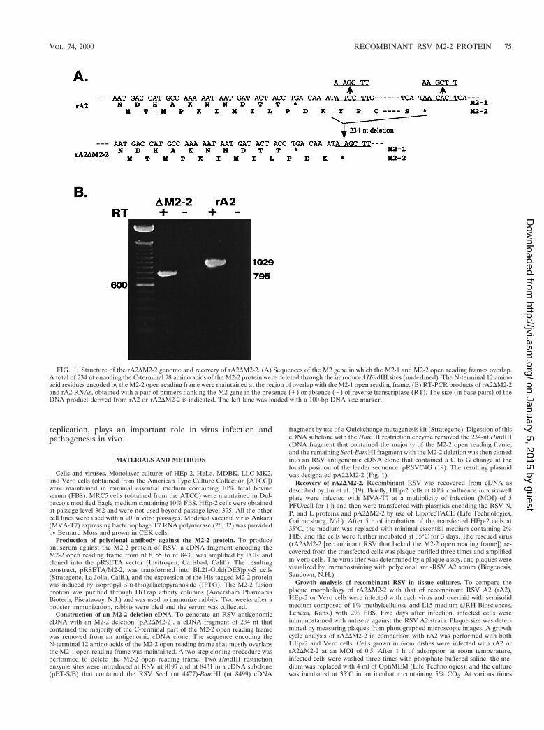
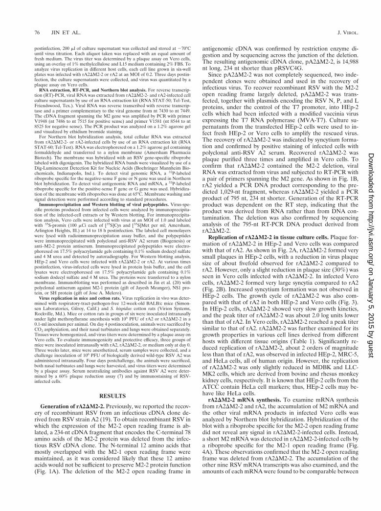
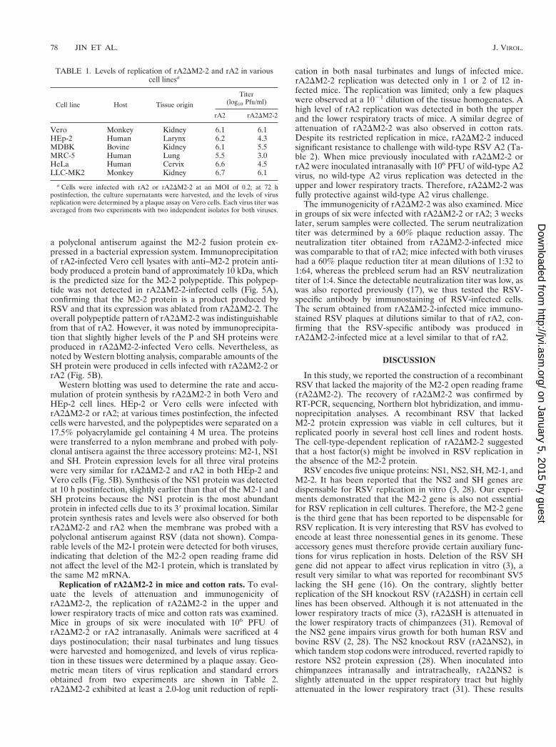
Replication of rA2DM2-2 in tissue culture cells. Plaque for-mation of rA2DM2-2 in HEp-2 and Vero cells was comparedwith that of rA2. As shown in Fig. 2A, rA2DM2-2 formed verysmall plaques in HEp-2 cells, with a reduction in virus plaquesize of about fivefold observed for rA2DM2-2 compared torA2. However, only a slight reduction in plaque size (30%) wasseen in Vero cells infected with rA2DM2-2. In infected Verocells, rA2DM2-2 formed very large syncytia compared to rA2(Fig. 2B). Increased syncytium formation was not observed inHEp-2 cells. The growth cycle of rA2DM2-2 was also com-pared with that of rA2 in both HEp-2 and Vero cells (Fig. 3).In HEp-2 cells, rA2DM2-2 showed very slow growth kinetics,and the peak titer of rA2DM2-2 was about 2.0 log units lowerthan that of rA2. In Vero cells, rA2DM2-2 reached a peak titersimilar to that of rA2. rA2DM2-2 was further examined for itsgrowth properties in various cell lines derived from differenthosts with different tissue origins (Table 1). Significantly re-duced replication of rA2DM2-2, about 2 orders of magnitudeless than that of rA2, was observed in infected HEp-2, MRC-5,and HeLa cells, all of human origin. However, the replicationof rA2DM2-2 was only slightly reduced in MDBK and LLC-MK2 cells, which are derived from bovine and rhesus monkeykidney cells, respectively. It is known that HEp-2 cells from theATCC contain HeLa cell markers; thus, HEp-2 cells may be-have like HeLa cells.
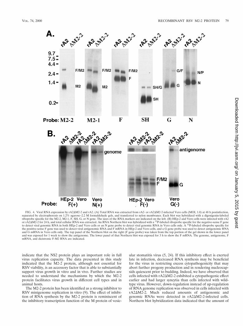
rA2DM2-2 mRNA synthesis. To examine mRNA synthesisfrom rA2DM2-2 and rA2, the accumulation of M2 mRNA andthe other viral mRNA products in infected Vero cells wasanalyzed by Northern blot hybridization. Hybridization of theblot with a riboprobe specific for the M2-2 open reading framedid not reveal any signal in rA2DM2-2-infected cells. Instead,a short M2 mRNA was detected in rA2DM2-2-infected cells bya riboprobe specific for the M2-1 open reading frame (Fig.4A). These observations confirmed that the M2-2 open readingframe was deleted from rA2DM2-2. The accumulation of theother nine RSV mRNA transcripts was also examined, and theamounts of each mRNA were found to be comparable between
76 JIN ET AL. J. VIROL.
on January 5, 2015 by guesthttp://jvi.asm
.org/D
ownloaded from
rA2DM2-2- and rA2-infected cells. Examples of Northern blotsprobed with riboprobes specific for the N, SH, G, or F genesare also shown in Fig. 4A. Slightly faster migration of F-M2bicistronic mRNA was also discernible due to the deletion ofthe M2-2 open reading frame.
The M2-2 protein was previously reported to be a potenttranscriptional negative regulator in a minigenome replicationassay. However, the lack of M2-2 protein expression did notappear to affect viral mRNA production in infected cells. Todetermine if the levels of viral antigenomic and genomic RNAsof rA2DM2-2 were affected by the M2-2 deletion, we examinedthe amounts of viral genomic and antigenomic RNAs pro-duced in infected Vero and HEp-2 cells by Northern hybrid-ization. Hybridization of the infected total cellular RNA with a32P-labeled F or N gene riboprobe specific for the negative-sense genomic RNA indicated that much less genomic RNAwas produced in cells infected with rA2DM2-2 than in cellsinfected with rA2 (Fig. 4B). A duplicate membrane was hy-bridized with a 32P-labeled F or G gene riboprobe specific forthe positive-sense RNA. Very little antigenomic RNA wasdetected in cells infected with rA2DM2-2; the amount of the For G mRNA in rA2DM2-2-infected cells was comparable to
that in rA2-infected cells. It is very striking that the levels ofboth genomic and antigenomic RNAs in rA2DM2-2-infectedcells were significantly reduced. Quantitation of the ratio ofgenomic and antigenomic RNA amounts to the viral mRNAamount indicated that at least a 10-fold reduction in antigeno-mic and genomic RNA amounts was observed in rA2DM2-2-infected cells. Therefore, it appears that RSV genome andantigenome syntheses were down-regulated due to the M2-2deletion. This down-regulation was seen in both Vero andHEp-2 cells and thus was not cell type dependent. This phe-nomenon has been observed with different riboprobes and twodifferent rA2DM2-2 isolates (Fig. 4B).
rA2DM2-2 protein synthesis. Since the putative M2-2 pro-tein has not been identified in RSV-infected cells previously, itwas necessary to demonstrate that the M2-2 protein is indeedencoded by RSV and produced in infected cells. We produced
FIG. 2. Comparison of the abilities of rA2 and rA2DM2-2 to form plaquesand syncytia. (A) Plaque morphology of rA2DM2-2 and rA2. HEp-2 or Vero cellswere infected with rA2DM2-2 or rA2 under a semisolid overlay composed of 1%methylcellulose and L15 medium containing 2% FBS for 5 days. Virus plaqueswere visualized by immunostaining with a goat polyclonal anti-RSV antiserumand photographed under a microscope. (B) Comparison of syncytium formation.Vero cells were infected with rA2 and rA2DM2-2 at an MOI of 0.5 and incubatedin liquid medium (OptiMEM) at 35°C for 40 h. The infected cell monolayerswere photographed without any treatment.
FIG. 3. Growth curves of rA2DM2-2 in HEp-2 and Vero cells. Vero cells orHEp-2 cells were infected with rA2DM2-2 or rA2 at an MOI of 0.5, and aliquotsof medium were harvested at 24-h intervals. The virus titers were determined bya plaque assay on Vero cells. The virus titer at each time point is an average fromtwo experiments with two independent isolates for both viruses.
VOL. 74, 2000 RECOMBINANT RSV M2-2 PROTEIN 77
on January 5, 2015 by guesthttp://jvi.asm
.org/D
ownloaded from
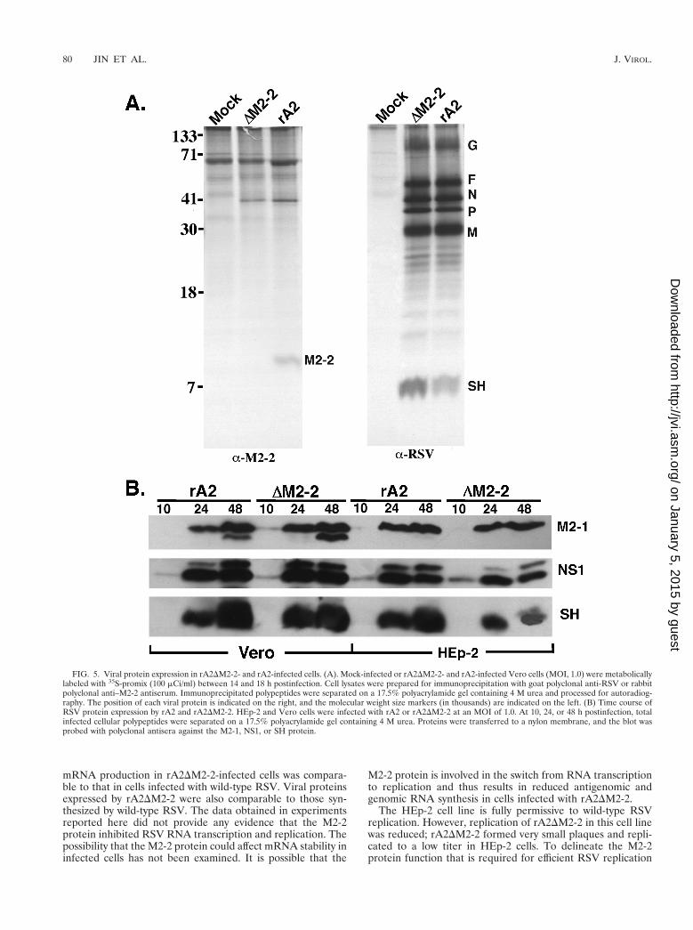
a polyclonal antiserum against the M2-2 fusion protein ex-pressed in a bacterial expression system. Immunoprecipitationof rA2-infected Vero cell lysates with anti–M2-2 protein anti-body produced a protein band of approximately 10 kDa, whichis the predicted size for the M2-2 polypeptide. This polypep-tide was not detected in rA2DM2-2-infected cells (Fig. 5A),confirming that the M2-2 protein is a product produced byRSV and that its expression was ablated from rA2DM2-2. Theoverall polypeptide pattern of rA2DM2-2 was indistinguishablefrom that of rA2. However, it was noted by immunoprecipita-tion that slightly higher levels of the P and SH proteins wereproduced in rA2DM2-2-infected Vero cells. Nevertheless, asnoted by Western blotting analysis, comparable amounts of theSH protein were produced in cells infected with rA2DM2-2 orrA2 (Fig. 5B).
Western blotting was used to determine the rate and accu-mulation of protein synthesis by rA2DM2-2 in both Vero andHEp-2 cell lines. HEp-2 or Vero cells were infected withrA2DM2-2 or rA2; at various times postinfection, the infectedcells were harvested, and the polypeptides were separated on a17.5% polyacrylamide gel containing 4 M urea. The proteinswere transferred to a nylon membrane and probed with poly-clonal antisera against the three accessory proteins: M2-1, NS1and SH. Protein expression levels for all three viral proteinswere very similar for rA2DM2-2 and rA2 in both HEp-2 andVero cells (Fig. 5B). Synthesis of the NS1 protein was detectedat 10 h postinfection, slightly earlier than that of the M2-1 andSH proteins because the NS1 protein is the most abundantprotein in infected cells due to its 39 proximal location. Similarprotein synthesis rates and levels were also observed for bothrA2DM2-2 and rA2 when the membrane was probed with apolyclonal antiserum against RSV (data not shown). Compa-rable levels of the M2-1 protein were detected for both viruses,indicating that deletion of the M2-2 open reading frame didnot affect the level of the M2-1 protein, which is translated bythe same M2 mRNA.
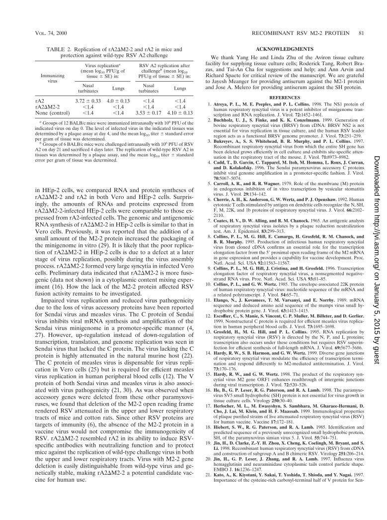
Replication of rA2DM2-2 in mice and cotton rats. To eval-uate the levels of attenuation and immunogenicity ofrA2DM2-2, the replication of rA2DM2-2 in the upper andlower respiratory tracts of mice and cotton rats was examined.Mice in groups of six were inoculated with 106 PFU ofrA2DM2-2 or rA2 intranasally. Animals were sacrificed at 4days postinoculation; their nasal turbinates and lung tissueswere harvested and homogenized, and levels of virus replica-tion in these tissues were determined by a plaque assay. Geo-metric mean titers of virus replication and standard errorsobtained from two experiments are shown in Table 2.rA2DM2-2 exhibited at least a 2.0-log unit reduction of repli-
cation in both nasal turbinates and lungs of infected mice.rA2DM2-2 replication was detected only in 1 or 2 of 12 in-fected mice. The replication was limited; only a few plaqueswere observed at a 1021 dilution of the tissue homogenates. Ahigh level of rA2 replication was detected in both the upperand the lower respiratory tracts of mice. A similar degree ofattenuation of rA2DM2-2 was also observed in cotton rats.Despite its restricted replication in mice, rA2DM2-2 inducedsignificant resistance to challenge with wild-type RSV A2 (Ta-ble 2). When mice previously inoculated with rA2DM2-2 orrA2 were inoculated intranasally with 106 PFU of wild-type A2virus, no wild-type A2 virus replication was detected in theupper and lower respiratory tracts. Therefore, rA2DM2-2 wasfully protective against wild-type A2 virus challenge.
The immunogenicity of rA2DM2-2 was also examined. Micein groups of six were infected with rA2DM2-2 or rA2; 3 weekslater, serum samples were collected. The serum neutralizationtiter was determined by a 60% plaque reduction assay. Theneutralization titer obtained from rA2DM2-2-infected micewas comparable to that of rA2; mice infected with both viruseshad a 60% plaque reduction titer at mean dilutions of 1:32 to1:64, whereas the prebleed serum had an RSV neutralizationtiter of 1:4. Since the detectable neutralization titer was low, aswas also reported previously (17), we thus tested the RSV-specific antibody by immunostaining of RSV-infected cells.The serum obtained from rA2DM2-2-infected mice immuno-stained RSV plaques at dilutions similar to that of rA2, con-firming that the RSV-specific antibody was produced inrA2DM2-2-infected mice at a level similar to that of rA2.
DISCUSSION
In this study, we reported the construction of a recombinantRSV that lacked the majority of the M2-2 open reading frame(rA2DM2-2). The recovery of rA2DM2-2 was confirmed byRT-PCR, sequencing, Northern blot hybridization, and immu-noprecipitation analyses. A recombinant RSV that lackedM2-2 protein expression was viable in cell cultures, but itreplicated poorly in several host cell lines and rodent hosts.The cell-type-dependent replication of rA2DM2-2 suggestedthat a host factor(s) might be involved in RSV replication inthe absence of the M2-2 protein.
RSV encodes five unique proteins: NS1, NS2, SH, M2-1, andM2-2. It has been reported that the NS2 and SH genes aredispensable for RSV replication in vitro (3, 28). Our experi-ments demonstrated that the M2-2 gene is also not essentialfor RSV replication in cell cultures. Therefore, the M2-2 geneis the third gene that has been reported to be dispensable forRSV replication. It is very interesting that RSV has evolved toencode at least three nonessential genes in its genome. Theseaccessory genes must therefore provide certain auxiliary func-tions for virus replication in hosts. Deletion of the RSV SHgene did not appear to affect virus replication in vitro (3), aresult very similar to what was reported for recombinant SV5lacking the SH gene (16). On the contrary, slightly betterreplication of the SH knockout RSV (rA2DSH) in certain celllines has been observed. Although it is not attenuated in thelower respiratory tracts of mice (3), rA2DSH is attenuated inthe lower respiratory tracts of chimpanzees (31). Removal ofthe NS2 gene impairs virus growth for both human RSV andbovine RSV (2, 28). The NS2 knockout RSV (rA2DNS2), inwhich tandem stop codons were introduced, reverted rapidly torestore NS2 protein expression (28). When inoculated intochimpanzees intranasally and intratracheally, rA2DNS2 isslightly attenuated in the upper respiratory tract but highlyattenuated in the lower respiratory tract (31). These results
TABLE 1. Levels of replication of rA2DM2-2 and rA2 in variouscell linesa
Cell line Host Tissue origin
Titer(log10 Pfu/ml)
rA2 rA2DM2-2
Vero Monkey Kidney 6.1 6.1HEp-2 Human Larynx 6.2 4.3MDBK Bovine Kidney 6.1 5.5MRC-5 Human Lung 5.5 3.0HeLa Human Cervix 6.6 4.5LLC-MK2 Monkey Kidney 6.7 6.1
a Cells were infected with rA2 or rA2DM2-2 at an MOI of 0.2; at 72 hpostinfection, the culture supernatants were harvested, and the levels of virusreplication were determined by a plaque assay on Vero cells. Each virus titer wasaveraged from two experiments with two independent isolates for both viruses.
78 JIN ET AL. J. VIROL.
on January 5, 2015 by guesthttp://jvi.asm
.org/D
ownloaded from
indicate that the NS2 protein plays an important role in fullvirus replication capacity. The data presented in this studyindicated that the M2-2 protein, although not essential forRSV viability, is an accessory factor that is able to substantiallysupport virus growth in vitro and in vivo. Further studies areneeded to understand the mechanisms by which the M2-2protein facilitates virus growth in different cell types and inanimal hosts.
The M2-2 protein has been identified as a strong inhibitor toRSV minigenome replication in vitro (9). The effect of inhibi-tion of RNA synthesis by the M2-2 protein is reminiscent ofthe inhibitory transcription function of the M protein of vesic-
ular stomatitis virus (5, 24). If this inhibitory effect is exertedlate in infection, decreased RNA synthesis may be beneficialfor the virus in restricting excess cytopathogenicity that mayabort further progeny production and in rendering nucleocap-sids quiescent prior to budding. Indeed, we have observed thatcells infected with rA2DM2-2 exhibited a cytopathogenic effectearlier and had larger syncytia than cells infected with wild-type virus. However, down-regulation instead of up-regulationof RNA genome replication was observed in cells infected withrA2DM2-2. Much reduced amounts of antigenomic andgenomic RNAs were detected in rA2DM2-2-infected cells.Northern blot hybridization data indicated that the amount of
FIG. 4. Viral RNA expression by rA2DM2-2 and rA2. (A) Total RNA was extracted from rA2- or rA2DM2-2-infected Vero cells (MOI, 1.0) at 48 h postinfection,separated by electrophoresis on 1.2% agarose–2.2 M formaldehyde gels, and transferred to nylon membranes. Each blot was hybridized with a digoxigenin-labeledriboprobe specific for the M2-2, M2-1, F, SH, G, or N gene. The sizes of the RNA markers are indicated on the left. (B) HEp-2 and Vero cells were infected with rA2or rA2DM2-2 for 24 h, and total cellular RNA was extracted. An RNA Northern blot was hybridized with a 32P-labeled riboprobe specific for the negative-sense F geneto detect viral genomic RNA in both HEp-2 and Vero cells or an N gene probe to detect viral genomic RNA in Vero cells only. A 32P-labeled riboprobe specific forthe positive-sense F gene was used to detect viral antigenomic RNA and F mRNA in HEp-2 and Vero cells, and a G gene probe was used to detect antigenomic RNAand G mRNA in Vero cells only. The top panel of the Northern blot on the right (F gene probe) was taken from the top portion of the gel shown in the lower paneland was exposed for 1 week to show the antigenome. The lower panel of that Northern blot was exposed for 3 h to show the F mRNA. The genome, antigenome, FmRNA, and dicistronic F-M2 RNA are indicated.
VOL. 74, 2000 RECOMBINANT RSV M2-2 PROTEIN 79
on January 5, 2015 by guesthttp://jvi.asm
.org/D
ownloaded from
mRNA production in rA2DM2-2-infected cells was compara-ble to that in cells infected with wild-type RSV. Viral proteinsexpressed by rA2DM2-2 were also comparable to those syn-thesized by wild-type RSV. The data obtained in experimentsreported here did not provide any evidence that the M2-2protein inhibited RSV RNA transcription and replication. Thepossibility that the M2-2 protein could affect mRNA stability ininfected cells has not been examined. It is possible that the
M2-2 protein is involved in the switch from RNA transcriptionto replication and thus results in reduced antigenomic andgenomic RNA synthesis in cells infected with rA2DM2-2.
The HEp-2 cell line is fully permissive to wild-type RSVreplication. However, replication of rA2DM2-2 in this cell linewas reduced; rA2DM2-2 formed very small plaques and repli-cated to a low titer in HEp-2 cells. To delineate the M2-2protein function that is required for efficient RSV replication
FIG. 5. Viral protein expression in rA2DM2-2- and rA2-infected cells. (A). Mock-infected or rA2DM2-2- and rA2-infected Vero cells (MOI, 1.0) were metabolicallylabeled with 35S-promix (100 mCi/ml) between 14 and 18 h postinfection. Cell lysates were prepared for immunoprecipitation with goat polyclonal anti-RSV or rabbitpolyclonal anti–M2-2 antiserum. Immunoprecipitated polypeptides were separated on a 17.5% polyacrylamide gel containing 4 M urea and processed for autoradiog-raphy. The position of each viral protein is indicated on the right, and the molecular weight size markers (in thousands) are indicated on the left. (B) Time course ofRSV protein expression by rA2 and rA2DM2-2. HEp-2 and Vero cells were infected with rA2 or rA2DM2-2 at an MOI of 1.0. At 10, 24, or 48 h postinfection, totalinfected cellular polypeptides were separated on a 17.5% polyacrylamide gel containing 4 M urea. Proteins were transferred to a nylon membrane, and the blot wasprobed with polyclonal antisera against the M2-1, NS1, or SH protein.
80 JIN ET AL. J. VIROL.
on January 5, 2015 by guesthttp://jvi.asm
.org/D
ownloaded from
in HEp-2 cells, we compared RNA and protein syntheses ofrA2DM2-2 and rA2 in both Vero and HEp-2 cells. Surpris-ingly, the amounts of RNAs and proteins expressed fromrA2DM2-2-infected HEp-2 cells were comparable to those ex-pressed from rA2-infected cells. The genomic and antigenomicRNA synthesis of rA2DM2-2 in HEp-2 cells is similar to that inVero cells. Previously, it was reported that the addition of asmall amount of the M2-2 protein increased the packaging ofthe minigenome in vitro (29). It is likely that the poor replica-tion of rA2DM2-2 in HEp-2 cells is due to a defect at a laterstage of virus replication, possibly during the virus assemblyprocess. rA2DM2-2 formed very large syncytia in infected Verocells. Preliminary data indicated that rA2DM2-2 is more fuso-genic (data not shown) in a cytoplasmic content mixing exper-iment (16). How the lack of the M2-2 protein affected RSVfusion activity remains to be investigated.
Impaired virus replication and reduced virus pathogenicitydue to the loss of virus accessory proteins have been reportedfor Sendai virus and measles virus. The C protein of Sendaivirus inhibits viral mRNA synthesis and amplification of theSendai virus minigenome in a promoter-specific manner (4,27). However, up-regulation instead of down-regulation oftranscription, translation, and genome replication was seen inSendai virus that lacked the C protein. The virus lacking the Cprotein is highly attenuated in the natural murine host (22).The C protein of measles virus is dispensable for virus repli-cation in Vero cells (25) but is required for efficient measlesvirus replication in human peripheral blood cells (12). The Vprotein of both Sendai virus and measles virus is also associ-ated with virus pathogenicity (21, 30). As was observed whenaccessory genes were deleted from these other paramyxovi-ruses, we found that deletion of the M2-2 open reading framerendered RSV attenuated in the upper and lower respiratorytracts of mice and cotton rats. Since other RSV proteins aretargets of immunity (6), the absence of the M2-2 protein in avaccine virus would not compromise the immunogenicity ofRSV. rA2DM2-2 resembled rA2 in its ability to induce RSV-specific antibodies with neutralizing function and to protectmice against the replication of wild-type challenge virus in boththe upper and lower respiratory tracts. Virus with M2-2 genedeletion is easily distinguishable from wild-type virus and ge-netically stable, making rA2DM2-2 a potential candidate vac-cine for human use.
ACKNOWLEDGMENTS
We thank Yang He and Linda Zhu of the Aviron tissue culturefacility for supplying tissue culture cells; Roderick Tang, Robert Bra-zas, and Tai-An Cha for suggestions and help; and Ann Arvin andRichard Spaete for critical review of the manuscript. We are gratefulto Jayesh Meanger for providing antiserum against the M2-1 proteinand Jose A. Melero for providing antiserum against the SH protein.
REFERENCES1. Atreya, P. L., M. E. Peeples, and P. L. Collins. 1998. The NS1 protein of
human respiratory syncytial virus is a potent inhibitor of minigenome tran-scription and RNA replication. J. Virol. 72:1452–1461.
2. Buchholz, U. J., S. Finke, and K. K. Conzelmann. 1999. Generation ofbovine respiratory syncytial virus (BRSV) from cDNA: BRSV NS2 is notessential for virus replication in tissue culture, and the human RSV leaderregion acts as a functional BRSV genome promoter. J. Virol. 73:251–259.
3. Bukreyev, A., S. S. Whitehead, B. R. Murphy, and P. L. Collins. 1997.Recombinant respiratory syncytial virus from which the entire SH gene hasbeen deleted grows efficiently in cell culture and exhibits site-specific atten-uation in the respiratory tract of the mouse. J. Virol. 71:8973–8982.
4. Cadd, T., D. Garcin, C. Tapparel, M. Itoh, M. Homma, L. Roux, J. Curran,and D. Kolakofsky. 1996. The Sendai paramyxovirus accessory C proteinsinhibit viral genome amplification in a promoter-specific fashion. J. Virol.70:5067–5074.
5. Carroll, A. R., and R. R. Wagner. 1979. Role of the membrane (M) proteinin endogenous inhibition of in vitro transcription by vesicular stomatitisvirus. J. Virol. 29:134–142.
6. Cherrie, A. H., K. Anderson, G. W. Wertz, and P. J. Openshaw. 1992. Humancytotoxic T cells stimulated by antigen on dendritic cells recognize the N, SH,F, M, 22K, and 1b proteins of respiratory syncytial virus. J. Virol. 66:2102–2110.
7. Coates, H. V., D. W. Alling, and R. M. Chanock. 1965. An antigenic analysisof respiratory syncytial virus isolates by a plaque reduction neutralizationtest. Am. J. Epidemiol. 83:299–313.
8. Collins, P. L., M. G. Hill, E. Camargo, H. Grosfeld, R. M. Chanock, andB. R. Murphy. 1995. Production of infectious human respiratory syncytialvirus from cloned cDNA confirms an essential role for the transcriptionelongation factor from the 59 proximal open reading frame of the M2 mRNAin gene expression and provides a capability for vaccine development. Proc.Natl. Acad. Sci. USA 92:11563–11567.
9. Collins, P. L., M. G. Hill, J. Cristina, and H. Grosfeld. 1996. Transcriptionelongation factor of respiratory syncytial virus, a nonsegmented negative-strand RNA virus. Proc. Natl. Acad. Sci. USA 93:81–85.
10. Collins, P. L., and G. W. Wertz. 1985. The envelope-associated 22K proteinof human respiratory syncytial virus: nucleotide sequence of the mRNA anda related polytranscript. J. Virol. 54:65–71.
11. Elango, N., J. Kovamees, T. M. Varsanyi, and E. Norrby. 1989. mRNAsequence and deduced amino acid sequence of the mumps virus small hy-drophobic protein gene. J. Virol. 63:1413–1415.
12. Escoffier, C., S. Manie, S. Vincent, C. P. Muller, M. Billeter, and D. Gerlier.1999. Nonstructural C protein is required for efficient measles virus replica-tion in human peripheral blood cells. J. Virol. 73:1695–1698.
13. Grosfeld, H., M. G. Hill, and P. L. Collins. 1995. RNA replication byrespiratory syncytial virus (RSV) is directed by the N, P, and L proteins;transcription also occurs under these conditions but requires RSV superin-fection for efficient synthesis of full-length mRNA. J. Virol. 69:5677–5686.
14. Hardy, R. W., S. B. Harmon, and G. W. Wertz. 1999. Diverse gene junctionsof respiratory syncytial virus modulate the efficiency of transcription termi-nation and respond differently to M2-mediated antitermination. J. Virol.73:170–176.
15. Hardy, R. W., and G. W. Wertz. 1998. The product of the respiratory syn-cytial virus M2 gene ORF1 enhances readthrough of intergenic junctionsduring viral transcription. J. Virol. 72:520–526.
16. He, B., G. P. Leser, R. G. Paterson, and R. A. Lamb. 1998. The paramyxo-virus SV5 small hydrophobic (SH) protein is not essential for virus growth intissue culture cells. Virology 250:30–40.
17. Herlocher, M. L., M. Ewasyshyn, S. Sambhara, M. Gharaee-Hermani, D.Cho, J. Lai, M. Klein, and H. F. Maassab. 1999. Immunological propertiesof plaque purified strains of live attenuated respiratory syncytial virus (RSV)for human vaccine. Vaccine 17:172–181.
18. Hiebert, S. W., R. G. Paterson, and R. A. Lamb. 1985. Identification andpredicted sequence of a previously unrecognized small hydrophobic protein,SH, of the paramyxovirus simian virus 5. J. Virol. 55:744–751.
19. Jin, H., D. Clarke, Z.-Y. H. Zhou, X. Cheng, K. Coelingh, M. Bryant, and S.Li. 1998. Recombinant human respiratory syncytial virus (RSV) from cDNAand construction of subgroup A and B chimeric RSV. Virology 251:206–214.
20. Jin, H., G. P. Leser, J. Zhang, and R. A. Lamb. 1997. Influenza virushemagglutinin and neuraminidase cytoplasmic tails control particle shape.EMBO J. 16:1236–1247.
21. Kato, A., K. Kiyotani, Y. Sakai, T. Yoshida, T. Shioda, and Y. Nagai. 1997.Importance of the cysteine-rich carboxyl-terminal half of V protein for Sen-
TABLE 2. Replication of rA2DM2-2 and rA2 in mice andprotection against wild-type RSV A2 challenge
Immunizingvirus
Virus replicationa
(mean log10 PFU/g oftissue 6 SE) in:
RSV A2 replication afterchallengeb (mean log10
PFU/g of tissue 6 SE) in:
Nasalturbinates Lungs Nasal
turbinates Lungs
rA2 3.72 6 0.33 4.0 6 0.13 ,1.4 ,1.4rA2DM2-2 ,1.4 ,1.4 ,1.4 ,1.4None (control) ,1.4 ,1.4 3.53 6 0.17 4.10 6 0.13
a Groups of 12 BALB/c mice were immunized intranasally with 106 PFU of theindicated virus on day 0. The level of infected virus in the indicated tissues wasdetermined by a plaque assay at day 4, and the mean log10 titer 6 standard errorper gram of tissue was determined.
b Groups of 6 BALB/c mice were challenged intranasally with 106 PFU of RSVA2 on day 21 and sacrificed 4 days later. The replication of wild-type RSV A2 intissues was determined by a plaque assay, and the mean log10 titer 6 standarderror per gram of tissue was determined.
VOL. 74, 2000 RECOMBINANT RSV M2-2 PROTEIN 81
on January 5, 2015 by guesthttp://jvi.asm
.org/D
ownloaded from

dai virus pathogenesis. J. Virol. 71:7266–7272.22. Kurotani, A., K. Kiyotani, A. Kato, T. Shioda, Y. Sakai, K. Mizumoto, T.
Yoshida, and Y. Nagai. 1998. Sendai virus C proteins are categorically non-essential gene products but silencing their expression severely impairs viralreplication and pathogenesis. Genes Cells 3:111–124.
23. Lamb, R. A., and D. Kolakofsky. 1996. Paramyxoviridae: the viruses and theirreplication, p. 1177–1204. In B. N. Fields, D. M. Knipe, P. M. Howley, et al.(ed.), Fields virology, 3rd ed. Lippincott-Raven Publishers, Philadelphia, Pa.
24. Peeples, M. E. 1991. Paramyxovirus M proteins: pulling it all together andtaking it on the road, p. 427–456. In D. W. Kingsbury (ed.), The paramyxo-viruses. Plenum Publishing Corp., New York, N.Y.
25. Radecke, F., and M. A. Billeter. 1996. The nonstructural C protein is notessential for multiplication of Edmonston B strain measles virus in culturedcells. Virology 217:418–421.
26. Sutter, G., M. Ohlmann, and V. Erfle. 1995. Non-replicating vaccinia vectorefficiently expresses bacteriophage T7 RNA polymerase. FEBS Lett. 371:9–12.
27. Tapparel, C., S. Hausmann, T. Pelet, J. Curran, D. Kolakofsky, and L. Roux.1997. Inhibition of Sendai virus genome replication due to promoter-in-creased selectivity: a possible role for the accessory C proteins. J. Virol.71:9588–9599.
28. Teng, M. N., and P. L. Collins. 1999. Altered growth characteristics ofrecombinant respiratory syncytial viruses which do not produce NS2 protein.J. Virol. 73:466–473.
29. Teng, M. N., and P. L. Collins. 1998. Identification of the respiratory syn-cytial virus proteins required for formation and passage of helper-dependentinfectious particles. J. Virol. 72:5707–5716.
30. Tober, C., M. Seufert, H. Schneider, M. A. Billeter, I. C. Johnston, S.Niewiesk, V. ter Meulen, and S. Schneider-Schaulies. 1998. Expression ofmeasles virus V protein is associated with pathogenicity and control of viralRNA synthesis. J. Virol. 72:8124–8132.
31. Whitehead, S. S., A. Bukreyev, M. N. Teng, C. Y. Firestone, M. St. Claire,W. R. Elkins, P. L. Collins, and B. R. Murphy. 1999. Recombinant respira-tory syncytial virus bearing a deletion of either the NS2 or SH gene isattenuated in chimpanzees. J. Virol. 73:3438–3442.
32. Wyatt, L. S., B. Moss, and S. Rozenblatt. 1995. Replication-deficient vacciniavirus encoding bacteriophage T7 RNA polymerase for transient gene ex-pression in mammalian cells. Virology 210:202–205.
33. Yu, Q., R. W. Hardy, and G. W. Wertz. 1995. Functional cDNA clones of thehuman respiratory syncytial (RS) virus N, P, and L proteins support repli-cation of RS virus genomic RNA analogs and define minimal trans-actingrequirements for RNA replication. J. Virol. 69:2412–2419.
82 JIN ET AL. J. VIROL.
on January 5, 2015 by guesthttp://jvi.asm
.org/D
ownloaded from


