Embed Size (px)
Citation preview
www.elsevier.com/locate/brainresBrain Research 992 (2003) 252–262
Research report
Modulatory effects of melatonin on behavior, hemolymph metabolites,
and neurotransmitter release in crayfish
Andrea R. Tildena,*, Rebecca Brauchb, Ryan Ballb, Aura M. Janzea, Ali H. Ghaffaria,Catherine T. Sweeneya, Jamie C. Yureka, Robin L. Cooperb
aDepartment of Biology, Colby College, 5720 Mayflower Hill, Waterville, ME 04901, USAbDepartment of Biology, University of Kentucky, Lexington, KY 40506-0225, USA
Accepted 27 August 2003
Abstract
Melatonin affects a variety of circadian processes such as behavior and neurotransmitter release in vertebrates. Crayfish melatonin
production occurs in the eyestalks, and the cycle of production may change seasonally. To date, however, melatonin’s roles and mechanisms
of action in crustacean physiology are unclear. We injected melatonin or saline into crayfish in scotophase and monitored activity and
hemolymph glucose/lactate over 24 h in early spring. Crayfish were significantly more active in photophase versus the expected scotophase,
and had concurrent glucose/lactate peaks. Melatonin reversed the activity pattern, causing a scotophase activity peak, but not the glucose/
lactate patterns. This study was repeated in late summer, during which control activity and glucose/lactate levels were elevated in scotophase.
Melatonin decreased the amplitude of scotophase activity and glucose/lactate, eliminating activity and glucose cycles. We also injected
melatonin or saline at various times of day in early summer and monitored locomotor activity for 1 h. Controls had high activity at 1200
(mid-photophase) and 2100 h (early scotophase), and melatonin increased activity at 1200 h but decreased it at 2100 h. Melatonin also
increased activity at 1500 h but not 1800 h (late photophase). Next, we examined the influence of melatonin on crayfish neurophysiology.
Melatonin (10 AM) enhanced synaptic transmission at the neuromuscular junction (NMJ). The presynaptic action resulted in more vesicles
being released during evoked stimulation. Our study indicates that melatonin may have a phylogenetically conserved role in the transduction
of circadian information in invertebrates as in vertebrates. Behavioral and physiological effects may be mediated by modulation of central
pathways, enhanced at the peripheral level via neuromodulation of the NMJ.
D 2003 Elsevier B.V. All rights reserved.
Theme: Neural basis of behavior
Topic: Biological rhythms and sleep
Keywords: Melatonin; Crayfish; Neuromuscular junction; Activity pattern; Locomotion; Metabolite
1. Introduction in the entrainment of circadian rhythms of behavior and
The presence of melatonin in crustaceans is now well
documented; however, the nature of its production and its
roles in crustacean physiology and behavior have not been
extensively investigated. Melatonin is found in high con-
centrations within the eyestalks, as is N-acetyltransferase,
the rate-limiting enzyme in vertebrate melatonin production
[51,59,60]. Vertebrates have a characteristic scotophase
increase in melatonin production, and melatonin is involved
0006-8993/$ - see front matter D 2003 Elsevier B.V. All rights reserved.
doi:10.1016/j.brainres.2003.08.053
* Corresponding author. Tel.: +1-207-872-3329; fax: +1-207-872-
3731.
E-mail address: [email protected] (A.R. Tilden).
physiology [41,56]. Crustaceans, on the other hand, exhibit
greater variability in melatonin production. The intertidal
fiddler crab Uca pugilator, for example, may have a tide-
associated melatonin cycle [50,51]. In the giant freshwater
prawn (Macrobrachium rosenbergii), females showed a
melatonin cycle with a photophase peak [59]. In addition,
early-photophase melatonin levels were different between
males and females, indicating possible sex differences in
melatonin cycles [60]. In the crayfish Procambarus clarkii,
melatonin levels were higher during photophase in one
study [1] and in scotophase in another study [6].
A variety of influences of melatonin have been reported
in crustaceans: In U. pugilator, melatonin increased the rate
A.R. Tilden et al. / Brain Research 992 (2003) 252–262 253
of limb regeneration in eyestalk-intact and -ablated crabs
[50]; it had both hypo and hyperglycemic effects and altered
glucose and lactate cycles [52]; and it influenced locomotor
activity [53]. In P. clarkii, melatonin caused both dark-
adapting [6] and light-adapting [45] responses in the visual
system. Together, these studies indicate a variable pattern of
production and a variable influence of melatonin that may
relate to seasonal or other changes. These studies suggest a
role of melatonin in modulating circadian, seasonal, and
other functions in crustaceans. Melatonin’s role in circadian
behavioral rhythms in vertebrates [8,25,28,55] is mediated
by neuromodulation of the nucleus accumbens [37] and
suprachiasmatic nucleus [27]. Much less is known about
melatonin and behavior in invertebrates. In the cricket
(Acheta domesticus), melatonin may entrain circadian loco-
motor rhythms [61]; and in U. pugilator, its influence may
be tide-based [53]. However, the mechanisms and locations
of action are not known.
In P. clarkii, the subject of our investigation, locomotor
activity, displays a circadian rhythm: locomotor activity
increases in scotophase and persists in constant conditions
[5,23]. The mechanism of control of locomotor rhythmic-
ity has not been clearly established in crustaceans. The x-
organ/sinus gland complex, located within the eyestalk,
releases neurodepressing hormone (NDH) which sup-
presses locomotor activity [3,4,26]. The entrainment of
locomotor activity to photoperiod persists in the absence
of the eyestalks, and the supraesophageal ganglion (SOG)
has been identified as a component of the circadian
pacemaker system [35,36]. A variety of other nervous
system locations may also be involved in the crayfish
circadian system [5]. Hemolymph glucose levels are also
rhythmic: an increase occurs during scotophase in noctur-
nally active Orconectes limosus. This increase is concom-
itant with the peak of the eyestalk x-organ-produced
crustacean hyperglycemic hormone (CHH) [31]. Lactate,
a metabolic endproduct of activities and stresses [22], may
also serve as a marker of locomotor activity. In the first
part of our study, we examined the influence of melatonin
on locomotor activity and on glucose and lactate patterns
in P. clarkii.
The neuromodulator serotonin (5-HT), a precursor of
melatonin, is also known to alter locomotor behavior in
crayfish [49] and has been shown to increase in titer
within the hemolymph during locomotor activity in crabs
[44]. It is established that 5-HT enhances the following
in crayfish: heart rate [32], neurotransmitter release at
neuromuscular junctions (NMJ) of skeletal muscles
[19,46], the drive of primary sensory neurons [14], and
intrinsic CNS drive of motor neurons [47]. Since recent
evidence showed that melatonin also causes an increase
in the frequency of cardiac activity [54] as well as being
correlated with locomotor activity, we used similar sen-
sory and skeletal NMJ preparations as previously used
for studies investigating 5-HT to compare the actions of
melatonin.
2. Methods
2.1. Animals
All experiments were performed using P. clarkii, mea-
suring 4–6 cm in body length (Atchafalaya Biological
Supply, Raceland, LA, or Fruge’s Cajun Crawfish, Branch,
LA).
2.2. Activity and metabolites
For the activity and glucose/lactate studies, animals
were held in aerated tanks and were fed daily at random
times to prevent entrainment to food availability. They
were acclimated to a 12L:12D photoperiod with lights
on at 0700 h and off at 1900 h; the temperature was
held at a constant 20 jC. Activity monitoring: male crayfish
were placed in 30� 40� 15 cm opaque Plexiglask boxes
divided into water-tight lanes, 30 cm (length)� 8 cm
(width)� 15 cm (height), with opaque dividers. Each lane
received aerated, conditioned tap water to a depth of 13 cm,
and one crayfish was placed into each lane; melatonin-
treated alternated with saline-treated crayfish. The bottom of
the tanks had gridlines placed 2 cm apart along the 30-cm
length of each lane for quantification of locomotor activity.
The activity monitoring room was a constant 20F 1 jC,with the same 12L:12D photoperiod as the acclimation
conditions; light was provided by broad-spectrum fluores-
cent bulbs with a light intensity of 422 lx at the water
surface. Sony 560� Digital8 Nightshot cameras were
suspended from a ceiling rack such that each camera’s field
of view encompassed one box, and these cameras were
connected to VCRs placed outside of the monitoring room.
Activity studies were conducted in early spring (March),
early summer (June), and late summer (August); glucose
and lactate levels were measured in early spring and late
summer only. In the early spring and late summer studies,
100 Al of a melatonin solution [melatonin (Sigma) was
dissolved in ethanol and diluted in crayfish saline [57] for
final ethanol concentration of 1%] was injected into the base
of a walking leg such that the final melatonin concentration
in crayfish was 1 Ag g� 1 (4.3 nmol g� 1). Controls received
100 Al of saline with 1% ethanol; all injections were at 1900
h, at the onset of scotophase. Activity was monitored at the
same time in 10 control and 10 experimental animals over a
24-h period, and different animals were used between early
spring and late summer studies. We recorded activity for a
1-h period every 3 h over 24 h. The early summer (June)
activity study was conducted similarly to the other two
studies; however, melatonin or saline was injected into 10
crayfish each at 1200, 1500, 1800, and 2100 h, with
different crayfish monitored for 1-h intervals (total: 80
animals). All activity data are reported as linear distance
moved per hour.
For the glucose/lactate studies, crayfish were injected
with melatonin (n = 12) or saline (n = 12) as described
A.R. Tilden et al. / Brain Research 992 (2003) 252–262254
previously, at 1900 h, with separate groups used in the early
spring versus late summer studies. Hemolymph (50 Al) wascollected from each animal at the base of a walking leg
every 3 h over a 24-h period. Glucose and lactate levels
were measured with a YSI 2300 STAT Plus glucose and
lactate analyzer (YSI, Yellow Springs, OH); validation of
this instrument for crustacean hemolymph has been previ-
ously determined [52].
2.3. Skeletal muscle preparations
All experiments were conducted using the opener muscle
or the extensor muscle of the first walking legs. Prepara-
tions were dissected according to standard procedures
[7,10]. The tissue was pinned out in a Slygard dish as to
expose the muscle in a taut position. The muscles were
viewed with a Nikon, Optiphot-2 upright fluorescent mi-
croscope using a 40� (0.55 NA) Nikon water immersion
objective. Dissected preparations were maintained in cray-
fish saline, a modified Van Harreveld’s solution (205 mM
NaCl; 5.3 mMKCl; 13.5 mM CaCl2 2H2O; 2.45 mM MgCl26H2O; 0.5 mM HEPES adjusted to pH 7.4) at 17 jC. Theentire opener muscle is innervated by a single excitatory
motor neuron [10], whereas the extensor muscle is inner-
vated by a single tonic and a single phasic excitatory motor
neuron [7].
2.4. Application of melatonin
To apply exogenous compounds to these preparations,
the bathing medium was rapidly exchanged with saline
containing melatonin (10 AM, Sigma). A fresh aliquot was
used before each experiment from a frozen stock (1 mM).
2.5. Intracellular recorded evoked postsynaptic potentials
(EPSPs)
Intracellular muscle recordings were made with a 3 M
KCl-containing microelectrode. Short-term facilitation
(STF) was induced by giving a 40-Hz train of 10 pulses
at intervals of 10 s. To determine a facilitation index for the
induced STF of the tonic EPSPs obtained from the opener
muscle, the amplitude of the 10th EPSP within a train is
compared with one of the preceding EPSPs amplitude
within the response [16]. The subtraction of the numeral
one ensures that if there is no facilitation occurring, the
facilitation index (FI) will then be zero. Responses were
recorded as previously described [15]. Stimulation of the
opener motor neuron was carried out as described by Dudel
and Kuffler [20].
2.6. Field recorded excitatory postsynaptic potentials
(f EPSPs)
Synaptic potentials were measured with focal macro-
patch electrodes, allowing for a determination of the effect
of melatonin on presynaptic vesicular events. The living
terminals were visualized by exposure to vital fluorescent
dye 4-Di-2-ASP (Molecular Probes [11,33]). The synaptic
potentials were obtained using the loose patch technique by
lightly placing a 10–20 Am fire polished glass electrode
directly over a spatially isolated varicosity along the nerve
terminal. The macropatch electrode is specific for record-
ings within the region of the electrode lumen. The seal
resistance was in the range of 100 kV to 1 MV. The evoked
field excitatory postsynaptic potentials (f EPSCs) and field
miniature excitatory postsynaptic potentials (fmEPSPs)
were recorded and analyzed to determine the mean quantal
content (m) [12,13]. Mean quantal content was determined
by the direct counting method for the low output tonic
terminals. By direct counts of the evoked quantal events, the
number of failures in evoked release was used as an index of
altering synaptic function. If only one single event occurred
after the spike, it was counted as one; when double events
occurred, they were referred to as two, etc.
Quantal release over the time was also monitored by
examining the area of the evoked potential. The time of the
peak in evoked events varies due to latency jitter, so when
multiple events occur, the measures of peak amplitude are
not as reliable as the area measure [12]. To monitor quanta
released over time, the area of the evoked potential was
measured for each event. This approach was also used to
quantify alterations in synaptic potentials for the tonic
terminals. The tonic nerve was stimulated at a rate of 1 or
2 Hz in order not to facilitate the responses between trials.
Since exposure to melatonin had a gradual effect on mean
quantal content (m), samples for every 200 events were used
[46,47]. Larger sample sizes of 800 to 1000 or more trials
were used to estimate the quantal parameters for the effects
of melatonin exposure on the number of releasing sites (n)
and the probability of release at a site ( p) [12,13,17].
2.7. Statistical analysis
Activity, glucose, or lactate levels were compared within
each group of the early spring and late summer study with a
one-way ANOVA with a Student–Newman–Keuls test.
Mean activity, glucose, or lactate levels were compared
between some groups of the above studies, and activity
was compared between melatonin-treated and control ani-
mals in the early summer study, with a Student’s t-test
(SigmaStat software, SPSS, San Rafael, CA).
To quantitatively compare the change in the EPSP
amplitudes and synaptic potentials from the f EPSPs, the
measurements were normalized to a percent difference. The
percent difference was calculated using the difference
among the average during baseline recording prior to
melatonin exposure and the average of the events at the
maximum response during the exposure. The average was
then divided by the baseline value as shown in the follow-
ing equation: [ABaseline�maximum responseA/Baseline]�100=%Difference. Numerical data were represented as the
A.R. Tilden et al. / Brain Research 992 (2003) 252–262 255
mean in the percent difference from exposure to saline.
When the basic assumption of parametric Student’s t-test
was valid, it was used; otherwise, the non-parametric Wil-
coxon rank sum test was used.
3. Results
3.1. Activity and metabolites
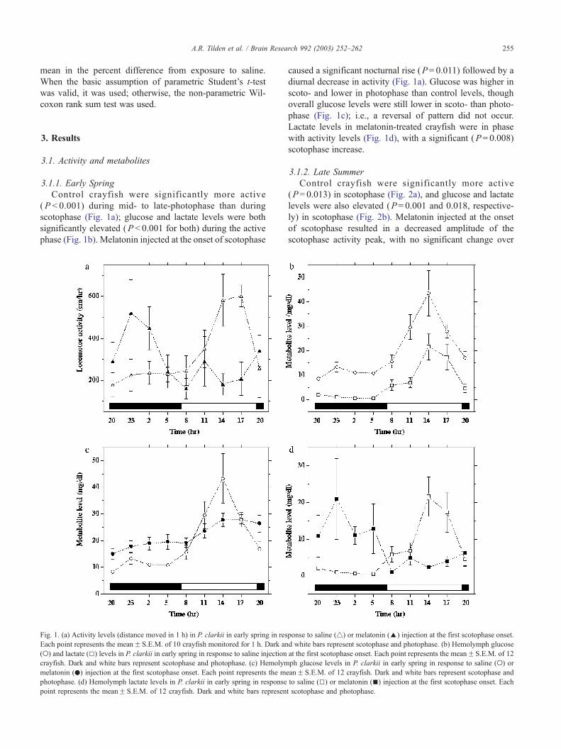
3.1.1. Early Spring
Control crayfish were significantly more active
(P < 0.001) during mid- to late-photophase than during
scotophase (Fig. 1a); glucose and lactate levels were both
significantly elevated (P < 0.001 for both) during the active
phase (Fig. 1b). Melatonin injected at the onset of scotophase
Fig. 1. (a) Activity levels (distance moved in 1 h) in P. clarkii in early spring in res
Each point represents the meanF S.E.M. of 10 crayfish monitored for 1 h. Dark an
(o) and lactate (5) levels in P. clarkii in early spring in response to saline injection
crayfish. Dark and white bars represent scotophase and photophase. (c) Hemolym
melatonin (.) injection at the first scotophase onset. Each point represents the m
photophase. (d) Hemolymph lactate levels in P. clarkii in early spring in response
point represents the meanF S.E.M. of 12 crayfish. Dark and white bars represen
caused a significant nocturnal rise (P= 0.011) followed by a
diurnal decrease in activity (Fig. 1a). Glucose was higher in
scoto- and lower in photophase than control levels, though
overall glucose levels were still lower in scoto- than photo-
phase (Fig. 1c); i.e., a reversal of pattern did not occur.
Lactate levels in melatonin-treated crayfish were in phase
with activity levels (Fig. 1d), with a significant (P= 0.008)
scotophase increase.
3.1.2. Late Summer
Control crayfish were significantly more active
(P= 0.013) in scotophase (Fig. 2a), and glucose and lactate
levels were also elevated (P= 0.001 and 0.018, respective-
ly) in scotophase (Fig. 2b). Melatonin injected at the onset
of scotophase resulted in a decreased amplitude of the
scotophase activity peak, with no significant change over
ponse to saline (4) or melatonin (E) injection at the first scotophase onset.
d white bars represent scotophase and photophase. (b) Hemolymph glucose
at the first scotophase onset. Each point represents the meanF S.E.M. of 12
ph glucose levels in P. clarkii in early spring in response to saline (o) or
eanF S.E.M. of 12 crayfish. Dark and white bars represent scotophase and
to saline (5) or melatonin (n) injection at the first scotophase onset. Each
t scotophase and photophase.
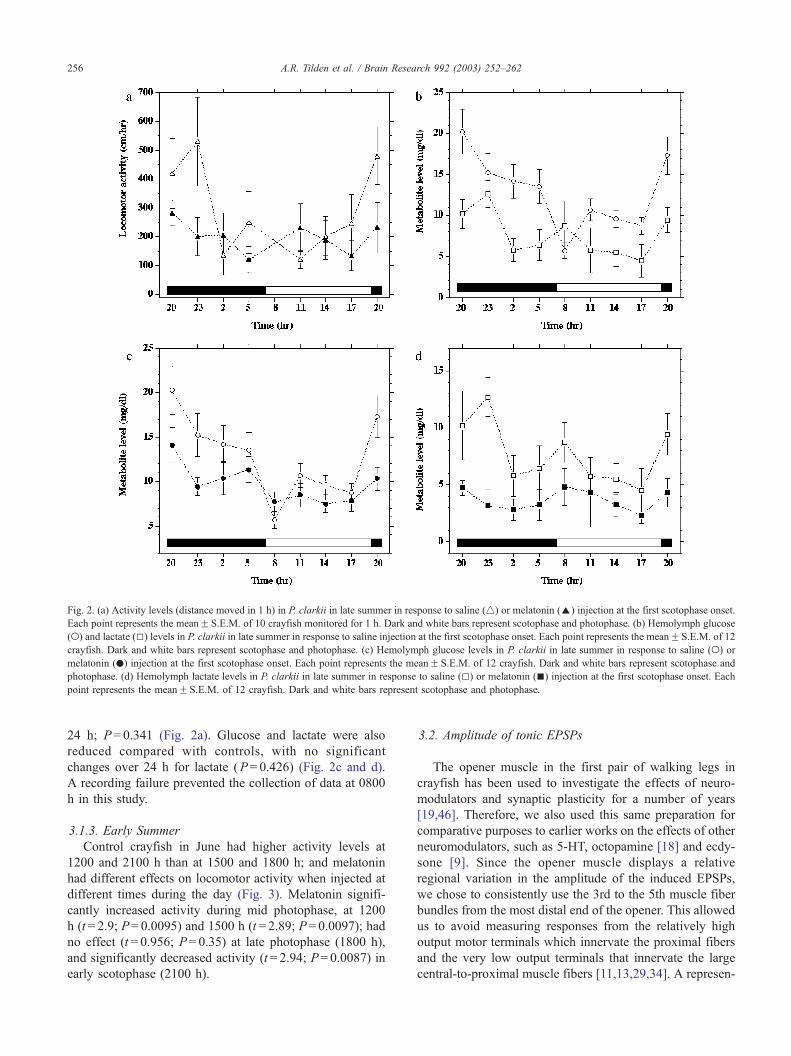
Fig. 2. (a) Activity levels (distance moved in 1 h) in P. clarkii in late summer in response to saline (4) or melatonin (E) injection at the first scotophase onset.
Each point represents the meanF S.E.M. of 10 crayfish monitored for 1 h. Dark and white bars represent scotophase and photophase. (b) Hemolymph glucose
(o) and lactate (5) levels in P. clarkii in late summer in response to saline injection at the first scotophase onset. Each point represents the meanF S.E.M. of 12
crayfish. Dark and white bars represent scotophase and photophase. (c) Hemolymph glucose levels in P. clarkii in late summer in response to saline (o) or
melatonin (.) injection at the first scotophase onset. Each point represents the meanF S.E.M. of 12 crayfish. Dark and white bars represent scotophase and
photophase. (d) Hemolymph lactate levels in P. clarkii in late summer in response to saline (5) or melatonin (n) injection at the first scotophase onset. Each
point represents the meanF S.E.M. of 12 crayfish. Dark and white bars represent scotophase and photophase.
A.R. Tilden et al. / Brain Research 992 (2003) 252–262256
24 h; P= 0.341 (Fig. 2a). Glucose and lactate were also
reduced compared with controls, with no significant
changes over 24 h for lactate (P= 0.426) (Fig. 2c and d).
A recording failure prevented the collection of data at 0800
h in this study.
3.1.3. Early Summer
Control crayfish in June had higher activity levels at
1200 and 2100 h than at 1500 and 1800 h; and melatonin
had different effects on locomotor activity when injected at
different times during the day (Fig. 3). Melatonin signifi-
cantly increased activity during mid photophase, at 1200
h (t = 2.9; P= 0.0095) and 1500 h (t = 2.89; P= 0.0097); had
no effect (t = 0.956; P= 0.35) at late photophase (1800 h),
and significantly decreased activity (t = 2.94; P= 0.0087) in
early scotophase (2100 h).
3.2. Amplitude of tonic EPSPs
The opener muscle in the first pair of walking legs in
crayfish has been used to investigate the effects of neuro-
modulators and synaptic plasticity for a number of years
[19,46]. Therefore, we also used this same preparation for
comparative purposes to earlier works on the effects of other
neuromodulators, such as 5-HT, octopamine [18] and ecdy-
sone [9]. Since the opener muscle displays a relative
regional variation in the amplitude of the induced EPSPs,
we chose to consistently use the 3rd to the 5th muscle fiber
bundles from the most distal end of the opener. This allowed
us to avoid measuring responses from the relatively high
output motor terminals which innervate the proximal fibers
and the very low output terminals that innervate the large
central-to-proximal muscle fibers [11,13,29,34]. A represen-
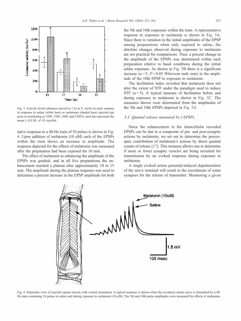
Fig. 3. Activity levels (distance moved in 1 h) in P. clarkii in early summer
in response to saline (white bars) or melatonin (shaded bars) injected just
prior to monitoring at 1200, 1500, 1800, and 2100 h; each bar represents the
meanF S.E.M. of 10 crayfish.
A.R. Tilden et al. / Brain Research 992 (2003) 252–262 257
tative response to a 40-Hz train of 10 pulses is shown in Fig.
4. Upon addition of melatonin (10 uM) each of the EPSPs
within the train shows an increase in amplitude. The
response depicted for the effects of melatonin was measured
after the preparation had been exposed for 10 min.
The effect of melatonin in enhancing the amplitude of the
EPSPs was gradual, and in all five preparations the en-
hancement reached a plateau after approximately 10 to 15
min. The amplitude during the plateau response was used to
determine a percent increase in the EPSP amplitude for both
Fig. 4. Schematic view of crayfish opener muscle with ventral orientation. A typic
Hz train containing 10 pulses in saline and during exposure to melatonin (10 AM).
the 5th and 10th responses within the train. A representative
response to exposure to melatonin is shown in Fig. 5A.
Since there is variation in the initial amplitudes of the EPSP
among preparations when only exposed to saline, the
absolute changes observed during exposure to melatonin
are not practical for comparisons. Thus, a percent change in
the amplitude of the EPSPs was determined within each
preparation relative to basal conditions during the initial
saline exposure. As shown in Fig. 5B there is a significant
increase (n = 5, P < 0.05 Wilcoxon rank sum) in the ampli-
tude of the 10th EPSP to exposure to melatonin.
The facilitation index revealed that melatonin does not
alter the extent of STF under the paradigm used to induce
STF (n = 5). A typical measure of facilitation before and
during exposure to melatonin is shown in Fig. 5C. The
measures shown were determined from the amplitudes of
the 5th and 10th EPSPs depicted in Fig. 5A.
3.3. Quantal release measured by f EPSPs
Since the enhancement in the intracellular recorded
EPSPs can be due to a composite of pre- and post-synaptic
actions by melatonin, we set out to determine the presyn-
aptic contribution of melatonin’s actions by direct quantal
counts of release [17]. This measure allows one to determine
if more or fewer synaptic vesicles are being recruited for
transmission by an evoked response during exposure to
melatonin.
A single evoked action potential-induced depolarization
of the nerve terminal will result in the recruitment of some
synapses for the release of transmitter. Monitoring a given
al response is shown when the excitatory motor nerve is stimulated by a 40-
The 5th and 10th pulse amplitudes were measured for effects of melatonin.
Fig. 5. Mean amplitudes of the intracellular recorded EPSPs for every 100 s
plotted versus time (A). The bars show the standard error. The addition of
melatonin (10 AM) causes a slight increase in the mean EPSP amplitude for
both the 5th and 10th EPSPs. A percent change in the amplitude of the
EPSPs for the 10th pulse was determined for the five preparations (B). The
average responses measured between 200 and 500 and between 1200 and
1500 periods were used to calculate the percent change as shown in part A.
Exposure to melatonin did not result in any change in facilitation (C).
Fig. 6. Influence of melatonin on synaptic currents as recorded with a focal
macropatch electrode from a spatially isolated varicosity. Representative
single traces are shown with one containing two evoked events and a
superimposed trace with a miniature excitatory postsynaptic potential (f
mEPSP). Note in the evoked field excitatory postsynaptic potentials (f
EPSPs) individual quanta can be counted (arrows) (A). A series of evoked
events induced at 1 HZ stimulation to the motor nerve before and after
application of melatonin. A typical time series for three preparations is
shown (B). The time bins are divided into 200 trials for assessment of
mean quantal content (m). The hatched area indicates the period taken, the
last three values, to obtain an average value to determine a percent change
due to melatonin’s action. A mean percent change in the mean quantal
content for five preparations indicates an excitatory presynaptic action of
melatonin (C). The individual values for each of the five preparations are
superimposed on the bar chart as closed circles.
A.R. Tilden et al. / Brain Research 992 (2003) 252–262258
length of the nerve terminal (f 10 Am) with a macropatch
electrode allows a subset of the nerve terminal to be
assessed and discrete quantal events to be detected for a
measure of synaptic efficacy. The vesicular glutamate re-
leased binds to ionotropic ligand-gated receptors of a
quisqualate type [21,43], resulting in a rapid inward current.
This current is detected by the focal macropatch electrode
[48], and since we use the axoclamp as a differential
amplifier (i.e., bridge mode), a field electrical potential is
measured from inside the electrode lumen to outside in the
surrounding bath.
Each single trace from an evoked stimulation of the
excitatory motor axon may result in a failure of response,
an evoked synaptic response with one quantal event, or
responses with multiple releases in the defined region of the
nerve terminal being monitored (Fig. 6A). With direct
counting of each quantal event for each trace, the gradual
changes in the observed quantal occurrences can be com-
piled (Table 1). These responses indicate an increased
number of single and multiple evoked events upon exposure
to melatonin (10 AM).
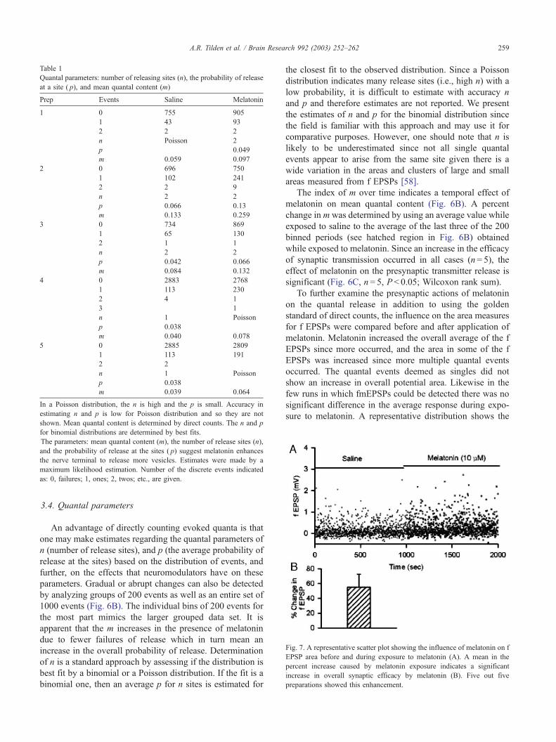
Table 1
Quantal parameters: number of releasing sites (n), the probability of release
at a site ( p), and mean quantal content (m)
Prep Events Saline Melatonin
1 0 755 905
1 43 93
2 2 2
n Poisson 2
p 0.049
m 0.059 0.097
2 0 696 750
1 102 241
2 2 9
n 2 2
p 0.066 0.13
m 0.133 0.259
3 0 734 869
1 65 130
2 1 1
n 2 2
p 0.042 0.066
m 0.084 0.132
4 0 2883 2768
1 113 230
2 4 1
3 1
n 1 Poisson
p 0.038
m 0.040 0.078
5 0 2885 2809
1 113 191
2 2
n 1 Poisson
p 0.038
m 0.039 0.064
In a Poisson distribution, the n is high and the p is small. Accuracy in
estimating n and p is low for Poisson distribution and so they are not
shown. Mean quantal content is determined by direct counts. The n and p
for binomial distributions are determined by best fits.
The parameters: mean quantal content (m), the number of release sites (n),
and the probability of release at the sites ( p) suggest melatonin enhances
the nerve terminal to release more vesicles. Estimates were made by a
maximum likelihood estimation. Number of the discrete events indicated
as: 0, failures; 1, ones; 2, twos; etc., are given.
Fig. 7. A representative scatter plot showing the influence of melatonin on f
EPSP area before and during exposure to melatonin (A). A mean in the
percent increase caused by melatonin exposure indicates a significant
increase in overall synaptic efficacy by melatonin (B). Five out five
preparations showed this enhancement.
A.R. Tilden et al. / Brain Research 992 (2003) 252–262 259
3.4. Quantal parameters
An advantage of directly counting evoked quanta is that
one may make estimates regarding the quantal parameters of
n (number of release sites), and p (the average probability of
release at the sites) based on the distribution of events, and
further, on the effects that neuromodulators have on these
parameters. Gradual or abrupt changes can also be detected
by analyzing groups of 200 events as well as an entire set of
1000 events (Fig. 6B). The individual bins of 200 events for
the most part mimics the larger grouped data set. It is
apparent that the m increases in the presence of melatonin
due to fewer failures of release which in turn mean an
increase in the overall probability of release. Determination
of n is a standard approach by assessing if the distribution is
best fit by a binomial or a Poisson distribution. If the fit is a
binomial one, then an average p for n sites is estimated for
the closest fit to the observed distribution. Since a Poisson
distribution indicates many release sites (i.e., high n) with a
low probability, it is difficult to estimate with accuracy n
and p and therefore estimates are not reported. We present
the estimates of n and p for the binomial distribution since
the field is familiar with this approach and may use it for
comparative purposes. However, one should note that n is
likely to be underestimated since not all single quantal
events appear to arise from the same site given there is a
wide variation in the areas and clusters of large and small
areas measured from f EPSPs [58].
The index of m over time indicates a temporal effect of
melatonin on mean quantal content (Fig. 6B). A percent
change in mwas determined by using an average value while
exposed to saline to the average of the last three of the 200
binned periods (see hatched region in Fig. 6B) obtained
while exposed to melatonin. Since an increase in the efficacy
of synaptic transmission occurred in all cases (n= 5), the
effect of melatonin on the presynaptic transmitter release is
significant (Fig. 6C, n = 5, P < 0.05; Wilcoxon rank sum).
To further examine the presynaptic actions of melatonin
on the quantal release in addition to using the golden
standard of direct counts, the influence on the area measures
for f EPSPs were compared before and after application of
melatonin. Melatonin increased the overall average of the f
EPSPs since more occurred, and the area in some of the f
EPSPs was increased since more multiple quantal events
occurred. The quantal events deemed as singles did not
show an increase in overall potential area. Likewise in the
few runs in which fmEPSPs could be detected there was no
significant difference in the average response during expo-
sure to melatonin. A representative distribution shows the
A.R. Tilden et al. / Brain Research 992 (2003) 252–262260
increase in the occurrences of the f EPSPs, as measured by
the EPSP area, over time due to melatonin exposure. Such
measures as the area or amplitude of the f EPSPs can be
undertaken when one is not confident in the direct quantal
counting method due to the lack of discerning multiple
releases within a single f EPSP. So by using both types of
analysis a clearer understanding of the mechanisms by
which melatonin influences synaptic transmission can be
ascertained. Since in a few preparations we were not
confident in the direct quantal counts, we utilized the index
for area of the f EPSP to assess a change in transmission
instead of the counting method. An average in the percent
increase in the area of the f EPSPs from before to during
melatonin exposure revealed approximately a 60% increase
in all five preparations (n = 5, P < 0.05 Wilcoxon rank sum)
(Fig. 7).
4. Discussion
Melatonin affects activity and metabolite levels, and has
an excitatory effect at the NMJ; however, the effects appear
to vary with the activity phase of the animals and the timing
of melatonin application. Crayfish are reportedly nocturnal-
ly active [2,23,24,38], with scotophase peaks of CHH and
glucose [30,31]; yet we observed diurnal activity in controls
in early spring. Two possible explanations for the diurnal
activity are: First, the crayfish may have entrained to a
daytime feeding regimen. Though we randomized feeding
and did not feed animals for 5 days preceding an experi-
ment, the crayfish were purchased from a farm-raised and
-fed population. Fernandez de Miguel and Arechiga [23,24]
showed that day-fed crayfish develop a photophase activity
pattern that can be instated with a single feeding and persists
for up to 2 weeks without food re-entrainment. Second, P.
clarkii is active in the daytime during early spring rain-
storms or cloudy days but not sunny days [38]. The activity
stimulus is not known but may involve levels of light
intensity. The light level in our lab (420 lx) is substantially
lower than that of bright sunlight (>20,000 lx) and may have
had a ‘cloudy day’ effect in the spring study.
Melatonin caused an increase in scotophase and a
decrease in photophase activity, glucose, and lactate in
the diurnally active early spring crayfish. Though we have
data for only a single 24-h cycle, melatonin may be able to
phase-shift an induced diurnal activity pattern to the more
typical nocturnal one. In separate studies, both a photo-
phase [1] and a scotophase [6] melatonin peak have been
shown in P. clarkii; these conflicting results could be
related to seasonal, acclimation, feeding schedule, sex, molt
stage, reproductive cycle, population, or other differences.
However, these studies do demonstrate that the timing of
the melatonin peak is variable. In our late summer study,
melatonin decreased the amplitude of the scotophase peaks
of activity, glucose, and lactate but did not affect the timing
of these events. In early summer, there were both photo-
phase and scotophase activity increases, with melatonin
enhancing activity in photophase but inhibiting activity in
scotophase. Likewise, in fiddler crabs melatonin increased
activity at some times of day but decreased it at others
[53].
Our contradictory early spring, early summer, and late
summer results may be due to seasonal or acclimational
changes in both the timing of melatonin production and the
timing of the animals’ sensitivity to melatonin. Similarly,
melatonin had opposing effects in two studies of visual
activity in P. clarkii: Melatonin affected both the period and
amplitude of the electroretinogram (ERG): the period was
shortened and the amplitude was increased, indicating dark-
adapting effects that were consistent with the finding of a
scotophase melatonin increase [6]. Yet in another study,
melatonin induced migration of retinal shielding pigments
and changes in photoreceptor potential that resembled light-
adapting responses [45], consistent with the finding of a
photophase melatonin peak [1].
Melatonin receptors in invertebrates have not yet been
studied, and the precise target tissues are not known. In
previous studies [52,53], we have demonstrated that mela-
tonin affects locomotor activity, glucose, and lactate in both
intact and eyestalk-ablated crabs, indicating that that these
effects probably are not mediated by eyestalk CHH and
NDH. Melatonin has direct receptor-mediated effects on
mammalian hepatocytes [40] and pancreatic islet cells [39],
and may likewise affect the crustacean hepatopancreas. In
addition to the hepatopancreas, muscle may be a source of
hemolymph glucose [42], and we have shown here that
melatonin has an excitatory effect on muscle cells. Melato-
nin may be a component of the complex system driving
rhythmic processes in invertebrates as it is in vertebrates.
The behavioral and physiological effects of melatonin are
likely due to central modulation of neural activity rather
than solely due to localized neuromodulation at the NMJ.
However, the crayfish NMJ provides an ideal model system
for the study of general nervous system neuromodulation
and provides information on possible central and peripheral
mechanisms of action.
Acknowledgements
Research and Creativity Summer Fellowship, University
of KY (Ryan Ball), Howard Hughes Medical Institute
(A.R.T.), Clare Boothe Luce Program of the Henry Luce
Foundation (A.R.T.), and NSF grants IBN-9808631 and ILI
DUE-9850907 (R.L.C.).
References
[1] M. Agapito, B. Herrero, M.I. Pablos, J.L. Miguel, J.M. Recio, Cir-
cadian rhythms of melatonin and serotonin-N-acetyltransferase ac-
tivity in Procambarus clarkii, Comp. Biochem. Physiol., A 112
(1995) 179–185.
A.R. Tilden et al. / Brain Research 992 (2003) 252–262 261
[2] H. Arechiga, A. Huberman, Hormonal modulation of circadian be-
havior in crustaceans, Front. Horm. Res. 6 (1980) 16–34.
[3] H. Arechiga, A. Huberman, A. Martinez-Palomo, Release of a neuro-
depressing hormone from the crustacean sinus gland, Brain Res. 128
(1977) 93–108.
[4] H. Arechiga, J.A. Williams, R.S.V. Pullin, E. Naylor, Cross-sensitivity
to neurodepressing hormone and its effect on locomotor rhythmicity
in two different groups of crustaceans, Gen. Comp. Endocrinol. 37
(1979) 350–357.
[5] H. Arechiga, F. Fernandez-Quiroz, F. Fernandez de Miguel, L. Ro-
drıguez-Sosa, The circadian system of crustaceans, Chronobiol. Int.
10 (1993) 1–19.
[6] I. Balzer, I.R. Espinola, B. Fuentes-Pardo, Daily variations of immu-
noreactive melatonin in the visual system of crayfish, Biol. Cell 89
(1997) 539–543.
[7] H. Bradacs, R. Cooper, M. Msghina, H. Atwood, Related articles,
links abstract differential physiology and morphology of phasic and
tonic motor axons in a crayfish limb extensor muscle, J. Exp. Biol.
200 (1997) 677–691.
[8] V.M. Cassone, W.S. Warren, D.S. Brooks, L. Lu, Melatonin, the
pineal gland, and circadian rhythms, J. Biol. Rhythms 8 (1993)
S73–S81.
[9] R.L. Cooper, M.E. Ruffner, Depression of synaptic efficacy at inter-
molt in crayfish neuromuscular junctions by 20-Hydroxyecdysone, a
molting hormone, J. Neurophys. 79 (1998) 1931–1941.
[10] R.L. Cooper, D. Hampson, H.L. Atwood, Synaptotagmin-like expres-
sion in the motor nerve terminals of crayfish, Brain Res. 703 (1995)
214–216.
[11] R.L. Cooper, L. Marin, H.L. Atwood, Synaptic differentiation of a
single motor neuron: conjoint definition of transmitter release, presy-
naptic calcium signals, and ultrastructure, J. Neurosci. 15 (1995)
4209–4222.
[12] R.L. Cooper, B.A. Stewart, J.M. Wojtowicz, S. Wang, H.L. Atwood,
Quantal measurement and analysis methods compared for crayfish
and Drosophila neuromuscular junctions and rat hippocampus,
J. Neurosci. Methods 61 (1995) 67–78.
[13] R.L. Cooper, C. Harrington, L. Marin, H.L. Atwood, Quantal release
at visualized terminals of crayfish motor axon: intraterminal and re-
gional differences, J. Comp. Neurol. 375 (1996) 583–600.
[14] R.L. Cooper, E. Ward, R. Braxton, H. Li, W.M. Warren, The effects of
serotonin and ecdysone on primary sensory neurons in crayfish, Mi-
crosc. Res. Tech. 60 (2003) 336–345.
[15] M.E. Crider, R.L. Cooper, The importance of the stimulation para-
digm in determining facilitation and effects of neuromodulation, Brain
Res. 842 (1999) 324–331.
[16] M.E. Crider, R.L. Cooper, Differentially facilitation of high- and low-
output nerve terminals from a single motor neuron, J. Appl. Physiol.
88 (2000) 987–996.
[17] J. Del Castillo, B. Katz, Quantal components of the end-plate poten-
tial, J. Physiol. 124 (1954) 560–573.
[18] S. Djokaj, R.L. Cooper, W. Rathmayer, Effects of octopamine, sero-
tonin, and cocktails of the two modulators on synaptic transmission at
crustacean neuromuscular junctions, J. Comp. Physiol., A 187 (2001)
145–154.
[19] J. Dudel, Facilitatory effects of 5-hydroxy-tryptamine on the crayfish
neuromuscular junction, Naunyn-Schmiedeberg’s Arch. Exp. Pathol.
Pharmakol. 249 (1965) 515–528.
[20] J. Dudel, S.W. Kuffler, The quantal nature of transmission and spon-
taneous miniature potentials at the crayfish neuromuscular junction,
J. Physiol. 55 (1961) 514–529.
[21] J. Dudel, C. Franke, H. Hatt, Rapid activation and desensitization of
transmitter-liganded receptor channels by pulses of agonists, in: T.
Narahashi (Ed.), Ion Channels, vol. 3, Plenum, New York, 1992,
pp. 207–260.
[22] M.L. Fanjul-Moles, T. Bosques-Tistler, J. Prieto-Sagredo, O. Cas-
tanon-Cervantes, L. Fernandez-Rivera-Rıo, Effect of variation in
photoperiod and light intensity on oxygen consumption, lactate
concentration and behavior in crayfish Procambarus clarkii and
Procambarus digueti, Comp. Biochem. Physiol., A 119 (1998)
263–269.
[23] F. Fernandez de Miguel, H. Arechiga, Entrainment by food of circa-
dian locomotor activity rhythm in the crayfish, Soc. Neurosci. Abstr.
14 (1988) 1299.
[24] F. Fernandez de Miguel, H. Arechiga, Circadian locomotor activity
and its entrainment by food in the crayfish Procambarus clarkii,
J. Exp. Biol. 190 (1994) 9–21.
[25] A. Foa, L. Minutini, A. Innocenti, Melatonin: a coupling device be-
tween oscillators in the circadian system of the ruin lizard Podarcis
sicula, Comp. Biochem. Physiol., A 103 (1992) 719–723.
[26] A. Huberman, H. Arechiga, A. Cimet, J. De La Rosa, C. Aramburo,
Isolation and purification of a neurodepressing hormone from the
eyestalk of Procambarus bouvieri (Ortmann), Eur. J. Biochem. 99
(1979) 203–208.
[27] A.E. Hunt, W.M. Al-Ghoul, M.U. Gillette, M.L. Dubocovich, Acti-
vation of MT2 melatonin receptors in rat suprachiasmatic nucleus
phase advances the circadian clock, Am. J. Physiol. 280 C (2001)
110–118.
[28] L.L. Hyde, H. Underwood, Daily melatonin infusions entrain the
locomotor activity of pinealectomized lizards, Physiol. Behav. 58
(1995) 943–951.
[29] J. Iravani, Membrandepolarisation der Muskelfasern des Offnermus-
kels des Flusskrebses auf Nervenreiz und Kaliumapplikation, Expe-
rientia 21 (1965) 609–610.
[30] J.L. Kallen, N.R. Rigiani, H.J.A. Trompenaars, Aspects of entrain-
ment of CHH cell activity and hemolymph glucose levels in crayfish,
Biol. Bull. 175 (1988) 137–143.
[31] J.L. Kallen, S.L. Abrahamse, F. Van Herp, Circadian rhythmicity of
the crustacean hyperglycemic hormone (CHH) in the hemolymph of
the crayfish, Biol. Bull. 179 (1990) 351–357.
[32] L. Listerman, J. Deskins, H. Bradacs, R.L. Cooper, Measures of heart
rate during social interactions in crayfish and effects of 5-HT, Comp.
Biochem. Physiol., A 125 (2000) 251–264.
[33] L. Magrassi, D. Purves, J.W. Lichtman, Fluorescent probes that stain
living nerve terminals, J. Neurosci. 7 (1987) 1207–1214.
[34] D.L. Mykles, S.A. Medler, A. Koenders, R.L. Cooper, Myofibrillar
protein isoform expression is correlated with synaptic efficacy in slow
fibres of the claw and leg opener muscles of crayfish and lobster, J.
Exp. Biol. 205 (2002) 513–522.
[35] T.L. Page, J.L. Larimer, Entrainment of the circadian locomotor ac-
tivity rhythm in the crayfish, J. Comp. Physiol. 78 (1972) 107–120.
[36] T.L. Page, J.L. Larimer, Neural control of circadian rhythmicity in
the crayfish: I. The locomotor rhythm, J. Comp. Physiol. 97 (1975)
59–80.
[37] D. Paredes, P. Rada, E. Bonilla, L.E. Gonzalez, M. Parada, L. Her-
nandez, Melatonin acts on the nucleus accumbens to increase acetyl-
choline release and modify the motor activity pattern of rats, Brain
Res. 850 (1999) 14–20.
[38] G.H. Penn, A study of the life history of the Louisiana red crawfish,
Cambarus clarkii Girard, Ecology 24 (1943) 1–18.
[39] E. Peschke, D. Peschke, T. Hammer, V. Csernus, Influence of mela-
tonin and serotonin on glucose-stimulated insulin release from peri-
fused rat pancreatic islets in vitro, J. Pineal Res. 23 (1997) 156–163.
[40] A.M.S. Poon, E.H.Y. Choy, S.F. Pang, Modulation of blood glucose
by melatonin: a direct action on melatonin receptors in mouse hep-
atocytes, Biol. Signals Recept. 10 (2001) 367–379.
[41] R.J. Reiter, Pineal melatonin: cell biology of its synthesis and of its
physiological interactions, Endocr. Rev. 12 (1991) 151–180.
[42] D. Sedlmeier, Mode of action of the crustacean hyperglycemic hor-
mone, Am. Zool. 25 (1985) 223–232.
[43] H. Shinozake, M. Ishida, Quisqualate action on the crayfish neuro-
muscular junction, J. Pharmacobio-dyn. 4 (1981) 42–48.
[44] L.U. Sneddon, A.C. Taylor, F.A. Huntingford, D.G. Watson, Agonis-
tic behaviour and biogenic amines in shore crabs Carcinus maenas,
J. Exp. Biol. 203 (2000) 537–545.
A.R. Tilden et al. / Brain Research 992 (2003) 252–262262
[45] S. Solorzano-Garcıa, A. De La O-Martınez, S. Hernandez-Munoz, B.
Fuentes-Pardo, Melatonin induces changes in the excitability of photo-
receptors in crayfish Procambarus clarkii, Freshwater Crayfish 12
(1999) 168–186.
[46] R.C. Southard, J. Haggard, M.E. Crider, S.W. Whiteheart, R.L. Coop-
er, Influence of serotonin on the kinetics of vesicular release, Brain
Res. 871 (2000) 16–28.
[47] J.R. Strawn, W.S. Neckameyer, R.L. Cooper, The effects of 5-HT on
sensory, central and motor neurons driving abdominal superficial
flexor muscles in the crayfish, Comp. Biochem. Physiol., B 127
(2000) 533–550.
[48] W. Strumer, W.M. Roberts, W. Almers, in: B. Sakmann, E.
Neher (Eds.), Single-Channel Recording, Plenum, New York, 1983,
pp. 123–132.
[49] A.J. Tierney, L.A. Mangiamele, Effects of serotonin and serotonin
analogs on posture and agonistic behavior in crayfish, J. Comp. Phys-
iol. 187 (2001) 757–767.
[50] A.R. Tilden, P. Rasmussen, R.M. Awantang, S. Furlan, J. Goldstein,
M. Palsgrove, A. Sauer, Melatonin cycle in the fiddler crab Uca
pugilator and influence of melatonin on limb regeneration, J. Pineal
Res. 23 (1997) 142–147.
[51] A.R. Tilden, J. Alt, K. Brummer, R. Groth, K. Herwig, A. Wilson, S.
Wilson, Influence of photoperiod on N-acetyltransferase activity and
melatonin in the fiddler crab Uca pugilator, Gen. Comp. Endocrinol.
122 (2001) 233–237.
[52] A.R. Tilden, L. McGann, J. Schwartz, A. Bowe, C. Salazar, Effect of
melatonin on hemolymph glucose and lactate levels in the fiddler crab
Uca pugilator, J. Exp. Zool. 290 (2001) 379–383.
[53] A.R. Tilden, J.K. Shanahan, Z.S. Khilji, J.G. Owen, T.W. Sterio, K.T.
Thurston, Melatonin and locomotor activity in the fiddler crab Uca
pugilator, J. Exp. Zool. 297 (2003) 80–87.
[54] A.R. Tilden, M.J. Crane, G.A. Cary, Melatonin modulation of tide-
associated processes in the green shore crab, Carcinus maenas, Bull.
MDIBL 42 (2003) (in press).
[55] H. Underwood, Circadian organization in the lizard, Anolis caro-
linensis: a multioscillator system, J. Comp. Physiol. 152 (1983)
265–271.
[56] H. Underwood, B. Goldman, Vertebrate circadian and photoperiodic
systems: role of the pineal gland and melatonin, J. Biol. Rhythms 2
(1987) 279–315.
[57] A. Van Harreveld, A physiological solution for freshwater crusta-
ceans, Proc. Soc. Exp. Biol. Med. 34 (1936) 428–432.
[58] K. Viele, A. Stromberg, R.L. Cooper, Estimating the number of re-
lease sites within the nerve terminal by statistical analysis of synaptic
charge, Synapse 47 (2003) 15–25.
[59] B. Withyachumnarnkul, K. Buppaniroj, A. Pongsa-Asawapaiboon, N-
acetyltransferase and melatonin levels in the optic lobe of giant fresh-
water prawns, Macrobrachium rosenbergii De Man, Comp. Biochem.
Physiol., A 102 (1992) 703–707.
[60] B. Withyachumnarnkul, S. Ajpru, S. Rachawong, A. Pongsa-sawa-
paiboon, A. Sumridthong, Sexual dimorphism in N-acetyltransferase
and melatonin levels in the giant freshwater prawn Machrobrachium
rosenbergii De Man, J. Pineal Res. 26 (1999) 174–177.
[61] H. Yamano, Y. Watari, T. Arai, M. Takeda, Melatonin in drinking
water influences a circadian rhythm of locomotor activity in the house
cricket, Acheta domesticus, J. Insect Physiol. 47 (2001) 943–949.


