Embed Size (px)
Citation preview

Regulatory elements of Xenopus col2a1 drive cartilaginousgene expression in transgenic frogs
RYAN KERNEY*,1, BRIAN K. HALL1 and JAMES HANKEN2
1Biology Department, Dalhousie University, Halifax, NS, Canada and2Museum of Comparative Zoology, Harvard University, Cambridge, MA, USA
ABSTRACT This study characterizes regulatory elements of collagen 2!1 (col2a1) in Xenopus thatenable transgene expression in cartilage-forming chondrocytes. The reporters described in thisstudy drive strong cartilage-specific gene expression, which will be a valuable tool for furtherinvestigations of Xenopus skeletal development. While endogenous col2a1 mRNA is expressed inmany embryonic tissues, its expression becomes restricted to tadpole and adult chondrocytes.This chondrocyte-specific expression is recapitulated by col2a1 reporter constructs, which weretested through I-SceI meganuclease-mediated transgenesis. These constructs contain a portionof the Xenopus tropicalis col2a1 intron, which aligns to a cartilage-specific intronic enhancer thathas been well characterized in mammals. Two overlapping regions of the first intron that are 1.5-Kb and 665-bp long, both of which contain this enhancer sequence, drove EGFP expression in bothlarval and adult chondrocytes when connected to an upstream promoter. However, neither atruncated 155-bp region that also contains the enhancer, nor a separate 347-bp intronic region thatlacks it, was able to drive cartilaginous transgene expression. The two cartilage-specific trans-genes are heritable in F1 progeny, which exhibit none of the background expression observed inthe injected founders. This study is the first to use the I-SceI technique to characterize an enhancerelement in Xenopus, and the first to generate chondrocyte-specific gene expression in a non-mammalian vertebrate. The creation of novel cartilage-specific gene expression provides a newtool for further studies of anuran skeletal development.
KEY WORDS: col2a1, chondrocyte, meganuclease, Xenopus, sox9
Introduction
The collagen 2!1 gene (col2a1) encodes type-II collagen, theprimary collagen protein of cartilage. The gene product, a pro!1(II) polypeptide, forms helical homotrimers through its collag-enous domain in the mature type-II collagen protein (reviewed inBoot-Handford and Tuckwell, 2003). The high copy number ofindividual polypeptides required for the collagen-rich extracellularmatrix of cartilage coincides with high copy number of precursormRNA within individual chondrocytes (Kosher et al., 1986a;Kosher et al., 1986b). Col2a1 mRNA is also expressed in thenotochord, somites, lateral plate mesoderm, and ventral neuraltube in Xenopus laevis (Su et al., 1991; Kerney et al., 2007) andmouse embryos (Thorogood et al., 1986; Wood et al., 1991).Expression of col2a1 in X. laevis becomes restricted to thecartilaginous skeleton of the tadpole and notochordal sheathduring skeletogenesis (Su et al., 1991). An alternatively splicedisoform, which lacks the second exon, also occurs during carti-
Int. J. Dev. Biol. 53: [In Press] (2009)(Use DOI or consult the journal web for the definitive reference of this article)doi: 10.1387/ijdb.092848rk
THE INTERNATIONAL JOURNAL OF
DEVELOPMENTAL
BIOLOGYwww.intjdevbiol.com
*Address correspondence to: Ryan Kerney. 1355 Oxford St., Biology Department, Dalhousie University, Halifax, NS, Canada Fax: +1-902-494-3736.e-mail: [email protected] - web: http://web.mac.com/ryankerney/iWeb/Site/Welcome.html
Accepted: 11 February 2009. Published online: 28 August 2009. Edited by: Edward de Robertis.
ISSN: Online 1696-3547, Print 0214-6282© 2009 UBC PressPrinted in Spain
Abbreviations used in this paper: AC, alary cartilage; AR, articular chondrocytes;BA, branchial arches; CB, ceratobranchial cartilages; CH, ceratohyal cartilage;col2a1, collagen 2!1 gene; EGFP, enhanced green fluorescent protein; EY,eye; FP, floor plate of the neural tube; GFP, green fluorescent protein; HC,hypertrophic chondrocytes; HU, humerus; HMG, high mobility group; IH,interhyoideus muscle; MC, Meckel’s cartilage; NC, nasal capsule; NF,Nieuwkoop Faber Stage; NO, notochord; NS, nasal septum; OV, otic vesicle;PA, planum antorbitale; PC, proliferating chondrocytes; PT, planumterminale; RU, radio-ulna; SO, somites; SPC, superior prenasal cartilage;UTR, untranscribed region.
lage-forming stages in Xenopus (Su et al., 1991) and mouse(Sandell et al., 1991).
Investigations into the regulation of col2a1 transcription inmammals have concentrated on its mRNA expression in chondro-cytes. These studies reveal an enhancer region in the first intronthat is necessary for chondrocyte-specific expression of col2a1mRNA. Successive truncations have reduced this intronic en-

2 R. Kerney et al.
hancer sequence from over 3-Kb to 800-bp (Horton et al., 1987),182-bp (Zhou et al., 1995), 48-bp (Zhou et al., 1998) and 18-bp(Lefebvre et al., 1996) regions, each of which is capable of drivingchondrocyte expression when connected to a col2a1 promoter.The mouse minimal enhancer contains the HMG recognitionsequence, CATTCAT, which binds to the HMG-box containingtranscription factor Sox9 (Lefebvre et al., 1997). However, mul-tiple copies of the truncated 48 bp and 18 bp enhancer regions arerequired for detectable expression of reporter genes, indicatingthe presence of additional regulatory information outside theSox9-binding region.
The tissue-specificity and high level of expression generatedby the col2a1 promoter/enhancer have provided a valuable toolfor studies of gene function within the chondrocyte lineages ofmice. Variations of this reporter construct have been used forover-expression of dominant-negative and full-length genes withinmouse chondrocyte lineages (Nakata et al., 1993; Tsumaki et al.,1999; Takeda et al., 2001; Ueta et al., 2001; Stricker et al., 2002).The mouse reporter construct also has been fused to bothdoxycycline and tamoxifen-inducible enhancers, allowing tempo-ral control of chondrocyte-specific gene expression (Grover andRoughley, 2006; Nakamura et al., 2006; Chen et al., 2007).
To date, the use of a cartilage-specific reporter has beenlimited to studies in mammals. No other vertebrate taxon has asimilar tool for driving transgene expression in chondrocytes. Thedevelopment of a col2a1 reporter in alternative species (andclasses) would allow investigations of both comparative gene
Fig. 1. In situ hybridizations during embryonic (A,B), larval(C), metamorphic (E,F) and post-metamorphic stages (D).(A,B) Whole-mount col2a1 in situ hybridization of a Nieuwkoop-Faber (NF) stage-33/34 embryo. (A) Lateral view, anterior is to theright. There is strong expression of col2a1 in the notochord (NO),otic vesicle (OV) and branchial arches (BA). Black line points tolevel of section depicted in (B). (B) Histological cross sectionthrough the trunk. Note diffuse staining in the somites (SO) andfloor plate of the neural tube (FP). (C) Whole-mount col2a1 in situhybridization; stage-46 tadpole head. Ventral view, anterior is tothe top. There is strong expression in early larval cartilages,including the ceratobranchials (CB), but not in the interhyoideusmuscle (IH). (D) Sectioned in situ hybridization of a stage-64froglet nasal capsule. Coronal section, anterior is to the top.Col2a1 is expressed again in the adult cranial cartilages, includingthe post-metamorphic nasal septum (NS), planum terminale (PT)planum antorbitale (PA), and alary cartilage (AC). (E,F) Mid-metamorphic, stage-57 tadpole. Coronal histological sections ofthe nasal capsule, stained by in situ hybridizations with col2a1 andsox9 probes. (E) Col2a1 expression increases in the alary (AC) andsuperior prenasal (SPC) cartilages as they form during metamor-phosis. (F) Up-regulation of col2a1 corresponds with heightenedexpression of sox9 in these cartilages during metamorphic chon-drogenesis. Additional abbreviations: NC, nasal capsule; EY, eye.Scale bar, 0.5 mm.
A B
C D
E F
regulation and comparative skeletal development. Metamorphos-ing anurans are ideal taxa for this type of investigation given theirunique skeletal development, which is associated with a biphasiclife history.
Metamorphic development of the anuran skeleton differs fromthat of amniotes in having two discrete periods of skeletogenesis.The first, embryonic phase of skeleton formation yields the strictlycartilaginous tadpole skeleton. The second, metamorphic phaseof skeleton formation results in both new cartilage and boneformation in the post-metamorphic froglet (reviewed in Hanken,1992). Previous investigations have concentrated on the cellularorigins of the tadpole skull (Stone, 1929; Sadaghiani and Thièbaud,1987; Olsson and Hanken, 1996) and the differentiation of tadpolechondrocytes (Spokony et al., 2002; Baltzinger et al., 2005;Kerney et al., 2007). Few studies, however, have investigated thecellular origins and differentiation of chondrocytes during thesecond—metamorphic—phase of anuran skull development (e.g.,Gross and Hanken, 2008). This lack of research is largely attrib-utable to the long period required for tadpoles to completemetamorphosis, and a lack of long-term genetic tools needed tomanipulate and analyze metamorphic processes.
This study investigates the regulation of col2a1 in Xenopususing I-SceI meganuclease-mediated transgenics and provides atool for future investigations of anuran skeletal development. Thistransgenic technique was originally used in a teleost, the Japa-nese medaka (Oryzias latipes; Thermes et al., 2002), and lateradapted for the creation of transgenic Xenopus (Ogino et al., 2006a,b; Pan et al., 2006). The technique allows for the efficienttransfection of embryos through simple enzyme digestion andmicroinjection. This study is the first to employ this transgenictechnique for the characterization of an enhancer element inXenopus. The cartilage-specific reporter construct described inthis study will facilitate further investigations into the genetic basisof anuran skeletal development and metamorphosis.
Cartilage reporter in Xenopus 3
Results
In situ hybridizationsAfter an initial distribution in several embryonic tissues, ex-
pression of col2a1 mRNA becomes restricted to the cartilaginousskeleton of the tadpole and later of the adult frog. In stage 33/34embryos, col2a1 is expressed in the floor plate of the neural tube,the notochord, branchial arches, somites, and otic vesicles (Fig.
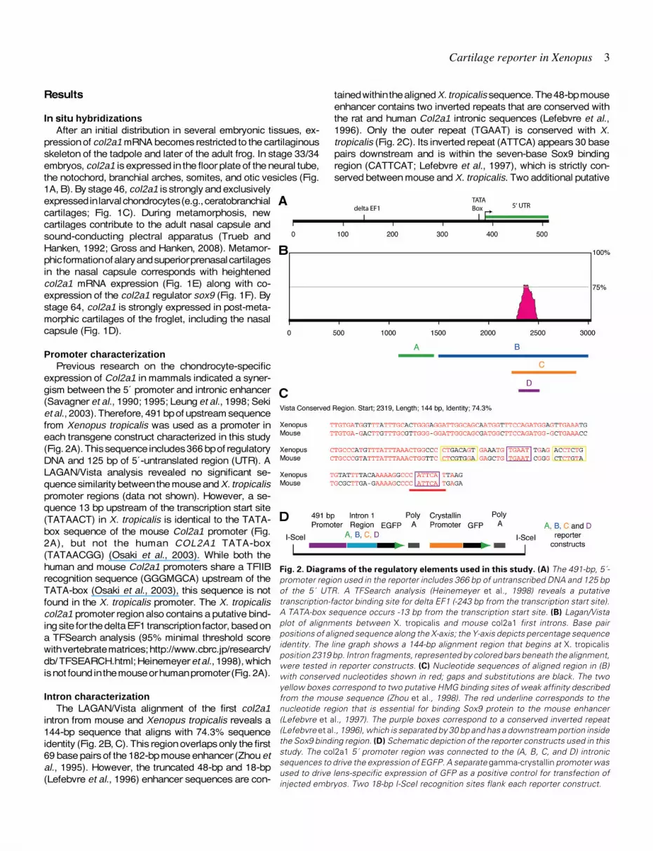
tained within the aligned X. tropicalis sequence. The 48-bp mouseenhancer contains two inverted repeats that are conserved withthe rat and human Col2a1 intronic sequences (Lefebvre et al.,1996). Only the outer repeat (TGAAT) is conserved with X.tropicalis (Fig. 2C). Its inverted repeat (ATTCA) appears 30 basepairs downstream and is within the seven-base Sox9 bindingregion (CATTCAT; Lefebvre et al., 1997), which is strictly con-served between mouse and X. tropicalis. Two additional putative
Fig. 2. Diagrams of the regulatory elements used in this study. (A) The 491-bp, 5´-promoter region used in the reporter includes 366 bp of untranscribed DNA and 125 bpof the 5´ UTR. A TFSearch analysis (Heinemeyer et al., 1998) reveals a putativetranscription-factor binding site for delta EF1 (-243 bp from the transcription start site).A TATA-box sequence occurs -13 bp from the transcription start site. (B) Lagan/Vistaplot of alignments between X. tropicalis and mouse col2a1 first introns. Base pairpositions of aligned sequence along the X-axis; the Y-axis depicts percentage sequenceidentity. The line graph shows a 144-bp alignment region that begins at X. tropicalisposition 2319 bp. Intron fragments, represented by colored bars beneath the alignment,were tested in reporter constructs. (C) Nucleotide sequences of aligned region in (B)with conserved nucleotides shown in red; gaps and substitutions are black. The twoyellow boxes correspond to two putative HMG binding sites of weak affinity describedfrom the mouse sequence (Zhou et al., 1998). The red underline corresponds to thenucleotide region that is essential for binding Sox9 protein to the mouse enhancer(Lefebvre et al., 1997). The purple boxes correspond to a conserved inverted repeat(Lefebvre et al., 1996), which is separated by 30 bp and has a downstream portion insidethe Sox9 binding region. (D) Schematic depiction of the reporter constructs used in thisstudy. The col2a1 5´ promoter region was connected to the (A, B, C, and D) intronicsequences to drive the expression of EGFP. A separate gamma-crystallin promoter wasused to drive lens-specific expression of GFP as a positive control for transfection ofinjected embryos. Two 18-bp I-SceI recognition sites flank each reporter construct.
A
B
C
D
1A, B). By stage 46, col2a1 is strongly and exclusivelyexpressed in larval chondrocytes (e.g., ceratobranchialcartilages; Fig. 1C). During metamorphosis, newcartilages contribute to the adult nasal capsule andsound-conducting plectral apparatus (Trueb andHanken, 1992; Gross and Hanken, 2008). Metamor-phic formation of alary and superior prenasal cartilagesin the nasal capsule corresponds with heightenedcol2a1 mRNA expression (Fig. 1E) along with co-expression of the col2a1 regulator sox9 (Fig. 1F). Bystage 64, col2a1 is strongly expressed in post-meta-morphic cartilages of the froglet, including the nasalcapsule (Fig. 1D).
Promoter characterizationPrevious research on the chondrocyte-specific
expression of Col2a1 in mammals indicated a syner-gism between the 5´ promoter and intronic enhancer(Savagner et al., 1990; 1995; Leung et al., 1998; Sekiet al., 2003). Therefore, 491 bp of upstream sequencefrom Xenopus tropicalis was used as a promoter ineach transgene construct characterized in this study(Fig. 2A). This sequence includes 366 bp of regulatoryDNA and 125 bp of 5´-untranslated region (UTR). ALAGAN/Vista analysis revealed no significant se-quence similarity between the mouse and X. tropicalispromoter regions (data not shown). However, a se-quence 13 bp upstream of the transcription start site(TATAACT) in X. tropicalis is identical to the TATA-box sequence of the mouse Col2a1 promoter (Fig.2A), but not the human COL2A1 TATA-box(TATAACGG) (Osaki et al., 2003). While both thehuman and mouse Col2a1 promoters share a TFIIBrecognition sequence (GGGMGCA) upstream of theTATA-box (Osaki et al., 2003), this sequence is notfound in the X. tropicalis promoter. The X. tropicaliscol2a1 promoter region also contains a putative bind-ing site for the delta EF1 transcription factor, based ona TFSearch analysis (95% minimal threshold scorewith vertebrate matrices; http://www.cbrc.jp/research/db/ TFSEARCH.html; Heinemeyer et al., 1998), whichis not found in the mouse or human promoter (Fig. 2A).
Intron characterizationThe LAGAN/Vista alignment of the first col2a1
intron from mouse and Xenopus tropicalis reveals a144-bp sequence that aligns with 74.3% sequenceidentity (Fig. 2B, C). This region overlaps only the first69 base pairs of the 182-bp mouse enhancer (Zhou etal., 1995). However, the truncated 48-bp and 18-bp(Lefebvre et al., 1996) enhancer sequences are con-

4 R. Kerney et al.
HMG binding sites (Zhou et al., 1998) are not conserved betweenmouse and X. tropicalis intronic sequences (Fig. 2C).
I-SceI meganuclease-mediated transgenesisA region of the col2a1 promoter and 5´ UTR was connected to
three separate regions of the first col2a1 intron and linked to egfp.Each construct was injected into zygotes of Xenopus laevis alongwith I-SceI meganuclease. The resulting embryos were screenedfor separate lens-specific GFP expression under the control of thegamma-crystallin promoter, which was used as a positive controlfor transgene uptake (Rankin et al., 2009). Injection of eachconstruct generates EGFP expression in a subset of epidermalcells early in development in most injected animals (Table 1). This
Fig. 3. Distribution of EGFP in transgenic Xenopus laevis tadpoles.(A–G) Ventral views, anterior to the top. (H) Dorsal view, anterior to thetop. (A) Cleared-and-stained tadpole showing Meckel’s (MC), ceratohyal(CH) and ceratobranchial (CB) cartilages stained with Alcian blue (stage42). All other images show EGFP/GFP expression in living tadpoles. (B)Transient transgenic tadpole with a reporter carrying the intronic regionA (stage 48). EGFP is not seen in the cartilaginous skeleton of any tadpolecarrying reporter construct A. GFP expression is apparent in the lens ofthe left eye (EY), under the control of the gamma-crystallin promoter. (C)Transient “half-transgenic” tadpole with a reporter carrying the intronicregion B (stage 42). Unilateral transgene expression is apparent in thesame cartilages labeled in (A), along with GFP expression in the eye. (D)An F1 transgenic tadpole (stage 48), bred from a half-transgenic mothercarrying construct B. (E) Transient half-transgenic tadpole carrying areporter with the C construct (stage 42) also exhibits unilateral cartilagi-nous expression, although expression is weaker than that seen intadpoles carrying the B construct. (F) An F1 transgenic tadpole (stage 40),bred from a half-transgenic mother carrying construct C. Early expressionof EGFP is strongest as the cartilages are forming. (G) Stage 48 of thesame tadpole in (F). None of the tadpole cartilages continues to expressEGFP under construct C during the later larval stages. However, cartilagi-nous expression of EGFP increases again during the metamorphicformation of adult cartilages (Fig. 4). (H) Merged bright-field and fluores-cent image of a transgenic tadpole carrying a reporter with the smallerintronic region D (stage 48). This region does not drive cartilaginous EGFPexpression during any stage in any injected tadpole. Auto-fluorescence isvisible in the yolk of stage 40–42 individuals (asterisks in C,E,F) and in thegallbladder of stage-48 tadpoles (asterisks in B,D,G). Scale bar, 1 mm.
shared expression may be due to expression driven by the 5´-promoter region itself, or it may be an artifact of the egfp injection(Pan et al., 2006). No construct drives detectable EGFP expres-sion similar to the col2a1 mRNA expression in the notochord, oticvesicle, somites, floor plate of the neural tube, or branchial archesduring early development (Fig. 1A, B).
The 347-bp intronic region ‘A’ does not overlap with the alignedregion from our Vista analysis (Fig 2B). This sequence was usedas a negative control and compared to the expression generatedby intronic sequences that contain the aligned enhancer. Areporter construct containing intronic sequence A does not driveexpression outside the lens and early epidermal cells (Fig. 3B,Table 1). Epidermal expression dissipates in later-stage tadpoles.No other pattern of EGFP is found in any additional larval tissue.
The reporter construct that contains the 1,517-bp region ‘B’ ofthe first intron (Fig. 2B) drives EGFP expression in the cartilagi-nous skeleton (Fig. 3C–D, 4A–F) in 24 of 70 transgenic animals(34.3%; Table 1). Twelve of these have bilateral mosaic expres-
Intronic Region Length Total Transgenic % Chondrocyte Expression A 347 bp 22 0 B 1,517 bp 70 34 C 665 bp 78 26 D 153 bp 22 0
TABLE 1
EXPRESSION OF EGFP IN TADPOLES INJECTED WITH FOURREPORTER CONSTRUCTS
“Total transgenic” refers to the number of injected tadpoles with GFP expressed in the lens underthe gamma-crystallin promoter (positive control). Chondrocyte expression in transgenic tadpoles(with lens-specific GFP expression) was scored between stages 42 and 47 for each intronicregion assayed.
A B
C D
E F
G H

Cartilage reporter in Xenopus 5
sion within cartilage (data not shown), while the other twelve shownearly complete expression within cartilage that is distributed ononly one side of the tadpole (Fig. 3C, 4B). The stronger unilateralexpression indicates continued integration of the transgene afterthe first cell division. Cartilaginous EGFP expression co-localizeswith collagen-II antibody reactivity in the ceratobranchial cartilages(Fig. 4A–B). Expression of EGFP driven by this construct is strongduring early cartilage-forming stages (40–42) but decreases later(stage 48–metamorphosis). This may be due to an increase inproduction of the extracellular matrix and spreading of the chon-drocyte lacunae, or to weaker activation of the col2a1 regulatory
Fig. 4. Chondrocyte expression of reporter constructs in histologicalsections. (A,B) Tadpole injected with a reporter construct that containsintronic region B. Coronal sections through a single larval ceratobranchialcartilage stained with anti-collagen II antibody (red in A) or anti-GFPantibody (green in B) with DAPI nuclear counter stain (blue). (C,E,G)Trichrome and (D,F,H) merged bright-field and fluorescent images ofanti-GFP antibody stained sections. (C,D) Coronal sections of the nasalcapsule of a post-metamorphic froglet that carries the reporter constructwith intronic region B. (D) EGFP is expressed in the alary cartilage (AC),nasal septum (NS) and planum terminale (PT). (E,F) Longitudinal sectionsthrough the leg of a froglet that carries a reporter construct with intronicregion B. Cartilaginous expression is seen in the humerus (HU) andproliferating chondrocytes (PC) of the radio-ulna (RU). EGFP is notexpressed in the hypertrophic (HC) or articular (AR) chondrocytes of theradio-ulna. (G,H) Serial coronal sections through the ethmoidal region ofa froglet that carries the C.1 reporter. Cartilaginous expression is seen inthe same post-metamorphic cartilages shown in D. Scale bar, 0.25 mm.
sequences in later stages. Cartilaginous EGFP expression in-creases in post-metamorphic cartilages of the nasal capsule (Fig.4 C–D), which also express col2a1 mRNA (Fig. 1D–E). Prolifera-tive chondrocytes of the radio-ulna and humerus also expressEGFP under this reporter (Fig. 4 E–F). EGFP is not expressed,however, in either articular or hypertrophic chondrocytes of theradio-ulna (Fig. 4F). Col2a1 mRNA expression diminishes inhypertrophic chondrocytes of mice and chickens, where type IIcollagen is largely replaced by type X collagen before endochon-dral ossification (reviewed in Goldring et al., 2006). Seven tad-poles express EGFP in a small number of axial myocytes; thisgradually diminishes in later tadpole stages.
A half-transgenic female carrying the B construct was bred toa wild-type male. Bilateral cartilaginous EGFP expression isobserved in 24 of 110 progeny. These 24 individuals consistentlyexpress EGFP in the cartilaginous skeleton with no variationamong individuals, which indicates a single integration of thetransgene (Marsh-Armstrong et al., 1999). Early chondrocyteexpression is strong (stages 40–42). Later EGFP expression(stage 48) is reticulate within individual cartilages (Fig. 3D).
The reporter construct with the 665-bp intronic region ‘C’ alsodrives EGFP expression in the cartilaginous skeleton, although itsexpression is less intense than that generated by intronic regionB (Figs. 3E–G, 4G–H). EGFP-expressing chondrocytes occur in20 of 78 tadpoles transfected with this reporter (25.6%; Table 1).Nine of these have stronger unilateral expression (Fig. 3E), whileeleven exhibit weaker sporadic expression that is distributedbilaterally in the early cartilaginous skeleton. Early cartilaginousexpression of EGFP dissipates by stage 48 and is undetectablein later-stage chondrocytes (Fig. 3G). As with construct B, how-ever, EGFP is strongly expressed in metamorphic cartilages ofthe nasal capsule in froglets that carry construct C (Fig. 4H).Myocyte expression also occurs in ten of the injected tadpoles,although it dissipates in later stages.
A half-transgenic female carrying the C construct, again withstrong unilateral EGFP expression, was bred to a wild-type male.The resulting progeny have bilateral cartilaginous expression ofEGFP by stage 40 (Fig. 3F), which dissipates entirely by stage 48(Fig. 3G). The transgene was inherited in approximately half (18of 30) of the F1 progeny. The F1 generation of construct C exhibitsmore variation in the distribution of EGFP and has twice theproportion of transgenic individuals compared to the F1 genera-
A B
C D
E F
G H

6 R. Kerney et al.
tion of construct B, which indicates multiple transgene insertionsin the maternal founder. Despite this increased number of inser-tions, cartilaginous expression of EGFP generated by construct Cis weaker than that generated by construct B in stage-matched,F1 progeny (Fig. 3D, G).
The reporter construct that carries the 155-bp intronic region‘D’ does not drive expression in any tissue outside the earlyepidermis (Fig. 3H). Although this construct contains the entirealigned intronic enhancer region (Fig. 2B), it fails to drive EGFPexpression in myocytes or chondrocytes of 22 transgenic tad-poles, all of which express GFP in the lens under the control of thegamma-crystallin promoter (Table 1).
Discussion
This study describes the expression of col2a1 mRNA in larval,metamorphic and post-metamorphic Xenopus laevis and charac-terizes reporter constructs that use a transcriptional enhancerelement from X. tropicalis’ col2a1 in X. laevis tadpoles and adults.This enhancer element is located in the 3’ region of the first intronand aligns closely with a well-characterized chondrocyte-specificmouse enhancer. The Xenopus enhancer, combined with afragment of the 5´ promoter, drives cartilaginous EGFP expres-sion in I-SceI meganuclease-mediated transgenic tadpoles andadults. These transgenes are heritable through the germ line andare expressed both in larval cartilages that form during embryo-genesis and in adult cartilages, which form de novo duringmetamorphosis and are not present in the larval stage.
Whereas several studies examine the early distribution ofcol2a1 mRNA in Xenopus laevis (Su et al., 1991; Bieker andYazdani-Buicky, 1992; Larrain et al., 2000; Kerney et al., 2007),this study is the first to investigate its expression through meta-morphosis (Fig. 1). An initially widespread embryonic expressionof col2a1 becomes restricted to the cartilaginous skeleton of thelarva and continues to be strongly expressed in chondrocytesduring subsequent stages. Formation of adult cartilages in thenasal capsule during metamorphosis coincides with an increasein mRNA expression of both col2a1 and the transcription factorsox9. This transcriptional regulator is essential for chondrocytedifferentiation (Akiyama et al., 2002) and early col2a1 expression(Lefebvre et al., 1997) in mice. Expression of sox9 during meta-morphic chondrogenesis may either coincide with the late differ-entiation of mesenchymal precursors into functioning chondro-cytes or reflect an increase of type II collagen production byexisting larval chondrocytes to create new cartilage. Post-meta-morphic cartilages of the nasal capsule continue to expresscol2a1 mRNA after they are fully formed.
Several studies in mammalian cell culture characterize otherpositive and negative regulatory elements in the Col2a1 promoterregion (Savagner et al., 1990, 1995; Seki et al., 2003; Osaki et al.,2003). An Sp1 binding region (Savagner et al., 1995) and two E-box silencer regions in the rat promoter (CIIS1 and CIIS2; Savagneret al., 1990) are not found in the 3.6 Kb upstream sequence of theX. tropicalis promoter (data not shown). However, the presenceof a putative delta EF1 binding site in the Xenopus tropicalispromoter (Fig. 2A) indicates a possible site of transcriptionalregulation. Delta EF1 is a zinc finger homeobox-1 transcriptionfactor (zfhx1). Null mutant mice that lack functional Delta EF1have fused limb cartilages and fusions of the carpal and tarsal
cartilages in neonates (Takagi et al., 1998). Delta ef1 mRNA isexpressed in paraxial mesoderm, migratory cranial neural crest,retina and neural tube of Xenopus laevis embryos (van Grusvenet al., 2006). Delta EF1 acts as both a repressor of E2-boxmediated transcription in mice (Sekido et al., 1994) and Smad-mediated BMP signaling in Xenopus (van Grusven et al., 2006).However, studies of the mouse Col2a1 promoter have not iden-tified Delta EF1 as a potential transcriptional silencer. Furtherresearch in the binding of Delta EF1 to the Xenopus promoter isneeded to validate its role in col2a1 regulation and anuranskeletogenesis.
Lagan/Vista alignment of the first intron from col2a1 in mouseand Xenopus tropicalis reveals a 144-bp region with 74.3%sequence identity. This sequence contains the seven-base Sox9binding region from the mouse intron (CATTCAT; Lefebvre et al.,1997), which is strictly conserved with the X. tropicalis sequence.Part of this recognition sequence exists as an upstream invertedrepeat, which also is found in the mouse enhancer (Lefebvre etal., 1996). Pairs of inverted Sox-binding sites have been charac-terized in the enhancer of a precursor gene for type XI collagen,col11a2. These are essential for chondrocyte-specific transcrip-tional activity in col11a2 as well (Bridgewater et al., 2003).However, additional sequences outside the repeated bindingdomains are required for the col11a2 enhancer to drive transgeneexpression in chondrocytes. Other regulatory information mayalso exist outside the conserved col2a1 enhancer region. To-gether, Sox6 and L-Sox5 cooperate with Sox9 to activate col2a1expression in chondrocytes (Bell et al., 1997). Their binding sitesin the col2a1 regulatory regions have not been identified in eithermammals (Lefebvre and Smits, 2005) or X. tropicalis.
While the one established Sox9 binding site in the col2a1enhancer is conserved between mouse and Xenopus, two addi-tional putative HMG binding sites (Zhou et al., 1998) are not (Fig.2C). The Sox9 protein does not bind to either of these putativesites in the mouse intron (Lefebvre et al., 1997). Absence of thesesites from the Xenopus sequence further suggests that thesesites are not part of the Sox9 regulation of col2a1.
Regulatory ability of the intronic enhancer from Xenopustropicalis was tested in X. laevis through I-SceI meganuclease-mediated transgenesis. A large 1.5-Kb region of the intron drovecartilaginous expression in both tadpole and adult cartilageswhen connected to an upstream promoter (Fig. 2B, region B).While a smaller 665-bp region also drove cartilage-specific ex-pression, its expression was weaker than the larger intronicfragment B (Fig. 2B, region C). A truncated 155-bp region contain-ing the entire aligned sequence from the LAGA/Vista analysisfailed to drive any expression (Fig. 2B, region D). These resultsare similar to previous studies of the mouse Col2a1 intronicenhancer, in which multiple copies of smaller enhancer regionsare required to drive cartilaginous gene expression (Lefebvre etal., 1996; Zhou et al., 1998). Multiple copies of region D were nottested in this study.
The negative-control construct A, which contains the 5´-pro-moter (491 bp) but not the conserved intronic enhancer, failed toshow any EGFP expression in chondrocytes. Intronic region A(347 bp) is shorter than B (1.5 Kb) and C (665 bp) regions that dodrive cartilage-specific expression. The small size of A mayaccount for its inability to drive cartilage-specific expression. Thedecreasing sizes of regions B, C and D correspond with a

Cartilage reporter in Xenopus 7
decrease in their effectiveness in driving cartilage-specific ex-pression (Table 1). This trend has been observed in studies ofmouse Col2a1 regulation, where smaller enhancer-containingfragments require multiple copies to activate transcription (Zhouet al., 1998; Lefebvre et al., 1996). However no study of amammalian col2a1 first intron has revealed regulatory informa-tion outside the Sox9-binding enhancer region. The rat col2a1 5´-promoter (310 bp) is capable of driving faint expression in chon-drocytes without the intronic enhancer (Horton et al., 1987).However, neither 6.1 Kb of the human promoter (Leung et al.,1998) nor 309 bp of the mouse promoter (Zhou et al., 1995) driveschondrocyte expression without the intronic enhancer in trans-genic mice. Xenopus, like human and mouse, appears to requireintronic enhancer sequence for chondrocyte-specific expression.
The mouse enhancer also can drive chondrocyte-specificexpression when connected to a minimal human "-globin pro-moter (Zhou et al., 1995). Surprisingly, the Col2a1 enhancer (4copies of 264 bp) from the extinct “marsupial wolf” Thylacinuscynocephalus can drive chondrocyte-specific transgene expres-sion in transgenic mice when connected to the "-globin promoter(Pask et al., 2008). However, a similar "-globin promoter con-struct, with 308 bp of the human COL2A1 intronic enhancer, doesnot drive chondrocyte-specific expression in mice (Leung et al.,1998).
Expression of EGFP in X. laevis tissues under the control of X.tropicalis regulatory information further supports the previouslysuggested compatibility of the two species (Khokha et al., 2002).Zygotes of X. laevis are larger and easier to microinject than thoseof X. tropicalis. However, the longer generation time in X. laevismakes germ line transmission of transgene constructs difficult totest within a reasonable time frame. Founder frogs bred in thisstudy required 18 months to reach sexual maturity in the labora-tory. Compatibility in regulatory sequence function between thesespecies also suggests the possibility that the col2a1 reportersdescribed here will drive transgene expression in cartilages ofother amphibians. The I-SceI meganuclease method of trans-gene integration works in the Mexican axolotl, Ambystomamexicanum (Sobkow et al., 2006), and similar microinjectiontechniques have been developed for the Oriental fire-bellied toad,Bombina orientalis (Olsson and Hanken, 1996; Olsson et al.,2001), which suggests the feasibility of testing the col2a1 reporterin a wide number of alternative amphibian models.
The meganuclease technique does yield some mosaic expres-sion of EGFP among individual chondrocytes for each reportertested. This mosaic expression is dramatically reduced in “half-transgenic” tadpoles, which strongly express EGFP unilaterallyon either the left or right side. EGFP expression is consistentlymore intense in the cartilages of half-transgenic tadpoles than in“full-transgenic” individuals, which express cartilaginous EGFPbilaterally (Fig. 3). Half-transgenic individuals result from incorpo-ration of the transgene after the first cellular division. The trans-gene is heritable provided the germ line cells carry the insertedconstruct (Pan et al., 2006).
Background expression from the reporter construct was ob-served in epidermal cells and larval myocytes. It is reduced inolder tadpoles and post-metamorphic froglets, and absent in F1progeny. This pattern indicates that the background is a result ofinjected egfp DNA, and not due to integrated transgene expres-sion.
Conclusions
Cartilage-specific expression generated by these reporterconstructs provides a useful tool for future investigations ofanuran skeletal development. These constructs can be used todrive dominant-negative protein expression or to over-expressvarious genes in the cartilaginous skeleton. Whereas severalstudies investigate the skeletal morphology of Xenopus laevisduring development (Bernasconi, 1951; Sedra and Michael,1957; Sokol, 1977; Trueb and Hanken, 1992), few examine theunderlying transcriptional regulators involved in skeletogenesis(Pasqualetti et al., 2000; Spokony et al., 2002; Moriishi et al.,2005; Kerney et al., 2007). Despite ongoing use of Xenopus asa model system of organogenesis (Thomsen, 2006) and thequickly growing field of skeletal developmental biology(Karsenty, 2003; Zelzer and Olsen, 2003; Hall, 2005; Goldringet al., 2006) few studies research skeletal development inXenopus. This lack of attention is due largely to the limitedarray of techniques available for investigations of later devel-opment in Xenopus (Blitz et al., 2005). Functional experimentsthrough the injection of morpholino oligonucleotides, antisenseRNA, or RNA interference are limited to early stages (Nutt etal., 2001; Khokha et al., 2002). Due to these limitations, thereare no current investigations into the function of skeletalregulatory genes during anuran metamorphosis. The ability todrive chondrocyte-specific gene expression in Xenopus willprovide a valuable tool for future investigations of gene func-tion and cell lineage in anuran skeletal development.
Materials and Methods
All animal protocols were approved by the Harvard UniversityFaculty of Arts and Sciences Standing Committee on the use ofAnimals in Research and Teaching (protocol 99-09) and DalhousieUniversity’s Committee on Laboratory Animals (protocol 07-126).
In situ HybridizationsAntisense mRNA probes were prepared from pCS2-AS-collagen II
(Larrain et al., 2000) and pGEMT:Sox9 (Spokony et al., 2002). Whole-mount in situ hybridizations were performed on albino embryos andhatchlings following the protocol of Sive et al. (2000). Embryos werefixed in MEMFA and stored in 70% methanol at -20 oC prior to initialuse. In situ hybridizations on sectioned tissues followed the protocolof Zelzer et al. (2001). Sectioned tissues were fixed in 4% paraform-aldehyde in phosphate-buffered saline (pH 7.4), dehydrated andembedded in Paraplast X-tra (Sherwood Medical, Deland, FL), and cutto a thickness of 7 µm on a rotary microtome (Leica 1512, Wetzlar,Germany).
Regulatory Region AnalysisThe Xenopus tropicalis col2a1 promoter and first intron sequence
are from scaffold 161 of the Joint Genome Institute X. tropicalis draftgenome assembly version 4.1 (http://genome.jgi-psf.org/cgi-bin/runAlignment?db=Xentr4). The transcription start site was determinedby aligning the complete coding sequence of X. tropicalis col2a1(Genbank Accession: BC063191) with the genomic sequence. ALagan/ Vista analysis (Brundo et al., 2003) aligned the enhancersequence between the first introns of mouse Col2a1 (NCBI accessionNW_001030577.1; base pair positions 19052638–19048835) and X.tropicalis col2a1 (JGI scaffold 161; base pair positions 125221–121349). The mouse-specific repeat masker was used for the mousesequence; the “softmask” option was used for the X. tropicalis se-

8 R. Kerney et al.
quence (http://lagan.stanford.edu).
Preparation of Plasmid ConstructsRegulatory sequences and egfp were inserted into the pDHG_I-SceI
plasmid (Daniel Buchholz, unpublished). This plasmid also contains agamma-crystallin promoter that drives gfp within the I-SceI recognitionsites (Fig. 2D–E). This strong promoter is used to screen injected animalsthat are successfully transformed with an injected construct (Pan et al.,2006). Egfp was taken from the pCS2+egfp-Bgl2 plasmid (Turner andWeintraub, 1994). The 5´-promoter region and first intron of the Xenopustropicalis col2a1 gene were amplified by PCR. The forward primer 5´-GCA CTG CAC ATG TTC TTG CT-3´ and reverse primer 5´-GCA GAATCT TTG TGG GGA AA-3´ were used to amplify ~3 Kb of the 5´ promoterregion. The forward primer 5´-GTT CGC AGC CAC ACA AGT C-3´ andreverse primer 5´-CCC AGT GTC ACA GAC ACA GAT T-3´ were used toamplify ~3.2 Kb of the first intron. Internal primers were used to amplify491 bp of the promoter with additional 5´-Hind III and 3´-Sac1 sites. Intron-specific internal primers were used to amplify four regions of the intronwith additional 5´-SacI and 3´-HindIII sites. The resulting intronic frag-ments are 347 bp (A), 1,517 bp (B), 665 bp (C), and 153 bp (D) (Fig. 2B).Whereas regions B–D each overlapped the sequence aligned by Vistaanalysis, region A did not; it was used as a negative control for enhanceractivity. Orientation of each promoter-intron was determined throughPCR and sequencing. The four reporter constructs were used for I-SceImeganuclease-mediated transgenics in Xenopus laevis zygotes.
I-SceI Meganuclease-Mediated TransgenicsThe I-SceI transgenic protocol followed those described previously for
Xenopus (Ogino et al., 2006 a,b; Pan et al., 2006). A total of 200 ngplasmid DNA was digested for 40 min at 37o C in a 23-µL I-SceImeganuclease reaction (New England Biolabs, Ipswich, MA). During thisdigest fertilized Xenopus laevis eggs were obtained and dejellied usingestablished protocols (Sive et al., 2000). A total of 4.5 nL of the reactionmix was injected into each single-celled zygote (40 pg DNA/ embryo)using a Femtojet microinjector (Eppendorf, New York). Embryos wereraised for 2 hr at room temperature. Individuals that failed to undergonormal cell division were culled. Remaining embryos were raised toembryonic stages 36–40 (Nieuwkoop and Faber, 1994) and screened forGFP expression in the lens under the control of the gamma-crystallinpromoter. Incorporation of the transgene after the first cell division yields“half-transgenic” embryos that exhibit unilateral fluorescence on eitherleft or right sides. Incorporation of the transgene before the first celldivision yields “full-transgenic” individuals with bilateral transgene ex-pression. Transgenic embryos were raised to early tadpole stages 42–47and screened for tissue-specific EGFP expression. Tadpoles with intenseEGFP expression were raised through metamorphosis to examine re-porter expression in adult cartilages.
Antibody Staining and HistologyAntibody staining was carried out on sectioned material following
established Xenopus protocols (Gross et al., 2006). Briefly, tadpoles werefixed overnight in 4% PFA at 4o C, embedded in OCT media (Sakura,Torrance, CA), frozen, and sectioned at 10 µm on a Leica CM 3050Scryostat. Primary antibodies included a FITC-conjugated goat polyclonalantibody to GFP (1:2000; Abcam, Cambridge, MA) and an anti-collagenII monoclonal antibody (1:100; Linsenmeyer and Hendrix, 1980). Slideswere counterstained with a 2 µg per mL DAPI nuclear stain solution(Invitrogen) before being cover-slipped with Fluoromount-G (Fisher Sci-entific, Waltham, MA). Serial sections of post-metamorphic tissues werestained with the Hall-Brunt-Quadruple stain, which colors cartilage blue(Hall, 1986).
AcknowledgementsSpecial thanks are due to Daniel Buchholz, who introduced RK to the
I-SceI meganuclease-mediated transgenics. Anne Everly helped with
microscopy and animal care. Jean-Pierre Saint-Jeannet and Edward DeRobertis generously donated the Xenopus laevis sox9 and col2a1 clones,respectively. Financial support was provided by a Goelet Summer Re-search Grant from the Museum of Comparative Zoology (RK), NSF grantEF-0334846 (AmphibiaTree; JH), and NSERC grant A5056 (BKH).
References
AKIYAMA H, CHABOISSIER MC, MARTIN JF, SCHEDL A, DE CROMBRUGGHEB. 2002. The transcription factor Sox9 has essential roles in successive stepsof the chondrocyte differentiation pathway and is required for expression ofSox5 and Sox6. Genes Dev 16:2813-2828.
BALTZINGER M, ORI M, PASQUALETTI M, NARDI I, RIJLI FM. 2005. Hoxa2knockdown in Xenopus results in hyoid to mandibular homeosis. Dev Dyn234:858-867.
BELL DM, LEUNG KK, WHEATLEY SC, NG LJ, ZHOU S, LING KW, SHAM MH,KOOPMAN P, TAM PP, CHEAH KS. 1997. SOX9 directly regulates the type-IIcollagen gene. Nat Genet 16:174-178.
BERNASCONI AF. 1951. Über den Ossifikationsmodus bei Xenopus laevis(Daudain). Mem Soc Helvet Sci Nat 79:191-252.
BIEKER JJ, YAZDANI-BUICKY M. 1992. Distribution of type II collagen mRNA inXenopus embryos visualized by whole-mount in situ hybridization. J HistochemCytochem 40:1117-1120.
BLITZ IL, ANDELFINGER G, HORB ME. 2005. Germ layers to organs: UsingXenopus to study «later» development. Semin Cell Dev Biol 17:133-145.
BOOT-HANDFORD RP, TUCKWELL DS. 2003. Fibrillar collagen: the key tovertebrate evolution? A tale of molecular incest. Bioessays 25:142-151.
BRIDGEWATER LC, WALKER MD, MILLER GC, ELLISON TA, HOLSINGER LD,POTTER JL, JACKSON TL, CHEN RK, WINKEL VL, ZHANG Z, MCKINNEY S,DE CROMBRUGGHE B. 2003. Adjacent DNA sequences modulate Sox9transcriptional activation at paired Sox sites in three chondrocyte-specificenhancer elements. Nuc Acid Res 31:1541-1553.
BRUNDO M, DO C, COOPER G, KIM M, DAVYDOV E, GREEN E, SIDOW A,BATZOGLOU S. 2003. LAGAN and Multi-LAGAN: efficient tools for large-scalemultiple alignment of genomic DNA. Genome Res 13:721-731.
CHEN M, LICHTLER AC, SHEU TJ, XIE C, ZHANG X, O’KEEFE RJ, CHEN D. 2007.Generation of a transgenic mouse model with chondrocyte-specific andtamoxifen-inducible expression of Cre recombinase. Genesis 45:44-50.
GOLDRING MB, TSUCHIMOCHI K, IJIRI K. 2006. The control of chondrogenesis.J Cell Biochem 97:33-44.
GROSS JB, HANKEN J, OGLESBY E, MARSH-ARMSTRONG N. 2006. Use of aROSA26:GFP transgenic line for long-term Xenopus fate-mapping studies. JAnat 209:401-413.
GROSS JB, HANKEN J. 2008. Segmentation of the vertebrate skull: neural-crestderivation of adult cartilages in the clawed frog, Xenopus laevis. Int Comp Biol.48:681-696.
GROVER J, ROUGHLEY PJ. 2006. Generation of a transgenic mouse in which Crerecombinase is expressed under control of the type II collagen promoter anddoxycycline administration. Matrix Biol 25:158-165.
HALL BK. 1986. The role of movement and tissue interactions in the developmentand growth of bone and secondary cartilage in the clavicle of the embryonicchick. J Embryol Exp Morphol 93:133-152.
HALL BK. 2005. Bones and Cartilage: Developmental and Evolutionary SkeletalBiology. Elsevier Academic Press, New York.
HANKEN J. 1992. Life history and morphological evolution. J Evol Biol 5:549-557.HEINEMEYER T, WINGENDER E, REUTER, I, HERMJAKOB H, KEL AE, KEL OV,
IGNATIEVA EV, ANANKO EA, PODKOLODNAYA OA, KOLPAKOV FA,PODKOLODNY NL, KOLCHANOV NA 1998. Databases on transcriptionalregulation: TRANSFAC, TRRD and COMPEL. Nuc Acid Res 26:362-367.
HORTON W, MIYASHITA T, KOHNO K, HASSELL JR, YAMADA Y. 1987. Identi-fication of a phenotype-specific enhancer in the first intron of the rat collagen IIgene. Proc Natl Acad Sci USA 84:8864-8868.
KARSENTY G. 2003. The complexities of skeletal biology. Nature 423:316-318.KERNEY R, GROSS JB, HANKEN J. 2007. Runx2 is essential for larval hyobranchial
cartilage formation in Xenopus laevis. Dev Dyn 236:1650-1662.

Cartilage reporter in Xenopus 9
KHOKHA MK, CHUNG C, BUSTAMANTE EL, GAW LW, TROTT KA, YEH J, LIMN, LIN JC, TAVERNER N, AMAYA E, PAPALOPULU N, SMITH JC, ZORN AM,HARLAND RM, GRAMMER TC. 2002. Techniques and probes for the study ofXenopus tropicalis development. Dev Dyn 225:499-510.
KOSHER RA, GAY SW, KAMANITZ JR, KULYK WM, RODGERS BJ, SAI S,TANAKA T, TANZER ML. 1986a. Cartilage proteoglycan core protein geneexpression during limb cartilage differentiation. Dev Biol 118:112-117.
KOSHER RA, KULYK WM, GAY SW. 1986b. Collagen gene expression during limbcartilage differentiation. J Cell Biol 102:1151-1156.
LARRAIN J, BACHILLER D, LU B, AGIUS E, PICCOLO S, DE ROBERTIS EM.2000. BMP-binding modules in chordin: a model for signaling regulation in theextracellular space. Development 127:821-830.
LEFEBVRE V, ZHOU G, MUKHOPADHYAY K, SMITH CN, ZHANG Z,EBERSPAECHER H, ZHOU X, SINHA S, MAITY SN, DE CROMBRUGGHE B.1996. An 18-base-pair sequence in the mouse proalpha1(II) collagen gene issufficient for expression in cartilage and binds nuclear proteins that are selec-tively expressed in chondrocytes. Mol Cell Biol 16:4512-4523.
LEFEBVRE V, HUANG W, HARLEY VR, GOODFELLOW PN, DE CROMBRUGGHEB. 1997. SOX9 is a potent activator of the chondrocyte-specific enhancer of thepro alpha1(II) collagen gene. Mol Cell Biol 17:2336-2346.
LEFEBVRE V, SMITS P. 2005. Transcriptional control of chondrocyte fate anddifferentiation. Birth Defects Res C Embryo Today 75:200-212.
LEUNG KH, NG LJ, HO KY, TAM PPL, CHEAH KE. 1998. Different cis-regulatoryDNA elements mediate developmental stage- and tissue-specific expression ofthe human COL2A1 gene in transgenic mice. J Cell Biol 141:1291-1300.
LINSENMEYER TF, HENDRIX MJ. 1980. Monoclonal antibodies to connectivetissue macromolecules: type II collagen. Biochem Biophys Res Commun92:440-446.
MORIISHI T, SHIBATA Y, TSUKAZAKI T, YAMAGUCHI A. 2005. Expression profileof Xenopus banded hedgehog, a homolog of mouse Indian hedgehog, is relatedto the late development of endochondral ossification in Xenopus laevis. BiochemBiophys Res Commun 328:867-873.
NAKAMURA E, NGUYEN MT, MACKEM S. 2006. Kinetics of tamoxifen-regulatedCre activity in mice using a cartilage-specific CreER(T) to assay temporalactivity windows along the proximodistal limb skeleton. Dev Dyn 235:2603-2612.
NAKATA K, ONO K, MIYAZAKI J, OLSEN BR, MURAGAKI Y, ADACHI E,YAMAMURA K, KIMURA T. 1993. Osteoarthritis associated with mild chondro-dysplasia in transgenic mice expressing alpha 1(IX) collagen chains with acentral deletion. Proc Natl Acad Sci USA 90:2870-2874.
NIEUWKOOP PD, FABER J. 1994. Normal table of Xenopus laevis (Daudin).Garland Publishing, New York.
NUTT SL, BRONCHAIN OJ, HARTLEY KO, AMAYA E. 2001. Comparison ofmorpholino based translational inhibition during the development of Xenopuslaevis and Xenopus tropicalis. Genesis 30:110-113.
OGINO H, MCCONNELL WB, GRAINGER RM. 2006a. High-throughput transgenesisin Xenopus using I-SceI meganuclease. Nat Protoc 1:1703-1710.
OGINO H, MCCONNELL WB, GRAINGER RM. 2006b. Highly efficient transgenesisin Xenopus tropicalis using I-SceI meganuclease. Mech Dev 123:103-113.
OLSSON L, FALCK P, LOPEZ K, COBB J, HANKEN J. 2001. Cranial neural crestcells contribute to connective tissue in cranial muscles in the anuran amphibian,Bombina orientalis. Dev Biol 237:354-367.
OLSSON L, HANKEN J. 1996. Cranial neural-crest migration and chondrogenicfate in the Oriental fire-bellied toad Bombina orientalis: defining the ancestralpattern of head development in anuran amphibians. J Morphol 1996:105-120.
OSAKI M, TAN L, CHOY BK, YOSHIDA Y, CHEAH KS, AURON PE, GOLDRINGMB (2003) The TATA-containing core promoter of the type II collagen gene(Col2a1) is the target of interferon-#-mediated inhibition in human chondro-cytes: requirement for Stat1!, Jak1 and Jak2. Biochem J 369:103-115.
PAN FC, CHEN Y, LOEBER J, HENNINGFELD K, PIELER T. 2006. I-SceImeganuclease-mediated transgenesis in Xenopus. Dev Dyn 235:247-252.
PASK AJ, BEHRINGER RR, RENFREE MB. 2008. Resurrection of DNA function invivo from an extinct genome. PLoS ONE 3:e2240
PASQUALETTI M, ORI M, NARDI I, RIJLI FM. 2000. Ectopic Hoxa2 induction afterneural crest migration results in homeosis of jaw elements in Xenopus. Devel-opment 127:5367-5378.
RANKIN SA, HASEBE T, ZORN AM, BUCHHOLZ DR. (2009). Improved Crereporter transgenic Xenopus. Dev Dyn 238: 2401-2408.
SADAGHIANI B, THIÉBAUD CH. 1987. Neural crest development in the Xenopuslaevis embryo, studied by interspecific transplantation and scanning electronmicroscopy. Dev Biol 124:91-110.
SANDELL LJ, MORRIS N, ROBBINS JR, GOLDRING MB. 1991. Alternativelyspliced type II procollagen mRNA’s define distinct populations of cells duringvertebral development: differential expression of the amino-propeptide. J CellBiol 114:1307-1319.
SAVAGNER P, MIYASHITA T, YAMADA Y. 1990. Two silencers regulate thetissue-specific expression of the collagen II gene. J Biol Chem 265:6669-6674.
SAVAGNER P, KREBSBACH PH, HATANO O, MIYASHITA T, LIEBMAN J,YAMADA Y. 1995. Collagen II promoter and enhancer interact synergisticallythrough Sp1 and distinct nuclear factors. DNA Cell Biol 14:501-510.
SEDRA S, MICHAEL M. 1957. The development of the skull, visceral arches, larynxand visceral muscles of the South African clawed toad, Xenopus laevis (Daudin)during the process of metamorphosis (from stage 55 to stage 66). Verh KonNederlandse Akad afg Natuurkunde Ser 2:1-80.
SEKI K, FUJIMORI T, SAVAGNER P, HATA A, AIKAWA T, OGATA N, NABESHIMAY, KAECHOONG L. 2003. Mouse snail family transcription repressors regulatechondrocyte, extracellular matrix, type II collagen, and aggrecan. J Biol Chem278:41862-41870.
SEKIDO R, MURAI K, FUNAHASHI J, KAMACHI Y, FUJISAWA-SEHARA A,NABESHIMA Y, KONDOH H. 1994. The delta-crystallin enhancer bindingprotein delta EF1is a repressor of E2-box-mediated gene activation. Mol CellBiol 14:5692-5700.
SIVE HL, GRAINGER RM, HARLAND RM. 2000. Early Development of Xenopuslaevis. Cold Spring Harbor Laboratory Press, Cold Spring Harbor, New York.
SOBKOW L, EPPERLEIN HH, HERKLOTZ S, STRAUBE WL, TANAKA EM. 2006.A germline GFP transgenic axolotl and its use to track cell fate: dual origin of thefin mesenchyme during development and the fate of blood cells during regen-eration. Dev Biol 290:386-397.
SOKOL OM. 1977. The free swimming Pipa larvae with a review of pipid phylogeny(Anura: Pipidae). J Morphol 154:357-426.
SPOKONY RF, AOKI Y, SAINT-GERMAIN N, MAGNER-FINK E, SAINT-JEANNETJP. 2002. The transcription factor Sox9 is required for cranial neural crestdevelopment in Xenopus. Development 129:421-432.
STONE LS. 1929. Experiments showing the role of migrating neural crest (mesec-toderm) in the formation of head skeleton and loose connective tissue in Ranapalustris. Wilhelm Roux Arch Entw Mech Org 118:40-77.
STRICKER S, FUNDELE R, VORTKAMP A, MUNDLOS S. 2002. Role of Runxgenes in chondrocyte differentiation. Dev Biol 245:95-108.
SU MW, SUZUKI HR, BIEKER JJ, SOLURSH M, RAMIREZ F. 1991. Expression oftwo nonallelic type II procollagen genes during Xenopus laevis embryogenesisis characterized by stage-specific production of alternatively spliced transcripts.J Cell Biol 115:565-575.
TAKEDA S, BONNAMY JP, OWEN MJ, DUCY P, KARSENTY G. 2001. Continuousexpression of Cbfa1 in nonhypertrophic chondrocytes uncovers its ability toinduce hypertrophic chondrocyte differentiation and partially rescues Cbfa1-deficient mice. Genes Dev 15:467-481.
THERMES V, GRABHER C, RISTORATORE F, BOURRAT F, CHOULIKA A,WITTBRODT J, JOLY JS. 2002. I-SceI meganuclease mediates highly efficienttransgenesis in fish. Mech Dev 118:91-98.
THOMSEN GH. 2006. A new century of amphibian developmental biology. SeminCell Dev Biol 17:78-79.
THOROGOOD P, BEE J, VON DER MARK K. 1986. Transient expression ofcollagen type II at epitheliomesenchymal interfaces during morphogenesis ofthe cartilaginous neurocranium. Dev Biol 116:497-509.
TRUEB L, HANKEN J. 1992. Skeletal development in Xenopus laevis (Anura:Pipidae). J Morphol 214:1-41.
TSUMAKI N, TANAKA K, ARIKAWA-HIRASAWA E, NAKASE T, KIMURA T,THOMAS JT, OCHI T, LUYTEN FP, YAMADA Y. 1999. Role of CDMP-1 inskeletal morphogenesis: promotion of mesenchymal cell recruitment and chon-drocyte differentiation. J Cell Biol 144:161-173.
TURNER DL, WEINTRAUB H. 1994. Expression of achaete-scute homolog 3 inXenopus embryos converts ectodermal cells to a neural fate. Genes Dev

10 R. Kerney et al.
8:1434-1447.UETA C, IWAMOTO M, KANATANI N, YOSHIDA C, LIU Y, ENOMOTO-IWAMOTO
M, OHMORI T, ENOMOTO H, NAKATA K, TAKADA K, KURISU K, KOMORI T.2001. Skeletal malformations caused by overexpression of Cbfa1 or its domi-nant negative form in chondrocytes. J Cell Biol 153:87-100.
WOOD A, ASHHURST DE, CORBETT A, THOROGOOD P. 1991. The transientexpression of type II collagen at tissue interfaces during mammalian craniofa-cial development. Development 111:955-968.
ZELZER E, GLOTZER DJ, HARTMAN C, THOMAS D, FUKAI N, SOKER S, OLSENB. 2001. Tissue specific regulation of VEGF expression during bone develop-ment requires CBFa-1/RUNX2. Mech Dev 106:97-106.
Further Related Reading, published previously in the Int. J. Dev. Biol.
See Special Issue Pattern Formation edited by Michael K. Richardson and Cheng-Ming Chuong at:http://www.ijdb.ehu.es/web/contents.php?vol=53&issue=5-6
Loss of Sox9 function results in defective chondrocyte differentiation of mouseembryonic stem cells in vitroGunnar Hargus, Ralf Kist, Jan Kramer, Daniela Gerstel, Angela Neitz, Gerd Scherer andJürgen RohwedelInt. J. Dev. Biol. (2008) 52: 323-332
BMP4 promotes chondrocyte proliferation and hypertrophy in the endochondralcranial base.Lillian Shum, Xibin Wang, Alex A Kane and Glen H NuckollsInt. J. Dev. Biol. (2003) 47: 423-431
Analysis of the odontogenic and osteogenic potentials of dental pulp in vivo using aCol1a1-2.3-GFP transgene.Alen Braut, Edward J Kollar and Mina MinaInt. J. Dev. Biol. (2003) 47: 281-29
Spatiotemporal pattern of the mouse chondromodulin-I gene expression and itsregulatory role in vascular invasion into cartilage during endochondral bone formation.C Shukunami, K Iyama, H Inoue and Y HirakiInt. J. Dev. Biol. (1999) 43: 39-49
Sequential synthesis of cartilage and bone marker proteins during transdifferentiationof mouse Meckel’s cartilage chondrocytes in vitro.K Ishizeki, Y Hiraki, M Kubo and T NawaInt. J. Dev. Biol. (1997) 41: 83-89
5 yr ISI Impact Factor (2008) = 3.271
ZELZER E, OLSEN B. 2003. Genetic basis of skeletal diseases. Nature 423:343-348.
ZHOU G, GAROFALO S, MUKHOPADHYAY K, LEFEBVRE V, SMITH CN,EBERSPAECHER H, DE CROMBRUGGHE B. 1995. A 182 bp fragment of themouse pro alpha 1(II) collagen gene is sufficient to direct chondrocyte expres-sion in transgenic mice. J Cell Sci 108:3677-3684.
ZHOU G, LEFEBVRE V, ZHANG Z, EBERSPAECHER H, DE CROMBRUGGHE B.1998. Three high mobility group-like sequences within a 48-base pair enhancerof the Col2a1 gene are required for cartilage-specific expression in vivo. J BiolChem 273:14989-14997.