Embed Size (px)
Citation preview
The Plant Cell, Vol. 11, 1651–1663, September 1999, www.plantcell.org © 1999 American Society of Plant Physiologists
Regional Expression of the Rice
KN1
-Type Homeobox Gene Family during Embryo, Shoot, and Flower Development
Naoki Sentoku,
a
Yutaka Sato,
a
Nori Kurata,
b
Yukihiro Ito,
b
Hidemi Kitano,
c
and Makoto Matsuoka
a,1
a
Nagoya University, BioScience Center, Chikusa, Nagoya 464-8601, Japan
b
Plant Genetics Laboratory, National Institute of Genetics, Yata, Mishima 411-0801, Japan
c
School of Agricultural Sciences, Nagoya University, Chikusa, Nagoya 464-8601, Japan
We report the isolation, sequence, and pattern of gene expression of members of the
KNOTTED1
(
KN1
)-type class 1 ho-meobox gene family from rice. Phylogenetic analysis and mapping of the rice genome revealed that all of the rice ho-meobox genes that we have isolated have one or two direct homologs in maize. Of the homeobox genes that we tested,all exhibited expression in a restricted region of the embryo that defines the position at which the shoot apical mer-istem (SAM) would eventually develop, prior to visible organ formation. Several distinct spatial and temporal expressionpatterns were observed for the different genes in this region. After shoot formation, the expression patterns of thesehomeobox genes were variable in the region of the SAM. These results suggest that the rice
KN1
-type class 1 ho-meobox genes function cooperatively to establish the SAM before shoot formation and that after shoot formation, theirfunctions differ.
INTRODUCTION
In animal embryogenesis, homeobox-containing genes workas spatial cues at all levels of the developmental hierarchy.For example, in Drosophila embryogenesis,
bicoid
is mater-nally transcribed, and its mRNA is specifically localized atthe anterior tip of the early embryo. Here, the
bicoid
geneproduct establishes a pattern in the anterior half of the em-bryo in a concentration-dependent manner (Driever andNüsslein-Volhard, 1988). A gap gene,
orthodenticle
, is ex-pressed in a circumferential stripe in the presumptive headregion of the blastoderm embryo and is necessary for theformation of anterior head structures (Finkelstein and Perrimon,1990). The
Distalless
transcript is expressed in the limb pri-mordia of the embryo and is required for the development ofall limb structures in the larva and the adult (Cohen, 1990).
eyeless
is transcribed in the embryonic primordia of the eye andis the master control gene for eye morphogenesis (Quiring etal., 1994). In each case study reported thus far, Drosophilahomeobox genes are involved in determining segmentalidentity, regionalization, or cell identity.
In addition, it is known that the product of one homeoboxgene can regulate the expression of other homeobox genesto create a more detailed positional information. The pairrule genes,
even-skipped
(
eve
) and
fushi-tarazu
(
ftz
), for ex-ample, are complementarily expressed in adjacent regions
of the blastoderm embryo (Lawrence et al., 1987). The posi-tion and width of the regions in which
eve
and
ftz
areexpressed coincide with the odd- and even-numbered para-segmental anlagen, respectively, indicating that the twogenes mutually but competitively define the parasegmentalregions. In this case, each homeobox gene mutually regu-lates the expression of the other to determine the more de-tailed regionalization after the broader regionalization hasbeen defined.
The involvement of plant homeobox genes in embryogen-esis was first demonstrated by the analysis of an Arabidop-sis embryogenesis-defective mutant,
shoot meristemless
(
stm
). Embryos of
stm
mutants lack shoot meristems,whereas other embryonic organs, such as cotyledons,hypocotyls, and radicles, develop normally (Barton andPoethig, 1993).
STM
encodes a KNOTTED1 (KN1)-type ho-meodomain protein and is expressed in the region of theshoot apical meristem (SAM) during embryogenesis (Long etal., 1996). Localized expression of other
KN1
-type ho-meobox genes has also been observed around the region inwhich the SAM develops early in embryogenesis (Smith etal., 1995; Sato et al., 1996, 1998). These findings demon-strate the involvement of this class of homeobox genes inSAM formation and maintenance during embryogenesis.
In a previous study, we found that expression of two ricehomeobox genes,
OSH1
and
OSH15
, during embryogenesisoverlaps in the region in which the SAM develops, prior toshoot formation (Sato et al., 1998). After shoot formation,the expression patterns of the two genes become distinct
1
To whom correspondence should be addressed. E-mail [email protected]; fax 81-52-789-5226.
1652 The Plant Cell
from one another. This observation suggests the possibilitythat homeobox genes in this class may act cooperatively inSAM formation, after which they may act independently orcompetitively to maintain the SAM or to form lateral organsin a fashion analogous to the Drosophila homeobox genes.
To test this possibility, we attempted to isolate all of themembers of the
KN1
-type class 1 homeobox gene familyfrom rice and to examine their expression patterns duringembryogenesis and in vegetative and floral shoots. In situmRNA localization analyses revealed that the rice
KN1
-typeclass 1 homeobox genes distinguish in some manner the re-gion in which the SAM develops from other regions prior toshoot formation. Based on these observations, we discussthe involvement of
KN1
-type homeobox genes in shoot for-mation during rice embryogenesis.
RESULTS
Cloning of Five Novel
KN1
-Type Genes from Rice
In previous studies, we isolated two rice clones,
OSH1
and
OSH15
, which are members of the class 1 family of
KN1
-type homeobox genes (Matsuoka et al., 1993; Sato et al.,1998). In this study, we attempted to isolate as many of theremaining
KN1
-type class 1 homeobox genes as possiblefrom rice. For this purpose, we constructed two cDNA librar-ies using poly(A)
1
mRNA extracted from the shoot apical re-gion and rachis, because most
KN1-
type class 1 homeoboxgenes are expressed mainly in these tissues (Kerstetter etal., 1994; M. Matsuoka, unpublished results). The cDNA li-braries were screened with the homeobox regions of
OSH1
and
OSH15
under low-stringency conditions, leading to theisolation of
.
40 clones that hybridized with the probes. Toconfirm that these clones represented
KN1
-type class 1genes, we amplified their homeobox regions using poly-merase chain reactions (PCRs) with primers flanking con-served regions of
KN1
-type class 1 homeobox genes.Approximately 90% of the clones produced DNA fragmentsof the expected size, and all of the amplified DNA fragmentswere cloned and sequenced. Sequence analysis revealedthat the clones represented six different
KN1
-type class 1homeobox genes. Two of these were
OSH1
and
OSH15
(Matsuoka et al., 1993; Sato et al., 1998). The remaining fourwere novel
KN1
-type class 1 homeobox genes that wenamed
OSH3
,
OSH6
,
OSH43
, and
OSH71
.We also tried to isolate novel homeobox genes by using
rice genomic DNA and PCR with primers flanking a con-served region of the previously reported
KN1
-type class 1homeobox genes. Because this conserved region includedparts of exons 4 and 5 in the
OSH1
and
OSH15
genes (seeFigure 1A), the sizes of the resulting PCR products were ex-pected to vary, depending on the length of intron 4 (assum-ing that other rice
KN1
-type class 1 homeobox genes
contain an intron at the same position or no intron). Sevenfragments of different sizes were amplified from rice ge-nomic DNA. All of the PCR products were cloned and se-quenced, revealing that six of the fragments correspondedto the previously identified
OSH
clones (
OSH1
,
OSH3
,
OSH6
,
OSH15
,
OSH43
, and
OSH71
).In the second approach, we screened a rice genomic li-
brary using the homeobox sequences of
OSH1
and
OSH15
as probes under low-stringency conditions. More than 100clones were characterized by using PCR with various prim-ers flanking the conserved ELK (glutamate, leucine, andlysine) homeodomain sequences present in the previouslyreported
KN1
-type class 1 genes (Kerstetter et al., 1994).Fragments of various sizes were amplified from all of theclones. Based on the sizes of the PCR products, we couldclassify the clones as corresponding to specific
OSH
genesbecause each
OSH
gene gives a band of unique size, whichis determined by the length of the intron sequence flankedby the primers. Among these clones, we identified one novel
KN1
-type class 1 gene,
OSH10
.We analyzed the entire nucleotide sequences of the
OSH3
,
OSH6
,
OSH43
, and
OSH71
cDNA clones. ThesecDNA clones contained open reading frames encoding 367,301, 341, and 311 amino acids, respectively. We also ana-lyzed the partial sequence of
OSH10
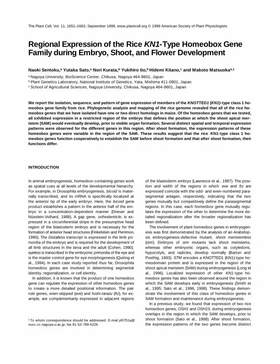
around the homeoboxregion. All of these clones encoded a 64–amino acid homeo-domain sequence that is well conserved throughout its en-tire length (Figure 1A). In particular, the third helix, which ispredicted to be a recognition helix, is totally conserved in allOSH homeodomain proteins. The flanking ELK domain,which has been found in all other plant KN1-type homeo-domain proteins (Kerstetter et al., 1994; Ito et al., 1999;Postma-Haarsma et al., 1999), was also conserved. More-over, there was another conserved region on the N-terminalside of the ELK homeodomain that has often been seen inthe KN1-type homeodomain proteins; this domain is re-ferred to as the KNOX domain.
To elucidate the structural relationship of the
OSH
genesto the maize KN1-type homeodomain proteins, we per-formed a phylogenetic analysis of the rice and maize homeo-domain sequences by using the unweighted pair groupmethod with arithmetic mean (UPGMA). Figure 1B demon-strates that each OSH protein corresponds to one or twocounterpart homeodomain protein(s) in maize. For example,OSH1 is paired with KN1, and OSH15 is paired with RS1and KNOX4. Similar pairing was also seen between OSH3and KNOX3, OSH6 and LG3, OSH43 and KNOX8, OSH10 andKNOX10, and OSH71 and KNOX5 or KNOX11.
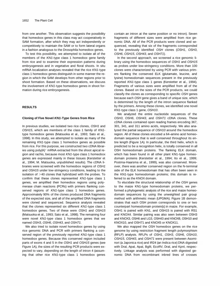
We also mapped the
OSH
homeobox genes on the ricegenome by using restriction fragment length polymorphism(RFLP) analysis. RFLPs of
OSH1
,
OSH3
,
OSH6
,
OSH10
,
OSH15
,
OSH43
, and
OSH71
were present between Asomi-nori (a Japonica rice) and IR24 (an Indica rice) DNA digestedwith DraI, ApaI, ApaI, BglII, EcoRV, DraI, and KpnI, respec-tively. Linkage analysis was performed with digested ge-nomic DNA from recombinant inbred lines of crosses
Expression Patterns of Rice Homeobox Genes 1653
between Asominori and IR24. Chromosomes 1, 5, and 7contained single
OSH
genes
OSH6
,
OSH71
, and
OSH15
, re-spectively. Chromosome 3 contained a small cluster of fourlinked
OSH
genes,
OSH1
,
OSH3
,
OSH10
, and
OSH43
(Fig-ure 2A).
According to the comparative linkage between the riceand maize genomes (Ahn and Tanksley, 1993), we were ableto compare the locations of the
OSH
homeobox genes on
the rice chromosomes with those of the maize homeoboxgenes (Figure 2A; Kerstetter et al., 1994). This comparativeanalysis demonstrated that the paired homeobox genes inthe phylogenetic tree (Figure 1B) mapped at analogous po-sitions on the rice and maize chromosomes (see Discussion).
Because
OSH1
and
OSH3
were found to be closely linked(Figure 2A), we investigated the relationship between thesegenes in more detail by hybridization of the genes to yeast
Figure 1. Deduced Amino Acid Sequences of OSH Genes.
(A) Comparison of OSH protein sequences. The deduced amino acid sequences of OSH3, OSH6, OSH10, OSH43, and OSH71 are comparedwith the previously reported sequences of OSH1 (Matsuoka et al., 1993) and OSH15 (Sato et al., 1998). Asterisks indicate the identical amino ac-ids in the ELK homeodomains among the OSH proteins. Arrows indicate the locations of primers used for PCR screening. The arrowhead indi-cates the position of an intron in the ELK homeodomain whose location is conserved in all OSH genes, with the exception of OSH3. Gaps,indicated by dashes, were introduced to facilitate alignment. GenBank/EMBL/DDBJ accession numbers of the proteins used are as follows:OSH1, D16507; OSH3, AB028882; OSH6, AB028883; OSH10, AB028886; OSH15, AB016071; OSH43, AB028884; and OSH71, AB028885.(B) Phylogenetic analysis of rice and maize homeodomain proteins. The tree was generated according to the UPGMA clustering strategy usingGenetyx Mac version 9.0 (Software Kaihatsu Co., Tokyo, Japan). The rice OSH homeodomains are shown in boldface. The sequences of OSH1and OSH15 are derived from Matsuoka et al. (1993) and Sato et al. (1998), respectively. The maize homeodomain sequences are derived fromKerstetter et al. (1994). The numbers above the junctions between pairs of clusters indicate the percentage of similarity between the clusters.
1654 The Plant Cell
artificial chromosomes. We found one yeast artificial chro-mosome clone, Y4583, that contained both genes. Thephysical map around the genes demonstrated that
OSH1
and
OSH3
lie within a 37-kb stretch of DNA. The two genesare oriented in the same direction, with
OSH3
lying up-stream of
OSH1
(Figure 2B).
In Situ Localization of the
OSH
mRNAs duringRice Embryogenesis
Rice embryos complete all morphogenetic events within 9days under normal conditions. The globular stage lasts until
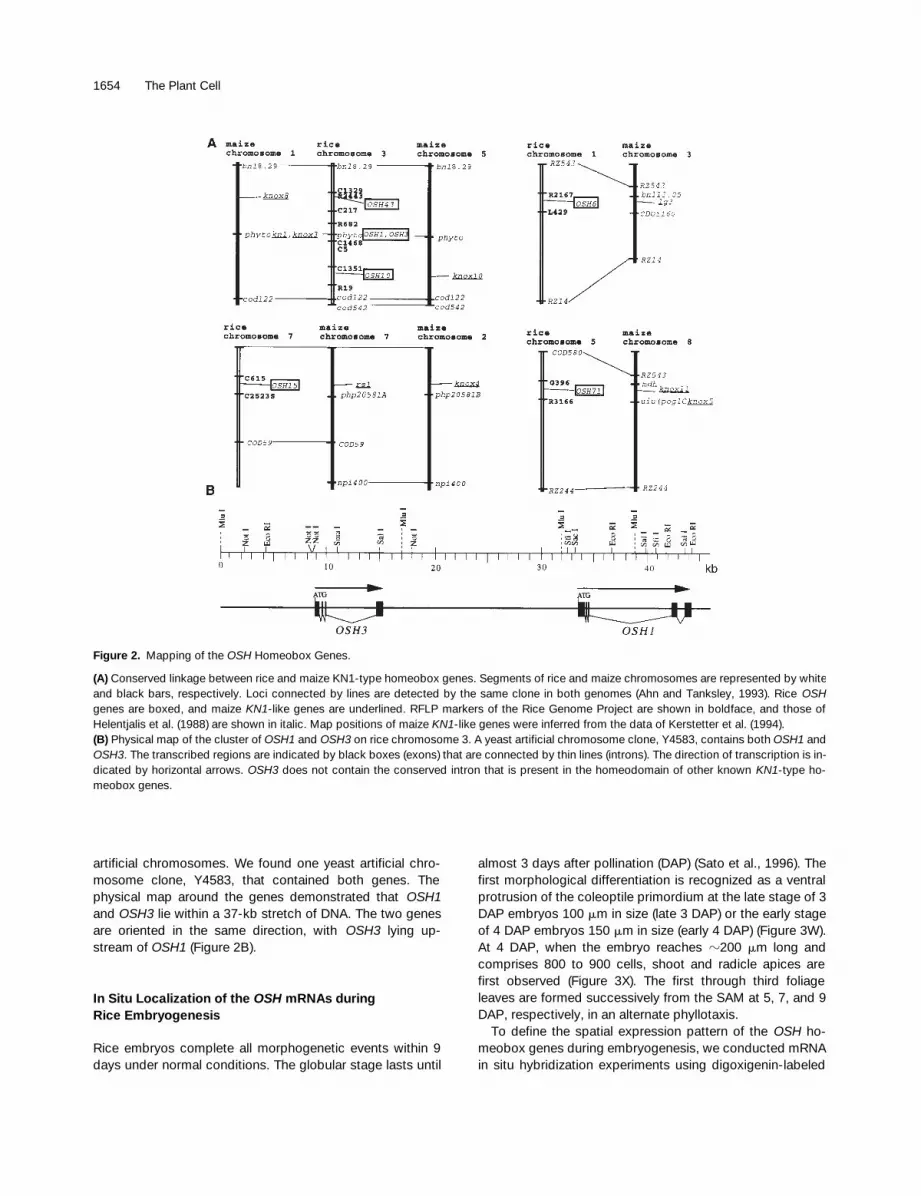
almost 3 days after pollination (DAP) (Sato et al., 1996). Thefirst morphological differentiation is recognized as a ventralprotrusion of the coleoptile primordium at the late stage of 3DAP embryos 100
m
m in size (late 3 DAP) or the early stageof 4 DAP embryos 150
m
m in size (early 4 DAP) (Figure 3W).At 4 DAP, when the embryo reaches
z
200
m
m long andcomprises 800 to 900 cells, shoot and radicle apices arefirst observed (Figure 3X). The first through third foliageleaves are formed successively from the SAM at 5, 7, and 9DAP, respectively, in an alternate phyllotaxis.
To define the spatial expression pattern of the
OSH ho-meobox genes during embryogenesis, we conducted mRNAin situ hybridization experiments using digoxigenin-labeled
Figure 2. Mapping of the OSH Homeobox Genes.
(A) Conserved linkage between rice and maize KN1-type homeobox genes. Segments of rice and maize chromosomes are represented by whiteand black bars, respectively. Loci connected by lines are detected by the same clone in both genomes (Ahn and Tanksley, 1993). Rice OSHgenes are boxed, and maize KN1-like genes are underlined. RFLP markers of the Rice Genome Project are shown in boldface, and those ofHelentjalis et al. (1988) are shown in italic. Map positions of maize KN1-like genes were inferred from the data of Kerstetter et al. (1994).(B) Physical map of the cluster of OSH1 and OSH3 on rice chromosome 3. A yeast artificial chromosome clone, Y4583, contains both OSH1 andOSH3. The transcribed regions are indicated by black boxes (exons) that are connected by thin lines (introns). The direction of transcription is in-dicated by horizontal arrows. OSH3 does not contain the conserved intron that is present in the homeodomain of other known KN1-type ho-meobox genes.
Expression Patterns of Rice Homeobox Genes 1655
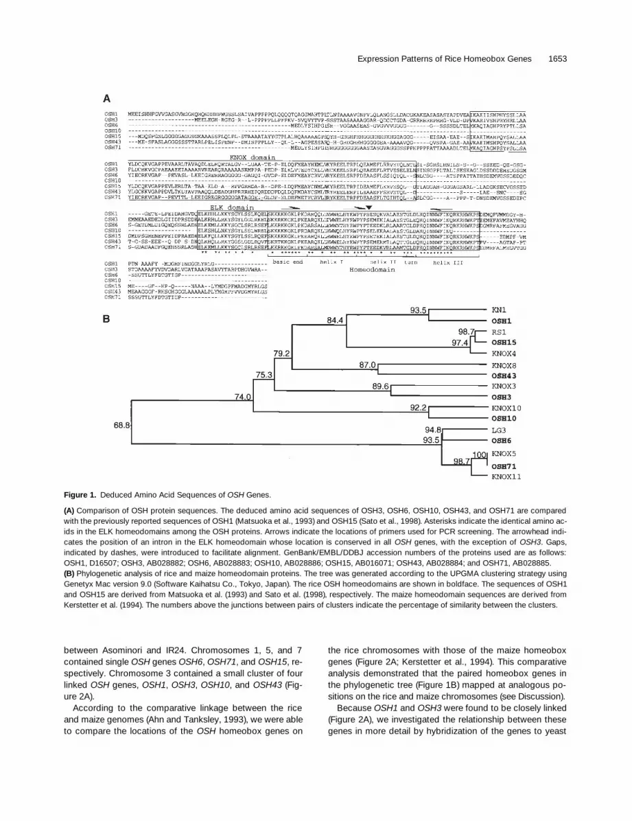
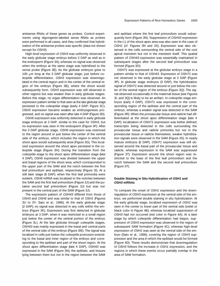
antisense RNAs of these genes as probes. Control experi-ments using digoxigenin-labeled sense RNAs as probeswere performed in all cases, and they confirmed that hybrid-ization of the antisense probes was specific (data not shownexcept for OSH3).
High-level expression of OSH3 was uniformly observed inthe early globular stage embryo before 2 DAP as well as inthe endosperm (Figure 3A), whereas no signal was observedwhen the embryo at the same stage was hybridized to thesense probe (Figure 3E). As the globular embryos grew to100 mm long at the 3 DAP globular stage, just before co-leoptile differentiation, OSH3 expression was downregu-lated in the central region and in the center of the ventral re-gion of the embryo (Figure 3B), where the shoot wouldsubsequently form. OSH3 expression was still observed inother regions but was weaker than in early globular stages.Before this stage, no organ differentiation was observed. Anexpression pattern similar to that seen at the late globular stagepersisted to the coleoptilar stage (early 4 DAP; Figure 3C).OSH3 expression became weaker as embryogenesis pro-gressed, and no signal was seen after late 4 DAP (Figure 3D).
OSH6 expression was uniformly detected in early globularstage embryos at 2 DAP, similar to the case for OSH3, butno expression was seen in the endosperm (Figure 3F). Afterthe 3 DAP globular stage, OSH6 expression was restrictedto the region around or just below the center of the ventralside of the embryo, which includes the region in which theshoot apex would subsequently arise (Figure 3G). This local-ized expression around the shoot apex persisted in the co-leoptile stage (Figure 3H). During the transition from thecoleoptile stage to the shoot apex differentiation stage (late4 DAP), OSH6 expression was divided between the upperand basal regions of the shoot area, which corresponded tothe upper part of the SAM and the notch between the firstleaf primordium and epiblast, respectively (Figure 3I). At astill later stage (6 DAP), when the first leaf primordia wereevident, OSH6 mRNA was localized to the notches betweenthe SAM and the first leaf primordium (Figure 3J) and the pu-tative second leaf primordium (Figure 3J) but was notpresent in the central part of the SAM (Figure 3J).
The expression pattern of OSH43 differed from those ofOSH3 and OSH6 and was similar to that of OSH1 (Figures3U to 3Y; Sato et al., 1996). At the early globular stage(2 DAP), no signal was detected in any cells within the em-bryo (Figure 3K). Expression was first detected in globularembryos at 3 DAP, when it was restricted to a small regionjust below the center of the ventral portion of the embryo(Figure 3L). At the late globular through coleoptile stages,OSH43 was mainly expressed in the basal and central partsof the ventral side of the embryo (Figure 3M). The signal waslocalized in cells just below the coleoptile protrusion extend-ing to the basal and inner region of the embryo and corre-sponding to the epiblast and part of the shoot region. At theshoot apex differentiation stage (late 4 DAP), OSH43 wasexpressed in the SAM (Figure 3N), the epiblast, and tissueslying between them but not in the region between the SAM
and epiblast where the first leaf primordium would subse-quently form (Figure 3N). Suppression of OSH43 expressionin the L1 of the shoot apex area was also seen in the case ofOSH1 (cf. Figures 3N and 3X). Expression was also ob-served in the cells surrounding the ventral side of the rootapical meristem but not in the meristem itself. This spatialpattern of OSH43 expression was essentially maintained insubsequent stages after the second leaf primordium wasformed (Figure 3O).
OSH71 was expressed at the globular embryo stage in apattern similar to that of OSH43. Expression of OSH71 wasnot observed in the early globular stage at 2 DAP (Figure3P). In globular stage embryos (3 DAP), the hybridizationsignal of OSH71 was detected around or just below the cen-ter of the ventral region of the embryo (Figure 3Q). The sig-nal observed occasionally in the maternal tissue (see Figures3L and 3Q) is likely to be an artifact. In coleoptile-stage em-bryos (early 4 DAP), OSH71 was expressed in the corre-sponding region of the epiblast and the central part of theembryo, whereas a weaker signal was detected in the SAM(Figure 3R). When the provascular tissue and radicle had dif-ferentiated at the shoot apex differentiation stage (late 4DAP), localization of OSH71 expression was better defined,transcripts being detected in the cells surrounding theprovascular tissue and radicle primordia but not in theprovascular tissue or radicle themselves; weaker hybridiza-tion signals were observed in the SAM (Figure 3S). In nearlymature embryos (6 DAP), OSH71 expression was still ob-served around the basal part of the provascular tissue andradicle, whereas expression in the SAM was suppressed(Figure 3T). Expression around the shoot region was re-stricted to the base of the first leaf primordium and thenotch between the SAM and the second leaf primordium(Figure 3T).
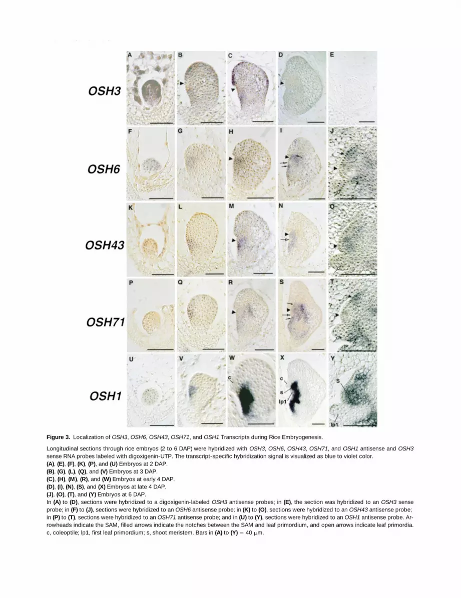
Double Staining in Situ Hybridization of OSH1 andOSH3 mRNAs
To compare the onset of OSH1 expression and the down-regulation of OSH3 expression at the ventral side of the em-bryo, we performed double staining in situ hybridization. Atthe early globular stage, localized expression of OSH1 wasseen in the center to lower part of the ventral side (violet orblack color in Figure 4B), whereas localized suppression ofOSH3 had not occurred (red color in Figure 4A). At a laterstage by which coleoptile differentiation had begun, sup-pression of OSH3 expression was observed in the region ofsubsequent SAM formation (Figure 4C), whereas high-levelexpression of OSH1 was seen at the ventral side of the em-bryo (Sato et al., 1996), covering the region of OSH3 sup-pression and the area in which the epiblast would be formed(Figure 4D). These results demonstrate that downregulationof OSH3 follows the increase in OSH1 expression, and theregions in which these events occur partially overlap in thearea of SAM formation.
1656 The Plant Cell
Figure 3. Localization of OSH3, OSH6, OSH43, OSH71, and OSH1 Transcripts during Rice Embryogenesis.
Longitudinal sections through rice embryos (2 to 6 DAP) were hybridized with OSH3, OSH6, OSH43, OSH71, and OSH1 antisense and OSH3sense RNA probes labeled with digoxigenin-UTP. The transcript-specific hybridization signal is visualized as blue to violet color.(A), (E), (F), (K), (P), and (U) Embryos at 2 DAP.(B), (G), (L), (Q), and (V) Embryos at 3 DAP.(C), (H), (M), (R), and (W) Embryos at early 4 DAP.(D), (I), (N), (S), and (X) Embryos at late 4 DAP.(J), (O), (T), and (Y) Embryos at 6 DAP.In (A) to (D), sections were hybridized to a digoxigenin-labeled OSH3 antisense probes; in (E), the section was hybridized to an OSH3 senseprobe; in (F) to (J), sections were hybridized to an OSH6 antisense probe; in (K) to (O), sections were hybridized to an OSH43 antisense probe; in (P) to (T), sections were hybridized to an OSH71 antisense probe; and in (U) to (Y), sections were hybridized to an OSH1 antisense probe. Ar-rowheads indicate the SAM, filled arrows indicate the notches between the SAM and leaf primordium, and open arrows indicate leaf primordia.c, coleoptile; lp1, first leaf primordium; s, shoot meristem. Bars in (A) to (Y) 5 40 mm.
Expression Patterns of Rice Homeobox Genes 1657
In Situ Localization of the OSH mRNAs inVegetative Tissues
The localization of the OSH mRNAs in vegetative tissueswas determined by using in situ hybridization. The expres-sion of five OSH genes, OSH3, OSH6, OSH43, OSH71, andOSH1, was examined in near-median longitudinal sectionsthrough the shoot apex of 1-month-old plants (Figures 5A,5D, 5H, 5L, and 5P, respectively). At this stage, the mer-istem had initiated approximately six to seven leaves.
The spatial expression patterns of four OSH genes,OSH6, OSH43, OSH71, and OSH1, around the shoot apexregion were essentially the same as those observed in thenearly mature embryo stage (cf. Figures 3J, 3O, 3T, and 3Y).Expression of OSH43 was observed throughout the corpusbut not in the tunica layer of the SAM or in leaf primordia.Such expression is very similar to that of OSH1 in vegetativeshoot apices (Figures 5H and 5P).
OSH6 and OSH71 showed more precisely localized ex-pression patterns around the vegetative SAM. Preferential ex-pression of OSH6 and OSH71 mRNA was observed withlower intensity in the periphery of the SAM but not in the cen-tral zone of the corpus or the tunica. Relatively strong signalsof both mRNAs were observed below the P0 leaf primordium(Figures 5D and 5L). Weaker signals were also observed atthe opposite side of the SAM, between the SAM and the P1
leaf primordium (Figures 5D and 5L) and the upper region ofthe P0 leaf primordium (Figures 5D and 5L). No expressionwas observed in leaf primordia themselves. The expressionpatterns of OSH6 and OSH71 in the basal region of leaveswere similar to that of OSH15 in rice (Sato et al., 1998) or rs1in maize (Jackson et al., 1994). No OSH3 expression wasseen in sections of the vegetative shoot apex (Figure 5A).
In Situ Localization of OSH mRNAs in Inflorescence and Floral Tissues
Expression of the OSH genes was also examined in inflores-cence and floral tissues. In longitudinal sections of inflores-cence shoots at the rachis primordium differentiation stage,we observed two expression patterns throughout the axillaryinflorescence meristem, whereas expression patterns of allfour genes changed with the transition between the inflores-cence and floral phases. OSH3 was expressed in the inflores-cence meristems but not in the floral meristem (Figures 5Band 5C, respectively). OSH6 and OSH71 were uniformly ex-pressed in the inflorescence meristem (Figures 5E and 5M),but after the transition from inflorescence to the floral phase,expression of these genes was located specifically in thenotches between the floral meristem and glume primordia,similar to the expression pattern in vegetative meristems (Fig-ures 5F and 5N, respectively). At later stages of flower devel-opment, OSH6 and OSH71 were uniformly expressedthroughout the corpus of the meristem (Figures 5G and 5O).Expression in the notches between glume primordia persisted
but was less well defined than in the previous stage (Figures5G and 5O, respectively).
Similarity of the expression patterns of OSH1 and OSH43was also observed during these stages. OSH1 and OSH43mRNA was detected only in the corpus of the rachis primor-dium but not in the tunica layer (L1) (Figures 5Q and 5I, re-spectively). After floral induction, OSH1 and OSH43 mRNAswere detected in both tunica and corpus (Figures 5R and 5J)but were not expressed in floral organ primordia. Differencesin the expression patterns of OSH1 and OSH43 were first ob-served later in flower development: expression of OSH1 con-tinued in the corpus of the floral meristem (Figure 5S),whereas OSH43 expression became undetectable (Figure 5K).
DISCUSSION
OSH Genes Are Members of the KN1-Type Class 1 Homeobox Gene Family
In this study, we have attempted to isolate as many novelKN1-type class 1 homeobox genes from rice as possible.
Figure 4. Localization of OSH1 and OSH3 Transcripts in Late Glob-ular to Coleoptile-Stage Embryos.
Double in situ hybridization using an OSH1 antisense RNA probe la-beled with fluorescein-UTP and an OSH3 antisense RNA probe labeledwith digoxigenin-UTP was performed with longitudinal sections through3 DAP ([A] and [C]) and early 4 DAP ([B] and [D]) rice embryos.(A) and (C) The OSH3-specific hybridization signal is visualized asred color (OSH3). The arrowhead indicates the predicted SAM region.(B) and (D) The hybridization signals are visualized as red (OSH3) orviolet to black (OSH1).Bars in (A) to (D) 5 100 mm.

1658 The Plant Cell
Figure 5. Localization of OSH6, OSH43, OSH71, and OSH1 Transcripts in Vegetative, Inflorescence, and Floral Shoots.
Longitudinal sections were hybridized with OSH3, OSH6, OSH43, OSH71, and OSH1 antisense RNA probes labeled with digoxigenin-UTP.(A), (D), (H), (L), and (P) Vegetative shoots.(B), (E), (I), (M), and (Q) Inflorescence shoots.(C), (F), (J), (N), and (R) Young floral shoots.(G), (K), (O), and (S) Developed floral shoots.Sections in (A) to (C) were hybridized to a digoxigenin-labeled OSH3 antisense probe. Those in (D) to (G) were hybridized to an OSH6 probe,those in (H) to (K) were hybrized to an OSH43 probe, those in (L) to (O) were hybrized to an OSH71 probe, and those in (P) to (S) were hybridizedto an OSH1 probe. Filled arrows indicate the notch between the SAM and leaf primordium, and open arrows indicate leaf primordia. gl, glume; lm,lemma; lp0, leaf primordium 0; lp1, leaf primordium 1; pl, palea; pr, primary rachis; r.gl, rudimentary glume; s, shoot meristem; st, stamen. Barsin (A) to (S) 5 100 mm.
Expression Patterns of Rice Homeobox Genes 1659
For this purpose, we screened two cDNA libraries fromshoot apices and rachis and a genomic library. Five novelKN1-like homeobox genes, OSH3, OSH6, OSH10, OSH43,and OSH71, were isolated. We also attempted to isolateclass 1 homeobox genes by PCR with primers flanking con-served sequences of the previously reported KN1-type ho-meobox genes. This approach also yielded five novel KN1-like homeobox genes that corresponded exactly to thoseisolated from the libraries. Interestingly, we were unable toisolate an OSH10 clone from the cDNA libraries. A similarsituation has been seen with knox10, the putative maizeortholog of OSH10 (S. Hake, personal communication), sug-gesting that the knox10 and OSH10 genes might be pseu-dogenes.
The products of the five novel genes and the two previ-ously isolated ones, OSH1 (Matsuoka et al., 1993) andOSH15 (Sato et al., 1998), shared a highly conserved homeo-domain with an invariant third helix. This invariant sequence,which includes 16 residues of the third helix, is conservedwithout exception in KN1-type homeodomain proteins fromother plants, including dicots, demonstrating that it is acharacteristic feature of the KN1-type homeodomain proteins(Kerstetter et al., 1994; Lincolin et al., 1994; Hareven et al.,1996; Tamaoki et al., 1997).
Invariant amino acid stretches were also observed in theregions between helix 1 and helix 2 and between helix 2and helix 3. These invariant stretches are conserved notonly in the KN1-type class 1 genes but also in the class 2genes (Tamaoki et al., 1995), indicating that they are char-acteristics of both class 1 and class 2 homeobox genes.Recently, Bürglin (1997) has shown that the invariant aminoacid stretch (proline–tyrosine–proline) between helix 1 andhelix 2 can be found not only in the plant KN1-type genesbut also in homeobox genes from other organisms. Thesethree amino acids appear as unique extra residues whenthese homeodomain proteins are compared with otherhomeodomain proteins such as Antennapedia. Based onthis unique feature, Bertolino et al. (1995) coined the termTALE (three–amino acid loop extension) to refer to this par-ticular homeobox group. Bürglin (1997) has pointed out thatsome TALE homeodomain proteins share a conservedamino acid sequence at the N-terminal side of the homeo-domain that is referred to as the KNOX domain. All OSHproteins also shared the KNOX domain with some ex-changes (Figure 1A).
It has been reported that the position of the intron in thehomeodomain is conserved in KN1-type homeobox genes.All of the OSH genes also contained an intron at the ex-pected position, with the exception of OSH3, which con-tained no introns in its homeodomain (Figure 2B). To thebest of our knowledge, this is a novel example of a KN1-type homeobox gene lacking an intron at this position.
The phylogenetic analysis based on the degree of similar-ity between deduced amino acid sequences from the riceand maize KN1-type class 1 homeobox genes demon-strated that each rice gene shares the highest degree of se-
quence similarity with one or two corresponding maizegenes (Figure 1B). The relationship between these pairs ofrice and maize genes was confirmed by the map positionsof the genes on the rice and maize genomes (Figure 2A). Ac-cording to the comparative linkage map of the rice andmaize genomes (Ahn and Tanksley, 1993), we can deducewhich rice homeobox genes are likely to correspond tomaize homeobox genes. For example, OSH1 and OSH3were mapped near a phytochrome (phy) gene on the shortarm of chromosome 3 (Figure 2A; Matsuoka et al., 1993).The comparative linkage maps reported by Ahn and Tanksley(1993) indicate that the position near a phy gene on theshort arm of rice chromosome 3 corresponds either to a po-sition near the phy A1 gene on the long arm of maize chro-mosome 1 or to a position near the phy A2 gene on theshort arm of chromosome 5. At these positions, we find KN1and KNOX3 on chromosome 1 and KNOX10 on chromo-some 5 in the maize genome, but we can deduce that OSH1and OSH3 correspond to KN1 and KNOX3 because OSH1and OSH3 are clustered within ,40 kb and KN1 and KNOX3are tightly linked on the maize genome (Kerstetter et al.,1994). In support of this deduction, the primary structures ofOSH1 and OSH3 show the highest similarity to KN1 andKNOX3, respectively. The comparative analysis of otherOSH genes showed that the maize paired genes in the phy-logenetic tree (Figure 1B) mapped at analogous positions onthe rice and maize chromosomes (Figure 2A), suggestingthat the pairs of rice and maize genes may have orthologousrelationships.
Involvement of OSH Homeobox Genes inSAM Formation
Molecular studies of Drosophila embryogenesis have re-vealed that homeobox genes are involved in morphogeneticprocesses, such as formation of the embryonic axis, seg-mentation, determination of segmental and cell identity, andcell differentiation. Before the morphogenetic events are vis-ible, homeobox genes are expressed in the presumptive re-gion in which the corresponding structures later arise.
By analogy to the involvement of Drosophila homeoboxgenes in developmental events in that species, we testedthe possibility that KN1-type homeobox genes in rice wouldshow localized expression in the rice embryo. All of the ho-meobox genes that we tested were expressed in limited ar-eas of the embryo but in different patterns. We havecategorized the homeobox genes into three groups basedon their comparative expression patterns in the globularstage (Figure 6A).
The first group includes four genes, OSH1 (Sato et al.,1996), OSH15 (Sato et al., 1998), OSH43, and OSH71,whose expression was not detected in any area in the earlyglobular stage at 2 DAP (,100 cells) but was detected at 3DAP (200 cells) in the limited area at which the SAM and epi-blast would subsequently develop. The second and third

1660 The Plant Cell
groups consist of single genes, namely, OSH6 and OSH3.Expression of these genes was uniformly observed in theearly globular stage at 2 DAP, but in the late globular stageat 3 DAP, expression of OSH6 became restricted to a regionpredicted to give rise to the SAM and epiblast, whereas ex-pression of OSH3 was downregulated in the ventral region.
It should be emphasized that we can visualize the specificregion of the globular embryo that later gives rise to theshoot by using different homeobox probes. Such regional-ization of homeobox gene expression suggests that thisclass of homeobox genes may contribute to the establish-ment of positional information in the presumptive shoot re-gion and/or to shoot formation itself. The involvement ofhomeobox genes in these processes could occur in twoways. The genes of the first and second groups, OSH1,OSH6, OSH15, OSH43, and OSH71, whose expression islocalized in the area of subsequent shoot development, maybe positively involved in shoot establishment. By contrast,OSH3, which represents a third group, could be involved inestablishment of positional information for the shoot regionbut not in shoot formation because its expression is specifi-cally suppressed in this area.
It is an interesting question whether localized expressionof the genes precedes the determination of the position ofshoot formation. Our intensive morphological investigationsindicate that the earliest expression of OSH1, OSH15,OSH43, and OSH71 occurs in embryos with 100 to 200cells, and localized expression of OSH6 begins at approxi-mately the same stage (data not shown). It is hardly possibleto find any differences in cell morphology in globular em-bryos at this stage, suggesting that the earliest expressionof OSH1, OSH15, OSH43, and OSH71 and the localized ex-pression of OSH6 precede the morphological developmentof the shoot. In contrast to these genes, downregulation ofOSH3 in the region of the SAM was observed at a laterstage than that of the first group. These observations indi-cate that downregulation of OSH3 is a later event than thelocalized expression of the first group of genes and may beclosely linked to the onset of shoot formation. However, it isstill possible that downregulation of OSH3 gives a positionalcue for shoot formation because it occurs before formationof the SAM (Figure 3B).
It is also interesting to speculate on the functional rela-tionships among the homeobox genes in the process ofshoot establishment. It may be relatively easy to infer thefunction(s) of the first group of genes, OSH1, OSH15,OSH43, and OSH71. The expression of these genes beginsduring the middle or late globular stages in the region of pre-sumptive shoot formation but before actual shoot formation.Thus, these genes may act cooperatively in shoot formationduring embryogenesis. Cooperative involvement of thesehomeobox genes with similar expression patterns and se-quences leads us to speculate that this group of genes maybe redundant for shoot formation during embryogenesis. In-deed, an OSH15 loss-of-function mutant did not show lossof shoot formation or abnormal shoot development in rice
embryogenesis but instead showed a defect in internodeelongation (Sato et al., 1999). Thus, loss-of-function ofOSH15 alone does not affect shoot formation. A similar ob-servation has been reported for the maize KN1 gene, whosepattern of expression in maize embryogenesis resemblesthat of the first group of OSH genes. Loss of function of KN1did not cause loss of shoot formation or abnormal shoot de-
Figure 6. Schematic Representation of Expression Patterns of theOSH Genes during Embryo, Shoot, and Flower Development.
(A) Expression patterns of the OSH genes (black) during embryo de-velopment.(B) Expression patterns of the OSH genes (black) during shoot andflower development.c, coleoptile; CP, corpus; e, epiblast; gl, glume; lm, lemma; lp1, firstleaf primordium; p0 to p2, leaf primordia; pl, palea; r, radicle; s,shoot meristem; sc, scutellum; TL, tunica layer.
Expression Patterns of Rice Homeobox Genes 1661
velopment but instead resulted in defective growth of lateralbuds in vegetative and reproductive stages (Kerstetter et al.,1997).
In contrast to the first group, the second and third groups,consisting of OSH6 and OSH3, respectively, may not befunctionally redundant because no other homeobox geneshad similar expression patterns, at least among the genesthat we investigated. Convergence of OSH6 expression tothe specific region in which the shoot later develops maysuggest that cells in the very early stage of embryo develop-ment possess the ability to become shoot primordia, but asembryogenesis progresses, such cells become restricted toa specific region through the transition to localized expres-sion of OSH6. Continuous expression of OSH6 in these cellsmay induce the expression of members of the first group ofhomeobox genes, which may then act cooperatively to pro-mote shoot formation. Thus, an epistatic relationship may existbetween OSH6 and the homeobox genes in the first group.
As previously mentioned, the disappearance of OSH3mRNA in the region of SAM formation occurred after the ap-pearance of expression of the first group of homeoboxgenes. This indicates that suppression of OSH3 does notcause the induction of expression of the other genes. Theclose map positions and opposing expression patterns ofOSH1 and OSH3 led us to speculate that these genes maybe cooperatively involved in shoot formation in a mannersimilar to the specification of segment identity by the animalhomeobox genes (McGinnis and Krumlauf, 1992). However,in situ hybridization analyses revealed that the regions ofOSH3 suppression and OSH1 expression only partially over-lapped (Figure 4). This observation suggests that OSH1 andOSH3 may not act as mutually competitive factors in the es-tablishment of the shoot region.
Separable Functions of OSH Genes afterShoot Formation
After shoot formation, the expression patterns of the sixOSH genes can be categorized into three groups. Expres-sion of the first group, which includes OSH1 and OSH43, ismaintained in the SAM (Figure 6B). Expression of OSH1 andOSH43 within the SAM is observed after seed germinationand continues until development of the inflorescence mer-istem. This suggests that homeobox genes of the first groupare involved in maintaining the indeterminate state of theSAM in vegetative and inflorescence stages, as is the casefor KN1 in maize (Kerstetter et al., 1997).
In contrast to the first group, the expression of genes inthe second group, which includes OSH6, OSH71, andOSH15, is downregulated in the SAM and in turn is localizedat the boundaries of the shoot lateral organs. Thus, the ex-pression pattern of the second group changes markedly be-fore and after formation of the SAM during embryogenesis.This change suggests that the second group of homeoboxgenes may have different functions before and after SAM
formation. The homeobox genes in this group may first func-tion in SAM formation, cooperating with genes in the firstgroup. However, after shoot formation, these homeoboxgenes are not directly involved in maintenance of the SAM,whereas the first group is continuously involved in this pro-cess. After shoot formation, the patchlike expression of thesecond group is always observed between the SAM andnewly formed determinate lateral organs, such as leavesand floral organs, of the vegetative and floral meristem.Such localized expression between lateral organs and theSAM indicates that the homeobox genes in the secondgroup may be involved in or respond to an early patterningevent that defines the segmental units of the plant bodydesignated phytomers, as proposed by Jackson et al. (1994)and Schneeberger et al. (1995) for maize rs1. Alternatively,the expression of these homeobox genes may mark the fu-ture internodes in the postembryonic stages of developmentand the mesocotyl during embryogenesis and may be in-volved in their differentiation.
In contrast to the patchlike expression in the vegetativeand floral meristem, the homeobox genes of the secondgroup are expressed uniformly in the inflorescence mer-istem. The uniform expression of these genes in the inflores-cence meristem is consistent with the hypothesis that theyare involved in internode differentiation, because the prod-ucts of the inflorescence meristem, rachis-branch primordia,are indeterminate organs that form indeterminate lateralbuds and inflorescence shoot internodes. In the floral mer-istem, in turn, lateral organs formed from the meristem aredeterminate, and so the homeobox genes in this group aredownregulated in the meristem, and the patchlike expres-sion is once again observed between the meristem and lat-eral organ. A third group consists of only one homeoboxgene, OSH3. The expression of this gene is observed in theinflorescence meristem but was not detected in the vegeta-tive or floral meristem by in situ hybridization.
In light of the collective data on the expression patternspresented here, all or some of the rice OSH homeoboxgenes may be involved in regionalization of the shoot areaand/or the establishment of the SAM itself before shoot for-mation early in embryogenesis. After shoot formation, how-ever, the functions of the homeobox genes appear todiffer. Some of these genes may maintain SAM activity inan indeterminate condition through continuous expressionin the SAM, whereas others may be involved in pattern for-mation of the segmental units of the plant body and/or in-ternode development. Prepatterning of specific cells by theexpression of homeobox genes before morphological or-gan formation is the same as in organ formation during an-imal embryogenesis. Even though the body structures ofplants and animals are quite different, there may be somecommon mechanisms in organ establishment and devel-opment in terms of homeobox function. The collection andanalysis of loss-of-function mutants of the OSH genes areessential to clarify the functions of these genes during em-bryogenesis and after seed germination.

1662 The Plant Cell
METHODS
Plant Material
Wild-type rice plants (Oryza sativa) grown in fields in Nagoya wereused for construction of cDNA and genomic libraries and for in situhybridization experiments.
Isolation of Homeobox Genes from Rice cDNA andGenomic Libraries
cDNA libraries were constructed from shoot apices and rachis. To-tal RNA was extracted from these tissues, and poly(A)1-enrichedRNA was purified by two passages through an oligo d(T) cellulosecolumn. The poly(A)1 RNA was used to synthesize double-strandedcDNA, which was cloned into the EcoRI site of lgt11 (Stratagene,La Jolla, CA).
Nuclear genomic DNA was isolated from 2-week-old seedlings.The DNA was partially digested with Sau3AI and enriched for frag-ments of z20 kb on a sucrose gradient. The fragments were clonedinto the BamHI site of EMBL3 (Stratagene).
Screening by hybridization was performed in 30% formamide, 6 3SSC (1 3 SSC is 0.15 M NaCl and 0.015 M sodium citrate), 5 3
Denhardt’s solution (1 3 Denhardt’s solution is 0.02% Ficoll, 0.02%PVP, and 0.02% BSA), 0.5% SDS, and 0.1 mg mL21 salmon spermDNA at 428C for 14 hr, using the homeobox sequence of OSH1 orOSH15 (150 bp) as a probe (Matsuoka et al., 1993; Sato et al., 1998).
For polymerase chain reaction (PCR) screening, three oligonucle-otide primers corresponding to the conserved amino acids of theELK (glutamate, leucine, and lysine) homeodomain were used (59-AA[A/G]AA[A/G]GG[A/C/G/T]AA[A/G]CT[A/C/G/T]CC-39 or 59-CA[C/T]TACCG[A/C/G/T]TGGCC[A/C/G/T]TA[C/T]CC[C/G]-39 and 59-TGG-TTGATGAACCAGTTGTT-39; the locations of the primers are indi-cated in Figure 1A). Amplified fragments were cloned into pCRII(Invitrogen, Leek, The Netherlands) and sequenced.
Sequence Analysis
Nucleotide sequences were determined by the dideoxynucleotidechain-termination method using an automated DNA sequencing sys-tem (ABI 373A; Applied Biosystems, Inc., Foster City, CA), accordingto the manufacturer’s protocol (Perkin-Elmer). cDNA clones werecompletely sequenced on both strands. Analyses of DNA and aminoacid sequences were performed using GENETYX computer software(Software Kaihatsu Co., Tokyo, Japan).
Mapping of the OSH Genes in Rice Recombinant Inbred Lines
To map the OSH genes, 71 recombinant inbred lines from a crossbetween two rice cultivars, Asominori (a Japonica rice) and IR24 (anIndica rice), were used. Restriction fragment length polymorphism(RFLP) analyses were performed with probes specific for each OSHgene. The linkage analysis was calculated using the MAPMAKERprogram (Whitehead Institute for Biomedical Research/Massachu-setts Institute of Technology Center for Genome Research, Wil-mington, NC).
In Situ Hybridization
Plant materials were fixed in 4% (w/v) paraformaldehyde and 0.25%glutaraldehyde in 0.1 M sodium phosphate buffer, pH 7.4, overnightat 48C, dehydrated through a graded ethanol series and thena t-butanol series (Sass, 1958), and finally embedded in ParaplastPlus (Sherwood Medical, St. Louis, MO). Microtome sections (8 to 10mm thick) were mounted on glass slides treated with silane.
Hybridization and immunological detection of the hybridizedprobes were performed according to the method of Kouchi andHata (1993). Digoxigenin-labeled RNA was produced from the cod-ing region of each gene without the ELK homeodomain region toexclude cross-hybridization among the OSH genes. Double in situhybridization stained with fluorescein-labeled OSH1 and digoxige-nin-labeled OSH3 probes was performed as described by Kouchi etal. (1995).
ACKNOWLEDGMENTS
We thank Akemi Tagiri for her advice and help on in situ hybridizationexperiments. We also thank the Rice Genome Research Program andDr. A. Yoshimura (Kyushu University, Fukuoka, Japan) for mapping ofthe OSH genes. This research was supported by a Grant-in-Aid forScientific Research on Priority Areas (Molecular Mechanisms ControllingMulticellular Organization of Plants) from the Ministry of Education,Science, and Culture (Japan) and by special coordination funds forpromoting science and technology from a Research Fellowship ofthe Japan Society for the Promotion of Young Scientists to N.S.
Received March 25, 1999; accepted July 5, 1999.
REFERENCES
Ahn, S., and Tanksley, S.D. (1993). Comparative linkage maps ofthe rice and maize genomes. Proc. Natl. Acad. Sci. USA 90,7980–7984.
Barton, M.K., and Poethig, R.S. (1993). Formation of the shoot api-cal meristem in Arabidopsis thaliana: An analysis of developmentin the wild type and in the shoot meristemless mutant. Develop-ment 119, 823–831.
Bertolino, E., Reimund, B., Wildt-Perinic, D., and Clerc, R.G.(1995). A novel homeobox protein which recognizes a TGT coreand functionally interferes with a retinoid-responsive motif. J. Biol.Chem. 270, 31178–31188.
Bürglin, T.R. (1997). Analysis of TALE superclass homeobox genes(MEIS, PBC, KNOX, Iroquis, TGIF) reveals a novel domain con-served between plants and animals. Nucleic Acids Res. 25, 4173–4180.
Cohen, S.M. (1990). Specification of limb development in the Dro-sophila embryo by positional cues from segmentation genes.Nature 343, 173–177.
Driever, W., and Nüsslein-Volhard, C. (1988). A gradient of bicoidprotein in Drosophila embryos. Cell 54, 83–93.

Expression Patterns of Rice Homeobox Genes 1663
Finkelstein, R., and Perrimon, N. (1990). The orthodenticle gene isregulated by bicoid and torso and specifies Drosophila headdevelopment. Nature 346, 485–488.
Hareven, D., Gutfinger, T., Parnis, A., Eshed, Y., and Lifschitz, E.(1996). The making of a compound leaf: Genetic manipulation leafarchitecture in tomato. Cell 84, 735–744.
Helentjalis, T., Weber, D., and Wright, S. (1988). Identification ofthe genomic locations of duplicate nucleotide sequences in maizeby analysis of restriction fragment length polymorphisms. Genet-ics 118, 353–363.
Ito, Y., Eiguchi, M., and Kurata, N. (1999). Expression of novelhomeobox genes in early embryogenesis in rice. Biochim. Bio-phys. Acta 1444, 445–450.
Jackson, D., Veit, B., and Hake, S. (1994). Expression of maizeKNOTTED1 related homeobox genes in the shoot apical meristempredicts patterns of morphogenesis in the vegetative shoot.Development 120, 405–413.
Kerstetter, R.A., Vollbrecht, E., Lowe, B., Veit, B., Yamaguchi, J.,and Hake, S. (1994). Sequence analysis and expression patternsdivide the maize knotted1-like homeobox genes into two classes.Plant Cell 6, 1877–1887.
Kerstetter, R.A., Laudencia-Chingcuanco, D., Smith, L.G., andHake, S. (1997). Loss-of-function mutations in the maizehomeobox gene, knotted1, are defective in shoot meristem main-tenance. Development 124, 3045–3054.
Kouchi, H., and Hata, S. (1993). Isolation and characterization ofnovel nodulin cDNAs representing genes expressed at earlystages of soybean nodule development. Mol. Gen. Genet. 238,106–119.
Kouchi, H., Sekine, M., and Hata, S. (1995). Distinct class ofmitotic cyclins are differentially expressed in the soybean shootapex during the cell cycle. Plant Cell 7, 1143–1155.
Lawrence, P.A., Johnstone, P., Macdonald, P., and Struhl, G.(1987). Borders of parasegments in Drosophila embryos aredelimited by the fushi tarazu and even-skipped genes. Nature 328,440–442.
Lincolin, C., Long, J., Yamaguchi, J., Serikawa, K., and Hake, S.(1994). A knotted-like homeobox gene in Arabidopsis isexpressed in the vegetative meristem and dramatically alters leafmorphology when overexpressed in transgenic plants. Plant Cell6, 1859–1876.
Long, J.A., Moan, E.I., Medford, J.I., and Barton, M.K. (1996). Amember of KNOTTED class of homeodomain proteins encodedby the STM gene of Arabidopsis. Nature 379, 66–69.
Matsuoka, M., Ichikawa, H., Saito, A., Tada, Y., Fujimura, T., andKano-Murakami, Y. (1993). Expression of a rice homeobox genecauses altered morphology of transgenic plants. Plant Cell 5,1039–1048.
McGinnis, W., and Krumlauf, R. (1992). Homeobox genes and axialpatterning. Cell 68, 283–302.
Postma-Haarsma, A.D., Verwoert, I.I.G.S., Stronk, O.P., Koster,J., Lamers, G.E.M., Hoge, J.H.C., and Meijer, A.H. (1999). Char-acterization of the KNOX class homeobox genes Oskn2 andOskn3 identified in a collection of cDNA libraries covering theearly stage of rice embryogenesis. Plant Mol. Biol. 39, 257–271.
Quiring, R., Walldorf, U., Kloter, U., and Gehring, W.J. (1994).Homology of the eyeless gene of Drosophila to the Small eye genein mice and Aniridia in humans. Science 265, 785–789.
Sass, A.E. (1958). Botanical Microtechnique, 3rd ed. (Ames, IA:Iowa State University Press).
Sato, Y., Hong, S.K., Tagiri, A., Kitano, H., Yamamoto, N.,Nagato, Y., and Matsuoka, M. (1996). A rice homeobox gene,OSH1, is expressed before organ differentiation in a specificregion during early embryogenesis. Proc. Natl. Acad. Sci. USA 93,8117–8122.
Sato, Y., Sentoku, N., Nagato, Y., and Matsuoka, M. (1998). Isola-tion and characterization of a rice homeobox gene, OSH15. PlantMol. Biol. 38, 983–998.
Sato, Y., Sentoku, N., Miura, Y., Hirochika, H., Kitano, H., andMatsuoka, M. (1999). Loss-of-function mutations in the ricehomeobox gene OSH15 affect the architecture of internodesresulting in dwarf plants. EMBO J. 18, 992–1002.
Schneeberger, R.G., Becraft, P.W., Hake, S., and Freeling, M.(1995). Ectopic expression of the knox homeobox gene roughsheath1 alters cell fate in the maize leaf. Genes Dev. 9, 2292–2304.
Smith, L.G., Jackson, D., and Hake, S. (1995). Expression ofknotted1 marks shoot meristem formation during maize embryo-genesis. Dev. Genet. 16, 344–348.
Tamaoki, M., Tsugawa, H., Minami, E., Kayano, T., Yamamoto,N., Kano-Murakami, Y., and Matsuoka, M. (1995). AlternativeRNA products from a rice homeobox gene. Plant J. 7, 927–938.
Tamaoki, M., Kusaba, S., Kano-Murakami, Y., and Matsuoka, M.(1997). Ectopic expression of a tobacco homeobox gene, NTH15,dramatically alters leaf morphology and hormone levels in trans-genic tobacco. Plant Cell Physiol. 38, 917–927.

DOI 10.1105/tpc.11.9.1651 1999;11;1651-1663Plant Cell
Naoki Sentoku, Yutaka Sato, Nori Kurata, Yukihiro Ito, Hidemi Kitano and Makoto MatsuokaFlower Development-Type Homeobox Gene Family during Embryo, Shoot, andKN1Regional Expression of the Rice
This information is current as of August 8, 2015
References http://www.plantcell.org/content/11/9/1651.full.html#ref-list-1
This article cites 29 articles, 16 of which can be accessed free at:
Permissions https://www.copyright.com/ccc/openurl.do?sid=pd_hw1532298X&issn=1532298X&WT.mc_id=pd_hw1532298X
eTOCs http://www.plantcell.org/cgi/alerts/ctmain
Sign up for eTOCs at:
CiteTrack Alerts http://www.plantcell.org/cgi/alerts/ctmain
Sign up for CiteTrack Alerts at:
Subscription Information http://www.aspb.org/publications/subscriptions.cfm
is available at:Plant Physiology and The Plant CellSubscription Information for
ADVANCING THE SCIENCE OF PLANT BIOLOGY © American Society of Plant Biologists


