Embed Size (px)
Citation preview

FEBS Letters 582 (2008) 177–184
Reciprocal regulation of the human sterol regulatoryelement binding protein (SREBP)-1a promoter by Sp1
and EGR-1 transcription factors
Ana Fernandez-Alvareza, Gemma Turb, Gerardo Lopez-Rodasb, Marta Casadoa,*
a Instituto de Biomedicina de Valencia (CSIC), Jaime Roig 11, E-46010 Valencia, Spainb Departamento de Bioquımica y Biologıa Molecular, Universidad de Valencia, Dr. Moliner, 50, E-46100 Burjassot, Valencia, Spain
Received 17 October 2007; revised 22 November 2007; accepted 28 November 2007
Available online 7 December 2007
Edited by Laszlo Nagy
Abstract Sterol regulatory element binding protein (SREBP)-1a is a transcription factor that is highly expressed in activelygrowing cells, and is involved in the biosynthesis of cholesterol,fatty acids and phospholipids. We have mapped the minimalhuman SREBP-1a promoter region to 75 bp upstream of the trans-lation start site where we discovered a functional role for the 3GC-boxes containing overlapping sites for the Sp1 and EGR-1transcription factors. Intact SP1-binding sites are essential forpromoter activity, whereas EGR-1 suppresses the transcriptionof the human SREBP-1a promoter. These results reveal a novelphysiologically relevant transcriptional mechanism for the reci-procal regulation of the SREBP-1a expression.� 2007 Federation of European Biochemical Societies. Publishedby Elsevier B.V. All rights reserved.
Keywords: SREBP-1a; Egr-1; Sp1; Gene expression; Promoter;Cell line
1. Introduction
Sterol regulatory element binding proteins (SREBPs) are
members of the basic helix–loop–helix family of transcription
factors that directly activate the expression of those genes
involved in cholesterol and fatty acid metabolism [1]. The
SREBP-1 gene encodes two almost identical proteins,
SREBP-1a and SREBP-1c, which are expressed from overlap-
ping mRNAs. A separate gene encodes a third isoform,
SREBP-2 [2]. SREBP-1a is the predominant isoform in most
cultured cell lines, spleen and intestine, whereas SREBP-1c
predominates in the liver and in most other tissues, such as
muscle and adipose tissue [3]. These data indicate that those
promoters that control SREBP-1 mRNAs initiation not only
respond independently to organ-specific factors, but also to
metabolic stresses.
The responsible promoter regions associated with the basal
transcription of the mouse SREBP-1a promoter have been
Abbreviations: ChIP, chromatin immunoprecipitation; egr-1, earlygrowth response gene 1; EGR-1, the early growth response 1 protein;EMSA, electrophoretic mobility-shift assay; HEK293T cells, humanembryonic kidney 293T cells; NFjB, nuclear factor kappa B; SREBP,sterol regulatory element binding protein
*Corresponding author. Fax: +34 96 369 0800.E-mail address: [email protected] (M. Casado).
0014-5793/$32.00 � 2007 Federation of European Biochemical Societies. Pu
doi:10.1016/j.febslet.2007.11.083
identified. The promoter for mouse SREBP-1a is contained
in a very small promoter-proximal region encompassing two
binding sites for transcription factor SP1 that are essential
for promoter activity. These two SP1 sites reside in a region
that is highly conserved within the human gene, emphasizing
these important cis-acting DNA elements [4]. In many promot-
ers, SP1 sites overlap with binding sites for the early growth re-
sponse 1 protein (EGR-1). This transcriptional factor, also
known as Zif268, NGFI-A, Krox24 or TIS8, is able to either
activate or repress the transcription of gene promoters at their
GC-rich elements [5]. Moreover, the early growth response
gene 1 (egr-1) expression augments the SP1 activation of
non-overlapping SP1+EGR-1 sites, but inhibits SP1 activity
when sites overlap by competing with SP1 for the binding site
[6].
The lack of detailed experimental data in the human
SREBP-1a promoter prompted us to not only investigate the
responsible regions in the 5 0 flanking region of the human
SREBP-1a promoter, but also the transcription factors associ-
ated with the human SREBP-1a transcript expression. We
mapped the basal promoter to 75 bp upstream of the ATG
start codon and demonstrated a functional role for three
GC-boxes containing overlapping EGR-1 and SP1 binding
sites. Therefore, both these factors are implicated in the regu-
lation of the human SREBP-1a promoter.
2. Materials and methods
2.1. Construction of the human SREBP-1a-lucifersase promoterplasmids
Two fragments containing 1545 bp and 1202 bp, corresponding tothe 5 0 upstream region of the human SREBP-gene, were amplifiedby PCR and cloned into pGEMTeasy vector (Promega, Madison,USA) to construct the pSPro-easy and pPro-easy vectors, respectively.The �2553, �1648, �1008, �889, �717, �360 and �75/+194lucplasmids were obtained by releasing restriction fragments from thepSPro-easy and pPro-easy constructs followed by subcloning into thepGL3-basic vector (Promega). Mutagenesis was performed by usingthe QuickChange Site-Directed Mutagenesis Kit (Stratagene, La Jolla,CA, USA), where pPro-easy was deployed as a template. All plasmidswere confirmed by nucleotide sequencing. Vectors pcDNA3-Egr1(Addgene plasmid 11729), pEBS14luc, pCMV-Sp1 (Addgene plasmid12097) and pAld-GCB4 were previously described [5,7].
2.2. Cell culture and luciferase assayHuman embryonic kidney 293T cells (HEK293T cells) were cultured
in DMEM containing 25 mmol/l glucose, 100 U/ml penicillin, and100 lg/ml streptomycin supplemented with 10% foetal bovine serum.
blished by Elsevier B.V. All rights reserved.

178 A. Fernandez-Alvarez et al. / FEBS Letters 582 (2008) 177–184
Cells were seeded in 6-well culture dishes and co-transfected usingLipofectamine 2000 (Invitrogen, Carlsbad, CA. USA) with 850 ng ofSREBP-1a reporter plasmids and 50 ng of a Renilla luciferase con-struct (pRL-TK) as an internal control. In some experiments, cellswere co-transfected with 200 ng of luciferase vectors, 100 ng ofpcDNA3-Egr1 or pCMV-Sp1 expression vector and 20 ng of pRL-TK. Transactivation activities were measured 24 h after transfectionin a Wallac 1420 VICTOR luminometer according to the technicalmanual of the Dual-Luciferase Reporter Assay System (Promega).
In the RNAi experiments, HEK293T cells were co-transfected with100 nM of synthetic pre-designed short interfering RNA (siRNA)against human egr-1 (siRNA ID#: 146223), human sp1 (siRNA ID#116547 and # 143158) or silencer negative control siRNA (Ambion,Austin, TX), with 800 ng of �75/+194luc plasmid and 40 ng of pRL-TK using Lipofectamine 2000 in Opti-MEM� 1 medium (Invitrogen)according to the manufacturer�s instructions. After 48 h of incubation,luciferase activities were determined as described above. The Egr-1, Sp-1 and SREBP-l levels were monitored by RT q-PCR using the secondderivative comparative Ct method. The primer sets and Taqmanprobes used to analyze cDNA were proprietary to Applied Biosystems(Assays-on-demand gene expression product; Applied Biosystems Fos-ter City, CA, USA).
2.3. RT-PCRTotal RNA was isolated from HeLa, HEK293T or HepG2 cells
using Trizol reagent (Invitrogen). First strand cDNA was synthesizedfrom 1 lg of total RNA using random hexamer and expand reversetranscriptase (Roche Diagnostics, Manheim, Germany). cDNA wasused as a template for conventional PCR, using specific primers of hu-
hSREBP -1a
hSREBP -1c
HeLa HEK293T
HepG2
-2553/+194
-1008/+194
-889/+194
-717/+194
-360/+194
-75/+194
-1648/+194
pGL3-basic 1
GAPDH
Fig. 1. Characterization of the early promoter region of the human SREBsubjected to RT-PCR. The expression of SREBP-1c was barely detectable inon the left-hand side, were ligated into the multiple cloning regions of the pGLrelative to pGL3-basic vector (±S.E.M.), and normalized to renilla lucifertransfections run in duplicate.
man SREBP-1 isoforms (sequence upon request). The GAPDH expres-sion was measured as an endogenous control.
2.4. Electrophoretic mobility shift assays (EMSA)Nuclear extracts from the HEK293T cells transfected with pcDNA3-
egr1 were prepared as previously described [8]. EMSA were performedusing double-stranded DNA oligonucleotides that were end-labelledwith [c-32P]ATP as described in [9]. Binding reactions were carriedout for 20 min at room temperature using recombinant human SP1protein (Promega), human EGR-1 protein synthesized from pcDNA3expression vectors using the TNT T7-coupled reticulocyte lysate sys-tem (Promega) or nuclear extracts, 9 fmol of probe, 2 lg dI/dC inthe binding buffer and 6 lg BSA. Supershifts were obtained by addingspecific antibodies to the reaction mix for 1 h on ice before adding theprobe. For the competition experiments, a 25-fold excess of unlabelledoligonucleotide was added to the reaction mixture. DNA–protein com-plexes were resolved on 6% (w/v) non-denaturing polyacrylamide gelsin 0.5· TBE buffer (1· TBE is 90 mM Tris, 90 mM boric acid and1 mM EDTA).
Protein analysis was carried out by Western blot as previously de-scribed [10] using antibodies to EGR-1 (Santa Cruz Biotechnology,Santa Cruz, CA, USA).
2.5. Chromatin immunoprecipitation assayChromatin immunoprecipitation (ChIP) and RNApol-ChIP proce-
dures, using isolated nuclei from formaldehyde cross-linked HEK293Tcells, were performed according to Sandoval et al. [11]. Briefly, isolatednuclei from formaldehyde cross-linked cell cultures were lysed andchromatin was sonicated in a Vibra-Cell VCX-500 sonicator to yield
0 5 10 15 20
Fold Activation
HEK293 cells
P-1a gene. (A) Total RNA was purified from different cell lines andHEK293T cells. (B) The sequences in the SREBP-1a promoter, shown3-basic vector and transfected into HEK293T cells. The fold induction
ase data, was calculated. The results shown are the average of four

rSREBP-1a GTGAGTTTCCTGTTCTCTGCTCCCACCCTTACGCCGGCACCCCC-TCCCTGGCCCTTTAG mSREBP-1a GTGAGTTTTCTGTTCTCTGCTCCCACCCTCATGCTGGGCCCCCCATCCCTGGCCCTTTAA hSREBP-1a AAGTTTGGGATGGGCCCAGGCCTGGGGCTTCCACTCGGCTTCCT-TGCTTGGTGCTGGAG -251 * * ** * * * * ** * * ** * * *** ** rSREBP-1a TCTAACGATGTCTGGGCTAGGTCTAG------------TCCTGTGGCTTCCATT----GG mSREBP-1a TCTAACGATGTCTGGGCTGGGTCTAG------------TCCTGTGGCTTCCATTC---GG hSREBP-1a AAACAGAGGCCCAGAGAGGGGGCTCGGCTTGCCCGCGTTCCCGCAGCAGCCGGCCAGAGG -191 * * * * ** ** * *** * ** ** ** rSREBP-1a AC---CCCTTGCCCTGGTG---CTGAAGAAACCCAGGCCCCGTGTCTGGGCACCCGCAGG mSREBP-1a TCCCTCCCTTGGCCTGATA---CAGAAGAAGCCAAGGCCCAGTGCCTGTGCACCCGCAGG hSREBP-1a CCGCTGCCATTGTGCGCGAGGCTGGATAAAATGAATGACTGGAGGGCGCTCTGGAGGAGG -131 * ** ** * * ** ** * * * * * * * *** rSREBP-1a AGGTGGCGGGAAAC------------CAGGCTCTGG--TAAGCAGTCTGAACCCGCT—-A mSREBP-1a AGATTGCTAGAAAC------------CAGGCTCGGG--CAGGCAGTCTCAACCCGCT--A hSREBP-1a GGCCGGCTGAGGGGAGATTTGTGGCGCAGACCGGGGATCAGGGGTCCCCCGCTCTCTCAA -71 * ** *** * ** * * * * * ** * rSREBP-1a GGTAAGCTGGCTGGATGTCCCGCTGGTNNNNNNNNNNNNNNNNNNNNNNCGCCCCTCCGC mSREBP-1a GGCGAGCTGTCAGGATGCAGGCTGGTGGGCGGGGCTTGATGGGAAACCCCGCCCCTCCGC hSREBP-1a GGTGGGGCGGGGCCGTCTATCTGGGAGGGCGGGTCCTCCCCGAAAGGCCCCGCCTCCGCC -11 ** * * * ****** * * ** *** ** * * rSREBP-1a TCAGGTGGCT----------CCGCCCGCGGAACCCGGTTTCCCAGGAACTTTTCGTTAAC mSREBP-1a TCAGGTGGCT----------CCGCCCGCGGAACCCAGTTTCCGGGGAACTTTTCCTTAAC hSREBP-1a TCGACCGCCCAGCAGAGCTGCGGCCGGGGGAACCCAGTTTCCGAGGAACTTTTCGCCGGC +50 ** * * * *** * ******* ****** ********** *
SP1 (C)SP1 (B)SP1 (A)
NF-Y NFκB (1)
NFκB (2)
-75/+194
-75/+194 mSp1A
-75/+194 mSp1B
-75/+194 mSp1C
-75/+194 mSp1AB
-75/+194 mSp1AC
-75/+194 mSp1BC
-75/+194 mSp1ABC
pGL3-basic
0 20 40 60 80 100 120
1
3
5
7
9
Fold Activation
A B C
****
****
**
-360/+194 mNFκB (2)
pGL3-basic
-360/+194
-360/+194 mNF-Y
-360/+194 mNFκB (1)
-75/+194
0 25 50 75 100 125
Fold Induction
Fig. 2. SP1 binding sites are critical for the human SREBP-1a promoter activity. (A) A partial sequence alignment of the rat, mouse and humanSREBP-1a promoters: the putative SP1, NF-Y and NFjB binding sites are underlined. The human SREBP-1a mRNA initiation site is denoted by anarrow. (B) Reporter gene analysis following the transfection of HEK293T cells with wild-type or indicated SP1 binding sites mutants. The data arerepresented as the mean firefly/renilla luciferase ratio relative to the activity of the p �75/+194luc construct (set at 100%). Each point is themeans ± S.E.M. of four assays runs in duplicate. **P < 0.01. (C) HEK293T cells were transiently transfected with p �360/+194luc plasmid or site-specific mutants, and were analyzed as above.
A. Fernandez-Alvarez et al. / FEBS Letters 582 (2008) 177–184 179

180 A. Fernandez-Alvarez et al. / FEBS Letters 582 (2008) 177–184
soluble fragments of �500 bp. Pre-cleared chromatin was incubatedwith 2 lg of the corresponding antibodies against SP1 (sc-59),EGR-1 (sc-110) and RNA pol II (sc-899) (Santa Cruz). Immunoselect-ed chromatin was eluted and formaldehyde cross-linking was reverted.The DNA from all samples was purified with a PCR purification kit(Qiagen) and used for PCR analysis. The PCR primers used todetect target sequences were as follows: SREBP-1a (promoter),5 0-ACTCGGCTTCCTTGCTTGGTGCTG-3 0 and 5 0-CGCCGGCG-AAAAGTTCCTCGGA-30; SREBP-1a (coding region), 5 0-AGCTTA-CAGCACAGAACTCCCCTG-3 0 and 5 0-CCCCTGTGGAGCACAT-GGTG-3 0; a-actin (promoter), 5-TCCAGGTCTAGCCAGTCCTG-3 0
and 5 0-AAAGCTGAGCCACGTCGACC-3 0; b-actin(promoter),5 0- TGCACTGTGCGGCGAAGC-3 0 and 5 0-TCGAGCCATAAAA-GGCAA-30. Each ChIP assay was performed at least twice to ensurereproducibility, and the linearity of the semi-quantitative PCR analysiswas routinely measured for the different genes.
2.6. Statistical analysisStatistical significance was estimated with the Student�s two-tailed t-
test for unpaired observations. A P value of less than 0.05 was consid-ered to be significant.
3. Results
3.1. Functional analysis of the 5 0 flanking region of the human
SREBP-1a promoter
We investigated the regulation of the human SREBP-1a
promoter by generating a series of progressive 5 0 deletion
reporter gene constructs, that extended over 2 kb of the
ATG translation start site (expressed as +1), and we then
proceeded to transfect these constructs into HEK293T cells.
Firstly, we confirmed that this cell line expressed the expected
SREBP-1 proteins with a majority SREBP-1a transcript
expression (Fig. 1A). Analysis of reporter constructs revealed
that a region at 75 bp upstream of the ATG start codon was
sufficient to retain full promoter activity (Fig. 1B). Fig. 2A
shows the human SREBP-1a promoter region between nucleo-
tides �310 and +285, along with some putative binding sites
mB
mC
mA CTCTCAAGGTctttgGaGGCCGTCTATCTGGGAGGG
CCGTCTATCTGGGAtGGaGG
-77 CTCTCAAGGTGGGGCGGGGCCGTCTATCTGGGAGGGCGG
SP1 (A) SP1 (B
Probe A
Probe B
- + + + + - + rhSP1- - wt mAAb - -Competitor
AProbe
Fig. 3. SP1 transcription factor binds to the minimal human SREBP-1a prompanel. The sequence of the human SREBP-1a core promoter and the sequencegenerated in the probes are noted in the lower case. The white arrow denoteSP1 binding signal.
for transcriptional regulators which were predicted by
TRANSFAC database analysis. This promoter sequence did
not contain a TATA box while various GC boxes were iden-
tified, which could be potential binding sites for SP1 family
members. In order to verify the contribution of the GC-boxes
to the overall activation, we generated single, double or triple
mutant reporter plasmids (Fig. 2B). Upon transient transfec-
tion into HEK293T cells, a dramatic drop in the reporter
activity was observed in the single mutant, which we named
C, or in the double or triple mutants, with a luciferase activ-
ity similar to the pGL3-basic vector. These data indicated a
major, if not exclusive, transcriptional activation from the
GC boxes. Furthermore, the 3 0-site appears to be crucial
for promoter activity in HEK293T cells. The computer anal-
ysis (TRANSFAC) of the human SREBP-1a promoter pre-
dicted two putative nuclear factor kappa B (NFjB) binding
sites and a CCAAT box (a NF-Y binding site) immediately
upstream of the GC boxes, that were not present in the
mouse or rat sequences (Fig. 2A). However, a mutation of
these putative elements did not reduce promoter activity sig-
nificantly (Fig. 2C).
3.2. Identification of SREBP-1a activators
In order to evaluate whether the GC boxes of the �77 to
�1 bp minimal promoter region bind SP1 transcription fac-
tors, three oligonucleotides (indicated as probes A, B or C in
Fig. 3) were used in the EMSA assay in the presence of recom-
binant human SP1 protein. The three SP1 binding sites were
well recognized by the SP1 factor since the complex was abol-
ished in the presence of unlabelled oligonucleotides, and was
partially supershifted with an anti-SP1 monoclonal antibody.
Moreover, competition was not observed when SP1-mutated
oligonucleotides were used (see lanes mA, mB and mC). These
results demonstrate that the putative SP1 elements, essential
for basal human SREBP-1a promoter activity, bind the SP1
transcription factor in vitro.
-1
GTCCTCCCCGAAAGGC
CCTCCCCGAAAGGaCtttCCTCCGCCTCGACCGCCC
GTCCTCCCCGAAAGGCCCCGCCTCCGCCTCGACCGCCC
) SP1 (C)
Probe C
+ + + - + + + +wt mBAb - - wt mCAb
CB
oter. An autoradiogram from a typical EMSA is shown in the bottoms for individual probes used in the EMSA studies are shown. Mutationss SP1 binding signal, while the black arrow represents the supershifted
A. Fernandez-Alvarez et al. / FEBS Letters 582 (2008) 177–184 181
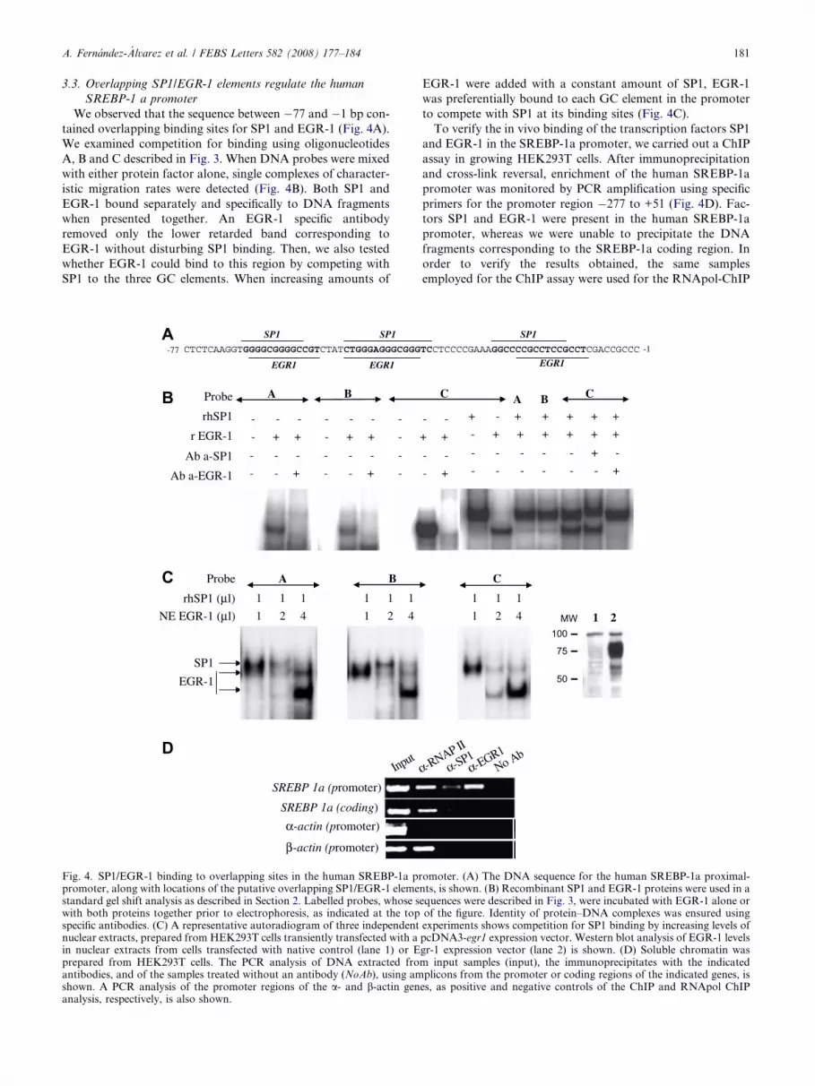
3.3. Overlapping SP1/EGR-1 elements regulate the human
SREBP-1 a promoter
We observed that the sequence between �77 and �1 bp con-
tained overlapping binding sites for SP1 and EGR-1 (Fig. 4A).
We examined competition for binding using oligonucleotides
A, B and C described in Fig. 3. When DNA probes were mixed
with either protein factor alone, single complexes of character-
istic migration rates were detected (Fig. 4B). Both SP1 and
EGR-1 bound separately and specifically to DNA fragments
when presented together. An EGR-1 specific antibody
removed only the lower retarded band corresponding to
EGR-1 without disturbing SP1 binding. Then, we also tested
whether EGR-1 could bind to this region by competing with
SP1 to the three GC elements. When increasing amounts of
CTCTCAAGGTGGGGCGGGGCCGTCTATCTGGGAGGGCGGG
SREBP 1a (promoter)
SREBP 1a (coding)
Input
-77
SP1
EGR1
SP1
EGR1
BAProbe
r EGR-1
rhSP1 - - -
- + +
- - -
- + +
-
-
Ab a-EGR-1
Ab a-SP1 - - -
- - +
- - -
- - +
-
-
α-actin (promoter)
β-actin (promoter)
BAProbe
NE EGR-1 (μl)
rhSP1 (μl) 1 1 1
1 2 4
1 1 1
1 2 4
SP1
EGR-1
Fig. 4. SP1/EGR-1 binding to overlapping sites in the human SREBP-1a prpromoter, along with locations of the putative overlapping SP1/EGR-1 elemestandard gel shift analysis as described in Section 2. Labelled probes, whose swith both proteins together prior to electrophoresis, as indicated at the topspecific antibodies. (C) A representative autoradiogram of three independentnuclear extracts, prepared from HEK293T cells transiently transfected with ain nuclear extracts from cells transfected with native control (lane 1) or Eprepared from HEK293T cells. The PCR analysis of DNA extracted froantibodies, and of the samples treated without an antibody (NoAb), using amshown. A PCR analysis of the promoter regions of the a- and b-actin genanalysis, respectively, is also shown.
EGR-1 were added with a constant amount of SP1, EGR-1
was preferentially bound to each GC element in the promoter
to compete with SP1 at its binding sites (Fig. 4C).
To verify the in vivo binding of the transcription factors SP1
and EGR-1 in the SREBP-1a promoter, we carried out a ChIP
assay in growing HEK293T cells. After immunoprecipitation
and cross-link reversal, enrichment of the human SREBP-1a
promoter was monitored by PCR amplification using specific
primers for the promoter region �277 to +51 (Fig. 4D). Fac-
tors SP1 and EGR-1 were present in the human SREBP-1a
promoter, whereas we were unable to precipitate the DNA
fragments corresponding to the SREBP-1a coding region. In
order to verify the results obtained, the same samples
employed for the ChIP assay were used for the RNApol-ChIP
TCCTCCCCGAAAGGCCCCGCCTCCGCCTCGACCGCCC
α-RNAP II
α-SP1α-EGR1
No Ab
-1
SP1
EGR1
C
- -
+ +
- -
- +
+ - +
- + +
+ + + +
+ + + +
- - -
- - -
- - + -
- - - +
A B C
C
1 1 1
1 2 4 MW100
75
50
1 2
omoter. (A) The DNA sequence for the human SREBP-1a proximal-nts, is shown. (B) Recombinant SP1 and EGR-1 proteins were used in aequences were described in Fig. 3, were incubated with EGR-1 alone orof the figure. Identity of protein–DNA complexes was ensured usingexperiments shows competition for SP1 binding by increasing levels ofpcDNA3-egr1 expression vector. Western blot analysis of EGR-1 levelsgr-1 expression vector (lane 2) is shown. (D) Soluble chromatin wasm input samples (input), the immunoprecipitates with the indicatedplicons from the promoter or coding regions of the indicated genes, is
es, as positive and negative controls of the ChIP and RNApol ChIP

182 A. Fernandez-Alvarez et al. / FEBS Letters 582 (2008) 177–184
assay, as previously described [11]. The a-RNA polymerase
antibody precipitated both promoter and coding chromatin
fragments, thus confirming that the SREBP-1a transcript is ex-
pressed in HEK293T cells.
In the light of the results obtained, we evaluated whether
EGR-1 directly affects the human SREBP-1a promoter activ-
ity. As Fig. 5A depicts, EGR-1 is able to inhibit the �75/
+194luc SREBP-1a luciferase activity. The EGR-1 overexpres-
sion was able to increase the luciferase activity of the pEB-
S14luc reporter vector as a positive control. This plasmid
encompasses four binding sites for EGR-1 derived from the
egr-1 promoter immediately upstream of the TATA box [5].
The essential role of SP-1 in the positive regulation of the hu-
man SREBP-1a promoter was also evaluated in HEK293T
cells in transient transfection experiments (Fig. 5B). The
expression of SP1 led to increased luciferase reporter activity
under the control of the SREBP-1a promoter. A reporter gene,
under the control of four copies of a GC-rich genetic element
0
15
30
45
60
75
90
0
5
10
15
20
25
Fold
indu
ctio
n
SREBP-1a luc pEBS14 luc
pcDNA3pcDNA3-Egr1
SREBP-1c luc
**
**
*
0
100
200
300
pAldGCB4
0
5
10
15
pCMVpCMV-Sp1
SREBP-1a luc pAld-GCB4
Fold
indu
ctio
n
*
*
Fig. 5. EGR-1 overexpression represses the human SREBP-1a pro-moter. Reporter plasmids and the pcDNA-egr1 (panel A) or pCMV-Sp1 (panel B) expression vector were transiently co-transfected intoHEK293T cells. After 24 h, cells were lysed to measure luciferaseactivities. The EGR-1 data are represented as the mean firefly/renillaluciferase ratio relative to the activity of the empty expression vector(set at 100%). For the Sp1 analysis, the mean firefly luciferase/proteinratio was considered. Values represent the means ± S.E.M. of fourtransfections run in duplicate; **P < 0.01; *P < 0.05.
of the aldolase C gene, a TATA box, and the luciferase open
reading frame (pALd-GCB4), was used as an SP1-dependent
luciferase expression vector [5].
To confirm the inhibitor role of EGR-1, HEK293T cells
were transfected with gene-specific siRNA. After transfection
with siRNA, the mRNA expression of egr-1 was specifically
knocked down by 75% (Fig. 6B). In contrast, the promoter
activity of the �75 bp fragment, or the endogenous SREBP-
1 expression, was significantly higher than those obtained in
cells transfected with the siRNA control (Fig. 6A and B).
However, the activity of the EGR-1 positive pEBS14luc repor-
ter vector diminished. The reciprocal effect was observed after
specifically knocking down SP1.
4. Discussion
In this report, we have identified the core human SREBP-1a
promoter region. The human promoter, like the mouse pro-
moter [4], is enclosed in a small region of DNA that is charac-
terized by a GC-rich region. Adamson et al. proposed three
‘‘levels of certainty’’ for the identification of EGR-1 target
genes [12]: (i) a correlation of the egr-1 expression with the
indicated gene using egr-1 inducible signal molecules or egr-1
expression vectors (level 1); (ii) the in vitro identification of
EGR-1 binding to the promoter of the gene (level 2) and (iii)
the in vivo verification of EGR-1 binding by the chromatin
immunoprecipitation assay (level 3). Having followed the three
aforementioned levels (Figs. 4 and 5), our studies revealed that
the EGR-1 and SP1 binding sites overlap within the SREBP-1a
proximal promoter. These overlapping sites are present in
other promoters, in most of which the binding of both tran-
scription factors is mutually exclusive [6]. The data in Fig. 4
show that elevated amounts of EGR-1 can displace pre-bound
SP1 from the GC elements. Moreover, the three GC boxes do
not have the same EGR-1 binding capacity. EGR-1 binds
more efficiently to probe C in comparison with probes A and
B (Fig. 4C). This main role of the 3 0 site was also described
in the mouse core SREBP-1a promoter [4], thus emphasizing
that this element is essential for the regulated expression of
the SREBP-1a transcript.
There are well documented examples of the dual role of
EGR-1 as either an activator or transcriptional repressor
[13]. Our evidence suggests that EGR-1 functions as a tran-
scriptional repressor for the human SREBP-1a promoter. This
inhibitor role was specific for the SREBP-1a promoter because
a forced expression of egr-1 slightly increased the activity of
the human SREBP-1c promoter (Fig. 5A).
Egr-1 is widely expressed and regulates a range of cellular
processes, including proliferation, growth and apoptosis [14].
The proposed role of EGR-1 in controlling cell growth is based
on the correlation between the mitogenic response and EGR-1
biosynthesis. However, the fact that the overexpression of egr-
1 neither induces cell death nor proliferation suggests that
EGR-1 alone is insufficient to induce changes in physiological
parameters [14]. Recently, Nakakuki et al. reported that
SREBP-1a regulates cell growth [15]. The authors pointed
out the biphasic effects of SREBP-1a, depending on its nuclear
amount; at low expression levels, it promotes proliferation,
whereas an overexpression of SREBP-1a blocks cell growth.
The regulation of the SREBP-1a expression exercised by

0
0.5
1
1.5
2
0
10
20
30
Fold
indu
ctio
n
SREBP-1a luc pEBS14 luc
siRNA-CN
siRNA-Egr1**
*
0
0.5
1
1.5
2
0
4
8 siRNA-CN
siRNA-Sp1
SREBP-1a luc pAld-GCB4
*
*m
RN
A le
vels
(a.
u.)
siRNA-CN
siRNA-Sp1
siRNA-CN
siRNA-Egr1
**
**
**
**
SREBP-1aEgr-1 Sp1 SREBP-1a
Fig. 6. Knockdown experiments confirm the reciprocal regulation of the human SREBP-1a by EGR-1 and Sp1. (A) HEK293T cells transfected withreporter plasmids and specific siRNAs. The fold induction relative to the pGL3-basic vector (±S.E.M.), normalized to renilla luciferase data, wascalculated. The results shown are the average of four transfections run in duplicate; **P < 0.01; *P < 0.05. (B) The Egr-1, Sp1 or SREBP-1aexpression, normalized to the GAPDH expression, on HEK293T cells after transfection with gene-specific siRNAs. Asterisks indicate a significantdifference (*P < 0.05; **P < 0.01) compared with cells with the negative siRNA control used as a calibrator.
A. Fernandez-Alvarez et al. / FEBS Letters 582 (2008) 177–184 183
EGR-1, and proved in our study, would reveal new aspects to
be taken into account in the control of cell cycle and growth.
In conclusion, we have performed a functional characteriza-
tion of the human SREBP-1a promoter, and have mapped the
basal promoter region required for transcriptional activation.
Furthermore, we have pointed out the functional role of the
three GC-boxes containing overlapping EGR-1/SP1 binding
sites.
Acknowledgements: We thank Gerald Thiel, Robert Tjian and EileenAdamson for their generous gifts of plasmids. This study was sup-ported by grants from Spanish Ministry of Education and Science(SAF2003-01262, SAF2006-06760 to M.C. and BFU2004-03616,CSD2006-49 to G.L.-R.). A.F-A. is supported by a fellowship fromthe Spanish Ministry of Education and Science.
References
[1] Horton, J.D., Shah, N.A., Warrington, J.A., Anderson, N.N.,Park, S.W., Brown, M.S. and Goldstein, J.L. (2003) Combinedanalysis of oligonucleotide microarray data from transgenic andknockout mice identifies direct SREBP target genes. Proc. Natl.Acad. Sci. USA 100, 12027–12032.
[2] Hua, X., Wu, J., Goldstein, J.L., Brown, M.S. and Hobbs, H.H.(1995) Structure of the human gene encoding sterol regulatoryelement binding protein-1 (SREBF1) and localization of SREBF1
and SREBF2 to chromosomes 17p11.2 and 22q13. Genomics 25,667–673.
[3] Shimomura, I., Shimano, H., Horton, J.D., Goldstein, J.L. andBrown, M.S. (1997) Differential expression of exons 1a and 1c inmRNAs for sterol regulatory element binding protein-1 in humanand mouse organs and cultured cells. J. Clin. Invest. 99, 838–845.
[4] Zhang, C., Shin, D.J. and Osborne, T.F. (2005) A simplepromoter containing two Sp1 sites controls the expression ofsterol-regulatory-element-binding protein 1a (SREBP-1a). Bio-chem. J. 386, 161–168.
[5] Al-Sarraj, A., Day, R.M. and Thiel, G. (2005) Specificity oftranscriptional regulation by the zinc finger transcription factorsSp1, Sp3, and Egr-1. J. Cell Biochem. 94, 153–167.
[6] Huang, R.P., Fan, Y., Ni, Z., Mercola, D. and Adamson, E.D.(1997) Reciprocal modulation between Sp1 and Egr-1. J. CellBiochem. 66, 489–499.
[7] Yu, J., de Belle, I., Liang, H. and Adamson, E.D. (2004)Coactivating factors p300 and CBP are transcriptionally cross-regulated by Egr1 in prostate cells, leading to divergent responses.Mol. Cell 15, 83–94.
[8] Azzout-Marniche, D., Becard, D., Guichard, C., Foretz, M.,Ferre, P. and Foufelle, F. (2000) Insulin effects on sterolregulatory-element-binding protein-1c (SREBP-1c) transcrip-tional activity in rat hepatocytes. Biochem. J. 350 (Pt 2), 389–393.
[9] Casado, M., Callejas, N.A., Rodrigo, J., Zhao, X., Dey, S.K.,Bosca, L. and Martin-Sanz, P. (2001) Contribution of cyclooxy-genase 2 to liver regeneration after partial hepatectomy. Faseb J.15, 2016–2018.
[10] Vernia, S., Eberle, D., Hernandez Mijares, A., Foufelle, F. andCasado, M. (2006) A rare missense mutation in a type 2 diabetes

184 A. Fernandez-Alvarez et al. / FEBS Letters 582 (2008) 177–184
patient decreases the transcriptional activity of human sterolregulatory element binding protein-1. Hum. Mutat. 27, 212.
[11] Sandoval, J. et al. (2004) RNAPol-ChIP: a novel application ofchromatin immunoprecipitation to the analysis of real-time genetranscription. Nucleic Acids Res. 32, e88.
[12] Adamson, E.D. and Mercola, D. (2002) Egr1 transcription factor:multiple roles in prostate tumor cell growth and survival. TumourBiol. 23, 93–102.
[13] Davis Jr., W., Chen, Z.J., Ile, K.E. and Tew, K.D. (2003)Reciprocal regulation of expression of the human adenosine 5 0-triphosphate binding cassette, subfamily A, transporter 2
(ABCA2) promoter by the early growth response-1 (EGR-1)and Sp-family transcription factors. Nucleic Acids Res. 31, 1097–1107.
[14] Thiel, G. and Cibelli, G. (2002) Regulation of life and death bythe zinc finger transcription factor Egr-1. J. Cell Physiol. 193,287–292.
[15] Nakakuki, M. et al. (2007) A transcription factor of lipidsynthesis, sterol regulatory element-binding protein (SREBP)-1acauses G(1) cell-cycle arrest after accumulation of cyclin-depen-dent kinase (cdk) inhibitors. Febs J. 274, 4440–4452.


