Embed Size (px)
Citation preview

R E V I E W A R T I C L E
Rathke’s cleft cysts
Raluca Trifanescu*, Olaf Ansorge†, John A. H. Wass*, Ashley B. Grossman* and Niki Karavitaki*
*Department of Endocrinology, Oxford Centre for Diabetes, Endocrinology and Metabolism, Churchill Hospital and †Neuropathology
Department, John Radcliffe Hospital, Oxford, UK
Summary
Rathke’s cleft cysts (RCCs) are benign, sellar and/or suprasellar
lesions originating from the remnants of Rathke’s pouch. Although
a common finding in routine autopsies (12–33% of normal pitui-
tary glands), symptomatic cases are rare and comprise 5–15% of all
surgically resected sellar lesions. Small, asymptomatic RCC do not
require surgical intervention, and their natural history is not clear.
In series of nonoperated presumed RCCs, 26–94% did not progress
during follow-up periods up to 9 years. In symptomatic ones, sur-
gery is indicated, aiming to drain the cyst content and safely
remove as much of the capsule as possible. Following surgical inter-
vention, headaches and visual field defects improve or resolve in a
significant number of patients (40–100% and 33–100%, respec-
tively) and partial hypopituitarism recovers in 14–50%. Data on
relapse rates published in the last 15 years are based on variable fol-
low-up periods and show wide variation (between 0% and 33%).
The lowest relapse rates have been described in reports with rela-
tively short mean observation periods (<3 years), whereas in those
with longer follow-up the relapse rates increase. Most of the
relapses occur within 5–6 years, suggesting that follow-up is
required for at least 5 years after surgery. Risk factors for relapse
include the presence of squamous metaplasia in the cyst wall, cyst
size and the presence of inflammation. Long-term sufficiently pow-
ered studies aiming to clarify the natural history of asymptomatic
RCCs and of those relapsing postoperatively are required.
(Received 31 July 2011; returned for revision 17 August 2011;
finally revised 19 September 2011; accepted 20 September 2011)
Introduction
Rathke’s cleft cysts (RCCs) are benign, sellar and/or suprasellar
lesions1 originating from the remnants of Rathke’s pouch.2 They
are named after the German anatomist Martin Heinrich Rathke,
who described the evagination process of the anterior foregut in
1838.3 Luschka in 1860 and then Goldzieher in 1913 published the
first cases of RCCs as incidental postmortem findings. Although a
common finding in routine autopsies (12–33% of normal pituitary
glands), symptomatic cases are relatively rare.4 Their presenting
manifestations are associated with the compression of adjacent
structures (usually headaches, visual disturbance and pituitary hor-
mone deficits).5–10 Treatment of the symptomatic cases is surgical.
The incidental detection of RCC has increased because of the
improvements in imaging techniques and the increased frequency
of brain scans, but at present their natural history and optimal
management protocols have not been clearly established.
In this review, we will highlight the clinical and laboratory char-
acteristics of RCCs, evaluate the current literature on their treat-
ment and propose a management protocol.
Epidemiology
Rathke’s cleft cysts are the most common incidentally discovered
sellar lesions, followed by pituitary adenomas. In routine autopsies,
they are encountered in 13–33% of normal pituitary glands.11,12 In
a retrospective review of 2598 subjects undergoing at least one pitu-
itary MRI scan from 1999 to 2009 in a tertiary pituitary centre,
nonadenomatous sellar masses accounted for 18% of visible
lesions, of which the most frequent were RCCs (19%).13 They have
been reported in all age groups, with a peak frequency between the
4th and 6th decades.8,14–19 Data from a nonselected autopsy series
suggested that RCCs measuring >2 mm were found in up to 4% of
subjects older than 30 years.12 Cases in paediatric populations have
been reported,20,21 but are generally rare. This may be attributed to
the fact that RCCs grow slowly with time and therefore, become
clinically evident later in life. Notably, in 44 autopsy specimens
from children aged <9 years, no RCC measuring >2 mm was
reported.12 However, MRIs performed in 341 children aged
<15 years revealed cystic pituitary lesions in four of them (1Æ2%);
these were sharply demarcated and situated just posterior to the
anterior pituitary lobe, highly suggestive of RCCs.20
Published reports have suggested a female preponderance with a
female/male ratio up to 3.9,10,14–16,18,19,22–24 Menstrual irregularity
may lead to earlier detection in women, possibly explaining this
finding.10 Furthermore, apoplexy of the pituitary gland on the
background of a RCC has been described more commonly in
women (female/male ratio of 15:5).25 No ethnic differences have
Correspondence: Niki Karavitaki, Department of Endocrinology, Oxford
Centre for Diabetes, Endocrinology and Metabolism, Churchill Hospital,
Old Road, Headington, Oxford OX3 7LJ, UK. Tel.: +44 186 585 7307;
Fax: +44 186 585 7311; E-mail: [email protected]
Clinical Endocrinology (2012) 76, 151–160 doi: 10.1111/j.1365-2265.2011.04235.x
� 2011 Blackwell Publishing Ltd 151

been recognized. Symptomatic cases are rare and constitute 5–15%
of all surgically resected sellar lesions.5,7,8,15,16,19,26–29.
Pathogenesis
Rathke’s cleft cysts originate from embryonic remnants of Rathke’s
pouch,30 which appears on the 24th day of embryonic life as a dor-
sal diverticulum from the ectodermal stomodeum (primitive oral
cavity) and is lined by simple ciliated columnar epithelium. Rath-
ke’s pouch extends cranially to form the craniopharyngeal duct
while, simultaneously, the infundibulum forms from the dienceph-
alon as a downgrowth of the neuroepithelium. By the 5th week of
gestation, Rathke’s pouch comes into contact with the infundibu-
lum,2,31 and by the 7th week, it separates from the oral cavity.
Between the 3rd and the 5th month, cells in the anterior wall of
Rathke’s pouch proliferate rapidly to form the pars anterior of the
pituitary and cells in the posterior wall differentiate into the pars
intermedia. Meanwhile, the infundibulum gives rise to the median
eminence, the pituitary stalk and the posterior pituitary lobe.32
Obliteration of the craniopharyngeal duct is associated with invo-
lution of the pouch. Failure of obliteration of the lumen results in
the development of a cyst between the pars distalis and the pars
nervosa.19,31,33 Remnants in the craniopharyngeal canal may result
in a spectrum of cystic lesions ranging from simple RCCs to com-
plex craniopharyngiomas. Notably, it has been proposed that
craniopharyngiomas and RCCs may represent a continuum of
ectodermally derived epithelial masses (also including neuro-
epithelial cysts, epidermoid and dermoid cysts).34
Other theories on the pathogenesis of RCCs propose an origin
from the neuroepithelium35 or from metaplasia of anterior pitui-
tary cells,36 but these explanations seem unlikely.32 Despite the
reported association of RCCs with pituitary adenomas, there is no
evidence for a direct metaplastic origin of these cystic lesions from
adenomas or vice versa.37
Pathology
Rathke’s cleft cysts are benign cystic lesions with a reported size
ranging from a few mm to 5 cm.15,32,38 Their content is usually a
thick, mucoid material consisting of cholesterol and protein,1
which in surgical series has been described as yellowish (15–37%),
mucoid (51–70%) or gelatinous (10%).5,8–10,22 Cerebrospinal
fluid-like or light brown and motor oil-like or milky contents have
also been reported.9,28
The cyst wall is lined by simple cuboidal or columnar epithelium
with or without cilia and occasionally goblet cells secreting mucous
into the cyst (Fig. 1).39 Pseudostratified, columnar, ciliated respira-
tory-type epithelium may be present in up to 49% of cases.8,9,28.
Squamous metaplasia (making them occasionally indistinguishable
from craniopharyngiomas)9 and nodules (at surgery, yellow, waxy,
solid masses) of mucinous material (containing mainly cholesterol
and protein) connected or not with the cyst wall8,11,14,40 may also
be seen. Notably, it has been demonstrated that the Ki-67 index
[the fraction of Ki-67 positive cells (Ki-67 protein: cellular marker
for proliferation)] of the squamous epithelium is higher than that
of the simple or pseudostratified epithelium observed in other parts
of the cyst.27 Other reported pathological findings include choles-
terol clefts (up to 23%), necrotic debris and fibrosis (up to 15%),
keratin nodules, haemosiderin and blood (when presenting with
haemorrhage). An inflammatory reaction penetrating the cyst
(a) (b)
(c) (d)
Fig. 1 The typical epithelial lining consists of tall epithelial cells with cilia (a, arrow). The epithelium expresses cytokeratins (b, Cam5.2 antibody, brown
reaction product, arrow) and may be mucin-rich (c, PAS, arrow). Long-standing cyst epithelium may show pressure atrophy (d, arrows), resulting in flat
epithelium without cilia (note clefted cyst contents). In some instances, squamous metaplasia may be pronounced, making distinction from other epithelial
lesions difficult. All magnifications · 600.
152 R. Trifanescu et al.
� 2011 Blackwell Publishing Ltd, Clinical Endocrinology, 76, 151–160

epithelium or subjacent stroma has been reported in up to 50% of
symptomatic RCCs. In such cases, the cyst wall undergoes changes
from a single-layered to stratified epithelium, depending on the
stage of inflammation.41 The inflammation is a foreign-body reac-
tion to the cystic content with the most prominent histological
findings at sites of rupture of the cyst wall, suggesting secondary
hypophysitis.5,9,41,42 Hypophysitis has been described sometimes
associated with pregnancy.42,43 The presence of inflammation and
associated oedema may result in destruction of the adjacent pitui-
tary gland, and it has been proposed that inflammation rather than
simple compression is responsible for the development of pituitary
hormone deficits.5,6,8,9,41 The formation of an abscess in the cyst
has been also reported.44
Overlapping pathological features between RCC and cranio-
pharyngiomas may pose difficulties in the differential diagnosis of
these two lesions. The distinction between a papillary craniophar-
yngioma and a RCC may be difficult, particularly in small biopsy
specimens, because of the capacity of the epithelial lining of the
RCCs to undergo squamous differentiation; in such cases, the lack
of a solid component and the presence of extensive ciliation and/or
mucin production are suggestive of Rathke’s.45 Furthermore, it has
been proposed that RCCs show a cytokeratin expression pattern
distinct to that of craniopharyngiomas: in contrast to craniophar-
yngiomas, RCCs express cytokeratins 8 and 20 (as also the pars
intermedia)46 and craniopharyngiomas show aberrant nuclear
staining for b-catenin37,47; these findings may be useful in the
differential diagnosis.
Co-existence of RCCs with pituitary adenomas has been
reported: GH secreting pituitary adenomas48 and prolactino-
mas9,10,49,50 are the most frequently described, followed by cortico-
troph4,9,37,49,51 or nonfunctioning pituitary adenomas.4,8,49 In a
large series of pituitary adenomas, co-existence with RCC has
been reported in 0Æ5–1Æ7% of the cases52; this is probably merely
co-incidental.
Location
Rathke’s cleft cysts typically present as intrasellar and/or suprasel-
lar cysts, rarely occupying only the suprasellar region.53 They typi-
cally lie in the pars intermedia, between the anterior and posterior
pituitary lobes.20,54 In a nonselected autopsy series of one thou-
sand pituitary glands, 87% of all medially situated lesions (33/38)
were RCCs; in contrast, 74% (17 out of 23) of the laterally located
ones were pituitary adenomas.12 Suprasellar extension has been
demonstrated in 16–97% of RCCs.5,8,10,15,17,27,49 Rare cases in
which the lesion is intrasphenoidal or with lateral extension into
the cavernous sinus, or extending into the frontal area, have been
reported. Interestingly, a tumour located in the cerebellopontine
angle and a second one with a giant retroclival RCC extending to
the lower prepontine cistern with brain stem compression have
also been published.38
Presenting manifestations
Small cysts are asymptomatic and their detection is usually inciden-
tal. When they are large enough to cause pressure effects on adja-
cent structures, they result in chronic clinical manifestations
mainly headaches, visual disturbance and pituitary dysfunction.
Occasionally, acute symptoms and signs may be present mimicking
pituitary apoplexy.8,18,25,49,55 The mean duration of clinical mani-
festations until diagnosis ranges from 3 days to 156 months (most
commonly between 9 and 24 months).4,8,10,14,25,39,49 The reported
signs and symptoms at presentation are shown in Table 1.
Headache is the most prevalent manifestation both at diagnosis
as well as in cases of recurrence, with reported frequencies between
33% and 81%.4,6,8–10,14–19,21,22,24,28,39,49,51,56 Frontal, episodic,
nonpulsating, bilateral headache or deep retroorbital pain are the
most common, but pain may also affect the occipital or temporal
region or involve the whole head.22 Chronic and/or continuous
headache is present in about 60% of the cases, and it may be the
only presenting manifestation in 40%.9,22 Headaches do not usually
correlate with the cyst size, location or the presence of pituitary
dysfunction, but are related to either high or isointense content on
T1-weighted MRI, mucous cyst content or intense chronic cyst wall
inflammation.9,22 Given that inflammation may result in pituitary
damage,8,9,28,41 it has been suggested that episodic frontal head-
aches may indicate an intermittent inflammatory reaction,22 and
surgical treatment is required to prevent further irreversible
endocrine dysfunction.22 Major visual field defects have been
demonstrated in up to 75% of the cases4,8–10,14–16,18,22,24,26,39,49 and
correlate with cyst size.9 Cranial nerve palsies have been rarely
described.18
Anterior pituitary hormone deficits at presentation vary widely
between 19% and 81% depending on the number of the affected
reported axes and the diagnostic tests used.6,8–10,14–
Table 1. Manifestations at presentation in patients with Rathke’s cleft cysts
References
No of
patients
Headache
(%)
Visual
disturbances
(%)
Pituitary
hormone
dysfunction
(%) (one or
more axes
affected)
Trifanescu et al.49 33 67 58 58
Xie et al.65 23 65 39 26
Wait et al.24 73 75 39 49
Madhok et al.17 35 81 – 19
Nishioka et al.22 46 59 35 24
Nishioka et al.9 37 49 38 24
Aho et al.26 118 – 49 66
Benveniste et al.5 62 71 20 55
Kim et al.8 53 81 47 –
Kasperbauer
et al.56
29 55 – 66
Isono et al.15 15 33 33 60
Shin et al.10 26 65 38 81
Mukherjee
et al.412 50 75 25
(panhypopituitarim)
Eguchi et al.6 19 – 47 47
Ross et al.23 43 44 12
Volker et al.19 155 49 56 39
Rathke’s cleft cysts 153
� 2011 Blackwell Publishing Ltd, Clinical Endocrinology, 76, 151–160

19,21,22,24,26,28,39,56 Hypogonadism with amenorrhoea/impotence or
low libido and galactorrhoea because of hyperprolactinaemia (10–
46%) were the most frequent hormonal manifestations.8,10,15,18,26,28
In a recently published series of surgically-treated RCCs, gonado-
trophin deficiency was found in 60%, ACTH deficiency in 36%,
TSH deficiency in 36% and hyperprolactinaemia in 31% of the
cases.49 GH deficiency (using variable criteria) has also been
reported in 12–79% of the patients.5,6,10,26 It has been suggested
that hypopituitarism does not correlate with cyst size and is found
only in patients with high or iso-intensity cyst on T1-weighted
MRI.9 Preoperatively, the frequency of diabetes insipidus varies in
the range 0–19%7–10,18,22,26,28,49 and is also found in patients with
high or iso-intensity cysts on T1-weighted MRI.9 Inappropriate
antidiuretic hormone secretion with hyponatraemia has also been
rarely reported.4,5,9,18
Apoplexy on the background of a RCC is an uncommon form of
presentation,8,18,25,55 and intracystic haemorrhage may or may not
be detected intraoperatively.55 The clinical picture is difficult to
distinguish from that of patients with apoplexy on the background
of a pituitary adenoma, and includes sudden onset of severe head-
ache or sudden increase in headache severity accompanied by
visual disturbance, nausea and vomiting, meningismus, oculomo-
tor palsies with diplopia, impairment of pituitary function25,55 and,
occasionally, alterations in mental status.25 The duration of head-
aches varies from hours to months, with most patients describing it
for 1–2 weeks.25
Aseptic meningitis followed by visual loss has been rarely
reported in adults5,22 and children21 and is attributed to rupture of
the cystic content into the subarachnoid space or to extension of
the inflammation into surrounding structures.22 Other rarely
described presenting manifestations include sphenoid sinusitis,
syncope, seizures,5,16 ataxia,57 mood disturbance16,57 and preco-
cious puberty.
Imaging features
Currently used tools for the radiological characterization of RCCs
include CT and MRI. They usually appear as well-circumscribed,
spherical, ovoid or dumb-bell sellar lesions in the region of the pars
intermedia7,32; lobular protrusion has been seldom reported. The
maximum diameter of the cyst ranges between 5 and 50 mm (usu-
ally between 10 and 20 mm).9,10,14,18,21,32,39 The normal pituitary
gland may be displaced in any direction, and its position is usually
around or under the cyst.9,28,32 In the latter case, the sign of ‘the egg
in a cup’ is very suggestive of an RCC.58
On CT, they usually appear as low or isodensity homogeneous
lesions or with slight hyperdensity relative to the brain paren-
chyma9,10,39 with or without enhancement. The enhancement is
ring-like or capsular.9 On MRI, they usually appear as well-demar-
cated cystic lesions with homogeneous intensity signal sometimes
combined or not with thin cyst wall enhancement.8,20,54,59 The thin
peripheral rim of enhancement has been attributed to inflamma-
(iii)
(a)
(b)
(i) (ii)
(i) (ii)
Fig. 2 MRIs of Rathke’s cleft cysts: (a) coronal (i) and sagittal (ii) T1-weighted postgadolinium images showing low signal intensity and rim enhancement.
The same cyst demonstrated hyperintense signal on T2-weighted images (shown with arrow) (iii). (b) Coronal (i) and sagittal (ii) T1-weighted images show-
ing hyperintense signal.
154 R. Trifanescu et al.
� 2011 Blackwell Publishing Ltd, Clinical Endocrinology, 76, 151–160

tion or deposition of haemosiderin or cholesterol crystals or to
squamous metaplasia of the cyst wall or to a circumferential rim of
displaced pituitary gland.8,14,32,58 The signal intensity varies widely
(hypo- iso- or hyperintense)10,15,28,53,60 and depends on the cyst
content (Fig. 2a,b).60 Lesions displaying hypointensity on T1-
weighted images and hyperintensity on T2-weighted ones contain
CSF-like fluid; those with hyperintensity on T1-weighted images
and isointensity on T2-weighted ones contain mucoid material and
hyperintensity on both T1- and T2-weighted images suggest the
presence of blood. Cysts with a high protein concentration show
high T1 signal intensity and usually have a low intracystic water
content leading to T2 signal decrease.9,54 Hyperintensity on T2
images has been described in about 70% of cases; nevertheless,
homogeneous T2 hypointense signal intensity is highly suggestive
of RCC,61 and this is useful in the differential diagnosis from chor-
domas, chondroid tumours, abscesses, mucoceles and lipomas, all
showing a hyperintense T2 signal.54 Small intracystic nodules, cor-
responding to proteinaceous concentrations, may be seen in 17–
77% of RCCs and present with low signal intensity on T2-weighted
and high signal intensity on T1-weighted images compared with
that of the surrounding cyst fluid.40,53,55 The nodules do not
enhance and are virtually pathognomonic for RCCs.54 In cases of
apoplexy on the background of a RCC, mixed signal intensities
have been described.55
A combination of a number of MRI findings may be useful for
the preoperative differential diagnosis of intra- and suprasellar
masses. An ovoid shape, small tumour volume, cystic characteris-
tics and little or no cyst wall enhancement suggest an RCC. A
‘snowman’ shape, solid characteristics and homogeneous enhance-
ment of the solid portion suggest a pituitary adenoma (Fig. 3a). A
superiorly lobulated shape, third ventricle compression, mixed
solid and cystic characteristics and reticular enhancement of the
solid portion suggest a craniopharyngioma (Fig. 3b).59 Particularly
in comparison with craniopharyngiomas, RCCs are smaller and
rarely show calcification (up to 13% of cases).10 Other cystic lesions
included in the differential diagnosis are dermoid cysts (on CT,
round or lobulated, often with negative density values and foci of
calcification; no contrast enhancement or surrounding oedema; on
T1-weighted MRI, high signal, because of the lipid content), epi-
dermoid cysts (on CT, a lobulated mass with attenuation values
similar to CSF; calcification may be present; MRI signal character-
istics are usually similar to CSF; it does not enhance, but shows
(a)
(b)
(i) (ii)
(i) (ii)
Fig. 3 (a) Coronal (i) and sagittal (ii) T1-weighted
images of pituitary adenoma arising out of the pitu-
itary fossa and compressing the optic chiasm show-
ing ‘snowman’ shape and uniform signal. (b)
Coronal (i) and sagittal (ii) T1-weighted postgado-
linium images of a craniopharygioma with superi-
orly lobulated shape and mixed solid and cystic
characteristics.
Rathke’s cleft cysts 155
� 2011 Blackwell Publishing Ltd, Clinical Endocrinology, 76, 151–160

restricted diffusion on diffusion weighted images), arachnoid cysts
(cystic lesion with a clearly defined border; the density on CT and
the signal intensity on MRI are similar to that of the CSF; it does
not show calcification and does not enhance after contrast admin-
istration) and an abscess (on CT, a central area of hypodensity sur-
rounded by a ring of increased density, which shows marked
enhancement following intravenous contrast material; on MRI, the
T1-weighted images show a marked hyperintense lesion with sig-
nificant enhancement of the abscess wall after contrast administra-
tion; T2-weighted scans show hyperintensity of the abscess
contents surrounded by an area of increased signal intensity indi-
cating oedema).45
Natural history
The natural history of both symptomatic and asymptomatic RCC
is not clear. In series of nonoperated presumed RCCs, 26–94% of
them did not progress (based on imaging studies or on the
clinical picture) during follow-up periods up to 9 years.9,21,26,62
Notably, in a cohort of 29 patients with symptomatic cystic sellar
lesions and imaging characteristics consistent with RCC managed
with surveillance, spontaneous involution was described in nine
cases (31%) that had a further follow-up of 9–46 months after
the regression. Furthemore, headaches spontaneously resolved in
five of seven patients presenting with this symptom.63 Possible
mechanisms of cyst regression include absorption of cystic fluid
greater than secretion or repeated cyst ruptures and leakage.63
On the other hand, Sanno et al.62 reported that 5Æ3% of inciden-
tally found presumed RCCs increased in size during a mean
follow-up period of 38Æ9 months (range 6–93). The change in the
cyst size is probably associated with an imbalance between secre-
tion and absorption of the cyst content, intracystic haemorrhage
or infection.
Treatment
Small, asymptomatic RCC do not require surgical intervention. In
symptomatic ones, surgery is indicated aiming to drain the cyst
content and safely remove as much of the capsule as possible.26
Cyst fenestration and drainage via an endonasal/sublabial trans-
sphenoidal approach are most commonly adopted5,17,21,26,29,39
and are usually successful in achieving improvement of the pres-
sure effects. The endoscopic transsphenoidal approach has also
been used.64,65 Intraoperative alcohol cauterization has been used
with, however, no proven benefit on relapse rates.5,16 Furthermore,
significant neurological complications from the alcohol cauteriza-
tion have been described (including seizures and cranial nerve pal-
sies), and hence, this treatment approach should be avoided.66
Radiotherapy is not routinely required, but it has been used in a
small number of recurrent cysts with promising results, suggesting
that this option may be considered for particularly multiple
relapses aiming to avoid the risks of repeated surgeries.4,16
The surgical management of RCC presenting with pituitary apo-
plexy depends on the clinical picture: for those with visual deficits
or endocrine disturbance transsphenoidal surgical intervention is
recommended, whereas in cases of headaches only without cranial
nerve deficits and no evidence of haemorrhage on gradient-refo-
cused T2-weighted images, conservative management may be an
option.55 The role of conservative therapy with glucocorticoids in
the acute phase has not been clarified. Recovery of pituitary func-
tion has been described after surgery.25
Postoperative complications include a CSF leak (up to
25%),8,9,18,65 transient and/or permanent diabetes insipidus (up to
67% and up to 20%, respectively),4,9,18,21,65 anterior pituitary hor-
mone deficits, hyponatraemia,5,16 meningitis and sinusitis. Cyst
infection has been reported reaching 12% in a series of 170 cases,
with the most common isolated organisms being Staphylococcus
Table 2. Recurrence rates of Rathke’s cleft cysts following surgical intervention
Author No of patients Years covered by the cohort of patients Recurrence rate (%) Follow-up (months)
Trifanescu et al.49 32 1978–2009 22
12 at 2 years
48 at 4 years
Mean 48 (range 2–267)
Xie et al.65 23 – 8Æ7 Range 3–36 months
Wait et al.24 73 1992–2010 10Æ9 Mean 27 months (range 0–129)
Madhok et al.17 35 1998–2008 6Æ3 Mean 19 months (range 1–60)
Zada et al.21 10 1999–2007 10 Median 34 months
Koutourousiou et al.51 14 2004–2007 14Æ3 Median 29 months (range 11–48)
Nishioka et al.22 33 – 21Æ2 Mean 42 months (range: 6–112)
Aho et al.26 118 1984–1995 18 At least 60 months
Sade et al.28 10 1994–2003 0 Mean 32 months
Benveniste et al.5 62 1989–2003 16 Mean 28 months (range: 1–166)
Kim et al.8 53 1990–2000 11Æ3 Mean 31 months (range: 2–95)
Billeci et al.14 14 – 14Æ3 Range: 6 months–10 years
Kasperbauer et al.56 29 1978–1998 28 –
Isono et al.15 15 1992–1999 6Æ7 Range 1–7 years
Shin et al.10 26 1974–1998 19 Mean 35 months
Mukherjee et al.4 12 1981–1996 33 Mean 30 months (range 1–168)
156 R. Trifanescu et al.
� 2011 Blackwell Publishing Ltd, Clinical Endocrinology, 76, 151–160

epidermidis and Propionibacterium acnes.67 There is no reported
perioperative mortality.5,9,10,16,18,26
Headaches5,8–10,14–17,21,22,24,26,28,39 and major visual field
defects5,8–10,14–17,22,24,26,28,39,49,56 improve or resolve in a significant
number of patients (40–100% and 33–100%, respectively), and hy-
perprolactinaemia is the most frequently resolved pituitary hor-
mone abnormality (in most series, in 100%).6–10,14,16,19,26,28,56
Partial hypopituitarism may recover postsurgery (14–
50%),9,10,16,17,21,26,39 whereas panhypopituitarism seldom
resolves.14,15,28 The poor endocrine prognosis could be due to
intense chronic inflammation of the adenohypophysis or neurohy-
pophysis.5,8,9,28,41 Notably, in one series, all patients with panhypo-
pituitarism had RCCs of high intensity on T1, contained mucous
material with high viscosity, demonstrated intense inflammation
on histology and did not improve their pituitary dysfunction after
surgery.9
New anterior pituitary hormone deficits following surgical inter-
vention have been reported in 4–30% of the cases5,7,8,10,17,19,21,26,28
and new diabetes insipidus between 2% and
67%.5,7,9,10,16,18,21,26,28,39,49,51,68 It has been proposed that aggres-
sive surgery with resection of the cyst wall is more commonly asso-
ciated with new endocrine morbidity.5,16,26 Interestingly, in a series
of 857 patients who underwent transsphenoidal microsurgery,
those with an intraoperative CSF leak, with a craniopharyngioma
or with an RCC were at higher risk for persistent diabetes insipidus
(DI).68 In this series, immediate postoperative and persistent DI
were found in 38Æ7% and 10%, respectively of the patients with
RCC.68
Data on the endocrine and visual outcome after long-term fol-
low-up are rather limited, but in a recently published series of 33
cases of RCCs with a mean follow-up of 48 months at last assess-
ment, 50% had gonadotrophin deficiency, 42% ACTH deficiency,
47% TSH deficiency, 39% required desmopressin treatment and
19% had at least quadrantinopia.49
Relapse rates and risk factors for relapse
The vast majority of the series published in the last 15 years report
recurrence rates only (and not survival data) during variable fol-
low-up periods and show wide variation (between 0% and 33%;
Table 2). Notably, the lowest relapse rates have been described in
reports with relative short mean observation periods (<3 years),
whereas in those with longer follow-up the relapse rate increases.
In a recently published series with a mean follow-up of 48 months,
one of the longest in the literature, survival analysis showed a recur-
rence rate of 48% at 4 years,49 suggesting that the previously
thought optimal course of RCCs may not reflect their true progno-
sis and long-term monitoring is needed. It should be also noted
that the different definitions of relapse (asymptomatic or symp-
tomatic, requiring or not surgical intervention) may explain the
wide variation in the relapse rates.
Relapses have been detected at intervals ranging between 4 weeks
up to 24 years4,5,8,10,14,15,56 with most of them occurring within the
first 5–6 years,8,10,14,16,18,21,26,51 suggesting that follow-up is
required for at least 5 years after surgery. It has been proposed that
there is a positive correlation between cyst size and time to recur-
rence.10 Although rare, multiple relapses have also been
described.5,14,16,18,49
Factors reported to be associated with an increased risk of recur-
rence include the presence of squamous metaplasia in the cyst
wall,5,8,16,24,26,32 cyst size,10 residual cyst demonstrated on postop-
erative MRI,8,18 enhancement of the lesion on MRI,5,8 use of
abdominal fat and/or fascial graft for closure (probably by increas-
ing the risk of loculation and reformation of the cyst),26 intraopera-
tive CSF leak16 and the presence of inflammation (probably
through promoting squamous metaplasia or encapsulation of the
cyst and facilitation of the accumulation of fluid – furthermore,
inflammation is considered to be a marker of more rapidly grow-
ing, highly secretory cysts).5,8 Suspected RCC infection is a strong
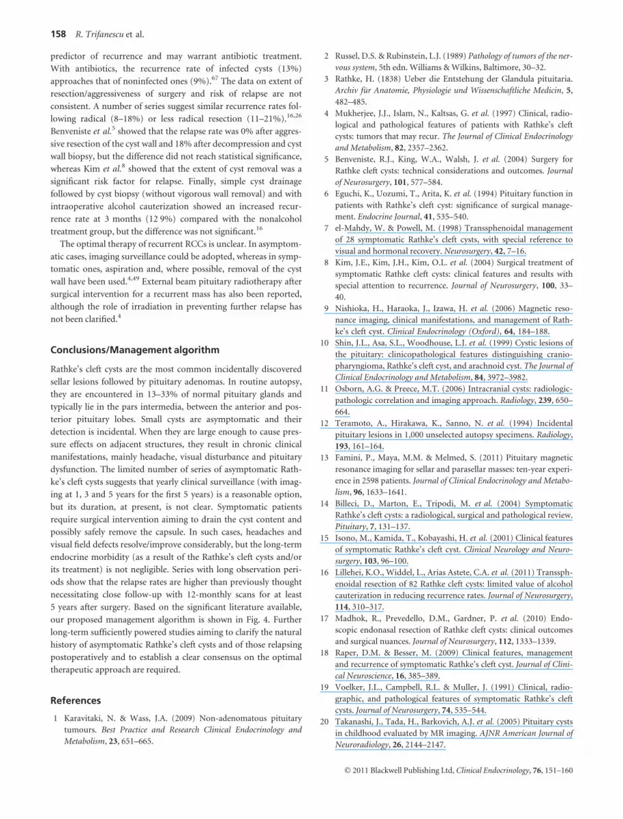
Presumed RCC on imaging
Pressure effects(headaches, visual deterioration, hypopituitarism)
No Yes
1. Yearly clinical follow-up and MRI at 1, 3 and 5 years.
1. Surgery (cyst aspiration saferemoval of cyst wall)*
2. Yearly clinical follow-up and formal visual fields assessment
3. Yearly clinical follow-up and formal visual fields assessment (imaging if indicated) for 5 years
Yes No
Increase in size
No Yes
Relapse
Clinical follow-up(duration?)
Clinical follow-up(duration?)
* In case of multiple relapses external irradiation may be considered
2. Yearly MRI for 5 years
Fig. 4 Management algorithm for Rathke’s cleft
cysts (RCCs).
Rathke’s cleft cysts 157
� 2011 Blackwell Publishing Ltd, Clinical Endocrinology, 76, 151–160
predictor of recurrence and may warrant antibiotic treatment.
With antibiotics, the recurrence rate of infected cysts (13%)
approaches that of noninfected ones (9%).67 The data on extent of
resection/aggressiveness of surgery and risk of relapse are not
consistent. A number of series suggest similar recurrence rates fol-
lowing radical (8–18%) or less radical resection (11–21%).16,26
Benveniste et al.5 showed that the relapse rate was 0% after aggres-
sive resection of the cyst wall and 18% after decompression and cyst
wall biopsy, but the difference did not reach statistical significance,
whereas Kim et al.8 showed that the extent of cyst removal was a
significant risk factor for relapse. Finally, simple cyst drainage
followed by cyst biopsy (without vigorous wall removal) and with
intraoperative alcohol cauterization showed an increased recur-
rence rate at 3 months (12Æ9%) compared with the nonalcohol
treatment group, but the difference was not significant.16
The optimal therapy of recurrent RCCs is unclear. In asymptom-
atic cases, imaging surveillance could be adopted, whereas in symp-
tomatic ones, aspiration and, where possible, removal of the cyst
wall have been used.4,49 External beam pituitary radiotherapy after
surgical intervention for a recurrent mass has also been reported,
although the role of irradiation in preventing further relapse has
not been clarified.4
Conclusions/Management algorithm
Rathke’s cleft cysts are the most common incidentally discovered
sellar lesions followed by pituitary adenomas. In routine autopsy,
they are encountered in 13–33% of normal pituitary glands and
typically lie in the pars intermedia, between the anterior and pos-
terior pituitary lobes. Small cysts are asymptomatic and their
detection is incidental. When they are large enough to cause pres-
sure effects on adjacent structures, they result in chronic clinical
manifestations, mainly headache, visual disturbance and pituitary
dysfunction. The limited number of series of asymptomatic Rath-
ke’s cleft cysts suggests that yearly clinical surveillance (with imag-
ing at 1, 3 and 5 years for the first 5 years) is a reasonable option,
but its duration, at present, is not clear. Symptomatic patients
require surgical intervention aiming to drain the cyst content and
possibly safely remove the capsule. In such cases, headaches and
visual field defects resolve/improve considerably, but the long-term
endocrine morbidity (as a result of the Rathke’s cleft cysts and/or
its treatment) is not negligible. Series with long observation peri-
ods show that the relapse rates are higher than previously thought
necessitating close follow-up with 12-monthly scans for at least
5 years after surgery. Based on the significant literature available,
our proposed management algorithm is shown in Fig. 4. Further
long-term sufficiently powered studies aiming to clarify the natural
history of asymptomatic Rathke’s cleft cysts and of those relapsing
postoperatively and to establish a clear consensus on the optimal
therapeutic approach are required.
References
1 Karavitaki, N. & Wass, J.A. (2009) Non-adenomatous pituitary
tumours. Best Practice and Research Clinical Endocrinology and
Metabolism, 23, 651–665.
2 Russel, D.S. & Rubinstein, L.J. (1989) Pathology of tumors of the ner-
vous system, 5th edn. Williams & Wilkins, Baltimore, 30–32.
3 Rathke, H. (1838) Ueber die Entstehung der Glandula pituitaria.
Archiv fur Anatomie, Physiologie und Wissenschaftliche Medicin, 5,
482–485.
4 Mukherjee, J.J., Islam, N., Kaltsas, G. et al. (1997) Clinical, radio-
logical and pathological features of patients with Rathke’s cleft
cysts: tumors that may recur. The Journal of Clinical Endocrinology
and Metabolism, 82, 2357–2362.
5 Benveniste, R.J., King, W.A., Walsh, J. et al. (2004) Surgery for
Rathke cleft cysts: technical considerations and outcomes. Journal
of Neurosurgery, 101, 577–584.
6 Eguchi, K., Uozumi, T., Arita, K. et al. (1994) Pituitary function in
patients with Rathke’s cleft cyst: significance of surgical manage-
ment. Endocrine Journal, 41, 535–540.
7 el-Mahdy, W. & Powell, M. (1998) Transsphenoidal management
of 28 symptomatic Rathke’s cleft cysts, with special reference to
visual and hormonal recovery. Neurosurgery, 42, 7–16.
8 Kim, J.E., Kim, J.H., Kim, O.L. et al. (2004) Surgical treatment of
symptomatic Rathke cleft cysts: clinical features and results with
special attention to recurrence. Journal of Neurosurgery, 100, 33–
40.
9 Nishioka, H., Haraoka, J., Izawa, H. et al. (2006) Magnetic reso-
nance imaging, clinical manifestations, and management of Rath-
ke’s cleft cyst. Clinical Endocrinology (Oxford), 64, 184–188.
10 Shin, J.L., Asa, S.L., Woodhouse, L.J. et al. (1999) Cystic lesions of
the pituitary: clinicopathological features distinguishing cranio-
pharyngioma, Rathke’s cleft cyst, and arachnoid cyst. The Journal of
Clinical Endocrinology and Metabolism, 84, 3972–3982.
11 Osborn, A.G. & Preece, M.T. (2006) Intracranial cysts: radiologic-
pathologic correlation and imaging approach. Radiology, 239, 650–
664.
12 Teramoto, A., Hirakawa, K., Sanno, N. et al. (1994) Incidental
pituitary lesions in 1,000 unselected autopsy specimens. Radiology,
193, 161–164.
13 Famini, P., Maya, M.M. & Melmed, S. (2011) Pituitary magnetic
resonance imaging for sellar and parasellar masses: ten-year experi-
ence in 2598 patients. Journal of Clinical Endocrinology and Metabo-
lism, 96, 1633–1641.
14 Billeci, D., Marton, E., Tripodi, M. et al. (2004) Symptomatic
Rathke’s cleft cysts: a radiological, surgical and pathological review.
Pituitary, 7, 131–137.
15 Isono, M., Kamida, T., Kobayashi, H. et al. (2001) Clinical features
of symptomatic Rathke’s cleft cyst. Clinical Neurology and Neuro-
surgery, 103, 96–100.
16 Lillehei, K.O., Widdel, L., Arias Astete, C.A. et al. (2011) Transsph-
enoidal resection of 82 Rathke cleft cysts: limited value of alcohol
cauterization in reducing recurrence rates. Journal of Neurosurgery,
114, 310–317.
17 Madhok, R., Prevedello, D.M., Gardner, P. et al. (2010) Endo-
scopic endonasal resection of Rathke cleft cysts: clinical outcomes
and surgical nuances. Journal of Neurosurgery, 112, 1333–1339.
18 Raper, D.M. & Besser, M. (2009) Clinical features, management
and recurrence of symptomatic Rathke’s cleft cyst. Journal of Clini-
cal Neuroscience, 16, 385–389.
19 Voelker, J.L., Campbell, R.L. & Muller, J. (1991) Clinical, radio-
graphic, and pathological features of symptomatic Rathke’s cleft
cysts. Journal of Neurosurgery, 74, 535–544.
20 Takanashi, J., Tada, H., Barkovich, A.J. et al. (2005) Pituitary cysts
in childhood evaluated by MR imaging. AJNR American Journal of
Neuroradiology, 26, 2144–2147.
158 R. Trifanescu et al.
� 2011 Blackwell Publishing Ltd, Clinical Endocrinology, 76, 151–160

21 Zada, G., Ditty, B., McNatt, S.A. et al. (2009) Surgical treatment of
rathke cleft cysts in children. Neurosurgery, 64, 1132–1137.
22 Nishioka, H., Haraoka, J., Izawa, H. et al. (2006) Headaches associ-
ated with Rathke’s cleft cyst. Headache, 46, 1580–1586.
23 Ross, D.A., Norman, D. & Wilson, C.B. (1992) Radiologic charac-
teristics and results of surgical management of Rathke’s cysts in 43
patients. Neurosurgery, 30, 173–178.
24 Wait, S.D., Garrett, M.P., Little, A.S. et al. (2010) Endocrinopathy,
vision, headache, and recurrence after transsphenoidal surgery for
Rathke cleft cysts. Neurosurgery, 67, 837–843.
25 Chaiban, J.T., Abdelmannan, D., Cohen, M. et al. (2011) Rathke
cleft cyst apoplexy: a newly characterized distinct clinical entity.
Journal of Neurosurgery, 114, 318–324.
26 Aho, C.J., Liu, C., Zelman, V. et al. (2005) Surgical outcomes in
118 patients with Rathke cleft cysts. Journal of Neurosurgery, 102,
189–193.
27 Ikeda, H. & Yoshimoto, T. (2002) Clinicopathological study of
Rathke’s cleft cysts. Clinical Neuropathology, 21, 82–91.
28 Sade, B., Albrecht, S., Assimakopoulos, P. et al. (2005) Manage-
ment of Rathke’s cleft cysts. Surgical Neurology, 63, 459–466.
29 Zada, G., Kelly, D.F., Cohan, P. et al. (2003) Endonasal transsphe-
noidal approach for pituitary adenomas and other sellar lesions: an
assessment of efficacy, safety, and patient impressions. Journal of
Neurosurgery, 98, 350–358.
30 Fager, C.A. & Carter, H. (1966) Intrasellar epithelial cysts. Journal
of Neurosurgery, 24, 77–81.
31 Horvath, E., Scheithauer, B.W., Kovacs, K. et al. (1997) Regional
neuropathology: hypothalamus and pituitary. In: D.I. Graham,
P.L. Lantos eds. Greenfield’s Neuropathology, Vol. 1. Arnold, Lon-
don, 1007–1082.
32 Zada, G., Lin, N., Ojerholm, E. et al. (2010) Craniopharyngioma
and other cystic epithelial lesions of the sellar region: a review of
clinical, imaging, and histopathological relationships. Neurosurgi-
cal Focus, 28, E4.
33 Kunwar, S. & Wilson, C.B. (2002) Cysts, hamartomas and vascular
tumours. In: J.A. Wass, S.M. Shalet eds. Oxford Textbook of
Endocrinology and Diabetes. Oxford University Press, Oxford, 225–
230.
34 Harrison, M.J., Morgello, S. & Post, K.D. (1994) Epithelial cys-
tic lesions of the sellar and parasellar region: a continuum of
ectodermal derivatives? Journal of Neurosurgery, 80, 1018–
1025.
35 Shuangshoti, S., Netsky, M.G. & Nashold, B.S. Jr (1970) Epithelial
cysts related to sella turcica. Proposed origin from neuroepitheli-
um. Archives of Pathology, 90, 444–450.
36 Asa, S.L. (1998) Tumors of the Pituitary Gland. In: J. Rosai ed. Atlas
of Tumor Pathology, 3rd Series, Washington DC, Armed Forces
Institute of Pathology, Fascicle 22.
37 Karavitaki, N., Scheithauer, B.W., Watt, J. et al. (2008) Collision
lesions of the sella: co-existence of craniopharyngioma with
gonadotroph adenoma and of Rathke’s cleft cyst with corticotroph
adenoma. Pituitary, 11, 317–323.
38 Chuang, C.C., Chen, Y.L., Jung, S.M. et al. (2010) A giant retrocli-
val Rathke’s cleft cyst. Journal of Clinical Neuroscience, 17, 1189–
1191.
39 Midha, R., Jay, V. & Smyth, H.S. (1991) Transsphenoidal manage-
ment of Rathke’s cleft cysts. A clinicopathological review of 10
cases. Surgical Neurology, 35, 446–454.
40 Binning, M.J., Gottfried, O.N., Osborn, A.G. et al. (2005) Rathke
cleft cyst intracystic nodule: a characteristic magnetic resonance
imaging finding. Journal of Neurosurgery, 103, 837–840.
41 Hama, S., Arita, K., Nishisaka, T. et al. (2002) Changes in the epi-
thelium of Rathke cleft cyst associated with inflammation. Journal
of Neurosurgery, 96, 209–216.
42 Schittenhelm, J., Beschorner, R., Psaras, T. et al. (2008) Rathke’s
cleft cyst rupture as potential initial event of a secondary perifocal
lymphocytic hypophysitis: proposal of an unusual pathogenetic
event and review of the literature. Neurosurgical Review, 31, 157–
163.
43 Sonnet, E., Roudaut, N., Meriot, P. et al. (2006) Hypophysitis asso-
ciated with a ruptured Rathke’s cleft cyst in a woman, during preg-
nancy. Journal of Endocrinological Investigation, 29, 353–357.
44 Bognar, L., Szeifert, G.T., Fedorcsak, I. et al. (1992) Abscess forma-
tion in Rathke’s cleft cyst. Acta Neurochirurgica, 117, 70–72.
45 Karavitaki, N., Cudlip, S., Adams, C.B. et al. (2006) Craniopharyn-
giomas. Endocrine reviews, 27, 371–397.
46 Xin, W., Rubin, M.A. & McKeever, P.E. (2002) Differential expres-
sion of cytokeratins 8 and 20 distinguishes craniopharyngioma
from Rathke cleft cyst. Archives of Pathology & Laboratory Medicine,
126, 1174–1178.
47 Hofmann, B.M., Kreutzer, J., Saeger, W. et al. (2006) Nuclear beta-
catenin accumulation as reliable marker for the differentiation
between cystic craniopharyngiomas and Rathke cleft cysts: a clinic-
o-pathologic approach. American Journal of Surgical Pathology, 30,
1595–1603.
48 Bader, L.J., Carter, K.D., Latchaw, R.E. et al. (2004) Simultaneous
symptomatic Rathke’s cleft cyst and GH secreting pituitary
adenoma: a case report. Pituitary, 7, 39–44.
49 Trifanescu, R., Stavrinides, V., Plaha, P. et al. (2011) Outcome in
surgically treated Rathke’s cleft cysts: long-term monitoring
needed. European Journal of Endocrinology, 165, 33–37.
50 Ikeda, H., Niizuma, H., Fujiwara, S. et al. (1987) [A case of prolac-
tinoma in close association with Rathke’s cleft cyst]. No Shinkei
Geka, 15, 999–1003.
51 Koutourousiou, M., Grotenhuis, A., Kontogeorgos, G. et al. (2009)
Treatment of Rathke’s cleft cysts: experience at a single centre. Jour-
nal of Clinical Neuroscience, 16, 900–903.
52 Nishio, S., Mizuno, J., Barrow, D.L. et al. (1987) Pituitary tumors
composed of adenohypophysial adenoma and Rathke’s cleft cyst
elements: a clinicopathological study. Neurosurgery, 21, 371–377.
53 Wen, L., Hu, L.B., Feng, X.Y. et al. (2010) Rathke’s cleft cyst: clini-
copathological and MRI findings in 22 patients. Clinical Radiology,
65, 47–55.
54 Bonneville, F., Cattin, F., Marsot-Dupuch, K. et al. (2006) T1 signal
hyperintensity in the sellar region: spectrum of findings. Radio-
graphics, 26, 93–113.
55 Binning, M.J., Liu, J.K., Gannon, J. et al. (2008) Hemorrhagic and
nonhemorrhagic Rathke cleft cysts mimicking pituitary apoplexy.
Journal of Neurosurgery, 108, 3–8.
56 Kasperbauer, J.L., Orvidas, L.J., Atkinson, J.L. et al. (2002) Rathke
cleft cyst: diagnostic and therapeutic considerations. The Laryngo-
scope, 112, 1836–1839.
57 Cao, Z., Lv, J., Ding, Z. et al. (2008) Pathological laughter in a
patient with Rathke cleft cyst. Journal of Clinical Neuroscience, 15,
1279–1282.
58 Brassier, G., Morandi, X., Tayiar, E. et al. (1999) Rathke’s cleft
cysts: surgical-MRI correlation in 16 symptomatic cases. Journal of
Neuroradiology, 26, 162–171.
59 Choi, S.H., Kwon, B.J., Na, D.G. et al. (2007) Pituitary adenoma,
craniopharyngioma, and Rathke cleft cyst involving both intrasellar
and suprasellar regions: differentiation using MRI. Clinical Radiol-
ogy, 62, 453–462.
Rathke’s cleft cysts 159
� 2011 Blackwell Publishing Ltd, Clinical Endocrinology, 76, 151–160

60 Hayashi, Y., Tachibana, O., Muramatsu, N. et al. (1999) Rathke
cleft cyst: MR and biomedical analysis of cyst content. Journal of
Computer Assisted Tomography, 23, 34–38.
61 Bonneville, F., Chiras, J., Cattin, F. et al. (2007) T2 hypointense sig-
nal of rathke cleft cyst. AJNR American Journal of Neuroradiology,
28, 397.
62 Sanno, N., Oyama, K., Tahara, S. et al. (2003) A survey of pituitary
incidentaloma in Japan. European Journal of Endocrinology, 149,
123–127.
63 Amhaz, H.H., Chamoun, R.B., Waguespack, S.G. et al. (2010)
Spontaneous involution of Rathke cleft cysts: is it rare or just
underreported? Journal of Neurosurgery, 112, 1327–1332.
64 Frank, G., Sciarretta, V., Mazzatenta, D. et al. (2005) Transsphe-
noidal endoscopic approach in the treatment of Rathke’s cleft cyst.
Neurosurgery, 56, 124–128.
65 Xie, T., Hu, F., Yu, Y. et al. (2011) Endoscopic endonasal resection
of symptomatic Rathke cleft cysts. Journal of Clinical Neuroscience,
18, 760–762.
66 Hsu, H.Y., Piva, A. & Sadun, A.A. (2004) Devastating complica-
tions from alcohol cauterization of recurrent Rathke cleft cyst. Case
report. Journal of Neurosurgery, 100, 1087–1090.
67 Tate, M.C., Jahangiri, A., Blevins, L. et al. (2010) Infected Rathke
cleft cysts: distinguishing factors and factors predicting recurrence.
Neurosurgery, 67, 762–769.
68 Nemergut, E.C., Zuo, Z., Jane, J.A. Jr et al. (2005) Predictors of dia-
betes insipidus after transsphenoidal surgery: a review of 881
patients. Journal of Neurosurgery, 103, 448–454.
160 R. Trifanescu et al.
� 2011 Blackwell Publishing Ltd, Clinical Endocrinology, 76, 151–160


