Embed Size (px)
Citation preview
Rapid tyrosine phosphorylation of focal adhesion kinase, paxillin,and p130Cas by gastrin in human colon cancer cells
Hong-Gang Yu, Henning Schrader, Jan-Michel Otte, Wolfgang E. Schmidt, Frank Schmitz*
Laboratory for Molecular Gastroenterology, Department of Medicine I, St. Josef-Hospital, Ruhr-University
of Bochum, Gudrunstr. 56, D-44791 Bochum, Germany
Received 13 June 2003; accepted 18 August 2003
Abstract
Although the expression of CCK2 receptors is widely reported in human colorectal cancers, little is known on its role in mediating the
proliferative effects of mature amidated gastrin (G17 amide) on colorectal cancers. The purpose of the present study was to determine the
effects of G17 amide on tyrosine phosphorylation of focal adhesion kinase (FAK), paxillin, and p130 Crk-associated substrate (p130Cas) in
Colo 320 cells, a human colorectal cancer cell line which expresses CCK2 receptors. By immunoprecipitation and immunoblotting, an
increase in tyrosine phosphorylation of FAK (tyrosine-397), paxillin (tyrosine-31), and p130Cas was detected in a time- and dose-
dependent manner. Overexpression of CCK2 receptors in Colo 320 cells (Colo 320 WT) by stable transfection with the human CCK2
receptor cDNA resulted in an increased tyrosine phosphorylation of FAK, paxillin, and p130Cas. After incubation with 1 mM L-365,260, a
specific CCK2 receptor antagonist, this increase was completely inhibited. Our results demonstrate that in human colon cancer cells,
gastrin caused a rapid tyrosine phosphorylation of FAK, paxillin, and p130Cas by activation of CCK2 receptor. The phosphorylation of
these proteins might be important in mediating gastrin effects on proliferation, apoptosis, and metastasis.
# 2003 Elsevier Inc. All rights reserved.
Keywords: Tyrosine phosphorylation; Colon cancer; Gastrin; CCK2 receptor; G protein-coupled receptor; Signal transduction
1. Introduction
Gastrin, a peptide hormone and trophic factor, has long
been recognized to stimulate mucosal growth in the upper
digestive tract [1,2]. More recent findings suggest that
gastrin has proliferative effects in colon cancer as well.
Many studies have shown that exogenously administered
gastrin stimulates the growth and proliferation of colon
cancer cells in culture [3,4], transplanted colon tumors
in mice [5], and carcinogen-induced tumors in rats [6].
In addition, antagonism of gastrin effects by antigastrin
antisera [7,8], gastrin receptor antagonists [3,9,10], and
antisense gastrin RNA [11] inhibited growth of colon
cancer cells in culture and in vivo in animal models.
The proliferative effect of gastrin has been shown to be
mediated by the CCK2 receptor in different cellular models
[12,13] including cells transfected with the CCK2 receptor
cDNA [14]. This receptor which has been cloned from
different species by several laboratories [15,16] belongs
to the family of G protein-coupled receptors which are
known to be linked to the phospholipase C/protein kinase C
(PLC/PKC) signaling pathway. Gastrin-dependent activa-
tion of the CCK2 receptors has been shown to induce
phosphatidyl-inositol bisphosphate (PIP2) hydrolysis by
phopholipase C (PLC) that results in inositol triphosphate
(IP3) and diacylglycerol (DAG) production leading to
intracellular Ca2þ mobilization and stimulation of PKC
[17,18]. Gastrin, like many ligands that bind to G protein-
coupled receptors, has also been shown to induce tyrosine
kinase activity [19,20].
A rapid increase in the tyrosine phosphorylation of the
non-receptor tyrosine kinase FAK and the adaptor proteins
p130Cas and paxillin, which localize to focal adhesion
plaques, has been identified as a prominent early event
in cells stimulated by diverse signaling molecules
that regulate cell proliferation, migration, and apoptosis
Biochemical Pharmacology 67 (2004) 135–146
0006-2952/$ – see front matter # 2003 Elsevier Inc. All rights reserved.
doi:10.1016/j.bcp.2003.08.015
* Corresponding author. Tel.: þ49-234-509-2378;
fax: þ49-234-509-2309.
E-mail address: [email protected] (F. Schmitz).
Abbreviations: CCK2R, CCK2 receptor (formerly CCK-B/gastrin
receptor); FAK, focal adhesion kinase; G17 amide, mature amidated
gastrin heptadecapeptide; GPCR, G protein-coupled receptor; p130Cas,
p130 Crk-associated substrate; PBS, phosphate buffered saline; PAGE,
polyacrylamide gel electrophoresis; P-tyr, phosphotyrosine.
[21,22]. These include CCK and other neuropeptides that
act via GPCR [23,24], LPA and other bioactive lipids
[25,26], and growth factors such as platelet-derived growth
factor (PDGF), insulin-like growth factor (IGF), and epi-
dermal growth factor (EGF) [27–29]. Thus, the tyrosine
phosphorylation of these focal adhesion proteins represents
a point of convergence in the action of GPCR agonists,
growth factors, integrins, and oncogenes [30].
It has been reported that activation of CCK2 receptors
stably transfected into mouse fibroblasts can cause tyrosine
phosphorylation of FAK and paxillin [14,31], however, it is
still unknown whether gastrin can cause tyrosine phos-
phorylation of FAK and paxillin in human cells, and
especially in human cancer cells. In the present study,
we have analyzed whether gastrin induces tyrosine phos-
phorylation of FAK and paxillin and p130Cas in a human
colon cancer cell line, Colo 320, which expresses low
levels of CCK2 receptors. Meanwhile, three different
clones of wild type CCK2 receptor stably transfected Colo
320 cells (Colo 320 WT) were generated to confirm and
amplify the effect of gastrin.
2. Materials and methods
Gastrin-17 amide was purchased from Bachem. The
gastrin receptor antagonist L-365,260 was kindly provided
by ML Laboratories. 125I-CCK-8 was obtained from NEN.
Rabbit polyclonal antibodies for FAK and paxillin, protein
A agarose were obtained from Santa Cruz Biotechnology,
mouse anti-phosphotyrosine monoclonal antibody (PY-20)
and anti-p130Cas monoclonal antibody from Transduction
Laboratories. The phosphospecific rabbit antibodies to
Tyr-397 of anti-FAK and Tyr-31 of paxillin were purchased
from BioSource International. Anti-mouse and anti-rabbit
secondary antibody, ECL reagents were obtained from
Amersham Pharmacia. Nitrocellulose membranes were
from Invitrogen GmbH. All other reagents were obtained
from Sigma. The rabbit antiserum against CCK2 receptor
was prepared as described by Schmitz et al. [32].
2.1. Cell culture
Stock culture of human colon cancer cell lines Colo 320
and SW787 were maintained at 378 in RPMI 1640 med-
ium, supplemented with 10% fetal bovine serum in a
humidified atmosphere containing 5% CO2 and 95% air.
For experimental purposes, cells were plated in 35 mm
dishes at 1 � 105 cells/dish and grown in RPMI 1640
medium containing 10% fetal bovine serum for 5–7 days.
2.2. Cell transfection
Colo 320 cells were stably transfected with the wild type
CCK2 receptor cDNA cloned into the eukaryotic expres-
sion vector pCR3.1 (Invitrogen) using the EffecteneTM
reagent (Qiagen) according to the manufacturer’s instruc-
tions. Following transfection, cells were seeded at very
low density to obtain single cells in individual wells of
96-well plates and further expanded in the presence of
500 mg/mL G418. G418 resistant clones were screened for
CCK2 receptor expression by 125I-CCK-8 radioligand
binding, semi-quantitative RT-PCR, and immunoblotting
as described below.
2.3. RNA extraction and reverse transcription
polymerase chain reaction (RT-PCR)
Total RNA was extracted from Colo 320 and Colo 320
WT using the RNeasy mini kit (Qiagen). First strand cDNA
was synthesized from 1 mg total RNA using murine molo-
ney leukemia virus (MuMLV) reverse transcriptase and the
first strand cDNA synthesis kit from Promega (Promega) in
a total volume of 20 mL. One microliter of each product
was subjected to PCR amplification with 20 cycles of 958for 30 s, 588 for 45 s, and 728 for 60 s using primers with
following specific primer for the CCK2 receptor: sense:
50-AACCAGTGGGGCCTCGTGT-30; antisense: 50-GAA-
GCGCGTGGTGCGAATGGT-30. The PCR products were
visualized in ethidium bromide-stained agarose gels (1%).
2.4. Radioligand binding studies
Colo 320 and Colo 320 WT cells were split into 24-well
dishes (5 � 104 cells/well). After 24 hr, competition bind-
ing experiments were performed in Hank’s balanced salt
solution supplemented with 25 mM HEPES (pH 7.3), 0.2%
bovine serum albumin and 0.15 mM phenymethysulfony-
fluoride. Twenty picomolar 125I-CCK-8 (NEN) and
increasing concentrations of unlabeled gastrin-17 amide
(0.05–500 nM) were added into the binding buffer. After
incubation for 80 min at 378, cell monolayers were washed
three times with Hank’s balanced salt solution, hydrolyzed
in 1 N NaOH, and bound radioactivity was quantified. All
IC50 values reported represent data obtained from at least
three independent experiments.
2.5. Stimulation and immunoprecipitation
Quiescent cultures of Colo 320 cells grown in 35 mm
dishes (1 � 105 cells) were serum starved overnight and
then treated with gastrin or CCK2 receptor antagonist
L-365,260. The stimulation was terminated on ice by aspir-
ating the medium and solubilizing the cells in 1 mL of ice-
cold RIPA buffer containing 1� PBS, 1% 1 pegal CA-630,
0.5% sodium deoxycholate, 0.1% SDS, 10 mg/mL phenyl-
methylsulfonyl fluoride, 60 mg/mL aprotinin, 100 mM
sodium orthovanadate.
Cell lysates were centrifuged at 10,000 g for 10 min.
The supernatants were transferred into new micro-centri-
fuge tubes, and proteins were immunoprecipitated at 48for at least 4 hr using specific antibodies and protein A
136 H.-G. Yu et al. / Biochemical Pharmacology 67 (2004) 135–146

agarose. The precipitates were washed three times with
ice-cold PBS buffer and subsequently solubilized in 2�SDS–polyacrylamide gel electrophoresis (SDS–PAGE)
sample buffer (128 mM Tris–HCl, pH 7.6, 4.6% SDS,
10% glycerol, 4% b-mercaptoethanol). Samples were
boiled and resolved by 10% SDS–PAGE.
2.6. Immunoblotting
Following SDS–PAGE, proteins were transferred to
nitrocellulose membranes. For detection of proteins, mem-
branes were blocked using 5% nonfat dried milk in Tris
buffer containing 0.1% Tween (TBS-T) and then incubated
Fig. 1. The expression and receptor binding characteristics of CCK2 receptor in Colo 320 and Colo 320 WT cells. (A) RT-PCR from Colo 320 and Colo 320
WT cells extracted RNA showing DNA amplified sequences with the expected size of 418 bp for gastrin receptor. The amplication of b-actin with the size of
318 bp was employed to show the equal amount of RNA. (B) Immunoblotting from Colo 320 and Colo 320 WT cells extracted protein showing CCK2
receptor. Blotting result of b-actin was used to show the equal loading. (C) Radioligand binding studies in Colo 320 and Colo 320 WT cells. Competition
binding experiments were performed in Hank’s balanced salt solution supplemented with 25 mM HEPES (pH 7.3), 0.2% bovine serum albumin and 0.15 mM
phenymethysulfonyfluoride. Twenty picomolar 125I-CCK-8 (NEN) and increasing concentrations of unlabeled G17 amide (0.05–500 nM) were added into
medium. After incubation for 80 min at 378, cell monolayers were washed three times with Hank’s balanced salt solution, hydrolyzed in 1 N NaOH, and
bound radioactivity was quantified. Binding studies were performed three times in duplicates.
H.-G. Yu et al. / Biochemical Pharmacology 67 (2004) 135–146 137

Fig. 2. Time-course of G17 amide stimulation of FAK, paxillin, and p130Cas phosphorylation in Colo 320 and Colo 320 WT cells. Colo 320 and Colo 320
WT cells are treated for various times as indicated with 10 nM G17 amide and then lysed. Whole cell lysates were immunoprecipitated by anti-FAK, paxillin,
and phosphotyrosine polyclonal or monoclonal antibody and immunoblotted with anti-phospho-FAK (Tyr-397), FAK; phospho-paxillin, paxillin, and
p130Cas. The blotting results of FAK and paxillin were used to show equal loading. These results are representative of at least three independent experiments.
Quantification of FAK phosphorylation at Tyr-397, paxillin phosphorylation at Tyr-31, and p130Cas phosphorylation was performed by densitometry. Values
shown are the mean � SD of at least three independent experiments and are expressed as the percentage of the maximal increase in FAK, paxillin and p130Cas
phosphorylation value. The results for Colo 320 WT cells were obtained from three different clones.
138 H.-G. Yu et al. / Biochemical Pharmacology 67 (2004) 135–146

at 48 overnight with specific antibodies diluted in TBS-T
containing 5% non-fat milk. Bound primary antibodies to
immunoreactive bands were visualized by enhanced che-
moluminescence (ECL) detection with horseradish perox-
idase conjugated anti-rabbit or anti-mouse antibodies.
2.7. Densitometry
Autoluminograms were scanned using the BioDocAna-
lysis system (Biometra GmbH) and the software for quan-
tity was provided by Biometra.
3. Results
The transfection efficiencies in Colo 320 and Colo 320
WT cells were evaluated by RT-PCR, immunoblotting, and
radioligand binding assay. Although none of these methods
is capable to discriminate between endogenous and trans-
fected CCK2 receptor expression, our results of RT-PCR
and immunoblotting showed that Colo 320 cells expressed
low levels of CCK2 receptor mRNA and protein. Stable
transfection with CCK2 receptor cDNA led to a 4-fold
overexpression of the CCK2 receptor at the protein and
message level (Fig. 1A and B). As shown in Fig. 2C, Colo
320 WT cells displayed significantly higher numbers of125I-CCK-8 binding sites. The IC50 values calculated for
Colo 320 and Colo 320 WT cells were 0:19 � 0:1 nM and
0:9 � 0:1 nM, respectively, and not statistically signifi-
cantly different. These results suggest that overexpression
of CCK2 receptors only increased the amount of CCK2
receptor at the cell surface without altering the binding
characteristics of the receptor protein.
To investigate whether the tyrosine kinase FAK can
serve as a substrate for gastrin-stimulated tyrosine phos-
phorylation in human colon cancer cells, Colo 320 and
Colo 320 WT cells were serum starved overnight and
subsequently incubated with 10 nM G17 amide for 0,
2.5, 5, 10 and 20 min. The lysates of the gastrin-treated
cells were immunoprecipitated with an anti-FAK polyclo-
nal antibody (C-20) and the immunoprecipitates were
analyzed by immunoblotting with a phosphospecific poly-
clonal rabbit antibodies directed against Tyr-397 of FAK.
Figure 2A shows that G17 amide caused a rapid increase in
tyrosine phosphorylation of FAK in Colo 320 cells with a
maximum (1.7-fold increase) at 2.5 min followed by a
gradual decrease after 5 min. In Colo 320 WT cells, G17
amide also induced a rapid but much more striking increase
of FAK phosphorylation with a maximum (4-fold increase)
at 5 min that was maintained for at least 5 hr (data not
shown).
To determine whether gastrin also promotes tyrosine
phosphorylation of the cytoskeleton-associated protein
paxillin, cell lysates were immunoprecipitated with a
polyclonal anti-paxillin antibody followed by immunoblot-
ting with phosphospecific polyclonal rabbit antibodies to
the Tyr-31 site of paxillin. Similar to its effect on FAK, G17
amide caused a striking increase in the amount of phos-
phorylated paxillin (Tyr-31) immunoreactivity in anti-pax-
illin immunoprecipitates of Colo 320 cells, which migrated
as a broad, diffuse band at a Mr 72–87 kDa. The maximum
was reached at 5 min and maintained for at least 20 min
Fig. 2. (Continued ).
H.-G. Yu et al. / Biochemical Pharmacology 67 (2004) 135–146 139

Fig. 3. Concentration-dependence of G17 amide stimulation of FAK (A), paxillin (B), and p130Cas (C) tyrosine phosphorylation in Colo 320 and Colo 320
WT cells. Colo 320 and Colo 320 WT cells are treated with various concentrations of gastrin-17 as indicated for 5 min and then lysed. FAK, paxillin, and
p130Cas tyrosine phosphorylation were determined by immunoprecipitation and immunoblotting as described in the legend to Fig. 2. The results were from
experiments with no addition or with various concentrations of gastrin-17. Values shown are the mean � SD of at least three independent experiments and are
expressed as the percentage of the maximal increase in FAK, paxillin and p130Cas phosphorylation. The results for Colo 320 WT cells were obtained from
three different clones.
140 H.-G. Yu et al. / Biochemical Pharmacology 67 (2004) 135–146

(Fig. 2B). Overexpression of CCK2 receptors significantly
increased the effect of gastrin on paxillin phosphorylation
(Fig. 2B), and the elevated phosphorylation persisted even
after 5 hr (data not shown).
Focal adhesion protein p130Cas is a potential target for
FAK and functions as adaptor protein in signal transduction.
To detect the effect of gastrin on phosphorylation of p130Cas,
cell lysates were immunoprecipitated by anti-phosphotyr-
oine antibody and immunoblotted with an anti-p130Cas
antibody. In Colo 320 cells, an elevated tyrosine phosphor-
ylation of p130Cas was detected in anti-phosphotyroine
immunoprecipitates. Figure 2C shows weak signals at a
Mr of 130 kDa which appeared at 2, 5, 10 min, and dis-
appeared after 20 min. In Colo 320 WT cells, prominent
signals at a Mr of 130 kDa were detected at 2.5 min which
remained stable for more than 20 min. As we immunopre-
cipitated with an anti-phosphotyroine antibody and Western
blotted with an antibody directed against p130Cas, we cannot
ultimately rule out that there is some enrichment of tyrosine
phosphorylated complexes including p130Cas. However,
our data clearly indicate that p130Cas phosphorylation is
under the regulatory control of gastrin in our model.
The effect of G17 amide on tyrosine phosphorylation of
FAK, paxillin and p130Cas was concentration-dependent
and the concentration-dependence was biphasic (Fig. 3).
From 0 to 10 nM, the effect of G17 amide on tyrosine
phosphorylation of FAK, paxillin, and p130Cas gradually
increased and the maximal effect occurred at 10 nM G17
amide both in Colo 320 and Colo 320 WT cells. A further
increase of the G17 amide concentration resulted in a
decrease of the maximal tyrosine phosphorylation of
FAK, paxillin and p130Cas.
To further characterize the effect of gastrin on tyrosine
phosphorylation of FAK, paxillin and p130Cas, serum
starved Colo 320 and Colo 320 WT cells were pre-incu-
bated with or without the specific CCK2 receptor antago-
nist L-365,260 for 30 min, followed by a treatment with
different concentrations of G17 amide (0.1, 10 nM) for 2.5
and 5 min, respectively. Cell lysates were immunopreci-
pitated and immunoblotted with phosphorylation state-
specific antibodies as described above. As shown in
Fig. 3, 1 mM L-365,260 alone had no effect on tyrosine
phosphorylation of FAK, paxillin, and p130Cas, but sig-
nificantly inhibited FAK (Fig. 4A, Colo 320 lanes 4, 6;
Colo 320 WT lanes 4, 6), paxillin (Fig. 4B, Colo 320 lanes
4, 6; Colo 320 WT lanes 4, 6) and p130Cas (Fig. 4C Colo
320 lanes 4, 6; Colo 320 WT lanes 4, 6) tyrosine phos-
phorylation in response to G17 amide.
To exclude the possibility that the vector itself
mediated the G17 amide-stimulated phosphorylation of
FAK, paxillin, and p130Cas, Colo 320 cells were trans-
fected temporarily by pCR3.1 vector and then pre-incu-
bated with or without various concentrations of G17
amide. Cell lysates were immunoprecipitated and immu-
noblotted with phosphorylation state-specific antibodies
as described above. The vector pCR3.1 did not induce any
phosphorylation of FAK, paxillin, and p130Cas in Colo
320 cells. In pCR3.1 vector transfected Colo 320 cells,
Fig. 3. (Continued ).
H.-G. Yu et al. / Biochemical Pharmacology 67 (2004) 135–146 141
G17 amide stimulated the same content of increase on
phosphorylation of FAK, paxillin, and p130Cas as those
without transfection.
In the present study, the effect of G17 amide on phos-
phorylation of FAK, paxillin, and p130Cas was also exam-
ined in a colorectal cancer cell line—SW787, in which no
expression of CCK2 receptor can be detected by RT-PCR or
immunoblotting. Our results showed that G17 amide did
not affect the phosphorylation of FAK, paxillin, and
p130Cas in SW787 cells.
4. Discussion
Tyrosine phosphorylations are believed to play a role in
the regulation of cellular growth induced by multiple recep-
tors, such as tyrosine kinase receptors, receptor coupled to
cytosolic tyrosine kinase (growth hormone and cytokine
receptors) as well as G protein-coupled receptors.
In the present study, we demonstrate for the first time
that G17 amide causes time- and concentration-dependent
tyrosine phosphorylation of FAK (tyrosine-397), paxillin
(tyrosine-31), and p130Cas in human colon cancer cells
(Colo 320). Furthermore, we show that overexpression of
the CCK2 receptor can enhance the effect of G17 amide on
phosphorylation of FAK, paxillin, and p130Cas. Our obser-
vations that (i) a specific CCK2 receptor inhibitor blocks
the effect of G17 amide and that (ii) G17 amide does not
affect the phosphorylation of FAK, paxillin, and p130Cas in
a CCK2 negative cell line suggest that CCK2 receptor
mediates gastrin-induced phosphorylation of FAK, paxil-
lin, and p130Cas.
The cellular function caused by the tyrosine phosphor-
ylation of FAK, paxillin and p130Cas in human colon
cancer cells has not been addressed in this study. In other
cell lines, FAK, paxillin and p130Cas were reported to
function as regulator of cell motility [33,34], which
is related to invasion and metastasis of human cancer.
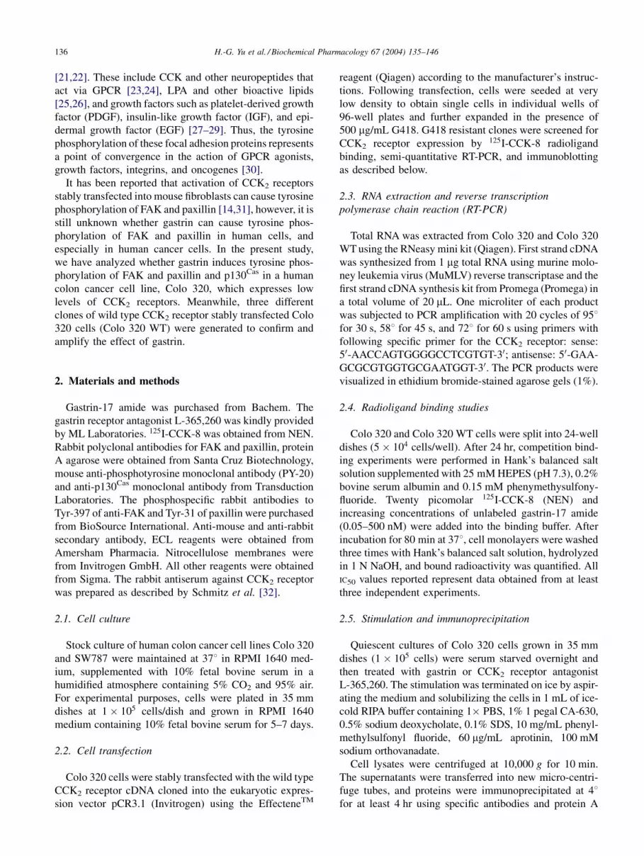
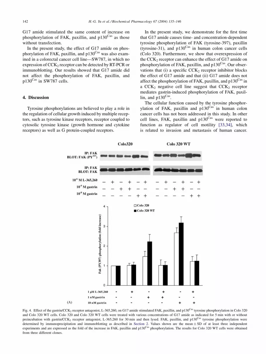
Fig. 4. Effect of the gastrin/CCK2 receptor antagonist, L-365,260, on G17 amide stimulated FAK, paxillin, and p130Cas tyrosine phosphorylation in Colo 320
and Colo 320 WT cells. Colo 320 and Colo 320 WT cells were treated with various concentrations of G17 amide as indicated for 5 min with or without
preincubation with gastrin/CCK2 receptor antagonist, L-365,260 for 30 min and then lysed. FAK, paxillin, and p130Cas tyrosine phosphorylation were
determined by immunoprecipitation and immunoblotting as described in Section 2. Values shown are the mean � SD of at least three independent
experiments and are expressed as the fold of the increase in FAK, paxillin and p130Cas phosphorylation. The results for Colo 320 WT cells were obtained
from three different clones.
142 H.-G. Yu et al. / Biochemical Pharmacology 67 (2004) 135–146
Phosphorylation of FAK at the site tyrosine-397 can
enhance cell motility by association with the SH2/SH3-
adaptor protein Crk, inducing p130Cas phosphorylation and
formation of p130Cas/Crk [35,36]. Therefore, FAK/Src-
dependent activation of the p130Cas/Crk signaling complex
is a central feature of both matrix and growth factor-
induced cell migration. Meanwhile, some investigators
also show that phosphorylation of the cytoskeletal protein
paxillin at the tyrosine-31 site increases cell motility by
formation of paxillin/Crk complexes [37].
FAK has also been proposed to function as a positive
regulator of cell growth. Overexpression of FAK can
modestly enhance DNA synthesis in fibroblasts after
the cells have been starved and stimulated with serum
[38,39]. Conversely, mutants of FAK can function in a
dominant negative fashion to inhibit progression through
the cell cycle following serum starvation [38,40]. There is
some evidence that these effects correlate with expression
of cyclin D1 and the cyclin-dependent kinase inhibitor
p21 [38]. Furthermore, these FAK-mediated cell cycle
effects are dependent on phosphorylation of tyrosine-397
and are believed to require Src and/or PI3-kinase binding
to this site. In addition, FAK is predicted to mediate
integrin-dependent regulation of the ERK family of
MAP kinases through several pathways [41], and these
pathways are suggested to act synergistically with mito-
genic signaling pathways to regulate cell growth [42,43].
However, the mechanism of FAK function in controlling
cell cycle progression has not been completely elucidated
yet.
Several studies demonstrate a role for phosphorylation of
FAK in promotion of cell survival. FAK has been shown to
play an important role in the cell survival of anchorage-
dependent cells [44]. Proteolytic cleavage of FAK by cas-
pase-3 has been reported during growth factor deprivation-
induced apoptosis in human umbilical vein endothelial cells
[45], which implies an association between FAK and apop-
tosis. FAK was also found to be tyrosine-phosphorylated
Fig. 4. (Continued )
H.-G. Yu et al. / Biochemical Pharmacology 67 (2004) 135–146 143

by oxidative stress before apoptosis occurred [46]. Further-
more, protein kinase B/Akt, which has been implicated in
the pathway of survival signal, was serine-phosphorylated
following tyrosine phosphorylation of FAK. FAK phosphor-
ylation at tyrosine-397 is required to prevent apoptosis,
because tyrosine-397 is an autophosphorylation site and a
high-affinity binding site for Src homology 2 domains of Src
family kinases [47]. PI3-kinase and phospholipase C also
interact with this site [48].
In conclusion, our results clearly demonstrate that acti-
vation of CCK2 receptors by gastrin induces phosphoryla-
tion of the non-receptor tyrosine kinase FAK, the
cytoskeletal protein paxillin, and the adaptor protein
p130Cas in human colon cancer cells. These findings
indicate that FAK, paxillin, and p130Cas could serve as
pharmacological downstream targets of gastrin-associated
growth mediated by CCK2 receptors during metastasis,
proliferation and apoptosis in colon cancer cells. These
findings also suggest a new approach to explore the
potential role of gastrin and its receptor in human colon
cancer. An important task in the future will be to elucidate
the interactions between FAK, paxillin, and p130Cas, to
close the gap between G protein binding and FAK phos-
phorylation and to define the cross-talks and synergistic
interactions between the tyrosine and serine/threonine
phosphorylation cascades in the promotion of biologic
responses stimulated by gastrin.
Acknowledgments
The work was supported in part by research grants from
the FoRUM program of Ruhr-University Bochum and the
Deutsche Forschungsgemeinschaft (DFG Schm 1073/4-1)
to F. Schmitz.
References
[1] Johnson LR. New aspects of the trophic action of gastrointestinal
hormones. Gastroenterolgy 1977;72:788–92.
[2] Walsh JH. Role of gastrin as a trophic hormone. Digestion 1990;47:
11–6.
[3] Smith J, Solomon T. Effects of gastrin, proglumide, and somatostatin
on growth of human colon cancer. Gastroenterology 1988;95:1541–8.
Fig. 4. (Continued ).
144 H.-G. Yu et al. / Biochemical Pharmacology 67 (2004) 135–146

[4] Watson S, Durrant L, Crosbie J, Morris D. The in vitro growth
response of primary human colorectal and gastric cancer cells to
gastrin. Int J Cancer 1989;43:692–6.
[5] Winsett O, Townsend C, Glass E, Thompson J. Gastrin stimulates
growth of colon cancer. Surgery 1986;99:302–7.
[6] Lamote J, Willems G. Stimulating effect of pentagastrin on cancer cell
proliferation kinetics in chemically induced colon cancer in rats.
Regul Pept 1988;20:1–9.
[7] Hoosein N, Kiener P, Curry R, Rovati L, McGilbra D, Brattain M.
Antiproliferative effects of gastrin receptor antagonists and antibodies
to gastrin on human colon carcinoma cell lines. Cancer Res
1988;48:7179–83.
[8] Watson SA, Michaeli D, Grimes S, Morris T, Crosbee D, Wilkinson M,
Robinson G, Robertson J, Steele R, Hardcastle J. Anti-gastrin anti-
bodies raised by gastrimmune inhibit growth of the human colorectal
tumour AP5. Int J Cancer 1995;61:233–40.
[9] Beauchamp R, Townsend C, Singh P, Glass E, Thompson J.
Proglumide, a gastrin receptor antagonist, inhibits growth of
colon cancer and enhances survival in mice. Ann Surg 1985;202:
303–7.
[10] Romani R, Howes LG, Morris DL. Gastrin receptor antagonist CI-988
inhibits growth of human colon cancer in vivo and in vitro. Aust NZ J
Surg 1996;66:235–7.
[11] Singh P, Owlia A, Varro A, Dai B, Rajaraman S, Wood T. Gastrin gene
expression is required for the proliferation and tumorigenicity of
human colon cancer cells. Cancer Res 1996;56:4111–5.
[12] Seva C, Dickinson CJ, Yamada T. Growth-promoting effects of
glycine-extended progastrin. Science 1994;265:410–2.
[13] Sethi T, Herget T, Wu SV, Walsh JH, Rozengurt E. CCKA and
CCKB receptors are expressed in small cell lung cancer lines and
mediate Ca2þ mobilization and clonal growth. Cancer Res 1993;
53:5208–13.
[14] Seufferlein T, Withers DJ, Braod S, Herget T, Walsh JH, Rozengurt E.
The human CCKB/gastrin receptor transfected into rat1 fibroblasts
mediates activation of MAP kinase, p74raf-1 kinase, and mitogenesis.
Cell Growth Differ 1995;6:383–93.
[15] Nakata H, Matsui T, Ito M, Taniguchi T, Naribayashi Y, Arima N,
Nakamura A, Kinoshita Y, Chihara K, Hosoda S. Cloning and
characterization of gastrin receptor from ECL carcinoid tumor of
Mastomys natalensis. Biochem Biophys Res Commun 1992;187:
1151–7.
[16] Wank SA, Harkins R, Jensen RT, Shapira H, de Weerth A, Slattery T.
Purification, molecular cloning, and functional expression of the
cholecystokinin receptor from rat pancreas. Proc Natl Acad Sci
USA 1992;89:3125–9.
[17] Seva C, Scemama JL, Pradayrol L, Sarfati PD, Vaysse N. Coupling of
pancreatic gastrin/cholecystokinin-B (G/CCKB) receptors to phos-
pholipase C and protein kinase C in AR4-2J tumoral cells. Regul Pept
1994;52:31–8.
[18] Bertrand V, Bastie MJ, Vaysse N, Pradayrol L. Inhibition of gastrin-
induced proliferation of AR4-2J cells by calcium channel antagonists.
Int J Cancer 1994;56:427–32.
[19] Dehez S, Daulhac L, Kowalski-Chauvel A, Fourmy D, Pradayrol L,
Seva C. Gastrin-induced DNA synthesis requires p38-MAPK activa-
tion via PKC/Ca(2þ) and Src-dependent mechanisms. FEBS Lett
2001;496:25–30.
[20] Rozengurt E, Walsh JH, Gastrin CCK. Signaling, and cancer. Annu
Rev Physiol 2001;63:49–76.
[21] Hanks SK, Polte TR. Signaling through focal adhesion kinase. Bioes-
says 1997;19:137–45.
[22] Rozengurt E. Gastrointestinal peptide signaling through tyrosine
phosphorylation of focal adhesion proteins. Am J Physiol 1998;
275:177–82.
[23] Rosado JA, Salido GM, Garcia LJ. A role for phosphoinositides in
tyrosine phosphorylation of p125 focal adhesion kinase in rat pan-
creatic acini. Cell Signal 2000;12:173–82.
[24] Leyton J, Garcia-Marin LJ, Tapia JA, Jensen RT, Moody TW. Bom-
besin and gastrin releasing peptide increase tyrosine phosphorylation
of focal adhesion kinase and paxillin in non-small cell lung cancer
cells. Cancer Lett 2001;162:87–95.
[25] Salazar EP, Rozengurt E. Src family kinases are required for integrin-
mediated but not for G protein-coupled receptor stimulation of focal
adhesion kinase autophosphorylation at Tyr-397. J Biol Chem
2001;276:17788–895.
[26] Derkinderen P, Siciliano J, Toutant M, Girault JA. Differential reg-
ulation of FAK and PYK2/Cakbeta, two related tyrosine kinases, in rat
hippocampal slices: effects of LPA, carbachol, depolarization and
hyperosmolarity. Eur J Neurosci 1998;10:1667–75.
[27] Leopoldt D, Yee Jr HF, Saab S, Rozengurt E. Tyrosine phosphoryla-
tion of p125(Fak), p130(Cas), and paxillin does not require extra-
cellular signal-regulated kinase activation in Swiss 3T3 cells
stimulated by bombesin or platelet-derived growth factor. J Cell
Physiol 2000;183:208–20.
[28] Cheng HL, Steinway ML, Russell JW, Feldman EL. GTPases and
phosphatidylinositol 3-kinase are critical for insulin-like growth
factor-I-mediated Schwann cell motility. J Biol Chem 2000;275:
27197–204.
[29] Lu Z, Jiang G, Blume-Jensen P, Hunter T. Epidermal growth factor-
induced tumor cell invasion and metastasis initiated by dephosphor-
ylation and downregulation of focal adhesion kinase. Mol Cell Biol
2001;21:4016–31.
[30] Rozengurt E. Convergent signalling in the action of integrins,
neuropeptides, growth factors and oncogenes. Cancer Surv 1995;
24:81–96.
[31] Taniguchi T, Matsui T, Ito M, Murayama T, Tsukamoto T, Katakami Y,
Chiba T, Chihara K. Cholecystokinin-B/gastrin receptor signaling
pathway involves tyrosine phosphorylations of p125FAK and
p42MAP. Oncogene 1994;9:861–7.
[32] Schmitz F, Otte JM, Techele HU, Reimann B, Banasiewicz T, Folsch
UR, Schmidt WE, Herzig KH. CCK-B/gastrin receptors in human
colorectal cancer. Eur J Clin Invest 2001;31:812–20.
[33] Ilic D, Furuta Y, Kanazawa S, Takeda N, Sobue K, Nakatsuji N,
Nomura S, Fujimoto J, Okada M, Yamamoto T. Reduced cell motility
and enhanced focal adhesion contact formation in cells from
FAK-deficient mice. Nature 1995;377:539–44.
[34] Cary LA, Chang JF, Guan JL. Stimulation of cell migration by
overexpression of focal adhesion kinase and its association with
Src and Fyn. J Cell Sci 1996;109:787–94.
[35] Owen JD, Ruest PJ, Fry DW, Hanks SK. Induced focal adhesion kinase
(FAK) expression in FAK-null cells enhances cell spreading and
migration requiring both auto- and activation loop phosphorylation
sites and inhibits adhesion-dependent tyrosine phosphorylation of
Pyk2. Mol Cell Biol 1999;19:4806–18.
[36] Sieg DJ, Hauck CR, Schlaepfer DD. Required role of focal adhesion
kinase (FAK) for integrin-stimulated cell migration. J Cell Sci
1999;112:2677–91.
[37] Schaller MD. Biochemical signals and biological responses elicited
by the focal adhesion kinase. Biochim Biophys Acta 2001;1540:
1–21.
[38] Zhao JH, Reiske H, Guan JL. Regulation of the cell cycle by focal
adhesion kinase. J Cell Biol 1998;143:1997–2008.
[39] Zhao J, Zheng C, Guan J. Pyk2 and FAK differentially regulate
progression of the cell cycle. J Cell Sci 2000;113:3063–72.
[40] Oktay M, Wary KK, Dans M, Birge RB, Giancotti FG. Integrin-
mediated activation of focal adhesion kinase is required for signalling
to Jun NH2-terminal kinase and progression through the G1 phase of
the cell cycle. J Cell Biol 1999;145:1461–9.
[41] Schlaepfer DD, Jones KC, Hunter T. Multiple Grb2-mediated
integrin-stimulated signaling pathways to ERK2/mitogen-activated
protein kinase: summation of both c-Src- and focal adhesion
kinase-initiated tyrosine phosphorylation events. Mol Cell Biol
1998;18:2571–85.
H.-G. Yu et al. / Biochemical Pharmacology 67 (2004) 135–146 145

[42] Eliceiri BP, Klemke R, Stromblad S, Cheresh DA. Integrin alphavbeta3
requirement for sustained mitogen-activated protein kinase activity
during angiogenesis. J Cell Biol 1998;140:1255–63.
[43] Short SM, Talbott GA, Juliano RL. Integrin-mediated signaling
events in human endothelial cells. Mol Biol Cell 1998;9:1969–80.
[44] Xu LH, Yang X, Bradham CA, Brenner DA, Baldwin Jr AS, Craven
RJ, Cance WG. The focal adhesion kinase suppresses transformation-
associated, anchorage-independent apoptosis in human breast cancer
cells. Involvement of death receptor-related signalling pathways. J
Biol Chem 2000;275:30597–604.
[45] Fukai F, Mashimo M, Akiyama K, Goto T, Tanuma S, Katayama T.
Modulation of apoptotic cell death by extracellular matrix proteins and a
fibronectin-derived antiadhesive peptide. Exp Cell Res 1998;242:92–9.
[46] Ben Mahdi MH, Andrieu V, Pasquier C. Focal adhesion kinase
regulation by oxidative stress in different cell types. IUBMB Life
2000;50:291–9.
[47] Sonoda Y, Matsumoto Y, Funakoshi M, Yamamoto D, Hanks SK,
Kasahara T. Anti-apoptotic role of focal adhesion kinase (FAK).
Induction of inhibitor-of-apoptosis proteins and apoptosis suppression
by the overexpression of FAK in a human leukemic cell line, HL-60. J
Biol Chem 2000;275:16309–15.
[48] Chan PC, Lai JF, Cheng CH, Tang MJ, Chiu CC, Chen HC. Suppres-
sion of ultraviolet irradiation-induced apoptosis by overexpression of
focal adhesion kinase in Madin–Darby canine kidney cells. J Biol
Chem 1999;274:26901–6.
146 H.-G. Yu et al. / Biochemical Pharmacology 67 (2004) 135–146


![Novel Glycated [ 99m Tc(CO) 3 ]-Labeled Bombesin Analogues for Improved Targeting of Gastrin-Releasing Peptide Receptor-Positive Tumors](https://img.dokumen.tips/doc/110x75/63350c0b6c27eedec6060564/novel-glycated-99m-tcco-3-labeled-bombesin-analogues-for-improved-targeting.jpg)


