Embed Size (px)
Citation preview
Fax +41 61 306 12 34E-Mail [email protected]
Basic Science Investigations
Respiration 2009;77:427–439 DOI: 10.1159/000176209
Proteomic Investigation in A549 Lung Cell Line Stably Infected by HPV16E6/E7 Oncogenes
Marco Ciotti a Valeria Marzano b–d Laura Giuliani c, e Marzia Nuccetelli b–d
Simona D’Aguanno b–d Barbara Azzimonti f Sergio Bernardini b–d
Carlo Federico Perno a Andrea Urbani b, g, h Cartesio Favalli a Giorgio Federici b–e
a Laboratory of Clinical Microbiology and Virology and b Laboratory of Clinical Biochemistry, University Hospital Tor Vergata, c Department of Internal Medicine, University of Rome Tor Vergata, d IRCCS-Santa Lucia Foundation, Laboratory of Proteomics, and e Pediatric Hospital ‘Bambino Gesù’ – IRCCS, Rome , f Department of Clinical and Experimental Medicine, Medical School of Novara, University of Eastern Piedmont, Novara , and g Foundation‘G. D’Annunzio’, Center for Aging Research, and h Department of Biomedical Sciences, University of Chieti-Pescara ‘G. D’Annunzio’, Chieti , Italy
sity was statistically significant (p ! 0.05) and differed by 2-fold. Relationships between differentially expressed proteins and the HPV-induced infection mechanism have been clus-tered by knowledge-base database functional association network analysis. Conclusion: The impact of Hsp27, annexin III, annexin IV, Gp96 and TPT1 on the cellular response mech-anism to HPV infection is presented and discussed.
Copyright © 2008 S. Karger AG, Basel
Introduction
Lung cancer is one of the leading causes of cancer-re-lated deaths in western countries [1] . Smoking, environ-mental pollution and exposure to asbestos are considered the main risk factors; however, some 15% of nonsmokers develop bronchial cancer [2] . Therefore, other etiological factors including genetic factors, functional inactivation of tumor suppressor genes such as p53 or pRb and infec-
Key Words
Papillomaviruses, human � Lung cancer � Proteomics � Molecular networks � Mass spectrometry � Oncogene
Abstract
Background: Data have accumulated implicating the in-volvement of oncogenic human papillomaviruses (HPVs) in bronchial carcinogenesis. We recently described the pres-ence of oncogenic HPV transcripts in non-small cell lung cancers. Objective: To investigate the role of oncogenic HPVs in lung carcinogenesis. Material and Methods: The lung cell line A549 stably infected with HPV16E6, HPV16E7 and HPVE6/E7 constructs was used to investigate the protein profile changes associated with the expression of these on-cogenes. Replicated two-dimensional gel electrophoresis gels from uninfected and stably HPV16E6-, E7-, and E6/E7-infected A549 cells were compared for changes in protein profile. Protein identification was achieved by peptide mass fingerprinting by MALDI-TOF-MS and nLC-ESI-Q-TOF-MS/MS peptide ladder sequencing. Results: We identified 17 dif-ferent polypeptides whose average normalized spot inten-
Received: April 10, 2008 Accepted after revision: September 10, 2008 Published online: November 20, 2008
Dr. Marco Ciotti Laboratory of Clinical Microbiology and Virology University Hospital Tor Vergata, Viale Oxford 81 IT–00133 Rome (Italy) Tel. +39 06 2090 2087, Fax +39 06 2090 2078, E-Mail [email protected]
© 2008 S. Karger AG, Basel0025–7931/09/0774–0427$26.00/0
Accessible online at:www.karger.com/res
M.C. and V.M. contributed equally to this work.

Ciotti et al. Respiration 2009;77:427–439428
tion with oncogenic human papillomaviruses (HPVs) have been implicated [3] . HPV is the etiological agent of cervical cancer, but it has been estimated that about 15–20% of all human malignancies could be related to onco-genic HPVs [4–6] . In two recent studies, we have revealed the presence of oncogenic HPVs (HPV16, 18, 31 and 53) and their transcripts in a series of non-small cell lung cancers [7, 8] , reinforcing the relationship between HPV and lung cancer and supporting the hypothesis that on-cogenic HPVs might play a role in lung carcinogenesis. The oncogenic action of the virus is exerted through the oncoproteins E6 and E7 by binding to p53 and pRb, re-spectively, interfering with cell cycle control [9–11] . E6 protein promotes cell proliferation by degradation of p53 protein, which forms a complex with E6 and the cellular ubiquitination enzyme E6-AP. E7 binds to pRb displac-ing the E2F transcription factor. The release of E2F influ-ences the expression of cellular genes involved in mitosis and cell cycle control [12] . Complex formation between viral oncoproteins and the two oncosuppressor proteins is believed to lead to cell immortalization and transfor-mation [13, 14] .
The aim of our study was to identify proteins whose expression is modulated by the HPV16E6 and E7 onco-genes in a lung cancer cell line, A549. We generated stably infected HPV16E6-, HPV16E7- and HPV16E6/E7-A549 cell lines and used a proteomic approach to separate and identify differentially expressed protein.
Materials and Methods
Cell Culture and Retroviral Infection The human lung adenocarcinoma epithelial cell line A549 was
cultured in Dulbecco’s modified Eagle’s medium (Sigma-Aldrich, Milan, Italy) supplemented with 10% heat-inactivated fetal bo-vine serum (Sigma-Aldrich). Cells were incubated at 37 ° C in a humidified atmosphere containing 5% CO 2 and 95% air.
The retroviral vector pLXSN was obtained from Clontech (Palo Alto, Calif., USA). The empty pLXSN vector, carrying a neo-mycin resistance gene, was used as a control.
The retroviral vectors pLXSN containing the HPV16E6, E7 and E6/E7 ORFs were kindly provided by Massimo Tommasino (International Agency for Research on Cancer, Lyon, France) and prepared as previously described [15] .
High-titer retrovirus-containing supernatants ( 1 5 ! 10 6 IU/ml) were generated by transient transfection of second-generation retrovirus producer Phoenix cells (amphotropic viruses) and used to infect the cells as previously described [16] . After infec-tion, A549 cells were selected in 800 � g/ml G418 for 10 days, used as pooled population, and designated A549 pLXSN empty vec-tor, A549 pLXSN-HPV16E6, A549 pLXSN-HPV16E7 and A549 pLXSN-HPV16E6/E7, respectively.
RT-PCR Total RNA was extracted from 2 ! 10 6 A549 cells infected
with pLXSN-HPV16E6, pLXSN-HPV16E7, pLXSN-HPV16E6/E7 and mock-infected cells, respectively. One milliliter of Trizol (In-vitrogen, SRL, Milan, Italy) was added and total RNA was ex-tracted following the protocol’s instructions. The absence of DNA in the extracted RNA samples was checked using primers for the � -actin gene located at the exon 4-intron 5-exon 5 junction [17] . All mRNAs present in 1.5 � g of total RNA extracted were reverse transcribed with the oligo-dT primer using the Omniscript kit (Qiagen, Hilden, Germany) in a final volume of 20 � l. The reac-tion tubes were incubated at 42 ° C for 1 h, heat-inactivated at 95 ° C for 5 min, and then placed on ice. Five microliters of each cDNA were used in the following PCR reaction as previously described [7] .
Western Blot Analysis For crude extract preparation, cells were lysed in 3% sodium
dodecyl sulfate (SDS)-lysis buffer containing 125 m M Tris-HCl pH 6.8, 3% SDS, 10 m M dithiothreitol (DTT), 10% glycerol with the addition of protease inhibitors [0.2 m M phenylmethanesulfo-nyl fluoride (PMSF), 1 mg/ml pepstatin, 0.1 m M benzamidine, 2 mg/ml aprotinin] and briefly sonicated. Insoluble material was removed by centrifugation at 13,000 rpm for 5 min. Protein con-centration was determined by the Bio-Rad Dc Protein Assay (Bio-Rad Laboratories, Milan, Italy). Twenty micrograms of proteins were loaded and separated on 8.5% SDS-polyacrylamide gels and transferred onto polyvinyl difluoride membranes (GE Healthcare Europe, Milan, Italy) according to the instruction manual. Mem-branes were blocked in a blocking solution [10 m M Tris-HCl pH 7.5, 0.1 M NaCl, 0.1% Tween-20, 5% (w/v) nonfat dry milk] over-night at 4 ° C, and incubated with the following mouse monoclo-nal primary antibodies: anti-p53 (clone DO-1, Santa Cruz, Calif., USA; diluted 1: 1,000) and antiactin (clone C4, Chemicon, Tem-ecula, Calif., USA; diluted 1: 5,000). Appropriate secondary anti-mouse antibodies, conjugated with horseradish peroxidase, were used (GE Healthcare Europe; diluted 1: 4,000) and the reactions were visualized by enhanced chemiluminescence (ECL Western Blotting Detection Reagents; GE Healthcare, Little Chalfont, Buckinghamshire, UK), according to the manufacturer’s instruc-tions.
Two-Dimensional Gel Electrophoresis and Image Analysis Cell pellets were dissolved in lysis buffer containing 7 M urea,
2 M thiourea, 40 m M Tris base, 50 m M DTT, 4% CHAPS and 0.5% Pharmalyte 3–10 (GE Healthcare). After sonication and incuba-tion at 37 ° C for 1 h the protein concentration in the lysates was measured using a densitometric method (adapted TCA proce-dure) [18] . The proteins were precipitated with 80% acetone over-night at –20 ° C and then washed extensively with cold 80% ace-tone. For the first dimension, 100 � g of total proteins were loaded onto pH 3–10 nonlinear 18-cm gel strips (IPG; GE Healthcare) with a rehydration solution (6 M urea, 2 M thiourea, 4% CHAPS, 15 m M DTT, 0.5% IPG buffer 3–10 and trace amounts of bromo-phenol blue). After rehydration for 12 h, isoelectric focusing was performed at 20 ° C using an Ettan IPGphor II IEF System (Amer-sham Bioscience, Uppsala, Sweden). The second-dimension sepa-ration was performed 12 gels at a time on gradient (9–14%) SDS-PAGE gels (20 ! 26 cm) using an EttanDALTtwelve System (Am-ersham Bioscience). After fixing, the protein spots in analytical
A549 Cell Line Stably Infected by HPV16E6/E7 Oncogenes
Respiration 2009;77:427–439 429
gels were visualized by Vorum silver staining (modified for MS) [19] and the stained gels were scanned with a UMAX Image Scan-ner (Amersham Bioscience). The image analysis was done using the PDQuest software (version 7.1.1; Bio-Rad).
For further protein identification larger amounts (1 mg) of total proteins, after treatment with DNAseI (GE Healthcare), RNAse A (USB, Cleveland, Ohio, USA) and 2-D Clean-Up Kit (GE Healthcare), were loaded on preparatory gels and subjected to isoelectric focusing to a total of 160 kVh. Following two-di-mensional gel electrophoresis (2-DE), gels were stained using col-loidal Coomassie brilliant blue staining. For the image analysis we chose the three highest quality gels, based on protein spot res-olution and number, to allow experimental statistical comparison between the A549 cell line and A549 infected with the HPV con-structs. A reference gel was selected from one of the experimental gels and unmatched protein spots of the remaining gels were manually added to this reference gel to create the master gel, a virtual image comprehensive of all matched spots derived from all analyzed samples. Subsequent to automatic spot detection and spot filtering, the matching of spots between gels was manually reviewed and adjusted as necessary.
Protein Identification Protein spots of interest were excised from 2-DE gels using a
robotic instrument (Proteineer SpotPicker; Bruker Daltonik, Bre-men, Germany). In-gel digestion for peptide mass fingerprinting was carried out manually with porcine trypsin (Promega, Madi-son, Wisc., USA) in 50 m M ammonium bicarbonate at 37 ° C for 16–18 h. The reaction was stopped by adding a final concentration of 0.1% TFA. For the mass-spectrometric analysis of tryptic di-gests the samples were prepared by reverse phase extraction using ZipTip C18 (Millipore, Bedford, Mass., USA). Elution and spot-ting on 600- � m AnchorChip target plates were obtained as de-scribed [20] . Each spectrometric analysis was performed using a Reflex IV MALDI time-of-flight (MALDI-TOF; Bruker Dalton-ik) operating in a positive ion mode with a reflectron setup. Da-tabase searches with the measured monoisotopic peptide masses were performed against the National Center for Biotechnology Information (NCBInr) human database using the peptide search routine MASCOT (Matrix Science, London, UK, at http://www.matrixscience.com). Carbamidomethylation of cysteine was set as a fixed modification and oxidation of methionine was allowed as variable modification of the peptides. The query was performed with the maximal tolerance of 100 ppm. For positive identifica-tion of the peptide mass fingerprinting, protein scores greater than 65 were considered significant (p ! 0.05), as calculated by the MASCOT scoring algorithm.
Tryptic digests of some gel spots selected for validation were subjected to analysis by nanoliquid chromatography-electrospray ionization-tandem MS (nLC-ESI-MS/MS; Waters, Milford, Mass., USA). The generated data were screened in NCBInr database with the MASCOT search engine using a mass tolerance ̂ 50 ppm for parent and 0.2 Da for fragments. An MS/MS ion score 1 35 indi-cates identity or extensive homology (p ! 0.05).
Pathway Analysis Relationships between differentially expressed genes were as-
sessed using Ingenuity Pathways Analysis (IPA; IPA 5.0; Ingenu-ity � Systems, www.ingenuity.com) that enables mining, visual-ization and exploration of relevant functional associations sig-
nificant to the experimental results. We employed a data set containing gene identifiers corresponding to MS and MS/MS-identified proteins and their corresponding relative abundance derived from 2D-PAGE experiments. Only IPA interconnections with statistical significance lower than p ! 0.05 were used to cre-ate the functional clusters employed to design the network graph. The network graphically displays genes/gene products as nodes and the biological relationships between the nodes as edges.
Results
RT-PCR In order to investigate the expression level of proteins
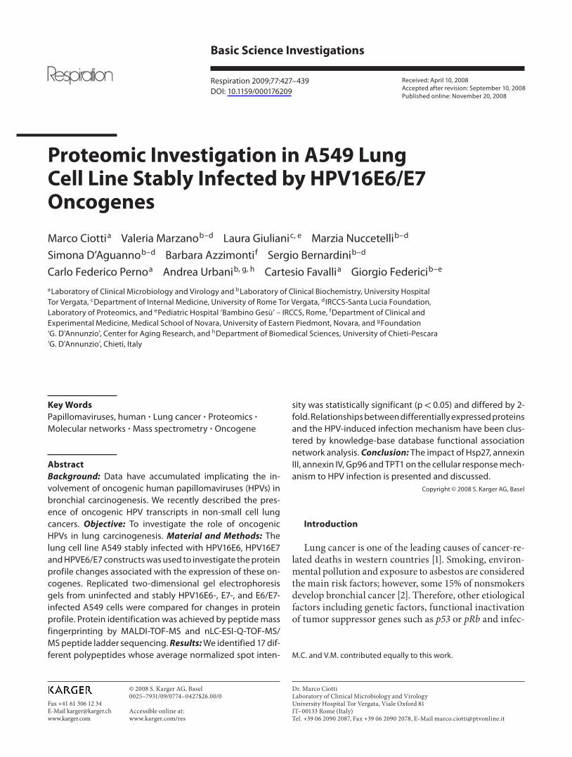
regulated by HPV16E6 and E7 oncogenes singularly and/or combined, we stably infected the A549 cell line with the retroviral vector pLXSN carrying HPV16E6, HPV16E7 and HPV16E6/E7 ORFs. Mock-infected cells were included as control. The expression of the HPV16E6 and E7 oncogenes, singularly or combined, is shown in figure 1 .
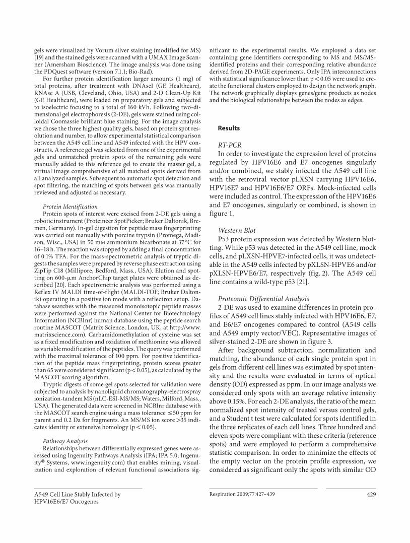
Western Blot P53 protein expression was detected by Western blot-
ting. While p53 was detected in the A549 cell line, mock cells, and pLXSN-HPVE7-infected cells, it was undetect-able in the A549 cells infected by pXLSN-HPVE6 and/or pXLSN-HPVE6/E7, respectively ( fig. 2 ). The A549 cell line contains a wild-type p53 [21] .
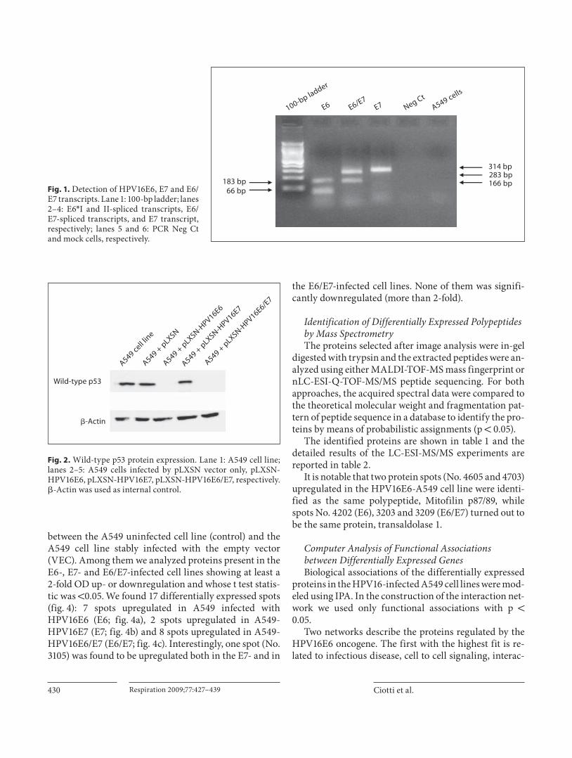
Proteomic Differential Analysis 2-DE was used to examine differences in protein pro-
files of A549 cell lines stably infected with HPV16E6, E7, and E6/E7 oncogenes compared to control (A549 cells and A549 empty vector/VEC). Representative images of silver-stained 2-DE are shown in figure 3 .
After background subtraction, normalization and matching, the abundance of each single protein spot in gels from different cell lines was estimated by spot inten-sity and the results were evaluated in terms of optical density (OD) expressed as ppm. In our image analysis we considered only spots with an average relative intensity above 0.15%. For each 2-DE analysis, the ratio of the mean normalized spot intensity of treated versus control gels, and a Student t test were calculated for spots identified in the three replicates of each cell lines. Three hundred and eleven spots were compliant with these criteria (reference spots) and were employed to perform a comprehensive statistic comparison. In order to minimize the effects of the empty vector on the protein profile expression, we considered as significant only the spots with similar OD
Ciotti et al. Respiration 2009;77:427–439430
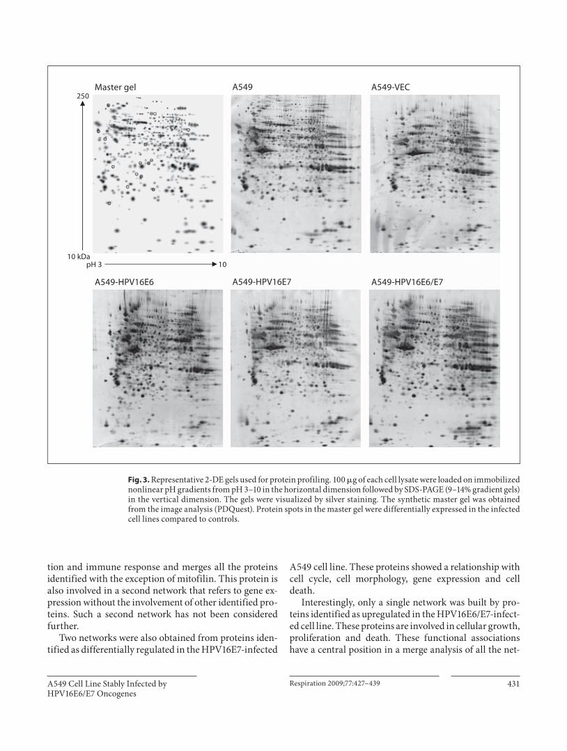
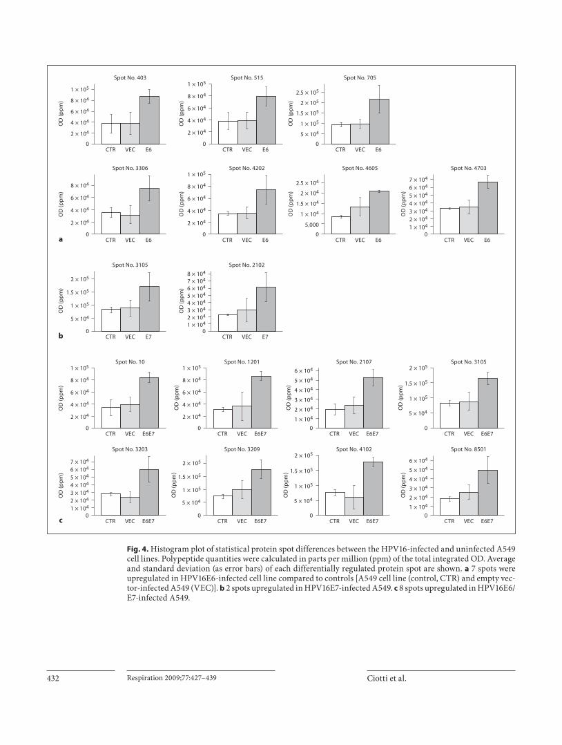
between the A549 uninfected cell line (control) and the A549 cell line stably infected with the empty vector (VEC). Among them we analyzed proteins present in the E6-, E7- and E6/E7-infected cell lines showing at least a 2-fold OD up- or downregulation and whose t test statis-tic was ! 0.05. We found 17 differentially expressed spots ( fig. 4 ): 7 spots upregulated in A549 infected with HPV16E6 (E6; fig. 4 a), 2 spots upregulated in A549-HPV16E7 (E7; fig. 4 b) and 8 spots upregulated in A549-HPV16E6/E7 (E6/E7; fig. 4 c). Interestingly, one spot (No. 3105) was found to be upregulated both in the E7- and in
the E6/E7-infected cell lines. None of them was signifi-cantly downregulated (more than 2-fold).
Identification of Differentially Expressed Polypeptides by Mass Spectrometry The proteins selected after image analysis were in-gel
digested with trypsin and the extracted peptides were an-alyzed using either MALDI-TOF-MS mass fingerprint or nLC-ESI-Q-TOF-MS/MS peptide sequencing. For both approaches, the acquired spectral data were compared to the theoretical molecular weight and fragmentation pat-tern of peptide sequence in a database to identify the pro-teins by means of probabilistic assignments (p ! 0.05).
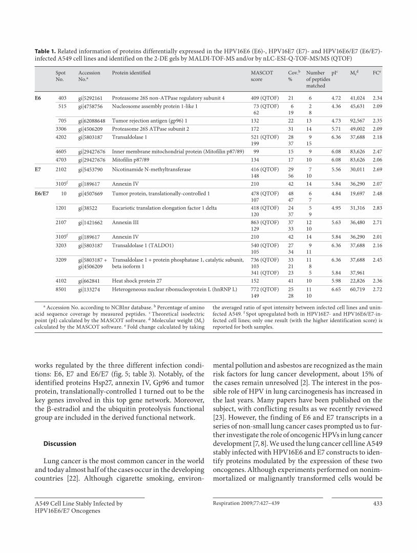
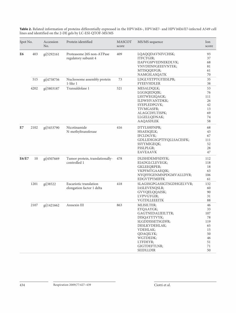
The identified proteins are shown in table 1 and the detailed results of the LC-ESI-MS/MS experiments are reported in table 2 .
It is notable that two protein spots (No. 4605 and 4703) upregulated in the HPV16E6-A549 cell line were identi-fied as the same polypeptide, Mitofilin p87/89, while spots No. 4202 (E6), 3203 and 3209 (E6/E7) turned out to be the same protein, transaldolase 1.
Computer Analysis of Functional Associations between Differentially Expressed Genes Biological associations of the differentially expressed
proteins in the HPV16-infected A549 cell lines were mod-eled using IPA. In the construction of the interaction net-work we used only functional associations with p ! 0.05.
Two networks describe the proteins regulated by the HPV16E6 oncogene. The first with the highest fit is re-lated to infectious disease, cell to cell signaling, interac-
183 bp
100-bp ladder
E6 E6/E7E7 Neg Ct
A549 cells
66 bp
314 bp283 bp166 bp
Fig. 1. Detection of HPV16E6, E7 and E6/E7 transcripts. Lane 1: 100-bp ladder; lanes 2–4: E6 * I and II-spliced transcripts, E6/E7-spliced transcripts, and E7 transcript, respectively; lanes 5 and 6: PCR Neg Ct and mock cells, respectively.
Wild-type p53
�-Actin
A549 + p
LXSN-HPV16E7
A549 + p
LXSN-HPV16E6
A549 + p
LXSN-HPV16E6/E
7
A549 + p
LXSN
A549 cell lin
e
Fig. 2. Wild-type p53 protein expression. Lane 1: A549 cell line; lanes 2–5: A549 cells infected by pLXSN vector only, pLXSN-HPV16E6, pLXSN-HPV16E7, pLXSN-HPV16E6/E7, respectively. �-Actin was used as internal control.
A549 Cell Line Stably Infected by HPV16E6/E7 Oncogenes
Respiration 2009;77:427–439 431
tion and immune response and merges all the proteins identified with the exception of mitofilin. This protein is also involved in a second network that refers to gene ex-pression without the involvement of other identified pro-teins. Such a second network has not been considered further.
Two networks were also obtained from proteins iden-tified as differentially regulated in the HPV16E7-infected
A549 cell line. These proteins showed a relationship with cell cycle, cell morphology, gene expression and cell death.
Interestingly, only a single network was built by pro-teins identified as upregulated in the HPV16E6/E7-infect-ed cell line. These proteins are involved in cellular growth, proliferation and death. These functional associations have a central position in a merge analysis of all the net-
A549-HPV16E6 A549-HPV16E7 A549-HPV16E6/E7
Master gel A549 A549-VEC
10pH 310 kDa
250
Fig. 3. Representative 2-DE gels used for protein profiling. 100 � g of each cell lysate were loaded on immobilized nonlinear pH gradients from pH 3–10 in the horizontal dimension followed by SDS-PAGE (9–14% gradient gels) in the vertical dimension. The gels were visualized by silver staining. The synthetic master gel was obtained from the image analysis (PDQuest). Protein spots in the master gel were differentially expressed in the infected cell lines compared to controls.
Ciotti et al. Respiration 2009;77:427–439432
1 × 1042 × 1043 × 1044 × 1045 × 1046 × 1047 × 1048 × 104
0CTR
OD
(pp
m)
VEC
Spot No. 2102
E7
5 × 10⁴
1 × 10⁵
1.5 × 10⁵
2 × 10⁵
0
2 × 10⁴
4 × 10⁴
6 × 10⁴
8 × 10⁴
1 × 10⁵
0
2 × 10⁴
4 × 10⁴
6 × 10⁴
8 × 10⁴
1 × 10⁵
0
0CTR
OD
(pp
m)
VEC
Spot No. 4703
E6
1 × 1042 × 1043 × 1044 × 1045 × 1046 × 1047 × 104
2 × 10⁴
4 × 10⁴
6 × 10⁴
8 × 10⁴
1 × 10⁵
0CTR
OD
(pp
m)
VEC
Spot No. 4202
E60
CTR
OD
(pp
m)
VEC
Spot No. 4605
E6
5,000
1 × 10⁴
1.5 × 10⁴
2 × 10⁴
2.5 × 10⁴
2 × 10⁴
4 × 10⁴
6 × 10⁴
8 × 10⁴
0
2 × 10⁴
4 × 10⁴
6 × 10⁴
8 × 10⁴
1 × 10⁵
0
2 × 10⁴
4 × 10⁴
6 × 10⁴
8 × 10⁴
1 × 10⁵
0CTR
OD
(pp
m)
VEC
Spot No. 1201
E6E7
2 × 10⁴
1 × 10⁴
3 × 10⁴
4 × 10⁴
5 × 10⁴
6 × 10⁴
0CTR
OD
(pp
m)
VEC
Spot No. 2107
E6E7
2 × 10⁴
1 × 10⁴
3 × 10⁴
4 × 10⁴
5 × 10⁴
6 × 10⁴
0CTR
OD
(pp
m)
VEC
Spot No. 8501
E6E7
5 × 10⁴
1 × 10⁵
1.5 × 10⁵
2 × 10⁵
0CTR
OD
(pp
m)
VEC
Spot No. 3105
E6E7
5 × 10⁴
1 × 10⁵
1.5 × 10⁵
2 × 10⁵
0CTR
OD
(pp
m)
VEC
Spot No. 3209
E6E7
5 × 10⁴
1 × 10⁵
1.5 × 10⁵
2 × 10⁵
0CTR
OD
(pp
m)
VEC
Spot No. 4102
E6E7
2 × 1043 × 1044 × 1045 × 1046 × 1047 × 104
0
CTR
OD
(pp
m)
OD
(pp
m)
OD
(pp
m)
OD
(pp
m)
VEC
Spot No. 3105
E7
CTR
OD
(pp
m)
VEC
Spot No. 403
E6 CTR
OD
(pp
m)
VEC
Spot No. 515
E60
CTR
OD
(pp
m)
VEC
Spot No. 705
E6
5 × 10⁴
1 × 10⁵
1.5 × 10⁵
2 × 10⁵
2.5 × 10⁵
CTR VEC
Spot No. 3306
E6a
b
CTR VEC
Spot No. 10
E6E7
CTR VEC
Spot No. 3203
E6E7
1 × 10⁴
c
Fig. 4. Histogram plot of statistical protein spot differences between the HPV16-infected and uninfected A549 cell lines. Polypeptide quantities were calculated in parts per million (ppm) of the total integrated OD. Average and standard deviation (as error bars) of each differentially regulated protein spot are shown. a 7 spots were upregulated in HPV16E6-infected cell line compared to controls [A549 cell line (control, CTR) and empty vec-tor-infected A549 (VEC)]. b 2 spots upregulated in HPV16E7-infected A549. c 8 spots upregulated in HPV16E6/E7-infected A549.
A549 Cell Line Stably Infected by HPV16E6/E7 Oncogenes
Respiration 2009;77:427–439 433
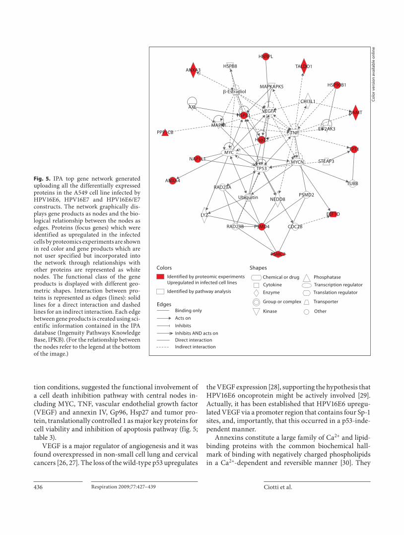
works regulated by the three different infection condi-tions: E6, E7 and E6/E7 ( fig. 5 ; table 3 ). Notably, of the identified proteins Hsp27, annexin IV, Gp96 and tumor protein, translationally-controlled 1 turned out to be the key genes involved in this top gene network. Moreover, the � -estradiol and the ubiquitin proteolysis functional group are included in the derived functional network.
Discussion
Lung cancer is the most common cancer in the world and today almost half of the cases occur in the developing countries [22] . Although cigarette smoking, environ-
mental pollution and asbestos are recognized as the main risk factors for lung cancer development, about 15% of the cases remain unresolved [2] . The interest in the pos-sible role of HPV in lung carcinogenesis has increased in the last years. Many papers have been published on the subject, with conflicting results as we recently reviewed [23] . However, the finding of E6 and E7 transcripts in a series of non-small lung cancer cases prompted us to fur-ther investigate the role of oncogenic HPVs in lung cancer development [7, 8] . We used the lung cancer cell line A549 stably infected with HPV16E6 and E7 constructs to iden-tify proteins modulated by the expression of these two oncogenes. Although experiments performed on nonim-mortalized or malignantly transformed cells would be
Table 1. Related information of proteins differentially expressed in the HPV16E6 (E6)-, HPV16E7 (E7)- and HPV16E6/E7 (E6/E7)-infected A549 cell lines and identified on the 2-DE gels by MALDI-TOF-MS and/or by nLC-ESI-Q-TOF-MS/MS (QTOF)
SpotNo.
AccessionNo.a
Protein identified MASCOTscore
Cov.b%
Numberof peptidesmatched
pIc Mrd FCe
E6 403 gi�5292161 Proteasome 26S non-ATPase regulatory subunit 4 409 (QTOF) 21 6 4.72 41,024 2.34515 gi�4758756 Nucleosome assembly protein 1-like 1 73 (QTOF)
626
1928
4.36 45,631 2.09
705 gi�62088648 Tumor rejection antigen (gp96) 1 132 22 13 4.73 92,567 2.353306 gi�4506209 Proteasome 26S ATPase subunit 2 172 31 14 5.71 49,002 2.094202 gi�5803187 Transaldolase 1 521 (QTOF)
1992837
915
6.36 37,688 2.18
4605 gi�29427676 Inner membrane mitochondrial protein (Mitofilin p87/89) 99 15 9 6.08 83,626 2.474703 gi�29427676 Mitofilin p87/89 134 17 10 6.08 83,626 2.06
E7 2102 gi�5453790 Nicotinamide N-methyltransferase 416 (QTOF)148
2956
710
5.56 30,011 2.69
3105f gi�189617 Annexin IV 210 42 14 5.84 36,290 2.07
E6/E7 10 gi�4507669 Tumor protein, translationally-controlled 1 478 (QTOF)107
4847
67
4.84 19,697 2.48
1201 gi�38522 Eucariotic translation elongation factor 1 delta 418 (QTOF)120
2437
59
4.95 31,316 2.83
2107 gi�1421662 Annexin III 863 (QTOF)129
3733
1210
5.63 36,480 2.71
3105f gi�189617 Annexin IV 210 42 14 5.84 36,290 2.013203 gi�5803187 Transaldolase 1 (TALDO1) 540 (QTOF)
1052734
911
6.36 37,688 2.16
3209 gi�5803187 +gi�4506209
Transaldolase 1 + protein phosphatase 1, catalytic subunit, beta isoform 1
736 (QTOF)103341 (QTOF)
332123
1185
6.36
5.84
37,688
37,961
2.45
4102 gi�662841 Heat shock protein 27 152 41 10 5.98 22,826 2.368501 gi�133274 Heterogeneous nuclear ribonucleoprotein L (hnRNP L) 772 (QTOF)
1492528
1110
6.65 60,719 2.72
a Accession No. according to NCBInr database. b Percentage of amino acid sequence coverage by measured peptides. c Theoretical isoelectric point (pI) calculated by the MASCOT software. d Molecular weight (Mr) calculated by the MASCOT software. e Fold change calculated by taking
the averaged ratio of spot intensity between infected cell lines and unin-fected A549. f Spot upregulated both in HPV16E7- and HPV16E6/E7-in-fected cell lines; only one result (with the higher identification score) is reported for both samples.
Ciotti et al. Respiration 2009;77:427–439434
Spot No. AccessionNo.
Protein identified MASCOTscore
MS/MS sequence Ionscore
E6 403 gi�5292161 Proteasome 26S non-ATPaseregulatory subunit 4
409 LQAQQDAVNIVCHSK;ITFCTGIR;IIAFVGSPVEDNEKDLVK;VNVDIINFGEEEVNTEK;MTISQQEFGR;NAMGSLASQATK
933768816170
515 gi�4758756 Nucleosome assembly protein 1-like 1
73 LDGLVETPTGYIESLPR;FYEEVHDLER
3538
4202 gi�5803187 Transaldolase 1 521 MESALDQLK;LGGSQEDQIK;LSSTWEGIQAGK;ILDWHVANTDKK;SYEPLEDPGVK;TIVMGASFR;ALAGCDFLTISPK;LLGELLQDNAK;AAQASDLEK
5376
111264213697458
E7 2102 gi�5453790 Nicotinamide N-methyltransferase
416 DTYLSHFNPR;HSAESQILK;IFCLDGVK;GDLLIDIGSGPTIYQLLSACESFK;SSYYMIGEQK;FSSLPLGR;EAVEAAVK
684367
111522847
E6/E7 10 gi�4507669 Tumor protein, translationally-controlled 1
478 DLISHDEMFSDIYK;EIADGLCLEVEGK;GKLEEQRPER;VKPFMTGAAEQIK;NYQFFIGENMNPDGMVALLDYR;EDGVTPYMIFFK
112118
1863
10661
1201 gi�38522 Eucariotic translation elongation factor 1 delta
418 SLAGSSGPGASSGTSGDHGELVVR;IASLEVENQSLR;GVVQELQQAISK;LVPVGYGIR;VGTDLLEEEITK
13260903188
2107 gi�1421662 Annexin III 863 MLISILTER;EYQAAYGK;GAGTNEDALIEILTTR;DISQAYYTVYK;SLGDDISSETSGDFR;DESLKVDEHLAK;VDEHLAK;QDAQILYK;WGTDEDK;LTFDEYR;GIGTDEFTLNR;SEIDLLDIR
4633
10778
11965155046517150
Table 2. Related information of proteins differentially expressed in the HPV16E6-, HPV16E7- and HPV16E6/E7-infected A549 cell lines and identified on the 2-DE gels by LC-ESI-QTOF-MS/MS
A549 Cell Line Stably Infected by HPV16E6/E7 Oncogenes
Respiration 2009;77:427–439 435
more informative, this cell line is commonly used in pro-teomic and mRNA studies [24, 25] instead of primary cultures because of technical problems in isolating, char-acterizing, growing and transfecting primary cultures.
To analyze the target molecules modulated by HPV16E6 and E7 oncogenes, singularly or combined, we used a proteomic approach based on 2-DE, peptide mass fingerprinting by MALDI-TOF-MS and MS/MS sequenc-ing by nLC-ESI-Q-TOF-MS/MS.
The global protein content in the A549 cell lines ex-pressing the oncogenes was compared with that of a con-trol A549 cell line and A549 mock cells, respectively, to
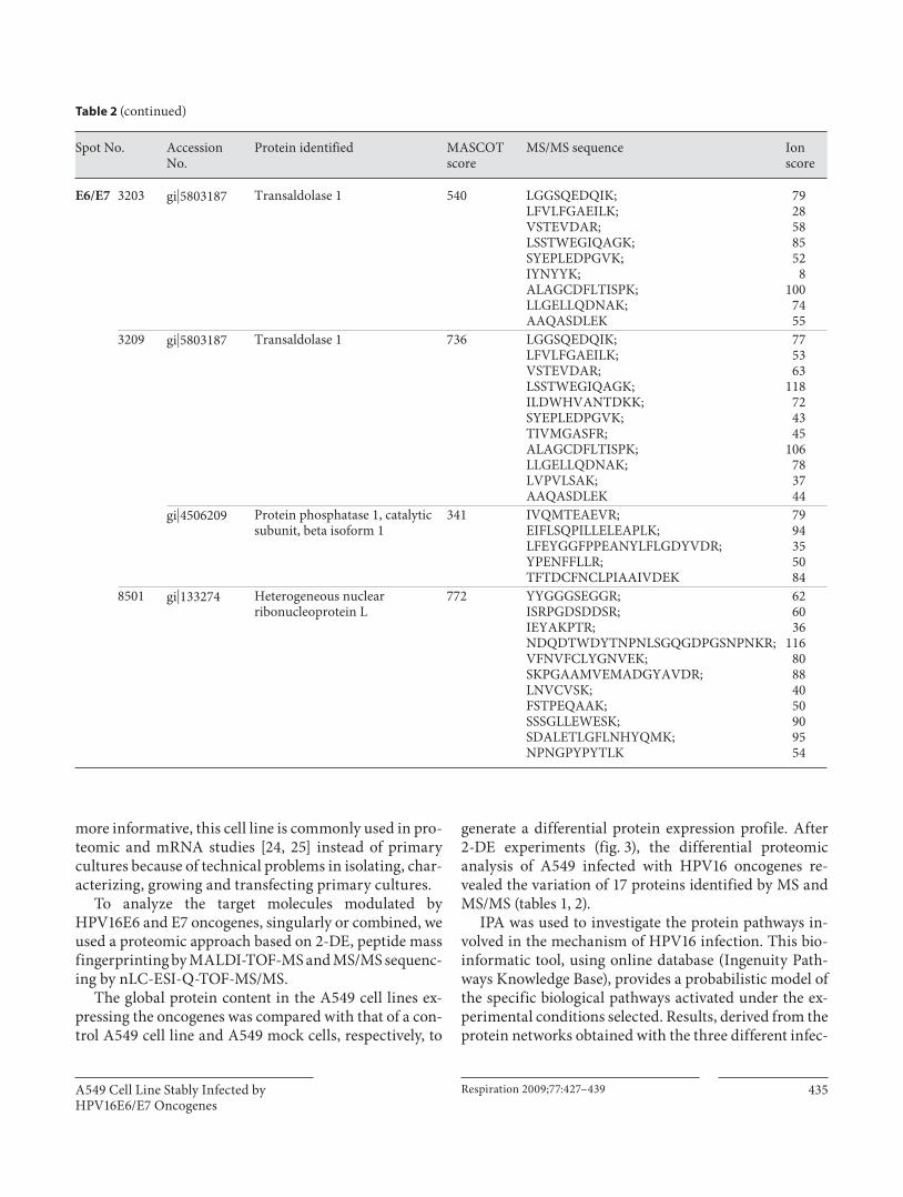
generate a differential protein expression profile. After 2-DE experiments ( fig. 3 ), the differential proteomic analysis of A549 infected with HPV16 oncogenes re-vealed the variation of 17 proteins identified by MS and MS/MS ( tables 1 , 2 ).
IPA was used to investigate the protein pathways in-volved in the mechanism of HPV16 infection. This bio-informatic tool, using online database (Ingenuity Path-ways Knowledge Base), provides a probabilistic model of the specific biological pathways activated under the ex-perimental conditions selected. Results, derived from the protein networks obtained with the three different infec-
Spot No. AccessionNo.
Protein identified MASCOTscore
MS/MS sequence Ionscore
E6/E7 3203 gi�5803187 Transaldolase 1 540 LGGSQEDQIK;LFVLFGAEILK;VSTEVDAR;LSSTWEGIQAGK;SYEPLEDPGVK;IYNYYK;ALAGCDFLTISPK;LLGELLQDNAK;AAQASDLEK
7928588552
8100
7455
3209 gi�5803187 Transaldolase 1 736 LGGSQEDQIK;LFVLFGAEILK;VSTEVDAR;LSSTWEGIQAGK;ILDWHVANTDKK;SYEPLEDPGVK;TIVMGASFR;ALAGCDFLTISPK;LLGELLQDNAK;LVPVLSAK;AAQASDLEK
775363
118724345
106783744
gi�4506209 Protein phosphatase 1, catalyticsubunit, beta isoform 1
341 IVQMTEAEVR;EIFLSQPILLELEAPLK;LFEYGGFPPEANYLFLGDYVDR;YPENFFLLR;TFTDCFNCLPIAAIVDEK
7994355084
8501 gi�133274 Heterogeneous nuclearribonucleoprotein L
772 YYGGGSEGGR;ISRPGDSDDSR;IEYAKPTR;NDQDTWDYTNPNLSGQGDPGSNPNKR;VFNVFCLYGNVEK;SKPGAAMVEMADGYAVDR;LNVCVSK;FSTPEQAAK;SSSGLLEWESK;SDALETLGFLNHYQMK;NPNGPYPYTLK
626036
11680884050909554
Table 2 (continued)
Ciotti et al. Respiration 2009;77:427–439436
tion conditions, suggested the functional involvement of a cell death inhibition pathway with central nodes in-cluding MYC, TNF, vascular endothelial growth factor (VEGF) and annexin IV, Gp96, Hsp27 and tumor pro-tein, translationally controlled 1 as major key proteins for cell viability and inhibition of apoptosis pathway ( fig. 5 ; table 3 ).
VEGF is a major regulator of angiogenesis and it was found overexpressed in non-small cell lung and cervical cancers [26, 27] . The loss of the wild-type p53 upregulates
the VEGF expression [28] , supporting the hypothesis that HPV16E6 oncoprotein might be actively involved [29] . Actually, it has been established that HPV16E6 upregu-lated VEGF via a promoter region that contains four Sp-1 sites, and, importantly, that this occurred in a p53-inde-pendent manner.
Annexins constitute a large family of Ca 2+ and lipid-binding proteins with the common biochemical hall-mark of binding with negatively charged phospholipids in a Ca 2+ -dependent and reversible manner [30] . They
Identified by proteomic experimentsUpregulated in infected cell lines
Identified by pathway analysis
Colors
Transcription regulator
Translation regulator
Shapes
Chemical or drug
Cytokine
Enzyme
Group or complex
Kinase
Phosphatase
Transporter
Other
Direct interactionIndirect interaction
EdgesBinding only
Inhibits
Inhibits AND acts on
Acts on
LYZ
RAD23B PSMD4
PSMC2
CDC2B
EEF1D
PSMD2NEDD8
TP53
Hsp27
EIF2AK3
MYC
Ubiquitin
RAD23AANXA4
NAP1L1
PPP1CB
AXL
MAPK1
HSPB1
�-EstradiolMAPKAPK5
VEGFA
ANXA3HSPB8
HNRPL
TUBB
STEAP3
TPT1
NNMT
HSP90B1
CHI3L1
TALDO1
MYCN
TNF
Fig. 5. IPA top gene network generated uploading all the differentially expressed proteins in the A549 cell line infected by HPV16E6, HPV16E7 and HPV16E6/E7 constructs. The network graphically dis-plays gene products as nodes and the bio-logical relationship between the nodes as edges. Proteins (focus genes) which were identified as upregulated in the infected cells by proteomics experiments are shown in red color and gene products which are not user specified but incorporated into the network through relationships with other proteins are represented as white nodes. The functional class of the gene products is displayed with different geo-metric shapes. Interaction between pro-teins is represented as edges (lines): solid lines for a direct interaction and dashed lines for an indirect interaction. Each edge between gene products is created using sci-entific information contained in the IPA database (Ingenuity Pathways Knowledge Base, IPKB). (For the relationship between the nodes refer to the legend at the bottom of the image.)
Co
lor v
ersi
on
avai
lab
le o
nlin
e
A549 Cell Line Stably Infected by HPV16E6/E7 Oncogenes
Respiration 2009;77:427–439 437
have been implicated in a wide range of biological pro-cesses. Annexin IV has been directly identified in our 2-DE maps and is recognized as an early marker of apopto-sis [31] . Annexin IV also plays a role in drug resistance as reported in the lung cancer cell line H460. In this model, annexin IV is one of the earliest proteins induced in H460 cells in response to cytotoxic stress due to antimitotic drug treatment [32] . These and our results suggest a link between annexin IV and cell proliferation. Annexin III has also been directly profiled in this study. This protein is a potential angiogenic factor; it induces migration and tube formation of vascular endothelial cells by stimulat-ing the hypoxia-inducible factor-1 (HIF-1), which in turn, may induce increased secretion of VEGF [33] , one of the nodal points of our protein function clustering analysis ( fig. 5 ). We speculate that upregulation of annexin III en-courages tumor progression through an increased vascu-larization of the tumoral mass.
The glucose-regulated proteins (GRPs) constitute a family of highly conserved stress response proteins that operate as molecular chaperones with both translocase and foldase activities. GRP94 (= gp96), identified in our analysis, is a Ca 2+ -binding protein localized in the endo-plasmic reticulum. It has previously been identified as tumor-specific antigen and, more recently, as regulator of antigen-presenting cell activation and so represents a po-tentially ideal candidate for cancer immunotherapy [34] . It can be considered an early marker of lung carcinogen-esis [35] .
The heat shock protein Hsp27 is an additional chaper-one molecule found in our study, which is expressed in response to a wide variety of physiological and environ-mental insults including anticancer therapy [36] . The molecular mechanisms involving Hsp27 in resistance to cancer therapies can be explained in the following ways: (1) as molecular chaperone it can confer cytoprotection by repairing more efficiently the damaged proteins re-sulting from cytotoxic drug administration, (2) protect-ing cancer cells from apoptosis [37] , (3) protecting the microvasculature inside tumors, because Hsp27 is found in endothelial cells [38] , and (4) enhancing DNA repair [39, 40] . In rodent models, overexpression of Hsp27 in-creases tumor growth and metastatic potential, while its inhibition reduces tumor size [41] . Therefore, Hsp27 has become a new target for anticancer therapy [42] .
Translationally controlled tumor protein (TPT1, also known as p23, fortilin and human recombinant hista-mine releasing factor) has been described as binding partner of MCL1 (Bcl-2 homologue) [43] , Bcl-xL [44] and identified as an antiapoptotic protein. The most convinc-
Table 3. Ingenuity pathway system gene product designations of the top gene network identified in the Ingenuity Pathways Analy-sis
Top function Score Focus genes
Cell deathp value:6.92 ! 10–6
36 13
Proteins in the network
Gene symbol Protein name
ANXA3 Annexin IIIANXA4 Annexin IVAXL AXL receptor tyrosine kinaseCDC2B Cell division cycle 2 homolog B (S. pombe)CHI3L1 Chitinase 3-like 1EEF1D Eukaryotic translation elongation factor 1 deltaEIF2AK3 Eukaryotic translation initiation factor 2-alpha
kinase 3HNRPL Heterogeneous nuclear ribonucleoprotein LHsp27 Protein family of heat shock protein 27HSP90B1 Tumor rejection antigen (gp96) 1HSPB1 Heat shock 27-kDa protein 1HSPB8 Heat shock 22-kDa protein 8LYZ LysozymeMAPK1 Mitogen-activated protein kinase 5MAPKAPK5 Mitogen-activated protein kinase-activated
protein kinase 5MYC v-myc myelocytomatosis viral oncogene homolog
(avian)MYCN v-myc myelocytomatosis viral-related oncogene,
neuroblastoma derived (avian)NAP1L1 Nucleosome assembly protein 1-like 1NEDD8 Neural precursor cell expressed, developmentally
downregulated 8NNMT Nicotinamide N-methyltransferasePPP1CB Protein phosphatase 1, catalytic subunit,
beta isoformPSMC2 Proteasome 26S, ATPase subunit 2PSMD2 Proteasome 26S, non-ATPase subunit 2PSMD4 Proteasome 26S, non-ATPase regulatory subunit 4RAD23A RAD23 homolog A (S. cerevisiae)RAD23B RAD23 homolog B (S. cerevisiae)STEAP3 STEAP family member 3TALDO1 Transaldolase 1TNF Tumor necrosis factorTP53 Tumor protein p53TPT1 Tumor protein, translationally controlled 1TUBB Beta tubulinVEGFA Vascular endothelial growth factor A
The score based on the number of the focus genes and network size is used for ranking the created network, the given p value represents the lower calculated threshold value for the involved proteins.
Ciotti et al. Respiration 2009;77:427–439438
References
1 Ferlay J, Bray F, Pisani P, Parkin D: GLOBO-CAN 2000: Cancer Incidence, Mortality and Prevalence Worldwide. IARC Cancer Base. Lyon, IARC Press, 2001.
2 Alberg AJ, Samet JM: Epidemiology of lung cancer. Chest 2003; 123: 21S–49S.
3 Stancu M, King T, Maizel A: Molecular biol-ogy of lung cancer: a primer; in WeitbergAB, Klastersky J (eds): Cancer of the Lung. Totowa, Humana Press, 2002.
4 Syrjanen K, Syrjanen S: Papillomavirus In-fections in Human Pathology. New York, Wiley, 2000.
5 zur Hausen H: Cervical carcinoma and hu-man papillomavirus: on the road to prevent-ing a major human cancer. J Natl Cancer Inst 2001; 93: 252–253.
6 zur Hausen H: Papillomaviruses and cancer: from basic studies to clinical application. Nat Rev Cancer 2002; 2: 342–350.
7 Ciotti M, Giuliani L, Ambrogi V, Ronci C, Benedetto A, Mineo TC, Syrjanen K, Favalli C: Detection and expression of human papil-lomavirus oncogenes in non-small cell lung cancer. Oncol Rep 2006; 16: 183–189.
8 Giuliani L, Jaxmar T, Casadio C, Gariglio M, Manna A, D’Antonio D, Syrjanen K, Favalli C, Ciotti M: Detection of oncogenic viruses (SV40, BKV, JCV, HCMV, HPV) and p53 co-don 72 polymorphism in lung carcinoma. Lung Cancer 2007; 57: 273–281.
9 Scheffner M, Werness BA, Huibregtse JM, Levine AJ, Howley PM: The E6 oncoprotein encoded by human papillomavirus types 16 and 18 promotes the degradation of p53. Cell 1990; 63: 1129–1136.
10 Werness BA, Levine AJ, Howley PM: Asso-ciation of human papillomavirus types 16 and 18 E6 proteins with p53. Science 1990; 248: 76–79.
11 Dyson N, Howley PM, Munger K, Harlow E: The human papilloma virus-16 E7 oncopro-tein is able to bind to the retinoblastoma gene product. Science 1989; 243: 934–937.
12 Scheffner M, Romanczuk H, Munger K, Huibregtse JM, Mietz JA, Howley PM: Func-tions of human papillomavirus proteins. Curr Top Microbiol Immunol 1994; 186: 83–99.
13 Hawley-Nelson P, Vousden KH, Hubbert NL, Lowy DR, Schiller JT: HPV16 E6 and E7 proteins cooperate to immortalize human foreskin keratinocytes. EMBO J 1989; 8: 3905–3910.
14 Barbosa MS, Schlegel R: The E6 and E7 genes of HPV-18 are sufficient for inducing two-stage in vitro transformation of human keratinocytes. Oncogene 1989; 4: 1529–1532.
15 Caldeira S, Zehbe I, Accardi R, Malanchi I, Dong W, Giarre M, de Villiers EM, Filotico R, Boukamp P, Tommasino M: The E6 and E7 proteins of the cutaneous human papil-lomavirus type 38 display transforming properties. J Virol 2003; 77: 2195–2206.
16 Pear WS, Nolan GP, Scott ML, Baltimore D: Production of high-titer helper-free retrovi-ruses by transient transfection. Proc Natl Acad Sci USA 1993; 90: 8392–8396.
17 Ciotti M, Sesti F, Paba P, Benedetto A, Patrizi L, Criscuolo A, Piccione E, Branca M, Syrjän-en K, Favalli C: Human papillomavirus (HPV) testing in the management of women with abnormal Pap smears. Experience of a colposcopy referral clinic. Eur J Gynaecol Oncol 2004; 25: 577–584.
18 Cheung CK, Mak YT, Swaminathan R: Au-tomated trichloroacetic acid precipitation method for urine total protein. Ann Clin Biochem 1987; 24: 140–144.
19 Shevchenko A, Wilm M, Vorm O, Mann M: Mass spectrometric sequencing of proteins silver-stained polyacrylamide gels. Anal Chem 1996; 68: 850–858.
20 Gharahdaghi F, Kirchner M, Fernandez J, Mische SM: Peptide-mass profiles of poly-vinylidene difluoride-bound proteins by matrix-assisted laser desorption/ionization time-of-flight mass spectrometry in the presence of nonionic detergents. Anal Bio-chem 1996; 233: 94–99.
21 Hall RA, Dix BR, O’Carroll SJ, Braithwaite AW: p53-dependent cell death/apoptosis is required for a productive adenovirus infec-tion. Nat Med 1998; 4: 1068–1072.
22 Parkin DM, Bray F, Ferlay J, Pisani P: Global cancer statistics, 2002. CA Cancer J Clin 2005; 55: 74–108.
ing point in favor of a link between TPT1 and cancer was provided by the demonstration that during reversion of cells from the malignant phenotype, TPT1 levels are con-siderably reduced. Such an inhibition of TPT1 expression results in suppression of the malignant phenotype with TPT1 levels downregulated through activation of the tu-mor suppressor protein p53 [45, 46] . Notably, TPT1 spe-cifically antagonizes the eEF-1 � -mediated GDP/GTP ex-change reaction in the elongation reaction of protein synthesis in higher cells [47, 48] . In our study the HPV16E6/E7-infected A549 cell line shows an upregula-tion of both TPT1 and EEF1D. Elongation factors of translation have been implicated in cell transformation [49] and overexpression of EEF1D resulted in anchorage-independent growth and in the formation of tumors in nude mice [50] . A lot of evidence indicates that participa-tion of EF-1 subunits in the control of virus expression can be part of a complex strategy for infected-cell protein synthesis optimization. In fact, EEF1D has been shown to interact with HIV Tat protein and this interaction re-
duces the translational efficiency of cellular but not viral mRNAs [51] .
Finally, our results on the upregulation of the proteins annexin III, gp96, transaldolase 1, elongation factor � 1, proteasome 26 subunit have been confirmed at the tran-scriptional level [52, 53] .
In conclusion, although careful molecular epidemio-logical studies are needed to establish a causal link be-tween HPV infection and lung carcinogenesis, proteomic analysis could be a valuable tool for identifying the mo-lecular pathways involved in the transformation process induced by HPV and for understanding its possible role as cocarcinogen in lung carcinogenesis.
Acknowledgement
The project was in part supported by the Progetto speciale Ministero della Salute-Regione Abruzzo 2003 (Meccanismi di cancerogenesi da HPV; Problemi correlati al processo di inte-grazione virale e all’infezione con multipli genotipi).

A549 Cell Line Stably Infected by HPV16E6/E7 Oncogenes
Respiration 2009;77:427–439 439
23 Giuliani L, Favalli C, Syrjanen K, Ciotti M: Human papillomavirus infections in lung cancer. Detection of E6 and E7 transcripts and review of the literature. Anticancer Res 2007; 27: 2697–2704.
24 Yim EK, Meoyng J, Namkoong SE, Um SJ, Park JS: Genomic and proteomic expression patterns in HPV-16 E6 gene transfected sta-ble human carcinoma cell lines. DNA Cell Biol 2004; 23: 826–835.
25 Choi YP, Kang S, Hong S, Xie X, Cho NH: Proteomic analysis of progressive factors in uterine cervical cancer. Proteomics 2005; 5: 1481–1493.
26 Zygalaki E, Tsaroucha EG, Kaklamanis L, Lianidou ES: Quantitative real-time reverse transcription PCR study of the expression of vascular endothelial growth factor (VEGF) splice variants and VEGF receptors (VEGFR-1 and VEGFR-2) in non small cell lung can-cer. Clin Chem 2007; 53: 1433–1439.
27 Branca M, Giorgi C, Santini D, Di Bonito L, Ciotti M, Benedetto A, Paba P, Costa S, Bon-ifacio D, Di Bonito P, Accardi L, Favalli C, Syrjanen K: Aberrant expression of VEGF-C is related to grade of cervical intraepithelial neoplasia (CIN) and high risk HPV, but does not predict virus clearance after treatment of CIN or prognosis of cervical cancer. J Clin Pathol 2006; 59: 40–47.
28 Kieser A, Weich HA, Brandner G, Marme D, Kolch W: Mutant p53 potentiates protein ki-nase C induction of vascular endothelial growth factor expression. Oncogene 1994; 9: 963–969.
29 Lopez-Ocejo O, Viloria-Petit A, Bequet-Romero M, Mukhopadhyay D, Rak J, Kerbel RS: Oncogenes and tumor angiogenesis: the HPV-16 E6 oncoprotein activates the vascu-lar endothelial growth factor (VEGF) gene promoter in a p53 independent manner. On-cogene 2000; 19: 4611–4620.
30 Gerke V, Creutz CE, Moss SE: Annexins: linking Ca 2+ signalling to membrane dy-namics. Nat Rev Mol Cell Biol 2005; 6: 449–461.
31 Sohma H, Ohkawa H, Hashimoto E, Sakai R, Saito T: Ethanol-induced augmentation of annexin IV expression in rat C6 glioma and human A549 adenocarcinoma cells. Alcohol Clin Exp Res 2002; 26: 44S–48S.
32 Han EK, Tahir SK, Cherian SP, Collins N, Ng SC: Modulation of paclitaxel resistance by annexin IV in human cancer cell lines. Br J Cancer 2000; 83: 83–88.
33 Park JE, Lee DH, Lee JA, Park SG, Kim NS, Park BC, Cho S: Annexin A3 is a potential angiogenic mediator. Biochem Biophys Res Commun 2005; 337: 1283–1287.
34 Nicchitta CV, Carrick DM, Baker-Lepain JC: The messenger and the message: gp96 (GRP94)-peptide interactions in cellular im-munity. Cell Stress Chaperones 2004; 9: 325–331.
35 Wang Q, An L, Chen Y, Yue S: Expression of endoplasmic reticulum molecular chaperon GRP94 in human lung cancer tissues and its clinical significance. Chin Med J 2002; 115: 1615–1619.
36 Urbani A, Poland J, Bernardini S, Bellincam-pi L, Biroccio A, Schnolzer M, Sinha P, Fed-erici G: A proteomic investigation into eto-poside chemo-resistance of neuroblastoma cell lines. Proteomics 2005; 5: 796–804.
37 Arrigo AP, Paul C, Ducasse C, Manero F, Kretz-Remy C, Virot S, Javouhey E, Mounier N, Diaz-Latoud C: Small stress proteins: novel negative modulators of apoptosis in-duced independently of reactive oxygen spe-cies. Prog Mol Subcell Biol 2002; 28: 185–204.
38 Ciocca DR, Rozados VR, Cuello Carrion FD, Gervasoni SI, Matar P, Scharovsky OG: Hsp25 and Hsp70 in rodent tumors treated with doxorubicin and lovastatin. Cell Stress Chaperones 2003; 8: 26–36.
39 Mendez F, Sandigursky M, Kureekattil RP, Kenny MK, Franklin WA, Bases R: Specific stimulation of human apurinic/apyrimidin-ic endonuclease by heat shock protein 70. DNA Repair 2003; 2: 259–271.
40 Nadin SB, Vargas-Roig LM, Cuello-Carrion FD, Ciocca DR: Deoxyribonucleic acid dam-age induced by doxorubicin in peripheral blood mononuclear cells: possible roles for the stress response and the deoxyribonucleic acid repair process. Cell Stress Chaperones 2003; 8: 361–372.
41 Garrido C, Brunet M, Didelot C, Zermati Y, Schmitt E, Kroemer G: Heat shock proteins 27 and 70: anti-apoptotic proteins with tu-morigenic properties. Cell Cycle 2006; 5: 2592–2601.
42 Ciocca DR, Calderwood SK: Heat shock pro-teins in cancer: diagnostic, prognostic, pre-dictive, and treatment implications. Cell Stress Chaperones 2005; 10: 86–103.
43 Liu H, Peng HW, Cheng YS, Yuan HS, Yang-Yen HF: Stabilization and enhancement of the antiapoptotic activity of mcl-1 by TCTP. Mol Cell Biol 2005; 25: 3117–3126.
44 Yang Y, Yang F, Xiong Z, Yan Y, Wang X, Nishino M, Mirkovic D, Nguyen J, Wang H, Yang XF: An N-terminal region of transla-tionally controlled tumor protein is required for its antiapoptotic activity. Oncogene 2005; 24: 4778–4788.
45 Tuynder M, Fiucci G, Prieur S, Lespagnol A, Geant A, Beaucourt S, Duflaut D, Besse S, Susini L, Cavarelli J, Moras D, Amson R, Tel-erman A: Translationally controlled tumor protein is a target of tumor reversion. Proc Natl Acad Sci USA 2004; 101: 15364–15369.
46 Tuynder M, Fiucci G, Prieur S, Lespagnol A, Geant A, Beaucourt S, Duflaut D, Besse S, Susini L, Cavarelli J, Moras D, Amson R, Tel-erman A: Translationally controlled tumor protein is a target of tumor reversion. Proc Natl Acad Sci USA 2004; 101: 15364–15369.
47 Cans C, Passer BJ, Shalak V, Nancy-Porte-bois V, Crible V, Amzallag N, Allanic D, Tu-fino R, Argentini M, Moras D, Fiucci G, Goud B, Mirande M, Amson R, Telerman A: Translationally controlled tumor protein acts as a guanine nucleotide dissociation in-hibitor on the translation elongation factor eEF1A. Proc Natl Acad Sci USA 2003; 100: 13892–13897.
48 Langdon JM, Vonakis BM, MacDonald SM: Identification of the interaction between the human recombinant histamine releasing factor/translationally controlled tumor pro-tein and elongation factor-1 delta (also known as eElongation factor-1B beta). Bio-chim Biophys Acta 2004; 1688: 232–236.
49 Sonenberg N: Translation factors as effectors of cell growth and tumorigenesis. Curr Opin Cell Biol 1993; 5: 955–960.
50 Joseph P, Lei YX, Whong WZ, Ong TM: On-cogenic potential of mouse translation elon-gation factor-1 delta, a novel cadmium-re-sponsive proto-oncogene. J Biol Chem 2002; 277: 6131–6136.
51 Xiao H, Neuveut C, Benkirane M, Jeang KT: Interaction of the second coding exon of Tat with human EF-1 delta delineates a mecha-nism for HIV-1-mediated shut-off of host mRNA translation. Biochem Biophys Res Commun 1998; 244: 384–389.
52 Kuner R, Vogt M, Sultmann H, Buness A, Dymalla S, Bulkescher J, Fellmann M, Butz K, Poustka A, Hoppe-Seyler F: Identification of cellular targets for the human papilloma-virus E6 and E7 oncogenes by RNA interfer-ence and transcription analyses. J Mol Med 2007; 85: 1253–1262.
53 Rosty C, Sheffer M, Tsafrir D, Stransky N, Tsafrir I, Peter M, de Cremoux P, de La Ro-chefordière A, Salmon R, Dorval T, Thiery JP, Couturier J, Radvanyi F, Domany E, Sas-tre-Garou X: Identification of a proliferation gene cluster associated with HPV e6/E7 ex-pression level and viral DNA load in invasive cervical carcinoma. Oncogene 2005; 24: 7094–7104.


