Embed Size (px)
Citation preview
c o r t e x 4 6 ( 2 0 1 0 ) 1 1 3 2 – 1 1 3 7
ava i lab le a t www.sc iencedi rec t .com
journa l homepage : www.e lsev ie r . com/ loca te /cor tex
Research report
Pre-saccadic perceptual facilitation can occur without covertorienting of attention
Annabelle Blangeroa,b, Aarlenne Z. Khana,b,c, Romeo Salemmea,b, Heiner Deubeld,Werner X. Schneidere, Gilles Rodea,b, Alain Vighettoa,b, Yves Rossettia,b and Laure Pisellaa,b,*aEspace et Action – Inserm 864; Universite Claude Bernard, Lyon, FrancebInstitut Federatif des Neurosciences de Lyon (IFNL), FrancecSmith-Kettlewell Eye Research Institute, San Francisco, California, USAdDepartment Allgemeine und Experimentelle Psychologie, Ludwig-Maximilians-Universitat Munchen, GermanyeDepartment Allgemeine und Neuro-Cognitive Psychologie, Bielefeld Universitat, Germany
a r t i c l e i n f o
Article history:
Received 10 June 2008
Reviewed 30 September 2008
Revised 5 March 2009
Accepted 24 June 2009
Action editor Jane Riddoch
Published online 15 July 2009
Keywords:
Attention
Saccades
Parietal cortex
Pre-motor theory
* Corresponding author. Universite Claude BE-mail address: [email protected] (L
0010-9452/$ – see front matter ª 2009 Elsevidoi:10.1016/j.cortex.2009.06.014
a b s t r a c t
The pre-motor theory of attention suggests that the mechanisms involved in target
selection for eye movements are the same as those for spatial attention shifts. The pre-
saccadic facilitation of perceptual discrimination at the location of a saccadic goal (para-
digm of Deubel and Schneider, 1996) has been considered as an argument for this theory.
We compared letter discrimination performance in a saccade (overt attention – pre-
saccadic facilitation) and a fixation (covert attention) task in a patient with right posterior
parietal damage and 4 controls.
In the overt attention condition, the patient was instructed by a central cue to make
a saccade to a target located at a peripheral location. During the saccade latency (in
a period of time of 250 msec following the presentation of the cue), a letter was presented
at the target location. Accuracy of leftward saccades was impaired compared to rightward
saccades. To evaluate letter discrimination performance in this saccade task (i.e., the
presence of pre-saccadic facilitation), we selected only those leftward saccades that were
equivalent in accuracy (and latency) to the rightward ones. Within these selected trials, the
patient was able to discriminate letters equally well in both visual fields. In contrast, he
performed at chance level during the fixation task (covert attention condition) for letters
presented at the same peripheral location with the same timing with respect to the cue
presentation. The patient could thus discriminate the letter presented at 8� of visual
eccentricity while he was preparing a saccade, whereas he was unable to perceive the letter
in the fixation task.
Remarkably, in the left visual field, letter discrimination was impossible even when
a letter was presented as close as 2.5� of visual eccentricity in the fixation task. Altogether,
these results suggest that pre-saccadic perceptual facilitation does not rely on the same
processes as those of covert attention, as tested by fixation task. Instead, we propose that
pre-saccadic perceptual facilitation results from a form of attention specific to action,
which could correspond to a pre-saccadic remapping process.
ª 2009 Elsevier Srl. All rights reserved.
ernard, Lyon 1, Espace et Action – Inserm U864, Bron, Rhone-Alpes, France.. Pisella).er Srl. All rights reserved.
c o r t e x 4 6 ( 2 0 1 0 ) 1 1 3 2 – 1 1 3 7 1133
1. Introduction
between left and right visual fields in a covert version andVision provides the brain with one of the most fundamental
forms of information for perception and motor behaviour.
Despite the subjective sensation of an instantaneous, global
and detailed perception of the visual scene, the structural
organisation of the retina allows good visual acuity only in
a restricted area, the fovea. Eye movements (known as
saccades or overt attention) can bring a new part of the visual
scene to the fovea, allowing it to be analysed more precisely by
the retina and the visual cortex. Shifts of attention without
eye displacements (known as covert attention) have also been
shown to improve spatial resolution and contrast sensitivity
in peripheral vision (Yeshurun and Carrasco, 1998; Carrasco
et al., 2000).
Brain imaging (e.g., Corbetta et al., 1998, Corbetta, 1998;
Perry and Zeki, 2000; Nobre et al., 2000) show largely, but not
completely, overlapping networks for covert shifts of visual
attention towards peripheral objects and saccadic eye move-
ments. Specifically, saccade target selection has been
considered to rely on the same processing mechanisms as
covert attention (Shepherd et al., 1986; Rizzolatti et al., 1987;
Sheliga et al., 1994, 1995; Hoffman and Subramaniam, 1995;
Kowler et al., 1995; Deubel and Schneider, 1996; Awh et al.,
2006). For example, Deubel and Schneider (1996) designed an
elegant dual-task paradigm, which combined a target-directed
saccade task with a letter discrimination task. Their results
showed that visual discrimination in periphery was best when
the discrimination stimulus and saccade target were at the
same location in space. This phenomenon called pre-saccadic
perceptual facilitation occurs before the eye movement takes
place, while the eyes are still at the fixation location.
The usual interpretation of pre-saccadic perceptual facili-
tation is that saccade preparation automatically induces
a covert attentional shift to the location of the saccadic goal,
which has been shown to improve spatial resolution in the
visual periphery (Yeshurun and Carrasco, 1998). In other
words, a single mechanism of spatial attention would select
objects both for perceptual processing and for motor action;
a saccade cannot occur without without a preceding atten-
tional shift (Deubel and Schneider, 1996; Schneider and
Deubel, 2002). An alternative interpretation could be that pre-
saccadic perceptual facilitation is a separate mechanism from
covert attention, even though, in a normally functioning
brain, it is impossible to dissociate the two using psycho-
physical tasks, since both show similar spatial enhancements.
These two abovementioned interpretations are actually
very different. The first one involves a common substrate for
‘covert’ (fixation) and ‘overt’ (saccade) shifts of attention and
thus predicts that if performance of the former is impaired, for
example by a brain lesion, it will also be impaired for the
latter. In contrast, the second interpretation suggests that pre-
saccadic perceptual facilitation and covert attention process-
ing mechanisms rely on distinct neural substrates and thus
implies a possible dissociation between fixation and saccade
tasks. Specifically, a brain-damaged patient may be able to do
one task but not the other. Here we investigate these two
alternatives by comparing the performance of a patient with
a unilateral lesion in the posterior parietal cortex (PPC)
a classical (but simplified) version of the pre-saccadic facili-
tation paradigm introduced by Deubel and Schneider (1996).
2. Material and methods
2.1. Subjects
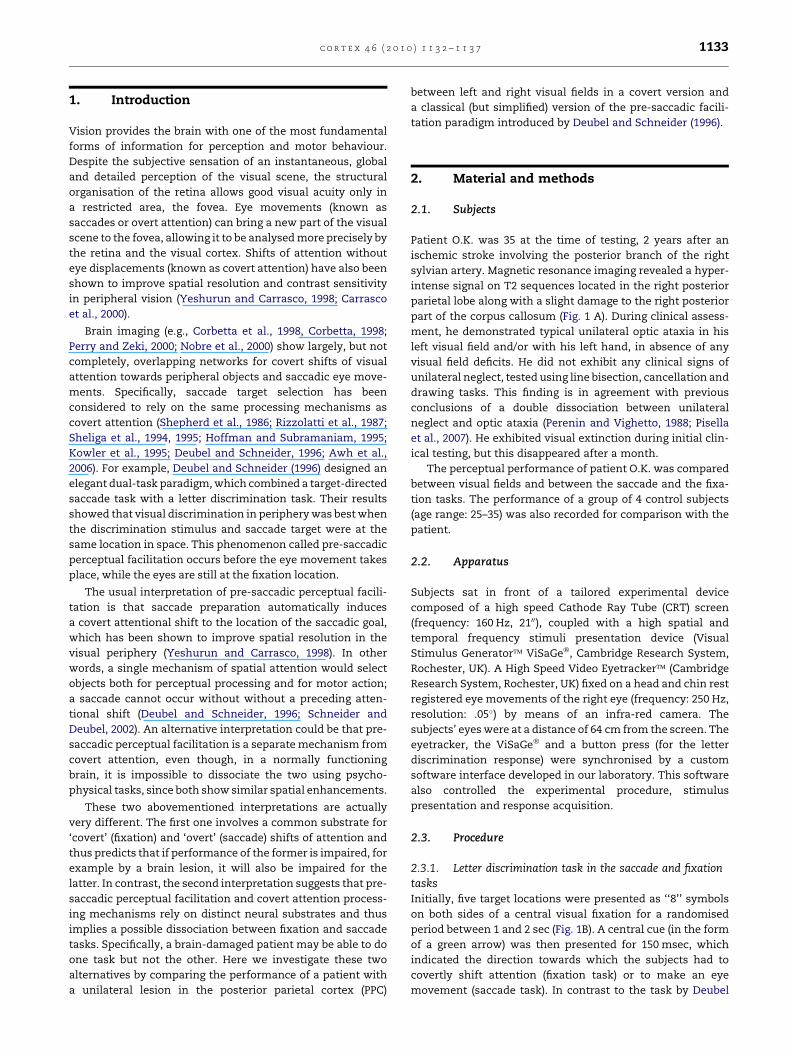
Patient O.K. was 35 at the time of testing, 2 years after an
ischemic stroke involving the posterior branch of the right
sylvian artery. Magnetic resonance imaging revealed a hyper-
intense signal on T2 sequences located in the right posterior
parietal lobe along with a slight damage to the right posterior
part of the corpus callosum (Fig. 1 A). During clinical assess-
ment, he demonstrated typical unilateral optic ataxia in his
left visual field and/or with his left hand, in absence of any
visual field deficits. He did not exhibit any clinical signs of
unilateral neglect, tested using line bisection, cancellation and
drawing tasks. This finding is in agreement with previous
conclusions of a double dissociation between unilateral
neglect and optic ataxia (Perenin and Vighetto, 1988; Pisella
et al., 2007). He exhibited visual extinction during initial clin-
ical testing, but this disappeared after a month.
The perceptual performance of patient O.K. was compared
between visual fields and between the saccade and the fixa-
tion tasks. The performance of a group of 4 control subjects
(age range: 25–35) was also recorded for comparison with the
patient.
2.2. Apparatus
Subjects sat in front of a tailored experimental device
composed of a high speed Cathode Ray Tube (CRT) screen
(frequency: 160 Hz, 2100), coupled with a high spatial and
temporal frequency stimuli presentation device (Visual
Stimulus Generator� ViSaGe�, Cambridge Research System,
Rochester, UK). A High Speed Video Eyetracker� (Cambridge
Research System, Rochester, UK) fixed on a head and chin rest
registered eye movements of the right eye (frequency: 250 Hz,
resolution: .05�) by means of an infra-red camera. The
subjects’ eyes were at a distance of 64 cm from the screen. The
eyetracker, the ViSaGe� and a button press (for the letter
discrimination response) were synchronised by a custom
software interface developed in our laboratory. This software
also controlled the experimental procedure, stimulus
presentation and response acquisition.
2.3. Procedure
2.3.1. Letter discrimination task in the saccade and fixationtasksInitially, five target locations were presented as ‘‘8’’ symbols
on both sides of a central visual fixation for a randomised
period between 1 and 2 sec (Fig. 1B). A central cue (in the form
of a green arrow) was then presented for 150 msec, which
indicated the direction towards which the subjects had to
covertly shift attention (fixation task) or to make an eye
movement (saccade task). In contrast to the task by Deubel
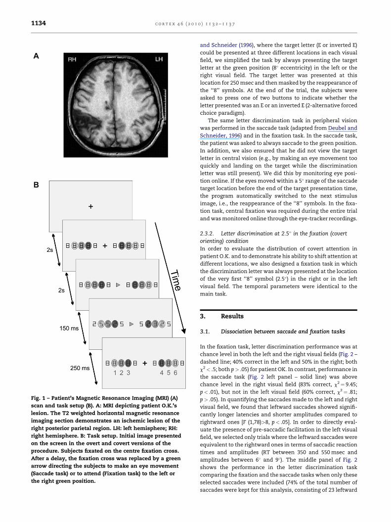
Fig. 1 – Patient’s Magnetic Resonance Imaging (MRI) (A)
scan and task setup (B). A: MRI depicting patient O.K.’s
lesion. The T2 weighted horizontal magnetic resonance
imaging section demonstrates an ischemic lesion of the
right posterior parietal region. LH: left hemisphere; RH:
right hemisphere. B: Task setup. Initial image presented
on the screen in the overt and covert versions of the
procedure. Subjects fixated on the centre fixation cross.
After a delay, the fixation cross was replaced by a green
arrow directing the subjects to make an eye movement
(Saccade task) or to attend (Fixation task) to the left or
the right green position.
c o r t e x 4 6 ( 2 0 1 0 ) 1 1 3 2 – 1 1 3 71134
and Schneider (1996), where the target letter (E or inverted E)
could be presented at three different locations in each visual
field, we simplified the task by always presenting the target
letter at the green position (8� eccentricity) in the left or the
right visual field. The target letter was presented at this
location for 250 msec and then masked by the reappearance of
the ‘‘8’’ symbols. At the end of the trial, the subjects were
asked to press one of two buttons to indicate whether the
letter presented was an E or an inverted E (2-alternative forced
choice paradigm).
The same letter discrimination task in peripheral vision
was performed in the saccade task (adapted from Deubel and
Schneider, 1996) and in the fixation task. In the saccade task,
the patient was asked to always saccade to the green position.
In addition, we also ensured that he did not view the target
letter in central vision (e.g., by making an eye movement too
quickly and landing on the target while the discrimination
letter was still present). We did this by monitoring eye posi-
tion online. If the eyes moved within a 5� range of the saccade
target location before the end of the target presentation time,
the program automatically switched to the next stimulus
image, i.e., the reappearance of the ‘‘8’’ symbols. In the fixa-
tion task, central fixation was required during the entire trial
and was monitored online through the eye-tracker recordings.
2.3.2. Letter discrimination at 2.5� in the fixation (covertorienting) conditionIn order to evaluate the distribution of covert attention in
patient O.K. and to demonstrate his ability to shift attention at
different locations, we also designed a fixation task in which
the discrimination letter was always presented at the location
of the very first ‘‘8’’ symbol (2.5�) in the right or in the left
visual field. The temporal parameters were identical to the
main task.
3. Results
3.1. Dissociation between saccade and fixation tasks
In the fixation task, letter discrimination performance was at
chance level in both the left and the right visual fields (Fig. 2 –
dashed line; 40% correct in the left and 50% in the right; both
c2< .5; both p> .05) for patient OK. In contrast, performance in
the saccade task (Fig. 2 left panel – solid line) was above
chance level in the right visual field (83% correct, c2¼ 9.45;
p< .01), but not in the left visual field (60% correct, c2¼ .81;
p> .05). In quantifying the saccades made to the left and right
visual field, we found that leftward saccades showed signifi-
cantly longer latencies and shorter amplitudes compared to
rightward ones [F (1,78)>8, p< .05]. In order to directly eval-
uate the presence of pre-saccadic facilitation in the left visual
field, we selected only trials where the leftward saccades were
equivalent to the rightward ones in terms of saccadic reaction
times and amplitudes (RT between 350 and 550 msec and
amplitudes between 6� and 9�). The middle panel of Fig. 2
shows the performance in the letter discrimination task
comparing the fixation and the saccade tasks when only these
selected saccades were included (74% of the total number of
saccades were kept for this analysis, consisting of 23 leftward
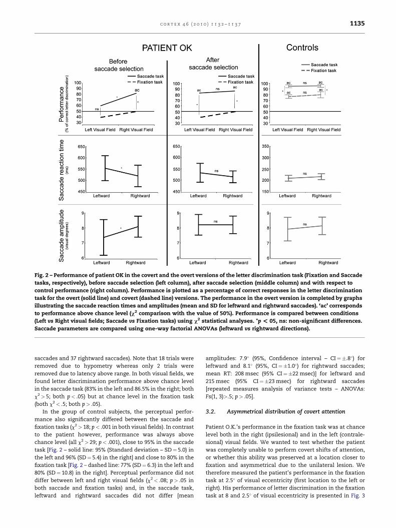
Fig. 2 – Performance of patient OK in the covert and the overt versions of the letter discrimination task (Fixation and Saccade
tasks, respectively), before saccade selection (left column), after saccade selection (middle column) and with respect to
control performance (right column). Performance is plotted as a percentage of correct responses in the letter discrimination
task for the overt (solid line) and covert (dashed line) versions. The performance in the overt version is completed by graphs
illustrating the saccade reaction times and amplitudes (mean and SD for leftward and rightward saccades). ‘ac’ corresponds
to performance above chance level (c2 comparison with the value of 50%). Performance is compared between conditions
(Left vs Right visual fields; Saccade vs Fixation tasks) using c2 statistical analyses. *p < 05, ns: non-significant differences.
Saccade parameters are compared using one-way factorial ANOVAs (leftward vs rightward directions).
c o r t e x 4 6 ( 2 0 1 0 ) 1 1 3 2 – 1 1 3 7 1135
saccades and 37 rightward saccades). Note that 18 trials were
removed due to hypometry whereas only 2 trials were
removed due to latency above range. In both visual fields, we
found letter discrimination performance above chance level
in the saccade task (83% in the left and 86.5% in the right; both
c2> 5; both p< .05) but at chance level in the fixation task
(both c2< .5; both p> .05).
In the group of control subjects, the perceptual perfor-
mance also significantly differed between the saccade and
fixation tasks (c2> 18; p< .001 in both visual fields). In contrast
to the patient however, performance was always above
chance level (all c2> 29; p< .001), close to 95% in the saccade
task [Fig. 2 – solid line: 95% (Standard deviation – SD¼ 5.0) in
the left and 96% (SD¼ 5.4) in the right] and close to 80% in the
fixation task [Fig. 2 – dashed line: 77% (SD¼ 6.3) in the left and
80% (SD¼ 10.8) in the right]. Perceptual performance did not
differ between left and right visual fields (c2< .08; p> .05 in
both saccade and fixation tasks) and, in the saccade task,
leftward and rightward saccades did not differ [mean
amplitudes: 7.9� (95%, Confidence interval – CI¼�.8�) for
leftward and 8.1� (95%, CI¼�1.0�) for rightward saccades;
mean RT: 208 msec (95% CI¼�22 msec)] for leftward and
215 msec (95% CI¼�23 msec) for rightward saccades
[repeated measures analysis of variance tests – ANOVAs:
Fs(1, 3)>.5; p> .05].
3.2. Asymmetrical distribution of covert attention
Patient O.K.’s performance in the fixation task was at chance
level both in the right (ipsilesional) and in the left (contrale-
sional) visual fields. We wanted to test whether the patient
was completely unable to perform covert shifts of attention,
or whether this ability was preserved at a location closer to
fixation and asymmetrical due to the unilateral lesion. We
therefore measured the patient’s performance in the fixation
task at 2.5� of visual eccentricity (first location to the left or
right). His performance of letter discrimination in the fixation
task at 8 and 2.5� of visual eccentricity is presented in Fig. 3
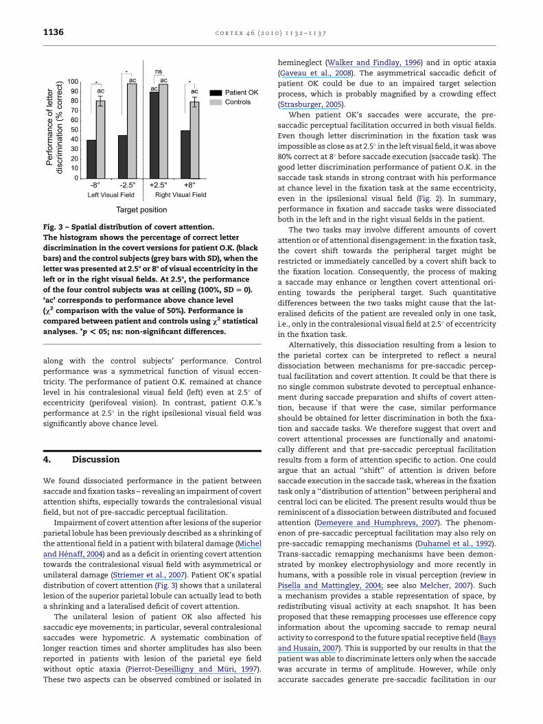
Fig. 3 – Spatial distribution of covert attention.
The histogram shows the percentage of correct letter
discrimination in the covert versions for patient O.K. (black
bars) and the control subjects (grey bars with SD), when the
letter was presented at 2.58 or 88 of visual eccentricity in the
left or in the right visual fields. At 2.58, the performance
of the four control subjects was at ceiling (100%, SD [ 0).
‘ac’ corresponds to performance above chance level
(c2 comparison with the value of 50%). Performance is
compared between patient and controls using c2 statistical
analyses. *p < 05; ns: non-significant differences.
c o r t e x 4 6 ( 2 0 1 0 ) 1 1 3 2 – 1 1 3 71136
along with the control subjects’ performance. Control
performance was a symmetrical function of visual eccen-
tricity. The performance of patient O.K. remained at chance
level in his contralesional visual field (left) even at 2.5� of
eccentricity (perifoveal vision). In contrast, patient O.K.’s
performance at 2.5� in the right ipsilesional visual field was
significantly above chance level.
4. Discussion
We found dissociated performance in the patient between
saccade and fixation tasks – revealing an impairment of covert
attention shifts, especially towards the contralesional visual
field, but not of pre-saccadic perceptual facilitation.
Impairment of covert attention after lesions of the superior
parietal lobule has been previously described as a shrinking of
the attentional field in a patient with bilateral damage (Michel
and Henaff, 2004) and as a deficit in orienting covert attention
towards the contralesional visual field with asymmetrical or
unilateral damage (Striemer et al., 2007). Patient OK’s spatial
distribution of covert attention (Fig. 3) shows that a unilateral
lesion of the superior parietal lobule can actually lead to both
a shrinking and a lateralised deficit of covert attention.
The unilateral lesion of patient OK also affected his
saccadic eye movements; in particular, several contralesional
saccades were hypometric. A systematic combination of
longer reaction times and shorter amplitudes has also been
reported in patients with lesion of the parietal eye field
without optic ataxia (Pierrot-Deseilligny and Muri, 1997).
These two aspects can be observed combined or isolated in
hemineglect (Walker and Findlay, 1996) and in optic ataxia
(Gaveau et al., 2008). The asymmetrical saccadic deficit of
patient OK could be due to an impaired target selection
process, which is probably magnified by a crowding effect
(Strasburger, 2005).
When patient OK’s saccades were accurate, the pre-
saccadic perceptual facilitation occurred in both visual fields.
Even though letter discrimination in the fixation task was
impossible as close as at 2.5� in the left visual field, it was above
80% correct at 8� before saccade execution (saccade task). The
good letter discrimination performance of patient O.K. in the
saccade task stands in strong contrast with his performance
at chance level in the fixation task at the same eccentricity,
even in the ipsilesional visual field (Fig. 2). In summary,
performance in fixation and saccade tasks were dissociated
both in the left and in the right visual fields in the patient.
The two tasks may involve different amounts of covert
attention or of attentional disengagement: in the fixation task,
the covert shift towards the peripheral target might be
restricted or immediately cancelled by a covert shift back to
the fixation location. Consequently, the process of making
a saccade may enhance or lengthen covert attentional ori-
enting towards the peripheral target. Such quantitative
differences between the two tasks might cause that the lat-
eralised deficits of the patient are revealed only in one task,
i.e., only in the contralesional visual field at 2.5� of eccentricity
in the fixation task.
Alternatively, this dissociation resulting from a lesion to
the parietal cortex can be interpreted to reflect a neural
dissociation between mechanisms for pre-saccadic percep-
tual facilitation and covert attention. It could be that there is
no single common substrate devoted to perceptual enhance-
ment during saccade preparation and shifts of covert atten-
tion, because if that were the case, similar performance
should be obtained for letter discrimination in both the fixa-
tion and saccade tasks. We therefore suggest that overt and
covert attentional processes are functionally and anatomi-
cally different and that pre-saccadic perceptual facilitation
results from a form of attention specific to action. One could
argue that an actual ‘‘shift’’ of attention is driven before
saccade execution in the saccade task, whereas in the fixation
task only a ‘‘distribution of attention’’ between peripheral and
central loci can be elicited. The present results would thus be
reminiscent of a dissociation between distributed and focused
attention (Demeyere and Humphreys, 2007). The phenom-
enon of pre-saccadic perceptual facilitation may also rely on
pre-saccadic remapping mechanisms (Duhamel et al., 1992).
Trans-saccadic remapping mechanisms have been demon-
strated by monkey electrophysiology and more recently in
humans, with a possible role in visual perception (review in
Pisella and Mattingley, 2004; see also Melcher, 2007). Such
a mechanism provides a stable representation of space, by
redistributing visual activity at each snapshot. It has been
proposed that these remapping processes use efference copy
information about the upcoming saccade to remap neural
activity to correspond to the future spatial receptive field (Bays
and Husain, 2007). This is supported by our results in that the
patient was able to discriminate letters only when the saccade
was accurate in terms of amplitude. However, while only
accurate saccades generate pre-saccadic facilitation in our
c o r t e x 4 6 ( 2 0 1 0 ) 1 1 3 2 – 1 1 3 7 1137
patient, our data do not allow us to rule out that covert
attention also contributes to pre-saccadic facilitation in
healthy subjects.
Remapping activity as well as activity related to attention
and saccade planning has been recorded in monkeys within
the lateral part of the intraparietal sulcus (LIP) (Andersen and
Buneo, 2002; Colby and Goldberg, 1999; Colby et al., 1995) but
not necessarily from the same neurons. Unfortunately, the
limited spatial resolution of brain imaging techniques does
not allow us to distinguish separate modules for these three
functions in the human. In addition, psychophysical tasks are
only able to determine behavioural consequences, which may
be similar in healthy controls, such as a spatial resolution
enhancement due to covert attention and pre-saccadic
perceptual enhancement, but cannot determine the under-
lying neuronal processes. However, the diversity of the visual
semiology after parietal injury (Balint’s syndrome, see Pisella
et al., 2007, 2008 for reviews) strongly supports the existence
of a complex functional network involving separate modules
for attention, eye movement and trans-saccadic integration.
r e f e r e n c e s
Awh E, Armstrong KM, and Moore T. Visual and oculomotorselection: Links, causes and implications for spatial attention.Trends in Cognitive Science, 10: 124–130, 2006.
Andersen RA and Buneo CA. Intentional maps in posterior parietalcortex. Annual Review of Neuroscience, 25: 189–220, 2002.
Bays PM and Husain M. Spatial remapping of the visual worldacross saccades. NeuroReport, 18: 1207–1213, 2007.
Carrasco M, Penpeci-Talgar C, and Eckstein M. Spatial covertattention enhances contrast sensitivity: Support for signalenhancement. Vision Research, 40: 1203–1215, 2000.
Colby CL, Duhamel JR, and Goldberg ME. Oculocentric spatialrepresentation in parietal cortex. Cerebral Cortex, 5: 470–481,1995.
Colby CL and Goldberg ME. Space and attention in parietal cortex.Annual Review of Neuroscience, 22: 319–349, 1999.
Corbetta M, Akbudak E, Conturo TE, Snyder AZ, Ollinger JM,Drury HA, et al. A common network of functional areas forattention and eye movements. Neuron, 21: 761–773, 1998.
Corbetta M. Frontoparietal cortical networks for directingattention and the eye to visual locations: Identical,independent, or overlapping neural systems? ProceedingNational Academy Sciences United States of America, 95: 831–838,1998.
Demeyere N and Humphreys GW. Distributed and focusedattention: Neuropsychological evidence for separateattentional mechanisms when counting and estimating.Journal of Experimental Psychology: Human Perception andPerformance, 33: 1076–1088, 2007.
Deubel H and Schneider WX. Saccade target selection and objectrecognition: Evidence for a common attentional mechanism.Vision Research, 36: 1827–1837, 1996.
Duhamel JR, Colby CL, and Goldberg ME. The updating of therepresentation of visual space in parietal cortex by intendedeye movements. Science, 255: 90–92, 1992.
Gaveau V, Pelisson D, Blangero A, Urquizar C, Prablanc C,Vighetto A, et al. Saccade control and eye-handcoordination in optic ataxia. Neuropsychologia, 46: 475–486,2008.
Hoffman JE and Subramaniam B. The role of visual attentionin saccadic eye movements. Perception and Psychophysics,57: 787–795, 1995.
Kowler E, Anderson E, Dosher B, and Blaser E. The role ofattention in the programming of saccades. Vision Research,35: 1897–1916, 1995.
Melcher D. Predictive remapping of visual features precedessaccadic eye movements. Nature Neuroscience, 10: 903–907,2007.
Michel F and Henaff MA. Seeing without the occipito-parietalcortex: Simultagnosia as a shrinkage of the attentional visualfield. Behavioural Neurology, 15: 3–13, 2004.
Nobre AC, Gitelman DR, Dias EC, and Mesulam MM. Covert visualspatial orienting and saccades: Overlapping neural systems.NeuroImage, 11: 210–216, 2000.
Pierrot-Deseilligny C and Muri RM. Posterior parietal cortexcontrol of saccades in humans. In Thier P and Karnath H-O(Eds), Parietal Lobe Contribution to Orientation in 3D Space.Heidelberg: Springer Verlag, 1997: 135–148.
Perenin MT and Vighetto A. Optic ataxia: A specific disruptionin visuomotor mechanisms. I. Different aspects of the deficitin reaching for objects. Brain, 111: 643–674, 1988.
Perry RJ and Zeki S. The neurology of saccades and covert shiftsin spatial attention: An event-related fMRI study. Brain,123: 2273–2288, 2000.
Pisella L, Ota H, Vighetto A, and Rossetti Y. Optic ataxia and Balintsyndrome: Neurological and neurophysiological prospects. InGoldenberg G and Miller B (Eds), Handbook of Clinical Neurology,3rd series. Neuropsychology and Behavioral Neurology, vol. 88.UK: Elsevier, 2008: 393–416. (Chapter 20).
Pisella L, Striemer C, Blangero A, Gaveau V, Revol P, Salemme R,et al. Perceptual deficits in optic ataxia? In Haggard P,Rossetti Y, and Kawato M (Eds), Attention and Performance XXI:Sensorimotor Foundations of Higher Cognition. Oxford: OxfordUniversity Press, 2007: 47–71. (Chapter 3).
Pisella L and Mattingley JB. The contribution of spatial remappingimpairments to unilateral visual neglect. Neuroscience andBiobehavioral Reviews, 28: 181–200, 2004.
Rizzolatti G, Riggio L, Dascola I, and Umilta C. Reorientingattention across the horizontal and vertical meridians:Evidence in favor of a premotor theory of attention.Neuropsychologia, 25: 285–297, 1987.
Schneider WX and Deubel H. Selection-for-perception andselection-for-spatial-motor-action are coupled by visualattention: A review of recent findings and new evidence fromstimulus-driven saccade control. In Prinz W and Hommel B(Eds), Attention and Performance, vol. XIX. Oxford: OxfordUniversity Press, 2002: 609–627.
Sheliga BM, Riggio L, and Rizzolatti G. Spatial attention and eyemovements. Experimental Brain Research, 105: 261–275, 1995.
Sheliga BM, Riggio L, and Rizzolatti G. Orienting of attention andeye movements. Experimental Brain Research, 92: 507–522, 1994.
Shepherd M, Findlay JM, and Hockey GRJ. The relation shipbetween eye movements and spatial attention. QuarterlyJournal of Experimental Psychology, 38A: 475–491, 1986.
Strasburger H. Unfocused spatial attention underlies thecrowding effect in indirect form vision. Journal of Vision,5: 1024–1037, 2005.
Striemer C, Blangero A, Rossetti Y, Boisson D, Rode G, Salemme R,et al. Deficits in peripheral visual attention in patients withoptic ataxia. NeuroReport, 18: 1171–1175, 2007.
Walker R and Findlay JM. Saccadic eye movement programmingin unilateral neglect. Neuropsychologia, 34: 493–508, 1996.
Yeshurun Y and Carrasco M. Attention improves or impairsvisual performance by enhancing spatial resolution. Nature,396: 72–75, 1998.


