Embed Size (px)
Citation preview

http://www.jhltonline.org
1053-2498/$ - see fronhttp://dx.doi.org/10.10
E-mail address: rin
Reprint requests:Medical Center, Depa168th St, PH 9, Rm 20Fax: 212-202-4018.
ORIGINAL CLINICAL SCIENCE
Pre-operative Echocardiographic Features Associated WithPersistent Mitral Regurgitation After Left Ventricular AssistDevice Implantation
Shuichi Kitada, MD, PhD,a Tomoko S. Kato, MD, PhD,a Sunu S. Thomas, MD,a SuzanneD Conwell, BA,a Cesare Russo, MD,a Marco R. Di Tullio, MD,a Maryjane Farr, MD,a P. ChristianSchulze, MD, PhD,a Nir Uriel, MD,a Ulrich P. Jorde, MD,a Hiroo Takayama, MD, PhD,b
Yoshifumi Naka, MD, PhD,b Shunichi Homma, MD, PhD,a and Donna M. Mancini, MDa
From the aDepartment of Medicine, Division of Cardiology, Columbia University Medical Center, New York, New York; and thebDepartment of Surgery, Division of Cardiothoracic Surgery, Columbia University Medical Center, New York, New York.
KEY WORDS:heart failure;mitral regurgitation;ventricular assist de-vice;echocardiography
t matter r 2013 I16/j.healun.2013.0
Tomoko S. Kato,rtment of Medicin8, New York, NY
BACKGROUND: Previous studies have shown remarkable decrease in size of the left ventricle after leftventricular assist device (LVAD) implantation due to mechanical unloading. However, a certain numberof patients continue to have significant mitral regurgitation (MR) under LVAD support. We investigatedpre-operative echocardiographic features associated with persistent MR after LVAD implantation.METHODS: We retrospectively reviewed 82 consecutive patients undergoing continuous-flow LVADimplantation between 2007 and 2010. We obtained echocardiograms performed within 2 weeks beforeand 1 week after surgery. We investigated the pre-operative echocardiographic findings associated withsignificant MR post-LVAD and compared 1-year mortality after LVAD surgery between patients withand without significant MR post-LVAD.RESULTS: MR was significant in 43 patients (52.4%) before LVAD surgery. Among those, 5 underwentconcomitant mitral valve repair (MVr) at the time of LVAD implantation. Of the remaining 38 patients,25 (65.8%) showed improvement of MR, whereas 13 patients (34.2%) continued to have significant MRpost-LVAD. Multivariate analysis revealed that posterior displacement of the coaptation point of mitralleaflets was significantly associated with significant MR post-LVAD (hazard ratio, 1.335; 95%confidence interval, 1.035–1.721; p ¼ 0.026) even after adjusting for the amount of pre-operative MRflow. Post-LVAD 1-year survival of patients with and without significant MR post-LVAD was notsignificantly different (92.3% vs 89.1%, p ¼ 0.826).CONCLUSIONS: Pre-LVAD posterior displacement of mitral leaflets may be indicative of post-operativesignificant MR, which would help identify echocardiographic features of functional MR refractory tosimple volume reduction of the ventricle.J Heart Lung Transplant ]]]];]:]]]–]]]r 2013 International Society for Heart and Lung Transplantation. All rights reserved.
nternational Society for Heart and Lung6.004
MD, PhD, Columbia Universitye, Division of Cardiology, 622 W10032. Telephone: 212-342-4523.
The left ventricular (LV) assist device (LVAD) hasevolved into a standard therapy for patients with advancedheart failure (HF).1–3 The management of native valvedysfunction in LVAD recipients has been discussed mainlywith regards to aortic valve and tricuspid valve dysfunc-tion.4–8 Aortic insufficiency after LVAD implantation limits
Transplantation. All rights reserved.
The Journal of Heart and Lung Transplantation, Vol ], No ], Month ]]]]2
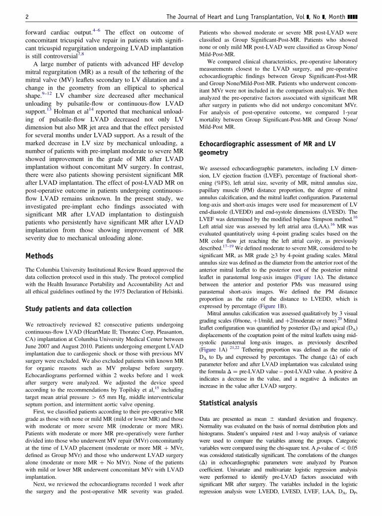
forward cardiac output.4–6 The effect on outcome ofconcomitant tricuspid valve repair in patients with signifi-cant tricuspid regurgitation undergoing LVAD implantationis still controversial7,8
A large number of patients with advanced HF developmitral regurgitation (MR) as a result of the tethering of themitral valve (MV) leaflets secondary to LV dilatation and achange in the geometry from an elliptical to sphericalshape.9–12 LV chamber size decreased after mechanicalunloading by pulsatile-flow or continuous-flow LVADsupport.13 Holman et al14 reported that mechanical unload-ing of pulsatile-flow LVAD decreased not only LVdimension but also MR jet area and that the effect persistedfor several months under LVAD support. As a result of themarked decrease in LV size by mechanical unloading, anumber of patients with pre-implant moderate to severe MRshowed improvement in the grade of MR after LVADimplantation without concomitant MV surgery. In contrast,there were also patients showing persistent significant MRafter LVAD implantation. The effect of post-LVAD MR onpost-operative outcome in patients undergoing continuous-flow LVAD remains unknown. In the present study, weinvestigated pre-implant echo findings associated withsignificant MR after LVAD implantation to distinguishpatients who persistently have significant MR after LVADimplantation from those showing improvement of MRseverity due to mechanical unloading alone.
Methods
The Columbia University Institutional Review Board approved thedata collection protocol used in this study. The protocol compliedwith the Health Insurance Portability and Accountability Act andall ethical guidelines outlined by the 1975 Declaration of Helsinki.
Study patients and data collection
We retroactively reviewed 82 consecutive patients undergoingcontinuous-flow LVAD (HeartMate II; Thoratec Corp, Pleasanton,CA) implantation at Columbia University Medical Center betweenJune 2007 and August 2010. Patients undergoing emergent LVADimplantation due to cardiogenic shock or those with previous MVsurgery were excluded. We also excluded patients with known MRfor organic reasons such as MV prolapse before surgery.Echocardiograms performed within 2 weeks before and 1 weekafter surgery were analyzed. We adjusted the device speedaccording to the recommendations by Topilsky et al,15 includingtarget mean atrial pressure 4 65 mm Hg, middle interventricularseptum portion, and intermittent aortic valve opening.
First, we classified patients according to their pre-operative MRgrade as those with none or mild MR (mild or lower MR) and thosewith moderate or more severe MR (moderate or more MR).Patients with moderate or more MR pre-operatively were furtherdivided into those who underwent MV repair (MVr) concomitantlyat the time of LVAD placement (moderate or more MR þ MVr,defined as Group MVr) and those who underwent LVAD surgeryalone (moderate or more MR þ No MVr). None of the patientswith mild or lower MR underwent concomitant MVr with LVADimplantation.
Next, we reviewed the echocardiograms recorded 1 week afterthe surgery and the post-operative MR severity was graded.
Patients who showed moderate or severe MR post-LVAD wereclassified as Group Significant-Post-MR. Patients who showednone or only mild MR post-LVAD were classified as Group None/Mild-Post-MR.
We compared clinical characteristics, pre-operative laboratorymeasurements closest to the LVAD surgery, and pre-operativeechocardiographic findings between Group Significant-Post-MRand Group None/Mild-Post-MR. Patients who underwent concom-itant MVr were not included in the comparison analysis. We thenanalyzed the pre-operative factors associated with significant MRafter surgery in patients who did not undergo concomitant MVr.For analysis of post-operative outcome, we compared 1-yearmortality between Group Significant-Post-MR and Group None/Mild-Post MR.
Echocardiographic assessment of MR and LVgeometry
We assessed echocardiographic parameters, including LV dimen-sion, LV ejection fraction (LVEF), percentage of fractional short-ening (%FS), left atrial size, severity of MR, mitral annulus size,papillary muscle (PM) distance proportion, the degree of mitralannulus calcification, and the mitral leaflet configuration. Parasternallong-axis and short-axis images were used for measurement of LVend-diastole (LVEDD) and end-systole dimensions (LVESD). TheLVEF was determined by the modified biplane Simpson method.16
Left atrial size was assessed by left atrial area (LAA).16 MR wasevaluated quantitatively using 4-point grading scales based on theMR color flow jet reaching the left atrial cavity, as previouslydescribed.17–19 We defined moderate to severe MR, considered to besignificant MR, as MR grade ≥3 by 4-point grading scales. Mitralannulus size was defined as the diameter from the anterior root of theanterior mitral leaflet to the posterior root of the posterior mitralleaflet in parasternal long-axis images (Figure 1A). The distancebetween the anterior and posterior PMs was measured usingparasternal short-axis images. We defined the PM distanceproportion as the ratio of the distance to LVEDD, which isexpressed by percentage (Figure 1B).
Mitral annulus calcification was assessed qualitatively by 3 visualgrading scales (0/none, þ1/mild, and þ2/moderate or more).20 Mitralleaflet configuration was quantified by posterior (DP) and apical (DA)displacements of the coaptation point of the mitral leaflets using mid-systolic parasternal long-axis images, as previously described(Figure 1A) 21,22 Tethering proportion was defined as the ratio ofDA to DP and expressed by percentages. The change (Δ) of eachparameter before and after LVAD implantation was calculated usingthe formula Δ ¼ pre-LVAD value – post-LVAD value. A positive Δindicates a decrease in the value, and a negative Δ indicates anincrease in the value after LVAD surgery.
Statistical analysis
Data are presented as mean � standard deviation and frequency.Normality was evaluated on the basis of normal distribution plots andhistograms. Student’s unpaired t-test and 1-way analysis of variancewere used to compare the variables among the groups. Categoricvariables were compared using the chi-square test. A p-value ofo 0.05was considered statistically significant. The correlations of the changes(Δ) in echocardiographic parameters were analyzed by Pearsoncoefficient. Univariate and multivariate logistic regression analysiswere performed to identify pre-LVAD factors associated withsignificant MR after surgery. The variables included in the logisticregression analysis were LVEDD, LVESD, LVEF, LAA, DA, DP,

Figure 1 The measurements of echocardiographic parameters toquantify the mitral leaflet configurations are shown in the parasternal2-dimensional echocardiogram (A) long-axis and (B) short-axisviews. DA, apical displacement; DP, posterior displacement; LVEDD,left ventricular end-diastolic dimension.
Kitada et al. MR after Left Ventricular Assist Device Surgery 3
tethering area, annulus size, PM distance proportion, tetheringproportion, and annulus calcification.
Variables that achieved statistical significance in the univariateanalysis were subsequently included in a multivariate analysis. Theratio of MR area to LAA was included in the multivariate analysis toadjust for the effect of pre-operative MR on significant MR post-LVAD. Post-operative mortality at 1 year was assessed by Kaplan-Meier analysis with log-rank test. Interobserver and intraobserver
Figure 2 The prevalence of mitral regurgitation (MR) before and aftindicates the number of patients in each group according to the classificmitral valve repair.
variability for echocardiographic parameters was determined using10 randomly selected patients by 2 independent observers (S.K. andT.S.K.) and the interclass correlation coefficient was calculated.Statistical analyses were performed using SPSS 20.0 software (IBMCorp, Armonk, NY).
Results
Pre-LVAD and post-LVAD MR grade and associatedpatients’ characteristics
Figure 2 illustrates the distribution of patients with andwithout MR before and after surgery. Among the 82 patientsstudied, 39 (47.6%) had none or mild MR before LVADimplantation, and 43 (52.4%) had moderate or severe MRpre-operatively. None of the patients with pre-operativenone/mild MR underwent concomitant MVr with LVADimplantation, and none developed worsening or newlydeveloped significant MR after surgery. Among the 43patients who had pre-operative moderate or severe MR, 5(11.6% [6.1% of total patients]) underwent concomitantMVr with LVAD implantation (Group MVr), consisting of4 MV repairs and 1 MV replacement. Therefore, 38 patientshad moderate or more MR before surgery but did notreceive concomitant MVr. Among those, 25 patientsshowed a marked decrease in MR severity to the grade ofmild or none after LVAD surgery (58.1% of those with pre-operative moderate or more MR [30.5% of total patients]),and the remaining 13 patients still had moderate or severeMR even after LVAD implantation (30.2% of those withpre-operative moderate or severe MR [15.9% of totalpatients]). Sixty-four patients were classified as GroupNone/Mild-Post-MR and the remaining 13 were classified asGroup Significant-Post-MR.
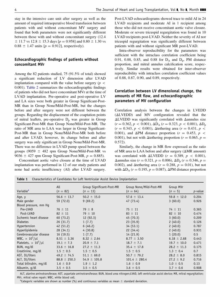
Clinical characteristics and laboratory data of each groupare summarized in Table 1. None of the variables differedsignificantly among the groups. We compared the length of
er left ventricular assist device (LVAD) implantation. The diagramation based on the pre-LVAD and post-LVAD MR severity. MVr,
The Journal of Heart and Lung Transplantation, Vol ], No ], Month ]]]]4
stay in the intensive care unit after surgery as well as theamount of required intraoperative blood transfusion betweenpatients with and without concomitant MV surgery andfound that both parameters were not significantly differentbetween those with and without concomitant surgery (12.4� 11.7 vs 12.8 � 15.3 days [p ¼ 0.958] and 0.80 � 1.30 vs0.88 � 1.47 units [p ¼ 0.912], respectively).
Echocardiographic findings of patients withoutconcomitant MVr
Among the 82 patients studied, 75 (91.5% of total) showeda significant reduction of LV dimension after LVADimplantation compared with the value before surgery (p o0.001). Table 2 summarizes the echocardiographic findingsof patients who did not have concomitant MVr at the time ofLVAD implantation. Pre-operative and post-operative LVand LA sizes were both greater in Group Significant-Post-MR than in Group None/Mild-Post-MR, but the changesbefore and after surgery were not different between thegroups. Regarding the displacement of the coaptation pointsof mitral leaflets, pre-operative Dp was greater in GroupSignificant-Post-MR than Group None/Mild-Post-MR. Theratio of MR area to LAA was larger in Group Significant-Post-MR than in Group None/Mild-Post-MR both beforeand after LVAD; however, its change before and aftersurgery was only significant in Group None/Mild-Post-MR.There was no difference in LVAD pump speed between thegroups (9059 � 482 rpm Group None/Mild-Post-MR vs9036 � 427 rpm Group Significant-Post-MR, p ¼ 0.885).
Concomitant aortic valve closure at the time of LVADimplantation was performed in 12 of our study patients andnone had aortic insufficiency (AI) after LVAD surgery.
Table 1 Characteristics of Candidates for Left Ventricular Assist Dev
All Group Significant-Post-Variablea (n ¼ 82) (n ¼ 13)
Age, y 56.6 � 13.7 50.2 � 15.0Male gender 59 (72.0) 9 (69.2)Blood pressure, mm Hg
Pre-LVAD 76 � 11 79 � 8Post-LVAD 82 � 11 79 � 10
Ischemic heart disease 60 (73.2) 12 (92.3)Diabetes 26 (31.7) 1 (7.7)Hypertension 42 (51.2) 6 (46.2)Hyperlipidemia 28 (34.1) 4 (30.8)Cerebrovascular disease 16 (19.5) 1 (7.7)WBC, � 103/ml 8.51 � 3.36 8.32 � 2.64Platelets, � 103/ml 19.1 � 7.3 20.9 � 7.1BUN, mg/dl 33.6 � 16.8 27.2 � 11.3Creatinine, mg/dl 1.5 � 0.5 1.5 � 0.5AST, IU/liters 49.2 � 74.5 51.1 � 69.0ALT, IU/liters 88.8 � 250.3 54.9 � 105.8Total bilirubin, mg/dl 1.6 � 0.9 1.9 � 1.2Albumin, g/dl 3.5 � 0.5 3.5 � 0.5
ALT, alanine aminotransferase; AST, aspartate aminotransferase; BUN, blood uMVr, mitral valve repair; WBC, white blood cell.
aCategoric variables are shown as number (%) and continuous variables as m
Post-LVAD echocardiograms showed trace to mild AI in 24LVAD recipients and moderate AI in 1 recipient amongthose who did not receive concomitant aortic valve closure.Moderate or severe tricuspid regurgitation was found in 10LVAD recipients post-LVAD. Neither the severity of AI nortricuspid regurgitation was significantly different betweenpatients with and without significant MR post-LVAD.
Intra-observer reproducibility for the parameters wassufficient with the intraclass correlation coefficient being0.91, 0.88, 0.85, and 0.88 for DA and Dp, PM distanceproportion, and mitral annulus calcification score, respec-tively. Similar results were obtained for inter-observerreproducibility with intraclass correlation coefficient valuesof 0.88, 0.87, 0.90, and 0.89, respectively.
Correlation between LV dimensional change, theamounts of MR flow, and echocardiographicparameters of MV configuration
Correlation analysis between the changes in LVEDD(ΔLVEDD) and MV configuration revealed that theΔLVEDD was significantly correlated with Δannulus size(r ¼ 0.362, p o 0.001), ΔDA (r ¼ 0.323, p o 0.001), ΔDp
(r ¼ 0.343, p o 0.001), Δtethering area (r ¼ 0.431, p o0.001), and ΔPM distance proportion (r ¼ 0.453, p o0.001), but not with Δtethering proportion (r ¼ 0.065, p ¼0.572).
Similarly, the change in MR flow expressed as the ratioof MR area to LAA before and after surgery (ΔMR amount)was correlated with ΔLVEDD (r ¼ 0.389, p o 0.001),Δannulus size (r ¼ 0.321, p ¼ 0.006), ΔDp (r ¼ 0.346, p ¼0.002), and Δtethering area (r ¼ 0.288, p ¼ 0.01), but notwith ΔDA (r ¼ 0.195, p ¼ 0.087), ΔPM distance proportion
ice Implantation
MR Group None/Mild-Post-MR Group MVr(n ¼ 64) (n ¼ 5) p-value
57.6 � 13.4 59.8 � 12.0 0.20447 (73.4) 3 (60.0) 0.926
75 � 11 74 � 11 0.36583 � 11 82 � 10 0.47445 (70.3) 3 (60.0) 0.20923 (35.9) 2 (40.0) 0.12634 (53.1) 2 (40.0) 0.78722 (34.4) 2 (40.0) 0.93114 (21.9) 1 (20.0) 0.58.77 � 3.50 6.18 � 2.68 0.44318.7 � 7.1 19.7 � 10.0 0.47135.4 � 17.8 28.2 � 11.3 0.1751.5 � 0.5 1.3 � 0.4 0.750.7 � 79.2 28.2 � 8.0 0.815101.4 � 280.4 27.2 � 9.2 0.7161.6 � 0.9 1.0 � 0.5 0.133.6 � 0.5 3.7 � 0.6 0.908
rea nitrogen;LVAD, left ventricular assist device; MR, mitral regurgitation;
ean � standard deviation.
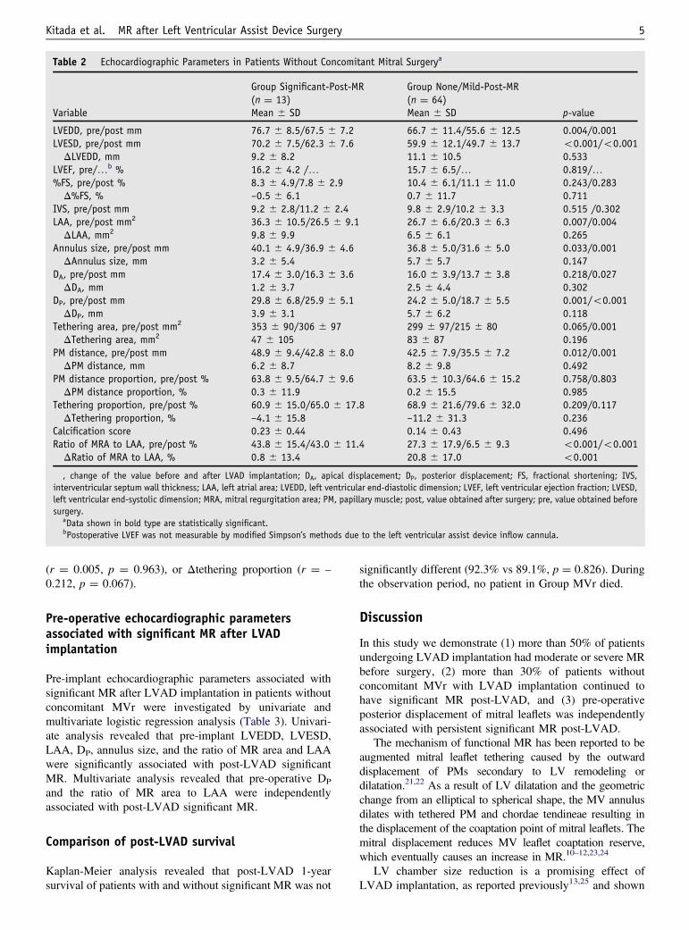
Table 2 Echocardiographic Parameters in Patients Without Concomitant Mitral Surgerya
Group Significant-Post-MR Group None/Mild-Post-MR(n ¼ 13) (n ¼ 64)
Variable Mean � SD Mean � SD p-value
LVEDD, pre/post mm 76.7 � 8.5/67.5 � 7.2 66.7 � 11.4/55.6 � 12.5 0.004/0.001LVESD, pre/post mm 70.2 � 7.5/62.3 � 7.6 59.9 � 12.1/49.7 � 13.7 o0.001/o0.001
ΔLVEDD, mm 9.2 � 8.2 11.1 � 10.5 0.533LVEF, pre/…b % 16.2 � 4.2 /… 15.7 � 6.5/… 0.819/…%FS, pre/post % 8.3 � 4.9/7.8 � 2.9 10.4 � 6.1/11.1 � 11.0 0.243/0.283
Δ%FS, % –0.5 � 6.1 0.7 � 11.7 0.711IVS, pre/post mm 9.2 � 2.8/11.2 � 2.4 9.8 � 2.9/10.2 � 3.3 0.515 /0.302LAA, pre/post mm2 36.3 � 10.5/26.5 � 9.1 26.7 � 6.6/20.3 � 6.3 0.007/0.004
ΔLAA, mm2 9.8 � 9.9 6.5 � 6.1 0.265Annulus size, pre/post mm 40.1 � 4.9/36.9 � 4.6 36.8 � 5.0/31.6 � 5.0 0.033/0.001
ΔAnnulus size, mm 3.2 � 5.4 5.7 � 5.7 0.147DA, pre/post mm 17.4 � 3.0/16.3 � 3.6 16.0 � 3.9/13.7 � 3.8 0.218/0.027
ΔDA, mm 1.2 � 3.7 2.5 � 4.4 0.302DP, pre/post mm 29.8 � 6.8/25.9 � 5.1 24.2 � 5.0/18.7 � 5.5 0.001/o0.001
ΔDP, mm 3.9 � 3.1 5.7 � 6.2 0.118Tethering area, pre/post mm2 353 � 90/306 � 97 299 � 97/215 � 80 0.065/0.001
ΔTethering area, mm2 47 � 105 83 � 87 0.196PM distance, pre/post mm 48.9 � 9.4/42.8 � 8.0 42.5 � 7.9/35.5 � 7.2 0.012/0.001
ΔPM distance, mm 6.2 � 8.7 8.2 � 9.8 0.492PM distance proportion, pre/post % 63.8 � 9.5/64.7 � 9.6 63.5 � 10.3/64.6 � 15.2 0.758/0.803
ΔPM distance proportion, % 0.3 � 11.9 0.2 � 15.5 0.985Tethering proportion, pre/post % 60.9 � 15.0/65.0 � 17.8 68.9 � 21.6/79.6 � 32.0 0.209/0.117
ΔTethering proportion, % –4.1 � 15.8 –11.2 � 31.3 0.236Calcification score 0.23 � 0.44 0.14 � 0.43 0.496Ratio of MRA to LAA, pre/post % 43.8 � 15.4/43.0 � 11.4 27.3 � 17.9/6.5 � 9.3 o0.001/o0.001
ΔRatio of MRA to LAA, % 0.8 � 13.4 20.8 � 17.0 o0.001
, change of the value before and after LVAD implantation; DA, apical displacement; DP, posterior displacement; FS, fractional shortening; IVS,interventricular septum wall thickness; LAA, left atrial area; LVEDD, left ventricular end-diastolic dimension; LVEF, left ventricular ejection fraction; LVESD,left ventricular end-systolic dimension; MRA, mitral regurgitation area; PM, papillary muscle; post, value obtained after surgery; pre, value obtained beforesurgery.
aData shown in bold type are statistically significant.bPostoperative LVEF was not measurable by modified Simpson’s methods due to the left ventricular assist device inflow cannula.
Kitada et al. MR after Left Ventricular Assist Device Surgery 5
(r ¼ 0.005, p ¼ 0.963), or Δtethering proportion (r ¼ –
0.212, p ¼ 0.067).
Pre-operative echocardiographic parametersassociated with significant MR after LVADimplantation
Pre-implant echocardiographic parameters associated withsignificant MR after LVAD implantation in patients withoutconcomitant MVr were investigated by univariate andmultivariate logistic regression analysis (Table 3). Univari-ate analysis revealed that pre-implant LVEDD, LVESD,LAA, DP, annulus size, and the ratio of MR area and LAAwere significantly associated with post-LVAD significantMR. Multivariate analysis revealed that pre-operative DP
and the ratio of MR area to LAA were independentlyassociated with post-LVAD significant MR.
Comparison of post-LVAD survival
Kaplan-Meier analysis revealed that post-LVAD 1-yearsurvival of patients with and without significant MR was not
significantly different (92.3% vs 89.1%, p ¼ 0.826). Duringthe observation period, no patient in Group MVr died.
Discussion
In this study we demonstrate (1) more than 50% of patientsundergoing LVAD implantation had moderate or severe MRbefore surgery, (2) more than 30% of patients withoutconcomitant MVr with LVAD implantation continued tohave significant MR post-LVAD, and (3) pre-operativeposterior displacement of mitral leaflets was independentlyassociated with persistent significant MR post-LVAD.
The mechanism of functional MR has been reported to beaugmented mitral leaflet tethering caused by the outwarddisplacement of PMs secondary to LV remodeling ordilatation.21,22 As a result of LV dilatation and the geometricchange from an elliptical to spherical shape, the MV annulusdilates with tethered PM and chordae tendineae resulting inthe displacement of the coaptation point of mitral leaflets. Themitral displacement reduces MV leaflet coaptation reserve,which eventually causes an increase in MR.10–12,23,24
LV chamber size reduction is a promising effect ofLVAD implantation, as reported previously13,25 and shown
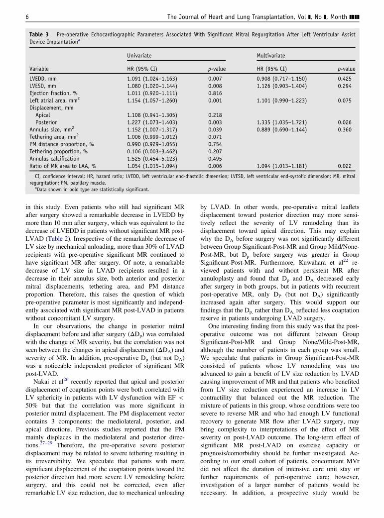
Table 3 Pre-operative Echocardiographic Parameters Associated With Significant Mitral Regurgitation After Left Ventricular AssistDevice Implantationa
Univariate Multivariate
Variable HR (95% CI) p-value HR (95% CI) p-value
LVEDD, mm 1.091 (1.024–1.163) 0.007 0.908 (0.717–1.150) 0.425LVESD, mm 1.080 (1.020–1.144) 0.008 1.126 (0.903–1.404) 0.294Ejection fraction, % 1.011 (0.920–1.111) 0.816Left atrial area, mm2 1.154 (1.057–1.260) 0.001 1.101 (0.990–1.223) 0.075Displacement, mm
Apical 1.108 (0.941–1.305) 0.218Posterior 1.227 (1.073–1.403) 0.003 1.335 (1.035–1.721) 0.026
Annulus size, mm2 1.152 (1.007–1.317) 0.039 0.889 (0.690–1.144) 0.360Tethering area, mm2 1.006 (0.999–1.012) 0.071PM distance proportion, % 0.990 (0.929–1.055) 0.754Tethering proportion, % 0.106 (0.003–3.462) 0.207Annulus calcification 1.525 (0.454–5.123) 0.495Ratio of MR area to LAA, % 1.054 (1.015–1.094) 0.006 1.094 (1.013–1.181) 0.022
CI, confidence interval; HR, hazard ratio; LVEDD, left ventricular end-diastolic dimension; LVESD, left ventricular end-systolic dimension; MR, mitralregurgitation; PM, papillary muscle.
aData shown in bold type are statistically significant.
The Journal of Heart and Lung Transplantation, Vol ], No ], Month ]]]]6
in this study. Even patients who still had significant MRafter surgery showed a remarkable decrease in LVEDD bymore than 10 mm after surgery, which was equivalent to thedecrease of LVEDD in patients without significant MR post-LVAD (Table 2). Irrespective of the remarkable decrease ofLV size by mechanical unloading, more than 30% of LVADrecipients with pre-operative significant MR continued tohave significant MR after surgery. Of note, a remarkabledecrease of LV size in LVAD recipients resulted in adecrease in their annulus size, both anterior and posteriormitral displacements, tethering area, and PM distanceproportion. Therefore, this raises the question of whichpre-operative parameter is most significantly and independ-ently associated with significant MR post-LVAD in patientswithout concomitant LV surgery.
In our observations, the change in posterior mitraldisplacement before and after surgery (ΔDp) was correlatedwith the change of MR severity, but the correlation was notseen between the changes in apical displacement (ΔDA) andseverity of MR. In addition, pre-operative Dp (but not DA)was a noticeable independent predictor of significant MRpost-LVAD.
Nakai et al26 recently reported that apical and posteriordisplacement of coaptation points were both correlated withLV sphericity in patients with LV dysfunction with EF o50% but that the correlation was more significant inposterior mitral displacement. The PM displacement vectorcontains 3 components: the mediolateral, posterior, andapical directions. Previous studies reported that the PMmainly displaces in the mediolateral and posterior direc-tions.27–29 Therefore, the pre-operative severe posteriordisplacement may be related to severe tethering resulting inits irreversibility. We speculate that patients with moresignificant displacement of the coaptation points toward theposterior direction had more severe LV remodeling beforesurgery, and this could not be corrected, even afterremarkable LV size reduction, due to mechanical unloading
by LVAD. In other words, pre-operative mitral leafletsdisplacement toward posterior direction may more sensi-tively reflect the severity of LV remodeling than itsdisplacement toward apical direction. This may explainwhy the DA before surgery was not significantly differentbetween Group Significant-Post-MR and Group Mild/None-Post-MR, but Dp before surgery was greater in GroupSignificant-Post-MR. Furthermore, Kuwahara et al22 re-viewed patients with and without persistent MR afterannuloplasty and found that Dp and DA decreased earlyafter surgery in both groups, but in patients with recurrentpost-operative MR, only DP (but not DA) significantlyincreased again after surgery. This would support ourfindings that the Dp, rather than DA, reflected less coaptationreserve in patients undergoing LVAD surgery.
One interesting finding from this study was that the post-operative outcome was not different between GroupSignificant-Post-MR and Group None/Mild-Post-MR,although the number of patients in each group was small.We speculate that patients in Group Significant-Post-MRconsisted of patients whose LV remodeling was tooadvanced to gain a benefit of LV size reduction by LVADcausing improvement of MR and that patients who benefitedfrom LV size reduction experienced an increase in LVcontractility that balanced out the MR reduction. Themixture of patients in this group, whose conditions were toosevere to reverse MR and who had enough LV functionalrecovery to generate MR flow after LVAD surgery, maybring complexity to interpretations of the effect of MRseverity on post-LVAD outcome. The long-term effect ofsignificant MR post-LVAD on exercise capacity orprognosis/comorbidity should be further investigated. Ac-cording to our small cohort of patients, concomitant MVrdid not affect the duration of intensive care unit stay orfurther requirements of peri-operative care; however,investigation of a larger number of patients would benecessary. In addition, a prospective study would be

Kitada et al. MR after Left Ventricular Assist Device Surgery 7
required to determine which patients would get the mostbenefit from concomitant MVr.
The study contains several limitations worth noting dueto its retrospective nature based on a small cohort of patientsfrom a single center. The major limitation of the analysis isthat we could not obtain echocardiograms recorded in achronic phase after surgery, such as 3 months after surgery,from all patients. Because of this we used the echocardio-grams recorded 1 week after the surgery. Post-operativeoptimization of volume status may typically take more than1 week from the time of surgery, and echocardiograms at1 week post-operatively may not represent the physiologyunder adequate mechanical unloading by LVAD; therefore,our analysis of MR may be overestimated.
Further, we could not obtain enough post-operativeinformation such as exercise tolerance and quality of lifeindices. Therefore, the effect of persistent MR on post-operative functional statuses other than survival was notfully assessed.
The decision of whether to perform concomitant MVr inour cohort was made by the surgeons according to the pre-operative clinical information and peri-operative trans-esophageal echocardiographic findings. Our cohort includedpatients with ischemic and non-ischemic etiology. Inaddition, we did not fully assess the configuration of MVleaflet and the quantitative assessment of MR flow. Using 3-dimensional echocardiography or cardiac magnetic reso-nance imaging could allow us to evaluate MV configurationin more detailed manner.30,31
Finally, our study included patients who did not havepre-operative MR in order to find the features associatedwith post-operative MR. Indeed, none of these patientsdeveloped MR post-operatively. However, we includedthese patients because we initially considered the possibilitythat the grade of MR might worsen if a patient’s LVcontraction dramatically improved or LV dyssynchronybecame obvious under LVAD support, even in a patientswithout MR before LVAD.
In conclusion, pre-LVAD posterior displacement of thecoaptation points of mitral leaflets detected by echocardio-grams was associated with persistent MR after LVADimplantation. Our observation may help to identifymorphologic features of the MV associated with functionalMR refractory to volume reduction of the LV.
Disclosure statement
None of the authors has a financial relationship with a commercialentity that has an interest in the subject of the presented manuscriptor other conflicts of interest to disclose.
References
1. Rose EA, Gelijns AC, Moskowitz AJ, et al. Randomized Evaluation ofMechanical Assistance for the Treatment of Congestive Heart Failure(REMATCH) Study Group. Long-term use of a left ventricular assistdevice for end-stage heart failure. N Engl J Med 2001;345:1435-43.
2. Slaughter MS, Rogers JG, Milano CA, et al. Heart Mate IIInvestigators. Advanced heart failure treated with continuous-flow leftventricular assist device. N Engl J Med 2009;361:2241-51.
3. Birks EJ, Yacoub MH, Banner NR, Khaghani A. The role of bridge totransplantation: should LVAD patients be transplanted? Curr OpinCardiol 2004;19:148-53.
4. Rao V, Slater JP, Edwards NM, Naka Y, Oz MC. Surgical managementof valvular disease in patients requiring left ventricular assist devicesupport. Ann Thorac Surg 2001;71:1448-53.
5. Pak SW, Uriel N, Takayama H, et al. Prevalence of de novo aorticinsufficiency during long-term support with left ventricular assistdevices. J Heart Lung Transplant 2010;29:1172-6.
6. Goda A, Takayama H, Pak SW, et al. Aortic valve procedures at thetime of ventricular assist device placement. Ann Thorac Surg2011;91:750-4.
7. Krishan K, Nair A, Pinney S, Adams DH, Anyanwu AC. Liberal use oftricuspid-valve annuloplasty during left-ventricular assist deviceimplantation. Eur J Cardiothorac Surg 2012;41:213-7.
8. Saeed D, Kidambi T, Shalli S, et al. Tricuspid valve repair with leftventricular assist device implantation: is it warranted? J Heart LungTransplant 2011;30:530-5.
9. Pino PG, Galati A, Terranova A. Functional mitral regurgitation inheart failure. J Cardiovasc Med (Hagerstown) 2006;7:514-23.
10. Di Salvo TG, Acker MA, Dec GW, Byrne JG. Mitral valve surgery inadvanced heart failure. J Am Coll Cardiol 2010;55:271-82.
11. Kono T, Sabbah HN, Rosman H, Alam M, Jafri S, Goldstein S. Leftventricular shape is the primary determinant of functional mitralregurgitation in heart failure. J Am Coll Cardiol 1992;20:1594-8.
12. Sabbah HN, Kono T, Rosman H, Jafri S, Stein PD, Goldstein S. Leftventricular shape: a factor in the etiology of functional mitralregurgitation in heart failure. Am Heart J 1992;123:961-6.
13. Kato TS, Chokshi A, Singh P, et al. Effects of continuous-flow versuspulsatile-flow left ventricular assist devices on myocardial unloadingand remodeling. Circ Heart Fail 2011;4:546-53.
14. Holman WL, Bourge RC, Fan P, Kirklin JK, Pacifico AD, Nanda NC.Influence of left ventricular assist on valvular regurgitation. Circulation1993;88:II309-18.
15. Topilsky Y, Hasin T, Oh JK, et al. Echocardiographic variables afterleft ventricular assist device implantation associated with adverseoutcome. Circ Cardiovasc Imaging 2011;4:648-61.
16. Lang RM, Bierig M, Devereux RB, et al. Chamber QuantificationWriting Group. American Society of Echocardiography’s Guidelinesand Standards Committee; European Association of Echocardiography.Recommendations for chamber quantification: a report from theAmerican Society of Echocardiography’s Guidelines and StandardsCommittee and the Chamber Quantification Writing Group, developedin conjunction with the European Association of Echocardiography, abranch of the European Society of Cardiology. J Am Soc Echocardiogr2005;18:1440-63.
17. Chen C, Thomas JD, Anconina J, et al. Impact of impinging wall jet oncolor Doppler quantification of mitral regurgitation. Circulation1991;84:712-20.
18. Enriquez-Sarano M, Tajik AJ, Bailey KR, Seward JB. Color flowimaging compared with quantitative Doppler assessment of severity ofmitral regurgitation: influence of eccentricity of jet and mechanism ofregurgitation. J Am Coll Cardiol 1993;21:1211-29.
19. Zoghbi WA, Enriquez-Sarano M, Foster E, et al. Recommendations forevaluation of the severity of native valvular regurgitation with two-dimensional and Doppler echocardiography. J Am Soc Echocardiogr2003;16:777-802.
20. Salarifar M, Rezvanfard M, Sadeghian H, Safir-mardanloo A, Shafii N.Mitral annular calcification predicts immediate results of percutaneoustransvenous mitral commissurotomy. Cardiovasc Ultrasound 2011;9:29.
21. Zhu F, Otsuji Y, Yotsumoto G, et al. Mechanism of persistent ischemicmitral regurgitation after annuloplasty: importance of augmented posteriormitral leaflet tethering. Circulation 2005112(9 Suppl):I396-401.
22. Kuwahara E, Otsuji Y, Iguro Y, et al. Mechanism of recurrent/persistent ischemic/functional mitral regurgitation in the chronic phaseafter surgical annuloplasty: importance of augmented posterior leaflettethering. Circulation 2006114(1 Suppl):I529-34.

The Journal of Heart and Lung Transplantation, Vol ], No ], Month ]]]]8
23. Tsukiji M, Watanabe N, Yamaura Y, et al. Three-dimensionalquantitation of mitral valve coaptation by a novel software systemwith transthoracic real-time three-dimensional echocardiography. J AmSoc Echocardiogr 2008;21:43-6.
24. Sabbah HN, Kono T, Stein PD, Mancini GB, Goldstein S. Leftventricular shape changes during the course of evolving heart failure.Am J Physiol 1992;263:H266-70.
25. Kato TS, Chokshi A, Singh P, et al. Markers of extracellular matrixturnover and the development of right ventricular failure afterventricular assist device implantation in patients with advanced heartfailure. J Heart Lung Transplant 2012;31:37-45.
26. Nakai H, Kaku K, Takeuchi M, et al. Different influences of leftventricular remodeling on anterior and posterior mitral leaflet tethering.Circ J 2012;76:2481-7.
27. Otsuji Y, Handschumacher MD, Schwammenthal E, et al. Insightsfrom three-dimensional echocardiography into the mechanism of
functional mitral regurgitation: direct in vivo demonstration of alteredleaflet tethering geometry. Circulation 1997;96:1999-2008.
28. Chaput M, Handschumacher MD, Guerrero JL, et al. Mitral leafletadaptation to ventricular remodeling: Prospective changes in a modelof ischemic mitral regurgitation. Circulation 2009120(11 Suppl):S99-103.
29. Saito K, Okura H, Watanabe N, et al. Influence of chronic tethering ofthe mitral valve on mitral leaflet size and coaptation in functional mitralregurgitation. JACC Cardiovasc Imaging 2012;5:337-45.
30. Rukosujew A, Klotz S, Welp H, et al. Surgery of secondary mitralinsufficiency in patients with impaired left ventricular function. JCardiothorac Surg 2009;4:36.
31. Heitner J, Bhumireddy GP, Crowley AL, et al. Clinical application ofcine-MRI in the visual assessment of mitral regurgitation compared toechocardiography and cardiac catheterization. PLoS One 2012;7:e40491.


