Embed Size (px)
Citation preview

J Investig Allergol Clin Immunol 2012; Vol. 22(2): 133-153 © 2012 Esmon Publicidad
Practitioner’s Corner
PRACTITIONER’S CORNER
Malignancy Phenotype in Common Variable Immunodefi ciency
H Abolhassani1, A Aghamohammadi,1,2 A Imanzadeh,1 P Mohammadinejad,1 B Sadeghi,1 N Rezaei1,2
1Research Center for Immunodefi ciencies, Children’s Medical Center, Tehran University of Medical Sciences, Tehran, Iran2Molecular Immunology Research Center; and Department of Immunology, School of Medicine, Tehran University of Medical Sciences, Tehran, Iran
Key words: Malignancy phenotype. Common variable immunodef ic iency. Non-Hodgkin lymphoma. Gastr ic adenocarcinoma. Palabras clave: Fenotipo maligno. Inmunodefi ciencia común variable. Limfoma no-Hodgkin. Adenocarcinoma gástrico.
Common variable immunodeficiency (CVID) is a heterogeneous group of disorders, characterized by hypogammaglobulinemia and defective specifi c antibody production [1,2]. Although the hallmark of the disease is frequent infections, primarily affecting the respiratory
cancer; there were 4 cases of non-Hodgkin lymphoma and 3 cases of gastric adenocarcinoma. The demographic data and laboratory fi ndings for these 7 patients are presented in the Table. The mean (SD) age at onset of symptoms was 7 (1.7) years and the age at diagnosis was 13 (2.4) years; these ages were signifi cantly higher than those recorded in all Iranian CVID databases (P<.001). Surprisingly, all the patients with lymphoma had childhood onset, while those with gastric cancer were in their fourth decade of life when the cancer was diagnosed. Although no population-based epidemiological studies have analyzed demographics or epidemiological indices for malignancy in patients with CVID, the incidence of all types of cancer has been estimated to be 1.8 to 13 times higher than in the general population [5]. According to our fi ndings, the incidence of cancer in Iranian CVID patients is 10%, which is 2.5 times higher than the risk of cancer in the general population [6]. The increased prevalence of cancer in immunodefi ciency may be explained by an increased risk of immune dysregulation, increased susceptibility to an infectious agent involved in carcinogenesis, or chromosomal instability due to the nature of the disease [7]. Moreover, lower total and CD4+ T cells and natural killer cells in terms of number and function have been strongly associated with risk for subsequent development of malignancy in CVID patients [8]. In our study, the most common forms of cancer were non-Hodgkin lymphoma and gastric adenocarcinoma. In previous studies, patients with CVID, and particularly female patients, have been found to have an 15.2- to 438-fold increased risk of developing non-Hodgkin lymphoma compared to the general population [5,7,9]. In another study of 120 CVID patients, 46% had non-Hodgkin lymphoma, while 7% had Hodgkin lymphoma [10]. Most of the patients in our study developed
Table. Demographics and Disease Characteristics in 93 Iranian Patients With Common Variable Immunodefi ciency (CVID)
Patients Patients Hodgkin Gastric Without With Lymphoma AdenocarcinomaParameters Cancer Cancer P Value (n=4) (n=3)
P Value
(n=84) (n=7)
Mean age, (SD), y 12.9 (7.2) 16.7 (11.4) .001 14.38 (5.5) 51.3 (5.8) <.001
Male/female, No. 49/15 4/3 .23 2/2 2/1 .57
Mean IgG, (SD), mg/dL 140.3 (135) 124.0 (122) .43 220.2 (43.2) 88.66 (21.2) .096
Mean IgA, (SD) (range), mg/dL 9.1 (16.3) 10.5 (17.2) .51 6.37 (2.9) 4.2 (0.9) .034
Mean IgM, (SD), mg/dL 16.8 (16.1) 16.0 (15.5) .70 67.62 (14.8) 12.33 (4.3) .14
Mean CD3, (SD), % 73.7 (14.8 ) 72.2 (14.2) .48 70.39 (43.2) 80.59 (30.5) .016
Mean CD4, (SD), % 33.0 (14.4) 36.7 (16.9) .41 29.0 (15.2) 44.51 (16.4) .007
Mean CD8, (SD), % 38.7 (13.5) 35.1 (12.4) .32 37.41 (12.3) 38.49 (14.5) .75
Mean CD19, (SD), % 10.8 (6.3) 10.0 (7.7) .14 16.73 (4.6) 8.56 (6.7) .15
Follow up, (SD), y 5.0 (4.9) 6.5 (3.6) .77 5.12 (4.1) 5.67 (2.8) .81
Delay in diagnosis, (SD), y 6 (5.9) 10 (7.3) .07 7.25 (4.9) 20.66 (11.0) .012
Mortality 26/84 4/7 .72 3/4 1/3 .3
and gastrointestinal tracts, the clinical spectrum of CVID is vast and includes malignancy [3]. The incidence of malignancy in CVID is around 11% to 13% and usually occurs during the fi fth or sixth decade of life [4]. The most common sites of involvement are the gastrointestinal tract and the lymphoid tissues. In the following study, we will discuss our experience with different cancers observed in CVID patients.
Ten percent of the clinical phenotypes observed in 93 patients diagnosed with CVID at the Children’s Medical Center Hospital (Pediatrics Center of Excellence in Tehran, Iran) were malignant phenotypes. The diagnosis of non-Hodgkin lymphoma was based on the American College of Radiology Appropriateness Criteria while that of gastric adenocarcinoma was based on pathologic findings of gastric epithelial neoplasia. Four male and 3 female patients, with a median age of 16 years (range, 12-59 years), had

Practitioner’s Corner
J Investig Allergol Clin Immunol 2012; Vol. 22(2): 133-153© 2012 Esmon Publicidad
❚ Manuscript received July 11, 2011; accepted for publication, August 8, 2011.
Asghar AghamohammadiChildren’s Medical Center Hospital
62 Qarib St., Keshavarz Blvd.Tehran 14194, Iran
E-mail: [email protected]
lymphoma at an early age. Lymphoma as a complication of CVID is well known and usually consists of nodal lymphoma, non-Hodgkin lymphoma, or B-cell lymphoma, which present in female patients an average of 9 years after diagnosis of CVID; the median age at diagnosis is 23 years and the lymphoma is normally found in association with T-cell defects [3,9]. In addition, the stage of lymphoma in young CVID patients is low grade B-cell tumors without bone marrow involvement. The observed prevalence of lymphoma may be due to Epstein-Barr virus (EBV) infection or reactivation of weakness of the immune system. However, EBV is not detectable in most patients. Furthermore, CVID patients with systemic and local granulomatosis are more susceptible to developing lymphoma; the diseases may have the same origin, ie, immune dysregulation and infection with B-cell lymphotropic human herpes virus type 8 (HHV8) [7].
Patients with CVID have a 10- to 47-fold increased risk of gastric adenocarcinoma [5,7,9]. In one study, 16% of 120 patients with CVID in the Immunodefi ciency Cancer Registry had this type of cancer [10]. In fact, our study suggests that elderly patients may be more vulnerable to gastric adenocarcinoma and that symptoms tend to present at an advanced stage of disease, with catastrophic clinical consequences. Furthermore, achlorhydria and suppression or lack of secretory immunoglobulin (Ig) A, which compromise defense against Helicobacter pylori infection in CVID patients, suggest a relative cause for gastric cancer, which is markedly increased in these patients. In our series, there was a female patient with breast cancer who had a large number of chromosome aberrations, despite receiving low radiation doses. Accordingly, it is possible that the immune impairment in CVID may have a role in the development of breast cancer by increasing susceptibility to infection with the mouse-mammary-tumor-virus-env-gene-like 660 base pair sequence, which has been found in 38% of human breast cancers. CVID patients should thus receive appropriate cancer surveillance including frequent imaging. Two patients in our study had some evidence of radiosensitivity before they developed cancer. As a matter of fact, these patients had statistically signifi cant results for chromatid gaps, total number of aberrations per cell at a radiation dose of 0.5 Gy, and for all types of aberrations except chromosome breaks at a radiation dose of 1 Gy. There is controversy regarding the effects of the use of multiple diagnostic tools (eg, chest radiographs, computed tomography scans, mammograms, and barium swallow and enema procedure) to screen for malignancy in CVID patients, as it has been suggested that this might actually increase risk. Unnecessary radiographic diagnostic tests should thus be avoided or replaced by alternative tests, and minimum radiation doses should be ensured in all cases. There is also controversy surrounding the use of radiotherapy in CVID patients with cancer. The toxic effects of radiation in CVID patients, however, are dose-dependent and it would be interesting to establish a threshold for radiation-induced aberrations in CVID patients as this would help to ensure the use of safe doses for diagnostic and therapeutic procedures.
Finally, 2 of the 7 patients in our series died; the 15-year survival rate was 72%, which is signifi cantly lower than that for a reference CVID group without cancer matched for age, race, and sex (82%) (P<.001) [1].
References
1. Aghamohammadi A, Farhoudi A, Moin M, Rezaei N, Kouhi A, Pourpak Z, Yaseri N, Movahedi M, Gharagozlou M, Zandieh F, et al. Clinical and immunological features of 65 Iranian patients with common variable immunodefi ciency. Clin Diagn Lab Immunol. 2005;12:825-32.
2. Cunningham-Rundles C, and Bodian C. Common variable immunodefi ciency: clinical and immunological features of 248 patients. Clin Immunol. 1999;92:34-48.
3. Hermaszewski R A, and Webster A D. Primary hypogammaglobulinaemia: a survey of clinical manifestations and complications. Q J Med. 1993;86:31-42.
4. Knight A K, and Cunningham-Rundles C. Infl ammatory and autoimmune complications of common variable immune defi ciency. Autoimmun Rev. 2006;5:156-9.
5. Mellemkjaer L, Hammarstrom L, Andersen V, Yuen J, Heilmann C, Barington T, Bjorkander J, and Olsen J H. Cancer risk among patients with IgA defi ciency or common variable immunodefi ciency and their relatives: a combined Danish and Swedish study. Clin Exp Immunol. 2002;130:495-500.
6. Montazeri A. Quality of life data as prognostic indicators of survival in cancer patients: an overview of the literature from 1982 to 2008. Health Qual Life Outcomes. 2009;7:102.
7. Cunningham-Rundles C, Siegal F P, Cunningham-Rundles S, and Lieberman P. Incidence of cancer in 98 patients with common varied immunodefi ciency. J Clin Immunol. 1987;7:294-9.
8. Chua I, Quinti I, and Grimbacher B. Lymphoma in common variable immunodefi ciency: interplay between immune dysregulation, infection and genetics. Curr Opin Hematol. 2008;15:368-74.
9. Kinlen L J, Webster A D, Bird A G, Haile R, Peto J, Soothill J F, and Thompson R A. Prospective study of cancer in patients with hypogammaglobulinaemia. Lancet. 1985;1:263-6.
10. Filipovich A H, Heinitz K J, Robison L L, and Frizzera G. The Immunodefi ciency Cancer Registry. A research resource. Am J Pediatr Hematol Oncol. 1987;9:183-4.
134

J Investig Allergol Clin Immunol 2012; Vol. 22(2): 133-153 © 2012 Esmon Publicidad
Practitioner’s Corner135
Importance of Controlled Sting Challenge and Component-Resolved Diagnosis in the Success of Venom Immunotherapy
G Dalmau Duch,1 V Gázquez García,1 P Gaig Jané,1 A Galán Nieto,2 RI Monsalve Clemente2
1Allergology Section, Hospital Universitari de Tarragona Joan XXIII, Institut d’Investigació Sanitària Pere Virgili, Tarragona, Spain2ALK-Abelló, S.A., Madrid, Spain
Key words: Component-resolved diagnosis. Cross-reactivity. Polistes dominulus. Sting challenge. Palabras clave: Diagnóstico por componentes. Reactividad cruzada. Polistes dominulus. Repicadura.
30
10
5
0
sIgE,
kU/
L
T0
T1
T1-a
T1-b T1-c
Serum Samples
Figure. Pol d 1 specifi c immunoglobulin (sIg) E values measured with ADVIA-Centaur, after the fi rst controlled sting challenge (T0), after venom immunotherapy (VIT) with P dominulus (T1), and after inhibition of the latter serum sample with identical concentrations of P dominulus extract (T1-a), APmix extract (T1-b), and the APmix used in the initial VIT (T1-c).Polistes dominulus and Polistes gallicus are very common
species of wasps in many European countries, especially in the Mediterranean area [1]. Traditionally, allergies to these species were diagnosed and treated using the American Polistes species venom mixture (APmix). However, recent papers have shown that cross-reactivity between American and European species is quite limited [2] and, for this reason, the use of European species such as P dominulus improves diagnostic capacity and the success of venom immunotherapy (VIT) in patients who experience an anaphylactic reaction after being stung by these wasps [3].
We report the case of a 58-year-old woman who experienced 2 anaphylactic reactions after being stung by a wasp in 2 consecutive years. In the summer of 2005, she experienced a grade IV anaphylactic reaction [4] after a wasp sting (the insect was identifi ed by the patient), and a similar episode occurred the following summer. After this second episode, the patient went to the hospital for allergy testing. Intradermal tests were negative for Apis meliifera and Vespula species and positive for Polistes species (0.001 μg/mL). Specifi c immunoglobulin (sIg) E was negative for Apis meliifera, mildly positive for Vespula species (1.38 kU/L), and clearly positive for Polistes species (19 kU/L). Based on these results, VIT was immediately initiated with APmix. The maintenance dose of 100 μg was achieved without problems. After 6 months of treatment, the patient returned to the hospital for assessment by a controlled sting challenge. According to our protocol, serum tryptase and total and sIgE were measured before the challenge. The results were 6.94 μg/L for tryptase, 23.3 IU/mL for total IgE, and 0.01 kU/L for Apis meliifera, 0.21 kU/L for Vespula species, and 5.14 kU/L for Polistes species. A few minutes after the sting challenge, the patient experienced a grade IV anaphylactic reaction, which was treated with adrenaline. We repeated the serum tests (samples taken after 90-120 minutes) and obtained the following results: 17.2 μg/L for tryptase, 21.6 IU/mL for total IgE, and 0.02 kU/L for Apis mellifera, 0.20 kU/L for Vespula species, and 4.72 kU/L for Polistes species. VIT was discontinued immediately and sIgE to the main allergenic components (phospholipases and antigen 5s from Vespula vulgaris and P dominulus) were measured with
the ADVIA-Centaur platform [5]. The results were negative for all allergens but the phospholipases Ves v 1 (0.54 kU/L) and Pol d 1 (35.79 kU/L). With these results, we decided to initiate a second cycle of VIT with a P dominulus extract (Pharmalgen; ALK-Abelló, S.A.) in October 2007. Six months after initiating this new phase of treatment, we repeated the sting challenge at the hospital. The patient experienced just a local reaction. The sIgE to Pol d 1 decreased to 12.96 kU/L (and 0.23 for the cross-reactive Ves v 1). In addition, a Pol d 1 sIgE inhibition assay was performed with 3 extracts (Figure): the APmix sample used in the fi rst VIT, an APmix extract, and a P dominulus extract (both at the same concentration and preincubated with the biotinylated Pol d 1). Inhibition of Pol d 1 sIgE was only achieved with the P dominulus extract, demonstrating its specifi city. After inhibition, sIgE was 9.2 kU/L. There was no inhibition whatsoever with the other extracts (Figure), demonstrating that only the P dominulus extract contained allergenic components. This species-specifi c composition must be the reason for the anaphylactic reaction that occurred after the fi rst sting challenge with P dominulus, since the initial VIT had been performed using APmix. Our results indicate that in patients allergic to Polistes, P dominulus must be considered as mandatory for diagnosis as well as a fi rst-line treatment option for VIT. Controlled sting challenge and component-resolved diagnosis are essential tools in the diagnosis and control of these patients.
Acknowledgments
We are grateful to Fernando de la Torre (ALK-Abelló, S.A.) for his critical review of the manuscript.
Confl icts of Interest
Agustín Galán and Rafael I. Monsalve work in the research department of ALK-Abelló.
≈

Practitioner’s Corner
J Investig Allergol Clin Immunol 2012; Vol. 22(2): 133-153© 2012 Esmon Publicidad
136
References
1. Fernández J. Distribution of vespid species in Europe. Curr Opin Allergy Clin Immunol. 2004;4:319-24
2. Severino MG, Campi P, Macchia D, Manfredi M, Turillazzi S, Spadolini I, Biló MB, Bonifazi F. European Polistes venom allergy. Allergy. 2006; 61:860-3.
3. Bonadonna P, Caruso B, Labardi D, Dama A, Senna G, Passalacqua G. Treatment with American Polistes venom was ineffective in an Italian patient allergic to European Polistes. Allergy. 2007;62:966-7.
4. Müeller U R. Insect Sting Allergy: clinical picture, diagnosis and treatment. Gustav Fisher Verlag/VCH Publishers. Stuttgart, New York;1990.
5. Müeller U R, Johansen N, Petersen A B, Fromberg-Nielsen J, Haeberli G. Hymenoptera venom allergy: analysis of double positivity to honey bee and Vespula venom by estimation of IgE antibodies to species-specifi c major allergens Api m1 and Ves v5. Allergy. 2009; 64:543-8.
❚ Manuscript received May 26, 2011; accepted for publication, August 30, 2011.
Gaspar DalmauHospital Universitari de Tarragona
Joan XXIIIc/ Dr. Mallafré Guasch, 4
C.P. 43007 Tarragona, SpainE-mail: [email protected]
Differences Between General Practitioners and Allergists in Treating Moderate to Severe Persistent Rhinitis
M Branco FerreiraImmunoallergology Department, Hospital de Santa Maria, Faculty of Medicine, University of Lisbon, Lisbon, Portugal
Key words: Allergic rhinitis. ARIA. General Practitioners. Immunoallergologists. Treatment.
Palabras clave: Rinitis alérgica. ARIA. Médicos Generalistas. Inmunoalergólogos. Tratamiento.
Adherence to the Allergic Rhinitis and its Impact on Asthma (ARIA) guidelines on the treatment of allergic rhinitis is generally considered to be poor among general practitioners (GPs), particularly regarding the prescription of nasal corticosteroids [1]. A recent study has even suggested frequent nonadherence to international guidelines among specialists, based on the answers to a questionnaire featuring 4 clinical case scenarios [2].
We evaluated the treatment choices of allergy specialists and GPs for allergic rhinitis using 2 clinical case scenarios (vignettes) in the form of a written questionnaire. The vignettes dealt with the treatment of moderate to severe persistent allergic rhinitis in adult patients without bronchial symptoms. Case 1 had predominant obstructive symptoms while case 2 had predominant histamine-induced symptoms (sneezing, itching, and rhinorrhea). The questionnaire was given to 467 physicians (397 GPs and 70 allergy specialists) who volunteered to participate in this study. The physicians all considered that they were used to treating allergic rhinitis and were familiar with the different treatment options available.
For each clinical vignette, the physicians were asked the following question “What would you prescribe to this patient on his/her fi rst visit?”. They were able to choose from a combination of 5 different drug classes as well as different doses and regimens. Specifi c immunotherapy was not an option.
The Table shows the drugs chosen for each case by the GPs and the allergy specialists. While maintenance daily nasal corticosteroids were prescribed by both groups for the 2 cases of moderate to severe persistent rhinitis, they were prescribed more frequently by specialists (88% vs 67%). The difference was statistically signifi cant (P<.05). Specialists also used double doses of nasal corticosteroids more frequently (25% vs 11%).
Oral H1-antihistamines were prescribed as daily medication by 77% of specialists and by 66% of GPs (not signifi cantly different). Topical nasal H1-antihistamines were rarely prescribed (4% in each group). Combined daily therapy with nasal corticosteroids and oral H1-antihistamines was prescribed by 41% of GPs and 66% of specialists (P<.01).
The Table also shows that specialists prescribed nasal lavages with saline more frequently, and almost always in association with nasal corticosteroids. Nasal decongestants were just slightly more popular with specialists than with GPs, but over 75% recommended their use for less than 15 days; a smaller proportion of GPs made this recommendation.
Systemic corticosteroids (oral and/or injectable) were prescribed signifi cantly less frequently by specialists than by GPs. They were chosen by 6% of specialists for case 1 (predominant nasal obstruction) and by none for case 2 (predominant histamine-induced symptoms). The GPs used systemic corticosteroids in both cases, although less frequently in the second case.
Leukotriene receptor antagonists were prescribed more often in case 1 by both specialists and GPs, but the differences between the 2 groups were not signifi cant.
One limitation of our study is that we used a questionnaire about theoretical patients rather than an analysis of real prescriptions. However, this is the only way to directly compare treatment choices by different professionals for the same patient. This approach has already been advocated and used by others [3,4].
In our study, nasal corticosteroids were prescribed by 67% of GPs. Although this percentage is signifi cantly lower than that observed for the specialists (88%), our GPs performed reasonably well if we compare the results with those of other studies [5], possibly because we only included GPs with

J Investig Allergol Clin Immunol 2012; Vol. 22(2): 133-153 © 2012 Esmon Publicidad
Practitioner’s Corner137
Abbreviation: ns, nonsignifi cant.aResults are shown by % of specialists and GPs.bPredominant obstructive symptoms.cPredominant histamine-induced symptoms.
Table. Choice of Drugs by Allergy Specialists (n=70) and General Practitioners (GPs) (n=397) for Treating Moderate to Severe Persistent Allergic Rhinitisa
Specialists GPs
Drugs Case 1b Case 2c Mean Case 1b Case 2c Mean P Value Oral H1-antihistamines 60 95 77 47 88 66 ns
Nasal H1-antihistamines 1 7 4 1 7 4 ns
Nasal corticosteroids (standard dose) 86 89 88 61 74 67 <.05
Nasal corticosteroids (double dose) 30 20 25 11 11 11 <.01
Nasal corticosteroids + oral H1-antihistamines 54 77 66 37 44 41 <.01
Leukotriene receptor antagonists 47 6 26 38 22 30 ns
Decongestants (vasoconstrictors) 22 11 16 18 9 13 ns
Systemic corticosteroids (oral/injectable) 6 0 3 13 7 10 <.05
Saline nasal lavage 27 33 30 24 16 20 <.05
experience in treating allergic rhinitis. In a Belgian study, 66% of 95 GPs used nasal corticosteroids for the treatment of moderate to severe persistent allergic rhinitis while 54% used these corticosteroids in association with oral H1-antihistamines [6]. It is worth mentioning that even among specialists, adherence to the use of nasal corticosteroids for moderate to severe rhinitis was not 100%, a fi nding that coincides with previous reports [2].
In our study, combined daily therapy with nasal corticosteroids and oral H1-antihistamines was frequently prescribed to treat moderate to severe persistent allergic rhinitis by both GPs and specialists. The rate of prescription is similar to that reported by a Spanish study involving GPs and specialists, although that study did not distinguish between the prescription habits of GPs and specialists [7].
The use of such a combination therapy, even on a fi rst visit, is in line with ARIA recommendations [8], which suggest that if symptoms are severe (as they were in our 2 cases), oral H1-antihistamines should be added at the beginning of treatment. Step-down should only be undertaken after assessment of clinical improvement, at least after 2 to 4 weeks of treatment.
Our study provides confi rmatory evidence that allergy specialists prescribe nasal corticosteroids signifi cantly more frequently than GPs in the management of moderate to severe allergic rhinitis. However, it also indicates that approximately 67% of GPs with clinical experience in allergic rhinitis prescribe nasal corticosteroids as fi rst-line therapy, at least for severe allergic rhinitis, and that 41% prescribe nasal
corticosteroids in combination with oral H1-antihistamines. Nonetheless, our study also shows that not all specialists (only 88% in our case) prescribe nasal corticosteroids to treat moderate to severe persistent allergic rhinitis, as would be expected.
Once again our results draw attention to the need for additional educational efforts to improve adherence to international guidelines, even among specialists, in order to provide patients with moderate to severe allergic rhinitis with the best treatment options available.
Acknowledgments
This study was supported by an unrestricted grant from Almirall.
Confl icts of Interest
The author has received speaker’s fees from the following companies: AstraZeneca, Almirall, Bial, Glaxo Smith Kline, Merck Sharp & Dohme, and Stallergenes.
References
1. Fokkens WJ. Nasal corticosteroids, fi rst choice in moderate to severe allergic rhinitis. What prevents general practitioners from using them? Allergy. 2003;58:724-6.

Practitioner’s Corner
J Investig Allergol Clin Immunol 2012; Vol. 22(2): 133-153© 2012 Esmon Publicidad
138
2. Van Hoecke H, van Cauwenberge P, Thas O, Watelet JB. The ARIA guidelines in specialist practice: a nationwide survey. Rhinology. 2010;48:28-34.
3. Peabody JW, Luck J, Glassman P, Jain S, Hansen J, Spell M, Lee M. Measuring the quality of physician practice by using clinical vignettes: a prospective validation study. Ann Intern Med. 2004;141:771-80.
4. Veloski J, Tai S, Evans AS, Nash DB. Clinical vignette-based surveys: a tool for assessing physician practice variation. Am J Med Qual. 2005;20:151-7.
5. Bousquet J, Lund VJ, van Cauwenberge P, Bremard-Oury C, Mounedji N, Stevens MT, El-Akkad T. Implementation of guidelines for seasonal allergic rhinitis: a randomized controlled trial. Allergy. 2003;58:733-41.
6. Van Hoecke H, Vastesaeger N, Dewulf L, Sys L, van Cauwenberge P. Classifi cation and management of allergic rhinitis patients in general practice during pollen season. Allergy. 2006;61:705-11.
7. Mullol J. A survey of the burden of allergic rhinitis in Spain. J Investig Allergol Clin Immunol. 2009;19:27-34.
8. Bousquet J, Khaltaev N, Cruz AA, Denburg J, Fokkens WJ, Togias A, Zuberbier T, Baena-Cagnani CE, Canonica GW, van Weel C, Agache I, Aït-Khaled N, Bachert C, Blaiss MS, Bonini S, Boulet LP, Bousquet PJ, Camargos P, Carlsen KH, Chen Y, Custovic A, Dahl R, Demoly P, Douagui H, Durham SR, van Wijk RG, Kalayci O, Kaliner MA, Kim YY, Kowalski ML, Kuna P, Le LT, Lemiere C, Li J, Lockey RF, Mavale-Manuel S, Meltzer EO, Mohammad Y, Mullol J, Naclerio R, O’Hehir RE, Ohta K, Ouedraogo S, Palkonen S, Papadopoulos N, Passalacqua G, Pawankar R, Popov TA, Rabe KF, Rosado-Pinto J, Scadding GK, Simons FE, Toskala E, Valovirta E, van Cauwenberge P, Wang DY, Wickman M, Yawn BP, Yorgancioglu A, Yusuf OM, Zar H, Annesi-Maesano I, Bateman ED, Ben Kheder A, Boakye DA, Bouchard J, Burney P, Busse WW, Chan-Yeung M, Chavannes NH, Chuchalin A, Dolen WK, Emuzyte R, Grouse L, Humbert M, Jackson C, Johnston SL, Keith PK, Kemp JP, Klossek JM, Larenas-Linnemann D, Lipworth B, Malo JL, Marshall GD, Naspitz C, Nekam K, Niggemann B, Nizankowska-Mogilnicka E, Okamoto Y, Orru MP, Potter P, Price D, Stoloff SW, Vandenplas O, Viegi G, Williams D; World Health Organization; GA2LEN; AllerGen. Allergic Rhinitis and its impact on asthma (ARIA) 2008 update (in collaboration with the World Health Organization, GA2LEN and AllerGen). Allergy. 2008;63(Suppl. 86):8-160.
❚ Manuscript received July 23, 2011; accepted for publication, September 2, 2011.
Manuel Branco FerreiraServiço de Imunoalergologia, Hospital de
Santa Maria-CHLNAv. Prof Egas Moniz
1649 Lisboa, PortugalE-mail: [email protected]
Necrobiotic Cutaneous Granulomas in Nijmegen Breakage Syndrome
S Pasic,1 L Kandolf-Sekulovic,2 S Djuricic,1 L Zolotarevski,2 R Simic,1 M Abinun3
1Departments of Pediatric Immunology, Pathology and Surgery, Mother & Child Health Institute, Medical School, University of Belgrade, Serbia 2Departments of Dermatology and Pathology/Forensic Medicine, Medical Military Academy, Belgrade, Serbia3Department of Pediatrics, Newcastle General Hospital, Newcastle upon-Tyne, United Kingdom
Key words: Nijmegen breakage syndrome. Immunodefi ciency. Necrobiotic granuloma.
Palabras clave: Síndrome de Nijmegen. Inmunodeficiencia. Granuloma necrobiótico.
Nijmegen breakage syndrome (NBS) is a rare syndrome of chromosomal instability characterized by microcephaly, immunodefi ciency, and predisposition to cancer at a young age [1]. Café-au-lait spots, vitiligo, and sun sensitivity of the eyelids are common skin fi ndings [2]. Multiple pigmented nevi, cutaneous telangiectasia, and cavernous or fl at hemangiomas are occasionally observed.
Skin and/or visceral granulomas are occasionally encountered in primary immunodeficiencies such as common variable immunodeficiency (CVID) and ataxia-telangiectasia [3,4]. In chronic granulomatous disease, an inherited defi ciency of phagocytes, granulomas of visceral organs are common but skin granulomas are rarely observed. Recently, both necrotizing and sarcoid-like cutaneous granulomas were reported in 2 pediatric patients with NBS [5,6].
We report on a young adult patient with NBS who developed necrobiotic cutaneous granulomas.
A female patient with NBS came to our attention at 8 years of age when she was investigated for recurrent infections. Microcephaly (head circumference, 44 cm; <p3) and a bird-like facial appearance raised suspicion of immunodefi ciency. The diagnosis of NBS was confi rmed by mutation analysis of the NBS1 gene, which revealed the typical, homozygous 5 base-pair deletion (del.657A). She was started on monthly intravenous immunoglobulin (IVIG) replacement therapy. At 10 years of age she developed T-cell lymphoblastic leukemia/lymphoma, which was successfully treated with standard chemotherapy.
At 21 years of age she developed subcutaneous livid and hyperpigmented nodules on her calves associated with inguinal lymph node enlargement. She had no fever, weight loss, or other signs of systemic infection. Laboratory investigations revealed an erythrocyte sedimentation rate of 30 mm/h, a hemoglobin level of 116 g/L, a white blood cell count of 2.0 � 109/L, and a platelet count of 118 � 109/L. Immunophenotyping of bone marrow aspirate excluded lymphoproliferative disease. Phenotypic analysis of peripheral blood lymphocytes revealed
J Investig Allergol Clin Immunol 2012; Vol. 22(2): 133-153 © 2012 Esmon Publicidad
Practitioner’s Corner139
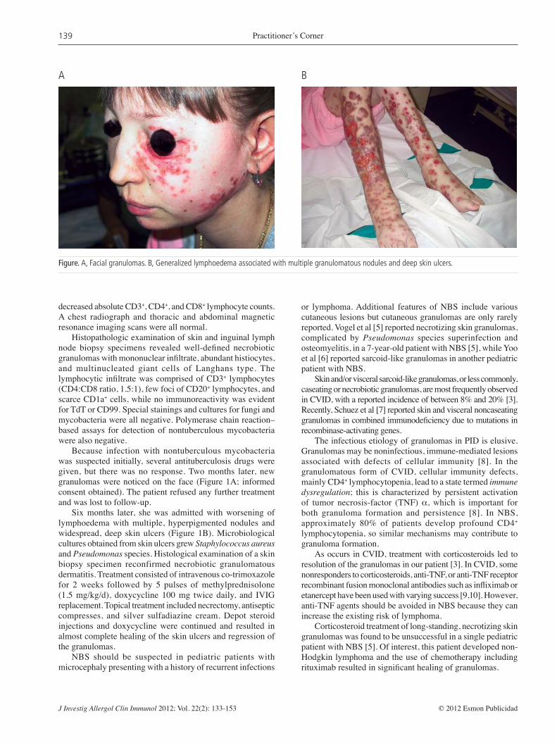
Figure. A, Facial granulomas. B, Generalized lymphoedema associated with multiple granulomatous nodules and deep skin ulcers.
A B
decreased absolute CD3+, CD4+, and CD8+ lymphocyte counts. A chest radiograph and thoracic and abdominal magnetic resonance imaging scans were all normal.
Histopathologic examination of skin and inguinal lymph node biopsy specimens revealed well-defi ned necrobiotic granulomas with mononuclear infi ltrate, abundant histiocytes, and multinucleated giant cells of Langhans type. The lymphocytic infi ltrate was comprised of CD3+ lymphocytes (CD4:CD8 ratio, 1.5:1), few foci of CD20+ lymphocytes, and scarce CD1a+ cells, while no immunoreactivity was evident for TdT or CD99. Special stainings and cultures for fungi and mycobacteria were all negative. Polymerase chain reaction–based assays for detection of nontuberculous mycobacteria were also negative.
Because infection with nontuberculous mycobacteria was suspected initially, several antituberculosis drugs were given, but there was no response. Two months later, new granulomas were noticed on the face (Figure 1A; informed consent obtained). The patient refused any further treatment and was lost to follow-up.
Six months later, she was admitted with worsening of lymphoedema with multiple, hyperpigmented nodules and widespread, deep skin ulcers (Figure 1B). Microbiological cultures obtained from skin ulcers grew Staphylococcus aureus and Pseudomonas species. Histological examination of a skin biopsy specimen reconfirmed necrobiotic granulomatous dermatitis. Treatment consisted of intravenous co-trimoxazole for 2 weeks followed by 5 pulses of methylprednisolone (1.5 mg/kg/d), doxycycline 100 mg twice daily, and IVIG replacement. Topical treatment included necrectomy, antiseptic compresses, and silver sulfadiazine cream. Depot steroid injections and doxycycline were continued and resulted in almost complete healing of the skin ulcers and regression of the granulomas.
NBS should be suspected in pediatric patients with microcephaly presenting with a history of recurrent infections
or lymphoma. Additional features of NBS include various cutaneous lesions but cutaneous granulomas are only rarely reported. Vogel et al [5] reported necrotizing skin granulomas, complicated by Pseudomonas species superinfection and osteomyelitis, in a 7-year-old patient with NBS [5], while Yoo et al [6] reported sarcoid-like granulomas in another pediatric patient with NBS.
Skin and/or visceral sarcoid-like granulomas, or less commonly, caseating or necrobiotic granulomas, are most frequently observed in CVID, with a reported incidence of between 8% and 20% [3]. Recently, Schuez et al [7] reported skin and visceral noncaseating granulomas in combined immunodefi ciency due to mutations in recombinase-activating genes.
The infectious etiology of granulomas in PID is elusive. Granulomas may be noninfectious, immune-mediated lesions associated with defects of cellular immunity [8]. In the granulomatous form of CVID, cellular immunity defects, mainly CD4+ lymphocytopenia, lead to a state termed immune dysregulation; this is characterized by persistent activation of tumor necrosis-factor (TNF) α, which is important for both granuloma formation and persistence [8]. In NBS, approximately 80% of patients develop profound CD4+ lymphocytopenia, so similar mechanisms may contribute to granuloma formation.
As occurs in CVID, treatment with corticosteroids led to resolution of the granulomas in our patient [3]. In CVID, some nonresponders to corticosteroids, anti-TNF, or anti-TNF receptor recombinant fusion monoclonal antibodies such as infl iximab or etanercept have been used with varying success [9,10]. However, anti-TNF agents should be avoided in NBS because they can increase the existing risk of lymphoma.
Corticosteroid treatment of long-standing, necrotizing skin granulomas was found to be unsuccessful in a single pediatric patient with NBS [5]. Of interest, this patient developed non-Hodgkin lymphoma and the use of chemotherapy including rituximab resulted in signifi cant healing of granulomas.

Practitioner’s Corner
J Investig Allergol Clin Immunol 2012; Vol. 22(2): 133-153© 2012 Esmon Publicidad
140
Allergy to Red Currant: Immunoglobulin E–Mediated Hypersensitivity to Lipid Transport Proteins (Pru p 3)
M Tomás,1 A Álvarez-Perea,1 A Ledesma,2 ML Baeza,1 M de Barrio1
1Allergy Service, Hospital General Universitario Gregorio Marañón, Madrid, Spain2Departamento de I+D, ALK Abelló SA, Madrid, Spain
Key words: Red currant. Rosaceae fruit allergy. LTP. Pru p 3.
Palabras clave: Grosella. Alergia a frutas rosáceas. LTP. Pru p 3.
We have reported a case of necrobiotic cutaneous granulomas in an adult patient with NBS who responded favorably to intensive antibiotic and immunosuppressive treatment with topical and systemic corticosteroids.
Acknowledgments This study was partially funded through a Serbian Ministry
of Science and Technology grants (No. 175065 and 175073) awarded to S Pasic.
References
1. Weemaes CMR, Hustinx TWJ, Scheres JMJC, et al. A new chromosomal instability disorder: Nijmegen breakage syndrome. Acta Paediatr Scand. 1981; 70: 557-64.
2. International Nijmegen breakage syndrome study group. Nijmegen breakage syndrome. Arch Dis Child. 2000; 82:400-6.
3. Ordeniz A, Cunningham-Rundles C. Granulomatous disease in common variable immunodefi ciency. Clin Immunol. 2009; 133:198-207.
4. Paller AS, Massey RB, Curtis MA, et al. Cutaneous granulomatous lesions in patients with ataxia-telangiectasia. J Pediatr. 1991; 119:917-22.
5. Vogel CA, Stratman EJ, Reck SJ, Lund JJ. Chronic noninfectious necrotizing granulomas in a child with Nijmegen breakage syndrome. Pediatr Derm. 2010; 27: 285-9.
6. Yoo J, Wolgamot G, Torgenson TR, Sidbury R. Cutaneous noncaseating granulomas associated with Nijmegen breakage syndrome. Arch Dermatol. 2008; 144:418-9.
7. Schuetz C, Huck K, Gudowius S, et al. An immunodefi ciency disease with RAG mutations and granulomas. N Engl J Med. 2008; 358:2030-8.
8. Aukrust P, Lien E, Kristoffersen AK, et al. Persistent activation of tumor necrosis factor system in a subgroup of patients with common variable immunodefi ciency - possible immunologic and clinical consequences. Blood. 1996; 87:674-81.
9. Lin HJ, Liebhaber M, Roberts RL, Dyer Z, Stiehm ER. Etanercept treatment for cutaneous granulomas in common variable immunodefi ciency. J Aller Clin Immunol. 2006; 117:878-82.
10. Tatayatikom A, Tatayatikom S, White AJ. Infl iximab treatment for severe granulomatous disease in common variable immunodefi ciency. Ann Allerg Asthma Immunol. 2005; 95:293-300.
❚ Manuscript received March 28, 2011; accepted for publication, September 8, 2011.
Srdjan Pasic Mother & Child Health Institute
11070 Belgrade, 8 R. Dakica StreetSerbia
E-mail:[email protected]
Individuals who are allergic to vegetables commonly react to other phylogenetically unrelated foods as a consequence of sensitization to panallergens. Profi lin and lipid transport protein (LTP) are the most frequent panllergens.
In Spain, the most frequent cause of food allergy is the Rosaceae family. Peach LTP (Pru p 3) is a major allergen in southern Europe. It causes severe reactions in allergic patients and has been associated with allergy to other Rosaceae, mainly apricot, cherry, and walnut.
Red currant is the fruit of Ribes rubrum, a deciduous bush from western Europe that belongs to the Grosuraliceae family. It is widely used in western countries because of its high content in vitamin C and carotenes and its powerful antioxidant properties.
Allergy to red currant is rare. We present a case of red currant–induced anaphylaxis that was diagnosed based on a suggestive history, positive skin test results, and determination of specific immunoglobulin (Ig) E. The presence of panallergens accounting for concomitant presence of food allergy and pollinosis was also studied.
A 19-year-old woman with a history of urticaria-angioedema in childhood due to sunfl ower seeds and peanut consulted after developing malaise, palpebral and genital angioedema, erythema on her face and neck, systemic hives, pruritus, and dysphonia immediately after eating duck meat with red currant sauce and lettuce with walnuts and goat cheese. After this episode, she tolerated almonds, sunfl ower seeds, pistachio, duck and other bird meats, onions peach, lettuce, and walnut.
Skin prick tests (SPT) were positive to grass pollen, Artemisia vulgaris and Platanus acerifolia pollen, peanut, walnut, chestnut, sunfl ower seed, apple, peach, and onion; prick-by-prick testing was positive to red currant and plum. SPTs with lettuce, pistachio, soy, strawberry, carrot, mustard, Anisakis simplex, and other pollens were negative.
Total IgE was 138 kUA/L. Specifi c IgE (CAP, Phadia, Uppsala, Sweden) levels (kUA/L) were as follows: Artemisia vulgaris, 1.54; Chenopodium album, 0.38; Platanus acerifolia, 0.59; strawberry, 0.67; apple, 3.63; peach, 7.71; plum, 0.70; onion, 0.45; peanut, 1.13; chestnut, 1.24; walnut, 2.82; and pistachio, 0.38. Specifi c IgE to Pru p 3 by CAP Pharmacia was 17.03 kUA/L.
IgE to red currant was positive by enzyme-linked immunosorbent assay. An allergenic extract was prepared, and IgE immunoblot of the patient’s serum revealed a single 14-kD band. Polyclonal rabbit serum was used to reveal the

J Investig Allergol Clin Immunol 2012; Vol. 22(2): 133-153 © 2012 Esmon Publicidad
Practitioner’s Corner141
25015010075
5037
252015
10
250150100755037
252015
10
1 2 M 1 2 M
A B
Figure. A, Immunoglobulin (Ig) E immunodetection with the currant extract: lane 1, negative control; lane 2, patient’s serum. B, Immunoblotting with polyclonal antiserum: lane 1, red currant polyclonal antiserum; lane 2, negative control. M indicates molecular weight (kDa).
presence of Pru p 3 in the red currant extract (14 kD) (Figure). The 14-kD band specifi cally recognized by the patient’s serum seemed likely to be an LTP.
Fruit and nuts, followed by legumes and vegetables, are the most common cause of food allergy in adults in Spain [1]. Many patients are sensitized to multiple foods, and in some cases sensitization is associated with pollen allergy. The sensitization pattern varies depending on the aerobiology of the area where the patient lives.
Birch pollen has been related to food allergy in northern Europe and in some regions of North America and Australia. However, birch trees are uncommon in Spain, and allergy to Rosaceae is more usually linked to grass pollen–fruit allergy syndrome.
These associations are based on the existence of IgE-mediated cross-reactivity. The most well known panallergens are Bet v 1 counterparts, profi lins, and LTP [2], which have been identifi ed as major allergens of some Rosaceae fruits. These proteins are highly stable, thus enabling them to cause severe systemic reactions (anaphylaxis, generalized urticaria).
Increasing levels of IgE to peach LTP are associated with skin reactivity to nuts, peanut, maize, rice, onion, orange, celery, and tomato [3]. Some studies suggest that high levels of IgE target common allergenic determinants of LTP, causing cross-reactivity to botanically unrelated vegetable foods in LTP-allergic patients [3].
However, although sensitization to LTP is not uncommon in pollen-allergic patients [4] and cross-reactivity in skin or in vitro tests is frequent in this population, the clinical signifi cance of this cross-reactivity is often unknown, and a complete clinical history or challenge test is often necessary to determine whether a certain food must be avoided [5]. Skin test results can remain positive, even when the culprit food is tolerated, as was the case with our patient.
Allergy to red currant is rare, and only 3 cases of systemic reaction have been reported [6-8]. Our group reported a case of anaphylaxis after ingestion of red currant in a patient with pollinosis,
globus sensation, and dysphagia with peach, apricot, and nectarine. The patient had specifi c IgE to currant and tolerated other Rosaceae fruits. Cross-reactivity between pollens and Rosaceae fruit allergens was demonstrated using CAP and immunoblot [8].
We present the case of a patient with subclinical pollen sensitization who suffered an episode of anaphylaxis after ingestion of red currant and was diagnosed with allergy by skin and in vitro tests. These studies demonstrated the presence of panallergens (LTP) in our red currant extract. Specifi c IgE to LTP is very likely to be the causative agent of concomitant pollen and food allergy sensitization.
Further studies are necessary to describe other allergenic components of red currant, since the increasing importance of this fruit in western society may cause a higher rate of allergic reactions in the near future.
Acknowledgments
Amalia Ledesma works at ALK Abelló SA. The remaining authors have no fi nancial relationship with pharmaceutical manufacturers.
References
1. Fernández-Rivas M. Reactividad cruzada en frutas y vegetales. Allergol et Immunopathol. 2003;31:141-6.
2. Fernández-Rivas M, Van Ree R, Cuevas M. Allergy to Rosaceae fruits without related pollinosis. J Allergy Clin Immunol. 1997;100:728-33.
3. Peach allergen components. In: Steiman H, Ruden S. Native & recombinant allergen components. Allergy-Which allergens? Uppsala: Phadia; 2010.
4. González-Mancebo E, González-de-Olano D, Trujillo MJ, Santos S, Gandolfo-Cano M, Meléndez A et al. Prevalence of sensitization to lipid transfer proteins and profi lins in a population of 430 patients in the south of Madrid. J Investig Allergol Clin Immunol. 2011;21:278-82.
5. Asero R. Lipid transfer protein cross-reactivity assessed in vivo and in vitro in the offi ce: pros and cons. J Investig Allergol Clin Immunol. 2011;21:129-36.
6. Kühl P, Kalveran CM, Gall H. Anaphylaxie auf rote Johannisbeere. Allergo J. 1997;7:17-9.
7. Zollner TM, Schmidt P, Kalveram CM, Emman AC, Boehncke WH. Anaphylaxis to red currants. Allergy. 2000;55:511.
8. Pérez-Ezquerra P, Vazquez M, De Barrio M, Flores V, Alvarez-Santullano A, Baeza ML. Currant allergy and the Rosaceae-grass pollen allergy syndrome: a case report. Ann Allergy Asthma Immunol. 2007;98:480-2.
❚ Manuscript received May 11, 2011; accepted for publication September 8, 2011.
Margarita Tomás, MD Servicio de Alergología
Hospital General Universitario Gregorio Marañón
Doctor Esquerdo, 4628007 Madrid, Spain
E-mail: [email protected]

Practitioner’s Corner
J Investig Allergol Clin Immunol 2012; Vol. 22(2): 133-153© 2012 Esmon Publicidad
142
Occupational Allergic Contact Urticaria to Crustacean in a Cook
G Pala,1 P Pignatti,1 L Perfetti,1 E Gentile,2 G Moscato1
1Allergy and Immunology Unit, Fondazione ‘Salvatore Maugeri’, Institute of Care and Research, Scientifi c Institute of Pavia, Pavia, Italy2Specialization School in Occupational Medicine, University of Pavia, Pavia, Italy
Key words: Occupational allergy. Contact urticaria. Crustacean. Seafood allergy. Cook.
Palabras clave: Alergia ocupacional. Urticaria de contacto. Crustáceos. Alergia marisco. Cocinero.
Abbreviations: Ig, immunoglobulin; NP, not performed; S, scratched skin; SI, stimulation index; SPT, skin prick test; U, unimpaired skin.aPercentage of CD63-positive basophils. bCD63% with extract/CD63% with wash buffer.
Table. Diagnostic Test Results Patch Test Basophil Activation Test SPTs
sIgE, 20 min 48 h CD63%a SIb
kU
A/L
Commercial allergens U S U S Lobster (Palinurus vulgaris) + <0.10 – – – – 52.8 21.1Shrimp + 0.44 – + – – 82.3 32.9Crab ++ 0.36 – – – – 83.1 33.2Dermatophagoides farinae – <0.10 – 1.7 0.7Dermatophagoides pteronyssinus – <0.10 – 1.5 0.6Anisakis simplex NP <0.10 – NP NP
Freshly prepared Crab 1:1 NP – + – – Crab 1:10 ++ – – – – Crab 1:100 + – – – – Lobster (Homarus vulgaris) 1:1 NP – – – – Lobster (Homarus vulgaris) 1:10 ++ – – – – Lobster (Homarus vulgaris) 1:100 + – – – – Recombinant allergens rPen a1 (tropomyosin) <0.10
Contact urticaria (CU) to crustacean is common in the general population, but its prevalence in occupational settings is largely unstudied. Although reactions to seafood have been documented, mainly among consumers, immune-mediated reactions have also been reported at work, especially in the fi shing and seafood processing industry [1,2]. The few cases reported among cooks were associated with oral allergy syndrome [3,4]. We report on a 33-year-old man who experienced wheals and fl ares after contact with the juice of frozen crab, water used for cooking lobster, and fresh shrimp.
No symptoms were reported after ingestion of crustacean or after contact with or ingestion of other foods. Symptoms appeared 17 years after working as a cook. We performed skin prick tests (SPT), specific immunoglobulin (Ig) E determination, and the basophil activation test with common allergens and crustacean (Table). We also performed SPTs and patch tests with freshly prepared extracts of crab and lobster juice. Patch tests were performed on the back, both on unimpaired skin and after scratching with a cotton swab drenched in alcohol (Table). The blood differential test revealed 6.2% eosinophils, and total serum IgE was 19.7 kUA/L. The results of SPTs and patch tests with freshly prepared extracts performed on 3 healthy controls were negative. In addition, immunoblotting with commercial crustacean extracts revealed no IgE-binding protein in serum. A diagnosis of occupational CU induced by crustacean was made, and the patient was advised to avoid any contact with this food.
To our knowledge, this is the fi rst report of occupational CU due to crustacean (crab, lobster, and shrimp) in a cook with no symptoms after ingestion. Denaturation of proteins during digestion may explain the absence of symptoms after ingestion. The absence of IgE-binding proteins in immunoblotting could account for the low total IgE levels. Positive skin test results with both raw and cooked crustacean (Table) suggest the involvement of a heat-resistant protein. Finally, the absence of sensitization to mite and detection of the negative tropomyosin (rPen a1) suggest primary sensitization to a cross-reactive crustacean allergen other than tropomyosin.

J Investig Allergol Clin Immunol 2012; Vol. 22(2): 133-153 © 2012 Esmon Publicidad
Practitioner’s Corner143
Acknowledgments
No funding was obtained for this study. The authors have no confl icts of interest to declare.
References
1. Desjardins A, Malo JL, L’Archevêque J, Cartier A, McCants M, Lehrer SB. Occupational IgE-mediated sensitization and asthma caused by clam and shrimp. J Allergy Clin Immunol. 1995;96:608-17.
2. Jeebhay MF, Robins TG, Lehrer SB, Lopata AL. Occupational seafood allergy: a review. Occup Environ Med. 2001:58:553-62.
3. Conde-Salazar L, Vazquez-Cortes S, Gonzalez de Olano D, Gonzalez-Guerra E, Raya Aguado C, Martinez Sanchez MJ, Canello Heranz MJ, Blancas M, Cuevas M. Occupational contact urticaria caused by seafood handling. Contact Dermatitis. 2005:53:178.
4. Yamaguchi J, Inomata N, Hirokado M, Shimakura K, Shiomi K, Ikezawa Z. [A case of occupational contact urticaria and oral allergy syndrome due to seafood]. Arerugi. 2007:56:49-53. Japanese.
❚ Manuscript received August 2, 2011; accepted for publication September 8, 2011.
Gianni Pala, MDAllergy and Immunology Unit
Fondazione ‘Salvatore Maugeri’, Institute of Care and Research, Scientifi c Institute of
PaviaVia Maugeri 10
Pavia, ItalyE-mail: [email protected]
Hyperimmunoglobulin E Syndrome Presenting With Renal Abscess
H Yuksekkaya,1 O Ozbek,2 F Goncu,3 M Keser,4 H Artac,5 P Harun,5 S Keles5
1Department of Pediatric Gastroenterology, Hepatology and Nutrition, Meram Medicine of Faculty, Selcuk University, Konya, Turkey2Department of Radiology, Meram Faculty of Medicine, Selcuk University, Konya, Turkey 3Department of Pediatrics, Meram Faculty of Medicine, Selcuk University, Konya, Turkey 4Department of Infectious Disease, Meram Faculty of Medicine, Selcuk University, Konya, Turkey 5Department of Pediatric Immunology, Meram Faculty of Medicine, Selcuk University, Konya, Turkey
Key words: Renal abscess. Hyper-IgE syndrome. Palabras clave: Absceso renal. Síndrome de hiper-IgE.
Hyperimmunoglobulin E syndrome (HIES), also known as Job syndrome, is a very rare primary immunodefi ciency
characterized by the clinical triad of high serum immunoglobulin (Ig) E levels (>2000 IU/mL), recurring staphylococcal skin abscesses, and pneumonia with formation of pneumatocele. Host defense defi ciencies in HIES include reduced neutrophil chemotaxis, impaired phagocytosis, and variably impaired T-cell function. Infections are mainly caused by Staphylococcus aureus and Streptococcus pneumoniae [1,2]. We report a case of multiple renal abscesses in a patient with HIES.
A 4-year-old girl was referred to our clinic with abdominal distension, diarrhea, fever, and failure to thrive. Her clinical history revealed 2 episodes of persistent diarrhea lasting 3-4 weeks 2 years previously. An impetigo-like skin lesion appeared intermittently on her chin. Her parents were not consanguineous, and she had 3 healthy siblings. Physical examination revealed her height and weight to be in the third percentile. Hepatosplenomegaly was not observed, and a review of her body systems was unremarkable. Interestingly, respiratory function was not tested before admission. Nevertheless, chest x-rays and computed tomography of the thorax revealed bronchiectasis in the right lung. Blood pressure was within normal ranges, but she had tachycardia and fever. Abnormal blood test results included the following: hemoglobin, 5 g/dL; white blood cell count, 16 200/mm3; erythrocyte sedimentation rate, 140 mm/h; and C-reactive protein, 120 mg/L (reference range, 0-5 mg/L). Other test results were normal. Urinalysis revealed large numbers of leukocytes and bacteria. The results of liver and kidney function testing were normal. Peripheral blood smear revealed an eosinophil percentage of 30%. The remaining test results were as follows: iron <10 μg/dL; iron-binding capacity, 136 μg/dL; ferritin, 268 μg/L (28-365); vitamin B12, 192 pg/mL (190-450); and folic acid, 2.4 ng/mL (3-12). Stool samples and blood cultures were negative. Ceftriaxone was started empirically because of fever. The urine culture antibiogram revealed methicillin-resistant S aureus (100 000 colonies/mL), which was sensitive to clindamycin. Ceftriaxone was switched to clindamycin. We initially considered the possibility of celiac disease because of the abdominal distension, severe anemia, and low folic acid level, but the results of celiac antibody testing were negative. Abdominal ultrasound and subsequent computed tomography revealed bilateral nephromegaly, bilateral multiple parenchymal abscesses, and an abscess measuring 4�5 cm in diameter affecting the calyces of the left kidney (Figure). Ultrasound-guided percutaneous nephrostomy was performed and a drainage tube inserted for 2 weeks. S aureus was isolated in the drainage fl uid and in urine. The immunological workup revealed elevated IgE titers (up to 13 000 IU/mL) and an eosinophil count of up to 3900 cells/μL. Titers for IgG, IgA, IgM, and IgG were normal. Analysis of the lymphocyte subpopulation revealed normal B- and T-lymphocyte counts. Delayed-type hypersensitivity reaction to the tuberculin skin test was negative. Lymphocyte proliferation was normal on stimulation with mitogen. Neutrophil functional responses were assessed using fl ow cytometry. Neutrophil chemotaxis was signifi cantly impaired. The patient had a homozygous deletion starting in exon 28 and extending to the terminal exon; there was no mutation in STAT3 gene. The patient was diagnosed with HIES (HIES score, 31). The abscesses began to decrease in size after

Practitioner’s Corner
J Investig Allergol Clin Immunol 2012; Vol. 22(2): 133-153© 2012 Esmon Publicidad
144
A B
Figure. Bilateral multiple renal abscesses. A, T1-weighed magnetic resonance image of the abdomen. B, Computed tomography image of the abdomen.
10 days with clindamycin and had disappeared by the third week on ultrasound. Clindamycin was continued for 2 months. The patient was discharged, low doses of prophylactic antibiotic (cotrimoxazole, 4 mg/kg/d) were started, and intravenous immunoglobulin was continued every 3 weeks. The follow-up examination at 15 months was unremarkable.
Renal abscess is a rare disease in childhood, although it involves potentially fatal complications (urinary tract infections or bacteremia). It can result from hematogenous spread or as a complication of infection from the lower urinary tract. S aureus and Escherichia coli are the most commonly isolated pathogens [3,4]. In HIES, however, infections are caused mainly by S aureus or S pneumoniae [1]. In our case, there was no vesicoureteral refl ux or anatomical problem. S aureus was isolated in both urine and abscess drainage fl uid. In addition to classic fi ndings, HIES is characterized by conditions such as tuberculous brain abscess [5], endocarditis, and epidural abscess [6], although disseminated renal abscesses have not been described. To our knowledge, this is the fi rst case of disseminated multiple renal abscesses in a patient with HIES to be reported in the English-language literature.
No cure is available for HIES. When abscesses, skin lesions, and pneumonia have developed, long-term high-dose intravenous antibiotics are required to eliminate infectious agents, and surgical drainage may occasionally be necessary [7]. Therapy with intravenous immunoglobulin can affect IgE levels owing to increased immunoglobulin catabolism or IgE neutralization via an anti-idiotype network [8]. Renal abscesses can be managed by medical treatment alone; interventional treatment should be reserved for large collections of abscesses and patients with clinical impairment. Perinephric and mixed
abscesses can be successfully managed by interventional treatment. Abscesses >5 cm or those that fail to respond to antibiotic treatment should be considered for drainage guided by ultrasound or computed tomography [4,9]. Our patient had both an abscess of approximately 5 cm in diameter and immunodeficiency. We performed percutaneous drainage and administered intravenous immunoglobulin. We did not consider a surgical intervention because of the disseminated parenchymal renal abscesses.
Vancomycin and cefepime are excellent choices to treat suspected renal abscess in a previously healthy child [10]; however, we preferred to use clindamycin, which completely penetrates the abscess. The role of long-term prophylactic antibiotic therapy has not been closely investigated in patients with HIES, but the consensus of opinion favors prophylactic therapy with an antistaphylococcal antibiotics such as cotrimoxazole or cephalosporin [2]. We prescribed cotrimoxazole as prophylaxis and therapy with intravenous immunoglobulin; our patient did not have to be hospitalized during her 15 months of follow-up.
In conclusion, disseminated renal abscess rarely progresses to HIES. The most important causal agent of renal abscess is S aureus, both in HIES and in other tissue abscesses. Clindamycin with drainage is a sound choice for the treatment of renal abscesses.
Acknowledgments
We thank Talal Chatila, MD, from University of California at Los Angeles for the mutation analysis.

J Investig Allergol Clin Immunol 2012; Vol. 22(2): 133-153 © 2012 Esmon Publicidad
Practitioner’s Corner145
References
1. Grimbacher B, Holland SM, Gallin JI, Greenberg F, Hill SC, Malech HL, Miller JA, O’Connell AC, Puck JM. Hyper-IgE syndrome with recurrent infections–an autosomal dominant multisystem disorder. N Engl J Med. 1999;340:692-702.
2. Grimbacher B, Holland SM, Puck JM. Hyper-IgE syndromes. Immunol Rev. 2005; 203:244-50.
3. Wang YT, Lin KY, Chen MJ, Chiou YY. Renal abscess in children: a clinical retrospective study. Acta Paediatr Taiwan. 2003;44:197-201.
4. Cheng CH, Tsai MH, Su LH, Wang CR, Lo WC, Tsau YK, Lin GJ, Huang YC, Chiu CH, Lin TY. Renal abscess in children: a 10-year clinical and radiologic experience in a tertiary medical center. Pediatr Infect Dis J. 2008;27:1025-7.
5. Metin A, Uysal G, Güven A, Unlu A, Oztürk MH. Tuberculous brain abscess in a patient with hyper IgE syndrome. Pediatr Int. 2004;46:97-100.
6. Cunha BA, Krol V, Kodali V. Methicillin-resistant Staphylococcus aureus (MRSA) mitral valve acute bacterial endocarditis (ABE) in a patient with Job’s syndrome (hyperimmunoglobulin E syndrome) successfully treated with linezolid and high-dose daptomycin. Heart Lung. 2008;37:72-5.
7. Freeman AF, Kleiner DE, Nadiminti H, Davis J, Quezado M, Anderson V, Puck JM, Holland SM. Causes of death in hyper-IgE syndrome. J Allergy Clin Immunol. 2007;119:1234-40.
8. Bilora F, Petrobelli F, Boccioletti V, Pomerri F. Moderate-dose intravenous immunoglobulin treatment of Job’s syndrome. Case report. Minerva Med. 2000; 91: 113–116.
9. Siegel JF, Smith A, Moldwin R. Minimally invasive treatment of renal abscess. J Urol. 1996;155:52-5.
10. Dalla Palma L, Pozzi-Mucelli F, Ene V. Medical treatment of renal and perirenal abscesses: CT evaluation, Clin Radiol. 1999;54:792-7.
❚ Manuscript received March 30, 2011; accepted for publication September 9, 2011.
Hasan YüksekkayaAlavardı Mah. Hekim Sadri Sok. No: 21 D:
5, Meram42080 Konya, Turkiye
E-Mail: [email protected]
A Case of Allergic Bronchopulmonary Aspergillosis Treated With Omalizumab
I Sastre, J Blanco, H Mata, F GarcíaUnidad de Alergología, Complejo Asistencial Universitario de Burgos, Burgos, Spain
Key words: Aspergilosis. Aspergillus. Omalizumab. Palabras clave: Aspergillosis. Aspergillus. Omalizumab.
Allergic bronchopulmonary aspergillosis (ABPA) is an inflammatory lung disease of immune origin that usually occurs as a complication of allergic asthma or cystic fi brosis. It is characterized by reversible airway obstruction, transient pulmonary infi ltrates, eosinophilia, and fever and is caused by an immunoglobulin (Ig) E–mediated hypersensitivity response to Aspergillus fumigatus, which colonizes the bronchial tree [1].
We present the case of a 33-year-old woman with a 6-month history of dyspnea and wheezing. Her blood count was normal, and there were no relevant fi ndings in the chest x-ray or paranasal sinuses. Total IgE was >1000 IU/mL. Spirometry revealed an obstructive pattern, and the result of the bronchodilation test was positive. Skin tests were positive for A fumigatus (12 mm) and Penicillium notatum (10 mm). Precipitins against A fumigatus were also measured, and the results were negative.
The patient was diagnosed with allergic bronchial asthma. In the following years she reported worsening of clinical symptoms associated with cough, brown expectoration, and dyspnea on exertion. Determinations were repeated and disclosed eosinophilia (1100/mm³). Chest x-ray revealed an infi ltrate in the middle lobe of the right lung.
Skin tests were positive to A fumigatus (10 mm). Total IgE was 3090 IU/mL, specifi c IgE to A fumigatus was 86 kUA/L, and determination of IgG and precipitins to A fumigatus was positive. The patient was diagnosed with ABPA and started treatment with prednisone 0.5 mg/kg/d.
In the following years, her progress was unsatisfactory, with several acute exacerbations. She continued to receive prednisone at a maintenance dose of 15 mg every other day. Spirometry revealed the following values: forced vital capacity (FVC), 2250 mL (75%); forced expiratory volume in the fi rst second of expiration (FEV1), 1370 mL (60%); and FEV1/FVC, 60%. Determination of IgE specifi c to recombinant A fumigatus antigens provided the following values: rAsp f 1, 9.45 kUA/L; rAsp f 2, 2.12 kUA/L; rAsp f 3, 6.94 kUA/L; rAsp f 4, 2.39 kUA/L; and rAsp f 6, <0.35 kUA/L.
Thoracic computed tomography (CT) revealed bronchiectasis in the middle and right upper lobes.
In order to reduce systemic corticosteroids and improve disease outcome, treatment was started with itraconazole [2] at 200 mg/12 h, although it was discontinued because of lack of improvement in clinical, laboratory, and spirometry parameters. The patient experienced an acute exacerbation and prednisone-induced side effects (cushingoid facies), and liver enzyme values increased.
In July 2009, at 53 years of age, she started treatment with

Practitioner’s Corner
J Investig Allergol Clin Immunol 2012; Vol. 22(2): 133-153© 2012 Esmon Publicidad
146
A B
Figure. Computed tomography scan showing reduced bronchiectasis after 12 months of treatment with omalizumab. Left, Before therapy with omalizumab. Right, After therapy with omalizumab..
omalizumab at 375 mg/2 wk. After 3 months of treatment, her clinical condition improved, with reduced cough and expectoration. The dose of prednisone was reduced to 5 mg/48 h of prednisone. The last spirometry test, performed in July 2010, disclosed the following values: FVC, 2890 mL (97.5%); FEV1, 1620 mL (72.6%); and FEV1/FVC, 70.7%.
No new acute exacerbations were detected in this 12-month period.
A second thoracic CT scan revealed persistent bilateral bronchiectasis at multiple levels with less bronchial wall thickening (indicative of remission of infl ammation), as well as a lower degree of bronchiectasis and a reduced number of micronodular peribronchial opacities (indicative of reduced impaction and infl ammation of the distal airways) (Figure).
ABPA is an infl ammatory lung disease of immune origin that manifests as an IgE-mediated type I hypersensitivity response to A fumigatus antigens in the bronchial tree. It is associated with the toxic action of enzymes released by the mold in the bronchial tree.
The disease consists of 5 stages, ranging from the acute stage (pulmonary infi ltrate, eosinophilia, and increased total IgE) to pulmonary fi brosis. Our patient’s disease was in stage IV (corticosteroid-dependent stage), in which treatment with systemic corticosteroids cannot be discontinued, as new acute exacerbations may occur.
Omalizumab is a humanized recombinant monoclonal antibody that selectively binds to IgE to inhibit the immune response to an allergen. It targets the high affi nity Fc receptor and prevents free serum IgE from binding to mast cells and other effector cells, in turn preventing IgE-mediated infl ammation. It is indicated mainly for the treatment of
persistent allergic bronchial asthma that is not well controlled with other treatments. However, it is increasingly successful in several IgE-mediated conditions. Five case reports of ABPA have been published to date, and all were recorded in patients with cystic fi brosis whose clinical condition improved. In all 5 cases, the dose of systemic corticosteroids was reduced without starting treatment with omalizumab [3-7].
We report a case of corticosteroid-dependent ABPA (stage IV) with unsatisfactory clinical progress and multiple acute exacerbations for which no response was obtained with antifungals. The patient’s condition improved considerably after starting treatment with omalizumab. No new acute exacerbations were recorded, and the dose of systemic corticosteroids was reduced by 70%. Furthermore, a signifi cant improvement in respiratory function and radiological fi ndings was observed in the last CT scan. This is the fi rst case in which such an improvement has been observed.
As ABPA is an infl ammatory disease of which the main cause is a type I hypersensitivity mechanism mediated by IgE against A fumigatus antigens, the use of omalizumab can improve disease control during the corticosteroid-dependent stage. Use of this agent could be extended in the event of recurrences in order to prevent progression to subsequent stages.
Acknowledgments
Laboratorios NOVARTIS translated the article from Spanish to English.

J Investig Allergol Clin Immunol 2012; Vol. 22(2): 133-153 © 2012 Esmon Publicidad
Practitioner’s Corner147
References
1. Knusten AP, Chauhan B, Slavin RS. Cell mediated immunology in allergic bronchopulmonary aspergillosis. Immunol Allergy Clin N Am. 1998;18:575-600.
2. Martinez A., Herrera. Aspergilosis broncopulmonar alérgica e itraconazol. Arch Bronconeumol. 1997;33(11):599-600.
3. Brinkmann F, Schwerk N, Hansen G, Ballmann M. Steroid dependency despite omalizumab treatment of ABPA in cystic fi brosis. Allergy. 2010;65(1):134-5.
4. Lebecque P, Leonard A, Pilette C. Omalizumab for treatment of ABPA exacerbations in CF patients. Pediatr Pulmonol. 2009;44(5):516.
5. Kanu A, Patel K. Treatment of allergic bronchopulmonary aspergillosis (ABPA) in CF with anti-IgE antibody (omalizumab). Pediatr Pulmonol. 2008;43(12):1249-51.
6. Zirbes JM, Milla CE. Steroid-sparing effect of omalizumab for allergic broncho-pulmonary aspergillosis and cystic fi brosis. Pediatr Pulmonol. 2008;43(6):607-10.
7. Van der Ent CK, Hoekstra H, Rijkers GT. Successful treatment of allergic bronchopulmonary aspergillosis with recombinant anti-IgE antibody. Thorax. 2007;62(3):276-7.
❚ Manuscript received February 7, 2011; accepted for publication September 14, 2011.
Iván José Sastre PérezUnidad de Alergología.
Complejo asistencial universitario de Burgos
Burgos, Spain E-mail: [email protected]
Threshold Doses in Specifi c Oral Tolerance Induction in Children With Egg Allergy
A Letrán, M Espinazo, MC López, FJ Caro, L Gómez, P Lobatón, J Dafonte, F Moreno Clínica Dr. Lobatón, Cádiz, Spain
Key words: Egg allergy. Egg immunotherapy. Food allergy. Specifi c oral tolerance induction.
Palabras clave: Alergia a huevo. Inmunoterapia frente a huevo. Alergia alimentaria. Inducción de tolerancia oral específi ca.
The current standard of care for egg allergy includes strict allergen avoidance and ready access to adequate pharmacotherapy in the event of accidental ingestion. Although most patients outgrow their allergy over time, some continue to have reactions over many years. Therefore, specifi c oral tolerance induction (SOTI) protocols have been developed. Tolerance is achieved by administering small doses, which are increased slowly to an amount equivalent to the usual daily oral intake. Subsequently, egg is given daily at a maintenance dose [1,2].
We present our experience with an open egg SOTI protocol in children with persistent egg allergy. We recorded age, sex, last documented adverse reaction with egg, skin prick test
results (mm), total-specifi c immunoglobulin (Ig) E titers (IU/mL), other allergic comorbid conditions, and threshold doses for each visit. The number and type of secondary effects of SOTI and their treatment were evaluated. The exclusion criteria were a history of anaphylaxis to egg within 6 months of beginning the protocol, uncontrolled asthma, and severe atopic dermatitis. Written informed consent was obtained in all cases.
With a target tolerance of 1 complete egg, we mixed a whole large size raw egg with cow or soymilk yogurt (0.09 g/mL of ovalbumin [OVA] and 0.02 g/mL of ovomucoid [OVM]) [3]. SOTI was initiated at our clinic with a placebo control from a low initial dose (0.01 mL of mixture), which was doubled every 20 minutes. Peak expiratory fl ow and blood pressure were recorded (build-up phase). Patients subsequently repeated the last tolerated dose every day at home (maintenance phase), thus providing the fi rst step for the next visit. Maintenance consisted of the daily administration of the corresponding tolerated part of a similar-size 1-egg omelet. Visits were programmed on a monthly basis.
The initial sample comprised 17 patients (11 boys, 6 girls); 6 patients completed the entire protocol and 3 patients dropped out. Mean age at enrollment was 8 years (range, 4-14 years). The most common symptoms related to prior exposure to egg were urticarial rash (7 patients) and anaphylaxis (6 patients). Thirteen patients (75%) had already had a positive response in an open challenge with raw egg white (average of 6 months before) and the rest (25%) had had a recent direct adverse reaction to egg. Five patients (29%) had atopic dermatitis.
In general, the build-up phase was well tolerated with no need for intramuscular adrenaline, intravenous fl uids, or oxygen treatment. At the fi rst visit, the median tolerated dose was 1.6 mL (corresponding to 0.284 accumulated grams of OVA and 0.063 accumulated grams of OVM: 1/65 of the whole egg). All the patients except 2 (patients 8 and 9) received more than 3 doses at this visit. One patient (patient 8) developed intense abdominal pain and vomited with the first dose; therefore, the protocol was stopped. Patient 13 experienced respiratory symptoms (0.2-mL dose) that resolved with ß2-agonists and did not prevent the protocol from continuing. The median tolerated dose at the second visit was 4.8 mL (0.869 accumulated grams of OVA and 0.195 accumulated grams of OVM: 1/17 of the whole egg). In 2 cases, the dose achieved at the fi rst visit could not be increased: in one patient (patient 8) from 0.01 mL, because of acute rhinoconjunctivitis, and in the other (patient 6) from 6.4 mL, because of abdominal pain and emesis. The fi rst patient received a short cycle of systemic corticosteroids. The median tolerated dose for the third and fourth visits were 9.6 mL (1/9) and 12.8 mL (1/8), respectively. No more than 7 visits were recorded.
Few severe adverse reactions were recorded during the maintenance phase. Eight children (47%) experienced intermittent self-limiting mild oral pruritus with no systemic symptoms. One patient (number 15) developed generalized urticaria at home with the previously tolerated dose of 12.8 mL when ingestion coincided with ibuprofen treatment (second dropout). Patient 3 had facial angioedema and dyspnea at the previous tolerated dose of 25.6 mL (1/4) and had to attend the emergency department. The parents did not report any concomitant fact that could explain the reaction.

Practitioner’s Corner
J Investig Allergol Clin Immunol 2012; Vol. 22(2): 133-153© 2012 Esmon Publicidad
148
Table. Sensitization Patterns and Threshold Doses (TD) in Milliliters of Mixture and Symptoms for Each Visit and Patient
Patient Skin Specifi c First Second Third Fourth Fifth Sixth Seventh Prick IgE Values, Visit TD Visit TD Visit TD Visit TD Visit TD Visit TD Visit TD Test, mm IU/mL and and and and and and and Symptoms Symptoms Symptoms Symptoms Symptoms Symptoms Symptoms 1 EW: 7 EW: 3.5 1.6 OVA: 4 OVA: 1.27 OP OVM: 4 OVM: 1.27 2 EW: 5 EW: 4.57 0.2 0.8 1.6 12.8 12.8 OVA: 4 OVA: 8.79 FE AP, E FE S, AP, E R, AP, E OVM: 3 OVM: 0.21 3 EW: 4 EW: 20.5 0.2 3.2 12.8 25.6 OVA: 4 OVA: 4.5 AP AP, D S, E, D R OVM: 5 OVM: 12.6 4 EW: 4 EW: 35 0.2 1.6 3.2 6.4 6.4 12.8 OVA: 4 OVA: 12.7 AP AP AP, E D, R AP, E OP, U OVM: 4 OVM: 0 5 EW: 6 EW: 0 1.6 3.2 6.4 25.6 OVA: 5 OVA: 0 AP AP AP AP OVM: 3 OVM: 0 6 EW: 5 EW: 0 6.4 6.4 OVA: 5 OVA: 0.65 OP, R AP, E, S OVM: 3 OVM: 0.45 7 EW: 4 EW: 30 0.4 51.2+33a
OVA: 3 OVA: 12 R None OVM: 3 OVM: 8 8 EW: 7 EW: 15 0.01 0.01 OVA: 8 OVA: 8 AP, E FE, R, OP OVM: 8 OVM: 6 9 EW: 7 EW: 100 0.1 0.8 1.6 3.2 6.4 25.6 51.2+33a
OVA: 6 OVA: 100 OP, FE AP E AP, N OP FE N OVM: 6 OVM: 100
10 EW: 8 EW: 12 1.6 6.4 12.8 OVA: 6 OVA: 10 E, D AP, OP E, D, R OVM: 5 OVM: 10 11 EW: 6 EW: 90 0.4 6.4 12.8 12.8 OVA: 7 OVA: 57 AP, E R, OP AP, S E, R, AP OVM: 9 OVM: 60 12 EW: 5 EW: 100 1.6 51.2+33a
OVA: 6 OVA: 35 OP None OVM: 4 OVM: 45 13 EW: 5 EW: 8.5 0.2 3.2 25.6 25.6 51.2 51.2+33a
OVA: 6 OVA: 4 W, OP AP, E S, E FE, E E N OVM: 6 OVM: 4.5 14 EW: 3 EW: 5 12.8 OVA: 3 OVA: 2.5 R, S OVM: 3 OVM: 3.2 15 EW: 4 EW: 20 1.6 12.8 OVA: 5 OVA: 20 OP, E AP OVM: 6 OVM: 22 16 EW: 4 EW: 30 1.6 51.2+33a
OVA: 4 OVA: 1.2 AP None OVM: 4 OVM: 0 17 EW: 6 EW: 15 51.2+33a
OVA: 4 OVA: 1.34 (Rest) OVM: 5 OVM: 1.42 None
Abbreviations: AP, abdominal pain; D, diarrhea; E, emesis; EW, egg white; FE, facial erythema; Ig, immunoglobulin; N, nausea; OP, oral pruritus; OVA, ovalbumin; OVM, ovomucoid; R, rhinorrhea; S, sneezing; U, urticaria; W, wheezing.aFinal dose of the protocol.

J Investig Allergol Clin Immunol 2012; Vol. 22(2): 133-153 © 2012 Esmon Publicidad
Practitioner’s Corner
❚ Manuscript received July 6, 2011; accepted for publication September 15, 2011.
Antonio LetránClínica Dr. Lobatón
Avenida Fernández Ladreda, 911008 Cádiz, Spain
Email: [email protected]
149
After starting with a low dose at the fi rst visit (1/65 of the egg), we observed that most patients were able to tolerate greater doses at each subsequent visit (3-fold from the fi rst to the second visit and 2.5-fold with the following steps until the fourth visit). Consistent with published data, it is recommended that food challenge should start with an individualized dose based on the clinical history that is generally lower than the quantity that caused the symptoms [4,5]. The position paper of the European Academy of Allergology and Clinical Immunology proposes starting food challenge with 1 mg of egg [6]. However, in our opinion, this approach is subject to practical drawbacks owing to an incomplete standardization procedure and the diffi culty involved in trying to divide a cooked or raw egg into equal parts and start the protocol with a high initial dose (1/16, 1/8). If the challenge result is positive, we should wait as long as is necessary until natural tolerance is achieved. Consequently, we are faced with a series of questions. First, even though food challenge is the current gold standard in food allergy, can we use protocols such as ours both to monitor and to induce tolerance? Is it always necessary to expose the patient to a risky amount of egg before SOTI? Is it so important to perform a food challenge before SOTI if both approaches involve controlled exposure? We used egg SOTI as the only challenge method and postulate that it could replace the classic food challenge.
References
1. Niggeman B, Staden U, Rolinck-Werninghaus C, Beyer K. Specifi c oral tolerance induction in food allergy. Allergy. 2006;61:808-11.
2. Skripak JM, Sampson HA. Towards a cure for food allergy. Curr Opin Immunol. 2008;20(6):690-6.
3. Benhamou AH, Caubet JC, Eigenmann PA, Nowak-Wegrzyn A, Marcos CP, Reche M, Urisu A. State of the art and new horizons in the diagnosis and management of egg allergy. Allergy. 2010;65:283-9.
4. Taylor SL, Hefl e SL, Bindslev-Jensen C, Bock SA, Burks AW, Christie CL, Hill DJ, Host A, Hourihane JB, Lack G, Metcalfe DD, Moneret-Vautrin DA,Vadas PA, Rance F, Skrypec DJ, Trautman TA, Yman IM, Zeiger RS. Factors affecting the determination of threshold doses for allergenic foods: How much is too much? J Allergy Clin Immunol. 2002;109:24-30.
5. Nowak-Wegrzyn A, Assa’ad AH, Bahna SL, Bock SA, Sicherer SH, Teuber SS. Work Group report: Oral food challenge testing. J Allergy Clin Immunol. 2009;123:S365-83.
6. Bindslev-Jensen C, Ballmer-Weber BK, Bengtsson U, Blanco C, Ebner C, Hourihane J, Knulst AC, Moneret-Vautrin DA, Nekam K, Niggemann B, Osterballe M, Ortolani C, Ring J, Schnopp C, Werfel T. Standardization of food challenges in patients with immediate reactions to foods – position paper from the European Academy of Allergology and Clinical Immunology. Allergy. 2004;59:690-7.
Diagnosis of Autosomal-dominant Hyperimmunoglobulin E Syndrome
P Garcés,1,3 F Garcia-Martin,2 D Detkova,1 T Espanol1
1Immunology Unit, University Hospital Vall d’Hebron, Barcelona. 2Infectious and Immunodefi ciencies Unit, Hospital Carlos Haya, Malaga, Spain 3Hospital Hernan Henriquez Aravena, Temuco, Chile
Key words: Hyper-IgE syndrome. STAT3 mutations. Autosomal-dominant inheritance Palabras clave: Síndrome de Hiper-IgE. Mutaciones en STAT3. Herencia autosómica-dominante.
Hyperimmunoglobulin E syndrome (HIES) is a primary immunodefi ciency with immunological and nonimmunological manifestations [1,2]. The main immunological manifestations comprise high serum immunoglobulin (Ig) E levels, eosinophilia, eczema, and skin and lung infections; the main nonimmunological features are facies, scoliosis, retained primary teeth, joint hyperextensibility, bone fractures following minimal trauma, and craniosynostosis. Such a broad spectrum can make diagnosis diffi cult, and typical features may become apparent over time, when bone and dental abnormalities appear. In pediatric patients, a differential diagnosis with severe atopy [3,4], eczema, and relatively high IgE titers is mandatory in children with no family history of this disease. Hence, any diagnostic algorithm is likely to underdiagnose younger patients. Furthermore, recently published score values [5] are not always helpful in pediatric patients. Both autosomal-dominant inheritance traits (AD-HIES) and autosomal-recessive inheritance traits (AR-HIES) have been described. Heterozygous mutations in the signal transducer and activator of transcription 3 gene (STAT3) have been identifi ed in patients with AD-HIES [6,7], whereas homozygous mutations in the dedicator of cytokinesis 8 gene (DOCK8) have been associated with the recessive form of the disease [8]. We report 3 pediatric cases of AD-HIES diagnosed at our center over the last 30 years and highlight the differences in presentation and prognosis.
We reviewed the clinical histories of 3 patients diagnosed with HIES from 1980 to the present. Ig levels (IgE and IgG) and lymphocyte subsets were measured using routine laboratory techniques. When possible, genetic testing for mutations in STAT3 was performed at the Royal Free Hospital, London, UK, after written informed consent was obtained from the parents (Table).
Patient 1
A 2-month-old boy presented with eczema that appeared during the fi rst month of life and led to severe infections (generalized impetigo and skin abscesses). He also suffered from recurrent episodes of thrush and respiratory infections. At the age of 13 months, he underwent lobectomy for removal of a pneumatocele in the left lung. The patient developed

Practitioner’s Corner
J Investig Allergol Clin Immunol 2012; Vol. 22(2): 133-153© 2012 Esmon Publicidad
150
Table. Clinical, Immunological, and Mutation Data
Patient No./Sex 1/Male 2/Female 3/Male Age at diagnosis/current age, y 7/died at 14 y 4/17 3/died at 4 y
NIH scorea 74 67 41
IgE, IU/mL >4000 13 800 880
2140 2562 3952Eosinophils/mL
Eczema Severe Moderate Severe
Infections Otitis, bronchitis, Recurrent otitis, Bronchitis, pneumonia, pneumonia, pyopneumothorax, pyopneumothorax, pneumothorax, pleuropneumonia, superinfected eczema recurrent lung abscess, impetigo, abscesses on bronchopulmonary neck and scalp aspergillosis
Microorganisms Unidentifi ed Streptococcus, Pneumocystis jiroveci, Unidentifi ed Pseudomonas aeruginosa, Salmonella, Streptococcus, Klebsiella, Acinetobacter anitratus, Haemophilus infl uenzae, Haemophilus infl uenzae, Candida albicans, Plasmocoagulase-positive Candida albicans, Aspergillus Staphylococcus, Plasmocoagulase-positive Pseudomonas aeruginosa, Staphylococcus Candida albicans
Pulmonary sequelae Bronchiectasis, Bronchiectasis, Multiple pneumatoceles pneumatocele multiple pneumatoceles
Other manifestations Arthralgia and Hyperlaxity, failure swelling of large and to shed primary teeth small joints, hyperlaxity
Treatments Occasional intramuscular Intravenous Antibiotic therapy of gammaglobulin, immunoglobulin, recurrent infections antibiotic therapy antibiotic and antifungal according to infections prophylaxis
STAT3 mutations Not analyzed c.1145 G>A Analyzed in the sister and her p.382Arg>Gln daughter, some years later c.1397 A>G N p466Asn>Ser
Abbreviation: NIH, National Institutes of Health.aAs described in Ref. 6 (score>40 high suspicion)
bronchiectasis; clinical manifestations included acropachy and episodes of hemoptysis. Despite antibiotics and therapy with intramuscular immunoglobulin, the patient died at the age of 14 years (1987), before the results of the genetic study became available. Given the severity of the disease, high National Institutes of Health score, and similarities with other cases, the patient was considered to have AD-HIES.
Patient 2
A 17-month-old girl (nonconsanguineous parents) developed Pneumocystis jiroveci–induced pneumonia at the age of 3 months. She had also had oral thrush and atopic eczema since birth. Severe combined immunodefi ciency and phagocytic disorder were ruled out at that point.
At 2 years of age, she presented with lung abscesses complicated by pneumothorax, subcutaneous emphysema, and pneumatocele. Segmentectomy and several courses of antibiotics were required and treatment with intravenous immunoglobulin was initiated. Laboratory analyses revealed hypereosinophilia and hypergammaglobulinemia.
Since then, the patient has presented several bacterial infections, aspergilloma, and recurrent life-threatening episodes of hemoptysis.
Today, she has typical HIES facies (high palate, wide nasal bridge) joint hyperextensibility, and discrete scoliosis and is receiving intravenous immunoglobulin and prophylaxis with antibiotics and antifungals. The mutation in STAT3 was confi rmed in 2007.

J Investig Allergol Clin Immunol 2012; Vol. 22(2): 133-153 © 2012 Esmon Publicidad
Practitioner’s Corner
Patient 3
A 3-year-old boy was diagnosed with HIES based on a clinical history of obstructive bronchitis, urethritis, pyodermatitis, and skin abscesses since the age of 10 months, with high IgE and IgG titers. The patient died from complications of chronic bullous lung disease at the age of 4 years.
His mother had died from chronic lung disease at the age of 40, although no defi nitive diagnosis was established. She had no history of asthma or smoking, and no hypogammaglobulinemia was detected. Plasma IgE levels were not tested. She could have had undiagnosed HIES.
The patient’s sister was diagnosed with HIES at the age of 30. She had had recurrent respiratory infections (with isolation of Staphylococcus aureus on 1 occasion), bronchiectasis, moderate eczema, recurrent oral candidiasis, spontaneous rib fractures (radiographic fi nding), and osteoporosis. IgE values were 32 000 IU/mL, and a blood test showed hypereosinophilia. In 2007, a molecular diagnosis confi rmed a mutation in STAT3. The second daughter of this patient (ie, the niece of Patient 3) was diagnosed with HIES at the age of 20 months based on family history, eczema since birth, occasional oral candidiasis, and 1 episode of respiratory tract infection. She also had the same mutation as her mother.
HIES is a very rare primary immunodefi ciency, representing less than 2% of cases in the registry of the European Society for Immunodefi ciencies (www.esid.org). It can present in childhood with heterogeneous clinical features and variable prognosis.
Clinical diagnosis can be diffi cult to establish in children, owing to similarities with other clinical entities, and a differential diagnosis between HIES and allergy in children with severe eczema and relatively high serum IgE levels is mandatory [3]. Neonatal rash could help to differentiate AD-HIES from AR-HIES. Genetic studies can confi rm the diagnosis and enable close follow-up and prompt antibiotic and antifungal prophylaxis and, in selected cases, IVIG therapy. Furthermore, molecular diagnosis facilitates prenatal diagnosis and genetic counseling.
Clinical heterogeneity with the same mutation in the same family (Patient 3) is not uncommon in other primary immunodefi ciencies [9] and probably refl ects the role of other nonimmunological factors in the development of the disease.
Diminished counts of CD4+ T lymphocytes secreting interleukin 17 (subtype 17 helper T cell [TH17]) have been observed in STAT3-defi cient patients, and a major role has been assigned to this subset [10]. However, the mechanisms underlying the pathological features of this disease have yet to be elucidated. Normal TH17 values have not been defi ned in healthy young children, and TH17 determinations are not yet widely available. The knowledge gained from further studies of this condition will help us to improve therapy and outcome.
Acknowledgments We are very grateful to Dr C Woellner for the genetic
studies and comments on the paper and to C O’Hara for the English language revision.
References
1. Freeman AF, Holland SM. Clinical manifestations, etiology and pathogenesis of the hyper-IgE syndromes. Pediatr Res. 2009;65(5 Pt2):32R-37R. Review.
2. Grimbacher B, Holland SM, Gallin JI, Greenberg F, Hill SC, Malech HL, Miller JA, O’Connell AC, Dent B, Puck JM. Hyper-IgE syndrome with recurrent infections--an autosomal-dominant multisystem disorder. N Engl J Med. 1999;340:692-702.
3. Schimke LF, Sawalle-Belohradsky J, Roesler J, Wollenberg A, Rack A, Borte M, Rieber N, Cremer R, Maass E, Dopfer R, Reichenbach J, Wahn V, Hoenig M, Jansson AF, Roesen-Wolff A, Schaub B, Seger R, Hill HR, Ochs HD, Torgerson TR, Belohradsky BH, Renner ED. Diagnostic approach to the hyper-IgE syndromes: immunologic and clinical key fi ndings to differentiate hyper-IgE syndromes from atopic dermatitis. J Allergy Clin Immunol. 2010;126(3):611-7.e1.
4. Joshi AY, Iyer VN, Boyce TG, Hagan JB, Park MA, Abraham RS. Elevated serum immunoglobulin E (IgE): when to suspect hyper-IgE syndrome. A 10-year pediatric tertiary care center experience. Allergy Asthma Proc. 2009;30(1):23-7.
5. Woellner C, Gertz EM, Schäffer AA, Lagos M, Perro M, Gloker EO, Glocker EO, Pietrogrande MC, Cossu F, Franco JL, Matamoros N, Pietrucha B, Heropolitańska-Pliszka E, Yeganeh M, Moin M, Español T, Ehl S, Gennery AR, Abinun M, Breborowicz A, Niehues T, Kilic SS, Junker A, Turvey SE, Plebani A, Sánchez B, Garty BZ, Pignata C, Cancrini C, Litzman J, Sanal O, Baumann U, Bacchetta R, Hsu AP, Davis JN, Hammarström L, Davies EG, Eren E, Arkwright PD, Moilanen JS, Viemann D, Khan S, Maródi L, Cant AJ, Freeman AF, Puck JM, Holland SM, Grimbacher B. Mutations in STAT3 and diagnostic guidelines for the hyper-IgE syndrome. J Allergy Clin Immunol. 2010;125(2):424-32, e8.
6. Holland SM, DeLeo FR, Elloumi HZ, Hsu AP, Uzel G, Brodsky N, Freeman AF, Demidowich A, Davis J, Turner ML, Anderson VL, Darnell DN, Welch PA, Kuhns DB, frutch DM, Malech HL, Gallin JI, Kobayashi SD, Whitney AR, Voyich JM, Musser JM, Woellner C, Schaffer AA, Puck JM, Grimbacher B. STAT3 mutations in the hyper-IgE syndrome. N Engl J Med. 2007;357:1608-19.
7. Minegishi Y, Saito M, Tsuchiya S, Tsuge I, Takada H, Hara T, Kawamura N, Ariga T, Pasic S, Stojkovic O, Metin A, Karasuyama H. Dominant-negative mutations in the DNA-binding domain of STAT3 cause hyper-IgE syndrome. Nature. 2007;448:1058-62.
8. Engelhardt KR, McGhee S, Winkler S, Sassi A, Woellner C, Lopez-Herrera G, Chen A, Kim HS, Lloret MG, Schulze I, Ehl S, Thiel J, Pfeifer D, Veelken H, Niehues T, Siepermann K, Weinspach S, Reisli I, Keles S, Genel F, Kutukculer N, Camcioğlu Y, Somer A, Karakoc-Aydiner E, Barlan I, Gennery A, Metin A, Degerliyurt A, Pietrogrande MC, Yeganeh M, Baz Z, Al-Tamemi S, Klein C, Puck JM, Holland SM, McCabe ER, Grimbacher B, Chatila TA. Large deletions and point mutations involving the dedicator of cytogenesis 8 (DOCK8) in the autosomal-recessive form of hyper-IgE syndrome. J Allergy Clin Immunol. 2009;124:1289-302.
9. Holinski-Feder E, Wies M, Brandau O, Jadele KB, Nore B, Backesjö CM, Vihinen M, Hubbard SR, Belohradsky BH, Smith CI, Meindl A. Mutation screening of the BTK gene in 56 families with X-linked agammaglobulinemia (XLA): 47 unique mutations without correlation to clinical course. Pediatrics. 1998;101(2):276-84.
10. Renner ED, Rylaarsdam S, Anover-Sombke S, Rack AL,
151

Practitioner’s Corner
J Investig Allergol Clin Immunol 2012; Vol. 22(2): 133-153© 2012 Esmon Publicidad
152
Reichenbach J, Carey JC, Zhu Q, Jansson AF, Barboza J, Schimke LF, Leppert MF, Getz MM, Seger RA, Hill HR, Belohradsky BH, Torgerson TR, Ochs HD. Novel signal transducer and activator of transcription 3 (STAT3) mutations, reduced T(H)17 cell numbers, and variably defective STAT3 phosphorylation in hyper-IgE syndrome. J Allergy Clin Immunol. 2008;122:181-7.
❚ Manuscript received May 20, 2011; accepted for publication September 15, 2011.
Paola GarcésHospital Vall d’Hebron and Hospital
Hernan Henriquez AravenaE-mail: [email protected]
Abbreviations: Ig, immunoglobulin; NP, not performed.aIncluding mustard, garlic, pepper, paprika, and oregano.bIncluding grass pollen, dog and cat epithelium, dust mites, and storage dust mites.cExtract obtained from the spores of Penicillium chrysogenum blanc used during the manufacture of dry sausage (provided by the manufacturer). Specifi c-IgE determinations against P chrysogenum extract were carried out using the enzyme allergosorbent test.
Table. Results of the Allergy Study in All 3 Patients Patient 1 Patient 2 Patient 3 Skin prick tests, mm– Alternaria alternata 7 0 0– Cladosporium herbarum 5 0 0– Penicillium notatum 4 10 4– Aspergillus fumigatus 4 0 0– Candida albicans 5 0 0
Pork meat 0 0 0Spicesa Negative Negative NegativeOther inhalantsb Pollen/cat Pollen Negative
Prick-by-prick, mmSausage outer skin 8 20 4 Sausage meat 0 0 0P chrysogenum blanc spores 12 8 6Additives:– Starter culture 0 0 0– Fast fermenting culture 0 0 0
Mushroom NP 17 NPCured cheese NP 10 NP
Saline 0 0 0Histamine 5 5 4
Specifi c IgE, kUA/L– Penicillium blancc 0.36 0.36 1.9– Alternaria alternata 58.5 <0.35 <0.35– Cladosporium herbarum 3.85 <0.35 <0.35– Penicillium notatum 0.88 2.7 1.8– Aspergillus fumigatus 1.98 <0.35 <0.35– Candida albicans 3.54 <0.35 <0.35
Edible mushrooms– Agaricus bisporus NP 1.2 NP– Boletus edulis NP <0.35 NP– Pleurotus ostreatrus NP <0.35 NP
Total IgE 192 47 28
IgE immunoblotting– IgE reactivity protein, kDa 55, 18 55, 21, 18 18
Different Patterns of Sensitization in Allergy to Dry Fermented Sausage
D González-de-Olano,1 M Gandolfo-Cano,1 E González-Mancebo,1 A Meléndez-Baltanás,1 R Juárez-Guerrero,1 B Bartolomé2 1Allergy Unit, Hospital de Fuenlabrada, Madrid, Spain2Research & Development Department, Bial-Arístegui, Bilbao, Spain
Key words: Allergy. Dry Fermented Sausage. Route of Sensitization.
Palabras clave: Alergia. Fuet. Ruta de sensibilización.
Manifestations of mold allergy are classically related to symptoms of asthma and other respiratory diseases; however, food allergy to molds is rare. It is common practice to add fl avor-enhancing molds to traditional foods. Cases of allergy after ingestion of fermented food in patients sensitized to molds have been described occasionally [1,2].
We report on 3 girls aged 15 years, 16 years, and 4 years (P1, P2, and P3, respectively) with a history of labial angioedema and oropharyngeal pruritus after eating a cured, dry Catalonian sausage of pork meat in a pork gut fermented with Penicillium chrysogenum blanc. None of the patients presented any symptoms after ingestion of pork meat or spices. Patient 2 had also presented adverse reactions to mushrooms and cured cheese since childhood and had pruritus with a wheal and fl are reaction when passing close to a charcuterie. Two of the patients (P1 and P2) had presented perennial asthma since childhood. P3 had never presented allergic symptoms.
The Penicillium chrysogenum samples used in the preparation o f t h e s a u s a g e , n a m e l y, P chrysogenum blanc, starter culture, and fast fermenting culture, were provided by the manufacturer. P chrysogenum blanc protein extracts (PCE) were prepared by homogenization in phosphate-buffered saline, dialyzation, and lyophilization. Skin prick tests to commercial extracts from common inhalants, spices, pork meat, and

J Investig Allergol Clin Immunol 2012; Vol. 22(2): 133-153 © 2012 Esmon Publicidad
Practitioner’s Corner
PCE extract were performed with positive results (wheal ≥3mm) to all the commercial molds and to PCE for P1, and PCE only for P2 and P3 (Table). Prick-by-prick tests with the sausage skin, sausage meat, and additives used during manufacture only showed a positive result to outer sausage skin and to spores of P chrysogenum blanc in the 3 patients. Serum specifi c immunoglobulin (Ig) E against commercial mold extracts (Pharmacia® CAP system) were >0.35 kUA/L for all the molds tested in P1 and only for P chrysogenum in P2 and P3. Specifi c-IgE determinations against PCE carried out using the enzyme allergosorbent test disclosed 0.36 kUA/L for P1 and P2 and 1.9 kUA/L for P3. P chrysogenum blanc extract was analyzed by sodium dodecyl-polyacrylamide gel electrophoresis (SDS-PAGE) as described by Laemmli [3], showing protein bands ranging between 80 and 12 kDa. SDS-PAGE revealed IgE reactivity with proteins of 55 and 18 kDa for P1, proteins of 55, 21, and 18 kDa for P2, and an 18-kDa protein band for P3 (Table).
P chrysogenum, also known as Penicillium notatum, is a widely distributed heterotrophic organism, since its characteristics allow it to live in multiple habitats (in indoor environments or growing on foods), and is one of the main causes of allergy. The symptoms of mold allergy usually affect the respiratory tract [4]; however, as P chrysogenum is frequently a contaminant of foods, beverages, and drugs, patients can also be exposed via the oral route [5].
We report 3 cases of allergy to a dry fermented sausage with P chrysogenum blanc. An IgE-mediated mechanism was demonstrated in all 3 cases, but the IgE-reactivity bands were different in each. Patients with respiratory and digestive symptoms (P1 and P2) were sensitized to proteins of 55 and 18 kDa, while the patient with digestive symptoms (P3) was sensitized to an 18-kDa protein. A comparison using databases revealed that none of these proteins have been reported as food allergens [6]. The authors hypothesize that sensitization to different allergens might induce different symptoms.
Nevertheless, further evaluation is necessary. Physicians should remember that the ingestion of aeorallergenic molds in food may induce allergic reactions regardless of previous related respiratory symptoms.
References
1. Morisset M, Parisot L, Kanny G, Moneret-Vautrin DA. Food allergy to moulds: two cases observed after dry fermented sausage ingestion. Allergy. 2003;58:1203-4.
2. Bobolea I, Barranco P, Jurado-Palomo J, Pedrosa M, Quirce S. Allergy to dry fermented sausage. J Investig Allergol Clin Immunol. 2009;19:324-5.
3. Laemmli UK. Cleavage of structural protein during assembly the head of the bacteriophage T4. Nature. 1970;227(5259):680-5.
4. Bush RK, Portnoy JM, Saxon A, Terr AI, Wood RA. The medical effects of mold exposure. J Allergy Clin Immunol. 2006;117(2):326-33.
5. Luccioli S, Malka-Rais J, Nsouli TM, Bellanti JA. Clinical reactivity to ingestion challenge with mixed mold extract may be enhanced in subjects sensitized to molds. Allergy Asthma Proc. 2009;30:433-42.
6. http://www.allergen.org
❚ Manuscript received July 18, 2011; accepted for publication September 30, 2011.
David González-de-OlanoAllergy Unit
Hospital FuenlabradaCamino del Molino, 2
28942 Fuenlabrada (Madrid), SpainE-mail: [email protected]
153


![Portable LDPC Decoding on Multicores Using OpenCL [Applications Corner]](https://img.dokumen.tips/doc/110x75/63356505cd4bf2402c0b104a/portable-ldpc-decoding-on-multicores-using-opencl-applications-corner.jpg)


