Embed Size (px)
Citation preview
Poster Session III-WednesdayWednesday, December 11, 2013
W2. Interoceptive Awareness in Meditators DuringCardiorespiratory Deviations in Body ArousalSahib Khalsa*, David Rudrauf, Richard Davidson,Daniel Tranel
UCLA Semel Institute for Neuroscience and HumanBehavior, Los Angeles, California
Background: Attention directed towards internal bodysensations is commonly practiced in many meditationtraditions, and is a core skill cultivated when learningmindfulness. This practice is commonly proposed toincrease ‘awareness’ of internal body states, despite a lackof supporting scientific evidence. In a previous study wefound that extensive meditation experience was notassociated with differences in the ability to perceiveheartbeat sensations at rest, despite the fact that meditatorsperceived their performance to be superior than nonmedi-tators. Since most individuals are unable to reliably detecttheir resting heartbeat, it is possible that we failed to assessthe intended function. Furthermore, since meditators mostcommonly direct attention towards breathing sensations, itis possible cardiac perception is a poor index of the type ofinteroception cultivated by meditation practice. In thecurrent study we extended our investigation to assessawareness of heartbeat and breathing sensations across awide range of arousal levels, using bolus isoproterenolinfusions. We hypothesized that meditators would displaygreater interoceptive awareness than nonmeditators. Wepredicted this would be indexed by increased discrimina-tion (ie, detection) of isoproterenol induced body changesat lower doses of isoproterenol than nonmeditators. We alsopredicted meditators would be more accurate than non-meditators at tracking the ongoing experience of inter-oceptive sensations at higher doses.Methods: Using a randomized, double-blinded and placebocontrolled design, we assessed interoceptive awareness in 15meditators and 15 nonmeditators who were individuallymatched on age-, gender- and body mass index. Recruit-ment emphasized Vipassana meditators as this traditionstrongly emphasizes cultivating attention to internal bodysensations, and specifically teaches that it results inincreased awareness of the body. Meditators were eligibleif they reported a daily or near daily meditation practiceduring the previous two years, and if they had also attendedone or more weeklong meditation retreats within theprevious year. Nonmeditators were eligible if they hadnever received formal or self-taught meditation or yogatraining. All were screened and excluded for psychiatric,neurological, cardiac or respiratory disease. Participantsrated the experience of internal body sensations duringmultiple bolus infusions of isoproterenol (0.1, 0.25, 0.5, 0.75,1.0 and 2.0 micrograms) and saline. During infusions,
participants indicated their ongoing experience of theintensity of heartbeat and breathing sensations by rotatinga dial. Discrimination was measured by a positive dialdeflection above baseline during an infusion trial. Accuracywas measured via maximum cross correlation between eachsubject’s dial ratings and their corresponding heart rateresponse. After each infusion participants also traced thebody locations where they had felt heartbeat sensations, ona manikin template.Results: Bolus isoproterenol infusions elicited equivalentincreases in heart rate in both groups (Repeated measuresANOVA; F(1, 5)¼ 50.1, po0.0001). There were no signifi-cant group (F(1, 28)¼ 1.27, p¼ 0.27) or group x doseinteractions (F(1, 5)¼ 0.04, p¼ 0.998). There were also nogroup differences in adrenergic sensitivity as measured byCD25 (dose required to elevate heart rate by 25 beats perminute; t(28)¼�0.35, p¼ 0.73). Examination of individualdial ratings revealed that both groups correctly detectedincreases in heartbeat and breathing sensations at increas-ing doses of isoproterenol compared with saline, with 100%of participants endorsing increased sensations at the highestdose (2.0 mcg). There were no significant differences in theproportions of meditators vs nonmeditators detectingincreased sensations, across all doses (w2, p40.05). At thehighest dose, peak sensation ratings were highly correlatedwith peak heart rate changes in nonmeditators (r¼ 0.864,p¼ 0.0001) but not meditators (r¼ 0.281, p¼ 0.310).Examination of accuracy revealed that all participantsgenerated greater maximum cross correlations at increasingdoses of isoproterenol (Repeated measures ANOVA;F(1, 5)¼ 39.92, po0.0001). However, there were no sig-nificant group (F(1, 28)¼ 0.00, p¼ 0.99) or group x doseinteractions (F(1, 5)¼ 2.23, p¼ 0.06). Overlap maps ofheartbeat sensation location revealed that nonmeditatorsperceived heartbeat sensations in the anterior chest,particularly in the lower left region. Meditators alsoperceived heartbeat sensations in the chest but with greatervariability in body localization, across the midline chest,neck, abdomen, head, back, arms and legs.Conclusions: Contrary to predictions, meditators did notdemonstrate increased awareness of heartbeat or breathingsensations across a range of levels of arousal. The absence ofan effect cannot be ascribed to an inability of most subjectsto perceive interoceptive sensations. Furthermore, thisfinding covers assessment of both heartbeat and respiratorysensations, the latter of which is more commonly the focusof attention during meditation. Although predominantlynegative, these findings address significant gaps in ourknowledge and suggest a revision of some basic assump-tions about the sensory effects of meditation may bewarranted.Keywords: interoception, meditation, interoceptive aware-ness, mindfulness, heartbeat, respiration, isoproterenol,attention, perception.Disclosures: S. Khalsa, Nothing to Disclose; D. Rudrauf,Nothing to Disclose; R. Davidson, Nothing to Disclose; D.Tranel, Nothing to Disclose.
Neuropsychopharmacology (2013) 38, S435–S593
& 2013 American College of Neuropsychopharmacology. All rights reserved 0893-133X/13
www.neuropsychopharmacology.org
ACNP 52nd Annual Conference
W3. Neural Mechanisms of Extinction Learning forMonetary Reward in Health and Cocaine AddictionAnna Konova*, Muhammad Parvaz, Nelly Alia-Klein,Rita Goldstein
Mount Sinai School of Medicine, New York, New York
Background: Addiction is characterized by continued drug-seeking despite reduced pleasure derived from the drug andeven in the face of catastrophic personal, social, and legalconsequences, suggesting that addicted individuals mayhave diminished ability to form and/or maintain newassociations for stimuli that previously predicted rewards.Therefore, here we used classical appetitive conditioning toexamine the psychophysiological and neural correlates ofextinction learning in healthy individuals and individualsaddicted to cocaine.Methods: 22 individuals with cocaine use disorders (CUD)and 13 healthy controls completed two days of testing. Onday 1, subjects learned to associate a cue, the conditionedstimulus (CS), with monetary reward ($4, CS + ) or noreward ($0, CS�) using partial reinforcement. Extinctiontraining was conducted immediately following acquisitionand involved repeated presentation of the CS without thepaired monetary reward. Retention of extinction learningwas assessed a day later (day 2). Skin conductanceresponses (SCR) and functional MRI were acquiredthroughout.Results: Differential SCR, indexing conditioned response tothe CS + vs CS�, did not differ between the groups or thelearning phases (reward acquisition, day 1 extinction, orday 2 extinction; Fo2.34, P40.11). Whole-brain analysesrevealed that, across subjects, extinction learning wasassociated with a progressive increase in activation of theright inferior frontal gyrus to the CS + vs CS� (acquisitio-noday 1 extinctionoday 2 extinction). In addition, whilein controls the opposite pattern was observed in thebilateral thalamus/globus pallidus and ventromedial pre-frontal cortex (VMPFC; CS +4CS�, acquisition4day 1extinction4day 2 extinction), in CUD it was not, and thisrepresented a significant group� learning phase interac-tion. CUD also had greater activation in the righthippocampus/amygdala compared with controls across thelearning phases. Follow-up whole-brain regression analysesindicated that suppression of activation in the rostralanterior cingulate and bilateral thalamus/striatum duringextinction learning was associated with reduced differentialSCR (CS +4CS�) across subjects and with lower frequencyof cocaine use in CUD (for all analyses, Po0.05 cluster-corrected).Conclusions: Consistent with studies demonstrating thatthe thalamus, striatum, and hippocampus/amygdala areinvolved in the expression of learned associations andarousal states, the lateral prefrontal cortex in emotionregulation, and the VMPFC/anterior cingulate is critical forthe success of extinction of aversive conditioning (eg,involving electric shock), our results provide preliminaryevidence for the involvement of these regions in theextinction of appetitive conditioning involving monetarygain, highlighting common neural correlates of aversive andappetitive conditioning in humans and respective abnorm-alities in individuals with CUD.
Keywords: addiction, extinction learning, money, reward,fMRI.Disclosures: A. Konova, Nothing to Disclose; M. Parvaz,Nothing to Disclose; N. Alia-Klein, Nothing to Disclose; R.Goldstein, Nothing to Disclose.
W4. Automated Analysis of DisorganizedCommunication Predicts Transition to PsychosisAmong Clinical High Risk PatientsGillinder Bedi, Facundo Carillo, Guillermo Cecchi, DiegoFernandez Slezak, Mariano Sigman, Jordan E DeVylder,Felix M Muchomba, Cheryl M Corcoran*
Columbia University Medical Center, New York, New York
Background: Subthreshold thought disorder has beenidentified as predictive of psychosis onset among patientsat clinical high risk (CHR) for psychosis (Bearden et al,2011). Assessment of thought disorder is achieved throughclinical ratings of speech production. Analyzing speech withautomated methods may present a direct, objective measureto complement existing methods, potentially offering aunique ‘window into the mind’. We evaluated the trajectoryof disorganized communication leading to psychosis usingclinical rating scales in a large cohort of clinical high risk(CHR) patients. We also assessed whether a novel,automated method of speech analysis could differentiatethose who went on to transition to psychosis from thosewho did not over a 2.5 year period.Methods: 100 patients at CHR for psychosis were ascer-tained and followed quarterly for up to 2.5 years, or untiltime of dropout or transition to psychosis. Disorganizedcommunication was assessed for predictive value forpsychosis both at baseline and as a latent trajectory overtime. A subcohort of 35 CHR patients had transcribedinterviews, which were analyzed for semantic and syntacticcoherence using a novel automated speech analysisapproach. We employed machine learning with leave-oneout cross validation to assess whether the semantic andsyntactic indices identified predicted conversion to psycho-sis over the period of follow up. To further validate themethod, we applied the classification algorithms developedto two separate cohorts of schizophrenia patients andhealthy controls.Results: Psychosis transition in the overall sample was 26%.Both baseline disorganized communication and a trajectoryof high persistent disorganized communication were pre-dictive of psychosis, with similar sensitivity and specificityB0.6. Automated speech-based analyses of speech from asingle time point predicted transition to psychosis with asensitivity of 0.8 and specificity of 0.93. The algorithmsdeveloped also accurately discriminated between schizo-phrenia patients and healthy controls in two separatecohorts.Conclusions: Persistent disorganized communication pre-dicts psychosis onset; importantly this feature of psychosisrisk can be accurately identified using automated speechanalyses. These findings are consistent with neural andelectrophysiological studies of psychosis risk, and haveimportant implications for treatment strategies and prog-nosis assessment in CHR individuals.
Abstracts
S436
ACNP 52nd Annual Conference
Keywords: psychosis; risk; thought disorder; graph theory;semantic.Disclosures: G. Bedi, Nothing to Disclose; F. Carillo,Nothing to Disclose; G. Cecchi, Nothing to Disclose; D.Fernandez Slezak, Nothing to Disclose; M. Sigman, Nothingto Disclose; J. DeVylder, Nothing to Disclose; F. Muchomba,Nothing to Disclose; C. Corcoran, Nothing to Disclose.
W5. BDNF, Synaptic Function and Cognitive Decline inHealthy Subjects and Pre-clinical Alzheimer’s DiseasePradeep J Nathan*
UCB Pharma, Brussels, Belgium
Background: Clinical trials in Alzheimer’s disease (AD) areextremely challenging. Its slow progression and hugepatient heterogeneity require lengthy studies with a largenumber of patients. Lack of sensitive and reliable biomar-kers for its core pathophysiology makes it difficult to trackdisease progression and drug efficacy. Increasing evidencesuggests that synaptic dysfunction is a key pathophysiolo-gical hallmark for AD. Success of a ‘synaptogenic’ therapydepends on whether synaptic dysfunction and repair/regeneration can be measured in the clinic. In this context,the discovery of the val66met polymorphism in the humanbrain-derived neurotropic factor (BDNF) gene provides aunique opportunity. Cellular, imaging and behavior studieshave revealed that the BDNF-met allele is associated with adecrease in activity-dependent BDNF secretion, leading toimpairments in synaptic plasticity and synaptogenesis, aswell as various cognitive functions. Taking advantages ofthe BDNF val66met polymorphism, the present study aimedto address two critical issues in AD clinical trial design.First, assuming that a reduction in BDNF secretion inBDNF-met carriers leads to proportional impairment insynaptic function in the brain, we systematically compareda number of ‘synaptic’ markers in individuals carrying val/val, val/met, met/met genotypes (Study 1). Second, giventhat Ab amyloid deposit is a key pathogenic factor for AD,we examined the interaction between BDNF val66metpolymorphism and Ab loading (measured by PiB PETimaging) in the brain (Study 2).Methods: For study 1, 60 healthy subjects (20 val/val; 20 val/met and 20 met/met) were recruited. For study 2, 165healthy older subjects (107 val/val and 58 met carriers) and34 patients with mild cognitive impairment (MCI) (24 val/val and 10 met carriers) were recruited. Synaptic activityand cognition were quantified using Imaging (electrophy-siology, functional magnetic resonance imaging) andbehavioural (cognitive performance) methods. Ab loadingwas quantified with PiB PET Imaging. Study 1 wasconducted at the GSK Clinical Unit Cambridge (CUC) whilestudy 2 was conducted as part of the Australian ImagingBiomarker and Lifestyle (AIBL) longitudinal study.Results: In study 1 we identified a number of BDNFsensitive markers of synaptic activity. Compared to valhomozygotes, met carries (val/met and met/met) showedevidence of ‘inefficient’ synaptic activity as demonstrated byimpaired EEG activity (ie decreased delta/theta power andphase synchrony) during cognitive processing in an errorrelated negativity task of executive function (all po0.05),
increased frontal and parietal resting slow wave EEG powerin the theta and alpha frequency (all po0.05), and increasedhippocampal (po0.05) and inferior frontal (p¼ 0.004) andparietal (p¼ 0.009) cortical activation during retrieval of anepisodic memory task. In study 2, we identified a number ofBDNF sensitive markers related to Ab amyloid. Comparedto val/val homozygotes with high Ab amyloid (ie PIB + ),healthy elderly subjects carrying BDNF-met genotypeshowed significant and moderate-to-large magnitude de-cline in episodic memory, executive function, and language,as well as greater reductions in hippocampal volume over 36months (all po0.05; cohen’s d between 0.73 and 0.8). Inpatients with MCI and high Ab amyloid (ie PIB + ), metcarriers showed significant and large decline in episodicmemory and hippocampal volume (all po0.05; cohen’s dbetween 0.83 and 0.95). In healthy elderly subjects with highAb amyloid (ie PIB + ) the odds ratio for conversation toMCI at 36 months was 3.57 for met carriers relative to valcarriers. In MCI patients with high Ab amyloid (ie PIB + )the odds ratio for conversation to AD at 36 months was 2.1for met carriers relative to val carriers.Conclusions: Using BDNF val66met polymorphism, we haveidentified several electrophysiological and imaging biomar-kers that could sensitively and reliably measure synapticchanges in the human brain, using relatively small numberof subjects. These endpoints may potentially be used in ADclinical trials to monitor both disease progression and drugeffects on synaptic changes. We have also demonstrated thatAb loading (ie PIB + ) combined with BDNF-met genotypeis associated with greater neurodegeneration and memorydecline as well as a greater odds ratio for conversion to MCIor AD. The latter finding suggests that Ab loading (ie PIB +) combined with BDNF-met genotype could be used ascriteria to select patients for a shorter clinical trial withfewer patients. Overall, these findings provide support forthe use of common genetic polymorphisms in biomarkerdevelopment and patient stratification for clinical studies ofcomplex brain diseases such as Alzheimer’s disease.Keywords: BDNF, Alzheimer’s disease, cognition, synapticactivity, Brain Imaging.Disclosures: P. Nathan, Part 1: I work for UCB Pharma, Part2: N/A, Part 3: I am currently emplyed by UCB Pharma.Prior to that I was employed by GlaxoSmithKline, Part 4: Ireceive grants from UCB Pharma for my research. I alsoreceived funding from GlaxoSmithKlne (my previousposition) while I was emplyed there until May 2013,Part 5: I am currently employed by UCB Pharma.
W6. Prolonged Temporal Interaction for PeripheralVisual Processing in Schizophrenia: Evidence from aThree-flash IllusionYue Chen*, Daniel Norton, Charles Stromeyer
McLean Hospital, Harvard Medical School, Belmont,Massachusetts
Background: Schizophrenia patients are impaired in theircapacity to organization of visual information. Coherentperception and understanding of the visual world requiresorderly processing of spatially and temporally distri-buted visual information in the central but also equally
Abstracts
S437
ACNP 52nd Annual Conference
importantly in peripheral visual fields. In schizophrenia, wepreviously showed visual temporal integration at the foveais prolonged in patients. It is however unclear how theprocessing of peripheral visual information is distinctlyaffected. In this study, we investigated this temporalinteraction in the periphery.Methods: We used a three-flash illusion paradigm in whichtwo light flashes are perceived by healthy individuals as one,two or three flashes depending on the time interval betweenthe flashes. In each trial, two brief, spatially-coincident lightpulses (each lasting 1 msec) were presented at the fovea orin periphery (341 in the right visual field). The inter-stimulus interval (ISI) of two light pulses ranged from 30 to310 msec. The task for patients (n¼ 28) and controls(n¼ 26) was to indicate the number of flashes (one, twoor three) perceived after the presentation of two lightpulses.Results: In the periphery, the peak of three-flash illusion wereshifted to longer ISIsF190 msec in controls (compared to110 msec at the fovea), and 270 msec in patients (compared to150 msec at the fovea). Patients had significantly greaterthree-flash illusion than controls for the ISIs of 230 and270 msec in the periphery and 270 msec at the fovea.Conclusions: Compared to central visual field, the range oftemporal interaction in periphery is prolonged to a greaterextent in schizophrenia. This expansion of temporal rangesuggests a coarse temporal resolution for visual interactiveprocesses in this mental disorder, especially when periph-eral visual processing is involved.Keywords: schizophrenic, temporal processing, periphery,fovea, vision, perceptual organization.Disclosures: Y. Chen, Nothing to Disclose; D. Norton,Nothing to Disclose; C. Stromeyer, Nothing to Disclose.
W7. Social Isolation Stress Markedly Reduces theResponse of Cortical Dopaminergic Neurons toPleasurable StimuliGiovanni Biggio*, Laura Dazzi
University of Caligari, Monserrato (CA), Italy
Background: Experimental evidences suggest that exposureto both acute and chronic stress cal deeply influence theamount of food eaten, inducing both decreases andincreases in food intake. Accordingly, decreased food intakeand weight loss have been used as reliable markers of stressseverity in rats. Social isolation (SI), a widely used animalmodel of depression, is able to induce anhedonia in rats,reducing the consume of sucrose. The mesocorticaldopaminergic system is involved in the coping responseto environmental stimuli. Accordingly, both stressful andpleasurable stimuli can induce an increase in the extra-cellular concentration of dopamine (DA) in the medialprefrontal cortex of rats. In light of these evidences wedecided to investigate the role of a chronic stress like SI, onthe sensitivity of mesocortical dopaminergic neurons toanticipation and consumption of both standard andpalatable food.Methods: For our experiments we measured extracellulardopamine concentration in freely moving rats by micro-dialysis. Rats were trained to consume their daily meal only
two hours a day (from 11:00 A.M. to 1:00 P.M.) and afterfour weeks of training dopamine was measured from 9:00A.M. to 3 P.M., thus including the 2 h before foodpresentation (anticipatory phase), the 2 h during foodconsumption (consummatory phase), and the following2 h (satiety). Dopamine extracellular concentration wasmeasured in group housed (GH), or socially isolated (SI)rats. Different groups of animal received a pellet ofchocolate (CH) or Imipramine (IMI 20 mg/kg) dissolvedin the CH pellet, once a day for 14 days one hour beforefood presentation.Results: In GH rats dopamine extracellular concentrationsignificantly decreased ( + 180% over basal values) as earlyas 80 min before food presentation, reached a maximumduring food consumption 8 + 350%) and returned to basalvalues when food was taken away. On the contrary, in SIrats the increase of dopamine output induced by both foodanticipation and consumption was almost completelyabolished with respect control, GH animals. As most ofthe SI-induced changes are reverted by housing the animalsin group, we measured DA output in a group of rats that,after SI for 4 weeks, have been GH for 4 more weeks. Inthese rats we found that, as in SI rats, neither foodanticipation nor consumption induced significant changesin DA output, suggesting that, at least in this parameter, SIinduced persistent changes. In order to try to restore theresponse of mesocortical DAergic neurons to food, we thenadministered the antidepressant drug IMI (20 mg/kg/day for14 days) in CH pellet that was presented to the animals onehour before food. This way of administration was chosen toavoid to handle the animals as handling has been shown toabolish the effect of SI. Control animals received only theCH pellet. Our results showed that in Si rats neither CHalone nor CH with IMI were able to restore the food-induced increase in DA extracellular concentration. In GHrats receiving the CH pellet, the anticipatory increase in DAoutput was shifted before CH presentation, while foodconsumption still induced an increase in DA output similarto that observed in control rats. In these rats, IMIadministered with CH was able to further enhance theincrease in DA output observed both in the anticipatory andconsummatory phase.Conclusions: Our data confirm the crucial role of meso-cortical dopaminergic neurons in the regulation of emotionand suggest that the alterations in mood state induced by Siare able to blunt the response of cortical dopaminergicneurons to pleasurable stimuli.Keywords: social isolation stress, cortical dopaminergicneurons, pleasurable stimuli.Disclosures: G. Biggio, Nothing to Disclose; L. Dazzi,Nothing to Disclose.
W8. Impaired Neural Functioning Following InternallyFocused Cognition in Obsessive-Compulsive DisorderEmily R Stern*, Alexandra F Muratore, Stephan F Taylor,James L Abelson, Patrick R Hof, Wayne K Goodman
Mount Sinai Hospital, New York, New York
Background: Obsessive-compulsive disorder (OCD) isassociated with excessive absorption in distressing
Abstracts
S438
ACNP 52nd Annual Conference
thoughts, images, or urges, which may be due to an inabilityto disengage flexibly from an internally focused (IF)cognitive state and attend to external information. Func-tional magnetic resonance imaging (fMRI) studies havelinked IF cognition to activation of the ‘default modenetwork’ (DMN), a large-scale brain system that ishyperactive in OCD. Externally focused (EF) cognition, bycontrast, is subserved by a fronto-parietal network (FPN)involved in detecting and acting on information in theenvironment. We previously reported widespread reduc-tions of FPN activity during target detection (TD) when isfollows a perceptually decoupled IF task compared to aperceptually guided EF task. This suggests that excessiveinternal absorption in OCD may impact patients’ subse-quent FPN functioning. To test this hypothesis, weexamined the association between brain activity duringTD and prior cognitive state in patients with OCD andcontrols.Methods: fMRI data have been obtained from 12 healthyindividuals and 9 OCD patients. Subjects imagined personalevent scenarios that were positive or negative (IF condition)or performed a color-word Stroop task (EF condition) for12–18 s before switching to a target detection (TD) task alsorequiring attention to external information (15 s). A blockdesign was employed to reduce collinearity betweensequential conditions, enabling the unique estimation ofneural activity associated with the IF, EF, and TD blocks.Behavioral analysis compared OCD patients with controlson reaction time (RT) and accuracy during TD usingindependent samples t-tests (alpha¼ 0.05). Neuroimaginganalysis employed two-sample t-tests (whole brain analysis,po0.005 voxelwise threshold with 20 contiguous voxels) tocompare patients and controls.Results: OCD patients made more errors during TD afterimagining negative personal scenarios (negative IF) thancontrols (t¼ 3.0, p¼ 0.008), with no significant groupdifferences in accuracy for TD following positive IF or TDfollowing EF. There were no group differences in RT for anyof the conditions. During the IF task, there were fewdifferences between OCD patients and controls, with bothgroups showing robust activation of DMN as would beexpected. However, on TD blocks following negative IF,OCD patients showed reduced activity compared tocontrols in FPN regions including dorsolateral prefrontalcortex (DLPFC)/frontal pole, dorsal anterior cingulatecortex, precuneus, cuneus, putamen, and thalamus. Thesegroup differences were not found for TD following positiveIF or TD following EF. Among OCD patients, greatersymptom severity as measured on the Yale-Brown Ob-sessive-Compulsive Scale (YBOCS) was associatedwith reduced activation of DLPFC during TD followingnegative IF.Conclusions: These preliminary findings reveal alteredbehavior and brain activity in OCD patients when directingattention to external information following engagementwith a negative, internally focused state. OCD may involve adeficit in the ability to activate FPN regions that arerequired for redirection of attention from internal states toexternal, perceptual information. This may help explaintheir failure to attend to or tendency to discount availableperceptual evidence that could otherwise counteract obses-sional thinking.
Keywords: obsessive-compulsive disorder neuroimagingattention imagination cognition.Disclosures: E. Stern, Nothing to Disclose; A. Muratore,Nothing to Disclose; S. Taylor, Nothing to Disclose; J.Abelson, Nothing to Disclose; P. Hof, Nothing to Disclose;W. Goodman, Nothing to Disclose.
W9. Hyperconnectivity of Default Mode and Emotion-salience Neural Networks in Late-adolescent, RemittedDepressionRachel Jacobs*, Laura Gabriel, Kelly Ryan, SaraWeisenbach, Amanda M Baker, Amy T Peters, RachelRingrose, Gloria Harrington, Jon-Kar Zubieta, K LuanPhan, Scott Langenecker, Robert Welsh
University of Illinois at Chicago, Chicago, Illinois
Background: Brain regions that are more active during restor during self-reflective thought than during cognitivelydemanding tasks have been labeled the default modenetwork (DMN). This describes the ‘default mode’ of thebrain when not attending to external stimuli. Analyses thatcapture inherent low frequency connectivity between nodesin this network suggest that it is stable and may be related toa number of diseases, such as Major Depressive Disorder(MDD). Functional connectivity MRI (fcMRI) analyses ofindividuals lying quietly in the scanner with eyes open havefocused primarily on those currently within a depressiveepisode. These studies have reported hyperconnectivity ofthe DMN and emotion/salience networks (ESN). Only a verysmall number of studies have examined whether theseabnormalities are a trait that can be observed outside ofepisode and not a state feature only present during theepisode. We examine low frequency neural networkconnectivity among a sample of young adults with remittedMDD (rMDD). These individuals are hypothesized to bearless of a lifelong cumulative burden of illness, and based onexisting literature we hypothesized that we would observehyperconnectivity between regions of the DMN and the ESNamong rMDD youth when compared to HCs.Methods: fcMRI data were acquired over eight minutes in a3.0 Tesla GE scanner using a forward-reverse spiralsequence and a 2 sec TR. Eighteen young adults (ages 18–22, mean age¼ 20.5, 68% Female) who had experiencedbetween 1–3 prior episodes of MDD (using DiagnosticInterview for Genetic Studies; DIGS) and who had beenremitted for at least one month were scanned and comparedwith 18 healthy control (HC) individuals with no personalor family history of a mood or anxiety disorders. Allparticipants were medication free for 30 days prior toenrollment. Participants rested with eyes open while view-ing a fixation crosshair to elicit coherence of the DMN atrest. Data were processed and analyzed using MATLAB andSPM8, including slice timing, realignment, coregistration,warping, and smoothing with a 5 mm FWHM. The leftposterior cingulate cortex (PCC, �5, �50, 36) and leftsubgenual anterior cingulate cortex (sgACC, �4, 21, �8)were used as seeds to analyze connectivity within the DMNand ESN based upon prior literature and confirmed with theaverage anatomy of the current sample of 36. Three-dimensional correlation coefficient images were trans-
Abstracts
S439
ACNP 52nd Annual Conference
formed to z scores and z images were used to conduct two-sample t-tests. AlphaSim was used with 1000 Monte Carlosimulations to determine whole brain correction with ajoint threshold of height and extent (po0.005, cluster extentof 440 mm3) for group comparisons. Images are displayedon an averaged brain anatomy of current subjects.Results: As expected, the use of the left PCC seed in lowfrequency fcMRI analyses indicated connectivity with themedial prefrontal cortex, posterior superior temporal gyrus,and bilateral hippocampus. rMDD participants demon-strated greater connectivity between the PCC and a numberof frontal and parietal regions, including the rostral anteriorcingulate cortex (rACC, 14, 46, 8, z¼ 3.82) and putamen/insula (20, �4, 0, z¼ 3.67) when compared to HCs. Whenevaluating the sgACC as a seed for the ESN, connectivitywas primarily with nearby areas including the OFC andventral striatum. rMDD participants demonstrated greaterconnectivity between the left sgACC and the bilateral medialthalamus (0, �10, 6; z¼ 4.65), bilateral dorsolateralprefrontal cortex (DLPFC; Right 42, 24, 54; z¼ 4.04; Left�48, 22, 48, z¼ 3.70), and additional medial and lateralprefrontal regions when compared with HCs.Conclusions: his is the first study to show hyperconnectivityof the DMN with fronto-limbic regions, and of the ESN withcognitive control regions in late-adolescents with a history ofdepression who are currently in remission. Findings ofabnormalities in DMN connectivity are similar toresultsderiving from currently depressedindividuals, suggesting thatdisturbances in the DMN may be likely contributors to thepersistence of depression across the lifespan and could betrait features (Pizzagalli, 2011). Given recent evidence that theDMN can be modulated with Serotonin Reuptake Inhibitors(SSRIs; Posner et al, 2013), understanding the role of thisnetwork in depressive relapse and acute illness is particularlyimportant. Future work will evaluate the clinical correlatesand other potential mediators of the pattern of hyperconnec-tivity of the DMN to cortical regions and the ESN to cognitivecontrol regions. In addition, critical future research can testwhether these abnormalities can be detected before the onsetof depression to guide preventive efforts.Keywords: depression, fmri, functional connectivity, remis-sion.Disclosures: R. Jacobs, Nothing to Disclose; L. Gabriel,Nothing to Disclose; K. Ryan, Nothing to Disclose; S.Weisenbach, Nothing to Disclose; A. Baker, Nothing toDisclose; A. Peters, Nothing to Disclose; R. Ringrose,Nothing to Disclose; G. Harrington, Nothing to Disclose; J.Zubieta, Nothing to Disclose; K. Phan, Nothing to Disclose;S. Langenecker, Nothing to Disclose; R. Welsh, Nothing toDisclose.
W10. Altered GABAergic Signaling in Prefrontal CortexContributes to Impaired Working Memory in AgedF344 RatsJennifer Bizon*, Cristina Banuelos, Sofia Beas, JosephMcQuail, Ryan Gilbert, Barry Setlow
University of Florida, Gainesville, Florida
Background: Prefrontal cortical (PFC) systems subserve awide range of complex executive functions that are essential
for adaptive behavior and necessary to maintain indepen-dence across the lifespan. Key among such functions is‘working memory’, which may be defined as the ability tobriefly store and act on a mental representation ofinformation, even when that information is no longerassociated with a persistent sensory input. Workingmemory is critically dependent upon the PFC, which iscomprised of excitatory glutamatergic pyramidal neuronsand inhibitory GABAergic interneurons. The balancebetween excitation and inhibition is crucial for optimalworking memory, and disruption of GABAergic signalingcontributes to working memory impairments in schizo-phrenia and related disorders. Across species, workingmemory deficits also emerge at advanced ages; however,surprisingly little is currently known regarding how PFCGABAergic systems are influenced by normal aging. Thecurrent study employed a rat model to determine if PFCGABAergic signaling alterations contribute to decline ofworking memory abilities at advanced ages, and ifpharmacological manipulations of PFC GABAergic signal-ing can attenuate age-related working memory impair-ments.Methods: Experiment 1 evaluated the relationship betweenexpression of GABAergic signaling proteins in the medialPFC of young adult (6 mo, n¼ 8) and aged (12 mo, n¼ 12)male F344 rats in relation to performance on a PFC-dependent delayed response test of working memory. Thistask was performed in operant test chambers and on eachtrial, rats were required to remember the location of asample lever over a delay period (0–24 s) in order to obtaina food reward. One week after completion of behavioraltesting, rats were sacrificed and brains extracted. In medialPFC homogenates, western blotting was used to evaluateexpression of (1) the GABA synthetic enzyme glutamic aciddecarboxylase (GAD67); (2) the neuronal GABA transporter(GAT-1); (3) the vesicular GABA transporter (vGAT); and(4) both subunits of the metabotropic GABA(B) receptor(GABA(B)R1 and GABA(B)R2). Experiment 2 used twocohorts of young (n¼ 8) and aged (n¼ 12) rats and acounterbalanced, within-subjects design to test the effects ofthe high-affinity GABA(B)R antagonist CGP55845 ondelayed response performance. In cohort 1, CGP55845 wasadministered systemically (0, 0.01, 0.1 mg/kg) and in cohort2, CGP55845 was administered directly into medial PFC (0,200, 600, 2000 nmol).Results: Performance in the delayed response task wasdelay-dependent, with working memory accuracy decreas-ing as a function of delay duration. Aged rats performedcomparably to young at short delays, but exhibited reliabledeficits relative to young at longer delays. In Experiment 1,immunoblots of PFC homogenates showed that whereasvGAT was unchanged by age, there was a significantincrease in GAD67 and a significant decrease in GAT-1 andin both subunits of the GABA(B)R in aged relative to youngrats (pso0.05). Linear regression analyses showed thatamong aged rats, expression of vGAT, GAD67 and GAT-1was not associated with working memory abilities (vGAT,GAD67, GAT-1: rso0.3, n.s.). However, GABA(B)R expres-sion was significantly and negatively associated withworking memory performance (r¼�0.60, po0.05), suchthat lower GABA(B)R expression predicted better workingmemory in aged rats. In Experiment 2, systemic adminis-
Abstracts
S440
ACNP 52nd Annual Conference
tration of CGP55845 in aged rats dose-dependentlyenhanced working memory relative to vehicle administra-tion (main effect of drug: po0.05). This effect wasmimicked in aged rats by direct intra-PFC administrationof this drug, which caused a similar enhancement inworking memory (main effect of drug: po0.05). Notably,neither systemic nor intra-PFC administration of CGP55845enhanced performance in young rats, suggesting that theenhancing effects of PFC GABA(B)R blockade in aged ratsare countering an age-specific deficit, rather than a result ofgeneral effects of this drug on PFC function.Conclusions: The data suggest that dysregulation of PFCGABAergic signaling during normal aging contributes toworking memory deficits. Specifically, the increase in GADexpression and reduction in GAT-1 expression suggest theremay be an increase in extracellular GABA availability andtonic inhibition in the PFC at advanced ages. Thishypothesis is supported by recently published work (Borieset al, 2013) and preliminary electrophysiological recordingsfrom our rat model in which greater GABA(B)R activation isobserved in PFC pyramidal neurons in aged rats (Carpenter,Kelly, Bizon, & Frazier, 2013, Soc. for Neurosci Abs.).Against this context, the finding that lower GABA(B)Rexpression significantly predicted better working memoryamong aged rats suggests that downregulation of GA-BA(B)Rs may represent an endogenous mechanism tocompensate for increased GABA availability in aging. Thefact that working memory in aged rats is restored byGABA(B)R blockade is consistent with this hypothesis, andsupports the potential of targeting GABA(B)Rs for amelior-ating age-related impairments in executive function.Keywords: aging, executive function, prefrontal cortex,working memory, GABA.Disclosures: J. Bizon, Nothing to Disclose; C. Banuelos,Nothing to Disclose; S. Beas, Nothing to Disclose; J.McQuail, Nothing to Disclose; R. Gilbert, Nothing toDisclose; B. Setlow, Nothing to Disclose.
W11. Negative Symptoms in the Early Course ofSchizophrenia: Their Longitudinal Stability andRelationship to Early Cognitive ProcessesJoseph Ventura*, Kenneth Subotnik, Michael J Gitlin,Denise Gretchen-Doorly, Gerhard S Hellemann, KathleenF Villa, Keith H Nuechterlein
UCLA Semel Institute for Neuroscience and HumanBehavior, Los Angeles, California
Background: Understanding the longitudinal course ofnegative symptoms in patients with a first episode ofschizophrenia is relevant to developing interventionapproaches. The longitudinal relationship between negativesymptoms and cognitive deficits may also impact interven-tion strategies. We examined the longitudinal course ofnegative symptoms following a first psychotic episode andthe longitudinal association between negative symptomseverity and cognitive deficits.Methods: The study included 149 patients with a firstepisode of schizophrenia, from 3 National Institute ofMental Health-funded protocols at the Aftercare ResearchProgram at the University of California, Los Angeles, who
had a mean age of 23.7 (standard deviation [SD]¼ 4.4)years and mean education level of 12.9 (SD¼ 2.2) years.Treatment programs included a range of psychosocialinterventions, including a novel vocational rehabilitationprogram for some patients, as well as either oral orinjectable atypical antipsychotic medications. Negativesymptom assessments (Brief Psychiatric Rating Scale[BPRS] and Schedule for Assessment of Negative Symptoms[SANS]) were conducted frequently by trained raters fromthe point of medication stability throughout the firstoutpatient year. Cognitive assessments (Degraded StimulusContinuous Performance Test [CPT], Span of ApprehensionTest [SPAN, 3–7 CPT]) were administered at baseline and at1-year follow-up. Cross-lagged panel analyses examinedrelationships between negative symptoms and cognitivefunctioning at baseline and 12 months.Results: After antipsychotic medication stabilization, negativesymptoms during the first outpatient year were moderatelystable (intraclass correlation coefficient [ICC]¼ 0.64). Inaddition, specific negative symptom domains were moder-ately stable: blunted affect (ICC¼ 0.61), emotional withdrawal(ICC¼ 0.53), and motor retardation (ICC¼ 0.63). Beyond thisoverall moderate stability, 24% of patients experienced at least1 period of negative symptom exacerbation, defined usingoperational criteria as remission followed by relapse orsignificant exacerbation, or persisting symptoms followed bysignificant exacerbation. Notably, 5% of patients had at least 2periods of negative symptom exacerbation. Furthermore,negative symptom severity at baseline significantly predictedpoorer sustained attention (Degraded Stimulus CPT) at 1 year(po0.01) and showed a similar tendency for early perceptualprocessing (SPAN) (po0.08).Conclusions: This study is among the first to systematicallyexamine longitudinal patterns of negative stability andfluctuations of negative symptoms in a sample of patientswith a first episode of schizophrenia. We found that negativesymptoms during the first outpatient year were generallystable over time but did exacerbate in a subset of patients. Inaddition, negative symptom severity at baseline appeared tocontribute to deficits in early cognitive processing at 1 year.We conclude that negative symptoms under current treat-ment conditions often continue during the first outpatientyear, may contribute to later cognitive functioning, and are animportant target for intervention to promote recovery.Keywords: early course of schizophrenia, SANS, negativesymptom stability, cognition, cross-lagged panel analysis.Disclosures: J. Ventura, Part 1: Research grants fromGenentech, Inc., and Janssen Scientific Affairs, LLC.; Con-sulting to Genentech Inc.; Part 4: Research grants fromGenentech, Inc., Janssen Scientific Affairs, LLC.; K. Subotnik,Part 1: Research grants from Janssen Scientific Affairs, LLC.;Consulting to Genentech Inc., Part 4: Research grants fromJanssen Scientific Affairs, LLC, and Genentech, Inc.; M.Gitlin, Part 1: Bristol Myers, Part 3: Bristol Myers; D.Gretchen-Doorly, Nothing to Disclose; G. Hellemann, Noth-ing to Disclose; K. Villa, Part 1: I own stock in Genentech,Inc/Roche., Part 5: I am an employee of Genentech, Inc.; K.Nuechterlein, Part 1: Research grants from Genentech, Inc.,and Janssen Scientific Affairs, LLC.; Consulting to GenentechInc.; research grants from Brain Plasticity, Inc., Part 3:Research grants from Genentech, Inc., Janssesn ScientificAffairs, LLC., and Brain Plasticity Inc.
Abstracts
S441
ACNP 52nd Annual Conference
W12. Error Monitoring in Autism: Correlates toSymptom SeverityMelisa Carrasco*, Gregory L Hanna, Catherine Lord,William J Gehring
University of Rochester, Rochester, New York
Background: Autism spectrum disorders (ASD) are asso-ciated with deficits in social function and communication,and the presence of repetitive behaviors (RBs). Childrenwith ASD also display significant variation in behavioralimpairment and often display co-occurring emotional andbehavioral symptoms. The electrophysiological mechanismsunderlying social deficits and RBs in ASD are still unclear.The purpose of this study was to evaluate two error-relatedERPs (the error-related negativity, ERN and the errorpositivity, Pe) and their behavioral correlates in ASD youthperforming the Eriksen flanker task.Methods: The dataset consisted of 26 ASD and 26 healthycontrols ages 10–17. ASD and control subjects wereequivalent for age, gender, and manual preference. ASDchildren were diagnosed using the ADOS and the diagnosiswas confirmed by clinical consensus.Results: There were no group differences in accuracy(t(50)¼ 1.050, p40.05), reaction time during error(t(50)¼ 1.283, p40.05) or correct trials (t(50)¼ 1.502,p40.05), or post-error slowing (t(50)¼ 1.377, p40.05)between ASD children and the healthy comparison subjects.After correcting for multiple comparisons, children withASD had a more negative ERN at Cz (t(50)¼ 2.416,po0.05), in comparison to healthy subjects. In addition,ASD children had a less positive early Pe (t(50)¼ 3.151,po0.05) and late Pe at Cz (t(50)¼ 2.820, po0.05), incomparison to healthy subjects.Conclusions: Results provide further support for groupdifferences in error processing between healthy and ASDyouth. Future analyses will seek to evaluate whether socialand repetitive behavior symptom severity, as measured bythe ADOS, significantly predict ERN and Pe amplitude.Keywords: autism, ERN, ERP, error monitoring.Disclosures: M. Carrasco, Nothing to Disclose; G. Hanna,Nothing to Disclose; C. Lord, Part 1: Dr Catherine Lord hasreceived royalties from the Western Psychological Servicesfor the Autism Diagnostic Observation Schedule (ADOS); allroyalties are donated to charity.; W. Gehring, Nothing toDisclose.
W13. Prospective Examination of Prepulse Inhibition inOIF/OEF Marines Suggests Reduced SensorimotorGating Is a Pre-existing Factor in Those That DevelopPTSD after Combat DeploymentVictoria Risbrough, Dean Acheson*, Dewleen G Baker,Caroline Nievergelt, Kate Yurgil, Mark A Geyer
University of California San Diego, La Jolla, California
Background: Development of combat-related posttraumaticstress disorder (PTSD) is one of the major health concernsarising following the wars in Iraq and Afghanistan (Smithet al, 2008; Polusny et al, 2011). To develop more effectivetreatment and prevention efforts, a greater understanding of
theneurobiological processes involved in the etiology andcourse of PTSD is needed (Baker et al, 2012). To understandPTSD etiology, it is critical to differentiate components ofPTSD-related phenotypes that are pre-existing factors fromthose that emerge specifically after trauma. This distinctioncan only be addressed by prospective studies. Here wetested the hypothesis that sensorimotor gating is a pre-existing factor in development of PTSD. Prepulse Inhibition(PPI) is a cross-species operational measure of sensorimo-tor gating and putative measureof pre-attentional informa-tion processing (Geyer and Braff, 1987). Presentation of aneutral acoustic ‘prepulse’ 30–300 ms before a more intense,startling stimulus reduces startle magnitude, possibly viadirection of attentional resources toward the prepulsecreating a ‘gate’ for the subsequent startle stimulus(Swerdlow et al, 1999). PPI has a well defined neural circuitand is modulated by both subcortical and cortical circuitssuch as the prefrontal cortex. PPI has been found to bedeficient in a number of psychiatric disorders (Swerdlowet al, 2006; Castellanos et al, 1996; Perry et al, 2001; Ahmariet al, 2012; Ludewig et al, 2002), however its role in PTSD iscurrently unclear (Kohl et al, 2013).Methods: These data are collected as part of the MarineResiliency Study (MRS), a prospective study of psycholo-gical and biological markers in a sample of Marinesdeployed to either Iraq or Afghanistan from 2008–2011.Marines completed the PPI test as well as a clinicianadministered PTSD symptom scale (CAPS) prior todeployment, 3 months post-deployment, and 6 monthspost-deployment. PPI was assessed as previously described(Acheson et al, 2012). In brief the session used 114 dBacoustic startle pulses, amd 86-dB prepulses (16 dB abovethe 70 dB background noise) that preceded the startle pulseby 30, 60 or 120 msec (ie interstimulus intervals).Results: Of the 1229 Marines that did not have PTSD beforedeployment and had complete EMG and CAPS data at the6-month time point, 46 (4%) developed PTSD after deploy-ment (CAPS score 465). A linear mixed effects modelfound that Marines who tested positive for PTSD 6-monthsafter return from combat deployment displayed signifi-cantly lower PPI performance across all pre- and post-deployment assessment periods relative to Marines who didnot test positive at 6 months. There were no main effects oftime point. PPI reductions in the PTSD group were greatestat 30 and 60 ms interstimulus intervals. No significantdifferences in either general startle magnitude or startlehabituation emerged at any assessment period, althoughthere was a trend for the PTSD group to show higherbaseline startle at the pre-deployment time point. Effect ofgroup remained when covarying for cohort, traumatic braininjury, and hearing loss. Further, post-deployment PPIperformance did not correlate with Marine self-report ofcombat experience, suggesting that PPI performance is astable trait unaffected by trauma experience itself.Conclusions: This study represents the first longitudinal testof PPI performance in relation to risk for later development ofPTSD following combat experience. These results suggest thatdeficient PPI performance may represent a pre-existing riskfactor for development of PTSD in response to traumaticexperience. Ongoing studies are now in progress to determineif environmental or genetic perturbations mediate the role ofPPI as a risk factor for PTSD.
Abstracts
S442
ACNP 52nd Annual Conference
Keywords: sensorimotor gating, PTSD, prospective study,risk factor, combat trauma.Disclosures: V. Risbrough, Part 4: Research grants awardedfrom Johnson and Johnson, Omeros Pharmaceuticals andSunovion Pharmaceuticals.; D. Acheson, Nothing to Dis-close; D. Baker, Nothing to Disclose; C. Nievergelt, Nothingto Disclose; K. Yurgil, Nothing to Disclose; M. Geyer, Part 1:Consulting compensation from Abbott, Acadia, Addex,Cerca, Lundbeck, Merck, Neurocrine, Omeros, Takeda,DART and Teva, Part 2: Equity interest in San DiegoInstruments, Part 3: Equity interest in San Diego Instru-ments, Part 4: Research grant support from IntracellularTherapeutics, Johnson & Johnson, NIDA, NIMH, and theU.S. Veteran’s Administration VISN 22 Mental IllnessResearch, Education, and Clinical Center.
W14. Relationship Between Peritraumatic Distress andAttentional Avoidance of Trauma-relevant Threat inthe Prediction of Posttraumatic Stress Disorder:Preliminary Results from a Prospective StudyCharmaine Thomas, Christopher Sears, Etienne Very,Juliette Salles, TH Eric Bui*
Massachusetts General Hospital, Boston, Massachusetts
Background: Although early interventions have been shownto be useful in preventing posttraumatic stress disorder(PTSD), reliably identifying those at risk for PTSD followinga potentially traumatic event remains elusive. Recentevidence suggests that two potential indicators may beuseful in this regard. First, peritraumatic distress, a measureof immediate emotional and physical response to traumaexposure (Brunet et al, 2001), has been consistently foundto prospectively predict PTSD symptoms in a number ofpopulations (eg, Bui et al, 2010). Second, while it has beenfound that individuals with chronic PTSD exhibit a threat-related attentional bias where they allocate more visualattention to threat-related information (eg Bar-Haim, et al,2007), recent preliminary evidence suggests that an atten-tional bias away from threat (threat avoidance) during orimmediately after trauma exposure may be a risk factor forlater PTSD symptoms (Wald et al, 2011). To date, however,little is known about the relationship between peritraumaticdistress and early attentional threat avoidance in theprediction of PTSD. The present study aims to examinethe relationship between these two factors in the immediatehours following trauma exposure, and their combinedability to predict future PTSD symptoms.Methods: Within six hours of admission to a hospitalemergency department, patients admitted for either aphysical assault or motor vehicle accident were approachedand provided consent to participate (current sampleenrolled n¼ 29, mean (SD) age¼ 36.6 (14.6), 34.5%(n¼ 10) women). Participants were assessed for priortrauma exposure and PTSD symptoms using the 17-itemself-report PTSD Checklist (PCL; range 17–85; Blanchardet al, 1996), and peritraumatic distress using the 13-itemself-report Peritraumatic Distress Inventory (PDI; range 0–52; Brunet et al, 2001). At baseline, participants were alsoassessed for threat-related attentional bias using a laptopcomputer administered dot-probe task (DPT; Bar-Haim
et al, 2007). The DPT consisted of 10 practice trials and 90experimental trials (60 trauma-relevant/neutral image pairs,and 30 neutral/neutral control image pairs, presented inrandomized order). Attentional bias scores were calculatedby subtracting participants’ response time to probesreplacing neutral images to response time to probesreplacing trauma-relevant images, so that decreased scoresindicated increased threat avoidance. Participants werereassessed for PTSD symptoms 1 and 3 months later. Allprocedures were approved by the Ethics Committee ofToulouse University Hospital and the Conjoint FacultiesResearch Ethics Board at the University of Calgary.Results: Preliminary analyses conducted among the first 29participants revealed that at baseline, mean (SD), PCL, PDI,and PDEQ scores were 30.7 (13.3), 18.0 (8.6) and 25.0 (9.5)respectively, and mean (SD) DPT score was 17.0 (39.1). At1-month, 25 participants completed the PCL (mean (SD)score¼ 35.0 (11.6)), with 7 scoring above the cut-off forprobable PTSD (444). Preliminary correlational analysesrevealed that 1-month PTSD symptoms were significantlyassociated with PTSD symptoms prior to emergencydepartment admission (r¼ 0.62, po0.01), with peritrau-matic distress (r¼ 0.64, po0.01), but not with threatavoidance (r¼ 0.08). The correlation coefficient betweenperitraumatic distress and threat avoidance was moderate(r¼ 0.30), although not quite significant (p¼ 0.14) due tolimited power. A linear regression analysis predicting1-month PTSD symptoms with these peritraumatic distressand threat avoidance, was significant (F(2, 22)¼ 10.48,po0.001, R2¼ 0.49) with peritraumatic distress predicting(beta¼ 0.73, p¼ 0.001), and attentional threat attendance(not avoidance) marginally predicting (beta¼ 0.30,p¼ 0.07) 1-month PTSD symptoms.Conclusions: First, the predictive ability of peritraumaticdistress measured in an emergency department adds to theliterature attesting to its clinical utility in a wide range ofsettings and populations (eg Bui et al, 2010). Second, wefailed to replicate Wald et al’s results (2011) of attentionalthreat avoidance during life threatening danger, and itsassociation with later PTSD. Instead, our findings suggestthat, when controlling for peritraumatic distress, those whogo on developing PTSD symptoms at one month mayexpress higher threat-related attentional hypervigilance, asopposed to avoidance, in the immediate hours followingtrauma exposure. These findings are however in line with alarge body of research indicating that chronic PTSD isassociated with threat-related attentional bias (eg Bar-Haim,et al, 2007). Thus, it is possible that the relationship betweenattentional bias towards threat and 1-month PTSD symp-toms may reflect an association with preexisting chronicPTSD symptom. Completion of the current study with thefull sample size will enable an examination of themoderating effects of pre-existing PTSD, and will furtherexpand current knowledge about the relationship betweenprior trauma, emotional, and attentional responses totraumatic stress in the immediate hours following exposureand the development of PTSD.Keywords: attentional biais, trauma, peritraumatic reac-tions, PTSD.Disclosures: C. Thomas, Nothing to Disclose; C. Sears,Nothing to Disclose; E. Very, Nothing to Disclose; J. Salles,Nothing to Disclose; T. Bui, Nothing to Disclose.
Abstracts
S443
ACNP 52nd Annual Conference
W15. Reduced Impairment, Yet Increased Reliability ofCognitive Control Measurements in Remitted MDDScott Langenecker*, Rachel Jacobs, Natania Crane, KellyRyan, Sara Weisenbach, Olusola Ajilore, Michelle Kassel,Laura B Gabriel, Jon-Kar Zubieta
University of Chicago, Chicago, Illinois
Background: Executive dysfunction is one of the mostconsistently reported effects of active-state, major depres-sive disorder (MDD). Despite many experiments in theactive illness state for MDD, few studies have investigatedwhether the Cognitive Control (CC) system is disrupted inthe remitted state of depression. Furthermore, studies oftenfail to account for the severity of illness, or the number andchronicity of episodes. One strategy to avoid these pastchallenges is to study individuals early in the course ofillness, in the remitted state, at a point where CC systemmaturity in development would not be expected to differbetween healthy control and MDD groups. To morespecifically address the question of whether CC deficitsare a trait risk factor for MDD, we studied remittedparticipants with former MDD using a Parametric Go/No-goTest (PGNG).Methods: Participants were recruited at 2 sites, either theUniversity of Michigan (n¼ 32) or the University of Illinoisat Chicago (n¼ 45). rMDD (n¼ 41) did not differ in age,education, verbal IQ, or sex from the HC group (n¼ 36,ps40.37). The remitted MDD group had minimal depres-sive symptoms on the HDRS (mean¼ 2.4, SD¼ 2.9) and hadbeen remitted for at least 1 month prior to evaluation. ThePGNG was completed as part of a neuropsychological testbattery. Repeat assessment at 4 weeks was used to evaluatefor stability of effects. The task was also completed duringfunctional MRI. The primary dependent variables for theCC were the PGNG variables; Go Accuracy, No-GoAccuracy, and Go Response Time. In active MDD, GoAccuracy and Go Response Time are typically impaired,with effect sizes in the medium to large range.Results: There were no significant differences betweenrMDD and HC groups in Go Accuracy with small effectsize (d¼ 0.31, p¼ 0.26). Reliability in measurementsbetween baseline and 4 weeks was weak (r¼ 0.50,p¼ 0.0001). Intraclass correlation coefficients (ICC) weremodest (ICC¼ 0.72, po0.0001). For No-Go Accuracy,reliabilities were improved over previous estimates(ICC¼ 0.66, po0.0001), yet still modest. Differencesbetween groups were borderline at baseline (d¼ 0.49,p¼ 0.065) and attenuated at 4 weeks (d¼ 0.16, p¼ 0.50).For Go Response Time, there were no differences bet-ween groups (d¼�0.07, p¼ 0.74), yet ICC was excellent(ICC¼ 0.85, po0.0001).Conclusions: CC measures demonstrated increased stabilityand reliability in a more homogenous sample of youngadults with and without remitted MDD. Those with remittedMDD do not appear to demonstrate significant abnormal-ities in CC function in the early, remitted state. Follow-upmeasures may indicate that this system is predictive forrecurrence of illness and risk for chronic illness as has beensuggested by longitudinal studies. fMRI may be moresensitive to baseline trait differences between HC and rMDDand these analyses are underway.
Keywords: depression, euthymia, cognitive control, relia-bility, trait.Disclosures: S. Langenecker, Nothing to Disclose; R. Jacobs,Nothing to Disclose; N. Crane, Nothing to Disclose; K. Ryan,Nothing to Disclose; S. Weisenbach, Nothing to Disclose; O.Ajilore, Nothing to Disclose; M. Kassel, Nothing to Disclose;L. Gabriel, Nothing to Disclose; J. Zubieta, Nothing toDisclose.
W16. Neural Substrates of Spatial Working MemoryDeficits in Patients with Neurofibromatosis 1 (NF1)Amira Ibrahim, Caroline A Montojo, Tena Rosser,Nicole Enrique, Katherine H Karlsgodt, Alcino Silva,Carrie Bearden*
UCLA Semel Institute for Neuroscience and HumanBehavior, Los Angeles, California
Background: Neurofibromatosis Type 1 (NF1) is a geneticdisorder resulting from a mutation in the neurofibromingene, which is part of the Ras/mitogen-activated proteinkinase (MAPK) pathway. As a single-gene disorder, NF1provides a unique genetic tool to identify and dissectmolecular and cellular mechanisms underlying cognitivedysfunction. Behavioral research suggests that NF1 isassociated with marked impairment in visuo-spatial work-ing memory, as well as other frontally-mediated cognitivefunctions. In healthy individuals, there is consistentevidence that neural activity within the intraparietal sulcus(IPS) is sensitive to manipulations of load levels withinworking memory (WM). Here we investigated the neuralunderpinnings of visuo-spatial deficits in NF1 patientsutilizing a spatial WM capacity task.Methods: Study participants consisted of 24 patientsmeeting clinical diagnostic criteria for NF1 and 25 healthycontrols matched on age and gender. BOLD fMRI data wereacquired while each participant performed the WM task. Inthis task participants were presented with target displaysconsisting of one, three, five, or seven dots; after a variabledelay period, a single probe dot was displayed andparticipants then judged whether it appeared in the sameposition as one of the target dots. fMRI data were analyzedusing a voxel-wise general linear model run in FSL.Results: The primary contrast of interest for WM load, highvs low load (Control mean, Load5-Load1), revealed thepredicted recruitment of IPS for healthy controls, inagreement with previous neuroimaging and electrophysio-logical research. NF1 patients exhibited a more diffusepattern of activation, with activity in IPS, as well as anteriorcingulate cortex, occipital cortex, and basal ganglia. Abetween-group contrast for WM load (NF14Controls,Load5-Load1) revealed that NF1 patients had significantlygreater activation than controls in multiple cortical regionsnot typically associated with spatial WM load, includingbilateral inferior and middle frontal gyrus, bilateral middletemporal gyrus, bilateral supramarginal gyrus, and cingu-late gyrus. There were no regions showing greater neuralactivity in controls relative to NF1 patients as a function ofincreased WM load.Conclusions: Together, these findings indicate that NF1patients exhibit a more diffuse pattern of neural activity
Abstracts
S444
ACNP 52nd Annual Conference
during spatial WM performance compared to controls,suggesting neural inefficiency, or possibly the engagementof compensatory mechanisms. Notably, these resultssuggest a possible neural substrate for previously reportedbehavioral findings of visuo-spatial deficits in NF1. We havepreviously observed defects in the regulation of Ras activity,resulting in increased GABA-mediated inhibition in pre-frontal/striatal networks and working memory impairment,in an Nf1 mouse model. These mechanisms may underlieneurophysiologic abnormalities during working memoryperformance in NF1 patients, a possibility which requiresfurther study in both human and animal models.Keywords: neurofibromin Rasopathy working memorycapacity intraparietal sulcus.Disclosures: A. Ibrahim, Nothing to Disclose; C. Montojo,Nothing to Disclose; T. Rosser, Nothing to Disclose; N.Enrique, Nothing to Disclose; K. Karlsgodt, Nothing toDisclose; A. Silva, Nothing to Disclose; C. Bearden, Nothingto Disclose.
W17. Positive and Negative Symptom Correlates ofSecond-generation Antipsychotic Adherence in Recent-onset SchizophreniaKenneth Subotnik*, Joseph Ventura, Denise Gretchen-Doorly, Gerhard S Hellemann, Elisha R Agee, Laurie RCasaus, John S Luo, Kathleen F Villa, Keith HNuechterlein
UCLA Semel Institute for Neuroscience and HumanBehavior, Los Angeles, California
Background: To determine to what extent initial severity ofpositive or negative symptoms in patients with recent-onsetschizophrenia is related to medication nonadherence duringthe first outpatient year.Methods: The study involved patients with first-episodeschizophrenia from 2 National Institute of Mental Health-funded longitudinal protocols (‘Sample 3’ and ‘Sample 4’)conducted at the Aftercare Research Program at theUniversity of California, Los Angeles, who received treat-ment with second-generation antipsychotic medication,regular psychiatrist visits, individual case management,and group psychosocial interventions and/or cognitivetraining interventions. Antipsychotic medication adherence,rated on a 1–5 scale (1¼ best, 5¼worst), was based on pillcounts every 2 weeks, plasma concentrations measuredevery 4 weeks, patient report, clinician assessment, and theMedication Event Monitoring System (MEMS-6 [Sample 4only]). Symptoms were evaluated using the Scale for theAssessment of Negative Symptoms (SANS) and the Scale forthe Assessment of Positive Symptoms (SAPS) completedevery 3 months. Correlations between medication adher-ence and symptoms were examined over each 3-monthinterval during 12 months of follow-through treatment. Forthese analyses, only the 64 patients receiving oral risper-idone and with complete 12-month follow-through datawere examined.Results: As expected, adherence with antipsychotic medica-tion was generally associated with lower levels of RealityDistortion (mean of SAPS Delusions and Hallucinations;Pearson correlation coefficients (r values) ranged from 0.22
(nonsignificant [ns]) to 0.36, po0.02). Adherence was alsoassociated with lower levels of Affective Flattening withineach 3-month interval (r values ranged from 0.08 (ns) to0.31, po0.02). Antipsychotic medication adherence wasassociated with lower Avolition-Apathy during the initial 3-month period (r¼ 0.27, po0.03), and with Alogia in the 3–6month interval (r¼ 0.31, po0.02), but was not significantlycorrelated with Anhedonia during any time interval.Medication adherence during the initial 3 months wasassociated with lower levels of Avolition-Apathy (r valuesranged from 0.16 (ns) to 0.31, po0.02) and of Alogia (rvalues ranged from 0.32, po0.01, to 0.38, po0.002) duringsubsequent time points. However, adherence was notassociated with decreases in Avolition-Apathy or Alogiaover time. It is possible that the relationship betweenantipsychotic medication adherence and negative symp-toms reflects their common relationship with 1 or moreadditional variables that are known to be associated withboth, such as insight, cognitive impairment, or possiblypsychotic symptoms. Indeed, in the current dataset, most ofthe significant correlations between antipsychotic medica-tion adherence and negative symptoms were nonsignificantafter controlling for Reality Distortion. Conversely, thecorrelations between adherence and Reality Distortion werestrengthened after controlling for Affective Flattening(r values ranged from 0.21 (ns) to 0.51, po0.001).Conclusions: In 64 patients with first-episode schizo-phrenia, antipsychotic medication adherence was signifi-cantly correlated with lower levels of positive andnegative symptoms. However, the associations betweenmedication adherence and lower levels of negative symp-toms appeared to be accounted for by the relationshipof both variables to positive psychotic symptoms. Thefindings suggest that the impact of second-generationantipsychotic medication on suppression of negativesymptoms might be mediated via a reduction in positivesymptoms. Future analyses will directly model this pro-posed mediation, and will examine the influence of poorinsight and cognitive impairment as additional commonthird variable influences.Keywords: schizophrenia; antipsychotic agents; patientcompliance; longitudinal studies.Disclosures: K. Subotnik, Part 1: I have received researchgrants from Janssen Scientific Affairs, LLC, and Genentech,Inc. I have received remuneration from Janssen ScientificAffairs, LLC, and Genentech, Inc., Part 4: I have receivedgrants from Janssen Scientific Affairs, LLC, and Genentech,Inc.; J. Ventura, Part 1: I have received research grants fromGenentech, Inc., Janssen Scientific Affairs, LLC, and BrainPlasticity, Inc. I have been a consultant for Genentech, Inc.,Part 4: I have received research grants from Genentech, Inc.,Janssen Scientific Affairs, LLC, and Brain Plasticity, Inc.; D.Gretchen-Doorly, Nothing to Disclose; G. Hellemann,Nothing to Disclose; E. Agee, Nothing to Disclose; L.Casaus, Nothing to Disclose; J. Luo, Nothing to Disclose; K.Villa, Part 1: I own stock in Genentech/Roche., Part 5: I aman employee of Genentech, Inc.; K. Nuechterlein, Part 1:I have received research grants from Genentech, Inc.,Janssen Scientific Affairs, LLC, and Brain Plasticity, Inc.I have been a consultant for Genentech, Inc., Part 4: I havereceived research grants from Genentech, Inc., JanssenScientific Affairs, LLC, and Brain Plasticity, Inc.
Abstracts
S445
ACNP 52nd Annual Conference
W18. Early Changes in Neural Responses to EmotionalInformation Predict Clinical Response to SSRITreatment in DepressionBeata Godlewska, Ray Norbury, Philip Cowen,Catherine J Harmer*
University of Oxford, Oxford, United Kingdom
Background: Antidepressant treatment has been shown tomodulate both behavioural and neural markers of negativeaffective bias in healthy volunteers and acutely depressedpatients. These effects can be measured early in treatmentbefore therapeutic changes in mood and subjective state areseen. It has been hypothesised, however, that these earlychanges in emotional processing are an important mechan-ism which mediates clinical improvement over time byallowing a more positive response to on-going environ-mental stimuli, stressors and social interactions. A keyprediction of this account is that early changes in emotionalbias should be predictive of later clinical changes insymptoms in depression.Methods: 32 unmedicated patients meeting DSMIV criteriafor major depression were prescribed the SSRI escitalopramat a dose of 10 mg over 6 weeks. The neural response toemotional facial expressions (fear, sad and happy) wasassessed both before and after 7 days of treatment.Depression severity was measured before treatment, 7 daysand after 6 weeks using the Beck Depression Inventory(BDI). Small volume correction was applied during fMRIanalysis to assess if early change in key areas identifiedthrough previous studies predicted clinical change after6 weeks.Results: BDI scores fell an average of 17.5 points after the6 weeks treatment. This response was predicted bydecreased amygdala response to both fearful and sad facialexpressions of emotion after 7 days of escitalopramtreatment. This decreased amygdala response was uniquelyrelated to 6 week clinical response and was not abolished byincluding either baseline or one week change in BDI scoresas a co-variate. The prediction was stronger for the leftcompared to the right amygdala, though a similar patternwas seen across both hemispheres. There were no otherstatistically significant predictors of clinical response at thewhole brain level.Conclusions: SSRIs have previously been reported todecrease amygdala response early in treatment in depressedpatients and compared to a placebo control (Godlewskaet al, 2012). The current study shows that these earlychanges are predictive of later clinical response assupported by the cognitive neuropsychological model ofantidepressant drug action. These data therefore addsupport to the idea that early changes in emotionalprocessing have clinical significance which is expressedwith a time delay, following environmental exposure and aperiod of learning in the context of a reduced negative bias.Keywords: depression, amygdala, antidepressants, seroto-nin, fmri.Disclosures: B. Godlewska, Nothing to Disclose; R. Norbury,Nothing to Disclose; P. Cowen, Part 1: Lundbeck (memberof advisory board and paid lecturer); C. Harmer, Part 1:Company Director of Oxford Psychologists Ltd., Consultantto P1vital Ltd, Speaker fees Servier, Lundbeck, Eli-Lilly,
Consultancy payments from Servier, Part 2: Consultant toP1vital Ltd, Part 3: Consultant to P1vital Ltd, Part 4: Grantincome from Servier, GSK, Lundbeck, J&J and Astra Zeneca.
W19. Differential Alterations of Internally-generatedBehavioral Responses and Dose-dependent TreatmentEffects in Antipsychotic Naıve First EpisodeSchizophrenia and Psychotic Mood Disorder PatientsSarah Keedy*, James Reilly, Jeffrey R Bishop,Margret Harris, Peter J Weiden, John A Sweeney
University of Chicago, Chicago, Illinois
Background: Laboratory studies in schizophrenia haveshown that antipsychotic treatment disrupts motor actsguided by internalized representations even when extra-pyramidal symptoms are not clinically evident, as seen foranticipatory responses during a predictive saccade task, atype of procedural learning task. This change from normalpretreatment responses is believed to implicate an effect ofD2 blockade on frontostriatal systems, where we havereported significant change after treatment in fMRI studiesusing saccade tasks. Here we seek to replicate the behavioraleffects, and examine their diagnostic specificity, doserelatedness, and neural function correlates.Methods: Antipsychotic naive first episode schizophrenia(n¼ 29) and bipolar patients with psychosis (n¼ 18) wereassessed before and after B5 weeks of similar, low-dosesecond generation antipsychotic treatment. Matchedhealthy controls (n¼ 59) were also assessed at a similarinterval. Participants performed the predictive saccade task,an oculomotor test of procedural learning. The task requirestracking a visual target alternating at a constant timeinterval between two locations, eliciting anticipatorysaccades, which are generated in accordance with inter-nalized representations of the target. Internally generatedanticipatory responses (o90 ms latency) and externally-guided responses were analyzed separately. A subsample ofthe schizophrenia patients also participated in fMRI studiesof predictive saccades before and after treatment.Results: Prior to treatment, only bipolar patients displayedimpaired task performance. After antipsychotic treatment,schizophrenia patients displayed reduced performance foranticipatory saccades as well, while bipolar patientsdisplayed no change, continuing to show a deficit. Higherantipsychotic doses were associated with worse perfor-mance posttreatment for only schizophrenia patients. In thescanned subsample of schizophrenia patients, pre-to-post-treatment activation changes included reduced right dorso-lateral prefrontal cortex activation and increased leftintraparietal sulcus and occipital cortex activation. Reducedperformance in the scanner pre-to-post treatment wasassociated with lower dorsolateral prefrontal cortex activa-tion and increased caudate activation.Conclusions: In this independent sample of first episode,antipsychotic naive schizophrenia patients, we replicatedthe finding of intact performance on a procedural learningtask prior to treatment as well as impaired performanceafter acute treatment with second generation antipsychoticmedication. The treatment effects were not seen in bipolardisorder; instead, deficits appeared to be more trait-like in
Abstracts
S446
ACNP 52nd Annual Conference
bipolar patients. Higher doses of antipsychotic medicationwere associated with more impaired performance only forschizophrenia patients, even in the narrow range of lowdoses typical of the initial weeks of treatment. The selectivenature of treatment-associated disruption of internally-guided responses, as opposed to externally-guided re-sponses, suggests an adverse effect on frontostriatalfunction supporting cognitive operations such as planningand spatial working memory in schizophrenia. Neuroima-ging analyses indicate that key correlates of posttreatmentalterations in internally generated responses in schizophre-nia are reduced activity in dorsolateral prefrontal cortexand increased activity in caudate.Keywords: schizophrenia, bipolar disorder, antipsychotic,fMRI, first episode.Disclosures: S. Keedy, Nothing to Disclose; J. Reilly,Nothing to Disclose; J. Bishop, Part 1: Advisory Board forPhysicians Choice Laboratory Services, Part 4: Ortho-McNeil Janssen; M. Harris, Nothing to Disclose; P. Weiden,Part 1: Consultant: Delpor, Johnson & Johnson (Janssen),Lundbeck, Otsuka USA, Merck, Novartis, Sunovion, Gen-entech/Roche, Vanda, Speaker Bureau: Johnson & Johnson(Janssen), Lundbeck, Merck, Otsuka USA Novartis, Suno-vion, Part 4: Genentech/Roche, Johnson & Johnson(Janssen), NIMH, Novartis, Sunovion, Neurocrine; J.Sweeney, Part 1: Consultant to Lilly, Roche, Takeda, Part4: Grant from Janssen.
W20. Progressive Reduction of Visual P300 Amplitudein Patients with First Episode SchizophreniaRobert McCarley*, Naoya Oribe, Yoji Hirano, ShigenobuKanba, Elisabetta del Re, Larry J Seidman, RaquelleMesholam-Gately, Martha Shenton, Jill M Goldstein,Kevin M Spencer, Margaret Niznikiewicz
Harvard Medical School/VA Boston Healthcare System,Brockton, Massachusetts
Background: To understand the underlying dynamicneurophysiological changes over the course of schizophre-nia, it is important to study subjects longitudinally from theearly stage of the illness. We previously reported that visualP300 was already impaired in first episode schizophreniapatients compared with healthy controls (Oribe et al,Schizophrenia Research, 2013). The present unpublishedstudy demonstrates how the visual P300 findings changed atthe 1 year follow-up after their initial measurement (Base-line). The P300 evoked potential is thought to representbrain activity associated with a cognitive updating.Although the auditory P300 has been frequently studied inschizophrenia, the visual P300 has been rarely examined incross- sectional studies of schizophrenia and, to ourknowledge, the visual P300 has not been previouslyexamined in longitudinal studies of schizophrenia.Methods: Visual P300 was recorded with the sameexperimental paradigm and the acquisition protocol atbaseline and 1 year follow-up in first episode schizophreniapatients (n¼ 18) or control subjects (n¼ 24) silentlycounted infrequent target stimuli (‘x’) amid standardstimuli (‘y’) presented on the screen while the 64-channelEEG was recorded.
Results: Schizophrenia patients showed visual P300 ampli-tude reduction and latency delay at baseline. Furthermore,patients showed progressive visual P300 amplitude reduc-tion in the course of the illness over the 1 year follow-up.P300 latency didn’t change over time in both patient andcontrol groups. There was a significant negative correlationbetween P300 peak amplitude at Pz with the BPRS positivescore at Baseline but this correlation lost significance atfollow-up. There was no correlation of P300 amplitude atbaseline or its change over time with medication dosage(chlorpromazine equivalents).Conclusions: Compared with controls, visual P300 ampli-tude was lower at the early (first episode) stage (baseline)and was further reduced at the 1 year follow-up. Patientswho had more positive symptoms showed more amplitudereduction at baseline, but no significant correlation withsymptoms at follow-up, perhaps because all patient groupmembers had more P300 reduction and positive symptomsat this follow-up recording, reducing the within groupdifferences and hence correlation. These data are signifi-cant, and we believe novel, in showing a visual modalityP300 progression in the early stage of schizophrenia, unlikethe unclear or absent progression in the auditory P300.These visual P300 findings support the concept of progres-sion of schizophrenia. They suggest the usefulness of thevisual P300 as a biological marker of progression inschizophrenia, both at onset and potentially even in theprodromal period.Keywords: neurophysiology first episode schizophreniavisual P300 longitudinal progression of schizophreniabiomarkers.Disclosures: R. McCarley, Nothing to Disclose; N. Oribe,Nothing to Disclose; Y. Hirano, Nothing to Disclose; S.Kanba, Nothing to Disclose; E. del Re, Nothing to Disclose;L. Seidman, Nothing to Disclose; R. Mesholam-Gately,Nothing to Disclose; M. Shenton, Nothing to Disclose; J.Goldstein, Nothing to Disclose; K. Spencer, Nothing toDisclose; M. Niznikiewicz, Nothing to Disclose.
W21. Adolescents’ Neural Response to PersonallyRelevant Social Reward: A Novel Paradigm withRelevance to Affective Symptoms and SensationSeekingErika E Forbes*, Lisa Sheeber, Nicholas Allen,Jennifer Silk, Marigrace Ambrosia
University of Pittsburgh, Pittsburgh, Pennsylvania
Background: Heightened reward seeking contributes to avariety of risky behaviors during adolescence, and altera-tions in neural response to reward have been implicated inboth substance use and affective problems (Bava andTapert, 2010; Forbes and Dahl, 2012). Development ofneural reward circuitry is linked to reward seeking and is alikely mechanism for these behaviors (Ernst et al, 2006).Notably, although adolescents’ risky behaviors most oftenoccur in peer social contexts (Steinberg, 2008), fewneuroimaging studies of adolescent reward function havefocused on personally relevant social rewards. To examineindividual differences in risky behavior during adolescence,we developed an innovative functional magnetic resonance
Abstracts
S447
ACNP 52nd Annual Conference
imaging (fMRI) task that uses ecologically valid, personallyrelevant social rewards as stimuli. Specifically, adolescentsview affective video clips of their closest, same-sex friend,with stimuli drawn from a recent conversation in which thetwo friends discussed their most positive mutual experi-ence. The goal of the task is to engage neural reward andsocial-processing circuitry that is relevant to peer socialexperiences and predictive of reward-related problemsduring adolescence. We particularly focused on the medialprefrontal cortex, a region critical to both reward proces-sing and social processing (Northoff, 2011).Methods: We assessed neural response to peer social rewardin 47 healthy adolescents who were age 14–18 years (meanage 16.3 years; 64% female; 34% African American, 66%European American). Approximately 2 weeks before thescan, participants took part in a laboratory assessment thatincluded a 10-min interaction about the most exciting orpleasant experience they had shared. Coding by a teamtrained to reliability identified segments of positive andneutral affect for each member of the dyad, and thesesegments were used to create stimuli for the fMRI task.During echoplanar imaging on a 3T Siemens TIM Trioscanner, participants performed a block design task inwhich they viewed video stimuli of their friends expressingpositive or neutral affect from the laboratory interaction, aswell as control stimuli of an unfamiliar adolescent expres-sing positive or neutral affect. Preprocessing and analysis ofdata were conducted in SPM8, and multiple comparisonswere addressed by computing a minimum cluster size inAlphaSim. Participants completed self-report measures ofmood symptoms (Mood and Feelings Questionnaire;Angold et al, 1995), sensation seeking (Sensation SeekingScale; Zuckerman, 1971), and risky behavior (Youth RiskBehaviors Survey; CDC, 2009).Results: The task successfully engaged regions relevant tosocial and reward processing, including the medial pre-frontal cortex (329 voxels, [Talairach: �6 52 6], t¼ 3.48,pcorrectedo0.001) and the ventral striatum (490 voxels, [�6 26], t¼ 3.20, pcorrected ¼ 0.001). Analyses with measures ofreward-related problems indicated that adolescents’ re-sponse in the medial prefrontal cortex was furtherassociated with depressive symptoms (1428 voxels,t¼ 3.51), manic symptoms (183 voxels, t¼ 3.24), sensationseeking (287 voxels, t¼ 2.97), and likelihood of having hadsexual intercourse (200 voxels, t¼ 3.69).Conclusions: Using an innovative fMRI task, we wereable to engage neural circuitry implicated in reward, social,and self-relevant processing, indicating that this task isvalid for assessing response to peer social reward.This fMRI task also allowed us to describe neuralmechanismsof individual differences in reward-relatedproblems. Specifically, adolescents’ response in a regioncritical to reward and social processing was correlated withtheir severity of affective symptoms, tendency to seek noveland intense experiences, and sexual behavior. This work istherefore relevant to investigating the development andpathophysiology of affective and substance use problems.Extensions of this work could be valuable to preventionefforts in several ways, including identifying contexts orcharacteristics that are promising targets for prevention;distinguishing adolescents who are likely to engage in riskybehavior or develop reward-related problems; or even
specifying neural regions for interventions such as neuro-cognitive training.Keywords: reward; brain development; depression; mania;sensation seeking.Disclosures: E. Forbes, Nothing to Disclose; L. Sheeber,Nothing to Disclose; N. Allen, Nothing to Disclose; J. Silk,Nothing to Disclose; M. Ambrosia, Nothing to Disclose.
W22. Startle Latency and Magnitude Predict ClinicalOutcome in the Psychosis Prodrome: Findings from theNorth American Prodromal Longitudinal Study(NAPLS)Kristin Cadenhead*, Jean Addington, Carrie Bearden,Tyrone Cannon, Barbara A Cornblatt, Daniel HMathalon, Thomas McGlashan, Diana Perkins,Larry J Seidman, Ming Tsuang, Elaine Walker,Scott Woods
University of California San Diego, La Jolla, California
Background: The NAPLS Consortium is a multisite long-itudinal study that investigates risk, prediction andmechanism of psychosis in individuals at clinical high risk(CHR) for psychosis. Startle reactivity and startle latency,candidate biomarkers for psychosis risk, were assessed aspart of the test battery.Methods: A total of 312 CHR subjects between the ages of 12and 30 were assessed at baseline. All CHR subjects thenreceived clinical follow-up for up to 2 years. CHR subjectswere subsequently classified as having unchanged prodro-mal symptoms (N¼ 95), a progression of prodromalsymptoms (N¼ 105), conversion to psychosis (N¼ 36) orclinically remitted (N¼ 76). The paradigm includes 3 blocksof startle pulse alone stimuli (115 dB white noise). Electro-myographic (EMG) responses were recorded using aBiosemi system equipped with EMG electrodes that recordeye blink at the obicularis oculi muscle. Baseline startlemagnitude and latency were compared using clinical statusat outcome as a grouping variable followed by post hoct-tests.Results: Greater startle latency predicted progression ofsymptoms (first block: F[3, 311]¼ 5.0, po0.002; secondblock: F[3, 311]¼ 2.70, po0.05), in that those CHR subjectswho either converted to psychosis or had symptomaticprogression had longer latencies relative to those whoremitted or remained symptomatically unchanged (allp’so0.05) at follow-up. Reduced startle reactivity predictedclinical outcome at a trend level (p¼ 0.09) with convertedand progressing CHR subjects having significantly smallerstartle magnitude compared to those whose symptomsremitted (p’so0.05).Conclusions: Greater startle latency and smaller magnitudeof response predict progression of symptoms and conver-sion to psychosis in subjects at CHR for psychosis. Inanimal models, startle latency is increased by dopamineagonists, suggesting that these findings may indicatehyperdopaminergia in CHR subjects destined to clinicallyprogress. If these results are replicated, the startle paradigmis a relatively simple test that has the potential for use in theclinical setting as a biomarker for psychosis risk.Keywords: biomarkers, prodrome, schizophrenia, startle.
Abstracts
S448
ACNP 52nd Annual Conference
Disclosures: K. Cadenhead, Nothing to Disclose; J. Adding-ton, Nothing to Disclose; C. Bearden, Nothing to Disclose;T. Cannon, Nothing to Disclose; B. Cornblatt, Nothing toDisclose; D. Mathalon, Part 1: Consultant to BristolMyersSquibb, Consultant to Amgen; T. McGlashan, Nothing toDisclose; D. Perkins, Part 1: Consultant: Genentech/Roche,Sunovion, Otsuka; L. Seidman, Nothing to Disclose; M.Tsuang, Nothing to Disclose; E. Walker, Nothing toDisclose; S. Woods, Nothing to Disclose.
W23. Developmental Trajectory of Facial EmotionRecognition in Normal Development and Across Stagesof SchizophreniaCheryl M Corcoran*, Pamela Butler Kahn, Elisa Dias,Daniel C Javitt
Columbia University Medical Center, New York,New York
Background: Schizophrenia patients have impairments insocial cognition, in both detection of prosody and sarcasm,and in facial emotion recognition and discrimination. It isnot clear when these deficits occur in the course of illnessand if they are already evident during prodromal stagesbefore the onset of psychosis. The normal developmentaltrajectory of social cognition and underlying sensoryprocessing also bears further study.Methods: Participants were schizophrenia patients andage-similar controls; clinical high risk (CHR) patientsand age-similar controls; and a population sample ofyouths ages 7–25. Assessments included demographicsand measures of social and other cognition, with a focuson the Penn Emotion Recognition Task or ER40. CHRpatients were followed for up to 2 years to determine‘conversion’ to psychosis. Social and other cognition wasassessed across groups and normal age-related trajectorieswere evaluated.Results: 15% of CHR patients ‘converted’ to psychosis.Facial affect recognition was a significant predictor ofconversion, wih an effect size 41.3 compared with controls,similar to schizophrenia. Both CHR converters and schizo-phrenia patients had greatest impairment in recognition ofthreat-based stimuli, and a similar pattern of error. Incontrast, CHR non-converters had performance equivalentto controls. Facial affect recognition was unrelated to ageand was stable across normal development.Conclusions: Impairment in facial affect recognitionappears to be a stable risk marker for schizophrenia,evident even before the onset of psychosis, consistent withthe relative stability of facial affect recognition, and itssubstrate of visual processing, over the course of normaladolescent development. A better understanding of theneural circuitry underlying this deficit is needed to optimizestrategies for its remediation.Keywords: psychosis; risk; facial affect recognition; visualprocessing; development.Disclosures: C. Corcoran, Nothing to Disclose; P. ButlerKahn, Nothing to Disclose; E. Dias, Nothing to Disclose; D.Javitt, Part 1: Honoraria from Sunovion, BMS, Eli Lilly,Takeda, Omeros, Otsuka, Consensus Medical Communica-
tions, Guidepoint global, American Capital, Clearpointcommunications, Vindico Medical Communication, andClearview Healthcare. Research support from Pfizer andRoche; equity in Glytech, Inc. and AASI; intellectualproperty rights for use of glycine, D-serine and glycinetransport inhibitors in schizophrenia, and serves on theadvisory board of Promentis. Part 2: Columbia, NYS OMH,Glytech, Part 4: Pfizer, Roche.
W24. Adults Recovered from Anorexia Nervosa ShowAltered Brain Response During Delayed Discounting inFasted and Fed StatesChristina Wierenga*, Amanda Bischoff-Grethe,Andrew Melrose, Laura Torres, Laura Irvine,Ursula F Bailer, Walter Kaye
University of California San Diego, San Diego, California
Background: Individuals with anorexia nervosa (AN)restrict food consumption and become emaciated. Alteredeating in AN may be related to enhanced executive ability toinhibit incentive motivational drives supported by thelimbic system, perhaps as a consequence of dysregulatedreward processing and/or awareness of homeostatic needs.Thus, individuals with AN may have an enhanced ability todelay rewards, thereby helping them to maintain persistentfood restriction.Methods: We used blood-oxygen level-dependent (BOLD)functional magnetic resonance imaging (fMRI) to examineeffects of satiety and hunger on neural processing related tothe motivational aspect of financial incentives to determinewhether women recovered from AN (RAN) have state-dependent altered limbic and executive neural function. Wecompared 23 recovered adult restricting-type AN to 17demographically matched control women (CW) during anfMRI time discounting task requiring a series of choicesbetween monetary reward options that varied by delay todelivery (McClure, 2004) during a Fast visit (after 16 h offasting) and a Fed visit (after being fed 30% of daily caloricneeds). Statistical analyses were performed on BOLD datausing two separate general linear models (GLM). The firstGLM included the beta regressor, which was defined asdecision trials in which the early reward option wasavailable immediately (ie, ‘Today’). The second GLMcontained the delta regressor, which represented alldecision trials in the task. We used a region of interest(ROI) analysis to examine group by visit interactions inexecutive-limbic circuitry. ROIs were based upon priorfindings (McClure, 2004) and included the ventral striatum,dorsal caudate, and dorsal anterior cingulate cortex (dACC)for beta trials and the superior parietal cortex, middlefrontal gyrus, ventrolateral prefrontal cortex (VLPFC), andinsula for delta trials. At the group level, we conducted agroup (RAN, CW) x visit (Fast, Fed) linear mixed effectsanalysis in R. Huber robust regressions were also conductedin R to examine the relationships between anxiety, asmeasured by the Speilberger State-Trait Anxiety Inventory,and the beta and delta regressor responses under Fast orFed visits within the respective ROIs. Small volumecorrection was determined with Monte-Carlo simulations,
Abstracts
S449
ACNP 52nd Annual Conference
giving an a posteriori ROI-wise of po0.05 for allcomparisons. To assess behavioral discounting rate, we fitthe participants’ choice data to the b-d discounting model(see Equation 1, McClure et al, 2007) to calculate eachsubject’s k value, which represents the rate of discounting.Higher k values correspond with greater discounting, andtherefore greater impulsivity (ie, steeper delay discountingcurve or increased preference for the early option).Results: No difference in discounting rate (k values) in agroup x visit ANOVA was found. For trials in which thereward option was available immediately (eg, beta regres-sor), a significant group x visit interaction was found withinthe bilateral ventral striatum, dorsal anterior caudate, anddACC. CW showed greater BOLD response on the Fast visitcompared to the Fed visit during trials that includedimmediate outcomes. In contrast, RAN had the oppositeresponse in these regions: they exhibited greater brainresponse in the Fed condition relative to the Fast condition.For the delta regressor, a significant group x visitinteraction was found in the left middle frontal gyrus, thebilateral insula, the right VLPFC, and bilateral superiorparietal cortex. Within the bilateral insula and right VLPFC,CW responded more strongly on the Fast visit relative to theFed visit. Within the left middle frontal gyrus, thesignificant interaction was associated both with the CWresponding more strongly to decision trials on the Fast visitrelative to the Fed visit, and for the RAN to respond morestrongly than the CW on the Fed visit. For RAN in the Fedstate, anxiety was negatively correlated with BOLD responsein the bilateral ventral striatum, dorsal caudate and dACCfor beta trials and the bilateral insula for delta trials. Incontrast, for RAN in the Fast state, anxiety was positivelycorrelated with BOLD response in the right ventral striatum,bilateral dorsal caudate, and left dACC for beta trials andthe right insula for delta trials.Conclusions: Most people are uncomfortable when hungryand experience pleasure when eating, whereas those withAN tend to be anxious when eating, and feel better whenstarving. Results showed that RAN and CW had oppositebrain responses during delayed discounting after being fedor fasting despite similar behavioral performance. RAN haddiminished responses in executive-limbic regions to im-mediate reward (beta) and across all decision trials (delta)when fasting and increased activation of these circuits aftereating, a response opposite to CW. A seemingly paradoxicalassociation between BOLD response and anxiety was foundfor RAN; increased anxiety corresponded to decreasedBOLD signal in the Fed state but to increased BOLDresponse in the Fast state. This study further supports thepossibility that AN adults have altered reward and executivecircuitry that is tied to metabolic state and may contributeto anxious and inhibited behaviors. Understanding theneurobiology of AN is critical for developing more effectivetreatments for this disorder.Keywords: anorexia nervosa, delayed discounting, fMRI,reward, eating disorders.Disclosures: C. Wierenga, Nothing to Disclose; A. Bischoff-Grethe, Nothing to Disclose; A. Melrose, Nothing toDisclose; L. Torres, Nothing to Disclose; L. Irvine, Nothingto Disclose; U. Bailer, Nothing to Disclose; W. Kaye,Nothing to Disclose.
W25. Implicit Cognition Towards Self-injury AmongTeenage Suicide Attempters vs Teens Engaged in Non-suicidal Self-injuryDaniel P Dickstein*, Megan Puzia, Grace Cushman, KerriKim, Karen Seymour, Alexandra Weissman, MatthewNock, Anthony Spirito
Bradley Hospital PediMIND Program, East Providence,Rhode Island
Background: Suicide is the third leading cause of deathamong 10–24 year olds, and the prevalence of suicideincreases six-fold rise from childhood to late teenage years.However, completed suicide attempts (SA) among adoles-cents are only the tip of the iceberg. According to the 2011Youth Risk Behavior Surveillance survey, 15.8% of highschool students seriously contemplated suicide during thepast year, while 12.8% made a suicide plan, 7.8% attemptedsuicide, and 2.4% sought medical attention for their SA.Non-suicidal self injury (NSSI)Fdefined as the deliberatedestruction of one’s body in the absence of intent to die (eg,cutting, burning, or hitting oneself)Fis another serious andgrowing problem among teens with prevalence estimatesranging from 15–45% among community and clinicalsamples.Theories abound about how NSSI may, or may not, berelated to SA. However, few studies have compared teensengaged in either SA or NSSI to improve our ability todiagnose, treat, and ultimately prevent both conditions. Inparticular, virtually none have examined attitudes towardssuicidality or NSSI among groups of teenagers engaged ineither behaviorFbut not bothFusing computerized beha-vioral tasks to tap implicit attitudes about suicidality andself-injury. To address this gap, we evaluated implicitattitudes towards NSSI and suicide using the computerizedSelf-Injury Implicit Association Task (SI-IAT) in threemutually exclusive groups of teenagers: (1) SAs who hadnever engaged in NSSI; (2) those engaged in NSSI who hadnever made an SA; (3) typically-developing controls (TDCs)who had no current or lifetime psychopathology. The SI-IAT was selected because this computerized task may tapimplicit attitudes for which people lack insight or may bemotivated to conceal, such as those towards NSSI andsuicide.Methods: This study was IRB approved and conducted atBradley Hospital. Participants ages 13–17 years old wereenrolled in three mutually exclusive groups: (1) teens whohad attempted suicide duirng the past 30 days who hadnever engaged in NSSI (n¼ 36) (2) teens meeting the DSM-V criteria for NSSI including 45 episodes of NSSI duringthe past year who had never made an SA (n¼ 42); (3)typically-developing controls (TDCs) who had no current orlifetime psychopathology (n¼ 40). The SI-IAT is a perfor-mance-based task founded on the assumption that it iseasier to respond behaviorally (ie, pressing a key on akeyboard) when two concepts are strongly associated thanwhen two concepts are weakly associated. Thus, the SI-IATcompares the reaction time for classifying one set ofsemantic stimuli related to either suicide or NSSI (eg,‘death’ ‘me;’ ‘cutting’ ‘me’) to the reaction time forclassifying another set of semantic stimuli (eg, ‘life’ ‘me;’
Abstracts
S450
ACNP 52nd Annual Conference
‘not cutting’ ‘me’). This comparison is quantified as astandardized difference score; higher values reflect strongeridentification with self-injury.Results: We found a main effect of group in our MANOVAof performance differences in three categories of SI-IATsemantic stimuli (ie, cutting/no cutting; death/life; andsuicide/life; Wilks’ l¼ 0.69, F (6, 240)¼ 8.03, po0.001).Decomposing these data, we found significant main effectsof group for all semantic categories (cutting/no cutting: F(2, 129)¼ 19.23, po0.001; death/life: F (2, 128)¼ 5.43,p¼ 0.005; suicide/life: F (2, 127)¼ 3.26 p¼ 0.04). Post-hoccomparisons showed that NSSI participants had strongeridentification with ‘cutting’ than did SAs or TDCs(pso0.001). Additionally, the NSSI group had strongeridentification with both death and suicide than did TDCs(p¼ 0.004 and 0.05, respectively), but did not differsignificantly from the SA group. The SAs did not differfrom TDCs in identification with any self-injury category.Conclusions: Our study, the first to evaluate implicitattitudes about suicide and NSSI in mutually exclusivesamples of teens engaged in either NSSI or SA, had twoimportant findings. First, as expected, teens engaged inNSSI had stronger implicit associations with self-cuttingthan teen SAs without history of NSSI. Second andunexpectedly, teens engaged in NSSI had the strongestassociations with death and suicide. Further work isnecessary to evaluate this relationship, including whatresilience factors enable teens engaged in NSSI to havestrong associations with death and suicide without actuallyattempting suicide, as well as to probe the neural under-pinnings of these differences.Keywords: suicide, non-suicidal self-injury, adolescence,cognition.Disclosures: D. Dickstein, Nothing to Disclose; M. Puzia,Nothing to Disclose; G. Cushman, Nothing to Disclose; K.Kim, Nothing to Disclose; K. Seymour, Nothing to Disclose;A. Weissman, Nothing to Disclose; M. Nock, Nothing toDisclose; A. Spirito, Nothing to Disclose.
W26. Decision Making in Avoidance-reward Conflict:Behavioral Performance in Non-human Primates andHumansDarin Dougherty*, Amanda R Arulpragasam, Andrew KCorse, Navneet Kaur, Demetrio Sierra-Mercado,Tina Chou, Alexandra Rodman, Amanda Duffy,Eric J McDonald, Christine A Eckhardt,Emad N Eskandar, Thilo Deckersbach
Harvard Medical School, Boston, Massachusetts
Background: The ability to avoid dangerous stimuli andseek reward is conserved across species. Converging lines ofevidence suggest a homology between non-human primates(NHPs) and humans in the brain circuits involved in rewardand aversion. There has been extensive research evaluatingreward and fear circuitry. However, there is little informa-tion known about the interaction of reward and aversion indecision making. The Avoidance-Reward Conflict (ARC)Task is the first paradigm that takes both these conditionsinto account. This study examined the interaction ofvarying levels of reward with varying levels of fear in
decision-making in both NHPs and humans in thisbehavioral task.Methods: 2 adult male NHPs (macaca mulatta) and 20healthy controls (12 females, AGE: M¼ 24.05, SD¼ 2.21;Education years: M¼ 16.4, SD¼ 1.1) participated in theARC task. The ARC paradigm is a two-choice discrimina-tion task, where the subject’s performance on each trial wasmodified by the expectation of both varying probability ofaversive stimuli and varying magnitude of reward. Trialonset began with the subject fixating on a central point on acomputer monitor. Next, two cues represented as circleswere presented on the periphery of the monitor. One cuewas the combined option cue that represented two distinctstimuli: the probability of aversion and an expected rewardof varying magnitude. The probability of aversion wasindicated by the color of the cue (Blue: low, 10%; Black:medium, 50%; Red: high, 90%) and consisted of an air-puffadministered to the eye by an air-puff stimulus device safefor use in NHPs and humans (Eyeblink ConditioningSystem, San Diego Instruments, Inc.). For reward, themagnitude was determined by the thickness of the cue(Thin: small; Medium: medium; Thick: large). For NHPs,increasing amounts of liquid juice were used for small,medium, or large reward. Pictures of money were used forincreasing amounts of reward in humans: small ($0.05),medium ($0.25), and large ($1.00). The other cue was alwaysa white ‘pass’ circle that predicted a smaller reward(nominal volume of juice for NHPs, $0.01 for humans) asa safe alternative with no aversion. The side of the screen inwhich the cues were presented in each trial was randomized.Data were analyzed using a random effects analysis ofvariance (ANOVA). Significant (po0.05) main effects werefollowed by simple F-tests. All statistical analysis wascarried out in Statistical Packages for the Social Sciences(SPSS v17).Results: Our results demonstrate that NHPs and humansperform similarly on the ARC task. For NHP #1 the ANOVAindicated a main effect condition between the reward andaversive conditions (F4, 36 ¼ 90.27, po0.001). Similarly, forNHP #2, the ANOVA indicated a main effect conditionbetween the reward and aversive conditions (F4, 36¼ 67.61,po0.001). For humans, the ANOVA indicated a main effectcondition between the reward and aversive conditions(F4, 76 ¼ 8.80, po0.001). Simple F-tests indicated that with ahigh probability of receiving an aversive stimulus, bothNHPs and humans are more likely to forgo reward if it issmall or medium, than when it is large (NHP#1:F1, 9¼ 102.92, po0.01; NHP #2: F1, 9¼ 168.48, po0.001;Humans: F1, 19¼ 4.560, p¼ 0.046).Conclusions: To our knowledge this is the first study thatinvestigates the interaction of aversive and rewardingstimuli in decision-making. We observed that both NHPsand humans were able to distinguish between varyingamounts of aversion and reward in the ARC task. Notably,NHPs and humans performed similarly on the samebehavioral task suggesting the reliability of animal modelsin predicting human behavior.Keywords: non-human primates, reward, aversion, deci-sion-making, humans.Disclosures: D. Dougherty, Part 1: Medtronic: ResearchSupport and Honoraria, Cyberonics: reserach support, EliLilly: Research Support, Roche: Research Support and
Abstracts
S451
ACNP 52nd Annual Conference
Travel expenses; A. Arulpragasam, Nothing to Disclose; A.Corse, Nothing to Disclose; N. Kaur, Nothing to Disclose; D.Sierra-Mercado, Nothing to Disclose; T. Chou, Nothing toDisclose; A. Rodman, Nothing to Disclose; A. Duffy, Nothingto Disclose; E. McDonald, Nothing to Disclose; C. Eckhardt,Nothing to Disclose; E. Eskandar, Nothing to Disclose; T.Deckersbach, Part 1: Dr Deckersbach’s research has beenfunded by NIMH, NARSAD, TSA, IOCDF, Tufts Universityand the Depression and Bipolar Disorder AlternativeTreatment Foundation. He has received honoraria, consulta-tion fees and/or royalties from the MGH PsychiatryAcademy, BrainCells Inc., Systems Research and Applica-tions Corporation, Boston University, the Catalan Agency forHealth Technology Assessment and Research, the NationalAssociation of Social Workers Massachusetts, the Massachu-setts Medical Society, Tufts University, NIDA, NIMH, andOxford University Press. He has also participated in researchfunded by NIH, NIA, AHRQ, Janssen Pharmaceuticals, TheForest Research Institute, Shire Development Inc., Medtro-nic, Cyberonics, Northstar, and Takeda.
W27. The Effects of Tobacco Smoking Status onTheta-gamma Coupling in Patients with Schizophreniaand Non-psychiatric ControlsMera S Barr*, Kristen M Mackowick, Reza Zomorrodi,Victoria C Wing, Caroline Wass, Zafiris J Daskalakis,Tony P George
Centre for Addiction and Mental Health, Toronto,Ontario, Canada
Background: Patients with schizophrenia smoke more cigar-ettes compared to the general population, and have greaterdifficulty quitting. Increased prevalence may be related to theunderlying pathophysiology of this disorder, and possibleremediation of cognitive deficits such as working memory.Working memory represents a core deficit in patients withschizophrenia that is associated with alterations oscillatoryactivity in the gamma frequency band (30–50 Hz) and in amechanism called theta (4–7 Hz)Fgamma coupling. Evaluat-ing oscillations mediating working memory in the dorsolateralprefrontal cortex (DLPFC) in smokers and non-smokers inpatients with schizophrenia compared to non-psychiatriccontrols may increase our understanding of the mechanismscontributing to co-morbid tobacco addiction in schizophrenia.Methods: In an on-going study, we have recruited 19patients with schizophrenia (smokers N¼ 9; non-smokersN¼ 10) and 13 non-psychiatric controls (smokers N¼ 8;non-smokers N¼ 5). All subjects completed the verbal N-back task administered at the 1- and 3-back workingmemory load while electroencephalography (EEG) wascollected with a 64-electrode cap and DC-coupled EEGsystem at 1000 Hz with a 0.3–200 bandpass filter. EEG datawas processed offline using MATLAB 7.04 (The Mathworks,Inc. Natick, MA, USA) and the EEGLAB toolbox customscripts developed by Co-author RZ. Oscillatory activity wasmeasured from the frontal electrodes (AF3/4, F1/2, F3/4, F5/6, and F7/8) encompassing the DLPFC for correct responsesto targets. EEG signals were filtered into theta (4–7 Hz) andgamma (30–50 Hz) frequencies with a zero-phase shift andthen Hilbert transformed to separate phase and amplitude
of the signal. Gamma power was calculated and averagedacross the entire epoch from �1000 to 3095 relative tostimulus onset. Theta-gamma coupling which evaluates therelationship between the phase of theta and amplitude ofgamma was measured using the Modulation Index (MI).With this method, the phase of theta is binned into 18 (201intervals) and mean gamma amplitude of each bin iscalculated and normalized. MI is a measure of thedivergence of the phase-amplitude distribution from theuniform distribution (MI¼ 0). The greater MI away from 0,the greater the coupling.Results: Notable differences were observed in the 3-back, butnot 1-back, conditions. Reduced gamma power was observedin smokers compared non-smokers with schizophrenia(Cohen’s d¼�1.0) though gamma power was still greater inthese patients compared to controls who were non-smokers(d¼�0.51). Evaluation of 3-back theta-gamma couplingshowed reduced coupling in controls who were smokerscompared to non-smokers (d¼ 1.8), while the opposite effectwas found in patients (d¼�0.76). Patients also demonstratedincreased coupling regardless of smoking status (smokersd¼�1.3; non-smokers d¼ 0.85) compared to the controls.Conclusions: Our preliminary findings suggest that oscilla-tions mediating working memory are modulated both bysmoking status and the diagnosis of schizophrenia. Furtherunderstanding of these mechanisms may have implicationsfor tobacco assessment and treatment in people with andwithout schizophrenia.Supported in part by Canadian Institute of Health Research(CIHR) operating grant MOP#115145 to Dr George.Keywords: schizophrenia, smoking, working memory,oscillations.Disclosures: M. Barr, Nothing to Disclose; K. Mackowick,Nothing to Disclose; R. Zomorrodi, Nothing to Disclose; V.Wing, Nothing to Disclose; C. Wass, Nothing to Disclose; Z.Daskalakis, Part 1: Roche Advisory Panel, Part 3: BrainswayInc Operating; T. George, Part 1: Data Monitoring Committee(DMC) for Novartis, Part 3: Pfizer Global Research.
W28. Baseline Neurocognitive Predictors of Respondersto Treatment at 12 Month Follow-up for MajorDepressive Disorder by Deep Brain StimulationShane McInerney, Sakina Rizvi, Heather McNeely,Helen S Mayberg, Lozano Andres, Peter Giacobbe,Joseph Geraci, Sidney Kennedy*
University Health Network, Toronto, Ontario, Canada
Background: Advances in understanding the neurobiologyand brain circuitry of depression are driving noveltreatment approaches for patients with treatment-resistantdepression (TRD). Deep brain stimulation (DBS) representsan adjustable and reversible method of focally modulatingthe activity of dysfunctional brain circuits with electricalstimulation. It has been reported that deep brain stimula-tion (DBS) of the subcallosal cingulate gyrus (SCG) mayprovide benet in treatment-resistant depression (TRD). In aprevious study on this sample, several areas of cognitionthat were below average or impaired at baseline improvedover 12 month follow-up. These changes were not correlatedwith improvements in mood. We now report the results of a
Abstracts
S452
ACNP 52nd Annual Conference
one-year follow-up of neurocognitive measures to assessbaseline predictors of cognitive functioning on response toDBS treatment for depression at 12 month follow up period.Methods: Twenty patients with TRD underwent neurocog-nitive assessments before and after SCG Deep BrainStimulation. All patients met criteria for major depressivedisorder (MDD) and were in a current major depressiveepisode (MDE) and had a minimum score of 20 on the 17-item Hamilton Rating Scale for Depression (HRSD-17). Wedetermined the response (50% or greater reduction in the17-item Hamilton Rating Scale for Depression [HRSD-17])after surgery. Performance in a battery of neuropsycholo-gical assessments tapping 4 domains of frontal lobefunction and general cognitive abilities were completedbefore implantation and at 12 months post-onset ofcontinuous DBS in 20 TRD patients.Results: There were no significant differences betweenresponder and non-responders in relation to age, gender,duration of depressive episode or number of episodes. 55%(N¼ 11) responded to DBS using the HRSD-17. Using non-parametric testing, Finger Tap test dominant hand(Z¼�2.1, P¼ 0.035), non-dominant hand (Z¼�2.6,P¼ 0.007) and Wisconsin Card Sorting Test total score(Z¼�2.0, P¼ 0.035) at baseline were predictive of responseto DBS treatment at 12 month follow up.Conclusions: Subcallosal cingulate gyrus DBS likely acts bymodulating brain networks whose dysfunction leads todepression. This study suggests that DBS is relatively safeand provides signicant improvement in some patients withTRD.DBS treatment for depression may provide positivebenefits in relation to executive and motor functioning tothose suffering from major depressive disorder.Keywords: deep brain stimulation, depression, neurocogni-tive testing, response to treatment.Disclosures: S. McInerney, Nothing to Disclose; S. Rizvi,Nothing to Disclose; H. McNeely, Nothing to Disclose; H.Mayberg, Nothing to Disclose; L. Andres, Nothing toDisclose; P. Giacobbe, Nothing to Disclose; J. Geraci,Nothing to Disclose; S. Kennedy, Nothing to Disclose.
W29. Relationship Between Frontal P300 Event-relatedPotential and Brain Glutamine/Glutamate RatioMeasured In VivoMei-Hua Hall*, Jordan W Smoller, Deborah L Levy,Mary Lohan, Kevin M Spencer, Dost Ongur
Harvard Medical School, Belmont, Massachusetts
Background: Deficits of the P300 event related potential(ERP) are one of the most replicated neurobiologicalabnormalities in schizophrenia. However, the neurochem-istry associated with the P300 deficit is unclear. Becausegamma-amino butyric acid (GABA) and glutamate (Glu) arethe primary inhibitory and excitatory neurotransmitters inthe brain and putatively play a role in informationprocessing and the pathophysiology of schizophrenia, it isreasonable to hypothesize that brain GABA and glutamatelevels may be related to features of the P300 ERP responses.Previous MRS studies have show that the Gln/Glu ratio inparticular is an index of glutamate neurotransmission. Weinvestigated the relationship between P300 ERP, Gln/Glu,
and GABA concentrations measured in vivo with protonmagnetic resonance spectroscopy (1H MRS). We hypothe-sized that higher GABA and Gln/Glu levels would associatewith larger P300 amplitude and shorter P300 latency.Methods: Target P300 ERPs measured at the Fz and Pz siteswere collected from 22 healthy participants who performed anauditory oddball task (20% 1000 Hz target tone, 80% 1500 Hzstandard tone). Measures of GABA and Gln/Glu ratio wereobtained on a 4 Tesla MR scanner in anterior cingulate (ACC)and posterior cingulate (POC) cortices. Linear regression andpartial correlations were used for statistical analysis.Results: Controlling for age, sex, and the time differencebetween EEG and MRS testing dates, a significant positivecorrelation was found between P300 amplitude at Fz andglutamine/glutamate ratio (but not GABA) in ACC (partialR¼ 0.49; P¼ 0.03). No significant relationship betweenP300 amplitude and latency at Pz and the glutamate orGABA levels in the parietal cortex were detected.Conclusions: These results indicate a specific connectionbetween glutamate neurotransmitter concentration in ACCand frontal amplitude of P300 response, providing a novelinsight into the relationship between the neurochemical andneurophysiological processes underlying cognition. Base-line glutamate neurotransmission may be the underlyingsubstrate for P300 generation, and its abnormalities mayexplain the abnormalities seen in P300 in schizophrenia.Keywords: event related potential, MRS, neurophysiology,glutamte and GABA neurotransmission.Disclosures: M. Hall, Nothing to Disclose; J. Smoller,Nothing to Disclose; D. Levy, Nothing to Disclose; M.Lohan, Nothing to Disclose; K. Spencer, Nothing toDisclose; D. Ongur, Nothing to Disclose.
W30. Ethnic Differences in Physiological Responses toFear Conditioned StimuliKaren G Martinez*, Jose A Franco, Mohammed R Milad,Gregory J Quirk
University of Puerto Rico, San Juan, Puerto Rico
Background: There is increasing evidence that people withanxiety disorders show exaggerated fear responses duringexperimental fear conditioning, in which a visual cue ispaired with a mild shock to elicit increases in skinconductance responses (SCR). Subjects’ sex and age havebeen shown to affect SCR, but it is unclear if there are ethnicdifferences in fear responses. We therefore compared SCRs ofhealthy Caucasians with those of healthy Hispanics in PuertoRico, during experimental fear learning and extinction.Methods: SCRs during a fear learning and extinctionprotocol were obtained at two sites: University of PuertoRico School of Medicine (PR) and Harvard School ofMedicine (Bos.) using the same methodology. A total of 78healthy subjects (39 Hispanics from PR and 39 Caucasiansfrom Bos.) were matched for sex, age, and educational level.All subjects were evaluated with the SCID and presented nopsychopathology. Subjects were trained in an establishedfear conditioning and extinction paradigm (Milad, et al,2005), employing visual CS + and CS� stimuli. On day 1,subjects received habituation, conditioning, and extinctiontrials. On day 2, subjects were tested for memory of
Abstracts
S453
ACNP 52nd Annual Conference
extinction (recall), and contextual renewal. ANOVAs withrepeated measures were used to evaluate ethnic and sexdifferences in SCRs. When significant, Tukey post-hoc testswere performed for individual trials.Results: The baseline skin conductance level (SCL) wassignificantly higher in the PR sample compared to the Bos.Sample, in both females (Bos. females¼ 2.17; PRfemales¼ 4.34; t(1, 38) ¼ 3.56, p¼ 0.001) and males (Bos.males¼ 4.38; PR males¼ 7.55; t(1, 36) ¼ 2.19, p¼ 0.034).There were no ethnic or sex differences to the uncondi-tioned responses to the shock. Despite the difference ininitial SCL values, PR and Bos. females did not significantlydiffer in their responses to the CS + or CS� during the anyphase of fear conditioning and extinction. In contrast, PRmales showed significantly higher CS + responses than Bos.males in multiple phases: habituation (main effect ofethnicity [F(1, 37) ¼ 9.787, p¼ 0.034]), early extinction (effectof ethnicity [F(1, 37) ¼ 8.138, p¼ 0.007] and trial[F(4, 37) ¼ 9.116, po0.001]), late extinction phase (effect ofethnicity: [F(1, 37) ¼ 5.545, p¼ 0.024]), recall of extinction(main effect of ethnicity [F(1, 37) ¼ 7.126, p¼ 0.011] andtrials [F(4, 37) ¼ 3.259, p¼ 0.014]), and renewal of condition-ing (main effect of ethnicity [F(1, 37) ¼ 11.224, p¼ 0.002] andtrials [F(4, 37) ¼ 17.332, po0.001]). The two ethnicities didnot show significant differences in differential learning (CS+ minus CS�), for either males or females. Compared toBos. males, PR males did not show increased anxiety (State/Trait anxiety inventory), nor did they choose significantlydifferent levels of shock intensity.Conclusions: In comparison to Caucasian males, Hispanicmales showed increased fear responses in most phases ofconditioning and extinction. Because no differences wereobserved between females, this difference in males isunlikely due to any procedural differences between sites.Subtracting out the differences between males observedduring the habituation phase, which precedes conditioning,eliminated significant differences in all subsequent phases,suggesting a heightened response to novelty rather thanheightened fear learning. Our findings are consistent withthe increased risk for post-traumatic stress disorder (PTSD)in Hispanic combat veterans (Lewis-Fernandez, et al, 2008)and suggest that Hispanic males, at least those living inPuerto Rico, show augmented physiological reactivity.Keywords: fear learning, anxiety, ethnic differences, sexdifferences, biological markers.Disclosures: K. Martinez, Nothing to Disclose; J. Franco,Nothing to Disclose; M. Milad, Nothing to Disclose; G.Quirk, Nothing to Disclose.
W31. Preventing the Return of Fear Using aRetrieval + Extinction Reconsolidation UpdateMechanism: The Integration of Fear-potentiated Startleand US-expectancy RatingsSeth D Norrholm*, Victor T Warren, Kemp Anderson,Cliffe Kwon, Lauren Bosshardt, Tanja Jovanovic,Bekh Bradley, Kerry J Ressler
Atlanta VA Medical Center, Atlanta, Georgia
Background: Disruption of the reconsolidation of conditionedfear memories has been suggested as a non-pharmacological
means of preventing the return of learned fear in humanpopulations. A reconsolidation update paradigm was devel-oped in which a reconsolidation window is opened by a singleisolated retrieval trial of a previously reinforced CS + which isthen followed by extinction training within that window(Monfils et al, 2009; Schiller et al, 2010). However, follow-upstudies in humans using different methods fear conditioningindices (eg, fear-potentiated startle, skin conductance, US-expectancy) have failed to replicate the retrieval + extinctioneffects (eg, Soeter and Kindt, 2013).Methods: In the present study, we further investigated theretrieval + extinction reconsolidation update paradigm bydirectly comparing the acquisition, extinction, and return offear-potentiated startle. Startle was measured through electro-myographic activity of the orbicularis oculi muscle. Theconditioned stimuli (CSs) were geometric shapes presentedon a computer screen and the unconditioned stimulus (US)was a 250-msec, 140 p.s.i. airblast directed at the larynx as inour previous work (eg, Norrholm et al, 2013). Fearconditioning sessions occurred in the absence or presenceof US-expectancy measures (using a trial-by-trial responsekeypad) with and without retrieval of a previously acquiredCS-US association. The experiment occurred over threeconsecutive days. On Day 1, participants were fear condi-tioned to two distinct CS + ’s (CS + a, CS + b). Twenty-fourhours later, one group (Retrieval) was administered one of thepreviously reinforced CS + ’s (CS + a) as a single, isolatedretrieval trial before extinction training. A second group (NoRetrieval) did not receive a retrieval trial and was extin-guished as usual. Extinction training (to both CS + ’s) wasfollowed 24 h later by an Extinction Test for fear recovery,Re-extinction, and finally a test of fear Reinstatement.Results: The results show that the inclusion of US-expectancy measures strengthens the CS-US association toprovide enhanced fear conditioning (Keypad x Trial Typeinteraction; F(2, 106)¼ 7.41, po0.001) and sustained ex-pression of fear memories over the experimental sessions(Main effect of Keypad; F(1, 50)¼ 11.44, p¼ 0.001). Inaddition, in the groups that used on-line US-expectancymeasures, the retrieval + extinction procedure reducedreinstatement of fear-potentiated startle (Main effect ofReinstatement; F(1, 8)¼ 6.56, p¼ 0.03) as compared to thegroup that did not receive a retrieval trial (p40.1). Theretrieval + extinction attenuation of reinstatement general-ized to both previously reinforced CS + ’s regardless ofwhether the CS + was used in the retrieval trial.Conclusions: These data support the further investigation ofreconsolidation update mechanisms as a potential non-pharmacological intervention for reducing relapse inanxiety disorders such as posttraumatic stress disorder(PTSD). The observed generalization of reinstatementblockade by retrieval + extinction has compelling clinicalimplications in that traumatic experiences can oftengeneralize to multiple cues, yet reactivation of a single cuemay significantly reduce re-experiencing symptoms.Keywords: fear extinction, reconsolidation, reinstatement,startle.Disclosures: S. Norrholm, Nothing to Disclose; V. Warren,Nothing to Disclose; K. Anderson, Nothing to Disclose; C.Kwon, Nothing to Disclose; L. Bosshardt, Nothing toDisclose; T. Jovanovic, Nothing to Disclose; B. Bradley,Nothing to Disclose; K. Ressler, Nothing to Disclose.
Abstracts
S454
ACNP 52nd Annual Conference
W32. Neural Correlates of Relational and Item-specificEncoding and Retrieval (RiSE) in SchizophreniaJ Daniel Ragland*, Charan Ranganath, Deanna M Barch,James M Gold, Michael P Harms, Evan Layher, Angus WMacDonald, Joshua Phillips, Andrew Poppe, Steve MSilverstein, Dennis Thompson, Cameron S Carter
University of California Davis, Sacramento, California
Background: The RiSE was developed as part of a multi-siteCognitive Neuroscience Task Reliability and ClinicalApplications Consortium (CNTRACS) to provide a time-efficient task (20 min) to dissociate specific encoding andretrieval processes and enhance discovery of pro-cognitiveinterventions in clinical trials research. A previous study(Ragland et al, 2012) established that the RiSE is valid andreliable, well tolerated, related to functional capacity, andhas good psychometric characteristics and alternative formsto facilitate treatment studies. It also revealed a three-wayinteraction in which memory performance in patients withschizophrenia was most preserved under conditions pro-moting item-specific encoding and familiarity-based recog-nition and was most impaired under relational encodingand recollection-based retrieval conditions. The goal of thecurrent study was to use fMRI to identify neural correlatesof these relative memory strengths and weaknesses.Methods: As part of a multi-site study, the RiSE wasadministered during event-related BOLD fMRI to ademographically matched sample of 50 patients withschizophrenia, and 55 healthy controls. Participants werevisually presented with a series of 54 pairs of commonvisual objects, and asked to alternate between two encodingconditions: during item-specific encoding, participantsindicated whether either object in the pair was living;during relational encoding, participants indicated whetherone of the objects in the pair could fit inside of the otherobject in the pair. This was followed by two recognitionmemory tasks: during item recognition, all 108 previouslystudied objects were presented one at a time together withan equal number of new objects and participants indicatedwhether the object was previously studied; during associa-tive recognition, participants viewed 27 original and 27 newobject pairings (of previously seen objects), and wererequired to determine whether or not the object pairing wasnew. fMRI data were pre-processed using standard proce-dures with the FMRI Expert Analysis Tool (FEAT) in theFMRIB Software Library (FSL version 4.1). Statisticalanalysis was performed using the general linear model inFEAT, with research site added as a co-variate to accountfor any site differences. Contrast images were calculated toidentify neural correlates of retrieval success (item recogni-tion hits minus misses) for each group and each encodingcondition. Resulting statistical maps were cluster correctedfor multiple comparisons at po0.05, with a height thresholdof po0.01 (Z¼ 2.3).Results: Recognition accuracy (d0) was impaired in patientsacross encoding and retrieval conditions, with a trend-levelinteraction between group and encoding condition for theitem recognition task [F(1, 103)¼ 3.09, p¼ 0.08]. This trendwas consistent with the previously observed dispropor-tionate patient impairment in relational vs item-specificmemory. fMRI results were consistent with these behavioral
findings. Controls had greater activation than patients inmemory specific medial temporal lobe regions (righthippocampus and parahippocampal gyrus) when success-fully retrieving (ie, hits4misses contrast) objects that hadundergone relational encoding. In addition, when partici-pants made errors following relational encoding (ie,misses4hits contrast), controls showed greater activationthan patients in a set of bilateral prefrontal regions (inferiorand middle frontal gyrus, anterior cingulate gyrus) oftenassociated with error detection. Conversely, in the itemencoding condition, there were no group differencesobserved in medial temporal or prefrontal activation duringeither successful or unsuccessful item recognition.Conclusions: Previous work on the RiSE revealed that it is areliable, time-efficient and easy to administer task that candissociate specific memory encoding and retrieval pro-cesses, and reveal patterns of relative memory strengths initem-specific memory and disproportionate weaknesses inrelational memory in patients with schizophrenia. InitialfMRI findings indicate that it can also be easily adminis-tered to patients during neuroimaging. fMRI results supportneural construct validity in revealing predictable mnemonicand error detection results in medial temporal andprefrontal brain regions. Moreover, fMRI results paralleledbehavioral findings in revealing group differences duringitem recognition primarily in the relational and not in theitem-specific memory condition. These combined findingssuggest that the RiSE is a promising behavioral and imagingbiomarker of declarative memory that may facilitatediscovery of cognitive enhancing interventions in clinicaltrials research.Keywords: declarative memory, episodic memory, func-tional neuroimaging, imaging biomarker.Disclosures: J. Ragland, Nothing to Disclose; C. Ranganath,Nothing to Disclose; D. Barch, Nothing to Disclose; J. Gold,Nothing to Disclose; M. Harms, Nothing to Disclose; E.Layher, Nothing to Disclose; A. MacDonald, Nothing toDisclose; J. Phillips, Nothing to Disclose; A. Poppe, Nothingto Disclose; S. Silverstein, Nothing to Disclose; D. Thomp-son, Nothing to Disclose; C. Carter, Nothing to Disclose.
W33. Differential Effects of Aging and High Fat Diet onCognitive Function in MiceJames Kesby*, Svetlana Semenova, Jane J Kim, Jerrold MOlefsky, Cristian L Achim, Virawudh Soontornniyomkij,Benchawa Soontornniyomkij, Dilip V Jeste
University of California San Diego, La Jolla, California
Background: Aging is associated with a decline in multipleaspects of cognitive function. Evidence suggests that highfat diet (HFD) consumption and presence of metabolicdysfunction lead to exacerbated brain aging and promotethe development of cognitive deficits. The present workinvestigated whether exposure to HFD accelerates age-related cognitive deficits in adult vs aged mice.Methods: Adult (6-month old; n¼ 32) and aged (16-monthold; n¼ 32) C57BL/6J male mice were exposed to regulardiet or HFD (60% fat) for 3 months prior to behavioraltesting and maintained on the HFD during testing for anadditional 2 months. Mice were tested in a battery of
Abstracts
S455
ACNP 52nd Annual Conference
cognitive tests including the novel object and novel locationrecognition tasks, and the Barnes maze. In addition,locomotor activity was assessed.Results: HFD resulted in a significant body weight gainwith adult mice gaining significantly more weight(72% increase from baseline body weight) than aged mice(35% increase from baseline body weight). Weight gainwas attributed to food calories sourced from fat andcholesterol, but not total calorie intake. Both age andexposure to HFD decreased locomotor activity. In thenovel object recognition task, all mice showed similardiscrimination index, indicating no effect of age or exposureto HFD on novel object recognition memory. In the novellocation recognition task, independent of exposure to HFD,adult mice showed significant increased discriminationindex for the novel location (mean±SEM; 0.26±0.04 testtrial vs �0.04±0.04 familiarization trial, po0.001);whereas, aged mice did not positively discriminate thenovel location (mean±SEM; 0.11±0.07 test trial vs0.01±0.04 familiarization trial, n.s.), indicating age-relateddeficits in spatial memory. During the 5-day Barnes mazetask acquisition (4 trials per day), mice exposed to HFDexhibited a slower rate of improvement (ie, decreasingerrors across days of training) compared to mice exposed toregular diet suggesting HFD-induced deficits in spatiallearning.Conclusions: The results indicate that both age andexposure to HFD produced deficits in cognitive func-tion mediated by the hippocampus but the observeddeficits were task-dependent. Specifically, exposure toHFD impaired novel location recognition memory; whereasaging was associated with impaired learning and memory inthe Barnes maze. Novel object recognition memorymediated by perirhinal cortex was unaffected by age orexposure to HFD. These findings highlight the compellinglink between age, diet-induced obesity, and cognitiveimpairments. Ongoing studies have been designed todetermine the neuropathologic and metabolic mechanismsunderlying the effects of aging and obesity on cognitivefunction.Keywords: aging obesity cognition learning mice.Disclosures: J. Kesby, Nothing to Disclose; S. Semenova,Nothing to Disclose; J. Kim, Nothing to Disclose; J. Olefsky,Nothing to Disclose; C. Achim, Nothing to Disclose; V.Soontornniyomkij, Nothing to Disclose; B. Soontornniyom-kij, Nothing to Disclose; D. Jeste, Nothing to Disclose.
W34. Age-at-Onset and Cognitive Control-related BrainCircuitry in First Episode SchizophreniaTara Niendam*, Emilio Ferrer, Tyler A Lesh, StefaniaAshby, Marjorie Solomon, J Daniel Ragland, Jong Yoon,Michael J Minzenberg, Cameron S Carter
University of California Davis, Sacramento, California
Background: Examining the impact of age at onset on brainfunction in schizophrenia may elucidate pathophysiologicalmechanisms contributing to individual differences inclinical course and outcome. Using fMRI and an establishedmeasure of cognitive control (AXCPT), we examinedrelationships between age at onset and prefrontal network
functioning across ages 12–24 years in individuals with firstepisode schizophrenia (SZ) and healthy controls (HC). Atbaseline, across the span of age at onset, we predicted thatfirst episode SZ participants would show poor performance(relative to controls) in conjunction with reduced DLPFCactivation under conditions of high cognitive control. Wealso hypothesized that earlier age at onset would beassociated with poorer cognitive control performance thanlater age at onset.Methods: HC (n¼ 82) and First Episode SZ (n¼ 73)participants from the UC Davis Early Psychosis Programswere identified using the Structured Clinical Interview forDSM-IV (SCID-I/P). Participants completed the AX-CPTduring fMRI on a 1.5 Tesla scanner at baseline. D’-contextwas used as the primary measure of participants’ ability toutilize context to guide response selection on the AX-CPT.Behavioral differences between groups were examined withANOVA using SPSS 21. Functional MRI data were processedusing SPM8 and focused on between-groups contrastsof the cue phase of high control (B cue) vs low control(A cue) trials, representing proactive control processes.Whole-brain voxel-wise and a priori DLPFC region ofinterest analyses were performed. Llinear contrasts acrossage bins identified activity associated with increasing ageat onset.Results: Consistent with previous findings with the AXCPT,First Episode SZ show impaired performance (d’context,po0.05) and lower activation in the DLPFC and parietalregions (po0.05 FWE) when compared to HCs underconditions of high cognitive control. When age at onset isconsidered, First Episode SZ continued to show a consistentpattern of impaired cognitive control performance acrossthe age range. Both SZ and HCs showed better cognitivecontrol performance as age increased. A linear age contrastacross both groups showed increased DLPFC activity withincreasing age (po0.005 uncorrected). Within groups, HCsshow a pattern of increased DLPFC activity with increasingage, but SZs do not.Conclusions: Individuals with SZ show impaired cognitivecontrol associated with reduced DLPFC activation whencompared to HCs, regardless of the age of illness onset. SZcognitive control performance is higher with later age atonset, suggesting that continued cognitive developmentprior to illness onset somewhat mitigates cognitive controldysfunction. However, it is not clear when the impairmentin cognitive control and associated changes in DLPFCfunction appears in illness course. Longitudinal imagingand clinical are ongoing and data will be presented todetermine if SZ cases show improvement, deterioration orstability in cognition over time that is associated withclinical or functional outcome as well as how this varieswith age of onset.Keywords: schizophrenia, psychosis, cognitive control,prefrontal, development.Disclosures: T. Niendam, Nothing to Disclose; E. Ferrer,Nothing to Disclose; T. Lesh, Nothing to Disclose; S. Ashby,Nothing to Disclose; M. Solomon, Nothing to Disclose; J.Ragland, Nothing to Disclose; J. Yoon, Nothing to Disclose;M. Minzenberg, Part 1: Stock held in Elan, Part 4: Researchfunding from GSK.; C. Carter, Part 1: served as a one-timeconsultant for Pfizer, Merck, Lilly, Servier., Part 4: ResearchFunding from GSK.
Abstracts
S456
ACNP 52nd Annual Conference
W35. Ecologically-valid Assessment of Attention inSchizophrenia in a Virtual EnvironmentGeorge Foussias*, Ishraq Siddiqui, Krysta McDonald,Eliyas Jeffay, Konstantine K Zakzanis, Gary Remington
Centre for Addiction and Mental Health, Toronto,Ontario, Canada
Background: Among the constellation of cognitive impair-ments that characterize schizophrenia, attentional deficitshave been identified as a reliable and enduring feature of theillness. In addition, attentional deficits have shown relation-ships to functional outcomes in schizophrenia, and havebeen identified as one important domain for the focus ofneurobiological and treatment research. However, assess-ments of attention to date have relied on paradigms usingabstract targets and tasks that have limited direct relevanceto everyday life. In recognition of this, we sought to evaluatea novel strategy for evaluating attention deficits inschizophrenia in an ecologically valid manner. To achievethis, we used a virtual reality (VR) environment thatresembles a typical factory setting, whereby participantsfunction as quality inspectors that must identify defective orincorrect objects on conveyor belts to either side of them.Within this environment, different task parameters enablethe examination of selective attention (SA), divided atten-tion (DA), and alternating attention (AA).Methods: Stable outpatients with schizophrenia (SZ) andhealthy controls (HC), between the ages of 18 and 55, withno other Axis I disorders or current substance abuse ordependence were recruited for this study. All participantsunderwent clinical assessments for positive and negativesymptom severity using the Scales for the Assessment ofPositive Symptoms (SAPS) and Negative Symptoms (SANS),motivational deficits with the Apathy Evaluation Scale(AES), and depression with the Calgary Depression Scale forSchizophrenia (CDSS). Cognitive function was assessedusing the Brief Assessment of Cognition in Schizophrenia(BACS), as well as the Trail Making Test (TMT) A & B.Community functioning was evaluated using the Quality ofLife Scale (QLS). Neurologic side effects from medications,and video game experience were also evaluated. Subse-quently, participants were administered the VR conveyorbelt tasks.Results: We recruited a total of 68 participants (37 SZ and31 HC) for this study. Examination of performance on theVR attention tasks revealed that SZ participants exhibitedsignificant deficits in DA and AA tasks compared to HCparticipants (z¼�4.28, po0.001, and z¼�4.82, po0.001,respectively), although with no difference in the SA task.Moreover, DA and AA task performance was significantlycorrelated with global cognitive performance as assessed bythe BACS (rho¼ 0.583, po0.001; and rho¼ 0.635, po0.001,respectively), as well as with TMT performance (TMT A:rho¼�0.521, po0.001, and rho¼�0.482, po0.001, re-spectively; TMT B: rho¼�0.486, po0.001, andrho¼�0.523, po0.001, respectively). Further, DA and AAtask performance was found to be significantly correlatedwith community functioning as measured by the QLS(rho¼ 0.386, p¼ 0.001, and rho¼ 0.505, po0.001, respec-tively). Finally, relationships between DA and AA taskperformance and community functioning remained signifi-
cant after controlling for severity of motivational deficitsand video game experience.Conclusions: In an effort to improve the ecological validityof tests of attention in schizophrenia, we evaluated a novelstrategy for evaluating attention in a virtual environmentusing a series of conveyor belt tasks. We found thatperformance in the VR attention tasks exhibited a relation-ship with standard tests of cognitive ability and specificallyattention. However, variance in performance on these taskswas not entirely captured by standard paper-and-penciltasks, suggesting that these VR tasks measure attentionalperformance beyond what is capable in standard tests.Further, the relationship between VR task performance andcommunity functioning offers some support for thepredictive validity of this approach. Finally, individualswith schizophrenia exhibited impairments in divided andalternating attention compared to healthy control partici-pants, although with no difference in selective attention.Overall, these findings offer preliminary support for the useof this ecologically valid assessment strategy for attentionaldeficits in schizophrenia, which may enable the neurobio-logical evaluation of attentional processes under conditionsresembling daily life.Keywords: schizophrenia cognitive impairment attentiondeficits virtual reality ecological validity.Disclosures: G. Foussias, Nothing to Disclose; I. Siddiqui,Nothing to Disclose; K. McDonald, Nothing to Disclose; E.Jeffay, Nothing to Disclose; K. Zakzanis, Nothing toDisclose; G. Remington, Nothing to Disclose.
W36. Effects of Amphetamine on Encoding andRetrieval of Memory for Emotional StimuliJessica Weafer*, David Gallo, Harriet de Wit
University of Chicago, Chicago, Illinois
Background: Stimulant drugs improve memory in animalswhen administered at either encoding or retrieval. However,little is known about effects of stimulants on memory inhumans. In this study we investigated the effects of d-amphetamine (AMP) on memory for emotional material inhealthy young adults under the following conditions: whenthe drug was administered at encoding, at retrieval, or atencoding and retrieval. We hypothesized that AMP wouldenhance memory for emotional material in all conditions.Methods: Participants attended an encoding session inwhich they viewed standardized positive, neutral, andnegative pictures from the International Affective PictureSystem (IAPS; Lang et al, 1999). Exactly 48 h later theyattended a retrieval session testing their memory of thesestimuli. Participants were randomly assigned to one of fourconditions (N¼ 20 each): Condition AP (20 mg AMP atencoding and placebo (PL) at retrieval); Condition PA (PLat encoding and AMP at retrieval); Condition AA (AMP atencoding and retrieval); or Condition PP (PL at encodingand retrieval). Conditions AP, PA, and AA were individuallycompared to Condition PP. Conditions AP and PA werecompared to Conditions AA and PP to test if memoryaccuracy was state-dependent.Results: AMP did not affect memory for positive, nega-tive, or neutral stimuli when administered at encoding
Abstracts
S457
ACNP 52nd Annual Conference
(AP compared to PP; ps40.25); retrieval (PA compared toPP; ps40.13); or at both encoding and retrieval (AAcompared to PP; ps40.53). Memory accuracy was not state-dependent (AP and PA compared to AA and PP, ps40.20).Conclusions: Contrary to hypotheses, AMP did not improvememory for emotional material when administered atencoding, retrieval, or both; nor was memory accuracystate-dependent. These findings do not support the use ofAMP to improve memory. This lack of effect on memorymay inform casual users, who take non-prescribed stimu-lants as study aids in academic settings.Keywords: amphetamine, memory, encoding, retrieval,state-dependent.Disclosures: J. Weafer, Nothing to Disclose; D. Gallo,Nothing to Disclose; H. de Wit, Nothing to Disclose.
W37. Training the Brain to Abstain from Alcohol:Towards the Neural Basis of Approach/AvoidanceTraining Effects in Hazardous DrinkersCharles Taylor*, Akanksha Shukla, Karalani Cross,Nader Amir, Murray B Stein, Martin Paulus
University of California San Diego, La Jolla, California
Background: A central defining feature of alcohol depen-dence is the inability to control alcohol use despiteawareness of harmful consequences. Research suggests thataddictive behaviors are maintained in part by an imbalancebetween (1) relatively strong automatic approach-orientedprocesses for drug-relevant stimuli that are governed bybrain systems involved in reward processing (eg, striatum)and (2) diminished regulatory processes to control drugurges that are governed by brain systems involved inmonitoring and resolution of conflict and cognitive control(eg, anterior cingulate cortex, ACC). Recently, researchershave developed a novel, computerized procedure (Ap-proach-Avoidance Training, AAT) that trains individuals toselectively avoid stimuli that are associated with alcohol use,thereby overriding habitual approach tendencies for alco-hol-relevant cues. Initial evidence suggests that this trainingcan improve an individual’s ability to abstain from alcoholuse compared to treatment-as-usual and may result in fewerindividuals developing significant long-term problem use(eg, Wiers et al, 2011). However, the neurobiologicalmechanisms that account for such effects have yet to bedelineated. As a first step toward addressing this issue, wecombined a well-established computerized approach/avoid-ance training procedure with functional brain imaging toinvestigate how modifying implicit approach responses foralcohol-related cues affects brain mechanisms that influencevulnerability to relapse.Methods: To date, n¼ 11 community volunteers endorsinghazardous alcohol use behaviors [Alcohol Use DisordersIdentification Test (AUDIT) X8] have completed the studyprocedures. Participants took part in a single functionalmagnetic resonance imaging (fMRI) session during whichthey completed a well-established alcohol cue reactivity taskbefore and after an approach/avoidance training (AAT)procedure. The alcohol cue reactivity task presentedparticipants with pictures of alcohol vs non-alcoholbeverages while changes in the blood-oxygen-level depen-
dent (BOLD) contrast were obtained. Previous studiesdemonstrate that individuals with alcohol-related problemstend to display exaggerated brain responses to alcoholstimuli in reward-related (eg, striatum), limbic (eg,amygdala and insula), and prefrontal regions (eg, ACC).During the computerized AAT, participants responded topictures of alcohol and non-alcohol beverages by pulling ajoystick toward themselves (approach) or pushing it awayfrom themselves (avoid). Participants were randomlyassigned to complete an AAT designed to facilitate implicitavoidance of alcohol stimuli by pushing alcohol stimuliaway from themselves on the majority of trials (avoid-alcohol AAT; n¼ 6) or a sham AAT (n¼ 5) in which therewas no contingency between the arm movement and alcoholstimuli (ie, 50% push; 50% pull). The fMRI analysis focusedon examining changes from before to after training in thecontrast (% signal difference) between processing of alcoholvs non-alcohol pictures. Given the small preliminarysample, we report effect size statistics illustrating between-group differences in brain activation to alcohol stimulifollowing training in several a priori regions of interestfollowing prior research.Results: Participants in the avoid-alcohol AAT groupdisplayed larger reductions in activation from pre- topost-training in the putamen, insula, amygdala, and ACCduring processing of alcohol stimuli relative to participantsin the sham training group. Between-group covariance(baseline) adjusted effect sizes at post-training revealedmedium to large effects of avoid-alcohol training on brainactivation during alcohol cue processing (putamen,d¼ 1.54; insula: d¼ 2.31; amygdala: d¼ 0.44; ACC:d¼ 2.01). Planned analyses in the final sample will examinewhether changes in neural activation on the alcohol cuereactivity task from pre- to post-training mediate between-group differences in subjective alcohol craving at post-assessment.Conclusions: Our preliminary evidence suggests thatapproach/avoidance training alters activity in brain regionsthat are important for modulating the processing of alcohol-relevant stimuli and that have been implicated in increasingvulnerability to relapse. Specifically, decreased activation inreward, limbic, and prefrontal neurocircuitry followingtraining may reflect reduced salience of alcohol cues aswell as enhanced efficiency of neural processing resourcesneeded to monitor and regulate the urge to use alcohol.Limitations of the present study include the use of a non-treatment-seeking sample, small sample size, and lack offollow-up assessments. The current findings support thenotion that integrating computerized cognitive trainingprocedures with fMRI may provide valuable informationabout the plasticity of neural systems that regulate theprocessing of alcohol-relevant stimuli. This study representsan initial step to identify new ways to ‘train the brain’ thatcan be used to improve existing interventions for substanceuse disorders and other psychiatric conditions character-ized by approach/avoidance system deficits.Keywords: translational; approach avoidance training;alcohol; cognitive bias modification; functional neuroima-ging.Disclosures: C. Taylor, Nothing to Disclose; A. Shukla,Nothing to Disclose; K. Cross, Nothing to Disclose; N. Amir,Part 1: Dr Nader Amir has a financial interest in Cognitive
Abstracts
S458
ACNP 52nd Annual Conference
Retraining Technologies, LLC, which has licensed elementsof the technology described in this study for commercialpurposes.; M. Stein, Nothing to Disclose; M. Paulus,Nothing to Disclose.
W38. Steady-state Gamma-band Responses in Childrenwith Autism Spectrum Disorders During an AuditoryOddball TaskKristina L McFadden*, Sarah Steinmetz, Susan Hepburn,Jason Tregellas, Donald Rojas
University of Colorado Denver, Aurora, Colorado
Background: Synchronous neuronal activity in the gammarange (30–80 Hz) has been shown to be abnormal inindividuals with autism spectrum disorders (ASD) andcould be a biological marker for the disorder. Whileprevious studies have found gamma-band abnormalitiesin response to passive stimuli in ASD, gamma-band activityin ASD has not been studied during tasks that requiresustained attention. The current study assessed gamma-band activity in children with ASD compared to controlchildren during a deviant detection task (auditory oddball)to determine how attention impacts gamma-band abnorm-alities in ASD.Methods: MEG recordings were obtained for a group ofchildren with ASD (N¼ 10) and a group of healthy controlchildren (N¼ 17) during completion of an auditory oddballtask. During this task, participants were asked to con-tinuously attend to the task and to respond via button pressto rare ‘oddball’ tones (1500 Hz sine waves, 25% of stimuli)and not to respond to the ‘frequent’ tones (1000 Hz sinewaves, 75% of stimuli). All tones were amplitude-modulatedat 40 Hz to elicit an auditory steady-state response in thegamma-band range. Participants completed a total of 600trials, with 150 ‘oddball’ and 450 ‘frequent’ tones. Evokedsteady-state gamma-band power was measured in the leftand right auditory cortex, inferior prefrontal cortex (PFC),and medial PFC, using source-space projection and time-frequency analysis.Results: Compared to the control group, the ASD groupdemonstrated significantly increased steady-state evokedgamma-band responses (200–500 ms) to both rare andfrequent stimuli in medial PFC (right and left hemispheres),inferior PFC (left), and auditory cortex (right and left),po0.05. The ASD group also showed increased evokedresponses compared to controls to frequent stimuli in rightinferior PFC, po0.05. However, while the control groupconsistently demonstrated increased evoked steady-stategamma-band responses to rare compared to frequentstimuli (po0.05), responses in the ASD group did notsignificantly differ between stimulus type.Conclusions: As expected from an auditory oddball task, anincreased steady-state evoked gamma-band response wasobserved during rare compared to frequent stimuli in thecontrol group. The ASD group demonstrated increasedresponses to both stimuli compared to controls, and did notshow the anticipated difference between frequent and rarestimuli. This suggests that the ASD group may have beenless responsive to deviant detection than control subjects. Itis possible that the increased response to frequent stimuli
observed in ASD indicates that this group was already in aheightened state of neuronal response, reducing their abilityto engage additional neuronal resources to attend to deviantstimuli. This could suggest that attention difficultiesobserved in ASD are indicative of disrupted inhibitorymechanisms in the brain (eg, impaired inhibitory neuro-transmission).Keywords: autism spectrum disorders; magnetoencephalo-graphy; steady-state response; gamma-band; auditory odd-ball.Disclosures: K. McFadden, Nothing to Disclose; S. Stein-metz, Nothing to Disclose; S. Hepburn, Nothing to Disclose;J. Tregellas, Nothing to Disclose; D. Rojas, Nothing toDisclose.
W39. Variation in Serotonin Transporter Gene PredictsNeural Activation in a Response Inhibition TaskRanjani Prabhakaran*, Roberta Rasetti, Ena Xiao,Bhaskar S Kolachana, Daniel R Weinberger,Venkata S Mattay, Karen F Berman
NIMH, Bethesda, Maryland
Background: Variation in the serotonin transporter gene,5HTT (SLC6A4, SERT), involving a variable number ofrepeats in the serotonin transporter-linked polymorphicregion, has been associated with individual variation inpersonality traits, cognitive performance, and neuralactivation. Specifically, the 5HTT short (s) allele has beenassociated with higher levels of anxiety and harm avoid-ance, increased amygdala reactivity to negative emotionalstimuli, and increased risk for depression compared to thelong (l) allele. Interestingly, recent work indicates that sallele carriers demonstrate better performance on tasksinvolving decision-making and attentional control com-pared to those homozygous for the (l) allele, suggestingpotential beneficial effects of the s variant for some domainsof cognition. However, the extent to which genetic variationin 5HTT impacts neural activation during cognitive controltasks is largely unknown. In this study, we investigated theeffects of variation in the 5HTT gene on the neuralmechanisms underlying response inhibition, a key aspectof cognitive control.Methods: Healthy Caucasian volunteers (N¼ 70; meanage¼ 30.5; 31 females) underwent fMRI scanning during amodified flanker task, including congruent, incongruent,neutral, and no-go conditions. During congruent, incon-gruent, and neutral trials, participants were asked toindicate the direction (right or left) of a centrally presentedarrow via a button-press. In the no-go condition, pairs ofX’s were presented on either side of the central arrow, andparticipants were asked to withhold their motor response.Participants completed a total of 145 trials with thefollowing number of each trial-type: 41 congruent 41 trials,40 incongruent trials, 31 neutral trials, and 33 no-go trials.The contrast of correct no-go and congruent trials was usedas an index of participants’ response inhibition perfor-mance. Participants were genotyped for the 5HTT (SLC6A4,SERT) polymorphism. Our sample included the followingnumber of participants in each 5HTT genotype group: 23 l/lhomozygotes (14 females), 31 s/l heterozygotes (14 females)
Abstracts
S459
ACNP 52nd Annual Conference
and 16 s/s homozygotes (3 females). A full factorial analysiswas conducted on first-level results to assess whether 5HTTgenotype groupsshowed differences in neural activation forthe contrast of correct no-go and congruent trials. Pairwiseanalyses comparing genotype cohorts were conductedwithin a mask of those voxels showing the main effect oftask for this contrast at po0.05, FDR corrected. l/lhomozygotes were compared to s carriers (s/l heterozygotesand s/s homozygotes combined), and l/l homozygotes werecompared to s/s homozygotes. Age, sex, IQ, and no-goaccuracy were included as nuisance covariates.Results: Accuracy was high for no-go trials across allgenotype groups (mean¼ 94.8%, SD¼ 5.2%), and no differ-ences were observed between genotypes for accuracy on no-go trials (l/l homozygotes: mean¼ 95.9%, s/l heterozygotes:mean¼ 93.5%, s/s homozygotes: mean¼ 95.8%; p¼ 0.15).However, differences in neural activation between genotypeswere observed. Greater response inhibition-related neuralactivation was found for l/l homozygotes compared to scarriers in the insula bilaterally, the right inferior frontalgyrus, and left supramarginal gyrus at po0.05, FDRcorrected. A similar pattern of neural activation was observedwhen l/l and s/s homozygotes were compared: greaterresponse inhibition-related neural activation for l/l homo-zygotes compared to s/s homozygotes was observed in theinsula bilaterally, the right inferior frontal gyrus and leftsupramarginal gyrus at po0.05, FDR corrected.Conclusions: These data provide novel evidence thatvariation in the 5HTT gene is associated with differentialrecruitment of neural regions involved in response inhibi-tion. Our results suggest that individuals homozygous forthe l allele have greater activation of neural regions involvedin response inhibition in order to achieve the same level ofbehavioral performance as individuals carrying at least ones allele. These results provide support for an importantcontribution of 5HTT genetic variation to individualdifferences in response inhibition-related neural activation.Keywords: serotonin transporter gene, response inhibition,fMRI, flanker task, individual differences.Disclosures: R. Prabhakaran, Nothing to Disclose; R.Rasetti, Nothing to Disclose; E. Xiao, Nothing to Disclose;B. Kolachana, Nothing to Disclose; D. Weinberger, Nothingto Disclose; V. Mattay, Nothing to Disclose; K. Berman,Nothing to Disclose.
W40. Fear Learning Deficits in Women with PTSD:Estrogen is Associated with Extinction of Fear-potentiated Startle but Not Dark-enhanced StartleEbony Glover*, Vas Michopoulos, Kristina B Mercer,Seth D Norrholm, Bekh Bradley, Kerry J Ressler,Tanja Jovanovic
Emory University School of Medicine, Atlanta, Georgia
Background: Although the prevalence of posttraumaticstress disorder (PTSD) is twice as high in women as it is inmen, the role of estrogen in the risk for PTSD is not wellunderstood. Using the fear-potentiated startle paradigm tomeasure conditioned fear, we have previously shown thatPTSD is associated with fear extinction deficits. Using thedark-enhanced startle paradigm to measure nonspecific
anxiety, we previously found a greater degree of dark-enhanced startle in women than men. To further under-stand the role of estrogen in conditioned and unconditionedfear regulation, the current study examined the influence ofestrogen levels on both fear extinction learning and dark-enhanced startle in women with and without PTSD.Methods: We first measured dark-enhanced startle and thenfear-potentiated startle during fear conditioning andextinction in women. The study sample (N¼ 81) wasrecruited from an urban, highly traumatized civilianpopulation at Grady Memorial Hospital in Atlanta, Georgia.We assayed serum estrogen levels in naturally cyclingwomen and used a median split to divide the sample intohigh and low estradiol (E2) groups. Seventeen of 41 women(41.5%) in the low E2 group and 15 of 40 women (37.5%)met criteria for PTSD in the high E2 group.Results: The results showed that all groups had equivalentlevels of fear conditioning and there was a significant increasein startle magnitude during the dark phase compared with thelight phase. However, we found significant interaction effectsbetween high vs low E2 groups and PTSD diagnosis[F(1, 71)¼ 4.55, po0.05] on extinction. Among women withlow estrogen levels, fear-potentiated startle was higher duringextinction in the PTSD group compared with traumatizedcontrol women [F(1, 38)¼ 5.04, po0.05]. This effect was absentin the High E2 group. There were no significant estrogen effectsassociated with dark-enhanced startle between groups.Conclusions: Modulation of the acoustic startle responseoccurs through different neural pathways in the limbic system:the amygdala mediates fear responses to specific cues, and thebed nucleus of the stria terminalis (BNST) mediates responsesto nonspecific anxiogenic cues. Our results suggest that lowerestrogen levels may contribute to risk for anxiety disordersthrough dysregulated conditioned fear responses but not viaunconditioned anxiety-related responses. Further research onthe role of estrogen in fear regulation may provide insight intonovel treatment strategies for PTSD.Keywords: PTSD, fear potentiated-startle, estrogen, extinc-tion, anxiety.Disclosures: E. Glover, Nothing to Disclose; V. Michopou-los, Nothing to Disclose; K. Mercer, Nothing to Disclose; S.Norrholm, Nothing to Disclose; B. Bradley, Nothing toDisclose; K. Ressler, Nothing to Disclose; T. Jovanovic,Nothing to Disclose.
W41. Pattern Separation Bias Deficit in Patients withSchizophreniaTheo Van Erp*, Adrian Preda, Steven Potkin, LawrenceFaziola, Lisa Thom, Dana Nguyen, Andrea Weideman,Charles Kessler, Craig Stark
University of California, Irvine, California
Background: Patients with schizophrenia show robustepisodic memory deficits, but the nature of these deficitsand their precise neural underpinnings remain to bedetermined. A recent hippocampus-based model of psy-chosis (Tamminga et al, 2010) posits that schizophreniapatients show a deficit in dentate gyrus (DG)-dependentpattern separation, required for the formation of non-overlapping memory representations, and an increase in
Abstracts
S460
ACNP 52nd Annual Conference
CA3/CA1-dependent pattern completion, required for associ-ate retrieval based on partial information; two processescritically important for the formation and retrieval ofdeclarative episodic memories. Based on this model, wehypothesized that patients with schizophrenia show patternseparation deficits when compared to healthy controls.Methods: We administered the Behavioral Pattern Separa-tion (BPS) paradigm developed by Stark and colleagues(2008) in 17 patients with schizophrenia and 15 controlsduring high-resolution imaging of the hippocampus. TheBPS is a continuous recognition task during whichparticipants are shown novel, repeated, or similar (lure)objects and are asked to indicate whether the current objectshown is ‘new’, ‘old’, or ‘similar’ to any of the objects shownduring the task. The behavioral data produced by the taskinclude the separation bias metric (SBM), computed as[p(‘Similar’[Lure]Fp(‘Similar’[Foil]), which quantifies theextent to which items are successfully separated whilecorrecting for response bias between groups. An initialgroup comparison of the SBM was performed using a t-test.Results: The patients with schizophrenia (mean age¼ 39,SD¼ 12, 14 males) did not differ in mean age (p¼ 0.36) andsex distribution (p¼ 0.38) compared with the healthyvolunteers (mean age¼ 35, SD¼ 11, 9 males). Patients withschizophrenia (mean¼ 0.24, SD¼ 0.20) had a significantlylower separation bias metric (SBM) than healthy volunteers(mean¼ 0.39, SD¼ 0.19; po0.02).Conclusions: Patients with schizophrenia show a deficit inthe Behavior Pattern Separation (BPS) task separation biasmetric (SBM) when compared with healthy volunteers. Thisdeficit may be at the core of patients’ episodic memorydeficits and provides initial cognitive neuroscience-basedbehavioral support for dentate gyrus dysfunction in patientswith schizophrenia.Keywords: schizophrenia, psychosis, memory, patternseparation, dentate gyrus.Disclosures: T. Van Erp, Nothing to Disclose; A. Preda,Nothing to Disclose; S. Potkin, Nothing to Disclose; L. Faziola,Nothing to Disclose; L. Thom, Nothing to Disclose; D. Nguyen,Nothing to Disclose; A. Weideman, Nothing to Disclose; C.Kessler, Nothing to Disclose; C. Stark, Nothing to Disclose.
W42. Change in Functional Activation DuringCognitive Control Across Childhood and Adolescenceas Related to Risk Taking BehaviorKatherine H Karlsgodt*, Bart D Peters, Toshikazu Ikuta,Pamela DeRosse, Kimberly Cameron, Angelica Bato,Philip R Szeszko, Anil Malhotra
Zucker Hillside Hospital, Glen Oaks, New York
Background: Adolescence is a time of growing indepen-dence in which the individual begins to be held responsiblefor increasingly important life decisions. The potential forincreased risky decision making in adolescence thereforehas special importance. Decision making is supported, inpart, by the frontal-parietal networks that support theexecutive functions, including cognitive control. This net-work has been demonstrated to continue maturation intolate adolescence and into early adulthood, and it is of greatinterest how this development is reflected in functional
activation changes. Here we have first investigated age-related differences in cognitive control in a large sample ofhealthy children and adolescents during functional mag-netic resonance imaging (fMRI). Secondly, we assesseddifferences in activation of the cognitive control network inindividuals exhibiting higher and lower risk taking behaviorbased on the hypothesis that those individuals with higherrisk taking may have deficits in cognitive control.Methods: We assessed 61 healthy individuals aged 8–18years using the Multi-Source Interference Test (MSIT)during functional magnetic resonance imaging (fMRI). Inthe MSIT a set of three numbers ranging from 1–3 areshown, two are identical and one is different. Theparticipant must use three fingers to indicate the numericalvalue (not the spatial location) of the number that differs.Interference is present in trials in which the value of thetarget does not match the spatial matching of its position. Incontrol trials there is only one number, which is paired withtwo O’s. In these trials, the number value always matchesthe position. Previous research has demonstrated that theMSIT typically activates an executive network includingfrontal cortex, parietal cortex, and anterior cingulate. Risktaking behavior was assessed using a subset of questionsfrom the rule-breaking scale of the Youth Self Report, thechild version of the Child Behavior Checklist.Results: Across the whole group, in an analysis controlledfor age and sex, the MSIT robustly engaged the fronto-parietal executive network, and activated superior parietalcortex, lateral frontal cortex and the anterior cingulate. Toinvestigate age effects we performed a voxel-wise regressionof age predicting functional activation, controlled for sex.We found an inverse correlation between age and inferiorfrontal, medial pre-frontal, and parietal activation, such thatyounger subjects showed higher activation than oldersubjects. This pattern may potentially indicate that theexecutive network is less efficient in younger subjects, whilethe default mode network is more suppressed during taskperformance as age increases. Finally, we demonstrated thatwhile controlling for age and sex, those individuals withgreater risk taking behavior showed higher activation in thecognitive control network, consistent with a less maturepattern of network activity.Conclusions: The current findings indicate that the MSIT isa useful tool for probing executive network development.An analysis assessing differences across age indicate thatyounger subjects perform more poorly than older subjects,yet show higher network activation, resulting in an overallless efficient pattern of functional activation. Furthermore,individuals with higher scores on the risk taking scaleshowed less efficient activation across the cognitive controlnetwork. This may indicate that one contribution toincreased risk taking behavior is immature function of thecognitive control network. This has implications for ourunderstanding of cognitive control and risk taking in bothhealthy and disordered populations.Keywords: development, cognitive control, fMRI, risktaking, adolescence.Disclosures: K. Karlsgodt, Nothing to Disclose; B. Peters,Nothing to Disclose; T. Ikuta, Nothing to Disclose; P.DeRosse, Nothing to Disclose; K. Cameron, Nothing toDisclose; A. Bato, Nothing to Disclose; P. Szeszko, Nothingto Disclose; A. Malhotra, Nothing to Disclose.
Abstracts
S461
ACNP 52nd Annual Conference
W43. Learning in the Absence of Intact CognitiveControl: The Neural Substrates of Transitive Inferencein Adolescents with Autism Spectrum DisordersMarjorie Solomon*, Tyler A Lesh, Tara Niendam,Jonathan Beck, John Matter, Nathan Whitmore, CameronS Carter, J Daniel Ragland
University of California Davis, Sacramento, California
Background: Individuals with autism spectrum disorders(ASD) display a unique pattern of learning strengths andchallenges. They show intact (or even enhanced) lower-levellearning of stimulus response associations, of single itemsof information, of facts, of habits, and of informationlearned implicitly. However, they display deficits ingeneralizing (or transferring) what they have learned duringtraining to new similar situations. Generalization problemshave a profound impact on the academic, social, andadaptive functioning of persons with ASD, and have notbeen well studied. The goal of the current research is toilluminate the neural mechanisms of learning differences inpersons with ASD using fMRI, and to translate findings intoclinically relevant insights related to classroom learning.Methods: To better understand the neural mechanismsunderlying generalization deficits characteristic of ASD, weused rapid event-related functional magnetic resonanceimaging (fMRI), and a newly designed transitive inference(TI) paradigm for pediatric populations, with a 5-stimuluspair hierarchy, a game format, frequent feedback, and theopportunity to earn prizes. TI involves learning a series ofordered stimulus pairs (AB, BC, CD, DE, EF whereA4B4C4D4E4F), and then transferring or generalizingthis learning about order to novel pairs (AC, AD, AE, BD,BE, CE). Previously, we examined TI in adults with ASD andTYP using a 4-pair hierarchy (Solomon, Frank, Smith, Ly, &Carter, 2011), and found that individuals with ASD used astrategy involving rote memorization, which we hypothe-sized was more hippocampally-mediated, whereas TYPemployed a more flexible reinforcement learning strategyusing information about the value of the hierarchy end-items, which we hypothesized relied more on reinforcementlearning involving interactions between the striatum andthe PFC. Due to the use of a rote memory strategy, the ASDgroup performed comparably to TYP on the harder BD pair,but worse on the easier AE pair. We extended theinvestigation of TI to adolescents with ASD and TYP usingneuroimaging. We also administered measures of academicachievement to participants. We examined their strategyuse, and correlations of performance with age and class-room learning problems. Hypotheses: (1) Replication of ourprior finding in adults with ASD that adolescents with ASDuse a rote memory strategy; (2) TI performance that isassociated with recruitment and functional connectivity ofthe hippocampus, striatum, prefrontal cortex (PFC), and theparietal cortex in TYP, and with recruitment of thehippocampus, striatum, and visual cortical brain regionsin ASD; and (3) Poor TI performance, and less recruitmentof the PFC and functional connectivity with the parietalcortex that is related to learning problems in individualswith ASD.Results: Individuals with ASD learned the task to compar-able rates as TYP by the end of training. In this format
involving frequent feedback and reinforcement, they werenot slower to learn. At test, the ASD group outperformedthe TYP group on the most difficult previously trainedinternal CD and DE pairs, suggesting they showed superiorrote memory. Whole brain and ROI analyses showed theASD group exhibited greater striatal and medial temporallobe activation during early learning that was related to taskperformance. At test, there was less activation in prefrontalregions in the ASD group on inference pairs, and reducedfronto-parietal functional connectivity. Indices of cognitivecontrol deficits were related to reading comprehension andmath problem solving scores on measures of academicachievement.Conclusions: Results suggest that individuals with ASDaccomplish transitive inference learning differently thanTYP. When making inferences, those with ASD recruitprefrontal regions less, and show reduced functionalconnectivity between frontal and parietal regions. Instead,adolescents with ASD recruit striatal, medial temporal, andvisual cortical regions to a greater extent, suggesting the useof these brain regions may be compensatory. Overall, thosewith ASD have cognitive control related learning deficits.Consequently they rely on a rote learning-based strategy asopposed to a more flexible one that can incorporate rapidupdating of reward contingencies, and integrate thisinformation in the service of goal directed behavior. Thisimpacts their academic performance. Our interpretation issupported by the systems-level computational modelingwork of Frank et al (2004, 2005, 2006).Keywords: autism spectrum disorders, adolescents, learn-ing, hippocampus, cognitive control.Disclosures: M. Solomon, Nothing to Disclose; T. Lesh,Nothing to Disclose; T. Niendam, Nothing to Disclose; J.Beck, Nothing to Disclose; J. Matter, Nothing to Disclose; N.Whitmore, Nothing to Disclose; C. Carter, Nothing toDisclose; J. Ragland, Nothing to Disclose.
W44. Attenuated Behavioral and Brain Responses toTrust Violations Among Assaulted Adolescent GirlsJosh Cisler*, Jennifer Lenow, Scott Steele, Clinton D Kilts
University of Arkansas for Medical Sciences, Little Rock,Arkansas
Background: Physical and sexual assault during adolescenceis a potent risk factor for mental health problems,particularly posttraumatic stress disorder (PTSD), depres-sion, and substance use disorders. Moreover, early lifeassault exposure is also associated with heightened risk forrevictimization later in life and deficits in interpersonalfunctioning, especially among females. While much re-search in the past two decades has focused on under-standing the emotional mechanisms that might mediate therelationship between early life trauma exposure andsubsequent risk for negative mental health outcomes, therehas been scarce research devoted to understanding thesocial-cognitive mechanisms associated with early lifetrauma. In order to shed light on this understudied domain,we conducted an exploratory study comparing assaultedand non-assaulted adolescent girls in behavioral and brainresponses during a social trust learning task.
Abstracts
S462
ACNP 52nd Annual Conference
Methods: Adolescent girls (14 assaulted, 16 non-assaulted)aged 12–16 engaged in a novel social learning task duringfMRI scanning that manipulated the probability with whichcomputer-simulated people delivered positive or negativesocial outcomes to the participant. The participant’s taskwas to pay attention to the behavior of the computer-simulated people and decide, on a trial-by-trial basis, whichis the the most trustworthy person. Contrasts of interests forbehavioral and brain analyses focused on comparingresponses during unexpected and expected social outcomes.Assaulted adolescent girls did not differ from control girlsin age, ethnicity, or IQ.Results: Behaviorally, we found that health control adoles-cent girls slowed their decision-making (ie, longer reactiontimes) following unexpected social outcomes (ie, a trustviolation; when a presumably trustworthy person delivereda negative outcome). By contrast, assaulted adolescentsdemonstrated significantly less behavioral slowing inresponse to unexpected negative social outcomes relativeto control girls. In regards to brain activation, healthycontrol girls demonstrated robust activations of a predic-tion error network in response to trust violations, includingactivation in the bilateral anterior insula and dorsal andperigenual anterior cingulate cortex (ACC). By contrast,assaulted adolescent girls demonstrated significantly lessactivation in these same regions. Furthermore, we foundthat the severity of participants’ exposure to assaultiveevents and PTSD symptom severity scaled negatively withrecruitment of these regions, but also that greater time sincethe last assault was actually associated with improved brainactivation in these regions, which highlights plasticity ofthis brain circuit in response to an improving socialenvironment.Conclusions: These preliminary results suggest that assaultvictims may generate diminished prediction error signalsupon unexpected negative social outcomes. The results alsoindicate that these deficits are worse among adolescent girlswith more severe trauma history and worse PTSDsymptoms, but that the deficits appear to improve in theabsence of on-going assault exposures. These findings haveimplications for understanding impaired social behavior,risk for revictimization, and clinical symptoms amongassault victims.Keywords: early life trauma, fMRI, social-cognitive-affectiveneuroscience, PTSD.Disclosures: J. Cisler, Nothing to Disclose; J. Lenow,Nothing to Disclose; S. Steele, Nothing to Disclose; C. Kilts,Nothing to Disclose.
W45. Independent and Additive Impact of Co-occurringAnxiety Disorders on Elevated Trait Impulsivity inBipolar Alcoholics: Implications for AlcoholismSeverity and Bipolar Course of IllnessBryan K Tolliver*, James J Prisciandaro, Delisa G Brown,Helena Brenner
Medical University of South Carolina, Charleston, SouthCarolina
Background: Alcohol dependence co-occurs with a lifetimeprevalence of roughly 50% in people with bipolar disorder
and is often associated with a more severe course of illness.Impulsivity, a core feature of both bipolar disorder andalcoholism, has been shown in previous research tocorrelate with lifetime suicide attempts, which occur inbipolar alcoholics at a rate that is roughly double the rate inbipolar patients without comorbid alcoholism. Anxietydisorders are also extremely common in those with bipolardisorder, especially in patients with comorbid substance usedisorders. Like alcoholism, comorbid anxiety disorders inpeople with bipolar disorder are associated with early age ofbipolar onset, a more severe course of affective illness, morefrequent mixed episodes, and higher rates of lifetime suicideattempts. Given that anxiety and substance use disordersoften co-occur together in bipolar disorder and sharesimilar associations with bipolar course of illness, it isreasonable to ask whether this diagnostic clustering isnonrandom and thus potentially related to other aspects ofbipolar clinical phenomenology. The current study evalu-ated the relationship between co-occurring anxiety dis-orders and trait impulsivity in a sample of adults withbipolar disorder, alcohol dependence, or both diagnoses.Additional exploratory analyses assessed whether impulsiv-ity and anxiety disorders may correlate with bipolar andalcoholism clinical characteristics.Methods: Adults aged 18–65 were enrolled in four DSM-IVdiagnostic groups as determined by the SCID-IV: (1)bipolar I or II disorder and lifetime alcohol dependence (BP+ AD), (2) bipolar I or II disorder without current orlifetime alcohol dependence (BP), (3) alcohol dependencewithout lifetime bipolar disorder (AD), and (4) healthycontrols without lifetime alcohol dependence or bipolardisorder (HC). Subjects in each group who met lifetimecriteria for generalized anxiety disorder (GAD), socialphobia, panic disorder, obsessive-compulsive disorder(OCD) and/or posttraumatic stress disorder (PTSD) asdetermined by the Mini International NeuropsychiatricInterview were recorded as having the diagnoses withoutregard to severity or past vs present status. Trait impulsivitywas evaluated using the Barratt Impulsiveness Scale (BIS-11). Alcoholism severity was evaluated using the AlcoholDependence Scale. Impulsivity data were analyzed usinggeneral linear models employing univariate linear regres-sion and/or factorial ANOVA as appropriate. Categoricalprevalence data were analyzed by logistic regression or Chi-square approaches as indicated.Results: In total 108 subjects were enrolled (n¼ 57 BP + AD,n¼ 14 BP, n¼ 18 AD, n¼ 19 HC). All AD subjects, and 72%(n¼ 41) of BP + AD subjects met criteria for currentalcoholism. Prevalence of anxiety disorders was higher insubjects with bipolar disorder (po0.01); a trend towardhigher prevalence in subjects with alcoholism did not reachsignificance (po0.07). Among bipolar patients, prevalenceof any anxiety disorder was 58% and did not differ in BP +AD vs BP subjects. Among alcoholics, only panic disorderwas significantly more prevalent in BP + AD (41.8%) than inAD (13.6%) subjects (po0.05). In the full sample,impulsivity per the BIS-11 Total score was significantlyand independently higher in subjects with bipolar disorder(po0.01) and with any anxiety disorder (po0.005), whereasthe effect of alcohol dependence on BIS-11 scores did notreach significance (po0.06). No interactions among thethree factors were evident. Exploratory analysis of bipolar
Abstracts
S463
ACNP 52nd Annual Conference
alcoholics alone indicated that BIS-11 Total and Attentionsubscale scores were significantly higher in those with at leastone comorbid anxiety disorder (Total po0.05, Attentionpo0.01). Analyzed by anxiety disorder, impulsivity washigher in BP + AD subjects with GAD, social phobia, andPTSD but not in those with panic disorder or OCD. Of note,in bipolar alcoholics the presence of 42 comorbid anxietydisorders was associated with higher impulsivity, with age ofbipolar onset, and with number of hospitalizations fordepression, but was not significantly associated with age ofonset or severity of alcoholism. BIS-11 score, but not loadingof comorbid anxiety disorders, was correlated with lifetimesuicide attempts in bipolar alcoholics.Conclusions: The current results confirm and extend previousresearch demonstrating a high prevalence of comorbid anxietydisorders and substance use disorders in individuals withbipolar disorder. Further exploration of the relationship oftrait impulsivity to each comorbid condition is warranted asimproved understanding may suggest novel therapeutictargets for this important clinical population.Keywords: addiction, comorbidity, depression, mania, suicide.Disclosures: B. Tolliver, Nothing to Disclose; J. Priscian-daro, Nothing to Disclose; D. Brown, Nothing to Disclose;H. Brenner, Nothing to Disclose.
W46. Fronto-parietal Dysfunction and CognitiveControl Deficits in First-episode Psychosis Patientswith Schizophrenia and Bipolar DisorderTyler A Lesh*, Benjamin Geib, Tara Niendam, Michael JMinzenberg, Jong Yoon, J Daniel Ragland, MarjorieSolomon, Cameron S Carter
University of California Davis, Sacramento, California
Background: Cognitive control deficits are consistentlyobserved in patients with chronic and first-episode schizo-phrenia, and appear to be distinct from those seen inpatients with unipolar depression or stable bipolar disorderwithout psychosis. However, recent meta-analyses revealthat individuals with bipolar disorder with psychoticfeatures show more severe deficits, primarily on neuropsy-chological tests of executive functioning. Given accumulat-ing evidence of genetic overlap in these disorders, as well assubstantial shared phenotypic features, we sought toexplore prefrontal-parietal circuit dysfunction and dis-rupted proactive cognitive control processes in patientswith first-episode schizophrenia and bipolar disorder withpsychotic features. We hypothesized that patients withbipolar disorder with psychotic features would show deficitsin cognitive control as measured by the AX-CPT at anintermediate level compared to schizophrenia patients andhealthy controls. We anticipated a similar pattern of resultsin brain activity during fMRI, with schizophrenia patientsshowing severely disrupted engagement of dorsolateralprefrontal cortex (DLPFC) and parietal cortex, and bipolarindividuals showing a significant but less severe deficit infronto-parietal activity, when compared to controls.Methods: First-episode schizophrenia (n¼ 67), bipolardisorder with psychotic features (n¼ 20), and healthycontrol (n¼ 64) participants were identified from referralsto the UC Davis Early Detection and Preventative Treatment
(EDAPT) clinic using the Structured Clinical Interview forDSM-IV. Participants completed the AX-CPT, an establishedmeasure of proactive cognitive control, while undergoingfMRI. D’-context was used as the primary measure ofparticipants’ ability to utilize context to guide responseselection on the AX-CPT. Functional MRI data wereprocessed using SPM8 and focused on contrasting the cuephase of high control (B cue) and low control (A cue) trials,representing proactive control processes. Whole-brain voxel-wise and a priori DLPFC region of interest analyses wereperformed. A voxel-wise threshold of po0.01 and FWEcluster threshold of po0.05 was utilized for all comparisons.Results: ANOVA revealed significant group differences ond’-context. Between group comparisons showed signifi-cantly lower performance in both bipolar and schizophreniapatients compared to control subjects. Bipolar subjects alsoshowed significantly higher performance compared topatients with schizophrenia. During fMRI, all groupsshowed the expected pattern of lateral PFC and parietalengagement. Schizophrenia subjects showed significantlyreduced activity in bilateral DLPFC and right parietal cortexcompared to controls. Similarly, bipolar subjects showedreduced activity in right DLPFC and parietal cortex, as wellas right supplementary motor area, compared to controls.Schizophrenia patients also demonstrated higher activity inright putamen compared to bipolar subjects, while bipolarpatients did not show any regions of significantly greateractivity compared to either group.Conclusions: These data suggest that first-episode bipolarpatients with psychosis show cognitive control deficitsanalogous to, but less severe than schizophrenia patients,when compared to control subjects. Furthermore, neuroi-maging data revealed similar fronto-parietal circuit ab-normalities in both patient groups compared to controlsubjects. These findings add to an expanding literatureexamining the common and unique factors underlying theetiology of bipolar disorder and schizophrenia.Keywords: schizophrenia bipolar cognition fmri dlpfc.Disclosures: T. Lesh, Nothing to Disclose; B. Geib, Nothingto Disclose; T. Niendam, Nothing to Disclose; M. Minzen-berg, Part 1: Owns stock in Elan, Part 4: Research fundingfrom GSK; J. Yoon, Nothing to Disclose; J. Ragland, Nothingto Disclose; M. Solomon, Nothing to Disclose; C. Carter,Part 1: one-time consultant for Pfizer, Merck, Lilly, Servier,Part 4: Research funding from GSK.
W47. Mismatch Negativity Predicts Psychosis Onset andis Associated with Plasma Markers of Inflammation inYouth at Clinical High Risk for PsychosisDaniel H Mathalon*, Diana Perkins, Kristin Cadenhead,Gregory A Light, Peter Bachman, Jason Johannesen,Aysenil Belger, Margaret Niznikiewicz, Erica Duncan,Ricardo Carrion, Jean Addington, Tyrone Cannon,Barbara A Cornblatt, Larry J Seidman, Elaine Walker,Scott Woods
University of CaliforniaSan Francisco, California
Background: The mismatch negativity (MMN), anevent-related brain potential (ERP) component elicitedpre-attentively by deviant auditory stimuli imbedded in a
Abstracts
S464
ACNP 52nd Annual Conference
stream of standard stimuli, has been repeatedly shown to bereduced in amplitude in schizophrenia. Prior studies fromseveral groups have shown MMN to be reduced inpatientsat clinical high risk (CHR) for psychosis, particularly inthose who subsequently develop a psychotic disorder,principally schizophrenia. MMN is thought to depend onneurotransmission at the N-methyl-d-aspartate (NMDA)sub-class of glutamate receptors based on human and non-human primate studies showing NMDA receptor antago-nists to disrupt MMN. Recent interpretations of the MMNhave emphasized its reflection of both short-term (seconds)and longer term (minutes to hours) synaptic plasticity inthe service of auditory sensory/perceptual learning, sincethe amplitude of the MMN to a deviant stimulus increasesas a function of the number of repetitions of the precedingstandard stimulus and also as a function of the emergenceof more enduring memory traces for complex soundsthrough repeated exposures to these sounds. A plethora ofbasic neuroscience data indicates that neuroinflammationand inflammatory cytokines compromise mechanisms ofsynaptic plasticity such as long-term potentiation. More-over, prior studies have shown schizophrenia and otherpsychiatric disorders to be associated with elevations inperipheral markers of inflammation. Using data collected aspart of the North American Prodromal Longitudinal Study(NAPLS), we examined whether MMN was reduced in CHRpatients, particularly those who went on to convert to apsychotic disorder, and further, whether blood plasmamarkers of inflammation were associated with reductions inMMN amplitude in CHR patients.Methods: As part of the ongoing multi-site NAPLS study,212 CHR youth meeting criteria for a psychosis prodromesyndrome (SIPS interview) and 152 age-matched healthycontrols (HC) underwent electroencephalographic (EEG)recording during a 3-deviant (pitch, duration, pitch +duration) MMN paradigm administered while subjectsperformed a visual distractor task. In addition, 27 CHRindividuals who subsequently converted to psychosis werecompared with 75 CHR individuals who had not convertedby the 24-month follow-up assessment. MMN was measuredat electrodes Fz and Cz, using linked mastoids as reference.Blood plasma samples were drawn from a sub-group ofsubjects including 32 CHR-converters, 40 CHR non-converters, and 35 healthy controls, and were subject toanalysis on a Luminexs multiplex platform. Analytes ofinflammatory markers that distinguished CHR convertersfrom non-converters were identified (n¼ 30) and aggre-gated (z-transformed) to form an ‘inflammatory thresholdindex’. For each subject, this index was the calculated as thenumber of analytes that exceeded a z-threshold set to flagdeviant analyte values.Results: Results showed that CHR patients had reducedMMN amplitudes at baseline relative to healthy controls forduration deviant MMN (po0.05), but not for pitch orcombination deviant MMNs (Group x Deviant Typeinteraction p¼ 0.011). Moreover, baseline MMN amplitude,irrespective of deviant type, was significantly reduced in theCHR converters relative to the CHR non-converters (Groupmain effect p¼ 0.024). As expected based on the methodused to derive it, the inflammatory threshold index wassignificantly elevated in CHR converters relative to CHRnon-converters (po0.00001). Of note, both the CHR con-
verters (po0.00001) and the CHR non-converters (po0.005)showed significant elevations of the inflammatory indexrelative to healthy controls. Reduced pitch-deviant MMNamplitudes were significantly associated with elevated scoreson the inflammatory threshold index in CHR patients(r¼ 0.38, p¼ 0.002, n¼ 66), but not in healthy controls(r¼ 0.11, p¼ 0.57, n¼ 35). Furthermore, while this MMN-inflammation relationship was strongly evident in the CHRconverters (r¼ 0.57, p¼ 0.007, n¼ 21), it was not evident inthe CHR non-converters (r¼ 0.16, p¼ 0.34, n¼ 39).Conclusions: Results to date replicate previous findingsshowing MMN amplitude to be reduced in CHR patients,particularly those who go on to convert to a psychoticdisorder. Accordingly, disruption of NMDA-dependent sy-naptic plasticity, as reflected by the MMN, appears to precedepsychosis onset and predicts the likelihood of conversion to apsychotic illness. In addition, the identification of a number ofplasma analyte markers of inflammation that are elevated inCHR converters suggests that an active inflammatory processprecedes psychosis onset in CHR patients who developpsychosis. Moreover, consistent with literature showingneuroinflammation to compromise mechanisms of synapticplasticity, baseline elevations in plasma markers of inflamma-tion were significantly associated with MMN amplitudereductions in the subgroup of CHR patients who subsequentlyconverted to a psychotic disorder.Keywords: mismatch negativity inflammation psychosisplasticity.Disclosures: D. Mathalon, Part 1: Consultant for Amgen,Consultant for BristolMyers Squibb; D. Perkins, Part 1:Consultant: Genentech/Roche, Sunovion, Otsuka; K. Caden-head, Nothing to Disclose; G. Light, Nothing to Disclose; P.Bachman, Nothing to Disclose; J. Johannesen, Nothing toDisclose; A. Belger, Nothing to Disclose; M. Niznikiewicz,Nothing to Disclose; E. Duncan, Part 4: Grant support fromBrain Plasticity, Inc.; R. Carrion, Nothing to Disclose; J.Addington, Nothing to Disclose; T. Cannon, Nothing toDisclose; B. Cornblatt, Nothing to Disclose; L. Seidman,Nothing to Disclose; E. Walker, Nothing to Disclose; S.Woods, Part 1: Investigator-initiated grant from Pfizer, Part4: Investigator-initiated grant from Pfizer.
W48. High-resolution fMRI Reveals RewardAnticipation Signaling of the Substantia Nigra that isModulated by Reward Magnitude in Healthy Subjectsand Blunted Responses in SchizophreniaJong Yoon*, Anthony Grandelis, Edward D Cui,Michael J Minzenberg, Tara Niendam, J Daniel Ragland,Tyler A Lesh, Marjorie Solomon, Cameron S Carter
Stanford University, Palo Alto, California
Background: The substantia nigra (SN) is a mid-brainnucleus and one of the main sources of neural dopamine(DA). Thus, the SN is well positioned to play a central rolein many of the cognitive and affective processes modulatedby DA. Furthermore, the SN may also play an importantrole in numerous neuropsychiatric conditions thought toinvolve impairments in DA function, such as schizophrenia.While the VTA has been the DAergic region mostcommonly associated with reward processing, the connec-
Abstracts
S465
ACNP 52nd Annual Conference
tivity pattern of nigral neurons with the ventral striatumsuggests that the nigra may also be involved in rewardprocessing. We tested this hypothesis by conducting a task-evoked high-resolution fMRI study with a novel method forlocalizing the nigral BOLD signal and one of the mostwidely used reward processing paradigms. We alsoconducted a pilot study with a small sample of subjectswith schizophrenia (SZ), to explore the possibility of nigraldysfunction during reward processing in this condition.Methods: 18 healthy subjects and 6 individuals with SZ onatypical antipsychotics performed the Monetary Incentive Delay(MID) Task while undergoing high-resolution fMRI. fMRIconsisted of a single-shot, T2*-weighted echo planar imaging(EPI) sequence (TR 2000 ms, TE 34 ms, flip angle 75 degrees,FOV 224 mm� 244 mm with 25 contiguous slices in the axialoblique plane, with a voxel size of 1.8 mm� 1.8 mm� 1.9 mm).Images underwent slice time correction and spatial realignmentto correct for in-scanner movement. Images did not undergospatial warping and all analyses were conducted in theindividual’s ‘native space.’ This was done to preserve as muchspatial resolution as possible so that for each individual wecould manually draw the SN based on its distinct signal qualitiesapparent in EPI based images. This SN mask was used to deriveestimates of the BOLD signal for hypothesis testing.Results: In healthy subjects, we observed significant modula-tion of nigral BOLD signal during the cue-delay interval as afunction of the amount of money to be won or lost. The orderof BOLD signal magnitude was: Lose $5¼Win $54Lose$1¼Win $14No reward. In the pilot study with patients, weobserved a different pattern of BOLD signal responses. Thenigra displayed above baseline activity only in conditions withthe largest amount of money to be won or lost. Compared tohealthy subjects, the magnitude of responses in theseconditions was decreased in SZ.Conclusions: The pattern of nigral responses in healthysubjects demonstrated in this study is very similar topublished reports of ventral striatal responses. This result,in conjunction with the known connectivity pattern betweenthese structures, suggests the possibility that the ventralstriatal responses are the downstream effects of nigralsignaling. The preliminary results from patients suggestblunting of reward related signaling of the nigra, particu-larly in the low monetary reward/loss conditions. However,the results also suggest some preservation of nigral rewardfunction during high reward conditions in SZ.Keywords: fMRI, substantia nigra, reward processing,schizophrenia, DAergic.Disclosures: J. Yoon, Nothing to Disclose; A. Grandelis; E. Cui,Nothing to Disclose; M. Minzenberg, Nothing to Disclose; T.Niendam, Nothing to Disclose; J. Ragland, Nothing to Disclose;T. Lesh, Nothing to Disclose; M. Solomon, Nothing toDisclose; C. Carter, Nothing to Disclose.
W49. Acute and Non-acute Effects of Cannabis onNeurocognitive FunctioningApril D Thames*, Natalie Arbid
University of California, Los Angeles, California
Background: Cannabis use has steadily increased in recentyears, with approximately 17.4 million individuals reporting
cannabis use within the past month of a National Survey onDrug Use and Health.1 While many studies generallysupport the adverse acute effects of cannabis on neurocog-nition, the non-acute effects are less clear. This may bepartially due to methodological differences across studiesthat include differences in participant characteristics,operational definition of ‘abstinence,’ and failure toconsider the role of other substances such as alcohol thatare commonly reported among cannabis users. The purposeof the current study was to examine the acute and non-acuteeffects of cannabis use in a sample of college students. Ourstudy hypotheses were as follows: (H1) Cannabis users willscore lower on global neurocognitive performance and indomains of attention/working memory, speed of informa-tion processing, learning and memory, and executivefunctioning compared to non-users, (H2) Time since lastuse (of cannabis) will be positively associated withneurocognitive performance, (H3) Recent users (ie, thosewho reported using cannabis in last 4-weeks) will demon-strate poorer neurocognitive performance than past users,(H4) Frequency of cannabis use in the last 4-weeks andamount of cannabis used per day will be negativelyassociated with neurocognitive performance.Methods: We recruited 76 college students who reportedrecent cannabis use (in last 4-weeks), remote cannabis use(prior to the last 4 weeks), and those who reported no use.We also examined the independent and synergistic effects ofcomorbid alcohol use. Participants were administered abrief cognitive test battery. Global neurocognitive perfor-mance was calculated by averaging T-scores from individualcognitive tests.Results: Cannabis users performed worse on globalcognitive performance (M¼ 42.02; SD¼ 13.42) than non-users (M¼ 47.63; SD¼ 7.2), F(3, 72)¼ 12.70, p¼ 0.001,Z2 ¼ 0.15. Groups also differed in domains of attention/working memory, F (3, 72)¼ 9.76, p¼ 0.003, Z2¼ 0.12;learning and memory, F(3, 72)¼ 8.55, p¼ 0.005, Z2 ¼ 0.11;information processing speed, F(3, 72)¼ 10.23, p¼ 0.002,Z2 ¼ 0.13; and executive functioning, F(3, 72)¼ 12.94,p¼ 0.001, Z2¼ 0.15. Multiple regression analyses revealedthat after controlling for cannabis use (r2¼ 0.428), alcohol(r2¼ 0.431) did not significantly predict neurocognitiveperformance, r2change¼ 0.003, F4(1, 72)¼ 0.362,p¼ 0.553. On the other hand, when alcohol use was enteredas a control variable, cannabis use significantly added to theprediction of neurocognitive performance, r2 change¼ 0.04,F (1, 72)¼ 5.52, p¼ 0.02. Results from these analysessuggest that cannabis use was a stronger predictor ofneurocognitive performance.Contrary to study hypotheses, we did not find significantcorrelations between time since last use and globalneurocognitive function, r (43)¼ 0.105, p¼ 0.502 or in-dividual cognitive domains. Recent users (n¼ 18) did notdemonstrate significant differences in global neurocognitiveperformance than past users (n¼ 25), F (2, 30)¼ 0.95,p¼ 0.33. However, recent users demonstrated worse per-formance on attention/working memory measures than pastusers, F (2, 30)¼ 4.30, p¼ 0.04, Z2¼ 0.09. Frequency ofcannabis use in the last 4-weeks was negatively associatedwith global neurocognitive performance, r (18)¼�0.76,po0.001 and cognitive domains of attention/workingmemory, r (18)¼�0.78, po0.001, learning/memory, r
Abstracts
S466
ACNP 52nd Annual Conference
(18)¼�0.66, p¼ 0.02, information processing speed, r(18)¼�0.68, p¼ 0.01 and executive functioning, r(18)¼�0.76, p¼ 0.004. Similarly, amount of cannabis useper day was negatively associated with global neurocogni-tive performance, r (18)¼�0.72, p¼ 0.01 and cognitivedomains of attention/working memory, r (18)¼�0.68,po0.02, learning/memory, r (18)¼�0.71, p¼ 0.02, infor-mation processing speed, r (18)¼�0.86, p¼ 0.001 andexecutive functioning, r (18)¼�0.74, p¼ 0.009.Conclusions: Our results support the widespread adverseeffects of acute cannabis use on neurocognitive functioning.Although some of these effects appear to attenuate overtime, lower performance on measures of attention andworking memory persisted among individuals who reportedremote use. With the recent debates over marijuanalegalization, it is critical to examine the long-term effectsof cannabis on cognition. Although age of onset was notexamined in the current study, more studies are needed todetermine how age of onset influence the relationshipbetween cannabis and neurocognitive functioning. Thecurrent study findings add to the extant literature byclarifying the acute and non-acute effects of cannabis onneurocognition.Keywords: cannabis, neurocognition, acute effects, non-acute effects.Disclosures: A. Thames, Nothing to Disclose; N. Arbid,Nothing to Disclose.
W50. Anger in Body and Brain: Elevated Blood PressureImpedes Reaction Time and Diminishes Neural Activityin Attention and Visual Areas During a DecisionMaking TaskSarah N Garfinkel*, Jos Brosschot, Julian Thayer,Hugo D Critchley
Brighton and Sussex Medical School, Sussex,United Kingdom
Background: Emotion and cognition are dynamicallycoupled with corresponding changes in physiology andneural activity: emotion states reflect and impact brain andphysiology, which in turn can affect cognitive function. Weexplore the neural and physiological basis of angerprocessing on lexical decisions using a subliminal primingparadigm.Methods: Two hundred neutral 7-letter strings are devisedto form either words (N¼ 100) or non-words (N¼ 100).Participants are instructed to make lexical decisions (iedetermine whether these strings form words or non-words)as quickly and as accurately as possible. To investigate theeffect of anger, the words ANGER or RELAX are presentedsubliminally (ie forward and backward masked) prior to theletter-strings. Experimental procedures are accompanied bysimultaneous beat-to-beat blood pressure monitoring[N¼ 40, Experiments 1 and 2] and fMRI [N¼ 19, Experi-ment 2] procedures.Results: Relax primes facilitate reaction time during lexicaldecisions, while anger primes impede reaction time, aneffect demonstrated in Experiment 1 [F(1, 20)¼ 13.031,p¼ 0.002] and replicated in Experiment 2 [t(18)¼�3.646,p¼ 0.002]. Blood pressure increased during trials with
subliminal presentation of ANGER [t(18)¼ 2.5, p¼ 0.02],and the magnitude of this increase directly correlated withprolonged reaction time following anger primes [r¼�498,p¼ 0.038]. In the brain, we show neural activation to angerprimes, specifically pons activation ([�6 �34 �30],Z¼ 3.33, K¼ 10, po0.001]). Linking brain activity tobehaviour, the facilitated effect of relax primes on decisionmaking corresponds with activation in areas of visual cortexand attention, such as cuneus ([22 �90 6], Z¼ 4.31,po0.001, k¼ 317) and parietal lobe ([34 �48 54],Z¼ 3.87, po0.001, k¼ 180).Conclusions: Implicitly induced emotion can have neuraland physiological consequences which impede decisionmaking, while speed of processing can be enhanced byrelaxed states through augmented visual processing andattention.Keywords: systolic blood pressure, stress, anger, parietalcortex, attention.Disclosures: S. Garfinkel, Nothing to Disclose; J. Brosschot,Nothing to Disclose; J. Thayer, Nothing to Disclose; H.Critchley, Nothing to Disclose.
W51. Input-information Processing During FearAcquisition in PTSD Using Dynamic Causal Modelingand fMRIHuijin Song*, Mohammed R Milad
Massachusetts General Hospital, Charlestown,Massachusetts
Background: Previous rodent studies have been investigat-ing fear circuit between regions of prefrontal and sub-regions of amygdala using fear conditioning. Also, manyhuman fMRI studies have been investigating to find fearcircuit using various functional connectivity analyses.However, these approaches couldn’t show effective con-nectivity that may reveal how information is processed fromone node to the other within the fear network. In this study,we used dynamic causal modeling (DCM) to investigatedeffective connectivity between thalamus, amygdala anddorsal anterior cingulate cortex(dACC) and effective con-nectivity between subregions of amygdala, dACC andventromedial prefrontal cortex(vmPFC) in healthy controlsand in patients diagnosed with post-traumatic stressdisorder (PTSD).Methods: A total of 18 PTSD and 19 trauma-exposed non-PTSD control(TENC) subjects were recruited from thecommunity. fMRI images were acquired during all subjectsperformed a 2-day fear conditioning and extinctionparadigm. In the data to be presented herein, the analyseswere focused on the fear acquisition phase. Temporal signaldata were extracted from each region of interest (ROI) inthe fMRI data of fear conditioning paradigm. ROIs ofinterest included amygdala subregion (centro medial (CM),laterobasal (LB), and superficial (SF), which were createdusing cytoarch-tectonic probablility map in Anatomy tool-box. DCM models were specified using extracted data fromeach ROI. Model expected and exceedance probability wereestimated by Baysian model selection (BMS). We testedinput-information-processing during fear acquisition inboth PTSD and TENC groups. One model tested informa-
Abstracts
S467
ACNP 52nd Annual Conference
tion flow from the thalamus. This model tested ifconditioned fear representation would move ‘upwards’ tothe dACC and then to the amygdala or from thalamus toamygdala to dACC. The second model tested informationflow from sub-regions of the amygdala as seeds and then‘upwards’ to either the vmPFC then the dACC or the dACCthen the vmPFC.Results: Starting with the thalamus as the seed, informationprocessing seem to better-fit the model starting with thethalamus to dACC via amygdala rather than thalamus toamygdala via dACC in both TENC and PTSD group. In themodel comparison between subregion of amygdala andprefrontal area, best-fit was the model of CM to vmPFC viadACC and model of LB to vmPFC via dACC were selected byBMS in TENC group. Importantly, effective connectivity ofthese two amygdala models was decreased in the PTSD group.Conclusions: Input representation and processing related tofear acquisition across different key nodes of the fearnetwork were tested in this study. Some information flowseem to be intact during fear acquisition with PTSD patientswhile others may not be. Continuation of this line ofinvestigation is currently being done to understand howthese abnormalities may or may not be related to fearextinction as well as to PTSD symptoms.Keywords: dynamic causal modeling, DCM, fMRI, effectiveconnectivity, PTSD, fear circuit, fear acquisition.Disclosures: H. Song, Nothing to Disclose; M. Milad,Nothing to Disclose.
W52. Deficits in Reward Prediction Error Signaling inCocaine Addiction: Evidence from the FeedbackNegativity and Relationship to Recency of Cocaine UseMuhammad Parvaz*, Anna Konova, Jonathan PDunning, Greg H Proudfit, Pias Malaker, Scott J Moeller,Nelly Alia-Klein, Rita Goldstein
Mount Sinai Hospital, New York, New York
Background: Reward-prediction-errors (RPE) reflect thedifference between expected and experienced outcomes, andtheir computation is vital to reinforcement learning andadaptive behavior. Addiction is a disorder characterized bydeficits in fronto-striatal dopaminergic neurotransmission,which contribute to maldaptive RPE computation. Never-theless, to date, neuroimaging studies have only indirectlyshown impaired RPE signaling in drug addiction. Wehypothesized that the feedback negativity (FN) componentof the event-related potentials (ERP) can be used to directlyinvestigate the RPE impairments in individuals with cocaineuse disorder (CUD). Indeed, FN is a reliable electrocorticalmarker of RPE signaling that reflects neural processesunderlying expectation violation.Methods: Subjects were 26 healthy controls (HC), 30 CUDpositive for cocaine on study day based on a urine test(CUD + ), and 23 CUD negative for cocaine on study day(CUD�); this urine test indicates use within the past 72 h,separating the groups by frequency of recent cocaine useand abstinence (former is higher and latter is shorter in theCUD + as compared to CUD�). Participants performed agambling task comprised of three reward probabilities (25,50, or 75%). Subjects indicated their expectations about
winning or losing on a trial-by-trial basis. Subjects were givenfeedback regarding the trial outcome on each trial (Win¼ -green up-arrow; Loss¼ red down-arrow). Electroencephalo-graphy was recorded during the task and averaged ERPs forall 4-conditions (predicted win, unpredicted win, predictedloss, and unpredicted) were computed offline. The differenceERP waveforms were created by subtracting the ERPsobserved following wins from the ERPs observed followinglosses, separately for predicted and unpredicted outcomes.Consistent with previous studies, FN amplitudes were scoredat Fz, FC1, FCz, and FC2 electrode sites as the most negativepeak in the difference waveform within a window between 200and 500 ms following feedback.Results: The FN amplitudes were analyzed using a 2[Prediction (predicted and unpredicted)] 3 [Group (HC,CUD + and CUD�)] repeated measures analysis ofvariance. Results showed an interaction between predictionand group [F(2, 76)¼ 3.77, p¼ 0.028], such that the FNamplitude was larger for unpredicted outcomes comparedto predicted outcomes in HC [t(25)¼ 2.62, p¼ 0.015] andCUD + [t(29)¼ 2.46, p¼ 0.020], but not in CUD�(p¼ 0.279). Moreover, across all CUD, the differential FNamplitude (predicted minus unpredicted) was correlatedwith days of current abstinence (rs ¼�0.324, p¼ 0.028) andthe frequency of current cocaine use (rs ¼ 0.310, p¼ 0.040),suggesting that attenuated modulation of the FN tounpredicted vs predicted outcomes was associated withlonger abstinence and less frequent cocaine use.Conclusions: The current results corroborated previousfindings in the HC and indicated that unlike HC and CUD + ,CUD� failed to show FN amplitude modulation to unpredictedvs predicted reward outcomes. The divergent results betweenCUD + and CUD� were further accentuated by the correla-tions that showed that attenuated RPE-related FN modulationwas associated with longer acute abstinence and less frequentcurrent cocaine use in CUD. Thus, using electrocorticalmarkers of reward sensitivity, the current results for the firsttime demonstrate RPE-related deficits in a subgroup of CUDwith less frequent current cocaine use/longer abstinence. Theseresults support the previously-documented ‘normalizing’effects of cocaine on cognitive function, extending them tothe activity of neural substrates underlying RPE signaling.Keywords: addiction cocaine EEG/ERP reward predictionerror abstinence.Disclosures: M. Parvaz, Nothing to Disclose; A. Konova,Nothing to Disclose; J. Dunning, Nothing to Disclose; G.Proudfit, Nothing to Disclose; P. Malaker, Nothing toDisclose; S. Moeller, Nothing to Disclose; N. Alia-Klein,Nothing to Disclose; R. Goldstein, Nothing to Disclose.
W53. Genetic Factors Contributing to Body Weight inAnorexia Nervosa and Bulimia NervosaAllan S Kaplan*, Zeynep Yilmaz #, Arun K Tiwari,Robert D Levitan, Jo Knight, Sara Piran, Sarah Gagliano,Andrew Bergen, Walter H Kaye, James Kennedy
Center for Addiction and Mental Health, Toronto,Ontario, Canada
Background: Anorexia nervosa (AN) is a serious eatingdisorder with substantial morbidity and a lifetime of
Abstracts
S468
ACNP 52nd Annual Conference
mortality as high as that associated with any psychiatricillness. Low weight or body mass index (BMI) is the sinequa non of AN and the primary target of initial treatment.Low weight and behaviours associated with reaching it,including purging and extreme caloric restriction, are alsothe primary reason for the high morbidity and mortality inthis illness. Bulimia Nervosa is an eating disorder thatshares the same psychopathology and disturbed eatingbehaviors as AN but is not associated with a low bodyweight and as such does not share the same high mortalityas AN. Genetic factors have been purported to play asignificant role in weight regulation in humans. The mainpurpose of this study is to determine the genetic factors thatcontribute to individuals with AN’s susceptibility to reachand sustain an abnormally low body weight compared toindividuals with bulimia nervosa (BN), who do not seemable to reach or sustain an abnormally low body weight.Methods: Only AN probands with no history of BN and BNprobands with no history of AN are included in thisanalysis, reducing phenotypic heterogeneity within groupsand possibly amplifying genetic differences between groups.The sample consisted of 745 AN probands with no historyof BN, 245 BN probands with no history of AN, and 321female nonpsychiatric controls. We conducted an analysisof on average two markers with known or putative functionper gene among 11 candidate genes with 21 SNPs in totalselected from the leptin (LEPR, LEP, GHRL, HRH1),melanocortin (AGRP, POMC, MC3R, MC4R) and neurotro-phin (BDNF, NTRK2, NTRK3) systems known to play a rolein the regulation of appetite and/or weight in humans. Acombination of Nyholt (gene-based) and Bonferroni (ex-periment-wise) corrections was applied to control formultiple testing.Results: Our case-control results suggest that a melanocor-tin 4 receptor (MC4R) genetic variant previously linked toantipsychotic medication-induced weight gain appears to beunderrepresented in AN probands compared to nonpsy-chiatric controls (p¼ 0.0027). We failed to find anystatisticallysignificant differences in the selected genesbetween AN and BN cases. Furthermore, the agouti relatedprotein (AGRP) gene was associated with lowest lifetimeBMI in AN (p¼ 0.0013), and a neurotrophic tyrosine kinase,receptor, type 2 (NTRK2) risk variant was linked to highestlifetime BMI in BN (p¼ 0.0018).Conclusions: To our knowledge, this is the first study toaddress the important issue of high crossover rates in eatingdisorders being a possible confounds in genetic studies. It isalso the first study to explore the role of various markerswith known or putative function in genetic systems knownto regulate appetite and weight in AN and BN. These geneticfindings may serve as an important first step toward gaininga better understanding of the regulation of weight, appetite,and energy balance in AN, BN, and healthy populations,including the possible identification of genetic factors thatmay protect or put at risk individuals for low weight and thepossibility of developing eating disorders. These findings onweight regulation in AN and BN also have the potential fordeveloping more effective treatment options and morespecifically for providing a highly specific target for thedevelopment of novel medications.Keywords: anorexia nervosa, bulimia nervosa, genetics,weight regulation.
Disclosures: A. Kaplan, Nothing to Disclose; Z. Yilmaz #,Nothing to Disclose; A. Tiwari, Nothing to Disclose; R.Levitan, Nothing to Disclose; J. Knight, Part 1: $ 2500 fromTeva Pharmaceuticals for genetic analysis; S. Piran, Nothingto Disclose; S. Gagliano, Nothing to Disclose; A. Bergen,Nothing to Disclose; W. Kaye, Nothing to Disclose; J.Kennedy, Part 1: Honoraria from Lilly, Roche, Novartis.
W54. FKBP5 Moderates Alcohol Withdrawal Severity:Human Genetic Association and Functional Validationin Knockout MiceMing-Chyi Huang, Melanie L Schwandt*, Julie A Chester,Aaron M Kirchhoff, Chung-Feng Kao, Tiebing Liang,Jenica Tapocik, Vijay A Ramchandani, David T George,Colin A Hodgkinson, David Goldman, Markus Heilig
NIH, Bethesda, Maryland
Background: Alcohol withdrawal is associated with hy-pothalamic-pituitary-adrenal (HPA) axis dysfunction. TheFKBP5 gene codes for a co-chaperone, FK506-bindingprotein 5, which regulates glucocorticoid receptor sensitiv-ity and exerts negative feedback on HPA axis function. Weinvestigated the effects of single-nucleotide polymorphisms(SNPs) in the FKBP5 gene in humans, and the effect ofFkbp5 gene deletion in mice, on alcohol withdrawalseverity.Methods: We genotyped FKBP5 gene SNPs (rs3800373,rs9296158, rs3777747, rs9380524, and rs9470080) in 399inpatients (274 males, 125 females) who had consumedalcohol within 48 h before they were admitted for treatment.All individuals were diagnosed with alcohol dependenceaccording to the Structured Clinical Interview for DSM-IV(SCID). Alcohol withdrawal severity was assessed using themaximum score obtained on the Clinical Institute With-drawal Assessment-Alcohol revised (CIWA-Ar) during thefirst four days of the inpatient stay. Genotype data wereanalyzed using HaploView to determine linkage disequili-brium (LD) and haplotype blocks. Association analyseswere conducted using linear regression in PLINK. Fkbp5gene knockout (KO) and wild type (WT) mice were assessedfor alcohol withdrawal using handling induced convulsions(HICs) following both acute and chronic alcohol exposure.Results: Minor alleles of rs3800373 (G), rs9296158 (A),rs3777747 (A), and rs9470080 (T) were significantlyassociated with lower CIWA-Ar scores, whereas the minorallele of rs9380524 (A) was associated with higher scores.These effects remained significant when adjusting forpotential confounding variables including age, gender,depression and anxiety symptoms, and treatment withbenzodiazepines. All five SNPs were found to be in strongLD, and two complementary haplotypes (rs3800373-rs9296158-rs3777747-rs9380524-rs9470080 GAACT andTGGAC) were associated with lower and higher CIWA-Arscores, respectively. Fkbp5 KO mice showed significantlygreater HICs during withdrawal from both acute andchronic alcohol exposure compared to WT controls.Conclusions: To our knowledge, this study is the first toshow a genetic effect of FKBP5 on alcohol withdrawalseverity. In humans, several FKBP5 SNPs found to be inhigh LD were associated with alcohol withdrawal severity,
Abstracts
S469
ACNP 52nd Annual Conference
while in mice the absence of the Fkbp5 gene enhancedsensitivity to alcohol withdrawal. Based on previous reports,we propose that FKBP5 variants might trigger differentadaptive changes in HPA axis regulation during alcoholwithdrawal by influencing FKBP5 expression levels, withconcomitant effects on withdrawal severity.Keywords: FKBP5 gene, alcohol withdrawal severity, HPAaxis, humans, mice.Disclosures: M. Huang, Nothing to Disclose; M. Schwandt,Nothing to Disclose; J. Chester, Nothing to Disclose; A.Kirchhoff, Nothing to Disclose; C. Kao, Nothing to Disclose;T. Liang, Nothing to Disclose; J. Tapocik, Nothing toDisclose; V. Ramchandani, Nothing to Disclose; D. George,Nothing to Disclose; C. Hodgkinson, Nothing to Disclose;D. Goldman, Nothing to Disclose; M. Heilig, Nothing toDisclose.
W55. Haplotype and SNP Variation in Genes Implicatedin GABA Synthesis, Synaptic Transmission andRe-uptake are Predictors for AlcoholismMary-Anne Enoch*, Colin A Hodgkinson, ElenaGorodetsky, Cheryl Marietta, Alec Roy, David Goldman
NIAAA, Rockville, Maryland
Background: As the primary inhibitory neurotransmitter inthe CNS, GABA has been implicated in the chronic effects ofalcohol, including tolerance, dependence and withdrawal. Inan earlier study using RNA-Seq we focused on expressionchanges in alcoholics and cocaine addicts relative tocontrols in 25 GABAergic pathway genes within thehippocampus that has been shown to be important in drugreinforcement behaviors in animals and craving and relapsein humans. Principal FDR-corrected findings were in genesinvolved in GABA synthesis (GAD1, GAD2), synaptictransmission (GABRG2, GPHN) and re-uptake (GABBR1,SLC6A1). This RNA-Seq study confirmed the involvementof the GABAergic system in alcoholism but also revealed ahippocampal GABA input in cocaine addiction. The aim ofthe current study was to determine whether haplotype andSNP variation within these same genes might be vulner-ability factors for the development of alcoholism andaddiction in general.Methods: African American men were recruited for thisstudy. Of the participants with DNA available there were360 treatment-seeking patients with lifetime DSM-IV singleand comorbid diagnoses of alcohol, cocaine and heroindependence, and 187 controls. Haplotype tagging SNPs forGAD1 (N¼ 12), GAD2 (N¼ 17), GABRG2 (N¼ 15) andGPHN (N¼ 23) were available on an existing addictionsarray (Illumina GoldenGate platform). This array alsoincluded 186 ancestry informative markers. A total of 9haplotype tagging SNPs spanning GABBR1 were genotypedusing an ABI open array. A functional SLC6A1 21 bppromoter insertion polymorphism, unique to Africanancestry, was genotyped.Results: GAD1: Of the five haplotypes in a distal 7 SNPhaplotype block, one haplotype was more common in thetotal group of alcoholics compared with controls (0.36 vs0.28) and another haplotype was more common in controlsthan in alcoholics (0.12 vs 0.07) (p¼ 0.007). This haplotype
association was driven by two SNPs: rs3791850 (p¼ 0.005)and rs2058725 (p¼ 0.027). GPHN: There was strong linkagedisequilibrium extending for at least 498 kb across this gene.For ease of data handling, this large haplotype block wasdivided into three: proximal block: the major haplotype(0.32 in controls) was significantly more common inpatients with alcohol dependence (AD) only (p¼ 0.003) orcocaine dependence only (p¼ 0.001); middle block: themajor haplotype (0.32 in controls) was significantly morecommon in patients with AD only (p¼ 0.002) and thisassociation was driven by one SNP rs10135701 (p¼ 0.005);distal block: the most common haplotype (0.34 in controls)was significantly more common in patients with AD only(p¼ 0.02) but also in all patients with heroin addiction(p¼ 0.006) [heroin dependence only (p¼ 0.012), alcoholism+ heroin addiction (p¼ 0.038) and heroin + cocaine addic-tion (p¼ 0.033)]. This association was driven by one SNPrs10141952 (p¼ 0.002 –0.008). GABBR1: There was a 7 SNPhaplotype block within the gene that included 5 haplotypes.Of the two most abundant haplotypes, one was morecommon in patients with AD only relative to controls (0.48vs 0.37) and the other was more abundant in controlsrelative to alcoholics (0.35 vs 0.27) (p¼ 0.021). SLC6A1: Theinsertion polymorphism was significantly more common incontrols than in patients with AD only (0.28 vs 0.13)(p¼ 0.028). GAD2, GABRG2: There were no haplotype orSNP associations with any addiction phenotype for eithergene.Conclusions: This study has identified associations betweenalcoholism and GAD1 that is involved in GABA synthesisfor phasic inhibition, GABBR1 encoding presynapticGABAB receptors that regulate GABA release, SLC6A1 thatencodes the neuronal GABA transporter GAT-1, and GPHN,that encodes gephyrin, a scaffolding protein that anchorsGABAA receptors to the postsynaptic skeleton. The resultsof our study suggest that variation in GABAergic pathwaygenes may predict vulnerability to the development ofalcoholism. There are also indications that GPHN mightplay a broader role in addiction.Keywords: GABA, GAD1, GPHN, GABBR1, SLC6A1.Disclosures: M. Enoch, Nothing to Disclose; C. Hodgkinson,Nothing to Disclose; E. Gorodetsky, Nothing to Disclose; C.Marietta, Nothing to Disclose; A. Roy, Nothing to Disclose;D. Goldman, Nothing to Disclose.
W56. Role of Genetic Variation in Host-parasiteInteraction Associated with Major Mental IllnessShinichi Kano*, Colin A Hodgkinson, Lorraine V Jones-Brando, Sharon Eastwood, Koko Ishizuka, Minae Niwa,Alec Roy, Nicola Cascella, Faith Dickerson, AnilMalhotra, David Goldman, Paul J Harrison, RobertYolken, Akira Sawa
Johns Hopkins School of Medicine, Baltimore, Maryland
Background: Recent clinical studies have reported thatpatients with major mental illness such as schizophreniaand bipolar disorder have co-morbid physical conditions,suggesting that systemic alterations affecting both brain andperipheral tissues might underlie the disorders. Themolecular mechanisms underlying such clinical observa-
Abstracts
S470
ACNP 52nd Annual Conference
tions, however, have rarely been addressed. One of the well-known systemic alterations is the elevated antibody levelagainst Toxoplasma gondii (T. gondii) in peripheral blood.Here we tested the hypothesis that genetic variationsrelevant for major mental illness may play a critical rolein host immune responses against T. gondii.Methods: Using multidisciplinary approaches of serology,cell biology, gene expression profiling, and genetics, westudied the role of genetic mutations in host immuneresponses against T. gondii.Results: We found that single nucleotide polymorphisms(SNPs) corresponding to amino acid residue 607 ofDisrupted in Schizophrenia 1 (DISC1) protein are involvedin host interaction with T. gondii. Individuals homozygousfor a rare variant Phe607 (Phe/Phe genotype) had elevatedlevels of serum anti-T. gondii antibodies. In vitro infectionexperiments revealed altered host-parasite interactions inDISC1 607 Phe/Phe cells in both gene expression profilingand parasite growth assay. In addition, DISC1 607 Phe/Phegenotype was associated with schizophrenia in African-Americans. Further studies on the effects of DISC1 607 SNPon brain immunity are in progress using animal models andcell culture.Conclusions: We demonstrate that a genetic variationrelevant for mental illness affects host immune responsesand thereby alters host-pathogen interaction. Our studyprovides mechanistic insight into one of the well-replicatedserological observations, and suggests that specific combi-nations of genetic factors and environmental insults mayunderlie systemic alterations in major mental illness.Keywords: Toxoplasma, immune, DISC1, SNP, lymopho-blastoid cells.Disclosures: S. Kano, Nothing to Disclose; C. Hodgkinson,Nothing to Disclose; L. Jones-Brando, Nothing to Disclose;S. Eastwood, Nothing to Disclose; K. Ishizuka, Nothing toDisclose; M. Niwa, Nothing to Disclose; A. Roy, Nothing toDisclose; N. Cascella, Nothing to Disclose; F. Dickerson,Nothing to Disclose; A. Malhotra, Nothing to Disclose; D.Goldman, Nothing to Disclose; P. Harrison, Nothing toDisclose; R. Yolken, Nothing to Disclose; A. Sawa, Part 1:Pfizer, Asubio, Sucampo, Eli Lilly, Taisho, Amgen, Astellas,Takeda, Tanabe-Mitsubishi, Dainippon-Sumitomo, Johnsonand Johnson, and Sucampo, Part 4: Astellas, Takeda,Tanabe-Mitsubishi, Dainippon-Sumitomo, Johnson andJohnson, and Sucampo.
W57. Genome-wide Association Study of SuperiorFrontal Volumes in SchizophreniaRyota Hashimoto*, Masashi Ikeda, Fumio Yamashita,Kazutaka Ohi, Hedenaga Yamamori, Yuka Yasuda,Michiko Fujimoto, Masaki Fukunaga, Kiyotaka Nemoto,Kiyoto Kasai, Norio Ozaki, Nakao Iwata, MasatoshiTakeda
United Graduate School of Child Development, OsakaUniversity, Suita, Japan
Background: Superior frontal gyrus (SFG), of which graymatter reductions have been repeatedly demonstrated inpatients with schizophrenia, is involved in self-awarenessand emotion. Self-awareness is a cognitive ability to
differentiate between self and non-self cues and is pivotalto understand the behavior of other human beings.Disturbance in self-awareness linked to social cognition isa core feature of schizophrenia. Emotional disturbancesincluding meaningless laughter are often seen in patientswith schizophrenia. Meaningless laughter was also seen inunaffected siblings of schizophrenia indicating its herit-ability and laughter is elicited by electrical stimulation ofSFG. Gray matter volumes of bilateral SFG have a stronggenetic component with estimated heritability of 76–80%.As there is considerable inter-individual variation in thedegree of reduced volume of SFG, it appears that geneticinfluences play a role in determining the severity of volumereduction of SFG in schizophrenia. Although genome-wideassociation studies (GWAS) of bilateral hippocampalvolume have recently been reported, no study hasinvestigated the other brain structures noted in patientswith schizophrenia.Methods: To identify single-nucleotide polymorphisms(SNP) related to SFG volumes, we conducted GWAS ofgray matter volume in right or left SFG of patients withschizophrenia and healthy subjects using the AffymetrixGenome-Wide Human SNP Array 6.0. The participants were158 patients with schizophrenia (95 males, mean age:35.3±11.8 years) and 378 healthy subjects (187 males,mean age: 36.5±12.7 years). We performed a multiplelinear regression analysis to compare the right or left SFGamong genotypes, with diagnosis status, age and gender ascovariates, using PLINK 1.07 software.Results: We observed the associations between five variantson 1p36.12 and the right SFG volume at a widely usedbenchmark for genome-wide significance (Po5.0� 10�8, r2
among SNPs40.8), the strongest association was observed atrs4654899, an intronic SNP in the eukaryotic translationinitiation factor 4 gamma, 3 (EIF4G3) gene on 1p36.12(P¼ 7.5� 10�9), however, no SNP has been found in thevolume of left SFG. The rs4654899 polymorphism was thesecond significantly associated with left SFG volume(P¼ 1.5� 10�6). Post hoc analyses separately assessed inpatients and controls also revealed slightly reduced associa-tions. Examining potential effects of the rs4654899 onexpression levels of genes at genome-wide region, significanteffects of the rs3767248 proxy SNP for rs4654899 (r2¼ 1.0)were identified in expressions of the heterochromatin protein1, binding protein 3 (HP1BP3) gene (P¼ 7.8� 10�6), whichlies 30 to EIF4G3, as a cis-acting effect (o200 kb) and thecalpain 14 gene (CAPN14) (P¼ 6.3� 10�6) as a trans-actingeffect (4200 kb). The both HP1BP3 and CAPN14 hasmoderate to high gene expression throughout the adulthuman SFG. To date, no study reported associations betweenthese genes and schizophrenia although the chromosomalregion (1p36.12) related to risk of schizophrenia has beenreported. Our results suggest that there are associations at thePo5.0� 10�8 level between SFG and variants in the EIF4G3gene, which play important roles in expression of theHP1BP3 and CAPN14 genes.Conclusions: We provide new insights into the geneticarchitecture of a brain structure closely linked to schizo-phrenia. Identification of causal variants and their func-tional effects of these genes may reveal yet unknown playersin the neurodevelopment and the pathogenesis of schizo-phrenia.
Abstracts
S471
ACNP 52nd Annual Conference
Keywords: GWAS, neuroimaging, superior frontal gyrus,schizophrenia, gray matter volume.Disclosures: R. Hashimoto, Nothing to Disclose; M. Ikeda,Nothing to Disclose; F. Yamashita, Nothing to Disclose; K.Ohi, Nothing to Disclose; H. Yamamori, Nothing toDisclose; Y. Yasuda, Nothing to Disclose; M. Fujimoto,Nothing to Disclose; M. Fukunaga, Nothing to Disclose; K.Nemoto, Nothing to Disclose; K. Kasai, Nothing to Disclose;N. Ozaki, Nothing to Disclose; N. Iwata, Nothing toDisclose; M. Takeda, Nothing to Disclose.
W59. A Trial Matching and Mismatching Ondansetronand Sertraline to 5-HTTLPR Alleles in Non-treatmentSeeking Alcohol Dependent IndividualsGeorge Kenna*, Lorenzo Leggio, Robert M Swift, WilliamH Zywiak, John McGeary, James Clifford, Jessica RShoaff, Samuel R Fricchione, Michael Brickley, KaylaBeaucage, Carolina Haass-Koffler, Cynthia Vuittonet
Brown University, Providence, Rhode Island
Background: Animal and human studies suggest thatserotonergic mechanisms are associated with the develop-ment and maintenance of alcohol dependence howeverstudies evaluating serotonergic medications in heteroge-neous populations of alcohol dependent individuals haveproduced conflicting clinical efficacy. The significance ofthis study is underscored by the focus on the ‘subtypehypothesis’ that proposes response and non- response toondansetron and sertraline is based on the 50-HTTLPRpolymorphisms LL/SS and SL (Johnson, 2000). Aims: LL-carriers of 5-HTTLPR receiving ondansetron compared toeither placebo or sertraline, will result in a significantreduction in alcohol consumption during the 7-day periodleading up to and during an alcohol self-administrationexperiment (ASAE). To evaluate the efficacy of sertraline forreducing drinking in participants who carry either the SL orSS variants of the 5-HTTLPR. Additionally that SL and SS-carriers receiving sertraline compared to either placebo orondansetron, and LL-carrier receiving ondansetron willresult in a significant reduction in alcohol consumption.Methods: A double-blind placebo controlled 2� 2 designhuman laboratory study randomized 78non-treatment-seeking alcohol dependent persons based on their 50-HTTLPR variant genotype (LL or SS/SL) into one of twocounterbalanced arms: the first arm (LL) received either200 mg/day of sertraline or ondansetron 0.5 mg/day for 3weeks followed by an alcohol self-administration experi-ment (ASAE), then received placebo for 3 weeks followed bya second ASAE. Participants received the second drug for 3weeks followed by a third ASAE. The second arm (SS/SL)received the same medications in the same balancedfashion.Results: 55 participants (38% women) were randomized by50-HTTLPR genotype, LL (44%) or SS/SL (56%) andcompleted the medication conditions. TLFB data was usedto calculate daily alcohol consumption in SDUs. As assessedby the TLFB, during the 7 days prior to the ASAEs, the genex order ANCOVA (controlling for gender and baselinedrinking) for alcohol use was significant [F(1, 47)¼ 8.42,p¼ 0.006]. Reduced drinking for subjects receiving ondan-
setron matched to LL alleles for the first ASAE wassignificant [F(1, 47)¼ 7.64, p¼ 0.008]. A trend for reducingdrinking was reported by subjects matching sertraline to SS/SL alleles [F(1, 26)¼ 3.87, p¼ 0.06]. For drinking during theASAEs the gene x order interaction using ANCOVA foralcohol consumed was not significant.Conclusions: There was modest support for the ‘subtypehypothesis’ confirming similar results we previously re-ported that ondansetron reduces drinking in the 7 dayperiod leading up to the first lab session (Kenna et al, 2008).This study adds growing support for the pharmacogeneticapproach particularly when using ondansetron in alcoholdependent subjects with LL 5-HTTLPR alleles. None of theauthors declare a conflict of interest with the informationprovided in this abstract. Supported by NIAAA Grant R01-AA016079 (Kenna).Keywords: alcoholism, 5-HTTLPR, ondansetron, sertraline,genetics.Disclosures: G. Kenna, Nothing to Disclose; L. Leggio,Nothing to Disclose; R. Swift, Nothing to Disclose; W.Zywiak, Nothing to Disclose; J. McGeary, Nothing toDisclose; J. Clifford, Nothing to Disclose; J. Shoaff, Nothingto Disclose; S. Fricchione, Nothing to Disclose; M. Brickley,Nothing to Disclose; K. Beaucage, Nothing to Disclose; C.Haass-Koffler, Nothing to Disclose; Vuittonet, Nothing toDisclose.
W60. Monoamine Polygenic Liability in Health andCocaine Addiction: Imaging Genetics StudyScott J Moeller*, Muhammad Parvaz, Elena Shumay,Salina Wu, Nicassia Beebe-Wang, Anna Konova, MichailMisyrlis, Nelly Alia-Klein, Rita Goldstein
Mount Sinai Hospital, New York, New York
Background: Gene polymorphisms that modulate serotoninsignaling may increase susceptibility to multiple psycho-pathologies marked by heightened emotional reactivity andpoor affect regulation. These symptoms characterize bothdrug addiction and depression, highly comorbid psychiatricillnesses that exhibit shared perturbations in brain regionsand circuits mediating emotional regulation. Of thecandidate serotonin regulator genes that could exacerbateemotional dysregulation in addiction, two genes likely toplay prominent roles include those encoding the serotonintransporter (SLC6A4) and monoamine catabolic enzymemonoamine oxidase A (MAOA). Because both gene poly-morphisms (when considered individually and collectively)modulate emotional reactivity, including responsiveness toaversive stimuli and experiences in health, we reasoned thatthese genes may also have implications for addiction. Thecurrent imaging genetics study therefore tested the com-bined effects of 5-HTTLPR and MAOA polymorphisms onreactivity to unpleasant stimuli in individuals with cocaineuse disorder (CUD) and healthy controls, and whether suchenhanced reactivity relates to higher depression sympto-matology.Methods: Using DNA extracted from peripheral blood, 62CUD and 57 healthy controls were genotyped for afunctional insertion-deletion polymorphism of the SLC6A4promoter (5-HTTLPR), which produces ‘short’ (S) and
Abstracts
S472
ACNP 52nd Annual Conference
‘long’ (L) alleles; and the repeat polymorphism (uVNTR, ie,variable number of tandem repeats) upstream of the MAOApromoter, which produces common alleles with highactivity (MAOA-H) and low activity (MAOA-L). Althoughwe examined these genes separately, our primary interestwas in creating a monoamine risk-allele profile: individualswith L/L 5-HTTLPR and MAOA-H were coded to have 0 riskvariants; individuals with L/S 5-HTTLPR, S/S 5-HTTLPR, orMAOA-Low were coded to have 1 risk variant; andindividuals with either L/S 5-HTTLPR or S/S 5-HTTLPRand MAOA-L were coded to have 2 risk variants. On studyday, participants also provided comprehensive diagnosticinformation [including depression diagnosis and currentdepression symptoms using the Beck Depression Inventory(BDI)], and underwent event-related potentials (ERPs) andfunctional magnetic resonance imaging (fMRI). ERPs werecollected via electroencephalography (EEG) as participantspassively viewed standardized unpleasant and neutralpictures (2000 ms per picture; 30 pictures per picturecategory). Functional MRI was collected while participantsperformed an event-related color-word Stroop task (duringwhich they had to press for ink color of color words printedin either their congruent or incongruent fonts). Dependentvariables were ERP response to unpleasant stimuli [speci-fically, the a priori defined late positive potential (LPP),thought to index stimulus salience] and fMRI response toerrors, negatively valenced task events.Results: Results of 2 (Diagnosis: CUD, control)� 3 (RiskVariants: 0, 1, 2) analyses of variance (ANOVAs) revealedthat having two risk variants was associated with increas-ingly greater LPP amplitudes to unpleasant images (vsneutral images) [F(2, 99)¼ 3.21, p¼ 0.045] and increasinglygreater fMRI reactivity to errors (vs correct responses) inthe anterior cingulate cortex (peak: x¼ 9, y¼ 47, z¼ 16,T¼ 3.67, corrected p¼ 0.038, 1 voxel). Importantly, only inCUD with two risk variants, the higher the LPP reactivity,the higher the BDI (Spearman r¼ 0.61, p¼ 0.005). Whenexamining these genes separately, however, the only effectto reach significance was the main effect of MAOA on LPPs(L4H) [F(1, 105)¼ 5.86, p¼ 0.017], speaking to the im-portance of combining these serotonin-related genes.Conclusions: This study provides novel evidence foradditive effects of the 5-HTTLPR and MAOA polymorph-isms on unpleasant picture reactivity in health and cocaineaddiction; more preliminary, yet complementary evidenceusing fMRI during error bolstered this conclusion. Anotherimportant finding was that uniquely in CUD with two riskvariants, heightened unpleasant LPPs tracked depressionsymptomatology. Thus, beyond a potential impact of theserisk alleles to initiate illness, in the presence of disease (eg,addiction) these risk alleles may exacerbate illness byincreasing sensitivity to unpleasant cues. Reducing suchaversive reactivity could be especially important duringearly abstinence/detoxification, when difficulties with emo-tion regulation in addicted individuals are accentuated.Taken together, our results support the important idea thatneuroimaging is well-positioned to bridge genetic risk andpsychopathology. They also provide support for theimplementation of future clinical intervention studies thatcan aim to leverage the combined power of genetic,neuroimaging, and possibly also clinical symptomatologyto investigate long-term outcomes and/or pharmacogenetic
therapies in drug addiction and other psychopathologies ofemotion dysregulation (eg, anxiety, eating disorders, inter-mittent explosive disorder, and/or borderline personality).Keywords: cocaine addiction; imaging genetics; 5-HTTLPR;MAOA; multimodal imaging.Disclosures: S. Moeller, Nothing to Disclose; M. Parvaz,Nothing to Disclose; E. Shumay, Nothing to Disclose; S. Wu,Nothing to Disclose; N. Beebe-Wang, Nothing to Disclose;A. Konova, Nothing to Disclose; M. Misyrlis, Nothing toDisclose; N. Alia-Klein, Nothing to Disclose; R. Goldstein,Nothing to Disclose.
W61. Brain eQTLs Shared by Multiple PsychiatricDiseasesChunyu Liu*, Chunling Zhang, Chao Chen, JudithBadner, Ney Alliey-Rodriguez, Elliot Gershon, EricGamazon, IOCDF-GC, TSAICG, Nancy J Cox
University of Illinois at Chicago, Chicago, Illinois
Background: Genome-wide association studies (GWASs)have detected some common variants associated withpsychiatric diseases. But very few of them impact proteincoding. Meanwhile, many common SNPs have been foundto be associated with gene expression levels in human brainby expression quantitative trait loci (eQTL) mapping. eQTLSNPs (eSNPs) were reported to be enriched in GWASsignals of multiple psychiatric diseases. Recently crossdisease analyses showed that some risk genes might beshared by multiple psychiatric diseases.Methods: We used brain eSNPs (po0.001) from cerebellumand parietal cortex to overlap GWAS signals (po0.01) ofbipolar disorder (BD), schizophrenia (SCZ), major depres-sion (MDD), autism (ASD), attention deficit and hyper-activity disorder (ADHD), obsessive compulsive disorder(OCD) and Tourette syndrome (TS) to generate lists ofeSNPs that were associated with specific diseases. Wefurther compared lists of eSNPs in disease GWAS signalsacross two or three diseases to identify eSNPs that areshared across multiple diseases. We used SimpleM tocalculate number of linkage-disequilibrium (LD) indepen-dent tests, further compute probability of chance sharingacross diseases. WTCCC type 2 diabetes (T2D) and bloodpressure (BP) GWAS data as control data to evaluatespecificity of eSNP sharing for psychiatric diseases.Results: Different diseases shared different amount of braineSNPs in their GWAS signals in pairwise comparison,ranging from 0 to 241. Numbers of LD-independent eSNPsshared by two diseases ranged from 0 to 73. SCZ and BDshared the most comparing to other disease combinations.Using eQTL data from cerebellum and parietal data, genelevel and exon level analyses showed consistent pattern ofsharing. In three-disease comparison, we found one eSNPshared by SCZ, BD and MDD; and eight eSNPs shared bySZ, BD and OCD. Fifteen genes were regulation targets ofthese SNPs. Most of these genes have their expression levelsassociated with mouse behavioral phenotypes. Interestingly,T2D and BP, as a control disease or trait, have the leastbrain eSNPs in its GWAS signals than psychiatric diseases.But the enrichment of brain eSNPs are still significantlyhigher than chance. One eSNP was shared between T2D and
Abstracts
S473
ACNP 52nd Annual Conference
SCZ at chance level. Diastolic BP and Sistolic BP shared 7Brain eSNPs.Conclusions: Our brain eQTL data helped to illustratefunctionality of SNPs and their target genes for multiplediseases. Different diseases share different amount of eSNPsthat can be captured by GWAS. Multiple novel risk geneswere identified as shared risk genes of psychiatric diseases.These findings suggest that genetic foundation of manycommon diseases could cross diagnosis boundaries. Devel-opment of a specific disease might be a product ofcombination of those disease-risk building blocks.Keywords: brain eQTL, SNP, cross dissease, association.Disclosures: C. Liu, Nothing to Disclose; C. Zhang, Nothingto Disclose; C. Chen, Nothing to Disclose; J. Badner,Nothing to Disclose; N. Alliey-Rodriguez, Nothing toDisclose; E. Gershon, Nothing to Disclose; E. Gamazon,Nothing to Disclose; I., Nothing to Disclose; T., Nothing toDisclose; N. Cox, Nothing to Disclose.
W62. Combined Brain Transcriptome Meta-analysisand Genome-wide Association Studies ProvideEvidence for Shared Genetic Risk Between Depressionand Other Brain DisordersEtienne Sibille*, Ying Ding, Lun-Ching Chang, XingbinWang, Jean-Philippe Guilloux, Jenna Parrish, HyunjungOh, David A Lewis, George C Tseng
University of Pittsburgh, Pittsburgh, Pennsylvania
Background: Major depression is a common and severedisorder that is significantly co-morbid with all majorpsychiatric disorders and often associated with selectedmedical illnesses, notably metabolic syndrome, cardiovas-cular disease and immune/inflammation-related diseases.Whether depression itself, or common upstream geneticfactors, underlie these reciprocal links with other illnesses,is the subject of investigation, as it has major implicationsfor health management. Using a top-down genetic ap-proach, we tested here the potential of depression-relatedgenes to predict risk for several categories of psychiatricand other medical illnesses.Methods: (1) MDD-related genes were identified by meta-analysis of eight gene array studies in three corticolimbicbrain regions in 101 postmortem brains (50.5% MDD); (2)The biological significance and functional coherence ofidentified genes was tested by coexpression network andchromosomal localization; (3) Prediction of shared risk wasperformed through enrichment analysis of MDD-relatedgenes in gene sets identified by nearby genetic variants inthe B1400 genome-wide association studies (GWAS) fromthe Catalog of Published GWAS and sorted by diseasecategories.Results: MDD-related genes (n¼ 308 at 20% false discoveryrate (FDR); 566 at 25%FDR) include novel and previously-implicated neuroplasticity- and stress-related genes, en-compass multiple synaptic and signaling pathways, andsuggests complex changes in cell structure and function.MDD-related genes display low connectivity and hubness incoexpression networks and are evenly distributed through-out the genome. Genetic variations nearby MDD-relatedgenes are associated with greater risk for neuropsychiatric
disorders (po0.0004), neurological disorders (p¼ 0.0058)and age-related phenotypes (brain changes and disease ageof onset; p¼ 0.0043), but not for metabolic syndrome,cardiovascular disease, immune and inflammation-relateddisorders, multiple cancer, and other multiple medicalillnesses (all p40.25).Conclusions: The set of identified MDD-related genesprovides firm leads for investigating the polygenic hetero-geneous biology of MDD, and implicates altered neuroplas-ticity and stress-related factors. The genetic predictionanalysis provides evidence for a genetic structure thatindependently and potentially directly confers risk formultiple other brain disorders. The risk associated withmultiple medical illnesses appears independent of thegenetic liability for brain disorders. Together these findingscontribute to delineating pathways for depression-relatedbiological risks across illnesses.Keywords: depression, gene array, transcriptome, postmor-tem, cingulate, amygdala, genetic, psychiatric disorders,cardiovascular disease, coexpression network, comorbi-dioty.Disclosures: E. Sibille, Nothing to Disclose; Y. Ding,Nothing to Disclose; L. Chang, Nothing to Disclose; X.Wang, Nothing to Disclose; J. Guilloux, Nothing to Disclose;J. Parrish, Nothing to Disclose; H. Oh, Nothing to Disclose;D. Lewis, Nothing to Disclose; G. Tseng, Nothing toDisclose.
W63. A Candidate Gene Analysis of Acoustic StartleLatency and PsychosisLauren Gensler, Tanja Jovanovic, Alicia K Smith, LynnAlmli, Seth D Norrholm, Ebony Glover, Kerry J Ressler,Bekh Bradley, Erica Duncan*
Atlanta VA Medical Center, Decatur, Georgia
Background: The acoustic startle response and its modula-tions have been very well studied in schizophrenia. Inhumans the eyeblink component of the startle response iseasily measured by electromyographic recording of the rightorbicularis oculi muscle. Latency of the acoustic startleresponse is the time required from the presentation of thestartling auditory stimulus until the startle response iselicited. Latency is determined by the time required for theauditory signal to travel through the 3-synapse subcorticalcircuit that mediates the startle response and therebyprovides an index of neural processing speed. Latency isprolonged in subjects with schizophrenia (SCZ) comparedto healthy controls (CON). We previously reported thatstartle latency is highly heritable (90%) among a sample ofSCZ and CON subjects and their first-degree relatives (1).For this study, we undertook an analysis of selected singlenucleotide polymorphisms (SNPs) to determine potentialgenetic association with latency as a potential inroad intogenetically based vulnerability to psychosis.Methods: The subjects were 245 individuals tested as part ofthe Grady Trauma Project (PI: K Ressler). Startle testing wasconducted by means of a Biopac M150 system according topublished methods from our group (2). Data on startlelatency were extracted from a session assessing startlemagnitude, fear potentiation of startle, and startle latency.
Abstracts
S474
ACNP 52nd Annual Conference
Subjects received diagnostic and symptom ratings, fromwhich data they were dichotomously classified as having ahistory of a psychotic disorder vs no history. DNA wasobtained from salivary samples for a genome-wide associa-tion study of post-traumatic stress disorder. Genotypingwas performed with the Illumina Human Omni1-Quad andOmniExpress BeadChips. Candidate SNPs were selectedfrom 12 genes associated with startle, startle latency and/orSCZ in the animal or human literature. All SNPs had callrates 495% and were consistent with Hardy-Weinbergproportions (p40.001). For associations between SNPs andlog(latency), separate linear regressions were conductedusing an additive model (eg homozygote 1 vs heterozygotevs homozygote 2), controlling for the following covariates:age, sex, chip type, and the top ten principal components(to correct for population substructure). If the minor allelein the additive model contained less than 10 subjects wefollowed up with a dominant model by combining theminor homozygote with the heterozygote. Follow up logisticregressions were run on the dichotomous history ofpsychosis variable, controlling for age, sex, chip type, andthe top ten principal components.Results: Of 73 SNPs tested, eight predicted slowing oflatency (po0.05). The SNP rs1912718 in the GRID2 genesignificantly predicted log(latency) in an additive model(Beta¼�0.263, p¼ 0.004), indicating that subjects with theAA genotype had slower mean latency (81.7±34.7 msec)than subjects with CA (69.2±20.9 msec) or CC(69.8±19.6 msec) genotypes. For this same SNP the AAhomozygotes were more frequent in the psychosis groupthan in the group without a history of psychosis(Beta¼�0.73, p¼ 0.004) after controlling for other vari-ables in the model. These results are not likely to be simplydue to sample size: when we ran similar dominant modelsby collapsing the minor allele with the heterozygote theresults were concordant for latency and for history ofpsychosis. The SNP rs980989 in the DISC1 gene had too fewsubjects with the minor allele for the additive modelregression on latency to be informative. A regression onlog(latency) indicated that genotype significantly predictedlatency: the GG subjects had longer mean latency(75.0±26.9 msec) than the mean latency of the TG and TTsubjects (63.2±16.8 msec; Beta¼�0.42, p¼ 0.009).Conclusions: Latency of the acoustic startle response, anindex of neural processing speed, is a potential endopheno-type for SCZ: latency is prolonged (ie slower) in SCZ andhighly heritable. A candidate gene approach yielded poten-tially promising SNPs that predicted slowing of latency,particularly rs1912718 (GRID2) and rs980989 (DISC1) as wellas several others. For rs1912718 the association of genotypewith latency was concordant with the association of genotypeand psychosis. Although these results obviously needreplication in larger datasets, these data suggest that startlelatency may be a useful biological probe for geneticcontributions to the risk of psychotic disorders. Supportedby NIMH (MH071537 and MH096764 to K.J.R. andMH085806 to A.K.S). (1) Hasenkamp W, Epstein MP, GreenA, Wilcox L, Boshoven W, Lewison B, Duncan E (2010)Heritability of acoustic startle magnitude, prepulse inhibitionand startle latency in schizophrenia and control families.Psychiatry Res 178(2):236–43 (2) Jovanovic T, Blanding NQ,Norrholm SD, Duncan E, Bradley B, Ressler KJ (2009)
Childhood abuse is associated with increased startle reactivityin adulthood. Depression and Anxiety 26:1018–26.Keywords: acoustic startle, latency, psychosis, genetics,single nucleotide polymorphisms.Disclosures: L. Gensler, Nothing to Disclose; T. Jovanovic,Nothing to Disclose; A. Smith, Part 4: Research supportfrom Schering-Plough Pharmaceuticals; L. Almli, Nothingto Disclose; S. Norrholm, Nothing to Disclose; E. Glover,Nothing to Disclose; K. Ressler, Nothing to Disclose; B.Bradley, Nothing to Disclose; E. Duncan, Part 4: Grantsupport from Brian Plasticity, Inc.
W64. DSM-IV and DSM-5 Alcohol, Cannabis, andMethamphetamine Use Disorders: Rates, Heritability,and Co-morbidity in an American Indian CommunitySampleDavid Gilder*, Ian Gizer, cindy L Ehlers
The Scripps Research Institute, La Jolla, California
Background: DSM-IV Alcohol Use Disorder (AUD), Can-nabis Use Disorder (CUD), and Amphetamine Use Disorder(MUD) defined as abuse or dependence are substantiallyheritable and co-morbid with anxiety, affective, ASPD, andsubstance use disorders in samples drawn from the generalU.S. population. Some American Indian tribes have highrates of DSM-IV AUD, CUD, and MUD. Little is knownabout to what extent DSM-5 AUD, CUD, and MUD areheritable and co-morbid with psychiatric and othersubstance use disorders in any population, including highrisk American Indian samples.Methods: This study examined rates of DSM-IV and DSM-5AUD, CUD, and MUD (considered without one yearclustering) and heritability of those disorders with age andgender as covariates in a sample of 847 (352 male, 495 female)adult American Indians living on contiguous reservations.DSM-IV and DSM-5 diagnoses were obtained using the Semi-Structured Assessment for the Genetics of Alcoholism(SSAGA). Heritability analyses were conducted using SOLAR,which uses a variance components approach to estimateheritability. Because the proposed DSM-5 substance usedisorder diagnosis, which requires X2 symptom criteria, alsoincludes a specifier for the severity of the disorder based onthe number of use disorder criteria (2–3 criteria, mild; 4–5criteria, moderate; and X6 criteria, severe), we assessedheritability of DSM-5 AUD, CUD, and MUD considered asX2 criteria, X4 criteria; and X6 criteria vs no diagnosis. Wealso assessed whether there was an association (OR, p-value)of DSM-IV anxiety and affective and cannabis and metham-phetamine use disorders with DSM-IV and DSM-5 AUD inthe presence of potential covariates, age, ASPD, and age offirst alcohol intoxication, in men and women separately.Results: The rates of use disorder diagnoses (men, women)were: DSM-IV AUD: 76.1%, 64.1%; CUD: 48.6%, 30.8%;MUD: 36.4%, 35.3%. The rates of DSM-5 use disorders(men, women) were: with X2 criteria: AUD: 75.6%, 65.5%;CUD: 51.1%, 30.4%; MUD: 35.5%, 35.3%; with X4 criteria:AUD: 59.7%, 45.2%; CUD: 32.7%, 20.6%; MUD: 32.1%,31.7%; and with X6 criteria: AUD: 40.9%, 32.1%; CUD:19.6%, 14.3%; MUD: 27.3%, 27.2%. Heritability (p-value) ofDSM-IV use disorders were: AUD: 0.13 (0.12); CUD: 0.26
Abstracts
S475
ACNP 52nd Annual Conference
(0.01); MUD: 0.23 (0.02). Heritability (p-value) of DSM-5use disorders were: with X2 criteria: AUD: 0.03 (0.40);CUD: 0.32 (0.01); MUD: 0.28 (0.01); with X4 criteria, AUD:0.29 (0.01), CUD: 0.19 (0.08), MUD: 0.38 (0.001); and withX6 criteria: AUD: 0.43 (o0.001), CUD: 0.09 (0.31), MUD:0.49 (o0.001). DSM-IV ASPD and both drug use disorderswere highly co-morbid (4.84–16.99, all p0.01) with bothDSM-IV and DSM-5 AUD in men and women. In men, noanxiety or affective disorder was co-morbid with DSM-IV orDSM-5 AUD. In women, only any anxiety disorder was co-morbid with DSM-IV AUD individually (2.12, o0.001) andin the presence of covariates (2.40, 0.011). However, anyanxiety disorder (1.93, 0.010), any affective disorder (1.72,o0.001), and major depressive disorder (1.56, 0.030) wereall individually co-morbid with DSM-5 AUD. In thepresence of covariates, any anxiety disorder (2.05, 0.032),any affective disorder (2.00, 0.010), and major depressivedisorder (1.81, 0.034) were co-morbid with DSM-5 AUD.Conclusions: The rates of DSM-IV and DSM-5 AUD, CUD,and MUD in this American Indian community sample aresubstantially higher than those reported for the general U.S.population. In this sample, the heritability of DSM-IV usedisorders is intermediate between DSM-5 low and moderateseverity diagnoses for AUD and CUD and below that of DSM-5low severity diagnosis for MUD. Heritability of DSM-5 AUDand MUD diagnoses increases as severity increases, whereasheritability of DSM-5 CUD decreases as severity increases.Differences in heritability, as well as differences in co-morbidity of anxiety and affective disorders with AUD inwomen assessed using DSM-5 as opposed to DSM-IV, mayresult from the inclusion of diagnostic orphans and/orinclusion of craving and exclusion of legal problems asdiagnostic criteria in DSM-5 and may identify genderdifferences in underlying heritable risk factors. Differences inpatterns of heritability for different substances using DSM-5severity criteria are consistent with the notion that environ-mental as well as underlying genetic risks may vary fordifferent substances within the same population. Requiringone year clustering for DSM-5 diagnoses may alter heritabilityand co-morbidity findings for these diagnoses. This studysuggests that the use of the new DSM-5 as compared to theprevious DSM-IV diagnostic system may influence the rates,heritability, and co-morbidity of substance use disorders, atleast in the high risk population from which this sample wasdrawn (supported by AA10201 and NIMHD).Keywords: DSM-IV and DSM-5, substance use disorders,American Indian.Disclosures: D. Gilder; I. Gizer, Nothing to Disclose; c. Ehlers,Part 1: I am a consultant for Neurocrine Biosciences Inc(biotechnology company), 12780 El Camino real, San Diego, CA.
W65. A Second Large-scale Candidate Gene Analysis ofEndophenotypes for Schizophrenia Further Implicatesthe Glutamate and Neuregulin-ErbB4 Signaling PathwaysTiffany A Greenwood*, Gregory A Light,Neal R Swerdlow, David L Braff
University of California San Diego, La Jolla, California
Background: We developed a second custom singlenucleotide polymorphism (SNP) array consisting of 1536
SNPs within 64 candidate genes for schizophrenia andrelated endophenotypes. Genes were selected based onthe results of our first custom array, originally developedfor use with the family-based sample collected by theConsortium on the Genetics of Schizophrenia (theCOGS chip), which implicated neuregulin-ErbB4 signalingand genes in the glutamate and GABA pathways asbeing of particular relevance to schizophrenia. Genesimplicated in recent genome-wide association and copynumber variation studies of schizophrenia were alsoincluded. We utilized this second custom SNP array toconduct association analyses of the same 127 schizophreniapatients and 92 controls of European ancestry collectedlocally by the UCSD Schizophrenia Research Program forwhom we have previously published data for the COGSchip.Methods: In addition to schizophrenia diagnosis, tenneurophysiological and neurocognitive endophenotypesthat tap into critical schizophrenia-related domains andclearly discriminate between patients and controls wereselected for analysis. These included prepulse inhibition(PPI), startle habituation, P50 S1 amplitude, the antisaccadetask, the Letter-Number Span (LNS) forward and re-ordered, the California Verbal Learning Test (CVLT-II)immediate and delayed recall, and the Wisconsin Card SortTest (WCST) perseverative responses and categoriescomplete. Of the 1536 SNPs, 1403 remained for analysisfollowing elimination based on quality control thresholdsfor call rate, cluster separation, and marker informativity.Association analyses were conducted using linear or logisticregression for the endophenotypes and diagnosis, respec-tively, covarying for age and/or sex included as necessarybased on significant correlations with the endophenotypes.The first two multidimensional scaling principal compo-nents were also used as covariates in all analyses to correctfor residual population stratification, and label-switchingpermutation procedures were utilized to estimate theempirical significance of the results.Results: A total of 29 genes were found to be associated withat least one endophenotype or schizophrenia at the po0.01level or less. The most significant finding in these analyseswas the association of TCF4 with WCST perseverativeresponses (p¼ 8.7� 10�5). Many of the associated genesinteract on a molecular level and, in combination with theresults from our initial custom chip, provide strongevidence for the involvement of the glutamate andneuregulin-ErbB4 signaling pathways in schizophrenia andrelated endophenotypes. Additionally, 14 genes displayedevidence for pleiotropy, revealing significant associationswith two or more endophenotypes. Among these genes wereANK3, CACNA1C, NOS1, NRXN1, and TCF4, furthersupporting a role for these genes in mediating schizo-phrenia susceptibility. We will expand on these resultsthrough the on-going analysis of an additional sample ofmore than 350 patients and controls for both custom arrays.Conclusions: These data extend our knowledge of thegenetic basis of schizophrenia and related endophenotypes.Detailed analyses of the genes associated with eachendophenotype are needed to elucidate the underlyinggenetic pathways involved in schizophrenia susceptibility.Identifying the causal genetic variants and elaborating theirmolecular interactions will ultimately facilitate the early
Abstracts
S476
ACNP 52nd Annual Conference
identification of schizophrenia and the development ofindividualized treatment strategies.Keywords: schizophrenia endophenotype genetics associa-tion.Disclosures: T. Greenwood, Nothing to Disclose; G. Light,Nothing to Disclose; N. Swerdlow, Part 1: I have receivedunrelated compensation for consulting services fromNeurocrine.; D. Braff, Nothing to Disclose.
W66. An Interactive Effect of the Two SNPs in theCatechol-O-Methyltransferase (COMT) Gene onDopamine Concentration in the Prefrontal CortexElena Shumay*, Joanna Fowler, Nora D Volkow
Brookhaven National Laboratory, Upton, New York
Background: COMT enzyme degrades dopamine (DA)regulating DA levels in prefrontal brain regions (PFC),therefore the COMT gene is of substantial interest toneuroscience and biological psychiatry. Numerous studieshave examined the association of the functional Val158Metpolymorphism (rs4680) with conditions of disruptedprefrontal activity including psychiatric disorders (schizo-phrenia and addiction) and age-related cognitive decline. Itremains unclear, however, whether the COMT genotypeaffects DA concentration in PFC. Here we explored therelationships between rs4680 and a neighboring SNP(rs4818) that was previously reported as a modulator ofthe COMT mRNA stability using high resolution sequencingof the population samples. We than tested the effect of thetwo SNPs on baseline DRD2 availability in the PFCmeasured by PET with 11C raclopride as single geneticmarkers and in their combination.Methods: All subjects were originally recruited to partici-pate in imaging studies at BNL Imaging Center andagreed to provide a genetic sample for the analysis.Genotyping of the 400 population samples was performedby PCR with in house flanking primers and optimizedconditions of amplification. Sequencing of the ampliconswas performed using an ABI 3130xl (Applied Biosystems).After the initial editing with the Sequencher softwareprogram (Gene Codes), individual sequences were alignedagainst the reference genomic sequence (hg18) usingCLUSTALW program (http://www.ebi.ac.uk/Tools/msa/clustalw2/). Individual genotypes were assigned based onthe amplicon reads. About 10% of the samples wereanalyzed as duplicates to ensure the reads quality.11Craclopride images of 68 African-American individuals(AA) who had participated as healthy controls for studiesevaluating DA D2 receptor availability were used to assessthe effect of the COMT genotypes on raclopride bindingpotential at preselected ROIs in cortical and subcorticalbrain regions.Results: In the population, the frequencies of the rs4680alleles were close to those predicted by the HWE (w2¼ 0.03),while we observed more occurrences of the rare G-allele ofrs4818 than expected (8 vs 5.6). SPSS analysis (GLM)revealed that although the main effect of both SNPs doesnot statistical significance, their interaction was associatedwith baseline DRD2 availability in dorsal PFC (BA 13:
F(2, 59)¼ 4.45, p¼ 0.016); anterior cingulate gyrus (BA 32:F(2, 59)¼ 3.82, p¼ 0.028); and inferior PFC (BA 45:F(2, 59)¼ 4.4, p¼ 0.017).Conclusions: Our preliminary data are in line with previousfinding, suggesting that the combinatorial effect of twoSNPs influences the local structure of the resulting mRNAtranscript and subsequently altering its translationalefficacy which manifests in variable levels of local COMTenzymatic activity.Keywords: COMT gene, 11Craclopride imaging, PFC, DRD2receptor.Disclosures: E. Shumay, Nothing to Disclose; J. Fowler,Nothing to Disclose; N. Volkow, Nothing to Disclose.
W67. Differential Allelic Expression and cis-RegulatorySites at Human Neuronal GenesQiaoping Yuan*, Seungeun Yeo, Zhifeng Zhou,Colin A Hodgkinson, David Goldman
NIAAA, Bethesda, Maryland
Background: Unknown cis-acting regulatory variants con-tribute to most heritable traits, including neuropsychiatricdiseases and behavioral differences. RNA sequencing(RNAseq) coupled with genome is a powerful tools todetect differential allele expression and can target the searchfor corresponding cis- acting regulatory sites at which thereis functional genetic variation. Recently, GEUVADIS(Genetic European Variation in Disease, A EuropeanMedical Sequencing Consortium) sequenced mRNAs iso-lated from lymphoblastoid cell lines of 462 humans whosegenomes had been sequenced in the 1000 Genomes Project.Using the GEUVADIS RNAseq data along with phasedgenotypes from the 1000 Genome Project, we evaluatedhuman neuronal genes for differential allele expression andbioinformatically identified potential cis-acting regulatoryvariants in core promoter regions. Initially 21 977 geneswith heterozygous exonic SNPs were evaluated, and genesexpressed both in lymphoblastoid cell lines and neuronalcells are the main focus of this report.Methods: The RNA sequencing (RNAseq) data for 462human lymphoblastoid cell lines from CEPH (CEU), Finn(FIN), British (GBR), Toscani (TSI) and Yoruba (YRI)samples in the 1000 Genomes sample collection wasobtained from GEUVADIS (Genetic European Variationin Disease, A European Medical Sequencing Consortium).For each sample, poly A + RNA had been sequenced byGEUVADIS on Illumina HiSeq instruments to a minimumdepth of 20 million mapped reads. The RNAseq reads thathad been mapped using GEM mapper v 1.349 weredownloaded as BAM files from ArrayExpress (ftp://ftp.ebi.ac.uk/pub/databases/microarray/data/experiment/GEUV/E-GEUV-1/processed/). Phased genotypes from auto-somal chromosomes were downloaded from ftp://ftp.ebi.ac.uk/pub/databases/microarray/data/experiment/GEUV/E-GEUV-1/genotypes/ and chrX from ftp://ftp-trace.ncbi.nih.gov/1000genomes/ftp/release/20110521/. To quantitatedifferential allele expression genome-wide, RNAseq readscontaining the reference base and alternative base in theBAM files were counted at each genotypically heterozygous
Abstracts
S477
ACNP 52nd Annual Conference
location within RefGene exonic regions. RNAseq allele-specific reads ratios on both phased chromosomes werecalculated for each gene and p-values based on the binomialmodel were calculated with the read counts at eachheterozygous exonic site. Candidate genes with deferentialallelic expression were selected based on RNAseq readsratios, p-values and different FDR cutoffs.Results: From the 462 human cell lines studied byGEUVADIS, a total of 549 368 heterozygous exonic SNPsites at 21 977 refGenes were surveyed. There were 281 982heterozygous exonic SNP sites at 14 955 genes with RNAseqread coverage of 4¼ 5� in at least one sample,representing the genes at which significant differential alleleexpression might be detected (for example, a 5/0 alleleexpression ratio). 138 533 heterozygous exonic SNPs in13 491 genes had a p-value o¼ 0.05 in at least one sampleand 98 960 SNPs survived 10% FDR cutoff. 268 of these wereat sites identified for 169 diseases/traits in the NHGRIcatalog of genome-wide association studies (GWAS). 27 ofthem were significant variants identified in the GWAS forneuropsych phenotypes, such as alcohol dependence andbipolar disorder. We were able to identify potential cisacting regulatory sites at many of the differentiallyexpressed selected genes based on correlation between thedifferentially expressed alleles and bioinformatically func-tional alleles in the phased gene promoters and upstream1 Kb regions. At neuronal genes that the focus of furtheranalysis and verification we were able to show that severalknown cis-acting regulatory sites including rs16147 in thepromoter region of NPY were detectable by this genomicapproach, and rs16147 had a effects on gene regulation aswe had observed previously in lymphoblastoid cell lines,brain and a reporter construct expressed in a neuronal cellline.Conclusions: Because gene expression and regulation arealtered by tissue and cell type specific factors, the detectionof cis-acting loci altering gene expression in braininvestigation ideally would use neuronal tissues or cells.Although it is difficult to obtain such samples from largenumbers of people, neurons derived from induced plur-ipotent stem cells may soon be used to capture effects ofcell-specific factors. Meanwhile we have taken advantage ofan existing resource of human RNASeq data derived fromperipheral lymphocytes, identifying evidence of cis-actingregulatory elements in genome wide fashion, findinggenetically correlated variants predicted to be regulatoryat many of the genes that are differentially expressed, andconfirming several. In several instances, including NPY,HTTLPR, and MAOA, regulatory variants altering geneexpression in brain have previously been shown to haveparallel effects in readily accessible lymphoblastoid cells.The genome wide detection of differential expression andcorrelation with potential cis-regulatory variants identifies apanel of genes and polymorphisms that can be furtherevaluated by studies on brain tissue, neuronal cells andreporter constructs.Keywords: human, expression, regulation, genetics.Disclosures: Q. Yuan, Nothing to Disclose; S. Yeo,Nothing to Disclose; Z. Zhou, Nothing to Disclose; C.Hodgkinson, Nothing to Disclose; D. Goldman, Nothing toDisclose.
W68. Application of Sequencing, Fatty Acid Profiling,and Metabolomics Investigations to ExplorePathogenesis and Treatment Strategy for AnorexiaNervosaPei-an Betty Shih*, Jun Yang, Christophe Morisseau,Ashley Van Zeeland, Toni-Kim Clarke, Andrew WBergen, Pierre Magistretti, Katherine Ann Halmi, WadeBerrettini, Nicholas Schork, Walter H Kaye, Bruce DHammock
University of California, San Diego, California
Background: Individuals with Anorexia Nervosa (AN)restrict eating and become emaciated. They tend to havean aversion to foods rich in fat. We have identified a novelAN susceptibility gene, Epoxide Hydrolase 2 (EPHX2),through a series of complementary genetic study designs(GWAS, exon-based sequencing, single-locus associationand replication studies) in 1205 AN and 1948 controls(p¼ 0.0004–0.00000016) (Molecular Psychiatry, 2013). Toassess the mechanisms by which EPHX2 influences AN risk,here we applied a multi-disciplinary approach usinglipidomics, metabolomics, and ex vivo techniques toevaluate the biological functions of EPHX2.Methods: EPHX2 codes for soluble epoxide hydrolase (sEH)which binds to specific epoxides and converts them to thecorresponding diols; thereby it plays a major role in themetabolism of endogenous lipid epoxides, such as theepoxyeicosatrienoic acids (EETs), a derivative of arachido-nic acid (ARA). We measured polyunsaturated fatty acids(PUFAs) and eicosanoids (bioactive lipid mediators that arederived from the metabolism of PUFAs) in 20 female ANcases and 17 age-, gender- and race-matched controls usingthe GC/MS and LC/MS/MS systems. EET-to-DHETs ratioswere calculated as proxy markers of in vivo sEH activity,whereas ex vivo sEH activity was directly measured in 36controls.Results: Omega 6 PUFAs (DGLA, ARA, and Osbond acids)and omega 3 PUFAs (ALA, SDA, EPA, and DHA) weresignificantly elevated in ANs compared to controls(p¼ 0.0003–0.00004). Controlling for effects of age andBMI, the 8.9.EpETrE of ARA and 8.9.EET-to-Diol ratio weresignificantly higher in ANs compared to controls(po0.0001) whereas the eicosanoids markers of LA, anotherPUFA substrate of sEH, were not significantly different. Theex vivo sEH activity measured in 13 controls showedmarginal association with 8.9.EET-to- Diol ratio (p¼ 0.07),suggesting 8.9.EpETrE of ARA may be a sensitive activitytarget of sEH. Variant allele carriers of an AN-associatedEPHX2 SNP in the 30-UTR region showed significantassociation with 11,12- EET-to-Diol ratio (p¼ 0.02) aftercontrolling for the effects of age, BMI, and disease status,further providing evidence for EPHX2 variation’s influenceon sEH activity and the subsequent effects on PUFAmetabolism and eicosanoid activity.Conclusions: This study suggests that EPHX2 influences ANrisk through biological interaction with the PUFA pathway,specifically the ARA. It demonstrates that an application ofmultiple genetic designs interrogating common and rarevariation is an effective approach to identify otherwiseunsuspected risk genes in AN; that joint investigations of
Abstracts
S478
ACNP 52nd Annual Conference
genetic mechanisms with their biological non-geneticpartners (eg: diet, stress) may lead to improved under-standing of pathophysiology, and new treatment strategiesfor AN.Keywords: anorexia nervosa, arachidonic acid, epoxidehydrolase, polyunsaturated fatty acids, eicosanoids.Disclosures: P. Shih, Nothing to Disclose; J. Yang, Nothingto Disclose; C. Morisseau, Nothing to Disclose; A. VanZeeland, Part 1: Dr Van Zeeland is a Co-founder and CEO ofCypherGenomics (http://www.cyphergenomics.com/) andhas stock as a result., Part 5: Cypher Genomics; T. Clarke,Nothing to Disclose; A. Bergen, Part 1: Dr Bergen has hadresearch supported by an Agreement with PerlegenBiosciences, by an Agreement with Medco Health Solutions,and with loans of equipment and reagents from Affymetrixand from Genisphere.; P. Magistretti, Nothing to Disclose;K. Halmi, Nothing to Disclose; W. Berrettini, Nothing toDisclose; N. Schork, Part 1: Dr Schork is a founder ofCypherGenomics (http://www.cyphergenomics.com/) andon the board of MD Revolution (http://mdrevolution.com/) and has stock as a result.; W. Kaye, Nothing to Disclose; B.Hammock, Nothing to Disclose.
W69. Evidence for Epistatic Interactions Between ANK3and Voltage Gated Ion Channels in InfluencingSchizophrenia RiskRebecca Birnbaum*, Fengyu Zhang, Daniel Weinberger
Lieber Institute for Brain Development, Baltimore,Maryland
Background: The ANK3 gene, which encodes the ankyrin Gscaffolding protein, has been implicated as a susceptibilitygene for both bipolar disorder and schizophrenia ingenome-wide association studies. The interactions ofankyrin G with voltage-gated ion channels is thought tobe critically important to neuronal structure and function,for the recruitment and localization of sodium andpotassium channels to the axon initial segment, and forneurons to initiate and propagate action potentials. Inter-estingly, many of the ion channels that are putative ankyrinG interactors have also been implicated as susceptibilitygenes for neuropsychiatric disorders, namely SCN2Aimplicated in autism spectrum disorders which encodesthe sodium Nav1.2 channel, and KCNQ2 implicated inbipolar disorder and schizophrenia, which encodes asubunit of the M potassium channel. Data also suggest thatankyrin G may interact with CACNA1C, a susceptibilitygene for both bipolar disorder and schizophrenia, whichencodes a subunit of a voltage gated calcium channel.KCNH2, a schizophrenia susceptibility gene which encodesthe brain selective potassium HERG channel, might also beconsidered a potential interactor. We hypothesized thatgiven the biological interaction of ankyrin G with ionchannels, that ANK3 variants interacting with variants atother genes would influence its clinical association. In thepresent analysis, we aimed to identify statistically signifi-cant interactions of SNPs in ANK3 with SNPs at any of fourgenes encoding ion channels (KCNH2, KCNQ2, SCN2A,CACNA1C) that influence schizophrenia risk in multiplecase-control cohorts. We then aimed to validate the two-
locus statistical epistasis in a biological model, demonstrat-ing ANK3 genetic epistasis with ion channels, at the level ofgene transcription.Methods: Criteria for inclusion were SNPs within the ANK3region or regions of the 4 genes encoding ion channels, withevidence of association in published schizophrenia GWASstudies, or with evidence of single SNP cis-eQTL associationin the BrainCloud expression database (Colantuoni et al,Nature 2011). A total of 84 independent SNPs (with r2o0.3)were selected, including 11 ANK3 SNPs, and 13 SCN2ASNPs, 10 KCNH2 SNPs, 44 CACNA1C SNPs, and 6 KCNQ2SNPs. Four schizophrenia case-control cohorts were used inthe clinical analysis: (1) LIBD/CBDB (Lieber Institute forBrain Development/Clinical Brain Disorders branch,NIMH) (413 cases, 2432 controls) (2) GAIN (GeneticAssociation Information Network) (936 cases, 1190 con-trols) (3) Scotland cohort (537 cases, 540 controls) (4)Germany cohort (518 cases, 517 controls). Clinical associa-tion was evaluated by regressing case status against SNP1(in ANK3), SNP2 (in ion channel gene), and the interactionSNP1*SNP2. Using logistic regression, each SNP and eachSNP pairwise interaction was evaluated in schizophreniacase-control cohorts individually and then in a meta-analysis. SNP pairwise interactions that were statisticallysignificant in the clinical datasets were then examined fortheir association with gene expression in postmortemDLPFC (dorsolateral prefrontal cortex) samples, using theBrainCloud expression database. The association wasevaluated using linear regression, modeling gene expressionat a microarray probe (that mapped to the ANK3 or the ionchannel gene transcript) as a function of SNP1, SNP2, andSNP1*SNP2.Results: 5 pairwise interactions between ANK3 SNPs andSNPs in ion channel genes were significant for schizo-phrenia risk association, within the 4 case-control cohortmeta-analysis (po0.01), including 3 ANK3-SNC2A interac-tions, 1 ANK3-KCNH2 interaction, and 1 ANK3-KCNQ2interaction. An ANK3-CACNA1C interaction was nearlysignificant with a combined p-value for association in themeta-analysis of 0.01 Analysis of the same pairwiseinteractions using the BrainCloud expression databasevalidated epistasis for ANK3-SCN2A, ANK3-KCNQ2, andANK3-CACNA1C. ANK3 transcripts sampled by a probe inthe microarray with no main effect when regressed againstthe single ANK3 SNP, rs1938526, showed significantepistatic association when combined with SCN2Ars3769931 (Interaction Coeff¼�0.3, p¼ 5� 10–3). Simi-larly, ANK3 demonstrated no cis-association with the singleANK3 SNP rs10994253 but showed significant associationwhen combined with KCNQ2 rs4603829 (InteractionCoeff¼ 0.51, p¼ 1.53� 10–2). Finally, the ANK3 SNPrs7902905, though not showing ANK3 cis- association,revealed an interaction with CACNA1C rs7971903 on ANK3expression (Interaction Coeff¼ 0.61, p¼ 9.29� 10–3).Conclusions: The present analysis suggests interactions ofANK3 with ion channel genes (SCN2A, KCNQ2, andCACNA1C) in influencing schizophrenia susceptibility.That gene products in a potential etiological causal pathwayare acting epistatically to influence risk, and that the non-synonymous risk variants are affecting gene expression,parallels findings of epistasis at other loci that mayinfluence schizophrenia risk (eg NRG1-ERBB4-PI3K). While
Abstracts
S479
ACNP 52nd Annual Conference
none of the clinical epistatic associations would surviveagnostic statistical correction for all tests performed, theseinteractions are predicted by the basic biology of ANK3 andtheir confirmation at the level of gene expression supporttheir validity. Ongoing work includes analysis of eachinteraction, and structural analysis of ANK3, via RNASequencing, to quantify gene expression and identify novelexons and transcripts that may help elucidate schizophreniarisk.Keywords: schizophrenia, ANK3, EPISTASIS.Disclosures: R. Birnbaum, Nothing to Disclose; F. Zhang,Nothing to Disclose; D. Weinberger, Nothing to Disclose.
W70. Suicidal Ideation and Suicidality in an AmericanIndian Community: Comorbidity with TraumaExposure, ASPD, Affective Disorders and DrugDependenceCindy L Ehlers*, David Gilder
The Scripps Research Institute, La Jolla, California
Background: American Indians appear to experience ahigher rate of trauma and suicide than what has beenreported in general population surveys. American Indiansalso suffer higher alcohol related death rates than any otherU.S. ethnic group in the U.S. population. Therefore effortsto delineate factors that may uniquely contribute toincreased likelihood of suicide and substance use disorders(SUD) over the lifetime in American Indians are importantbecause of the high burden of morbidity and mortality thatthey pose to American Indian communities. Therefore, theaims of the present study were: (1) to document the range ofsuicidal behaviors reported in an American Indian com-munity; (2) to study the relationship between suicidalbehaviors and demographic/psychosocial characteristicsincluding trauma exposure; (3) to determine the comorbid-ity of suicidal behaviors with substance dependence,affective disorder, and conduct disorder/antisocial person-ality disorder.Methods: Participants were 715 American Indians recruitedfrom reservations who were assessed with the semi-structured assessment for the genetics of alcoholism(SSAGA), data were collected on suicidal ideation, suicideplans, suicide attempts, and completed suicides. Multi-variate ANOVA and logistic regression were used toevaluate the data.Results: Of the 715 participants, 15% of the populationreported suicidal ideation only, 5% had reported creating aplan but had not attempted, 15% had attempted butsurvived and 3% had a fatal suicide attempt. There wereno differences in age between those who experienced any ofthe levels of suicidal thoughts/behaviors and those who didnot. Equal numbers of men and women reported ideation/plans, more women had made non-fatal attempts, where asmore men had fatal suicides. There was no associationbetween levels of suicidal thoughts/behaviors and measuresof cultural identification or frequency of thoughts ofhistorical trauma. A comparison of those individuals withno suicidal thoughts/behaviors to those with ideation onlyrevealed that family history of alcoholism (FHA), havingexperienced assaultive trauma, having a lifetime diagnoses
of any affective or anxiety disorder (ANY AXAF), posttraumatic stress disorder (PTSD), Alcohol dependence(ALCDEP4) and Marijuana dependence (MJ4) and symp-toms of antisocial personality disorder/conduct disorder(ASPD/CD) were all significantly associated with suicidalideation. Factors associated with having created a plan were:FHA, ANYAXAF, ALCDEP4, and ASPD/CD. Suicide at-tempts were associated with: gender, assaultive trauma,PTSD, ANYAXAF, ALCDEP4, MJDEP4, stimulant depen-dence (STIMDEP4), education and ASPD/CD. Fatal suicideswere associated with: gender, Native American heritage(NAH), economic level, ALCDEP4, ASPD/CD and sleepquality as assessed by the PSQI.Conclusions: These studies suggest that suicidal thoughtsand behaviors are highly prevalent this American Indiancommunity and that they are associated with similar riskfactors to what are seen in the general population, namely,affective and anxiety disorders and substance dependence.Additionally, sleep quality may be an important predictivemeasure of level of suicidality. These findings furthersuggest that suicide prevention programs for AmericanIndians may benefit from addressing issues related totrauma and substance dependence and their associatedsymptomatology.Keywords: suicide, native Americans, alcohol use disorders,affective disorders, sleep disorders.Disclosures: C. Ehlers, Part 1: consulting neurocrine corpo-ration; D. Gilder, Part 1: consulting neurocrine corporation.
W71. Whole Genome Sequencing of Schizophrenia in aFounder PopulationTodd Lencz*, Semanti Mukherjee, Shai Carmi,Anil Malhotra, Itsik Pe’er, Ariel Darvasi
Zucker Hillside Hospital, Glen Oaks, New York
Background: Amongst the most well-replicated and robustfindings in the schizophrenia literature are the highheritability (B80%) and elevated siblingrecurrence of thedisorder, indicating a strong genetic component. Never-theless, the identification of susceptibility genes for SZ hasproven challenging. While genomewide association studies(GWAS) have begun to yield replicable susceptibility loci,effect sizes are small and leave the majority of varianceunexplained by common genetic variants. With the adventof affordable whole genome sequencing (WGS), it is nowpossible to fully interrogate rare genetic variation as well.However, the examination of rare variation is potentiallyconfounded by subtle population stratification and randominter-individual variation. The present study is designed toreduce these confounds by examination of samples from ahomogeneous founder population, the Ashkenazi Jewish(AJ) population, which is marked by an extreme populationbottleneck in the relatively recent past; this bottleneckresults in a marked reduction of intrapopulation variance. Asmall number of schizophrenia cases and controls from theAJ population were sequenced with WGS, and thencandidate variants were subsequently genotyped in a largercohort of AJ cases and controls.Methods: We performed whole genome sequencing ofn¼ 40 AJ SZ cases and n¼ 128 AJ controls using the
Abstracts
S480
ACNP 52nd Annual Conference
Complete Genomics, Inc (CGI) platform. Median depth was55� , with 496% of the genome called, resulting in B3.4 Msingle nucleotide variants per sample, with 99.9% consis-tency with SNP array data. The transition/transversion (Ti/Tv) ratio of detected variants was B2.14 (±0.003) and wasconsistent across the entire genome, independent of localdepth of coverage or minor allele frequency, indicatingconsistent high-quality of variant calls and lack of GC orother bias. Moreover, 98.8% of the exome was called atX10� depth, indicating that the CGI platform lacks thehybridization biases associated with exome sequencing.Results: Despite the small sample size of our WGS pilot, wehave identified a number of highly promising candidateswhich are currently undergoing follow-up genotyping in ourlarger AJ cohort (nB1000 cases and 2000 controls). First,we utilized a method of detecting identity-by-descent in ourpreviously collected GWAS data (n¼ 904 cases and 1640controls) to identify extended haplotypes shared by X7cases (and 0 controls). For each such segment, wesequenced 2–3 carriers, and then interrogated the sequencedata for variants shared exclusively in those samples. Usingthis method, we found a missense variant, designated by B-SIFT as activating (gain-of-function) in EPB41L3, a stronglybrain-expressed gene that has three roles relevant to SZpathophysiology: (1) it is critical to myelination; (2) it isessential to recruitment of NMDA receptors to themembrane of hippocampal neurons; and (3) it binds D2/D3 dopamine receptors to the membrane. Additionally, weidentified several functional exonic variants, significantlyover-represented in cases vs controls, that were alsoreported as candidates in recent SZ sequencing studies.Conclusions: WGS conducted in an ethnically homogeneousfounder population has great potential to identify rarevariants associated with risk for schizophrenia. Additionalsequencing and follow-up genotyping are currently under-way, and updated results will be presented at the time of themeeting.Keywords: schizophrenia; whole genome sequencing; rarevariants.Disclosures: T. Lencz, Nothing to Disclose; S. Mukherjee,Nothing to Disclose; S. Carmi, Nothing to Disclose; A.Malhotra, Nothing to Disclose; I. Pe’er, Nothing to Disclose;A. Darvasi, Nothing to Disclose.
W72. A Genome-wide Association Study onAntipsychotic-induced Body Weight Gain Dissectingthe CATIE SampleDaniel J Mueller*, Eva J Brandl, Arun K Tiwari, ClementC Zai, Nabilah I Chowdhury, Tamara Arenovich,Jiangshan J Shen, James L Kennedy
Centre for Addiction and Mental Health, Toronto,Ontario, Canada
Background: Antipsychotic medications are widely usedacross major psychiatric disorders irrespective of classifica-tion criteria for mental disorders. However, the markedobserved inter-individual variability with respect to re-sponse and side effects often require lengthy treatmenttrials until the right medication is found for the rightpatient. This variability is thought to largely depend on
genetic factors and where pharmacogenetic effects aresupposedly larger and thus easier to replicate compared todisease genetics. We have identified several important genevariants in the past years which could be successfullyreplicated in independent samples investigating the geneticsof antipsychotic-induced weight gain (AIWG). AIWG is asevere and common side effect often leading to patient non-compliance and increased morbidity and mortality due tometabolic syndrome and cardiovascular events. Beyondthese negative effects for the patients, medical conditionscaused by AIWG lead to increased health care utilizationand costs. Genetic predictors would be extremely helpful toidentify subjects at risk prior to antipsychotic exposure andfirst genetic tests are now becoming commercially available.However, more research is required and the vast majority ofprevious studies have not used hypothesis-free genomewide strategies such as genome-wide association studies(GWAS). The Clinical Antipsychotic Trials of InterventionEffectiveness (CATIE) study is one of the largest clinicaltrials conducted for comparing the effectiveness of severalantipsychotic drugs and where genome-wide data isavailable. Notably, in a subset of the CATIE samples(n¼ 738) a GWAS has been conducted to assess associationof single nucleotide polymorphisms (SNPs) with metabolicside effects (Adkins et al, 2011). However, the study sampleused in that GWAS analysis was limited by severalimportant factors such as use of patients with differentethnicities and medications with different propensities tocause weight gain. This prompted us to conduct a new set ofanalyses using rigorous inclusion criteria in order to obtaina more homogeneous sub-sample derived from the CATIE-GWAS data.Methods: Our refined subsample of patients consistedexclusively of individuals who were not exposed to highrisk medication for weight gain prior to study inclusion,who did not show marked obesity (BMI440) at baseline(T0) or were exposed to low risk medication for weight gainduring the CATIE trial (eg ziprasidone). This refined sample(n¼ 358) eventually consisted of 251 individuals ofEuropean and 107 individuals of African-American ancestrywith well-documented information on weight changesavailable throughout the study. Exclusion criteria included(a) related individuals, (b) available genotype rate of lessthan 95%, (c) mismatch between genetic markers andassigned sex status and (d) heterozygosity of more than fourstandard deviations from the mean. SNPs with minor allelefrequencies of less than 5%, and genotypes which deviatedsignificantly from Hardy-Weinberg Equilibrium (po0.001)were removed. A series of mixed models was used includingall potentially relevant covariates (eg, baseline BMI) andchange in BMI over a period of three months as theoutcome variable. In addition, interactions between timeand all other predictors/covariates were considered. Thefinal model included a random intercept and slopeassociated with time, study medication group and baselineBMI. Standard quality control workflow was applied to thegenotype data. After quality control, we analyzed 328 733SNPs in each individual of our sample. In order to rule outthe effect of population stratification, we plotted the MDScomponents and selected patients within the clustercorresponding to European ancestry as the largest cohort.The GWAS analysis presented here was conducted on 189
Abstracts
S481
ACNP 52nd Annual Conference
individuals treated with risperidone, quetiepine, or olanza-pine. The SNP vs BMI association analysis was carried outusing the R package: ‘nlme’.Results: None of the SNPs was significant at the genome-wide threshold of p¼ 5� 10�8. The top hit of the GWASwas rs12924003 (p¼ 1.06� 10�5) located downstream of theSAL-1 gene on chromosome 16. The sal-like-1 genefunctions as a zinc finger domain containing transcriptionalrepressor and is associated with developmental syndromes.The second hit, SNP rs4771655 (p¼ 1.91� 10�5) is B194 kbupstream of IRS2 gene (insulin receptor substrate 2). IRS2mediates effects of insulin and several cytokines and hasbeen associated with insulin resistance, coronary arterydisease and cancer in the general population. The third hit,rs4751427 (p¼ 2.4� 10�5) is located B59 kb upstream ofthe Neuropeptide S gene. The 20 amino acid peptide codedby this gene has been shown to influence food intake,anxiety, locomotion, memory, and drug addiction.Conclusions: Our analyses did not detect an associationwhen considering the commonly applied genome-widecorrection threshold for multiple testing. However, ouranalysis presented here using stringent inclusion andexclusion criteria on the CATIE GWAS data has revealedinteresting new genes that may be associated withantipsychotic induced weight gain. Two of our top hits,IRS2 and NPS, were previously shown to be involved inregulation of insulin sensitivity and food intake in otherpopulations. Direct functional effects of the identified SNPsare yet unknown and functional studies as well asreplication in independent samples are required. Besidethe main limitations given by the relatively small samplesize, other limitations include previous antipsychoticexposure in most patients and heterogeneous medication.Nonetheless, our findings are an important contribution tounderstanding genetic mechanisms of AIWG by using agenome-wide approach.Keywords: antipsychotics, weight gain, GWAS, CATIE,genetics.Disclosures: D. Mueller, Nothing to Disclose; E. Brandl,Nothing to Disclose; A. Tiwari, Nothing to Disclose; C. Zai,Nothing to Disclose; N. Chowdhury, Nothing to Disclose; T.Arenovich, Nothing to Disclose; J. Shen, Nothing toDisclose; J. Kennedy, Nothing to Disclose.
W73. Pharmacogenetics of Obsessive-CompulsiveDisorder Candidate Genes and AntidepressantResponseGwyneth Zai*, Clement C Zai, Vanessa Goncalves, Eva JBrandl, Karen Wigg, James L Kennedy, Peggy MA Richter
Centre for Addiction and Mental Health, Toronto,Ontario, Canada
Background: Obsessive-compulsive disorder (OCD) is achronic and debilitating psychiatric disorder with a stronggenetic etiology. Genetic associations between OCD andseveral candidate genes including the glutamate trans-porter (SLC1A1), monoamine oxidase (MAOA), glutamateNMDA receptor 2B (GRIN2B), serotonin 2A receptor(5HTR2A), serotonin transporter (SLC6A4), brain-derivedneurotrophic factor (BDNF), and catecholamine-O-methyl
transferase (COMT) genes have previously been reported.Pharmacogenetics represents a valuable alternative strategyto define subtypes of OCD and to define clinically usefulinter-individual genetic variation in drug response.Methods: We investigated 14 genes including thosementioned above and serotonin 1B receptor (HTR1B) aswell as top hit genes from a recent OCD genome-wideassociation study: Disks Large (drosophila) homolog-associated protein 1 (DLGAP1), BTB (POZ) domaincontaining 3 (BTBD3), SLIT and NTRK-like family(SLITRK5), Fas apoptotic inhibitory molecule 2 (FAIM2),glutamate receptor, ionotropic, kainite 2 (GRIK2), andfucosyl-transferase 2 (FUT2). We examined a total of 32single nucleotide polymorphisms across these candidategenes and their regulatory regions using a custom-made 32-SNP OpenArrays chip and genotyping was performedusing the QuantStudiot 12K Flex Real-Time PCR System in222 OCD patients with retrospective response data onmultiple serotonin reuptake inhibitor (SRI) trials. Indivi-duals were grouped into those who improved following anadequate trial of one or more serotonin reuptake inhibitors[SRI(s)] as compared with those who reported ‘minimal’,‘no change’, or ‘worsening’. Genotypes and response datawere examined on a combined selective SRI (SSRI) and SRIbasis.Results: Interesting associations (P¼ 0.005–0.04) weredetected for DLGAP1, SLITRK5, BTBD3, HTR1B, andSLC1A1 in SSRI/SRI response.Conclusions: These results suggest that genetic variants mayplay a role in SRI response to OCD. Combination of thesevariants may be clinically useful in predicting treatmentresistance vs response in OCD.Keywords: obsessive-compulsive disorder; pharmacoge-netics; antidepressant response.Disclosures: G. Zai, Nothing to Disclose; C. Zai, Part 4:Fellowship from Eli Lilly; V. Goncalves, Nothing to Disclose;E. Brandl, Nothing to Disclose; K. Wigg, Nothing toDisclose; J. Kennedy, Part 1: Honoraria from Lilly, Rocheand Novartis.; P. Richter, Part 1: Honoraria from Lundbeck,Part 4: Research fellow funded by Eli Lilly, Research studiesfunded by Lundbeck, Roche study funding.
W74. The Genetic Basis of Neurocognitive Decline andReduced White-matter Integrity in Normal HumanBrain AgingDavid C Glahn*, Jack Kent, Rene L Olvera, Laura Almasy,Peter Kochunov, Ravi Duggirala, John Blangero
Yale University, Hartford, Connecticut
Background: Identification of genes associated with brainaging should markedly improve our understanding of thebiological processes that govern normal age-related decline.However, challenges to identifying genes that facilitatesuccessful brain aging are considerable including a lack ofestablished phenotypes and difficulties modeling the effectsof aging per se, rather than genes that influence theunderlying trait.Methods: In a large cohort of randomly selected pedigrees(n¼ 1246 subjects), we documented profound aging effectsfrom young adulthood to old age (18–97 years) on
Abstracts
S482
ACNP 52nd Annual Conference
neurocognitive ability and diffusion-based white-mattermeasures.Results: Despite significant phenotypic correlation betweenwhite-matter integrity and tests of processing speed, work-ing memory, declarative memory and intelligence, noevidence for pleiotropy between these classes of phenotypeswas observed. Applying an advanced quantitative gene byenvironment interaction analysis where age is treated as anenvironmental factor, we demonstrate a heritable basis forneurocognitive deterioration as a function of age. Further-more, by decomposing gene by aging (G�A) interactions,we infer that different genes influence some neurocognitivetraits as a function of age, while other neurocognitive traitsare influenced by the same genes, but to differential levels,from young adulthood to old age. In contrast, increasingwhite-matter incoherence with age appears to be non-genetic.Conclusions: These results clearly demonstrate that traitssensitive to the genetic influences on brain aging can beidentified, a critical first step in delineating the biologicalmechanisms of successful aging.Keywords: aging, gene by environment interaction, neuro-cognition, white matter.Disclosures: D. Glahn, Nothing to Disclose; J. Kent, Nothingto Disclose; R. Olvera, Nothing to Disclose; L. Almasy,Nothing to Disclose; P. Kochunov, Nothing to Disclose; R.Duggirala, Nothing to Disclose; J. Blangero, Nothing toDisclose.
W75. Variation in the ZNF804A Gene is Associated withStriatal Presynaptic Dopamine FunctionCatherine E Hegarty*, Daniel P Eisenberg, Philip D Kohn,Daniel R Weinberger, Joseph C Masdeu, Karen F Berman
Brown University, Providence, Rhode Island
Background: The rs1344706 single nucleotide polymorph-ism (SNP) in the ZNF804A gene has been associated withrisk for schizophrenia in a large GWAS study (O’Donovanet al, 2008), a finding supported by some subsequentinvestigations (International Schizophrenia Consortiumet al, 2009; Shi et al, 2009; Riley et al, 2010; Steinberget al, 2011; Zhang et al, 2011) but not others (Stefanssonet al, 2009; Rietschel et al, 2012). The function of theZNF804A protein remains incompletely understood, andtherefore candidate mechanisms underlying the gene’scontribution to illness risk are poorly defined. However,efforts to elucidate the gene’s role in illness have been made.For example, one study has shown that enhanced ZNF804Aexpression modulates transcription of several putativeschizophrenia risk genes, including the dopamine relatedgenes COMT and DRD2 in rat cortical progenitor cells(Girgenti et al, 2012). The risk allele (A) has also beenshown to be associated with increased white matter volume,reduced gray matter volume and impaired neurocognitiveperformance, all of which are phenotypes that have beenassociated with schizophrenia (eg, Lencz et al, 2010; Thurinet al, 2012). Furthermore, neuroimaging association studiesin humans have linked ZNF804A genotype to fMRI-measured functional coupling of the prefrontal cortex toparalimbic structures (Rasetti et al, 2011; Esslinger et al,
2009), which is another phenotype that has been linked toschizophrenia and is hypothesized to rely in part ondopaminergic transmission (Meyer-Lindenberg and Wein-berger, 2006). To more directly test the effect of ZNF804Aon the dopamine system, and in light of reports thatschizophrenia is associated with exaggerated striatal pre-synaptic dopamine, we used positron emission tomography(PET) in healthy volunteers to assess the relationshipbetween variation in ZNF804A and striatal dopaminesynthesis.Methods: DNA was obtained from 103 healthy controlsunder 55 years of age (mean age¼ 36.32±11.21; 46 females)who were genotyped for the ZNF804A rs1344706 SNPand who were also studied with the 18F-FDOPA PETtechnique for measuring presynaptic dopamine synthesisand storage. One hour prior to the PET scan, volunteerswere administered carbidopa (200 mg) by mouth todecrease peripheral tracer decarboxylation. 18F-FDOPA(8–16 mCi) was then administered intravenously, and a90 min dynamic emission scan on a GE Advance PETscanner was performed. Specific uptake levels (Ki) of 18F-FDOPA for the caudate, putamen and nucleus accumbenswere calculated using the Patlak-Gjedde method with acerebellar reference region. Ki values from each regionwere entered into a univariate general linear model withgenotype and sex as fixed factors and age as a co-variate. Post-hoc pairwise tests were performed to deter-mine the differences between individual genotypegroups. A whole brain, voxel-wise analysis for the maineffect of ZNF804A genotype on Ki value was also performedusing SPM5 (http://www.fil.ion.ucl.ac.uk/spm/software/spm5).Results: The sample of 103 healthy volunteers contained 38with the AA genotype, 52 AC heterozygotes, and 13 CCindividuals. ROI analysis of extracted Ki values revealedhigher Ki levels in risk allele carriers (AA & AC, combined)than in non-carriers (CC) in both the putamen and caudate,but not in the nucleus accumbens. While the effect in thecaudate survived correction for multiple comparisons, theeffect in the putamen did not reach a significance of po0.05after a Bonferroni correction. Pairwise post-hoc analyses ofthe three groups revealed that this effect was mostpronounced in heterozygotes, who showed a significantlyhigher Ki than the non-carriers (p¼ 0.035, corrected).Voxel-wise analysis supported our findings from theunivariate ROI analysis, whereby a main effect of ZNF804Awas seen in the caudate and putamen (po0.005). Therewere no regions in which the CC group had higher Ki levelsthan risk-allele carriers.Conclusions: The putative schizophrenia risk variant ofZNF804A is associated with the illness phenotype ofelevated striatal dopamine synthetic capacity, suggestingthat this variant, or one in close linkage disequilibrium withit, may impact dopamine regulation in humans. Thisfinding was supported by both a univariate ROI analysisand voxel-wise findings. Though these findings in healthyindividuals cannot directly inform ZNF804A’s role in riskfor schizophrenia, they do provide in vivo support forfurther investigation of potential dopaminergic mechanismsunderlying ZNF804A’s clinical associations.Keywords: ZNF804A, schizophrenia, dopamine, F-FDOPA,striatum.
Abstracts
S483
ACNP 52nd Annual Conference
Disclosures: C. Hegarty, Nothing to Disclose; D. Eisenberg,Nothing to Disclose; P. Kohn, Nothing to Disclose; D.Weinberger, Nothing to Disclose; J. Masdeu, Nothing toDisclose; K. Berman, Nothing to Disclose.
W76. Pharmacoepigenetics of DepressionFA Role ofMonoamine Oxidase A DNA Hypomethylation?Katharina Domschke*, Nicola Tidow, Jurgen Deckert,Volker Arolt, Peter Zwanzger, Bernhard Baune
University of Wuerzburg, Wuerzburg, Germany
Background: The monoamine oxidase A (MAO-A) gene(Xp11.4-p11.3) has been suggested to be involved in thepathogenesis as well as the pharmacological treatment ofMajor Depression: The more active alleles of a VNTRpolymorphism in the MAO-A promoter have been found tobe associated with major depression and impaired anti-depressant treatment response particularly in femalepatients [1,2]. Recently, MAO-A DNA hypomethylation,presumably leading to increased MAO-A expression, wasreported to be associated with depression in female patients[3]. In the present study, for the first time a pharmaco-epigenetic approach was applied by investigating theinfluence of DNA methylation patterns in the MAO-Aregulatory and exon1/intron1 region on antidepressanttreatment response.Methods: Ninety-four Caucasian patients with a MajorDepressive Episode (f¼ 61, m¼ 33) were analyzed forDNA methylation status at 43 MAO-A CpG sites in thepromoter region as well as exon 1 and parts of intron 1via direct sequencing of sodium bisulfate treated DNAextracted from blood cells. The obtained sequenceswere quantitatively analyzed by determining relativepeak heights (C/C + T) from the normalized sequencetrace files using Epigenetic Sequencing Methylation analysissoftware (ESME) [cf. 3]. Patients were additionallygenotyped for the functional MAO-A VNTR. The clinicalresponse to antidepressant pharmacological treatmentwith escitalopram (with individual co-medication) wasassessed by weekly changes of HAM-D-21 scores relativeto HAM-D at week 1 over the six weeks study periodadjusted for age, smoking status, lifetime duration ofdepression, lifetime hospitalisations, lifetime number ofMDD episodes and co-medication with neuroleptics andmood stabilizers.Results: In female patients, lower methylation at three CpGsites in the MAO-A promoter region (positions 43.514.063,43.514.574, 43.514.684; UCSC Human Genome Browser; Feb2009; GRCh37/hg19) was associated with a significantlyworse response under antidepressant treatment after weeks4, 5 and 6 (po0.04–0.001). After applying Bonferronicorrection for multiple testing yielding a corrected p-valueof p0.001, the result for CpG 43.514.684 in amplicon Bremained significant for response at week 5. MAO-A VNTRgenotype did not influence MAO-A methylation status. Malesubjects showed no or only very minor methylation acrossall CpG sites, which was not associated with antidepressanttreatment response.Conclusions: The present pilot data suggest that MAO-Agene hypomethylationFpossibly via consecutively
decreased serotonin and/or norepinephrine availabilityFnegatively influences antidepressant treatment responsein female patients. These findings are in line withMAO-A hypomethylation being associated with depression[2] and the more active MAO-A VNTR alleles conferringimpaired antidepressant treatment response [1]. Futurestudies are warranted to replicate the present results andto clarify the functional consequence of MAO-A DNAhypomethylation on an mRNA and protein level. Apharmacoepigenetic approach as applied in the presentstudy might eventually contribute to the development of amore individualized treatment concept of major depressionbased on epigenetic information, eg suggesting a potentiallybeneficial use of MAO-A inhibitors as an adjunct treatmentin patients displaying MAO-A DNA hypomethylation.References.[1] Domschke K, Hohoff C, Mortensen LS, Roehrs T,Deckert J, Arolt V, Baune BT (2008). Monoamine oxidase Avariant influences antidepressant treatment response infemale patients with Major Depression. Prog Neuropsycho-pharmacol Biol Psychiatry 32: 224–228.[2] Melas PA, Wie Y, Wong CCY, Sjoholm LK, Aberg E, MillJ, Schalling M, Forsell Y, Lavebratt C (2013). Genetic andepigenetic associations of MAOA and NR3C1 with depres-sion and childhood adversities. Int J Neuropsychopharmacol16: 1513–1528.[3] Domschke K, Tidow N, Kuithan H, Schwarte K, KlaukeB, Ambree O, Reif A, Schmidt H, Arolt V, Kersting A,Zwanzger P, Deckert J. (2012). Monoamine oxidase A genehypomethylationFa risk factor for panic disorder? Int JNeuropsychopharmacol 15: 1217–1228.Keywords: MAO-A; epigenetics; methylation; depression;gender.Disclosures: K. Domschke, Nothing to Disclose; N. Tidow,Nothing to Disclose; J. Deckert, Nothing to Disclose; V.Arolt, Nothing to Disclose; P. Zwanzger, Nothing toDisclose; B. Baune, Nothing to Disclose.
W78. High Transcriptional Plasticity of the AKT1Gene is Revealed by RNA Sequencing Analysis in theBrainGianluca Ursini*, Joo Heon Shin, Bin Xie, GiovannaPunzi, Yuan Gao, Joel E Kleinman, Thomas M Hyde,Keri Martinowich, Daniel R Weinberger
Lieber Institute for Brain Development, Baltimore,Maryland
Background: AKT1 signaling is a target of psychotropicdrugs and is altered in brain of individuals with schizo-phrenia. Although it has been reported that AKT1 proteinlevels are decreased in frontal cortex of patients withschizophrenia, it is unclear which are the specific molecularmechanisms that lead to an altered AKT1 signal. Here, weuse RNA sequencing to analyze AKT1 RNA transcription indorsolateral prefrontal cortex (DLPFC) of individuals withschizophrenia and controls subjects.Methods: PolyA enriched RNA was extracted from post-mortem human DLPFC grey matter (n¼ 107 controls,n¼ 107 patients with schizophrenia) and then purifiedand enriched with PCR to create a final cDNA library for
Abstracts
S484
ACNP 52nd Annual Conference
high throughput sequencing using the Illumina HiSeq2000.The Illumina Real Time Analysis (RTA) module was used toperform image analysis and base calling, followed by use ofBCL Converter (CASAVA v1.8.2) to generate FASTQ filescontaining sequence reads. Pair-end reads of cDNAsequences obtained by the HiSeq2000 were aligned to thehuman genome reference (UCSC hg19) by splice-readmapper (TopHat v2.0.4), providing known transcripts fromEnsembl Build GRCh37.67. To quantify abundance atthe gene-level and at the level of all expressed sequences,we used htseq-count v0.5.3 (with intersection-strictmode) to count the properly-paired and mapped readsand RPKM (Reads Per Kilobase per Million mapped reads)was calculated. Expression level was compared betweenpatients and controls by ANCOVA with age, sex, race andRIN as covariates. All novel expressed sequences werevalidated by PCR, with subsequent sequencing of the PCRproducts inserted into a plasmid vector for Sangersequencing.Results: Surprisingly, AKT1 mRNA expression as RPKM isgreater in patients, compared with controls (N¼ 214;F1, 205 ¼ 3.7; p¼ 0.05). However, no significant relationshipwas found between expression of known exons of AKT1 anddiagnosis (p40.25), thus suggesting that the greaterexpression of AKT1 in patients could be related to newtranscripts. New splicing events were detected in the regionof AKT1 gene, and are predicted to alter translation. Threepotential novel 50 exons are spliced with exons 2 or 3. Twotranscripts show novel exons spliced with exons 3–4, andexons 4–5, while both exons 5 and 6 are partially deleted inanother novel transcript. Exons 5, 7, 9 and 10 showextensions of 20, 32, 60 and 16 bp respectively, providinganother source of novel transcripts. An additional transcriptis characterized by a deletion starting at the rs1130233 SNPand spanning the adjacent exon 9. A novel exon overlapswith the central part of exon 11, while many splicing eventswere also detected in the 30end of the gene. Further, wefound a transcript with a deletion of 52 bp in the 30UTR, twotranscripts with a truncated 30 exon spliced with exon 14 orwith a novel exon, a transcript with a junction between the30 exon and a new exon overlapping the central part of exon14. Finally, different long non-coding RNAs span a largeregion of Chr 14 (104972633–105361511), which includeAKT1, one of which includes exon 6 of AKT1.Conclusions: Our RNA sequencing analysis shows multiplenovel expressed sequences in the AKT1 locus in humanbrain, demonstrating exceptionally complex transcriptionalregulation of AKT1. The high transcriptional plasticity ofAKT1 might be related with its increased expression inpatients with schizophrenia, given that many noveltranscripts are predicted to alter translation or representnon-coding RNAs. Further analyses are necessary to clarifythe function of each transcript.Keywords: AKT1, RNA sequencing, schizophrenia, pre-frontal cortex, transcription.Disclosures: G. Ursini, Nothing to Disclose; J. Shin, Nothingto Disclose; B. Xie, Nothing to Disclose; G. Punzi, Nothingto Disclose; Y. Gao, Nothing to Disclose; J. Kleinman,Nothing to Disclose; T. Hyde, Nothing to Disclose; K.Martinowich, Nothing to Disclose; D. Weinberger, Nothingto Disclose.
W79. Oxytocin and Vasopressin Peptide Gene Region:Associations with Autism Related PhenotypesSunday Francis, Emily Kistner-Griffin, Guter Stephen,Edwin H Cook, Suma Jacob*
University of Minnesota, Minneapolis, Minnesota
Background: There has been growing literature on theoxytocin genes and associations with human socialbehaviors that originated with studies of the oxytocinreceptor in autism spectrum disorders (ASD). Few studieshave focused on the peptide producing oxytocin gene(OXT) and its evolutionarily related, vasopressin (AVP)gene. The human OXT and AVP are closely linked onchromosome 20p13 and are positioned in opposite tran-scriptional orientations, while separated by only 12 kb ofDNA. We aimed to examine associations between singlenucleotide polymorphisms (SNPs) of the OXT and AVPgenes with ASD diagnosis and intermediate, quantitativephenotypes.Methods: 209 probands, including 157 trios and 52 singleparent families, were assessed for ASD using the AutismDiagnostic Interview-Revised, Autism Diagnostic Observa-tion Schedule and clinical DSM-IV-TR criteria. A EuropeanAmerican subsample was based on self-reported race/ethnicity and confirmed by Genetic Array Ancestry with121 ASD probands. Ten Tag-SNPs covering the adjacent OTand AVP gene regions were genotyped using TaqMan. Weexamined associations between the SNPs and ASD diagnosisas well as IQ measures. Given our previous findings aboutthe relationship between blood oxytocin and serotoninlevels in ASD and within related animal models (Hammocket al, 2012), we also examined how these SNPs related towhole blood serotonin levels. P-values that are reportedhere are uncorrected for multiple comparisons, so theseresults will need to be replicated in larger samples of ASDfamilies.Results: With the entire study sample, family basedassociation testing found an association with rs6084258(p¼ 0.001) in the 50 OXT region. In the EA ancestrysubgroup, the rs6084258 association (p¼ 0.005) suggeststhat this SNP with a minor allele frequency 0.366 may beassociated in Western European populations. With Full-Scale IQ examined as a quantitative phenotype within ASDprobands, rs6133010 (p¼ 0.008) was associated. With non-verbal IQ as a quantitative phenotype, both rs6133010(p¼ 0.01) and rs6084258 (p¼ 0.03) were associated. Thisreplicates previously published data showing SNPs in the 50
OXT region, such as rs6133010 to be associated with ASDdiagnosis overall or within OXT haplotypes using PBAT-mFBAT analyses models with IQ and the Vineland AdaptiveBehavior Scale (Ebstein, 2009). A subsample also has OTblood levels measured, and associations with OT-AVP SNPswill be examined and reported. When whole bloodserotonin was examined as a quantitative phenotype inthe EA subgroup with ASD, rs4813625 (p¼ 0.03) andrs877172 (p¼ 0.04) were associated with serotonin levelsand are also in the 50 OXT region (N¼ 98 probands).Conclusions: Our results suggest that polymorphisms nearand in the 50 promoter region of the OXT gene may beassociated with the diagnosis of ASD and related inter-
Abstracts
S485
ACNP 52nd Annual Conference
mediate phenotypes. Autism is heterogeneous disorder withmany genes and pathways that contribute to its develop-ment. Only a few published studies have looked at OXTgenes in social behavior and other disorders, although thereare more studies published on the receptor gene (OXTR)and effects of intranasal oxytocin drug administration. Ourgenetic findings need to be replicated larger samples, butsuggest that IQ and quantitative blood biomarkers need tobe further studied to examine how this particular region ofOXT may be related to phenotypes found in neurodevelop-mental disorders like ASD.Keywords: autism; ASD; oxytocin; vasopressin; quantitativephenotypes.Disclosures: S. Francis, Nothing to Disclose; E. Kistner-Griffin, Nothing to Disclose; G. Stephen, Nothing toDisclose; E. Cook, Part 1: Seaside TherapeuticsFConsul-tant and site investigator for clinical trial (with support toconduct the trial), Part 4: Seaside TherapeuticsFsponsoredclinical trial mentioned in 2 above; S. Jacob, Nothing toDisclose.
W80. Association of SCN2A Variants with CognitiveAbility in Schizophrenia, and Additional Support fromAnalyses of Unaffected Siblings, IndependentSchizophrenia Samples, fMRI, and mRNA Expression inBrainDwight Dickinson*, Richard Straub, Joey W Trampush,Yuan Gao, Ningping Feng, Gianluca Ursini, Kristin Bigos,Bhaskar Kolachana, Ryota Hashimoto, MasatoshiTakeda, Dan Rujescu, Joseph H Callicott, Thomas MHyde, Karen F Berman, Joel E Kleinman, Daniel RWeinberger
NIMH, Bethesda, Maryland
Background: Schizophrenia is a heritable neurodevelop-mental disorder characterized by disturbed patterns ofbehavior and abnormalities of brain function. Genome-wideassociation studies (GWAS) are beginning to yield insightsinto the genetic architecture of schizophrenia, althougheffect sizes for individual genes and markers are modest.However, few GWAS have examined behavioral or biologi-cal traits associated with the disorder, which may reflectmore penetrant effects of common genetic variation. Broadcognitive impairment is common in schizophrenia. Subtlecognitive differences are often measurable years beforepsychotic symptoms or exposure to medications, andimpairment is seen in attenuated form in unaffectedrelatives, suggesting that impaired cognition is an inter-mediate phenotype related to genetic risk for schizophrenia.We sought to identify single nucleotide polymorphismsassociated with general cognitive ability (‘g’) in people withschizophrenia and controls. Analyses identified a genome-wide association of SCN2A SNPs rs10174400 and rs10182570to general cognitive ability (‘g’) in a Caucasian schizo-phrenia sample from the NIMH/CBDB Genetic Study ofSchizophrenia. SCN2A encodes part of a sodium ionchannel that is widely expressed in the brain and involvedin the initiation and propagation of action potentials.SCN2A has been associated previously with epilepsy,intellectual disability and autism.
Methods: Initial GWAS analyses were conducted in adiscovery cohort of 339 people with schizophrenia and363 controls. Follow-up work sought to support and extendinitial findings. We tested the SCN2A rs10174400 genotypeassociation to g and related variables in unaffected siblingsand independent schizophrenia samples. BOLD fMRI wasused to examine differences in prefrontal cortex activationduring ‘Nback’ performance as a function of SCN2Agenotype in cases and controls. RNA sequencing data frompost-mortem PFC samples allowed examination of geno-type-related differences in SCN2A mRNA expression inschizophrenia and comparison samples.Results: In 334 Caucasians with schizophrenia, the SCN2ASNP rs10174400 association to g surpassed the genome-widesignificance threshold (p¼ 9.3e�10). SCN2A SNPrs10182570, showed a similar magnitude of association.Controls showed a trend for g/genotype association withreversed allelic directionality. The genotype-by-groupinteraction was also GWAS-significant (p¼ 1.75� 10�9).In unaffected siblings, there was a genotype association withg (t[146]¼�2.245, P¼ 0.026), accounting for 3.4% of gvariance and an association with education completed(t[146]¼�3.001, p¼ 0.003), accounting for 5.7% of educa-tion variance. In independent schizophrenia samples fromthe USA (n¼ 279) and Japan (n¼ 95), tests of a proxy SNPreplicated the main g finding (respectively: t[275]¼�2.115,p¼ 0.035, 1.5% variance; t[94]¼�2.217, p¼ 0.029, 4.8%variance, recessive model). In 397 community controls and87 schizophrenia cases studied with fMRI during workingmemory, rs10174400 was differentially associated with PFCefficiency (MNI coordinates: �36 27 33, interaction p¼ 0.02FWE-corrected). Analysis of RNA sequencing data frompost-mortem PFC grey matter tissue samples showedsignificantly reduced expression of SCN2A mRNA in theschizophrenia sample relative to controls for two of threeRefSeq transcripts and significant genotype effects andinteractions for these two transcripts that paralleled thedirectionality of the associations with cognition.Conclusions: We have identified common variants in a genethat, in the context of schizophrenia and risk for schizo-phrenia, show substantial associations with broad cognitiveperformance, educational attainment, brain physiology, andmRNA expression in the brain. The findings are plausible,both biologically and in terms of known clinical associa-tions. SCN2A encodes the pore-forming, a2 subunit of avoltage-gated sodium ion channel that is widely expressedin the brain. The ion channel contributes to the initiationand propagation of action potentials. Na(v)1.2 (the proteinencoded by SCN2A) is abundant in parvalbumin-positiveGABAergic inhibitory interneurons, at least in hippocam-pus and temporal lobe. GABA system abnormalities havebeen a particular focus of cognitive impairment research inschizophrenia. Multiple mutations in SCN2A, includingseveral identified in recent exome-sequencing studies, havebeen associated with childhood epilepsies, intellectualdisability and/or autism-like syndromes, and antiepilepticmedications that block sodium channels (eg, topiramate)have adverse cognitive effects. In sum, current findingspoint to novel mechanisms of cognitive impairment inschizophrenia, highlight the importance of genetic contextin shaping variant associations with complex traits, andsuggest new avenues for treatment development.
Abstracts
S486
ACNP 52nd Annual Conference
Keywords: SCN2A, schizophrenia, cognition, GWAS, fMRI,RNA sequencing.Disclosures: D. Dickinson, Nothing to Disclose; R. Straub,Nothing to Disclose; J. Trampush, Nothing to Disclose; Y.Gao, Nothing to Disclose; N. Feng, Nothing to Disclose; G.Ursini, Nothing to Disclose; K. Bigos, Nothing to Disclose;B. Kolachana, Nothing to Disclose; R. Hashimoto, Nothingto Disclose; M. Takeda, Nothing to Disclose; D. Rujescu,Nothing to Disclose; J. Callicott, Nothing to Disclose; T.Hyde, Nothing to Disclose; K. Berman, Nothing to Disclose;J. Kleinman, Nothing to Disclose; D. Weinberger, Nothingto Disclose.
W81. Variation in the Williams Syndrome GTF2i Geneand Anxiety-proneness Interactively Predict DLPFCResponse to Aversive Social Stimuli in HumansMbemba Jabbi*, Qiang Chen, Nicholas Turner, MichaelWhite, J Shane Kippenhan, Philip Kohn, DwightDickinson, Bhaskar Kolachana, Venkata Mattay, Daniel RWeinberger, Karen Berman
NIH, Bethesda, Maryland
Background: The general transcription factor gene, GTF2i,located in the ‘Williams syndrome (WS) region’ onchromosome 7q11.23, is hemizygously deleted in WS(MIM 194050) and duplicated in the recently discovered7q11.23 duplication syndrome (dup7q11.23; MIM 609757).Persons with these two different 7q11.23 copy numbervariants (1 copy vs multiple copies of affected genes,respectively) have contrasting anxiety phenotypes, andmouse models constructed to have one, two, or three copiesof Gtf2i have demonstrated relationships between anxietyand Gtf2i copy number that parallel observations in thesetwo human syndromes (Mervis et al, 2012). In light of theheritability of human anxiety (Smoller et al, 2008), theseresults suggest a role for GTF2i in the etiology of humananxiety, but the impact of this gene on neurobehavioralcorrelates of state and trait anxiety remains largelyunknown. Here, we examined the role of rs2527367, acommon intronic single nucleotide polymorphism (SNP) inthe GTF2i gene, and tested for its association with traitanxiety and with neural response to anxiety-laden socialstimuli in the general population.Methods: 260 healthy participants (115 homozygous for themajor allele, 118 heterozygotes, and 27 homozygous for theminor allele) completed fMRI and measures of thetridimensional personality subscale of harm avoidance(HA; a trait measure of anxiety-proneness [Cloninger,1987]). During fMRI, participants were shown a trio offaces and were asked to match a target face to one of theother two faces on the basis of emotional expression (angryor fearful). Blocks of these events were compared to blocksof a control condition involving the matching of a targetgeometric shape to one of two other shapes. This emotiondiscrimination task promotes implicit processing of aver-sive social stimuli and taps into the neural substrates ofinnate social anxiety (Hariri et al, 2003).After preprocessing & 1st-level analysis (aversive evalua-tion4shape evaluation), a second-level 1-sample t-testassessing neural response to aversive evaluation and its
association with HA was carried out. A flexible factorialdesign (3-levels of genotype) using HA as regressors, andsex, age and IQ as covariates of no interest was adopted toexamine the interactions of these factors on brain responseto angry and fearful faces.Results: We found no main or interaction effects betweenGTF2i genotype and HA, age, sex or IQ. A main effect ofaversive content relative to control was found in theamygdala, the DLPFC region implicated in top-downguidance of attention and thought (Arnsten and Rubia,2012), and in fusiform/visual areas at po0.05, corrected.However, using HA scores as predictors of whole-brainresponse to aversive social content, we found that the DLPFClocus responding to aversive content as a main effect of taskwas modulated by HA (po0.005 uncorrected). Given thatgenes influence human temperament and risk for neuropsy-chiatric disorders by acting upon brain circuits mediatingbehavior (Hamer, 2002), we next tested for an interactionbetween individuals’ HA scores and variations of GTF2igenotype on brain response to aversive social cognition.Using a flexible factorial design with HA scores as predictors,we found that variation in GTF2i was associated with DLPFCresponse to aversive social cues (in the same anatomicalregion predicted by HA) at po0.0001. To further examinethis combined effect of HA and GTF2i genotype on theDLPFC response, we extracted each individual’s mean signalvalue from the response cluster and, for each genotype groupseparately, performed a regression analysis using HA scoresas predictors. We found a stepwise genotype-related relation-ship such that individuals homozygous for the major alleleshowed a negative association between percent signal changein DLPFC response and their HA scores (t¼�2.589,p¼ 0.0109), the heterozygotes showed no association be-tween DLPFC response and HA (t¼ 1.252, p¼ 0.213), andindividuals homozygous for the minor allele showed asignificant positive association (t¼ 2.905, p¼ 0.00758).Conclusions: Here, we demonstrated an interactive effect ofGTF2i genotype and degree of HA (a trait measure ofanxiety-proneness) on right DLPFC responsivity to aversivesocial stimuli in a large adult cohort. The hemideletion ofGTF2i in WS, and its duplication in dup7q11.23 have beenimplicated in the expression of anxiety (Mervis et al, 2012)and in autism spectrum disorders (Sanders et al, 2011). Bydemonstrating that sequence variation at rs2527367, acommon intronic SNP in GTF2i, affects the relationshipbetween an anxiety trait measure and response to aversivestimuli in DLPFC, a brain region known to guide attentionand thought (Arnsten and Rubia, 2012), these results maysuggest a possible neurogenetic contribution to individualdifference in anxiety. Further characterization of thesefindings in healthy individuals and in pathological condi-tions may provide information to guide the search for newpharmacotherapeutic targets for anxiety.Keywords: genetics, anxiety proneness, GTF2i, DLPFCresponse.Disclosures: M. Jabbi, Nothing to Disclose; Q. Chen,Nothing to Disclose; N. Turner, Nothing to Disclose; M.White, Nothing to Disclose; J. Kippenhan, Nothing toDisclose; P. Kohn, Nothing to Disclose; D. Dickinson,Nothing to Disclose; B. Kolachana, Nothing to Disclose; V.Mattay, Nothing to Disclose; D. Weinberger, Nothing toDisclose; K. Berman, Nothing to Disclose.
Abstracts
S487
ACNP 52nd Annual Conference
W82. Generation of Serotonin Transporter Knock-inMice Carrying Ile425Val Coding Variant Associatedwith Obsessive-Compulsive Disorder and TouretteDisorderSammanda Ramamoorthy*, Padmanabhan Mannangatti,Kamalakkannan Naidu, Lankupalle Jayanthi,Dennis L Murphy
Virginia Commonwealth University, Richmond, Virginia
Background: Obsessive-Compulsive Disorder (OCD) is acomplex neuropsychiatric disorder. Neuroimaging studieshave indicated that OCD individuals have decreasedserotonin transporter (SERT) expression in the midbrainand brainstem. Moreover, Serotonin Selective ReuptakeInhibitors (SSRIs) are currently the only clinically effectivemedications for the treatment of OCD. A rare singlenucleotide polymorphism (SNP), isoleucine to valine atamino acid 425 (I425V) in SERT has been implicated in thepathogenesis of OCD and Tourette Disorder (TD). Expres-sion of I425V in heterologous cell models exhibits higher5-HT transport with higher Vmax and decreased Km. Ouradditional studies revealed that I425V alters the normalregulation by Protein Kinase G-mediated upregulation andphosphorylation. However, the physiological effects ofV425-SERT in serotonergic signaling and whether V425-linked dysregulation of SERT imposes behaviors contribut-ing to OCD, TD and possibly to other psychiatric disordersare unknown.Methods: To investigate the impact of V425-SERT in aphysiological context, we replaced isoleucine to valine atposition 425 of exon 9 in the SERT gene (Slc6a4). The V425-SERT knock-in mutation was generated by homologousrecombination in the C57BL/6 background mice. Bothfemale and male V425 homozygous mice were used forbehavioral and neuro-biochemical assays and comparedwith wild type littermate controls.Results: The mutation was identified by PCR using primersspecific for either wild type or the mutant allele and thepresence of the V425 mutation in SERT was confirmed bysequencing. V425-SERT mice have a birth rate consistentwith Mendelian inheritance ratios, indicating that the V425mutation in SERT did not create a trans-dominant lethalphenotype. Both female and male homozygous mice wereviable, displayed no major developmental abnormalitiesand exhibit normal size and growth. Results from neuro-chemical analysis, brain regional distribution of SERTactivity, expression, phosphorylation, PKG/p38 MAPKmediated regulation and anxiety related behaviors arecurrently under evaluation.Conclusions: Given the value of SERT offering a pharmaco-logical target in the treatment of OCD and the association ofSNP V425 in SERT with OCD and TD, in vivo studies using theSERT-V425 knock-in animal model may provide insights toour understanding of neuropsychiatric disorders and otherdisease states resulting from aberrant 5-HT transmission andan opportunity for the development of potential pharmaco-therapeutic strategies for the treatment of such disorders.Keywords: obsessive-compulsive disorder, tourette disorder,serotonin transporter, regulation, human coding varients.Disclosures: S. Ramamoorthy, Nothing to Disclose; P.Mannangatti, Nothing to Disclose; K. Naidu, Nothing to
Disclose; L. Jayanthi, Nothing to Disclose; D. Murphy,Nothing to Disclose.
W83. New Insight into Genetic Mechanism Underlyingthe Treatment Effect of Obsessive-Compulsive DisorderUsing SSRIsHaide Qin*, Jack Samuels, Ying Wang, Gerald Nestadt,Yin Yao
NIH, Bethesda, Maryland
Background: Selective serotonin reuptake inhibitors (SSRIs)are first line medications for the treatment of obsessive-compulsive disorder (OCD). Although SSRIs are currentlythe most frequently used drug therapy for OCD, approxi-mately 30% of OCD patients show limited or no response tothese medications, and 47% cannot tolerate side effects.Genetic predictors for OCD treatment responsiveness are indemand. We hypothesized that genetic variations in genesexpressed in human brain may influence SSRI response.Methods: To test the hypothesis, we conducted a genome-wide association study on SSRI drug effect in 1668 OCDcases from a family-based GWAS study. We used a powerfulQuasi-Likelihood Score Test, namely, MQLStest, to conductassociation test correcting for the relatedness coefficients(based on identity-by-descent, IBD).Results: The top significant SNPs are rs17162912, withP¼ 1.44� 10�8. The SNP rs17162912 is an intergenic variationnear DISP1 gene on 1q41-q42, a microdeletion region related tomental retardation. The 51 other significant variants are locatedin 20 genes, including IRAK3, PRKCH, GPC6, NTM, LIN7A,ENTPD5, SLC9A4, and RPS19. Interestingly, most of these genesare expressed in human brain and have significant biologicalrelevance to the mechanisms of psychiatric disorders, pharma-cogenomics, or neurological development, implying the essentialroles of genetic alterations in SSRI response in OCD patients.Conclusions: The importance of identification of drugresponse loci is for the development of ‘personalized medicaltreatment’ of OCD patients treated with SSRIs. The potentialresults would provide new targets for developing novel drugsfor the treatment of non-responders. More well-designed case-control studies with larger sample size with next-generationsequencing data are needed to investigate the role of causalexonic variations for the most significant genes.Keywords: selective serotonin reuptake inhibitors (SSRIs)obsessive-compulsive disorder (OCD) pharmacogenomicsGWAS study family-based design.Disclosures: H. Qin, Nothing to Disclose; J. Samuels,Nothing to Disclose; Y. Wang, Nothing to Disclose; G.Nestadt, Nothing to Disclose; Y. Yao, Nothing to Disclose.
W84. The Utility of DNA Extracted from Saliva forMethylation Studies of Psychiatric TraitsAlicia K Smith*, Varun Kilaru, Torsten Klengel, Kristina BMercer, Karen Conneely, Kerry J Ressler, Elisabeth Binder
Emory University, Atlanta, Georgia
Background: DNA methylation has become increasinglyrecognized in the etiology of psychiatric disorders. Because
Abstracts
S488
ACNP 52nd Annual Conference
brain tissue is not accessible in living humans, epigeneticstudies are most often conducted in blood. Thus, numerousstudies have been published associating specific DNAmethylation patterns with psychiatric conditions affectingadults. For example, epigenetic correlates of child abusehave been identified in adult samples and animal models,but comparable research is lacking in children because evena blood draw can be considered too invasive. Saliva isreadily collectable, but the proportion of epithelial cells andleukocytes varies between individuals and represents asignificant barrier to conducting studies in children. Thegoal of this study is to evaluate whether DNA isolated fromsaliva is comparable to DNA isolated from blood foroutcomes relevant to psychopathology, using child abuse asan example.Methods: Saliva and blood samples were collected from 64African American participants in the Grady Trauma Project(Atlanta, GA). Child abuse was assessed using the Child-hood Trauma Questionnaire (CTQ) and operationalized as areport of moderate to severe sexual, emotional or physicalabuse. DNA methylation was interrogated for each sampleusing the HumanMethylation450 BeadChip (Illumina). Themethod described by Houseman and colleagues was used toestimate the proportion of epithelial cells in saliva DNA andthe proportion of lymphocytes and neutrophils in bloodDNA. We examined the association of each CpG site withchild abuse using a linear model that adjusted confoundingfactors such as age, sex, race, batch effects and cellularheterogeneity. We also used linear models that adjusted forbatch effects and cellular heterogeneity to test whether theDNA methylation levels of saliva predicted those of blood.For all analyses, the false discovery rate was controlled at5%.Results: Tissue-specific DNA methylation patterns wereclearly indicated by hierarchical clustering of methylationlevels across the genome, which segregated blood samplesfrom saliva samples. DNA methylation from saliva pre-dicted that of blood in only 24% of CpG sites(2.3� 10�61opo3.8� 10�3) though there were correlatedCpG sites in almost every gene in the genome. CorrelatedCpG sites were more likely to occur in between genes or inCpG island shores (po2.2� 10–16), an area that is morelikely to harbor disease-related variation. While noindividual CpG site remained associated with child abuseafter adjustment for multiple testing, the test statistics forcomparable analyses of saliva and blood were moderatelycorrelated (r¼ 0.43).Conclusions: This analysis suggests that tissue-specificdifferences are more prominent than those related toindividual genetic or environmental factors. DNA derivedfrom saliva may be informative for research questions thatcan be assessed in blood, though only a small fraction ofCpG sites can be considered correlative across tissues.These results have applications for longitudinal andbiomarker studies as well as large-scale DNA methylationstudies of childhood psychiatric disorders. Further studieswill be necessary to quantify the correlation between salivaand brain methylation patterns.Keywords: epigenetic, DNA methylation, child abuse,trauma.Disclosures: A. Smith, Nothing to Disclose; V. Kilaru,Nothing to Disclose; T. Klengel, Nothing to Disclose; K.
Mercer, Nothing to Disclose; K. Conneely, Nothing toDisclose; K. Ressler, Nothing to Disclose; E. Binder, Nothingto Disclose.
W85. ADRB2 Gene Polymorphism Interacts withChildhood Trauma in Conveying Risk for AdultPosttraumatic Stress Disorder (PTSD)Anthony P King*, Kerry J Ressler, Lynn Almli,Greg Cohen, Marijo Tamburrino, Sandro Galea,Joseph R Calabrese, Israel Liberzon
University of Michigan, Ann Arbor, Michigan
Background: Posttraumatic stress disorder (PTSD) is adebilitating and highly prevalent (7.6%) consequence oftrauma exposure, of unknown etiology. However, only asubset of trauma-exposed individuals develops PTSD, andheritable factors, in interaction with environmental expo-sure (trauma), have been implicated by twin and familystudies. Targeted molecular genetic studies have identified anumber of candidate genes in PTSD vulnerability, howeverlarge studies containing replication cohorts and detailedphenotypic data are needed to examine gene by environ-ment interaction (G x E) models. Using two independenttrauma-exposed cohorts, we report novel findings of beta 2adreno-receptor gene (ADRB2) SNP’s conveying risk forPTSD in interaction with trauma exposure (childhood andlifetime).Methods: Participants wereselected from two independentsamples. The discovery sample is a prospective longitudinalstudy of Ohio National Guard soldiers, primarily European-American males, recruited while in training for deploymentto Iraq and Afghanistan (total N¼ 2616, genotype data forthis analysis N¼ 715 European ancestry by PCA). Thereplication sample (N¼ 2083) was from the Grady Traumastudy, a study of predominantly African-American femaleswith low income and high levels of trauma exposures. PTSDsymptoms, childhood adversity, and lifetime adult traumaexposures were assessed by diagnostic interviews and self-report measures. Genotyping was performed in thediscovery sample using a custom 3755 SNP IlluminaInfinium genotyping array, and in the replication sampleusing Illumina HumanOmniExpress BeadChip. Associationanalyses were performed in PLINK. Correction for admix-ture and population structure was performed by PCA of1500 markers in the discovery sample and genomewide inthe replication samples; Bonferroni correction for multiplecomparisons (threshold 1.33� 10–5) was applied to thediscovery sample.Results: We identified a SNP within the promoter region ofADRB2 gene associated PTSD symptoms in interaction withchildhood trauma (rs2400707, p¼ 1.02� 10–5, additivegenotype relative risk B1.5), controlling for level of life-time trauma exposure which was significant after Bonferonicorrection. Association of rs2400707 with PTSD in interac-tion with childhood adversity was confirmed in anindependent, predominantly female, African Americancohort (Grady Trauma Project N¼ 2083, rs2400707 xchildhood trauma interaction p¼ 5.01� 10–4).Conclusions: Altered adrenergic/noradrenergic function hasbeen long believed to play a key etiologic role in PTSD
Abstracts
S489
ACNP 52nd Annual Conference
development, however direct evidence to this link has beenmissing. The rs2400707 polymorphism has been linked tofunction of the adrenergic system6 and to the developmentof chronic pain, however this is the first report linking theADRB2 gene to PTSD or any psychiatric disorders. Thesefindings have important implication PTSD etiology, chronicpain and stress related comorbidity as well as for bothprimary prevention and treatment strategies.Keywords: PTSD, ADRB2, beta-adrenergic receptor, gene xenvironment iinteraction, childhood trauma.Disclosures: A. King, Nothing to Disclose; K. Ressler,Nothing to Disclose; L. Almli, Nothing to Disclose; G.Cohen, Nothing to Disclose; M. Tamburrino, Nothing toDisclose; S. Galea, Nothing to Disclose; J. Calabrese,Nothing to Disclose; I. Liberzon, Nothing to Disclose.
W86. Variation in the Human Fatty Acid AmideHydroxylase (FAAH) Gene and Threat ProcessingFrancisco J Amador*, Andrew Holmes, Carmen L Cadilla,Mohammed R Milad, Karen G Martinez, Gregory J Quirk
University of Puerto Rico, San Juan, Puerto Rico
Background: FAAH (fatty acid amide hydroxylase) breaksdown the endogenous endocannabinoid anandamide. Acommon genetic variation (FAAHC385A, A-allele) results in50% less enzyme activity and therefore increased endocan-nabinoid levels (Chiang et al, 2004). In humans, the A-alleleis associated with increased substance abuse (Sipe et al,2002). A recent publication reports that healthy CaucasianA-allele carriers, compared to C-allele (FAAHC385A, non-carriers), showed faster amygdala reactivity habituationduring threat (Hariri et al, 2009 and Gunduz-Cinar et al,2012). Our aim was to investigate A-allele carriers in fearconditioning/extinction, together with threat processing.Methods: Forty-eight consenting healthy Hispanic adults(31 female, mean age¼ 32) were screened with theStructural Clinical Interview for DSM-IV, matched bydemographics, and grouped as A-allele or C-allele fromsaliva samples. Subjects completed the NEO Five FactorInventory & State-Trait Anxiety Inventory (STAI) ques-tionnaires, performed the Emotional Stroop Test (EST) andthe Multisource Interference task (MSIT). They were thengiven fear conditioning and extinction, using a visual CS + ,CS� and mild shock US (Milad et al, 2005b), and returnedthe following day to assess recall of extinction. Skinconductance responses (SCR) was used as a measure of fear.Results: Our sample showed a high A-allele frequency(A¼ 0.40, C¼ 0.60, Hardy-Weinberg Equilibrium x2¼ 1.2,p¼ 0.297). There were no significant differences between C-allele & A-allele subjects in their SCR to conditioned stimuliin any phase of testing (habituation, conditioning, extinc-tion, recall of extinction, renewal). ANOVA showed nosignificant main effect of genotype allele or interaction.Matching for sex or age did not reveal any differences.Differential learning (CS + minus CS�) also did not differbetween groups. A-allele carriers, however, showed less of adifference in response times to threat vs neutral words inthe EST (differential: �4 ms (A) vs �26 ms (C); p¼ 0.024),and longer response times to non-congruent trials in theMSIT (non-congruent; 889 ms (C) vs 967 ms (A); p¼ 0.020).
Compared to C-allele, A-allele carriers showed higherneuroticism in the NEO (52 vs 43; p¼ 0.005), and chosesignificantly lower shock levels for fear conditioning (1.9 vs2.7 mA; p¼ 0.014). There were no group differences in stateor trait anxiety (STAI).Conclusions: Our findings from the EST and MSITsuggest that A-allele carriers are slower to respond toconflict. Moreover, their higher neuroticism and choiceof lower shock levels suggests increased anticipation ofthreat. However, they did not show increased physiologicalfear responses, suggesting that healthy A-allele subjectsshow behavioral rather than autonomic anticipation ofthreat.Keywords: FAAH threat fear extinction endocannabinoid.Disclosures: F. Amador, Nothing to Disclose; A. Holmes,Nothing to Disclose; C. Cadilla, Nothing to Disclose; M.Milad, Nothing to Disclose; K. Martinez, Nothing toDisclose; G. Quirk, Nothing to Disclose.
W87. Telomere Length Measurements in Post-mortemHuman Brain in Major Depressive DisorderFiroza Mamdani*, Brandi Rollins, William E Bunney,Richard M Myers, Jack David Barchas, Alan F Schatzberg,Stanley J Watson, Huda Akil, Marquis P Vawter,Pedro A Sequeira
University of California, Irvine, California
Background: Major depressive disorder (MDD) is adebilitating and chronic condition that affects between 5and 10% of the general population. Several studies haveshown that stressful life events, both during early childhoodand during adulthood, are risk factors for psychiatricdisorders. Telomeres, which are special sequences of DNAlocated at the end of chromosomes that preserve DNAintegrity, naturally shrink with age and eventually can leadto cell death. Stress has been shown to be associated withtelomere shortening in peripheral tissue, and there isevidence for decreased telomere length in mood disordersand schizophrenia. Telomere shortening occurs normally incells undergoing mitosis, although this also occurs in cellsnot undergoing active mitosis when exposed to stress. Todate the majority of investigations of telomere length inMDD have been performed in peripheral blood mono-nuclear cells (PBMCs), with only one study carried outusing post mortem occipital cortex samples, however, inboth cases no differences were found between MDDs andcontrols. The purpose of this study was to survey telomerelength in the brain, by first determining if differences inlength could be observed between MDD-associated brainregions, and second to ascertain if telomere length short-ening can be observed in MDD subjects.Methods: In this study we measured telomere length acrossfive brain regions (dorsolateral prefrontal cortex (DLPFC),hippocampus (HC), amygdala (AMY), nucleus accumbens(NAC), and substantia nigra (SN)) in both MDDs andcontrols (N¼ 10, each group). Postmortem brains wereobtained through the UCI Brain Bank. Telomere length wasdetermined with qPCR using two genomic assays, one forthe single copy human albumin gene and the other withprimers specific to the repetitive telomeric sequence. A ratio
Abstracts
S490
ACNP 52nd Annual Conference
of the relative quantities was then used as a quantitativemeasure of telomere length.Results: Statistically significant differences were observed intelomere length between brain regions (po0.001), with SNhaving the longest telomeres and DLPFC the shortest. Weobserved no significant difference between groups fortelomere length in the DLPFC, AMY, NAC, or SN. However,in the HC significantly lower telomere length was observedin MDDs compared to controls (p¼ 0.004). We alsoobserved as expected, a negative correlation between ageand telomere length, but the decreased telomere lengthremained significant even after controlling for age.Conclusions: Our results revealed regional differences intelomere length in the human brain and a significantdecrease of telomere length in the hippocampus of MDDs.These results provide further evidence linking stress toMDD and might help explain the reduction in hippocampalvolume observed in MDD.Keywords: major depressive disorder, post-mortem humanbrain, stress, telomeres.Disclosures: F. Mamdani, Nothing to Disclose; B. Rollins,Part 4: Pritzker Neuropsychiatric Disorders ResearchConsortium; W. Bunney, Part 4: Pritzker NeuropsychiatricDisorders Research Consortium; R. Myers, Part 4: PritzkerNeuropsychiatric Disorders Research Consortium; J.Barchas, Part 4: Pritzker Neuropsychiatric DisordersResearch Consortium; A. Schatzberg, Part 4: PritzkerNeuropsychiatric Disorders Research Consortium; S. Wat-son, Part 4: Pritzker Neuropsychiatric Disorders ResearchConsortium; H. Akil, Part 4: Pritzker NeuropsychiatricDisorders Research Consortium; M. Vawter, Part 4: PritzkerNeuropsychiatric Disorders Research Consortium; P. Se-queira, Part 4: Pritzker Neuropsychiatric Disorders Re-search Consortium.
W88. Dynamics of Transcriptional Coexpression BrainNetworks Over the Human LifespanClaudia C Wehrspaun*, Wilfried Haerty, Danielle Bassett,Joo Heon Shin, Daniel R Weinberger, Chris Ponting
Oxford University, Oxford, United Kingdom
Background: Brain development depends on the successfulfunction of many complex biological networks. Geneticinteraction networks are important for programmeddevelopment and plasticity, especially during critical devel-opmental stages. We therefore hypothesized that transcrip-tional coexpression networks would change across lifespan.In early developmental stages, transcriptional networks maybe more tightly controlled and therefore exhibit changesthat are more consequential to the developing organism,compared to increasingly random, and less consequential,change over later adult stages. Second, we hypothesized thatthe networks would differ between females and males, dueto gender differences in hormonal balance.Methods: Using RNA sequencing data from 200 humanpostmortem brain samples (prefrontal cortex), we testedour hypotheses of change across the lifespan by generatinggene regulatory networks (GRNs) from fetal to adult age(�0.52 weeks to 83 years). Gender comparison was doneusing samples of females and age-matched males (each
n¼ 55). We probed network dynamics across the humanlifespan by generating GRNs (n¼ 100) from fetal to adultage. We then used different network algorithms to identify(i) temporal changes in network metrics and (ii) networkmodules within different age groups. We applied variousnetwork metrics, including mean correlation, a measure ofnetwork cohesiveness, and modularity, a measure ofnetwork segregation. Secondly, we generated multilayernetworks across the lifespan to analyze which genes formedthe stable temporal core of the networks. The multilayernetwork allowed coupling of networks along a timeaxisFso that we could assess individual genes, specificallyto query which genes formed the stable ‘temporal core’ andwhich genes switched module assignment most often(‘flexible periphery’) in a time dependent manner.Results: Younger and older age groups’ GRNs exhibitedhigher modularity than those of middle age. However, onlynetwork modules in the younger groups showed strongevidence for functional enrichment (GO-terms and KEGG-pathways at a¼ 0.05; Bonferroni corrected), which likelyindicates the tight control of gene co-expression duringearly development. More specifically, modules whoseeigengenes (first principal component) correlated positivelywith age (18 modules in total) were enriched for neuronalsignaling (36 non-redundant GO-terms); while modulesnegatively correlated with age (199 modules in total) weremainly enriched for splicing regulation (27 non-redundantGO-terms). Comparison of gender differences across thelifespan revealed similar modules in both genders duringearly age: Two main modules were detected in the youngergroups, both showing strong enrichment, which decayedover the lifespan. Differences became obvious during olderage when an additional module was detected only in males,enriched for neurodegenerative disease-association. Corre-spondingly, the temporal core of the network was similar infemales and males and mainly consisted of the moduleenriched for neuronal signaling. In contrast, the genes withhighest flexibility, which switched module assignment mostoften during the lifespan (highest 5% of flexibility), showedstrong gender differences. Only in females, the genes withhighest flexibility were enriched for terms linked toneurodegenerative diseases.Conclusions: Our results suggest that splicing regulation isimportant during early development. In contrast, modulesdetected in older age groups show weaker enrichment ofsplicing regulation which could indicate less stringentcontrol of splicing and transcript abundance. The decay inenrichment was observed for females and males, although itwas slightly stronger in males. In addition to the weakerenrichment in later age, gender differences increased withage. Whereby the modules detected in both females andmales were highly similar in young age, they differed morestrongly, in terms of size, number and enrichment, in theolder age groups. The comparison of temporal core andflexible periphery of networks across time indicates thatgenes involved in neurodevelopment form a relatively stableinteraction pattern which is highly regulated in early life.This is true for both females and males. In contrast, genesassociated with neurodegenerative diseases exhibit stronggender differences, assembling to form an additionalmodule in late life in the male samples but not in thefemale samples. In contrast, genes enriched for neuro-
Abstracts
S491
ACNP 52nd Annual Conference
degenerative disease-association are the most flexible in thefemale sample. Future work will aim to identify gene sub-networks driving the changes in the described networkmetrics.Keywords: networks, neurodevelopment, gene expression,genetic interactions.Disclosures: C. Wehrspaun, Nothing to Disclose; W. Haerty,Nothing to Disclose; D. Bassett, Nothing to Disclose; J. Shin,Nothing to Disclose; D. Weinberger, Nothing to Disclose; C.Ponting, Nothing to Disclose.
W89. Large-scale RNA-sequencing of SchizophreniaBrains by the CommonMind ConsortiumPamela Sklar*
Mount Sinai School of Medicine, New York, New York
Background: Schizophrenia is a severe psychiatric diseasewith B1% lifetime prevalence in the general population.Although it is a highly heritable disease, a picture of thegenetics is only now emerging from large-scale studies thatis nowhere near complete. The specific genes and pathwaysinvolved are still largely unknown, and no individualvariant explains even a moderate fraction of the totalgenetic variance. Thus, two things have become increasinglyclear: (i) large-scale genetic studies are needed due todisease heterogeneity and polygenicity, and (ii) study of anysingle level of biology (genetics, expression, imaging, etc.)will be insufficient to fully unravel the biology of disease,but rather information must be collected and combinedacross multiple levels. The CommonMind Consortium(CMC, http://commonmind.org) is a public-private pre-competitive consortium that brings together disease areaexpertise, large and well-curated brain sample collections,and data management and analysis expertise. The goal ofthe CMC is to generate and analyze large scale data fromhuman subjects with neuropsychiatric and neurodevelop-mental disorders and to make this data and the analyticalresults broadly available to the public as a free resource.This is based on the belief that development of newtherapies is best met by pooling resources and by sharingdata with the entire research community. The consortiumconsists of four academic groups (Icahn School of Medicineat Mount Sinai, University of Pennsylvania, University ofPittsburgh, and University of Texas Southwestern), twopharmaceutical companies (Takeda Pharmaceutical Com-pany, and F. Hoffman-La Roche LTD), one non-profit group(Sage Bionetworks) and the NIMH.Methods: Phase I of the CMC project is generating whole-genome transcriptome data via RNA sequencing on theprefrontal cortex as well as high-density SNP genotypesfrom B600 postmortem brain samples from schizophreniaand control tissue collections. Currently it is envisaged thatthis data will become available to the public throughSynapse (www.synapse.org) in 2014. Subsequent phases ofthe project will expand the molecular data for the braincollection to include new brain regions and new types ofinformation beyond transcriptomic data (exome sequen-cing, epigenetic marks, etc.). Here we describe this Phase Ilarge-scale study of RNA-sequencing-based gene expressionin prefrontal cortex in brains of individuals with schizo-
phrenia, as compared to control subjects. The full sample iscomposed of post-mortem brain tissue of schizophreniapatients (NB300) and controls (NB300). The utility of thedata being generated here, beyond the large size of thesample, is that the data will be processed concurrently at thesame site (Icahn Medical School at Mount Sinai), whichpermits randomization of case and control samples intobatches for sample preparation and sequencing (eg, acrossdifferent sequencing lanes, etc.). Having a uniform datageneration and analysis pipeline from start to finish willallow us to be as highly powered as possible to drawconclusions about functional changes at the molecular levelin the brains of individuals with schizophrenia.Results: This project will generate a gene expression datasetfor schizophrenia far larger than any currently available todate. RNA sequencing is currently underway and expectedto be completed late 2013, so we present preliminary resultson the first B300 cases, B300 controls). We performedextensive QC of pilot samples (N¼ 14 controls), including 5samples for which RNA was prepared using both polyAisolation and RiboZero depletion of ribosomal RNA, inorder to calibrate the amount of sequencing necessary todetect both low- and high-expression transcripts. Similarly,we carried out an analysis of power for differentialexpression to inform our decision of how deeply tosequence. The analysis of the expression data will aim toidentify particular genes and pathways with differentialexpression between cases and controls, while correcting foravailable clinical (age at onset, medications) and technical(post-mortem interval, collection site) covariates. We willalso explicitly search for more homogeneous subsets ofaffected individuals, as compared to controls. Moreover, wewill curate a catalogue of brain-expressed genes and theirsplice forms in case and control individuals.Conclusions: The CommonMind Consortium (CMC) ispoised to expose signals of functional changes in theexpression levels of genes in the human brain as related toschizophrenia. Leveraging a large and uniformly processedand analyzed dataset that will also be made public, theschizophrenia research community will be empowered tomake novel discoveries relating neurobiology to risk ofdisease.Keywords: schizophrenia gene expression RNA sequencingbipolar disorder genetic.Disclosures: P. Sklar, Part 1: Takada Pharmaceuticals andRoche via Sage Bionetworks in Foundation grant to MountSinai, Part 4: Takada Pharmaceuticals and Roche via SageBionetworks in Foundation grant to Mount Sinai.
W90. Pharmacogenetics of Growth Effects ComplicatingADHD TreatmentErika L Nurmi*, Allyson Mallya, Karyn S Mallya,Gerhard S Hellemann, James McGough, Sandra K Loo,Robert M Bilder, James T McCracken
University of California, Los Angeles, California
Background: Pharmacogenetics matches individuals withoptimal treatment based on genetic background and canidentify individuals at risk for adverse medication events.However, psychiatric pharmacogenetics lags behind other
Abstracts
S492
ACNP 52nd Annual Conference
areas of medicine. Previously we reported monoaminergicvariants influencing treatment response to methylphenidateand guanfacine in children and adolescents with ADHD inthe UCLA Translational Research to Enhance CognitiveControl (TRECC) sample. We now report pharmacogeneticinfluences on stimulant-mediated growth slowing. Growtheffects of ADHD pharmacotherapies are important, treat-ment-limiting side effects. Due to monoamine and acet-ylcholine involvement in growth hormone regulation andenergy balance, we examined genetic variation in thesesignaling pathways for association with growth effects.Methods: 202 subjects between 7–14 years of age wererecruited for the acute phase of a randomized, double-blind,placebo-controlled trial of dexmethylphenidate (d-MPH)and guanfacine for pediatric ADHD. Three treatmentgroups included guanfacine monotherapy, d-MPH mono-therapy, and combination guanfacine plus d-MPH. Medica-tion responders continued in the trial for approximately 14months (n¼ 99) and height and weight were trackedregularly in addition to ADHD symptoms. We testedassociation of genetic variation in monoamine and energybalance candidate systems with height and BMI changesduring medication exposure. Complete common variationwas captured across dopaminergic (DRD1–5), adrenergic(ADRA2A, SLC6A2, HCN1), serotonergic (SLC6A4,HTR2C), cholinergic (CHRNA3, A4, A5, A7, CHRNB2),monoamine catabolic enzyme (MAO-A, MAO-B), andenergy balance (FTO, LEP, NPY, MC3R, MC4R, CNR1)pathways. A generalized linear mixed model (GLMM) wasused to analyze the three-way interaction of treatmentgroup by genotype by time on change in height (Z-score)and BMI (Z-score). Since small non-Caucasian group sizescould produce unstable effects, we limited our analyses tothe Caucasian subset (n¼ 147). Due to the large number ofmarkers tested (n¼ 95), we only report results meetinggenomewide significance.Results: In our sample, guanfacine monotherapy wasassociated with height and BMI acceleration compared tothat predicted from CDC growth charts (Z-score increase of0.14 for height and 0.41 for BMI). Both d-MPH mono-therapy (Z-score decrease of �0.12 for height and �0.84 forBMI) and combination treatment (Z-score decrease of�0.19 for height and �0.83 for BMI) were associatedgrowth slowing. Overall, variation in treatment grouptrajectories for height (p¼ 6.47� 10–15) and BMI(p¼ 5.58� 10–34) between were highly significant. Variantsin the cannabinoid receptor CNR1 (rs806378 p¼ 2.68� 10–9); the norepinephrine transporter SLC6A2 (rs36021p¼ 2.29� 10–15); the serotonin transporter SLC6A4(rs4251417 p¼ 3.75� 10–9); the dopamine DRD2 receptor(rs1079596 p¼ 1.49� 10–9, rs6277 p¼ 1.96� 10–9); andcholinergic receptors CHRNA4 (rs2236196 p¼ 5� 10–8),CHRNA5 (rs16969968 p¼ 1.96� 10–19) and CHRNA7(rs6494212 p¼ 2.51� 10–8, rs711 p¼ 6.00� 10–9) wereassociated with height but not BMI change across all 3treatment conditions. Effects specific to treatment conditionwere apparent. Individuals homozygous for the commonallele of DRD4 rs3758653 treated with guanfacine mono-therapy showed a 0.6 increase in height z-score compared tominor allele carriers (p¼ 5.54� 10–9), while no geneticeffects were present in MPH + groups. Conversely, in theMPH-only condition, DRD3 rs3732790 minor allele homo-
zygotes showed a 2.0 greater z-score weight loss thencommon allele carriers (p¼ 1.09� 10–8), while no geneticeffects were present in guanfacine + groups. Surprisingly,minor allele homozygotes at DRD1 rs4867798 demonstratedgreater height gain than predicted in both MPH + condi-tions, but not the guanfacine monotherapy group(p¼ 8.32� 10–21). Homozygotes for the common allele atADRA2A rs3750625 (p¼ 1.62� 10–8) and the rare allele atDRD3 rs2134655 (p¼ 2.18� 10–9) revealed opposite heighteffects in guanfacine and MPH conditions. Negative effectsof multiple NPY alleles on height change were predomi-nantly seen in the combination condition (rs16147p¼ 9.67� 10–10, rs16141 p¼ 4.02� 10–10, rs3905497p¼ 7.33� 10–17, rs16148 p¼ 3.98� 10–14).Conclusions: The power afforded by a repeated measuresmodel across 16 months of visits would allow us to detecteven small effects of guanfacine and stimulant treatment ongrowth. However, effect sizes between alleles are consistentlyin the moderate to strong range. Rare variants with extremeheight effects in CHRNA4, CHRNA5, and DRD3 certainlyrequire replication in larger samples, but are supported byfunctional data for variant effects on receptor signaling.Interestingly, the alleles associated with treatment responsein our prior analyses were also associated with growth effectshere, supporting a hypothesis of allele-specific increases indrug sensitivity. We are currently exploring methods forcombining genetic factors to produce clinically actionablepharmacogenetic-based treatment recommendations. Theresults presented here suggest that genetic factors contributesignificantly to stimulant-mediated growth slowing and areable to identify patients likely to be affected. Choice ofoptimal treatment in the future can be guided by bothresponse and side effect pharmacogenetics.Keywords: pharmacogenetics, ADHD, methylphenidate,guanfacine, growth.Disclosures: E. Nurmi, Nothing to Disclose; A. Mallya,Nothing to Disclose; K. Mallya, Nothing to Disclose; G.Hellemann, Nothing to Disclose; J. McGough, Part 4: ShirePharamceuticals Inc., Advisory Board; S. Loo, Nothing toDisclose; R. Bilder, Part 4: Shire Pharamceuticals Inc.,Consultant; J. McCracken, Part 4: Shire PharamceuticalsInc., Research Support, Novartis Pharmaceutical Corpora-tion, Consultant.
W91. Obsessive-Compulsive Traits in Children andAdolescents from the General Population: A Genome-wide Association StudyPaul D Arnold*, Christine Burton, Laura Park, BingbinLi, S-M Shaheen, Vanessa Sinopoli, Annie Dupuis,Andrew Paterson, Jennifer Crosbie, Russell Schachar
The Hospital for Sick Children, Toronto, Ontario,Canada
Background: Obsessive-Compulsive disorder (OCD) is acommon (1–2% lifetime prevalence), debilitating andphenotypically heterogeneous neuropsychiatric disorder.There is growing evidence that OCD is a multidimensionalsyndrome consisting of four to six underlying quantitativetraits (Bloch et al, 2008) that are temporally stable and differfrom one another in clinical course and neuroanatomical
Abstracts
S493
ACNP 52nd Annual Conference
correlates. Twin studies have demonstrated that both OCtraits (Van Groothest et al, 2005) and its componentdimensions (Iervolino et al, 2012) are highly heritable,particularly when symptoms begin in childhood or adoles-cence. Although genetic studies, including a recent genome-wide association study (Stewart et al, 2013) have yieldedsuggestive findings, no genetic associations have beenconclusively identified. One possible reason for the lack ofclear genetic findings to date is that previous studies haveignored the quantitative and multidimensional nature ofOCD, which may have reduced the power of associationstudies, particularly if they included controls who wereunscreened for psychiatric traits. We therefore set out toconduct a genome-wide study to identify variants associatedwith quantitative OC traits measured in a large, community-based sample of children.Methods: The sample consisted of 16 380 children andadolescents (mean age 11.0 years, 51% male) recruited froma science museum in Toronto, Ontario, Canada. A subset of220 twin pairs (60 MZ and 160 DZ) were collected and usedfor heritability analysis. Saliva DNA and informationregarding obsessive-compulsive (OC) symptoms was col-lected from all participants. To measure OC traits we usedboth the obsessive-compulsive scale of the Child BehaviorChecklist (CBCL-OCS) and a 21-item measure we developedto measure OC traits in the general population. Traitinformation was obtained from parents (80% of cases) and/or youth themselves (20% of cases). Principal componentsfactor analysis was used to identify OC trait dimensionsfrom these measures. For the genetic study, we selected 5940European Caucasian individuals and genotyped them usingthe Illumina HumanCoreExome beadchip. Quality controlanalyses were conducted using PLINK, including multi-dimensional scaling (MDS) for population structure and theCochrane-Armitage trend test for detection of association.Results: The two quantitative OC trait measures weremoderately correlated (r¼ 0.6). Factor analyses resulted insix factors consistent with previous studies of OCD and OCtraits. Twin analyses revealed that OC traits were 65%heritable, with the remainder of the variance mostlyaccounted for by non-shared environment. The GWAS isongoing and results will be available at the time of thepresentation. Quality control analyses on an initial set of475 individuals resulted in 9/475 exclusions due to sexdiscrepancy (n¼ 5) and non-European ancestry based onMDS plots (n¼ 4). Overall call rates were 99.9%.Conclusions: OC traits in the general child population arecontinuously distributed, heritable and have a dimensionalstructure consistent with that seen in clinical studies ofOCD. Our study is the first that demonstrates the utility ofperforming a genome-wide study of quantitative OC traitsin children. Genome-wide association analysis, currently inprogress promises to identify candidate variants for futurebiological investigation. We predict that a population-basedapproach will increase power for genetic studies of OCDand accelerate the identification of genetic risk factors forOCD and for other neuropsychiatric disorders (eg autismspectrum disorders) with prominent obsessive-compulsivefeatures.Keywords: genome-wide association study, obsessive-compulsive traits, dimensional traits, child psychiatricdisorders.
Disclosures: P. Arnold, Part 4: Unrestricted research grantreceived from DNA Genotek; C. Burton, Nothing toDisclose; L. Park, Nothing to Disclose; B. Li, Nothing toDisclose; S. Shaheen, Nothing to Disclose; V. Sinopoli,Nothing to Disclose; A. Dupuis, Nothing to Disclose; A.Paterson, Nothing to Disclose; J. Crosbie, Nothing toDisclose; R. Schachar, Nothing to Disclose.
W92. Leveraging Hyperserotonemia and Whole ExomeSequencing in Autism Families to Identify Genetic RiskFactorsJames S Sutcliffe, Nicholas Campbell, Emily L Crawford,Bingshan Li, Lea K Davis, Nancy J Cox, Edwin H Cook*
University of Illinois, Chicago, Illinois
Background: Autism spectrum disorder (ASD) is a neuro-developmental condition characterized by deficiencies incommunication, reciprocal social interaction, and restrictedand repetitive behaviors such as insistence on sameness (IS)and other rigid-compulsive behaviors (RCBs). Numerouslines of investigation implicate serotonergic dysfunction asan element of ASD etiology. Prime examples includehyperserotonemia, or significantly elevated levels of plateletserotonin (5-HT) in B35% of cases, and (2) efficacy ofselective serotonin reuptake inhibitors in amelioratinganxiety and irritability related to IS and RCBs in ASD.While ASD demonstrates a predominantly genetic etiology,it is complex and extremely heterogeneous. Commonvariants individually show small and thus far non-reproducible effects on risk, yet in aggregate, commongenetic variation contributes significantly to heritability.Both CNV and whole exome (WES) and whole genome(WGS) sequencing pointing to rare variants, an de novovariation as a major class of autism risk in 6–8% or more ofASD cases. Additionally, careful examination of inheritedrare variants from WES studies has shown that compoundheterozygous, or two-hit, loss-of-function (LOF) mutationscontribute significantly to ASD risk (Lim et al, 2013). Wehypothesize that examination of rare and common variationin autism families with hyperserotonemic probands willreduce genetic heterogeneity and offer insight into ASD riskfactors and those specifically relevant to serotonergicdysfunction.Methods: To tease out genetic factors related to RCBs and inparticular hyperserotonemia as a heritable biomarker inASD, we conducted WES of families selected on the basis ofthese traits. While most families were parent-child trios, onewas a large 4-generation, multiplex family with severalaffected members across generations. In addition to filteringWES data for de novo mutations (DNMs), partially reportedin Neale et al, 2012, we also filtered phased family exomedata to identify autosomal or X-linked (in females) genesharboring inherited compound heterozygous (CH; ‘2-hit’),or X-lined hemizygous (in males) ‘functional’ variants(missense, nonsense, consensus splice site and read-through). Application of various algorithms to multipledatabases (KEGG, GO, Reactome, InWeb, etc.) were used toexplore pathway, network or functional enrichment. Ad-ditionally, a gene-collapsing transmission disequilibriumtest (TDT) was employed to explore potential over-
Abstracts
S494
ACNP 52nd Annual Conference
transmission of variants across genes in this biomarker-selected sample.Results: A number of loci were identified to harbor‘functional’ CH genotypes and DNMs. Various analysesindicated an enrichment of genes in pathways related to celladhesion, extracellular matrix proteins and genes previouslyimplicated in ASD, intellectual disability or other neurolo-gical phenotypes. Analysis of the multiplex family revealedtwelve ‘functional’ variants shared across all affectedmembers and obligate (‘unaffected’) carriers. As expected,there is a bias towards larger genes harboring such variants,but pathway enrichment nevertheless suggested empiricalsignificance TDT analysis revealed an expected over-representation of under-transmitted functional variants,reflecting false negative calls in probands or false positivecalls in parents. We continue to explore the application ofconsensus calling pipelines that employ multiple algorithmsto call genotypes from WES data to dramatically reducesuch biases, and to integrate functional rare variants withCNV data. Our findings from DNMs and CH-harboringgenes continue to provide interesting clues into specificgene sets and functional pathways.Conclusions: We have used a hyperserotonemic and RC-selected subset of ASD to identify potential risk factors inASD and have found numerous genes harboring ‘functional’de novo mutations and inherited two-hit, compoundheterozygous variants that will contribute to our growingbody of knowledge regarding risk factors in ASD.Keywords: autism spectrum disorder, whole exome sequen-cing, serotonin, rare variants, de novo mutation.Disclosures: J. Sutcliffe, Nothing to Disclose; N. Campbell,Nothing to Disclose; E. Crawford, Nothing to Disclose; B. Li,Nothing to Disclose; L. Davis, Nothing to Disclose; N. Cox,Nothing to Disclose; E. Cook, Nothing to Disclose.
W93. A Genome-wide Association Study Identifies aGenetic Locus in the GRB10-Amino Acid DecarboxylaseRegion of 7p12.2 Associated with Caucasian TreatmentResistant Schizophrenia PatientsHerbert Y Meltzer*, Jiang Li
Northwestern Feinberg School of Medicine, Chicago,Illinois
Background: Approximately 30% of schizophrenia patientshave persistent moderate-to-severe positive symptoms aftertreatment with standard doses of typical or atypicalantipsychotic drugs (APD), and are considered treatmentresistant (TR). The remainder are considered non-TR (non-TRS). Once identified as TRS, the APD of choice isclozapine, which is effective in ameliorating positivesymptoms in 50–60% of TRS. The dose of clozapine whichis effective in such patients is higher than that for non-TRS.Some TRS patients respond within the usual time frame forAPD response, 2–6 weeks, but more require 3–6 monthtreatment. A further differentiation of patients with TRS iswhether they are TR from the first episode or only developTR subsequently. Thus, it is reasonable to suppose that TRS,based on course of illness and treatment response, let aloneneurobiology, is non-homgeneous. A biomarker for at leastsome TRS patients would be of value to more rapidly
initiate a trial with clozapine and to identify a subgroup forstudying the neurobiological basis for their refractoriness.Methods: We report here a genome-wide association study(GWAS) of Caucasian schizophrenic patients, diagnosed byDSM-IV criteria. Genome-wde SNP genotyhping wasperformed using Illumina 619K quad BeadChips. Taq-mans assay for the top hits was performed to validate theGWAS data. All GWAS association testing was conductedwith PLINK 1.0.7 software. The GWAS sample consisted of79 TR and 95 non-TR cases diagnosed prospectively byKane et al, (AGP, 1988) criteria.Results: Although the effect size was small, five SNPs withsignificant association (po10–5) with TRS were identifiedbased on chi-square analysis, supported by a Manhattanplot. No SNP associated with TRS survived Bonferronicorrection. Rs2237457, the top hit associated with TRS, islocated in a genomic region of 7p12.2 with an imprintedinheritance. Haploview disclosed a 30 kb haplotype blockflanking this SNP at intron 4 of GRB10. Growth factorreceptor-bound protein (Grb)10 is a protein that interactswith the IGF-I receptor and may thus regulate IGF-I whichis critical for normal human somatic growth and develop-ment. Grb10 is located 70 kb upstream of amino-aciddecarboxylase (AADC), an enzyme required for the synth-esis of dopamine (DA) and, although not usually consideredto be rate limiting. Nevertheless, it has been implicated inschizophrenia and the actions of APDs. We reanalyzed theGWAS data after excluding the heterozygous genotypes ofthis SNP because of unclear inheritance of the risk allele.The association with TRS was significantly enhanced:po10–11. This marker survived Bonferroni correctionamd FDR-BH correction. TT, the homozygous risk alleleof rs2237457, in the 103 homozybous schizophrenia patientsprovided sensitivity of 48% and specificity of 94%’. 88.89%positive predictive value of 88.9%value and 94.34%specificity. Rs2237457 identified 48% of TRS, just less thanhalf of the TRS in this sample Those so identified may be asubgroup of TRS. Braincloud gene expression data sug-gested that this SNP and related haplotype serves as anexclusive cis-acting eQTL for the gene expression of AADDin dorsolateral prefrontal cortex of normal controls.Conclusions: The risk allele for TRS leaded to significantlylower level of expression in AADD, indicating lesser abilityto respond to antipsychotic drug-induced upregulation ofAADD. Several studies of the effect of acute and chronictreatment with typical neuroleptic drugs and clozapine haveshown that both lead to upregulation of the mRNA andincreased expression of AADD protein and activity instriatum without affecting tyrosine hydroxylase, which isthe rate limiting enzyme in the synthesis of DA. Usingselective ligands. Neff et al (JPET, 2006) reported that D4, 5-HT1A, and 5-HT2A D1, D2 and D3 antagonists augmentedAADD activity in rat striatum. An increase in AADD activitymight also affect levels of serotonin (5-HT), which mightimpact on stimulation of 5-HT receptors important toantipsychotic drug action, or influence the levels ofkynurenic pathway tryptophan metabolites that affect drugresponse and psychopathology. The other four SNPs andclosest genes we found to be most strongly associated withTRS were rs10825879 (ZWINT), rs1421321 (CNPY1),rs6763175 (MRPS35P10 and rs6500291 (HEATR3). Insummary, we have identified a novel genetic signature for
Abstracts
S495
ACNP 52nd Annual Conference
some TRS patients and a genetic marker in 7p12.2 region,which may provide an initial screening test to identify someTR Caucasian schizophrenia. Replication studies are needed.Keywords: schizophrenia, genetic, clozapine, treatmentresistant.Disclosures: H. Meltzer, Part 1: SureGene, Teva, Sunovion,Dainippon Sumitomo, Alkermes, Lundbeck, EnVivo, Take-da, Janssen, Reviva, Part 2: SureGene, Part 3: SureGene,Part 4: Teva, Sunovion, Dainippon Sumitomo, Alkermes,Lundbeck, EnVivo, Takeda, Janssen, Reviva, Part 5: NOne;J. Li, Nothing to Disclose.
W94. Characterization of Autism-associated De NovoMutations Impacting Integrin Receptor Subunit GenesJames S Sutcliffe*, Emily L Crawford, Keaton Wadzinski,Ana Carneiro
Vanderbilt University, Nashville, Tennessee
Background: Autism spectrum disorder (ASD) is character-ized by a triad of impairments affecting development oflanguage and communication, social deficits, restrictedinterests and behavioral inflexibility. The high incidenceof ASD demands further studies on the biological processesunderlying the pathology. While ASD has a strong geneticcomponent, studies are further complicated by the geneticheterogeneity evident from numerous genetic studies. Geneenrichment studies reveal that rare variants can be clusteredinto conserved biological processes, including cell adhesionand motility. Cell adhesion is modulated by the expressionof cell surface receptors and signaling pathways, many ofwhich have been identified based on harboring de novovariants in ASD families. Examples of such ASD-associatedfocal adhesion genes include CTNNB1, ITGB3, FN1, ROCK1,ITGA2, LAMB2, ITGA5, PTEN, ITGA11 and RELN. Havingnoted several de novo mutations in integrin receptorsubunits and their ligands, we hypothesize that integrinsignaling is a conserved molecular pathway associated withASD. Integrins are heterodimeric plasma membrane recep-tors, composed of one a and one b subunit, that interactwith the extracellular matrix (ECM). In both mice andhumans there are 26 integrin genes: 18 genes encoding a-subunits, and eight genes that encode b-subunits. Early indevelopment, integrin signaling supports developmentalevents such as neuronal migration and synaptic differentia-tion. Here we studied the cellular mechanisms by which denovo mutations influence integrin function in vitro models.Methods: Integrin variants were expressed in heterologouscells to determine cell adhesion, survival and migratoryproperties. Attachment specificity was determined by celladhesion assays using different ECM proteins (laminin,vitronectin, fibronectin). ECM concentration curves (10–500 mg/ml) revealed changes in receptor affinity. We treatedcells with protein kinase C activator b-PMA, a knownmodulator of cell migration and attachment to determinewhether adhesion changes result from altered sensitivity togrowth-receptor mediated activation. Cell adhesion relativestrength was determined by attaching cells and inducingdetachment by centrifugation, where the number of cellsattached under increasing g forces is proportional to thestrength of the adhesion.
Results: Exome sequencing studies conducted by the NIHARRA Sequencing Consortium identified a de novo non-sense mutation in ITGA5 (E885*), and similar studiesconducted by groups working on the Simon SimplexCollection revealed a missense mutation in ITGB3(R378H), previously implicated in ASD and in regulationof the serotonin transporter. These are but two of otherintegrin or integrin-related mutations observed. The ITGA5mutation leads to a truncation in the extracellularmembrane-proximal domain of the integrin a5 protein,likely resulting in a non-functional protein. We expressedthe truncated vs, wiltype a5 in CHO cells to determine theinfluence of the truncated a5 on the endogenous a5b1function. CHO cells overexpressing the WT a5 show a time-dependent increase in attachment, while the truncatedisoform inhibits this increase. Receptors with truncated a5inhibit both MnCl2- and PKC-mediated attachment of theendogenous a5b1. The signaling properties of the endogen-ous a5b1 were also modified by co-expression of thetruncated a5, as measured by shear stress-induced celldeath (anoikis). The co-expression of the truncated a5inhibits cell death measured as reduction of cells attachedover time. We also examined a coding variant in ITGB3. Wefound no significant changes in basal adhesion, suggestingthat this mutation does not alter the conformation of avb3.We observed, however, a blunted response to b-PMA by theITGB3 variant.Conclusions: The transition between neuronal migration,axonal formation and synaptogenesis depends on thedifferential expression of integrin subunits together withthe extracellular guidance cues. The interaction betweenmigrating neurons and radial glia, and the timely detach-ment of neurons at the end of migration involves a3b1integrin. The expression of extracellular matrix proteins,such as reelin, laminin and fibronectin modulate the fate ofaxons and synapses. Both reelin (RLN), which binds a3b1and fibronectin (FN1), which binds both a5b1 and aVb3 areexpressed during development. For this study, we focusedon fibronectin-binding receptors and identified significan-treductions in cell adhesion in both variants studied. Thesignificance of this study extends from unpublished geneticdata to characterize a conserved biological pathwaymodified in ASD.Keywords: integrins, autism, de novo mutation, wholeexome sequencing, cell adhesion.Disclosures: J. Sutcliffe, Nothing to Disclose; E. Crawford,Nothing to Disclose; K. Wadzinski, Nothing to Disclose; A.Carneiro, Nothing to Disclose.
W95. RNA Editing Levels of 5-HT2C and GluA2 areIncreased in Suicides with Major DepressionMonsheel Sodhi*, Thomas M Hyde, Stefan Green,Joel E Kleinman
University of Illinois, Chicago, Illinois
Background: A completed suicide occurs every 14 minin the United States. Many individuals who completesuicide have previously been diagnosed with major depres-sion. Accumulating data indicate that a process known as‘RNA editing’ may contribute to the pathophysiology of
Abstracts
S496
ACNP 52nd Annual Conference
suicide. RNA editing is a post-transcriptional process thatcreates sequence changes in RNA. The most common formof RNA editing in the human brain is catalyzed by theADAR enzymes, ADARs1–3. This form of RNA editingcreates sequence changes of adenosine to inosine innumerous RNA targets. The most important RNA editedtargets characterized in the mammalian brain to dateinclude the 5-HT2C, AMPA and kainate glutamate recep-tors. The physiological functions of these receptorsare profoundly altered by the RNA editing process(reviewed by Nishikura, 2011). Some but not all reportshave found increased 5-HT2C RNA editing in the prefrontalcortex of postmortem subjects with mood disordersand in suicide cases. At least in part, these discrepanciesmay reflect small sample sizes. We have recently reportedincreased ADAR1 expression in the dorsolateral prefrontalcortex of major depressive suicide cases (Simmons et al,2010), indicating that increased RNA editing in thesepatients may be more generalized and not restrictedto 5-HT2C receptors. We now present data from analysesof all three ADAR enzymes, 5-HT2C RNA editing and GluA2RNA editing in a much larger cohort of postmortemsubjects (n¼ 114) to test the hypothesis that generalizedincreases in RNA editing occur in suicides with majordepression.Methods: RNA was extracted from the gray matter of theDLPFC of 80 postmortem major depressive suicide casesand 34 control subjects. All postmortem subjects includedin these analyses were tested for the presence of anti-depressants using toxicology screens, and were diagnosedby psychological autopsy (for details see Lipska et al, 2006).Expression analyses were conducted by quantitative poly-merase chain reaction (QPCR). QPCR data were normalizedto the expression of three housekeeping genes and analyzedusing the relative standard curve method. Subsequently,levels of RNA editing of the 5-HT2C receptor weremeasured using tagged next generation sequencing in thesesubjects. GluA2 RNA editing was assessed using RT-PCRfollowed by restriction enzyme analysis (RFLP). Data wereanalyzed by ANCOVA, covarying for age at death,postmortem interval and postmortem pH of the brain.When we detected association between the expression of thegene being analyzed and the presence of antidepressants orother drugs, the subjects testing positive for those drugswere excluded from the analyses.Results: ADAR expression and 5-HT2C RNA editing but notGluA2 RNA editing were associated with the presence ofantidepressants. Therefore the antidepressant positivesubjects were excluded from analyses of ADAR expressionand 5-HT2C RNA editing. In the antidepressant free MDDsuicides, there were increased levels of 5-HT2C RNAediting, resulting in increased expression of the 5-HT2C-VDV isoform in this subject group (F2, 73¼ 4.3,p¼ 0.01)and increased levels of RNA editing at the5-HT2C ‘B’ site (F2, 73¼ 3.8, p¼ 0.02). These increases inRNA editing were not observed in the entire MDD groupcompared with the controls. The levels of RNA editing ofGluA2 and 5-HT2C are indices of ADAR activity. GluA2RNA editing at the R/G site was measured using QPCRfollowed by RFLP. The expression of all three ADARenzymes was correlated with GluA2 RNA editing(F3, 74¼ 24.6, po10–10) and also 5-HT2C RNA editing at
the B site (F3, 74¼ 6.2, p¼ 0.001). Post-hoc tests showed thatGluA2 RNA editing was also increased in the majordepressive suicide group (F2, 112¼ 5.1, p¼ 0.008). Multi-variate analyses revealed a gender by diagnosis effect of allthree ADAR enzymes with major depression (F3, 68¼ 10.4,po10–4).Conclusions: We have found that both 5-HT2C and GluA2RNA editing are increased in major depressive suicidesrelative to major depression non- suicide cases and thecontrol group. These results were generated from analysesof antidepressant negative patients, since 5-HT2C RNAediting and ADAR expression was significantly associatedwith the presence of antidepressants on toxicology screens.RNA editing of both 5-HT2C and GluA2 was stronglycorrelated with the expression of all three ADAR enzymes.In addition, a gender by diagnosis interaction wasdetected between the expression of the three ADARenzymes and major depression. Taken together with ourprevious studies, these data indicate that the RNA editing of5-HT2C and GluA2 are increased in major depressivesuicide cases. Increases in the editing of these RNA targetswould be predicted to lead to reduced 5-HT2C signaltransduction (Niswender et al, 1999) and also reduced cellsurface trafficking and altered conductance of the AMPAreceptor ion channel (Greger et al, 2006). Therefore RNAediting may contribute to the pathophysiology of suicide inmajor depression. In addition to identifying potentialbiomarkers of suicide, these molecular changes may alsofacilitate the development of improved antidepressantdrugs.Keywords: gene expression, serotonin, AMPA receptor,ADAR, dorsolateral prefrontal cortex.Disclosures: M. Sodhi, Nothing to Disclose; T. Hyde,Nothing to Disclose; S. Green, Nothing to Disclose; J.Kleinman, Nothing to Disclose.
W96. Deep RNA Seq Characterization of NovelTranscripts in CACNA1CJoo Heon Shin*, Dewey Kim, Joshua Hurtado, Bin Xie,Thomas M Hyde, Joel E Kleinman, Daniel R Weinberger
Lieber Institute for Brain Development, Baltimore,Maryland
Background: The L-type calcium channel gene CACNA1Chas been identified by several independent studies as a riskgene for bipolar disorder and schizophrenia. It has alsobeen shown that healthy subjects who carry the CACNA1Crisk-associated genotype exhibit patterns of brain activitythat are associated with mental illness. Further, the riskassociated genotype is associated with increased mRNAexpression of a fetally expressed transcript of CACNA1C inthe prefrontal cortex of human post-mortem brain samplesbased on an oligonucleotide array expression analysis. Inorder to probe the function of CACNA1C in mental diseaseand the molecular mechanism of the clinical associations, adetailed characterization of transcript processing of CAC-NA1C is necessary.Methods: We analyzed RNA sequencing data from post-mortem dorsolateral prefrontal cortex (DLPFC) of 556 brainsamples (Control, Schizophrenia, MDD, Bipolar, Autism).
Abstracts
S497
ACNP 52nd Annual Conference
RNA-seq reads are achieved by two different libraryconstruction procedures, Poly-A selection or RibosomalRNA depletion (RiboZero) with strand specific protocolsfollowed by high-throughput sequencing. After sequencingrun on the HiSeq 2000, the Illumina Real Time Analysis(RTA) module to perform image analysis, base calling, andthe BCL Converter (CASAVA v1.8.2) were used to generatethe sequence reads. The sequencing depth is 100 bp paired-end 80–100 fold coverage of mapable sequencing readswhich are aligned back to the human genome by thespliced-read mapper (Tophat v2.0.4) based on knowntranscripts of Ensembl Build GRCh37.67. Then, relativeabundances of genes, exons, or transcript are estimated. Toenrich for novel transcript signals in CACNA1C, we selectedthe reads aligned to CACNA1C using samtools (v0.1.18) andcombined all the reads across all the samples. Then, for eachjunction read, we annotated it with the known transcripts ofEnsembl to check whether either end of a junction falls intoa known transcript. After that, all the novel expressedsequences, ie those not mapping to known transcripts,were validated by PCR, with subsequent Sanger sequencingof the PCR products inserted into a plasmid vector. Tocheck whether or not any of the novel expressed sequencesare brain-specific, we compared it with stem cell RNA-seqdata.Results: Two novel transcription start sites (TSS) werediscovered within the first canonical intron of CACNA1C:one in a CpG island, with low transcriptional abundance,and another 152 bp downstream from the CpG island, withrelatively high transcriptional abundance. The two novelsignals were confirmed by PCR using pooled adult and fetalbrain samples. Both of these signals have been confirmed inthe adult and fetal samples, but the CpG-island associatedTSS is absent in the fetal samples. To confirm the tissue-specificity of the novel TSS, we used 9 stem-cell RNA-seqsamples using the same protocol and analysis pipelines.There was no evidence of transcription within these twonovel regions.Conclusions: We describe the discovery of two potentiallybrain-specific TSS sites in CACNA1C. We have alsodiscovered similar novel TSS signals in other genes of theCACNA1 family (CACNA1A and CACNA1E) in the RNA-seq data that have yet to be confirmed by PCR. CACNA1Bdoes not have a novel TSS signal, but that may be due totruncation of the CACNA1B 50 end compared to its ancestralsequenceFthere is a CpG island upstream from CACNA1Bthat may be an analog to the CpG island that is beingtargeted in CACNA1A and CACNA1C. To explore thepossible relevance to psychiatric illness and genetic risk forpsychiatric illness, we are quantifying expression ofCACNA1c known and novel transcripts across diagnosticgroups using both the RNA seq data and Taq-man qPCRtechniques and examining the associations with GWASpositive risk genotypes.Keywords: CACNA1C, psychiatric illness, RNA-Seq, TSS,brain-specific.Disclosures: J. Shin, Nothing to Disclose; D. Kim, Nothingto Disclose; J. Hurtado, Nothing to Disclose; B. Xie,Nothing to Disclose; T. Hyde, Nothing to Disclose; J.Kleinman, Nothing to Disclose; D. Weinberger, Nothing toDisclose.
W97. Genetic Pathway Analyses of theEndocannabinoid System in a Sample of SocialDrinkers and Treatment-Seeking AlcoholicsJia Yan*, Bethany L Stangl, Mark A Reimers, Melanie LSchwandt, Hui Sun, Colin A Hodgkinson, DavidGoldman, Daniel W Hommer, David T George, KennethS Kendler, Markus Heilig, Vijay A Ramchandani
NIH, Bethesda, Maryland
Background: The endocannabinoid system has been impli-cated in a range of substance use and related phenotypes inboth animal and human models. In particular, singlenucleotide polymorphisms (SNPs) in the cannabinoid receptor1 (CNR1) and fatty acid amide hydrolase (FAAH) genes havebeen reported to be associated with a number of addictiondisorders, including cannabis, cocaine, heroin, nicotine, andalcohol dependence. Previous findings in our group suggestedthat rs2023239, a functional polymorphism in CNR1, may beinvolved in the severity of alcohol withdrawal. In a sample oftreatment-seeking individuals, carriers of the C allele ofrs2023239 had a lower mean peak alcohol withdrawal, asmeasured by the Clinical Institute Withdrawal Assessment-Alcohol Revised (CIWA-Ar) tool, compared with noncarriers.Here, we expand our investigation into the role of theendocannabinoid system in alcohol dependence by using apathway-based approach to examine the joint effect ofpolymorphisms in key endocannabinoid system genes.Genetic studies of alcohol dependence and other complexphenotypes have increasingly supported a polygenic model ofrisk in which individual genetic variants account for only asmall proportion of the variance in the trait. Pathway analysishas been developed as a systematic, targeted approach toanalyze functional gene sets. Based on the notion that variantsconferring small perturbations along a biological pathwaycould in concert have a larger influence on disease risk, testingvariants as a single statistical unit could increase power todetect association with complex disease phenotypes. In ouranalyses, we assessed four genes encoding proteins that playroles in the function of the endocannabinoid system in thecentral nervous system: CNR1, FAAH, transient receptorvanilloid 1 (TRPV1), encoding a receptor for the endogenouscannabinoid, anandamide, and the gene encoding N-acetyl-phosphatidylethanolamine-hydrolysing phospholipase D (NA-PEPLD), which is involved in the biosynthesis of anandamide.Methods: The sample in this study consisted of 551treatment-seeking alcoholics and 405 non-treatment seek-ing participants enrolled in studies at the National Instituteon Alcohol Abuse and Alcoholism Laboratory of Clinicaland Translational Studies. All samples were genotypedusing the Illumina OmniExpress BeadChip. We selectedSNPs genotyped on this array within a 50-kb window ofCNR1, FAAH, TRPV1, and NAPEPLD. SNPs with call ratesless than 90%, minor allele frequency less than 1%, anddeviation from Hardy Weinberg Equilibrium (po0.0001)and samples missing more than 10% of the SNPs of interestwere excluded from the analyses, resulting in a final set of142 SNPs. The association between the endocannabinoidpathway and alcohol disorder, determined by DSM-IVcriteria for current or past alcohol abuse or dependence(n¼ 707 cases, 287 controls), was assessed using logistic
Abstracts
S498
ACNP 52nd Annual Conference
regression with sex and self-reported African, European, orAsian ancestry as covariates. Pathway analysis was performedusing the PLINK v1.07 set-based test. For this test, single-SNPanalyses are first performed within the set to select a list ofSNPs that individually meet a nominal p-value threshold(po0.05) and an LD threshold (r2o0.50) for selection. Theselected set of SNPs is then used to create a test statistic basedon the mean of single-SNP tests in the selected set. The entireprocedure is then repeated for simulated datasets generated byshuffling the phenotype labels in the dataset. An empiricalp-value is generated based on the proportion of SNP sets fromsimulated data with test statistics exceeding that of theobserved data. In our analyses, we selected a maximum of 5SNPs for the set, and repeated the procedure using 10 000permutations to generate an empirical p-value. We furtherassessed the effects of the pathway within a subset of socialdrinkers who participated in an intravenous alcohol self-administration study using the computer-assisted self-infusionof ethanol (CASE) open bar paradigm (n¼ 80).Results: Set-based pathway analysis showed a nominalassociation of the endocannabinoid pathway with alcoholdisorder in the sample of treatment-seeking alcoholics andnon-treatment seeking participants (empirical p-value¼ 0.07169). The optimal set of SNPs meeting allthresholds consisted of rs1861726 (NAPEPLD), rs9911213(TRPV1), and rs224546 (TRPV1). Analysis of the samepathway in the CASE self-administration paradigm did notresult in significant association with study session measuressuch as peak and average breath alcohol content.Conclusions: Pathway analysis in a sample of socialdrinkers and individuals seeking treatment for alcoholdependence provided nominal support for the importanceof the endocannabinoid system in alcohol abuse anddependence. The lack of association within the subset ofsocial drinkers participating in the CASE paradigm suggestsvariability in influence on alcohol administration. Theseresults emphasize the need for further investigation into thebiological mechanisms by which subtle genetic effectsconfer changes in endocannabinoid function and subse-quent risk for alcohol use disorders.Keywords: alcohol dependence, pathway analysis, endocan-nabinoid system, genetics.Disclosures: J. Yan, Nothing to Disclose; B. Stangl, Nothing toDisclose; M. Reimers, Nothing to Disclose; M. Schwandt,Nothing to Disclose; H. Sun, Nothing to Disclose; C.Hodgkinson, Nothing to Disclose; D. Goldman, Nothing toDisclose; D. Hommer, Nothing to Disclose; D. George, Nothingto Disclose; K. Kendler, Nothing to Disclose; M. Heilig,Nothing to Disclose; V. Ramchandani, Nothing to Disclose.
W98. Clinical and Genetic Predictors of Length ofSobriety in Alcoholics Treated with AcamprosateJoanna Biernacka*, Jennifer Geske, Gregory Jenkins,Mark A Frye, Doo-Sup Choi, Daniel Hall-Flavin, TerrySchneekloth, Falk Kiefer, Karl F Mann, Victor Karpyak
Mayo Clinic, Rochester, Minnesota
Background: Acamprosate supports abstinence in somealcoholics. It is believed to counteract the ‘relief craving’associated with increased brain glutamate levels. Experi-
mental findings also indicate that glycine neurotransmis-sion, which modulates glutamate effects, may contribute toacamprosate response. However, predictors of inter-indivi-dual differences in acamprosate response are unknown. Weinvestigated associations between acamprosate responseand genetic variation in pathways responsible for glycineand glutamate neurotransmission, as well as candidategenes previously implicated in acamprosate response.Methods: 518 tag SNPs covering 40 candidate genes and 28ancestry informative markers were genotyped in 225alcoholics treated with acamprosate and followed-up for 3months (discovery sample). Cox proportional hazardmodels were used to test for association of genotype withthe length of abstinence (time to first alcohol use) duringthe first 3 months of acamprosate treatment. Data from 164German male alcoholics treated with acamprosate orplacebo in the PREDICT study was used for replication ofthe top SNP association findings. Baseline demographic,clinical and consumption variables were also tested forassociation with treatment outcome in both samples, andrelevant covariates were accounted for in statistical modelsused for testing pharmacogenetic effects.Results: In the discovery sample, increased intensity ofcraving was associated with shorter abstinence, whileincreased number of days since last drink before startingacamprosate was associated with longer abstinence. Afteradjustment for these variables and recruitment site, shorterabstinence was found to be associated with GRIN2Brs2058878 T (p¼ 4.6� 10–5, Bonferroni correctedp¼ 0.024) and GLRB rs17035723 A (p¼ 1.2� 10–4, Bonfer-roni corrected p¼ 0.064) alleles. In the replication sample,increased intensity of craving, depressive symptoms, andhigher alcohol consumption were associated with shorterabstinence. After adjustment for these covariates, GRIN2Brs2300272 G (proxy for rs2058878 A, r2¼ 0.9) wasassociated with shorter abstinence (p¼ 0.024).Conclusions: Our findings indicate that abstinence length inacamprosate-treated alcoholics is associated with theintensity of craving and depressive symptoms as well asthe GRIN2B rs2058878 variant and its proxy rs2300272.Future research should focus on replication of thesefindings and investigation of the potential functional roleof the identified GRIN2B variants or other SNPs in linkagedisequilibrium. These findings may help predict response toacamprosate treatment, leading to improved individualizedstrategies for treating alcohol dependence.Keywords: pharmacogenetics, acamprosate, alcohol depen-dence, abstinence, glutamate.Disclosures: J. Biernacka, Nothing to Disclose; J. Geske,Nothing to Disclose; G. Jenkins, Nothing to Disclose; M.Frye, Part 1: Allergan, Merck, Myriad, Sanofi-Aventis,Sunovion, Takeda Global Research, Teva Pharmaceuticals,United Biosource Corporation, Mitsubishi Tanabe PharmaCorporation, Part 4: Dr Frye has had grant support fromPfizer and Myriad.; D. Choi, Nothing to Disclose; D. Hall-Flavin, Nothing to Disclose; T. Schneekloth, Nothing toDisclose; F. Kiefer, Part 1: Falk Kiefer received honorariafrom Pfizer and Desitin, and is a consultant to Lundbeck,AWD.pharma and Roche.; K. Mann, Part 1: Karl Mann wasa consultant to Alkermes (injectable naltrexone) andreceived honoraria from Desitin (naltrexone). He is aconsultant to Lundbeck (Nalmefene)., Part 4: Karl Mann
Abstracts
S499
ACNP 52nd Annual Conference
received research grants from Schering Plough and Merck.;V. Karpyak, Nothing to Disclose.
W99. Alterations of BDNF Signaling and Splicing inViolent SuicideGiovanna Punzi*, Gianluca Ursini, Kristen Maynard, JooHeon Shin, Bin Xie, Yuan Gao, Joel E Kleinman, ThomasM Hyde, Keri Martinowich, Daniel R Weinberger
Lieber Institute for Brain Development, Baltimore,Maryland
Background: Impulsivity is frequently concomitant ofviolent suicide attempts and is associated with higherlethality. Psychotic patients tend to choose more violent andimpulsive methods of suicide. Previous evidence suggeststhat independent of psychiatric diagnosis, BDNF signalingmay be altered in individuals who commit suicide. Thepromoter of BDNF exon 1 contains a binding site forNPAS4, a transcription factor that has been involved inemotional dysregulation, suggesting that it might be a linkbetween BNDF and suicide. We analyzed BDNF expressionin post-mortem human brains of individuals who died ofsuicide and assessed aggressive behavior in a transgenicmodel of altered BDNF signaling to explore relationshipsbetween specific alterations of BDNF signaling with violentsuicide and impulsivity.Methods: We utilized a standard resident-intruder para-digm to assess aggressive behavior in mice engineered todisrupt BDNF expression generated specifically from Bdnfpromoter I (BDNF-e1 mice) to determine whether deletionof BDNF protein derived only from BDNF exon I containingtranscripts was sufficient to cause aggressive behavior. Weanalyzed RNA sequencing data from post-mortem dorso-lateral prefrontal cortex (DLPFC) of 106 patients withschizophrenia (77 non-suicide, 13 non-violent suicide, 16violent suicide). PolyA enriched RNA was extracted andthen purified and enriched with PCR to create a final cDNAlibrary for high throughput sequencing using the IlluminaHiSeq2000. The Illumina Real Time Analysis (RTA) modulewas used to perform image analysis and base calling,followed by BCL Converter (CASAVA v1.8.2) to generateFASTQ files containing sequence reads. Pair-end reads ofcDNA sequences obtained by the HiSeq2000 were aligned tothe human genome reference (UCSC hg19) by splice-readmapper (TopHat v2.0.4), providing known transcripts fromEnsembl Build GRCh37.67. To quantify the gene-level andthe exon-level expression, we used htseq-count v0.5.3 (withintersection-strict mode) to count the properly-paired andmapped reads and RPKM (Reads Per Kilobase per Millionmapped reads) was calculated. We used IGV software tovisualize RNA seq data and to search for novel transcripts ofthe BDNF gene. All the novel findings were validated byPCR, with subsequent sequencing of the PCR productsinserted into a plasmid vector. ANCOVA with age, sex, raceand RIN as covariates, was performed to analyze therelationship between gene expression and manner of death.Results: BDNF-e1 mice (n¼ 12 wild-type and n¼ 12 BDNF-e1, age 10 weeks), showed highly aggressive behavior in aresident intruder paradigm compared to wild-type litter-mates. BDNF-e1 mice showed decreased latency to attack
(Po0.001), increased number of fights (po0.001), in-creased number of tail rattles (po0.001), and abnormalaggressive tendencies including targeting vulnerable regionsof opponents. We therefore analyzed RNA seq data focusingon novel transcripts in the BDNF exon 1 region, potentiallyrelevant for violent suicide. New splicing events weredetected in the region of BDNF exon 1. Specifically, a newexon overlapping with the central part of exon 1(1truncated, 1t) is spliced with different exons, which havenot been previously described, also providing a source ofnovel transcripts. The expression of this novel exon 1t isincreased in violent suicide (VS), compared with nonsuicide (NS), with non violent suicide (NV) being inter-mediate (N¼ 106; F2, 99¼ 3.754; p¼ 0.02; post hoc withFisher’s least significant difference: NSoVS, po0.01;NSoNV, p¼ 0.31; NVoVS, po0.01), consistently withexperiments in mice showing an effect of exon 1 deletion onaggressive behavior. No significant difference was found inthe expression of the region of exon 1, known to be splicedwith the coding exon of BDNF (p40.1). Moreover, wecompared BDNF expression in VS, NS and NV, and wefound that manner of death is related to BDNF expressionas RPKM (N¼ 106; F2, 99¼ 7.235; po0.01). Specifically,BDNF expression is greater in VS and lower in NS, with NVbeing intermediate (post hoc with Fisher’s least significantdifference: NSoVS, po0.01; NSoNV, p¼ 0.05; NVoVS,po0.01). Consistently, manner of death is also related toexpression of genes involved in BDNF/WNT signaling,including GSK3B (N¼ 106; F2, 99¼ 2.973; p¼ 0.05) andCTNNB1 (N¼ 106; F2, 99¼ 7.306; po0.01).Conclusions: Detection of specific markers for short-termsuicidal risk is a potentially important insight for thedevelopment of high impact prevention strategies. Wedemonstrate that transcription at the BDNF locus is alteredin DLPFC of violent suicides. Specifically, our experiment inmice and our RNA sequencing analysis in humans areconsistent in demonstrating a role of BDNF exon 1alteration in aggressive behavior in mice and violent suicidein humans. The gene expression alterations related to theBDNF signaling in potentially impulsive behavior comestogether with gene expression alterations of the WNTpathway, known to play a role in the action of psychoactivedrugs.Keywords: suicide, impulsivity, BDNF, Wnt signal, schizo-phrenia.Disclosures: G. Punzi, Nothing to Disclose; G. Ursini,Nothing to Disclose; K. Maynard, Nothing to Disclose; J.Shin, Nothing to Disclose; B. Xie, Nothing to Disclose; Y.Gao, Nothing to Disclose; J. Kleinman, Nothing to Disclose;T. Hyde, Nothing to Disclose; K. Martinowich, Nothing toDisclose; D. Weinberger, Nothing to Disclose.
W100. Potential Methylation in the 5HT System:Analysis in Suicidal BehaviorVincenzo de Luca*, Ali Bani-Fatemi, Jiali Song,Aaron Howe, Nuwan Hettige, Ahmed Hassan
CAMH, Toronto, Canada
Background: Studies of various genes have shown thatSNPs are associated with suicidal behavior rather than
Abstracts
S500
ACNP 52nd Annual Conference
with primary psychiatric diagnosis. Given the aboverationale, the primary aims of the current study are:(1) To identify SNPs in the 5HT system that are asso-ciated with suicide attempt in schizophrenia; (2) Createa SNP map in our sample, by selecting SNPs thataffect the potential methylation within the 5HT system.Methods: We have collected detailed clinical informationand DNA samples from 221 schizophrenia patients,allowing us to perform genetic association analyses insuicide attempters and non-attempters. Using the theColumbia Suicide Severity Rating Scale, we deter-mined the presence of suicide attempts lifetime that canbe tested using DNA variants in the serotonin (5HT)system to detect the genetic markers associated withsuicide risk in schizophrenia. This cross-sectional DNAsample included subjects with a diagnosis of schizo-phrenia ascertained by structured interview, all carefullyre-assessed for lifetime suicide attempt by the meansof Beck Scale for Suicide Ideation (BSS). In the initialstep, we will apply a conventional genetic asso-ciation strategy in order to find any SNP associatedwith suicide attempt in schizophrenia. The associationstudy was performed using the duration of illness as maincovariate incorporated in an additive model. A novelmapping analysis will be conducted using a specificbioinformatic tool we have developed, which analyzesonly the polymorphic CpG sites in the 5HT system.This analysis will looks at the presence or absence ofmethylation sites affected by the SNP allele. Usingthis analysis, each subject can have zero, one or twomethylation sites for each SNP locus, which in turncan be translated in a methylation level of 0, 50 or 100%.This bioinformatic tool can detect the SNPs that areaffecting the polymorphic CpG sites across the genome.In this analysis, the SNPs will be studied under a differentperspective considering their direct contribution to theavailability of methylation sites within the gene ofinterest. Furthermore the total number of potentialmethylation sites at gene level and in the overall 5HTsystem was calculated.Results: Among the candidate 5HT genes we consi-dered none of them was significantly associated withsuicide attempt. There were approximately 33% of CpGSNPs in our sample that was investigated using a totalof 611 SNPs in the 5HT system. The overall potentialmethylation was 56.1% in the attempters and 56.3% in thenon-attempters. This difference was not significant(p¼ 0.865).Conclusions: The overall results show no associationbetween CpG SNPs in the 5HT system and suicide attempthowever the information of the SNP CpG potentialmethylation can be used as covariate in future methylationanalysis.Keywords: schizophrenia, epigenetics, 5ht, suicide, SNPs.Disclosures: v. de luca, Nothing to Disclose; A. Bani-Fatemi,Nothing to Disclose; j. song, Nothing to Disclose; a. howe,Nothing to Disclose; n. hettige, Nothing to Disclose; a.hassan, Nothing to Disclose.
W101. Discovery and Validation of Blood Biomarkersfor SuicidalityAlexander B Niculescu*, Helen Le-Niculescu, DanielLevey, Mikias Ayalew, Nitika Jain, Evan Winiger, GaneshShankar, Mark Radel, Elizabeth Belanger, HilaryDuckworth, Robert Schweitzer, Michael Yard, GeorgeSandusky, Anantha Shekhar, Nicholas Schork, DanielSalomon
Indiana University School of Medicine, Indianapolis,Indiana
Background: Suicides are a leading cause of death inpsychiatric patients, and in society at large. Developingmore quantitative and objective ways (biomarkers) forpredicting and tracking suicidal states would have immedi-ate practical applications, and positive societal implications.We undertook such an endeavor.Methods: First, building on our previous blood biomarkerwork in mood disorders and psychosis, we decided toidentify blood gene expression biomarkers for suicidality,looking at differential expression of genes in the blood ofsubjects with a major mood disorder (bipolar disorder), ahigh risk population prone to suicidality1. We compared nosuicidal ideation states and high suicidal ideation statesusing a powerful intra-subject design, as well as an inter-subject case-case design, to generate a list of differentiallyexpressed genes. Second, we used a comprehensive Con-vergent Functional Genomics (CFG) approach to identifyand prioritize from the list of differentially expressed genesbiomarkers of relevance to suicidality. CFG integratesmultiple independent lines of evidence- genetic andfunctional genomic data, as a Bayesian strategy foridentifying and prioritizing findings, reducing the false-positives and false-negatives inherent in each individualapproach. Third, we examined whether expression levels ofthe blood biomarkers identified by us in the live bipolarsubject cohort are actually altered in blood in an age-matched cohort of suicide completers collected from thecoroner’s office. Fourth, we look if the markers can predictfuture, as well as past hospitalizations for suicidality. Fifth,we combined clinical and biomarker data to develop multi-modal predictors.Results: We report that 13 out of the 41 top CFG scoringbiomarkers (32%) show step-wise significant change fromno suicidal ideation to high suicidal ideation and then to thesuicide completers group. 6 out of them (15%) remainedsignificant after strict Bonferroni correction for multiplecomparisons. We also show that blood levels of SAT1, thetop biomarker identified by us, at the time of testing for thisstudy, differentiated future as well as past hospitalizationswith suicidality, in a live cohort of bipolar disorder subjects,and exhibited a similar but weaker pattern in a live cohortof psychosis (schizophrenia/schizoaffective disorder) sub-jects. Three other (PTEN, MARCKS and MAP3K3) of the sixbiomarkers that survived Bonferroni correction showedsimilar but weaker effects. Taken together, the prospectiveand retrospective hospitalization data suggests SAT1, PTEN,MARCKS and MAP3K3 might be not only state biomarkersbut trait biomarkers as well. We then show how a multi-dimensional approach using SAT1 blood expression levelsand two simple visual-analog scales for anxiety and mood
Abstracts
S501
ACNP 52nd Annual Conference
enhances predictions of future hospitalizations for suicid-ality in the bipolar cohort (ROC curve with AUC of 0.813).Of note, this simple approach does not directly ask aboutsuicidal ideation, which some individuals may deny orchoose not to share with clinicians. Lastly, we conductedbioinformatic analyses to identify biological pathways,mechanisms, and medication targets. Overall, suicidalitymay be underlined, at least in part, by biological mechan-isms related to stress, inflammation and apoptosis.Conclusions: Taken together, our results have implicationsfor the understanding of suicide, as well as for thedevelopment of objective laboratory tests and tools to tracksuicidal risk and response to treatment.Keywords: convergent functional genomics; suicide; bipolardisorder; biomarkers; blood.Disclosures: A. Niculescu, Part 1: Speaker’s Bureau,Sunovion; H. Le-Niculescu, Nothing to Disclose; D. Levey,Nothing to Disclose; M. Ayalew, Nothing to Disclose; N.Jain, Nothing to Disclose; E. Winiger, Nothing to Disclose;G. Shankar, Nothing to Disclose; M. Radel, Nothing toDisclose; E. Belanger, Nothing to Disclose; H. Duckworth,Nothing to Disclose; R. Schweitzer, Nothing to Disclose; M.Yard, Nothing to Disclose; G. Sandusky, Part 2: Lilly stock;A. Shekhar, Nothing to Disclose; N. Schork, Nothing toDisclose; D. Salomon, Nothing to Disclose.
W102. Growth Factors as Biomarkers of MajorDepressive Disorder and Potential Predictors ofAntidepressant Drug ResponseAngelos Halaris*, Anne Clark-Raymond, Edwin Meresh,Aparna Sharma, Robin Kang, Brandon Hage,Kathryn Morrissey, Jawed Fareed, Ghanshyam Pandey
Loyola University Stritch School of Medicine, Maywood,Illinois
Background: Major Depressive Disorder (MDD) is a highlyprevalent mood disorder worldwide, carries significantmorbidity and mortality risk, and high co-morbidly with ahost of other disease entities. In spite of numerous scientificbreakthroughs, many questions remain about the patho-physiology of the disorder. Similarly, the precise mechan-isms of antidepressant drug action and the reasons whyonly a minority of patients remit after initial treatment areunclear. Recent targets of investigation in an effort toaddress these questions have included Growth Factors(GFs), notably, Vascular Endothelial Growth Factor (VEGF)and Brain Derived Neurotrophic Factor (BDNF). VEGF,while extensively characterized as an angiogenic mitogen, iswidely expressed in the body, and extensive research hasidentified its role in the brain. VEGF is involved inhippocampal neurogenesis and response to stress, and itexerts a neuroprotective effect. BDNF is densely distributedthroughout the brain and a variety of peripheral tissues. Inaddition to its role in early development and neuronalsurvival, BDNF also plays a critical role regulating activity-dependent plasticity mechanisms. Based on these findings,the neurotrophic model of depression has been proposed. Itpostulates that a decline in neurotrophins may potentiallycause atrophy of limbic structures that control mood,resulting in symptoms of depression. Antidepressant
treatment may reverse this atrophy and restore levels ofneurotrophins, such as VEGF and BDNF. The objective ofthis study was to investigate the potential utility of serumVEGF and BDNF as biomarkers for depression and aspredictors of response to treatment. We measured VEGFand BDNF levels in serum of MDD patients and compared itto age/sex-matched healthy controls. Additionally, weassessed whether baseline VEGF and BDNF levels correlatewith the response to treatment either with the SSRI,escitalopram (ESC), or the atypical antipsychotic, quetia-pine (QTP), used as monotherapeutic agents.Methods: MDD patients (N¼ 54) were evaluated with abattery of standard depression and anxiety scales, pertinentfamily history, blood chemistries, urinalysis, and toxicologyscreens. Healthy controls (HC) (N¼ 25) were evaluatedusing the same rating instruments. Once enrolled, baselineblood was analyzed for GFs and other biomarkers. Patientsreceived ESC or QTP starting immediately after the baselineblood drawing. Dosing for ESC began at 10 mg/day and wasmaintained in the range of 10–30 mg/day. Dosing for QTPwas started at 25 mg/day and was gradually increased up to300 mg/day at the discretion of the physician. Patientsreturned for follow-up assessments and blood measure-ments at weeks 1, 2, 4, 8, and 12. During the study, thepatients did not receive any other form of therapy.Correlations were sought between the pre- and post-treatment biomarkers and various clinical and psychologi-cal rating scales to gain more insight into the role of GFs indepression.Results:
� A statistically significant elevation in serum VEGF atbaseline was found in the MDD patients vs the healthycontrols (10.5 vs 5.9 pg/ml, p¼ 0.001) and in respondersto treatment vs non-responders (11.8 vs 5.5 pg/ml,po0.001), but no such differences were found for BDNF
� Baseline serum BDNF levels were lowest in patients withno history of prior treatment with antidepressant drugs,but no such trend was seen for VEGF
� Patients treated with a SSRI within the past 30 days hadsignificantly higher GF levels
� Patients with depressive episodes lasting greater than 6months had significantly higher BDNF levels (p¼ 0.04),but no such trend was seen for VEGF
� No correlation was found between baseline VEGF orBDNF and age, gender, or ethnicity, BMI, or depressionseverity
� Although symptoms of depression dissipated, there wasno change in GF levels
A w2 test comparing GF levels to treatment response showedonly patients with high GF levels were treatment respon-ders.Conclusions: We conclude that serum BDNF and VEGF mayhelp stratify subgroups of MDD patients and may also beuseful markers in predicting response to antidepressantdrug therapy. Furthermore, by elucidating the role of GFs indepression, we can better understand the pathophysiologyof MDD and the mechanism of action of its treatmentmodalities. To our knowledge, this is the largest study toanalyze serum VEGF in depressed vs healthy subjects andthe first to be done in North America. Additionally, to ourknowledge, this is the first study to demonstrate the
Abstracts
S502
ACNP 52nd Annual Conference
potential of these GFs as clinically useful biomarkers in thediagnosis and treatment of MDD.Keywords: growth factors, VEGF, BDNF, depression, anti-depressants.Disclosures: A. Halaris, Part 4: I received an Investigator-initiated grant from AstraZeneca that supported the studywith Quetiapine; A. Clark-Raymond, Nothing to Disclose; E.Meresh, Nothing to Disclose; A. Sharma, Nothing toDisclose; R. Kang, Nothing to Disclose; B. Hage, Nothingto Disclose; K. Morrissey, Nothing to Disclose; J. Fareed,Nothing to Disclose; G. Pandey, Nothing to Disclose.
W103. Task-related Brain Activation in Subjects withChronic, Stable Schizophrenia and the Effects of aSingle-dose a-7 Nicotinic Acetylcholine ReceptorAgonist (AQW051): A Placebo-controlled, Double-blind, Randomized StudyDeanna M Barch*, Stephen R Marder, Michael P Harms,Lars F Jarskog, Will Cronenwett, Li-Shiun Chen, MarkusWeiss, Ralph P Maguire, Nicole Pezous, DominikFeuerbach, Cristina Lopez-Lopez, Rhett B Behrje,Baltazar Gomez-Mancilla, Robert W Buchanan
Washington University, Saint Louis, Missouri
Background: There is evidence that differential expressionof nicotinic acetylcholine receptor (nAChR) is associatedwith cognitive impairment in schizophrenia. Furthermore,studies have shown that administration of AQW051, an a-7nAChR partial agonist, can improve cognitive functioningin rodent models of learning and memory. The primary goalof the current study in chronic, stable outpatients withschizophrenia was to evaluate task-related activation in keyregions of interest (ROI) in the brain during performance ofa working memory task (WMT) and an episodic memorytask (EMT), with both encoding and retrieval components,and to examine the effect of AQW051 on task-relatedfunctional brain activation.Methods: Subjects included males and females aged 18–60years with a diagnosis of schizophrenia according to theDiagnostic and Statistical Manual of Mental Disorders IV.The study was conducted at seven centers in the USA andwas a double-blind, randomized, placebo-controlled, stra-tified trial, employing a single-dose, two-period, cross-overdesign. Subjects were randomized (1 : 1 : 1) to receive anactive single dose of 7.5, 50 or 100 mg AQW051. Theprimary outcome of the study was the task-relatedactivation as measured by fMRI during performance ofworking and episodic memory tasks in specific pre-definedROI (WMT: dorsolateral pre-frontal cortex, inferior pre-frontal cortex, dorsal parietal cortex; EMT: anterior andposterior hippocampus, anterior and posterior parahippo-campal gyrus) under both placebo and AQW051 conditions.Effect sizes of AQW051 administration vs placebo werecalculated by dividing mean change in least square meansby the pooled total standard deviation of the blood-oxygen-level-dependent response. A moderate effect of the drug wasdefined as an effect size X0.4 occurring in two or more ROIduring a task, and a strong effect was defined as an effectsize X0.7 occurring in one or more ROI. Safety evaluationsincluded the recording of all adverse events (AEs).
Results: Of 68 subjects enrolled, 60 subjects completed thestudy. Baseline demographics were comparable betweendose groups. Significant and predicted changes in fMRIsignal (activation) were detected in the placebo conditionsduring performance of WMT and EMT. No effect on task-related brain activation was detected in response toAQW051 vs placebo during WMT. During the EMT-encoding phase, a strong effect on task-related brainactivation was detected in response to 7.5 mg AQW051 vsplacebo (anterior hippocampus, mean effect size [95% CI]:0.795 [0.228, 1.362]). No clinically relevant increase in brainactivation was detected during the EMT retrieval phase atROI in response to AQW051 administration. Reportedincidences of AEs during the study were numerically similarfor all three dose cohorts (7.5 mg AQW051 cohort; drug:36.4%, placebo: 47.4%. 50 mg AQW051 cohort; drug: 43.5%,placebo: 34.8%. 100 mg AQW051 cohort; drug: 45.0%,placebo: 50.0%).Conclusions: AQW051 administered in a single dose of 7.5,50 or 100 mg was generally well tolerated. Under placeboconditions, fMRI responses were robustly detected atmultiple target ROI during performance of WMT andEMT, illustrating the feasibility of implementing a multi-sitefMRI study to examine treatment effects on cognition inpsychiatric disorders. Brain activation in response toAQW051 administration only deviated from placebo duringEMT at the lowest dose, suggesting some sensitivity of thisapproach to measuring treatment efficacy in individualswith schizophrenia. Expanding the lower dose range andtesting longer treatment exposures may be required tounderstand the potential cognitive benefits of AQW051.Keywords: AQW051, schizophrenia, fMRI.Disclosures: D. Barch, Part 1: Research grants fromNovartis (though longer than 2 years), Allon (longer than2 years), and Dainippon Sumitomo Pharma, Consulting forPfizer and Amgen, Part 4: Research grants from Novartis(though longer than 2 years), Allon (longer than 2 years),and Dainippon Sumitomo Pharma; S. Marder, Part 1:Advisory Boards: Genentech, Roche, Brsitol Meyers Squibb,Otsuka, Abbott, Targacept, EnVivo, Boeringer Ingelheim,Lundbeck, Pfizer, Shire, Reckser, Stockholder: MedAvante,Part 4: Sunovion, Amgen, Novartis; M. Harms, Part 4:Research support from Novartis Pharmaceuticals for astudy involving a nicotinic acetylcholine receptor agonist.;L. Jarskog, Part 1: DSMB membership for Janssen, Part 4:Research grant funding from Sunovion, Amgen, Genentech,Merck; W. Cronenwett, Part 1: Will Cronenwett hasreceived research funding from Novartis Inc., SunovionPharmaceuticals Inc., Otsuka Pharmaceuticals Inc., andNaurex Inc., Part 4: Will Cronenwett has received researchfunding from Novartis Inc., Sunovion Pharmaceuticals Inc.,Otsuka Pharmaceuticals Inc., and Naurex Inc.; L. Chen,Nothing to Disclose; M. Weiss, Part 5: Novartis Pharma; R.Maguire, Part 5: Novartis Pharma AG; N. Pezous, Part 5:Employee of Novartis Pharma AG; D. Feuerbach, Part 1: Iam an employee of Novartis Pharma AG., Part 2: I am anemployee of Novartis Pharma AG., Part 3: I am an employeeof Novartis Pharma AG., Part 5: I am an employee ofNovartis Pharma AG.; C. Lopez-Lopez, Part 1: Permanentfull Novartis Employee., I am holding Novartis Shares/Stocks, Part 5: Novartis; R. Behrje, Part 5: Novartis PharmaAG; B. Gomez-Mancilla, Part 1: Novartis Employee, Part 5:
Abstracts
S503
ACNP 52nd Annual Conference
Novartis Institutes for Biomedical Research; R. Buchanan,Part 1: Advisory Boards: Abbott; Amgen; Janssen Pharma-ceutical, Inc.; NuPathe; Pfizer; Roche; Takeda, Consultant:Abbott; Amgen; Bristol-Myers Squibb; EnVivo; Omeros;Pfizer, DSMB: Otsuka; Pfizer.
W104. Opioid Antagonism Decreases HedonicResponses to Social Stimuli in Healthy AdultsMargaret C Wardle*, Anya K Bershad, Matt Pulaski,Harriet de Wit
Univeristy of Chicago, Chicago, Illinois
Background: Studies in laboratory animals suggest theopioid system is critical to social functioning, andparticularly social reward. In laboratory animals, opioidagonists enhance the rewarding effects of social contact, andmay even partially substitute for social reward, reducingmotivation for social interaction. In contrast, opioidantagonists increase motivation for social interaction, whilesimultaneously reducing the pleasure derived from socialcontact. However, few studies have examined the role ofopioids in responses to social stimuli in humans. Here weexamine the effect of naltrexone (NTX), a m-, d-, and k-opioid antagonist used to treat opioid and alcoholdependence, on subjective and psychophysiological re-sponses to emotional pictures containing social and non-social content. Pictures with social content effectivelyactivate the neural ‘social network’, including areasimplicated in m-opioid regulation of social reward. In thisstudy, emotional reactions were assessed using subjectiveand psychophysiological methods. We hypothesized thatNTX would preferentially decrease hedonic positive re-sponses to pictures with positive social content.Methods: Healthy normal adults (N¼ 26) participated in athree-session, within-participant, double-blind study inwhich they received oral NTX 25, 50 mg or placebo. Ateach session they self-reported subjective drug effects,mood, and physical symptoms, and viewed standardizedand matched positive, negative and neutral pictures withand without social content. Pictures with social contentcontained two or more people, while pictures without socialcontent contained no people. Participants rated the picturesfor subjective positivity immediately after each picture.Physiological responses to the pictures were quantifiedusing activity in the corrugator (frown) muscle, whichdeactivates during positive emotion, and activity in thezygomatic (smile) muscle, which activates during positiveemotion, collected during each picture.Results: NTX increased self-reported fatigue and nausea,but did not affect overall positive mood. NTX reducedzygomatic responses to all types of social pictures (positive,neutral and negative), without affecting responses to non-social pictures, suggesting reduced hedonic responses tosocial stimuli. This effect was significant even aftercontrolling for self-reports of fatigue and nausea. NTX didnot alter subjective positive responses to the pictures, orcorrugator responses.Conclusions: This study is the first demonstration that a non-specific opioid antagonist reduces hedonic responses to socialstimuli in humans. In contrast to our hypothesis, this effect
was not specific to positive social pictures, but was evidentregardless of picture valence (positive, neutral and negative).These findings are consistent with non-human studiessuggesting reduced reward from social stimuli during opioidblockade. This has implications for the use of NTX as atreatment for opioid and alcohol dependence, as the drug mayreduce the positive impact of social support, a critical factorin successful treatment of these addictions.Keywords: opioids, human psychopharmacology, naltrex-one, social cognition, psychophysiology.Disclosures: M. Wardle, Nothing to Disclose; A. Bershad,Nothing to Disclose; M. Pulaski, Nothing to Disclose; H. deWit, Nothing to Disclose.
W105. Inpatient Resource Utilization and Cost-relatedBenefits of Long-acting Injectable AntipsychoticsAcross Different Age Groups of Medicaid-insuredSchizophrenia PatientsCraig Karson*, Steve Offord, Ross A Baker, Anna Eramo,Jay Lin, Siddhesh Kamat
CNK Consulting Group LLC, Delray Beach, Florida
Background: Schizophrenia is associated with a substantialclinical and economic burden for patients and the healthcaresystem. An estimated 1.1% of adults (2.6 million) in the US haveschizophrenia (1-year prevalence rate; based on 2010 censusdata) with onset in early adulthood. Among patients withschizophrenia, relapse rates are as high as 40–50% at 1 year and80% at 5 years. Although preventing future relapses andworsening of symptoms is the main goal of antipsychotictherapy, up to 74% of patients are non-adherent to oralantipsychotics, which is associated with increased incidence ofrelapse and hospitalization. Relapse prevention may help topreserve function, especially in younger patients with schizo-phrenia. Long-acting injectable (LAI) formulations of antipsy-chotic agents were developed with the primary aim ofimproving treatment adherence, and thus, reducing the riskof future relapses and hospitalizations. Although LAIs havebeen shown to significantly prevent relapses among patientswith schizophrenia, they are used less often among youngerpatients, and are usually used among older patients who havefailed multiple oral treatments. The objective of this study is toassess the effectiveness of LAIs among different age categoriesof patients in terms of inpatient resource utilization and costs.Methods: Adult patients (X18 years) with at least 1inpatient or 2 outpatient healthcare claims on separatedates with a primary or secondary diagnosis of schizo-phrenia (ICD-9-CM codes 295.0X, 295.1X, 295.2X, 295.3X,295.5X, 295.6X, 295.8X, 295.9X) before initiating treatmentwith LAI antipsychotics were identified from the ThomsonReuters MarketScans Research Medicaid database (1/1/2006–12/31/2010). Patients were required to have 12 monthsof continuous medical and pharmacy health plan enroll-ment before (baseline period) and after (follow-up period)LAI initiation (index event). Adherence to oral medicationswas evaluated during the baseline period using theMedication Possession Ratio (MPR¼ sum of days’ supplyof oral antipsychotic therapy during the baseline 12-monthperiod/12 months; MPR of 1 indicates full adherence and 0indicates poor adherence) for four age categories: 18–30,
Abstracts
S504
ACNP 52nd Annual Conference
31–40, 41–50, and 51–60 years. Inpatient resource utiliza-tion and associated paid costs were evaluated and comparedwith the baseline and follow-up periods for the four agecategories described.Results: Of 3094 patients identified with schizophrenia andinitiated on LAIs, 983 (31.8%) were 18–30 years, 617 (19.9%)were 31–40 years, 877 (28.3%) were 41–50 years, and 617(19.9%) were 51–60 years. Adherence to oral medicationsduring baseline period was significantly lower among youngerpatients compared with other age categories: mean MPR was0.47 for patients 18–30 years, 0.49 for 31–40 years, 0.54 for41–50 years, and 0.53 for 51–60 years (p¼ 0.0014). Descriptiveanalysis indicated that before initiating LAIs, all-cause inpatientresource utilization (mean number of days spent in thehospital) ranged from 13.1 days in the 18–30 years age groupto 10.3 days among patients 31–40 and 41–50 years, andassociated baseline inpatient paid costs from $18 406 (51–60years) to $12 700 (31–40 years). All age groups experiencedsignificant (po0.0001) reduction in the mean number of daysspent in the hospital during the 12-month follow-up comparedwith baseline. In terms of inpatient paid costs associated withinpatient resource utilization, mean reduction in costs were$4369 (18–30, po0.0001), $3681 (31–40, po0.0001), $2051(41–50, p¼ 0.1332), and $4492 (51–60, p¼ 0.0107) during the12-month follow-up compared with baseline.Conclusions: For Medicaid-insured patients, these dataindicate that LAIs are as effective in younger patient groupsas they are for older patients. Based on the suggestion oflower compliance during the baseline period for youngerpatients, and because the usage of LAIs appears to have thesame favorable impact across all age groups on inpatientresource utilization and cost, these data provide real-worldevidence suggesting that prescribers should consider earlieruse of LAIs in younger patients with schizophrenia.Keywords: schizophrenia, adherence, relapse prevention,long-acting injectable, resource utilization and cost.Disclosures: C. Karson, Part 1: Otsuka America Pharmaceu-tical, Inc., Part 2: Otsuka America Pharmaceutical, Inc.; S.Offord, Part 1: Employee of Otsuka Pharmaceuticals, Part 2:Otsuka, Sanofi Stock, Part 3: Otsuka, Part 5: Otsuka AmericaPharmaceuticals, Inc.; R. Baker, Part 1: Employee of OtsukaPharmaceuticals, Part 5: Otsuka; A. Eramo, Part 5: Lundbeck;J. Lin, Part 1: OtsukaFconsultant, Part 2: OtsukaFconsul-tant, Part 3: OtsukaFconsultant; S. Kamat, Part 1: Employeeof Otsuka America Pharmaceutical, Inc., Part 3: OtsukaAmerica Pharmaceutical, Inc., Part 4: Indirectly throughgrants to previous employers AstraZeneca, Bristol-MyersSquibb, Pfizer, Part 5: Otsuka America Pharmaceutical, Inc.
W106. A Trial of D-Cycloserine to Treat the SocialDeficit in Older Adolescents and Young Adults withAutism Spectrum DisordersMaria R Urbano*, Leonore Okwara, Paul Manser,Kathrin Hartmann, Stephen Deutsch
Eastern Virginia Medical School, Norfolk, Virginia
Background: Social communication deficits, manifested bypoor knowledge of social skills or the inappropriate use ofthese skills, are a core symptom domain of ASDs; there arefew effective treatments that target this domain especially in
older adolescents and young adults (OAYA). A role forglutamatergic dysfunction in the pathophysiology of ASDshas been shown. GlycineB site agonists work cooperativelywith glutamate to promote channel opening and Ca2 +conductance. Importantly, D-cycloserine (DCS), which is apartial glycineB agonist, improved sociability in severalmouse models of ASDs. The sensitivity of the obligatoryglycineB co-agonist binding site may change with dailyadministration of DCS as a result of ‘agonist-induceddesensitization’. We compared the efficacy of a ‘pulsed’once weekly administration vs ‘daily’ administration of DCSin order to address the important issue of ‘tachyphylaxis’ orreceptor desensitization.Methods: The study was a double-blind, randomized 10week trial, consisting of 8 weeks of active drug with a 2 weekfollow-up visit. Two dosing strategies, 50 mg weekly(placebo taken for the remaining 6 days of the week) and50 mg daily, were compared. Subjects were required toattend 4 visits: baseline, midpoint of active drug adminis-tration at 4 weeks, end of active drug administration at 8weeks, and follow-up at 10 weeks. The study measures werecompleted at each visit unless otherwise indicated. Malesand females, ages 14–25, with a documented diagnosis ofASD and whose symptoms met a cutoff on a checklistcompleted by the parent were enrolled. All subjects wererequired to provide documentation of a normal physicalexamination, laboratory screen (Comprehensive MetabolicPanel-CMP and Complete Blood Count-CBC), EKG, and IQtest (IQ470) within a year prior to starting the study.Medication and therapy regimens were required to be stablefor 4 weeks prior to beginning the study. The AberrantBehavior Checklist (ABC){15–18}and the Social Respon-siveness Scale (SRS), were administered at every visit. TheSocial Perception (SP) test of the Advanced Solutionscognitive test battery were administered at every visit: (1)Affect Naming (AN)FSubjects shown pictures of adultsexperiencing various emotions are tasked with pointing towhich of several affective terms best describe the emotionsand (2) Prosody-Face Matching (PFM)FSubjects listeningto audiotapes of people speaking are tasked with pointing towhich of several faces best represents the feeling expressedon the audiotape. Summary statistics included mean andstandard deviation for continuous variables as well asfrequencies for categorical variables. Linear mixed effectsmodels were used to assess changes in clinical outcomesover time according to group assignment. T-tests were usedto test for differences in baseline demographics between thetwo groups. Fisher’s exact test was used to test fordifferences in gender and number of clinical respondersbetween the two groups. Principal component analysis wasused to produce an exploratory bi-plot of patient improve-ment from baseline to week 8. A significance level ofalpha¼ 0.05 was used for all analyses. P-values wereadjusted for multiple comparisons using the Bonferronicorrection. SAS version 9.2 was used for all analyses.Results: A total of 21 subjects were enrolled in the study andrandomized to either daily or weekly dosage of DCS using ablock randomization procedure. One subject discontinuedDCS after the baseline visit due to minor depression,resulting in 20 total subjects in trial. Our primary outcomemeasure of efficacy was the SRS. Both daily and weeklydosing strategies showed a significant downward trend
Abstracts
S505
ACNP 52nd Annual Conference
when modeled separately (P-value¼ 0.004, 0.001 respec-tively). However, we were unable to detect a significantdifference in linear trend between the two groups (P-value¼ 0.335). If we instead define a threshold for clinicalresponse equal to change greater than one standarddeviation (16 points), we again see no difference in theproportion of clinical responders in each group (3 of 6 indaily, 4 of 7 in weekly) as assessed by Fisher’s Exact test (P-value¼ 0.370). All other measures similarly showed statis-tically significant trends over time that was not differentbetween the two groups. No differentiation in response wasfound between subscales of the SRS. DCS was very welltolerated.Conclusions: Treatment of the core social deficit in ASDshas been elusive. The current study provides proof ofconcept/proof of principle that targeting the NMDAreceptor in the context of a comprehensive, individualizedinterdisciplinary treatment plan holds promise for addres-sing this core symptom domain. Positive therapeutic effectsof DCS were detected with parent rating scales (ie, ABC andSRS), as well as objective tests completed by the subjects (ie,SP-AN). The results suggest that a once weekly ‘pulsed’dosing strategy can be adopted in future clinical trials,which will enhance compliance, minimize the potential forside effects and reduce cost. DCS was safe and well-toleratedin this study sample. Importantly, medication trials thatinclude the OAYA population with ASDs are rare. Thecurrent investigation supports a double-blind, placebo-controlled trial of ‘pulsed’ weekly DCS administration in alarger sample of OAYA with ASDs.Keywords: autism, D-cycloserine, social deficits, NMDAreceptor.Disclosures: M. Urbano, Nothing to Disclose; L. Okwara,Nothing to Disclose; P. Manser, Nothing to Disclose; K.Hartmann, Nothing to Disclose; S. Deutsch, Nothing toDisclose.
W107. Selective Effects of the 5-HT2C Receptor AgonistMeta-chlorophenylpiperazine (mCPP) on Intake of aPalatable Snack Food in Healthy Female Volunteers:Correlation with Regional Brain Activations Measuredby BOPLD fMRIColin T Dourish*, Jason M Thomas, Suzanne Higgs
P1vital, Wallingford, United Kingdom
Background: The 5-HT2C receptoragonist meta-chlorophe-nylpiperazine (mCPP) has been reported to decrease foodintake in lean and obese volunteers although the behaviour-al selectivity of these effects and the brain mechanismsinvolved are unclear. In a recent study using a universaleating monitor to measure meal microstructure we showedthat mCPP caused a dose related decrease in appetite ratingsand enhanced the satiation quotient (change in hungerratings divided by caloric intake) in lean females. However,despite these changes in rating derived measures the effectsof mCPP on total food intake were not statisticallysignificant. Therefore, in the present study, we investigatedfor the first time the potential influence of palatability onthe response to mCPP by comparing the effects of the drugon the consumption of a pasta meal and a palatable snack in
lean females. In addition, to investigate the brain mechan-isms involved we used functional magnetic resonanceimaging (fMRI) to determine the effects of a dose of mCPPthat decreases appetite on blood oxygen level dependent(BOLD) signals.Methods: The study used a within-subject double blindplacebo controlled design and 24 healthy female volunteersreceived placebo and 30 mg mCPP in a counterbalancedorder one week apart. Participants were scanned usingBOLD fMRI pre and post oral dosing and after the secondscan were provided with a pasta meal and allowed to eat tosatiety. Food intake and meal microstructure were recordedusing a Sussex Ingestion Pattern Monitor (SIPM). The SIPMcomprises a concealed weighing system and computersoftware to enable detailed collection and analysis of humaneating behaviour and continuously monitors food intake inparallel with measures of appetite and satiety. Whenparticipants had finished their pasta meal they were offereda palatable snack (chocolate chip cookies) and were againallowed to eat to satiety. Cookie intake and snackmicrostructure were recorded using the SIPM.Results: mCPP significantly reduced hunger and the desireto eat but did not reduce the amount of pasta consumedduring the meal. However, the drug significantly reducedpasta eating rate and increased pause length betweenmouthfuls of pasta. In contrast, mCPP significantly reducedthe amount of cookies consumed. In addition, the drugreduced the size of cookie mouthfuls, the total number ofcookie portions eaten and the cookie eating rate andincreased the pause duration between mouthfuls of cookies.mCPP also decreased pleasantness ratings of both the pastameal and the cookie snack but the temporal patterns ofthese responses were significantly different. Thus, the effectof mCPP on pleasantness ratings of pasta had a slow onsetand increased throughout the meal whereas the effect of thedrug on pleasantness ratings of cookies was immediatelyapparent and was maintained at the same level throughoutthe snack intake. Analysis of the BOLD fMRI results showedthat mCPP attenuated activity in the hypothalamus, insula,brainstem, anterior cingulate cortex and dorsolateralprefrontal cortex. In addition, correlational analyses re-vealed that mCPP-induced BOLD changes in the hippo-campus, anterior cingulate cortex, midbrain andorbitofrontal cortex were significantly and negativelycorrelated with cookie eating rate. These correlations werenot apparent with pasta eating rate.Conclusions: These results show for the first time thatmCPP decreases the consumption of a highly palatablesnack food in humans. Furthermore, this was a selectiveeffect as the drug had no effect on the consumption of apasta meal suggesting that the effects of mCPP on eatingmay be determined by the hedonic properties of food. Thisinterpretation is consistent with the contrasting timecourses of the effects of mCPP on pleasantness ratings ofthe pasta and cookies during the meal. The fMRI resultsshow that mCPP attenuated BOLD signals in key areasinvolved in the processing of appetitive, rewarding andmotivational stimuli. Furthermore, changes in hippocam-pus, anterior cingulate cortex, midbrain and orbitofrontalcortex were negatively correlated with cookie eating rate butnot pasta eating rate suggesting that these brain regionswhich are known to be involved in reward and memory
Abstracts
S506
ACNP 52nd Annual Conference
processing could mediate the selective hedonic effectsobserved. Finally, as a selective 5-HT2C receptor agonisthas recently been approved by the FDA for the treatment ofobesity these findings could have important implicationsfor drug therapy in obese patients where the overconsumption of highly palatable foods may be an importantcontributory factor to the development and maintenance ofthe disease.Keywords: eating, fMRI, mCPP, reward, obesity.Disclosures: C. Dourish, Part 1: Employee, Director andshareholder of P1vital, Part 2: Employee, Director andshareholder of P1vital, Part 3: Employee, Director andshareholder of P1vital, Part 5: P1vital; J. Thomas, Part 1:PhD studentship part funded by P1vital, Part 3: PhDstudentship part funded by P1vital, Part 4: PhD studentshippart funded by P1vital; S. Higgs, Part 1: Academic Super-visor of PhD studentship part funded by P1vital, Part 4:Academic Supervisor of PhD studentship part funded byP1vital.
W108. Measurement of Immune Activation via BloodGene Expression Early and Accross Treatment of MajorDepressive DisorderMarisa Toups*, Thomas Carmody, Cobi Heijnen,Robert Dantzer, Madhukar Trivedi
UT Southwestern Medical Center, Dallas, Texas
Background: Increased activation of the immune system isfound in patients with Major Depressive Disorder (MDD)and may be related to its pathophysiology and treatmentoutcomes. Most studies have focused on peripheralcytokines, and show a decrease in some cytokines withSelective Serotonin Reuptake Inhibitors (SSRI) treatment.Early indications show similar changes in the expression ofgenes for inflammatory cytokines in whole blood. Changesover a whole course of treatment (generally 8–12 weeks)demonstrate that antidepressants are anti-inflammatory butit is not known if these changes represent a mediator effectbecause change in gene expression has not usually beenmeasured early in the course of treatment before clinicalimprovement can be detected.Methods: In this pilot study, twenty-one adults, age 21–55with untreated MDD provided baseline whole blood mRNAsamples and were treated with an SSRI for 12 weeks. Sixteensubjects had sufficient outcome data to analyze. Additionalsamples were collected at week 1 and week 12. Samples wereanalyzed using RT-PCR for expression of candidate genespreviously found to relate to immune activation indepressed patients: COX2, CREB1, FKBP5, IL-1b (inter-leukin 1-b), IL-6, IDO1 (indolamine 2.3-deoxygenase), KMO(kyneurenine 3-monooxygenase), NR3C1 (glucocorticoidreceptor), and S100A10 (p11). Gene expression levels werecompared at the three time points using the WilcoxonSigned Rank test of the median expression level. Spearman’scorrelations compared gene expression change with out-come status adjusted for age and type of SSRI. A mixedeffects model examined the effect of baseline geneexpression on change over time of depression symptomseverity on the Inventory of Depression SymptomatologyFClinician rated (IDS-C), Concise Associated Symptom
Tracking (CAST) scale, and the Concise Health RiskTracking (CHRT) scale. Alpha of 0.05 was consideredsignificant, and good correlations are considered to berho40.6.Results: IL-6 levels dropped significantly between baselineand week 1, and levels of KMO between baseline and week12. Baseline FKBP5 level was significantly correlated withoutcome on the IDS-C; baseline CREB1 was significantlyinversely correlated with CAST score. Week 1 drop in IL-6was significantly correlated with improvement of symptomson the IDS-C and suicidality on the CHRT over the 12 weekstudy period. Good correlation (after adjustment) betweenweek one change and outcome on the CAST was seen forNR3C1 and S100A10; on the CHRT for KMO and FKBP5.When comparing gene expression change over the entire12 week treatment period with symptom score change, wefound strong correlations between CAST score and severalgenes (CREB1, IDO1, IL-6, KMO, NR3C1, and S100A10) withsignificance for CREB1, IL-6 and S100A10 after adjustment;change in COX2 correlated well with CHRT scores. Goodcorrelations between adjusted IDS-C score and COX2,CREB1, IDO1, IL-6, and NR3C1 were found although nonereached significance.Conclusions: In this small pilot study we found evidence ofearly gene expression change in subjects undergoing SSRItreatment of MDD. Gene expression change over the firstweek of treatment produced good correlation with eventualchange in general symptom scores as measured on the IDS-C. We also found 12 week change in several genes correlatedwith activation symptoms as measured on the CAST,suggesting that these symptoms may be especially relatedto inflammatory activation. This is consistent with thetheory that inflammation is associated with neurovegetativesymptoms in depressed patients. This small study providessupport for future randomized trials examining earlychanges in immune function as mediators of antidepressantoutcome.Keywords: depression, inflammation, gene expression,SSRIs, biomarkers.Disclosures: M. Toups, Nothing to Disclose; T. Carmody,Nothing to Disclose; C. Heijnen, Nothing to Disclose; R.Dantzer, Nothing to Disclose; M. Trivedi, Nothing toDisclose.
W109. Pregnenolone for Depression in Outpatientswith Bipolar DisorderE Sherwood Brown*, John Park, Christine E Marx,Linda Hynan, Domingo Davila, Alyson Nakamura,Prabha Sunderajan, Alexander Lo, Traci Holmes
UT Southwestern Medical Center, Dallas, Texas
Background: Pregnenolone is a neurosteroid from whichmany other steroids are synthesized. Low cerebrospinalfluid (CSF) levels of pregnenolone are reported in depres-sion and bipolar disorder. Many psychiatric medicationsalter neurosteroid levels. Depression in bipolar disorder ischallenging to treat. We previously a reported reduction, ascompared to placebo, in depressive symptom severity inpatients with unipolar or bipolar depression given pregne-nolone (100 mg/day). We now report findings from a larger
Abstracts
S507
ACNP 52nd Annual Conference
trial of pregnenolone as an add-on therapy for bipolardepression that used a higher dose.Methods: A total of 80 outpatients with bipolar depressionwere enrolled in a 12 week randomized, double-blind,placebo-controlled trial of pregnenolone. Included weremen and women, age 18–75 years, bipolar I, II, or NOSdisorders currently meeting criteria for a major depressiveepisode confirmed by a structured clinical interview.Excluded were those with active suicidal ideation with planand intent, vulnerable populations or medically unstable,and those with psychiatric medication changes in the past10 days. Outcome measures included the Hamilton RatingScales for Depression (HAM-D) and anxiety (HAM-A),Inventory of Depressive Symptomatology-Self Report (IDS-SR) and Young Mania Rating Scale (YMRS). Neurosteroidlevels were obtained at baseline and week 12 and analyzedusing gas chromatography/mass spectrometry. Randomiza-tion was accomplished using a random number sequence.All staff members with participant contact were blinded totreatment assignment. Pregnenolone or placebo was in-itiated as add-on therapy at two capsules/day (50 mg twicedaily if active medication, 100 mg/d), titrated to 100 mg inthe morning and 200 mg in the evening (300 mg/d) at week 2and 200 mg in the morning and 300 mg in the evening(500 mg/d) at week 4 allowing slower upward titration ordecreased dose, based on clinician judgment, if side effectsreported. Assessments were performed every two weeks.Data were analyzed using a mixed model ANCOVA with abetween factor of treatment assignment, a within factor(repeated) of visit, and the baseline measure as a covariate.The best fitting covariance model for these data was a firstorder autoregressive.Results: Demographic characteristics of those with at leastone post-baseline visit (n¼ 38 pregnenolone, n¼ 35 place-bo) were similar in the pregnenolone and placebo groups(age 43±8 vs 44±10 years, bipolar II disorder 55 vs 48%,education 13±2 vs 13±2 years) with the exception ofgender (42 vs 69%) which was controlled for in the analysis.A significant group*visit effect for the HRSD (DF¼ 288,F¼ 2.61, p¼ 0.025) was observed. Remission rates on theIDS-SR (exit IDS-SRp12) were greater with pregnenolonethan placebo (61 vs 37% placebo (w2(1)¼ 3.99, p¼ 0.046)with no significant between-group differences in remissionon the HRSD. No differences in overall side effects burdenwas observed between groups. The study completion ratewas 71% with pregnenolone and 83% with placebo (p¼NS).Some significant relationships between neurosteroid levelswere observed. Baseline pregnenolone levels correlatedpositively with YMRS scores (r¼ 0.31, p¼ 0.028). Partici-pants randomized to pregnenolone (but not participantsrandomized to placebo) demonstrated significant increasesin pregnenolone post-treatment at week 12 compared tobaseline, as anticipated (mean pregnenolone change1216±2358 pg/ml in the pregnenolone group vs�38±148 pg/ml in the placebo group). In addition,participants randomized to pregnenolone showed signifi-cant increases in downstream GABAergic neurosteroidswith anxiolytic properties, including allopregnanolone(mean allopregnanolone increase 305±521 in the pregne-nolone group vs �4±125 pg/ml in the placebo group) andpregnanolone (mean increase of 694±2148 in the pregne-nolone group vs 7±114 pg/ml in the placebo group). A
modest decrease in androsterone was seen in the pregne-nolone group (�27±42 vs �7±37 pg/ml). In the pregne-nolone group, changes in the HRSA correlated with changesin allopregnanolone (r¼�0.43, p¼ 0.036) and pregnano-lone (r¼�0.48, p¼ 0.019) levels.Conclusions: The findings suggest that pregnenolone at500 mg/day is well tolerated in patients with bipolardepression and may be associated with an improvementin depressive symptoms.Keywords: neurosteroid, pregnenolone, bipolar disorder,depression, clinical trial.Disclosures: E. Brown, Part 1: Research support from ForestLaboratories, Inc. and Sunovion Pharmaceuticals. Part 4:Research support from Forest Laboratories, Inc. andSunovion Pharmaceuticals.; J. Park, Nothing to Disclose;C. Marx, Part 3: Pending patents on the use of neurosteroidsand derivatives in CNS disorders and for loweringcholesterol (no patents issued, no licensing in place).Unpaid scientific advisor, Sage Theraepeutics.; L. Hynan,Nothing to Disclose; D. Davila, Nothing to Disclose; A.Nakamura, Nothing to Disclose; P. Sunderajan, Nothing toDisclose; A. Lo, Nothing to Disclose; T. Holmes, Nothing toDisclose.
W110. Marijuana Withdrawal and Relapse in theHuman Laboratory: Effect of Zolpidem Alone and inCombination with NabiloneMargaret Haney*, Ziva Cooper, Gillinder Bedi,Stephanie Collins Reed, Divya Ramesh, Richard W Foltin
Columbia University, New York, New York
Background: Disrupted sleep is a robust marijuanawithdrawal symptom and is one of the few factorspredicting the severity of marijuana relapse, defined inour laboratory model as a resumption of marijuana self-administration after a period of abstinence (Haney et al,2013a). These observations suggest that alleviating with-drawal-related sleep disruption might decrease the like-lihood that abstinent marijuana smokers will return toactive marijuana use. Zolpidem, a widely used hypnoticwith low abuse liability, has been shown to improve sleepduring marijuana withdrawal (Vandrey et al, 2011), yet theeffects of zolpidem on models of marijuana relapse are notknown. The cannabinoid agonist, nabilone (4 mg BID, 6 mgonce/day) robustly decreased a range of marijuanawithdrawal symptoms (negative mood, marijuana craving,disrupted sleep) and decreased marijuana relapse inour laboratory model relative to placebo (Haney et al,2013b), but can also produce slight decrements in cognitivetask performance. The objective of this study was todetermine if zolpidem alone and in combination with alower acute dose of nabilone than previously tested, wouldattenuate marijuana withdrawal and relapse compared toplacebo.Methods: Nontreatment-seeking marijuana smokers com-pleted three 8-day inpatient phases. Each phase tested adifferent dose condition in counter-balanced order: placebo(0.0 mg zolpidem, 0.0 mg nabilone BID), zolpidem (12.5 mgonce/day, 0.0 mg nabilone BID), and zolpidem (12.5 mgonce/day) combined with nabilone (3.0 mg BID). On the
Abstracts
S508
ACNP 52nd Annual Conference
first inpatient day of each phase, participants repeatedlysmoked active marijuana (5.6% THC) under controlledconditions to standardize recent marijuana exposure. Forthe following 3 days (withdrawal), they had the option toself-administer inactive marijuana (0.0% THC). On thesubsequent 4 days (relapse), active marijuana (5.6% THC)was available for self-administration. Participants had topay for self-administered marijuana using study earnings.Ratings of mood, drug-related effects, sleep, food intake andcognitive task performance were assessed repeatedlythroughout each day.Results: Male, daily, nontreatment-seeking marijuana smo-kers (10 + 5 marijuana cigarettes/day) completed the study(n¼ 11). Marijuana withdrawal: Relative to placebo,zolpidem alone and in combination with nabilone decreasedobjective measures of sleep latency and increased subjectiveratings of ‘Fell Asleep Easily.’ Zolpidem plus nabilone alsosignificantly improved objective measures of sleep effi-ciency and ratings of ‘Slept Well,’ while decreasingratings of ‘Woke often.’ (po0.05). Zolpidem alone had noeffect on mood symptoms during marijuana withdrawal,replicating earlier findings (Vandrey et al, 2011), butzolpidem plus nabilone significantly reduced withdrawal-related increases in ratings of ‘Miserable,’ ‘Anxious,’ and‘Irritable.’ Neither medication condition altered cognitivetask performance or ratings of capsule ‘liking’ or desire totake the capsule again, but zolpidem plus nabiloneincreased ratings of capsule strength relative to placebo.Marijuana relapse: Neither medication condition altered theamount of active marijuana self-administered the first day itbecame available after 3 days of abstinence. However, afterthis initial relapse day, the zolpidem plus nabilonecondition significantly decreased marijuana self-adminis-tration (po0.05), while zolpidem alone had no effectrelative to placebo.Conclusions: These results suggest that although zolpidemalone decreased the amount of time it took to fallasleep during marijuana withdrawal, it did not reduce themood symptoms of withdrawal and did not reducemarijuana self-administration. Of note, quetiapine (Cooperet al, 2012) and mirtazapine (Haney et al, 2010) similarlyimproved sleep during marijuana withdrawal but had fewother promising effects. However, the combination ofzolpidem and nabilone produced a robust attenuation ofmarijuana withdrawal including normalizing sleep disrup-tion and reduced marijuana self-administration in dailymarijuana smokers, similar to our earlier findings withnabilone alone, while producing no decrements in cognitiveperformance as seen with the larger dose of nabilone. Weconclude that although improving sleep may be animportant characteristic of an effective treatment formarijuana use disorder, improving sleep alone does notappear to be sufficient to impact marijuana use. In light ofour earlier findings of nabilone’s effects when given alone(Haney et al, 2013b), these findings support future clinicaltesting with nabilone.Keywords: cannabis, cannabinoid, self-administration, hyp-notic, agonist therapy.Disclosures: M. Haney, Nothing to Disclose; Z. Cooper,Nothing to Disclose; G. Bedi, Nothing to Disclose; S. CollinsReed, Nothing to Disclose; D. Ramesh, Nothing to Disclose;R. Foltin, Nothing to Disclose.
W111. Relationship Between Tobacco Consumption andLifetime Cannabis Use Status in Outpatients withSchizophreniaTony P George*, Rachel A Rabin
Centre for Addiction and Mental Health, Toronto,Ontario, Canada
Background: While high prevalence of tobacco andcannabis use is well established in people with schizo-phrenia, reports on their co-morbid use are limited.Therefore, we explored cigarette smoking behaviors as afunction of cannabis use patterns in outpatients withschizophrenia.Methods: Using a cross-sectional design, cigarette smokingbehaviours were assessed in schizophrenia outpatients(N¼ 60) with current cannabis dependence (CD; n¼ 24),former cannabis dependence (in remission 46 months)(FD; n¼ 24), and those with no lifetime cannabis depend-nece (ND; n¼ 12).Results: We found significant differences in cigarettes perday (CPD) across the three groups: F (2, 57)¼ 15.77,po0.01. Post hoc tests revealed that CD patients(M¼ 11.6, SD¼ 6.3) smoked significantly less CPD thanND (M¼ 26.8, SD¼ 12.8; po0.01) and FD patients(M¼ 20.1, SD¼ 6.3; po0.01). There was a trend for FDpatients to smoke less CPD than ND (po0.07). Nicotinedependence scores assessed using the FTND also differedwith respect to cannabis use status, with CD patients beingthe least dependent and ND patients demonstrating thehighest level of nicotine dependence. Expired carbonmonoxide (CO) levels were similar across groups.Conclusions: Our preliminary findings support an effect ofcannabis use status on tobacco consumption in outpatientswith schizophrenia. In the presence of cannabis, patientsmay decrease cigarette smoking, suggesting state-dependenteffects of cannabis on tobacco. Our on-going studies utilizea prospective design using a 30-day cannabis abstinencemodel to further explore this relationship between tobaccoand cannabis use in schizophrenia patients vs non-psychiatric controls.Keywords: schizophrenia, tobacco, cannabis, co-morbidity,outpatients.Disclosures: T. George, Part 1: Member of a DataMonitoring Committee for Novartis, Part 4: Investigator-initiated and industry-sponsored grants from Pfizer; R.Rabin, Nothing to Disclose.
W112. Intravenous Methamphetamine Self-administration by Humans in a Modified Progressive-ratio ParadigmRajkumar J Sevak*, Carmen Freire-Cobo, Eric Wagreich,Edythe D London
University of California, Los Angeles, California
Background: Methamphetamine (MA) abuse and depen-dence are major public health concerns; however, a widelyaccepted pharmacotherapy has not yet been identified.Efforts in this regard have been limited, in part, by theinadequate sensitivity of human laboratory methods for
Abstracts
S509
ACNP 52nd Annual Conference
measuring abuse-related behavioral effects (eg, reinforcingeffects) of MA. Human laboratory procedures with en-hanced sensitivity to reveal reinforcing effects couldfacilitate the testing of pharmacological agents that canmodify these effects. Only a handful of studies havemeasured the reinforcing effects of MA in the humanlaboratory, and the procedures used (eg, choice betweendrug and monetary rewards) have yielded effects that weremoderate at best. The magnitude of reinforcing effects ofMA found with these procedures limited the extent ofpotential pharmacological modification. Alternatively, theprogressive-ratio procedure is an efficient method forassessing the reinforcing effects of abused drugs. In thisprocedure, the response requirement (ie, ratio) to obtainreinforcement progressively increases until the participantstops responding. The final ratio, ie the ‘break point’,reflects the maximum effort expended to receive thereinforcer. Progressive-ratio procedures have not yet beenused in studies of human MA self-administration althoughd-amphetamine self-administration has been examinedusing this procedure. Progressive-ratio studies with d-amphetamine, however, showed only modest effect, mostlikely due to certain experimental design parameters. Inthese studies, response requirements ranged from either 50–6400 or 25–3200 responses (ie, mouse clicks) to earncapsules. This contributed to high responses for placeboand low responses for d-amphetamine, resulting in small tomoderate magnitudes of the measured reinforcement. Here,wee used different response requirements (ie, 600–850clicks) under a progressive-ratio schedule, reasoning thatthe higher ‘minimal’ and lower ‘maximal’ ratios woulddecrease responding for placebo and increase respondingfor MA, with an enhanced magnitude of the reinforcingeffects of MA.Methods: Eight MA-dependent individuals completed a totalof 10 sessions (two practice sessions, four sampling sessionsand four self-administration sessions) during their inpatientstay at the Ronald Reagan UCLA hospital. At each samplingsession, participants sampled one of the i.v. doses of MA (0,6, 12, and 24 mg), and the next day during the self-administration session, they received opportunities to workfor the sampled dose on a progressive-ratio procedure.Subjective self-report and cardiovascular measures wereobtained before and at several time points after i.v.administration of MA. The order of MA dose administrationwas random, except that, for safety purposes, subjects neverreceived the highest dose (24 mg) before 6- or 12-mg doses.The area under the timecourse curve (AUC) data wereanalyzed using repeated-measures ANOVA with MA dose (0[PL], 6, 12, 24 mg) as the within-subjects factor. Whensignificant effects were observed in the ANOVA, Fishers LSDpost hoc tests were conducted to compare effects of 6, 12, or24 mg doses with those of 0 mg dose (placebo).Results: Placebo maintained low levels of responding, andall three active doses of MA increased the number ofinjections earned under the progressive-ratio proceduresignificantly above placebo levels. MA produced proto-typical subjective effects (eg, Good Effects, Like Drug) thatvaried with dose. MA produced a greater increase in heartrate and blood pressure compared to placebo. However, theincreases were not clinically significant, and there were nomajor adverse events.
Conclusions: The progressive-ratio procedure employedyielded low levels of placebo taking and a large differencebetween the number of placebo and MA injections earned,revealing a large magnitude of reinforcing effect of MA. Thisenhanced magnitude of reinforcing effects could facilitatethe detection of shift in the MA dose–response curve with apharmacological treatment. Thus, this research provides asensitive laboratory method for assessing reinforcing effectsof MA that could be well-suited for evaluating pharmaco-logical modification of the reinforcing effects of MA. Thesefindings, therefore, may facilitate the human laboratoryscreening of putative pharmacotherapies for managing MAdependence.Keywords: methamphetamine, progressive-ratio, humanlaboratory, reinforcing effects.Disclosures: R. Sevak, Nothing to Disclose; C. Freire-Cobo,Nothing to Disclose; E. Wagreich, Nothing to Disclose; E.London, Nothing to Disclose.
W113. Topiramate Treatment of Heavy Drinkers:Moderation by a GRIK1 PolymorphismHenry Kranzler*, Jonathan Covault, Richard Feinn,Stephen Armeli, Howard Tennen, Albert Arias,Joel Gelernter, Timothy Pond, Cheryl Oncken,Kyle Kampman
University of Pennsylvania Perelman School ofMedicine, Philadelphia, Pennsylvania
Background: Topiramate has been shown to reduce thefrequency and intensity of drinking in alcohol-dependentindividuals whose goal was to stop drinking. The presentstudy evaluated (1) the efficacy and tolerability oftopiramate in heavy drinkers whose treatment goal was toreduce drinking to safe levels and (2) the moderating effectof a single nucleotide polymorphism (rs2832407) in GRIK1,which encodes the kainate GluR5 subunit. We focused onGRIK1 because topiramate’s effects on AMPA/kainatereceptors are most potent and selective for those containingthe GluR5 subunit. We tested only rs2832407 as a moderatorbased on prior evidence of association of the C-allele at thatlocus with alcohol dependence.Methods: We randomly assigned 138 individuals (62.3%male) to receive 12 weeks of treatment with topiramate(N¼ 67), at a maximal daily dosage of 200 mg, or matchingplacebo (N¼ 71). Both groups received brief counseling thatfocused on adherence to the medication regimen, reducingheavy drinking, and increasing abstinent days.Results: The rate of treatment completion was high (84.9%)and did not differ by treatment group. Topiramatetreatment significantly reduced heavy drinking days(po0.001) more than placebo. In the last week of treatment,the odds of experiencing a heavy drinking day in theplacebo group was 5.33 (95%CI¼ 1.68–7.28) times that ofthe topiramate group. There were twice the number oftreatment responders (ie, no heavy drinking days in the last4 weeks of treatment) in the topiramate group (N¼ 24,35.8%) as in the placebo group (N¼ 12, 16.9%) (OR¼ 2.75,95%CI¼ 1.24–6.10). Topiramate also increased days absti-nent more than placebo (p¼ 0.032) and by the last week oftreatment, the odds of having an abstinent day in the
Abstracts
S510
ACNP 52nd Annual Conference
topiramate group was 2.57 (95%CI¼ 1.13–5.84) times thatof the placebo group. There were similar differencesfavoring topiramate in the concentration of GGTP(p¼ 0.06 at 6 weeks and p¼ 0.013 at 12 weeks) and ameasure of alcohol-related problems (SIP score; p¼ 0.001 at12 weeks). In the European-American subsample (N¼ 122),the effects of topiramate on heavy drinking days (p¼ 0.004)was significantly greater than placebo only in rs2832407C-allele homozygotes. In follow-up comparisons in patientswith the CC genotype, topiramate reduced heavy drinkingsignificantly more than placebo (po0.001), whereas inA-allele carriers, the difference between topiramate andplacebo was not significant (p¼ 0.37). The interaction ofmedication group by the genotype group on abstinent dayswas not statistically significant (p¼ 0.26).Conclusions: These findings support the use of topiramate200 mg/day to reduce the frequency of heavy drinking andincrease the number of abstinent days in heavy drinkers.There was a significant effect of rs2832407 in GRIK1 as amoderator of topiramate’s effects. If validated, thesefindings would facilitate the identification of heavy drinkerswho are likely to respond well to topiramate treatment andprovide an important personalized treatment option for thisundertreated patient population.Keywords: topiramate, genetic moderation, pharmacoge-netics, GRIK1.Disclosures: H. Kranzler, Part 1: Consulting or advisoryboard membership: Alkermes, Lundbeck, Lilly, Pfizer,Roche. Membership in the American Society of ClinicalPsychopharmacology’s Alcohol Clinical Trials Initiative,supported by Lilly, Lundbeck, Abbott, and Pfizer. Part 2:Lundbeck, Part 4: Adial; J. Covault, Nothing to Disclose; R.Feinn, Nothing to Disclose; S. Armeli, Nothing to Disclose;H. Tennen, Nothing to Disclose; A. Arias, Nothing toDisclose; J. Gelernter, Nothing to Disclose; T. Pond, Nothingto Disclose; C. Oncken, Part 4: Pfizer provided activemedication and placebo for a smoking cessation trial.; K.Kampman, Nothing to Disclose.
W114. Onset of Efficacy of Long-acting InjectablePaliperidone Palmitate for Negative Symptoms andAnxiety/Depression in Subjects With SchizophreniaDong-Jing Fu*, Cynthia A Bossie, Jennifer Kern Sliwa,Yi-Wen Ma, Larry Alphs
CNS Clinical Development, Titusville, New Jersey
Background: Negative symptoms (eg, those affectingmotivation, social interactions, and emotional expressionand responsiveness; apathy; slowed movements) are a corefeature of schizophrenia. They are significant contributorsto long-term functional disability, cognitive dysfunction,and poor prognosis. Negative symptoms are sometimesdifficult todifferentiate from depressive symptoms, whichalso are common in schizophrenia, complicating diagnosisand effective treatment. Paliperidone palmitate (PP), aonce-monthly injectable antipsychotic for schizophrenia,has been shown to be efficacious for the multiple symptomdomains of schizophrenia. This analysis examined the onsetof efficacy of PP specifically in the treatment of patientswith negative and depressive symptoms.
Methods: A post hoc analysis of a 13-week, randomized,double-blind study of fixed doses of PP vs placebo in 636subjects with schizophrenia (Pandina et al, J Clin Psycho-pharmacol. 2010;30:235–244 [NCT00590577]) examinedeffects on PANSS negative symptom and anxiety/depressionfactors and items. PP was administered at 234 mg on day 1,followed by 39, 156, and 234 mg on days 8, 36, and 64,respectively, with no oral antipsychotic supplementation.PANSS scores were collected at baseline and on days 4, 8,22, 36, 64, and 92/endpoint. Data for PP arms were pooledfor day 4 and day 8 measures (all received 234 mg on day 1).‘Symptom reduction’ was assessed as within-group meanchange from baseline using paired t-tests. ‘Treatment vsplacebo effect’ was assessed as between-group meanchangeusing ANCOVA models and LOCF methodologywithout adjustment for multiplicity.Results: PANSS negative symptom factors and items: Atbaseline, the mean negative symptom factor score was 33% ofmaximum. Symptom reduction within group: Mean changeswere significant by day 4 for all PP treatment groups(Po0.05) and only at day 22 for the placebo group (P¼ 0.01).Treatment vs placebo: A significant effect was observed forthe total PP group vs placebo by day 8 (P¼ 0.023; all received234 mg onday 1). For the 234- and 156-mg groups, asignificant treatment vs placebo effect was observed from day36 to endpoint (all Po0.05). For the 39-mg group, asignificant difference was observed only at endpoint(P¼ 0.032). For all individual items (blunted affect, emo-tional withdrawal, poor rapport, passive social withdrawal,lack of spontaneity, motor retardation, active social avoid-ance), significant improvement varied by item and by dose.PANSS anxiety/depression factors and items: At baseline, themean anxiety/depression factor score was 28% of maximum.Symptom reduction within group: Mean changes weresignificant by day 4 for all PP treatment groups and for theplacebo group (Po0.05). Treatment vs placebo: A significanteffect was observed for the PP 234-mg group vs placebo up today 36 (P¼ 0.048) and at endpoint (P¼ 0.010), and for the156-mg group by day 22 through endpoint (all Po0.05). Nosignificant differences were observed for the 39-mg PP groupvs placebo at any time point. For all individual items(anxiety, guilt, tension, depression), significant improvementvaried by item and by dose.Conclusions: Mean negative symptom and anxiety/depres-sion factor scores improved relative to baseline by day 4(within-groupchange) with PP and no oral antipsychoticsupplementation. Onset of symptom reduction with PPcompared with placebo varied by individual symptoms andby PP dose. A differential placebo response was notedamong factors and items. Findings illustrate the complexityof studying response to treatment for these relatedsymptoms.Support: Janssen Scientific Affairs, LLC.o/po3.8� 10.Keywords: paliperidone palmitate, long-acting injectableantipsychotic, negative symptoms, anxiety/depressivesymptoms, schizophrenia.Disclosures: D. Fu, Part 2: Johnson & Johnsomn Share-holder, Part 5: Janssen Scientific Affairs, LLC; C. Bossie,Part 1: I have been a full time employee of Janssen ScientificAffairs, LLC during this time period, and a Johnson &Johnson stockholder, Part 2: I have been a full timeemployee of Janssen Scientific Affairs, LLC during this time
Abstracts
S511
ACNP 52nd Annual Conference
period, and a Johnson & Johnson stockholder, Part 3: I havebeen a full time employee of Janssen Scientific Affairs, LLCduring this time period, and a Johnson & Johnson stock-holder, Part 5: I am full time employee of Janssen ScientificAffairs, LLC; J. Sliwa, Part 1: Employee of Janssen ScientificAffairs, LLC (Johnson & Johnson); Johnson & Johnsonstock-holder, Part 2: Employee of Janssen Scientific Affairs,LLC (Johnson & Johnson); Johnson & Johnson stock-holder,Part 3: Employee of Janssen Scientific Affairs, LLC (Johnson& Johnson); Johnson & Johnson stock-holder, Part 5:Employee of Janssen Scientific Affairs, LLC (Johnson &Johnson); Johnson & Johnson stock-holder; Y. Ma, Part 5:Johnson & Johnson, Jassen Pharmaceutical Companies; L.Alphs, Part 2: During this period I have been an employeeof Janssen Scientific Affairs, a division of Johnson &Johnson. I hold stock in Johnson & Johnson, Part 5:Janssen Scientific Affairs, LLC.
W115. Effects of Varenicline on Neural Correlates ofMotivation for Alcohol in Heavy Drinkers: The Alcohol-food Incentive Delay TaskVatsalya Vatsalya, Reza Momenan, Melanie L Schwandt,Marion Coe, Selena Bartlett, Daniel W Hommer,Markus Heilig, Vijay A Ramchandani*
NIAAA Bethesda, Maryland
Background: There is considerable evidence for a role of thenicotinic system in the rewarding effects of alcohol andalcohol consumption. Varenicline is a nicotinic partialagonist currently approved for smoking cessation, and pre-clinical and clinical studies suggest that varenicline mayattenuate the motivation for as well as the rewarding effectsof alcohol. The brain reward system, particularly thestriatum, has demonstrated activation during anticipationof working for reward as well as following rewardnotification. The Monetary Incentive Delay (MID) task hasbeen used extensively to study reward processing inpharmacological and neuroeconomic imaging research,and the BOLD signal change in the striatum duringanticipation for monetary reward is thought to measurethe incentive salience of reward cues (Knutson, 2001, Bjork,2004). The objective of this study was to examine the effectof varenicline on striatal activation and the incentivesalience of cues associated with alcohol administrationduring reward processing, using a variation of the MID task,in non-treatment-seeking heavy drinkers.Methods: In this double-blind randomized study, 36 maleand female heavy drinkers (20 smokers), aged 21–58 years,were randomized to receive varenicline (2 mg/day) orplacebo for 3 weeks. Following 2 weeks of treatment,participants underwent an fMRI scan while performing avariation of the MID task designed to evaluate the incentivesalience of alcohol cues. In this version called the Alcohol-Food Incentive Delay (AFID) task, instead of monetaryrewards, participants were presented with visual cuessignaling alcohol (intravenous alcohol infusion), food(highly palatable snacks) or no rewards. Participants wereinstructed to respond to targets to earn credits associatedwith their selection. BOLD responses to the presentation ofcue, target and feedback were measured. Group data was
evaluated using Mixed Effect Meta-Analysis (Chen, et al,2012) to examine effects of cue-type (alcohol vs food vsneutral) and treatment (varenicline or placebo). Linearcontrasts were computed using general linear models inAFNI (Cox, 1996), separately for the varenicline and placebogroups, as well as separately for responses for alcohol, foodand neutral cues by performing voxel wise t-tests betweenevent-related b coefficients of each response type. Clusterslarger than 10 voxels at an individual voxel threshold levelof po0.05 (corrected) were considered significant.Results: Results indicated that participants in the placebogroup (n¼ 17) showed significantly higher BOLD activationin striatal regions during anticipation of working for alcoholreward (alcohol cue) than the varenicline group (n¼ 19).The placebo group showed robust striatal activation toalcohol cue that was attenuated in the varenicline group. Adirect contrast of treatment groups showed significantlylower activation in the caudate for varenicline compared toplacebo group (TLRC �1, 8, 3, po0.05 corrected). Therewere no significant differences in response to food cuesbetween treatment groups. The placebo group also showedstriatal activation in response to notification of alcoholreward (alcohol hits), and this response was also attenuatedin the varenicline group. A direct contrast of treatmentgroups showed significantly lower activation in vareniclinecompared to placebo group (TLRC �6, 11, �5, po0.05corrected). Participants in the varenicline group alsoreported significantly lower feelings of happiness andexcitement on subjective mood scales in response to alcoholcues compared to the placebo group.Conclusions: These results indicate significant activation ofstriatal regions to alcohol cues and notification of alcoholreward in the placebo but not in the varenicline group. Thissuggests that varenicline may exert its effects by modulatingthe neural substrates underlying motivation for and incentivesalience of alcohol reward in heavy drinkers. Consistent withthese neural changes, subjective responses to alcohol cueswere also attenuated in response to alcohol cues in thevarenicline compared to the placebo group. These results alsosuggest that the AFID task may have utility as a brainbiomarker of motivation and incentive salience for alcoholand other rewards, as well as of the effect of medications onthe neural correlates of these reward processes.Keywords: varenicline, alcohol, fMRI, motivation, incentivesalience.Disclosures: V. Vatsalya, Nothing to Disclose; R. Momenan,Nothing to Disclose; M. Schwandt, Nothing to Disclose; M.Coe, Nothing to Disclose; S. Bartlett, D. Hommer, Nothingto Disclose; M. Heilig, Nothing to Disclose; V. Ramchanda-ni, Nothing to Disclose.
W116. Treatment with Paroxetine Increases Levels ofNociceptine in Cerebrospinal Fluid in Females withFibromyalgiaLars H Tanum*, Morten Vinje, Gunnar Ordeberg,Fred Nyberg
Akershus University Hopsital, Oslo, Norway
Background: Fibromyalgia syndrome is a chronic muscu-loskeletal disorder characterized by pain and tenderness,
Abstracts
S512
ACNP 52nd Annual Conference
and is often accompanied by fatigue, sleep disturbance, andmorning stiffness. The syndrome has been attributed tochanges in a number of neurochemical biomarkers includ-ing lower CSF levels of nociceptin. Nociceptin exerts apotent anti-nociceptive action through binding to opioide-like receptor complexes both in the brain and the spinalcord, and been postulated to take part in a broad range ofphysiological and behavioural responses. A lowered level ofnociceptin may contribute to induce an increased sensitivityfor pain involving development of hyperalgesia andallodynia generally observed in fibromyalgia females.However, it is still unclear to what extend CSF levels ofnociception may be altered by treatment with an anti-depressant. The aim of the present study was to investigateto which extend pharmacological treatment would couldalter the level of CSF nociceptin.Methods: Thirty-six females with a diagnosis of fibromyal-gia syndrome according to the ACR 1990 criteria (AmericanCollege of Rheumatology) but with no other axis-1 disorderaccording to the DSM-IV were included in the study. Allpatients underwent a 16 weeks randomized controlledtreatment with paroxetine (n¼ 19) or placebo (n¼ 17) aspart of a larger treatment study. CSF was collectedat baseline and after 16 weeks on fasting basis betweeneight and nine in the morning and immediately frozen.Pain was assessed by daily VAS diary and bi-weeklyClinical Global improvement Scale and McGill PainInventory. CSF was also collected from 12 healthy femalevolunteers under equal conditions. High Pressure LiquidChromatography (HPLC) was used to detect nociceptin incerebrospinal fluid. All analyses were performed at theDepartment of Pharmaceutical Biosciences, University ofUppsala, Sweden.Results: Patients receiving paroxetin for 16 weeks showed asignificant increase in nociceptin levels in contrast toplacebo treated patients who showed only a minor increase(p¼ 0.024). Mean delta value of nociceptin after treatmentwas 8.5 and 2.9 femtomol/ml for paroxetin and placebo,respectively. The mean nociceptin level in the paroxetingroup after treatment was still significantly lower than inhealthy controls (84.8 vs 98.0 femtomol/ml; p¼ 0.015). Thepatients age correlated inversely with change in nociceptin(RR �0.48; p¼ 0.036). However, reported clinical improve-ment on any pain scale or even major relief of symptomsdid not correspond with changes in individual nociceptinlevels.Conclusions: Our study showed that treatment withparoxetine but not placebo modulated nociceptin levelsin a number of females with fibromyalgia. We assumethat this effect is not a specific for paroxetin but forSSRI as a group, known to modulate genetic expressions fornumber of neuropeptids involved in pain and depression.A short-time limited increase of nociceptin may notbe sufficient to change its postulated role in pain modu-lation. This corresponds with previous inverse findings thatshort-time pain conditions do not modulate nociceptinCSF levels.Keywords: Fibromyalgia syndrome, chronic pain nocicep-tine, paroxetine, cerebrospinal fluid.Disclosures: L. Tanum, Nothing to Disclose; M. Vinje,Nothing to Disclose; G. Ordeberg, Nothing to Disclose; F.Nyberg, Nothing to Disclose.
W117. Efficacy of Lurasidone in the Treatment ofSchizophrenia with Prominent Negative Symptoms:A Post-hoc Analysis of Short-term TrialsNina R Schooler*, Andrei Pikalov, Jay Hsu, JosephineCucchiaro, Robert Goldman, Antony Loebel
SUNY Downstate Medical Center, Brooklyn, New York
Background: Negative symptoms in schizophrenia areassociated with impairment in quality of life and function-ing. Reviews indicate that negative symptoms are moredifficult to treat than positive symptoms (1). The aim of thispost-hoc analysis was to evaluate the efficacy of lurasidonein treating patients with prominent negative symptomshospitalized for an acute exacerbation of schizophrenia.Methods: This post-hoc analysis utilized pooled data fromthree large 6-week, double-blind, placebo-controlled multi-regional trials (2, 3) of patients (N¼ 1206) with an acuteexacerbation of schizophrenia who were randomized tofixed, once-daily, 40–160 mg oral doses of lurasidone.Efficacy assessments included PANSS total, PANSS positiveand negative subscale scores, and the CGI-severity score.Patients with prominent negative symptoms at baselinewere identified based on the following criteria: a PANSSnegative subscale score X25 (median score); and a PANSSpositive subscale score o26 (median score). MMRManalyses were performed for change in PANSS total,negative subscale and CGI-S scores. Treatment responderstatus at endpoint was evaluated for both the PANSS-totaland PANSS-negative subscale scores, defined as reductionfrom baseline of X20, X30, or X40% (week 6 LOCF-endpoint).Results: A total of 247/1206 (20.5%) patients met criteria forprominent negative symptoms. For the prominent vs less-prominent negative symptom subgroups, mean baselinescores were higher for the PANSS negative subscore (27.4 vs23.2), lower for the PANSS positive subscore (22.6 vs 26.8),and similar for the PANSS total (96.0 vs 96.9) and CGI-Sscores (4.9 vs 5.0). Treatment of the prominent negativesymptom subgroup with lurasidone (vs placebo) wasassociated with significantly greater improvement at week6 in the PANSS total score (�23.1 vs �16.2; po0.01),PANSS-negative subscale score (�6.7 vs �4.5; po0.01), andCGI-S (�1.4 vs �1.0; po0.01); but not the PANSS-positivesubscale score (�5.4 vs �3.9; p¼ 0.13). Treatment of theprominent negative symptom subgroup with lurasidone (vsplacebo) was associated with significantly greater endpointresponse using the PANSS total 20% improvement criterion(71.3 vs 52.5%; po0.01), 30% criterion (55.1 vs 37.5%;po0.01), and 40% criterion (42.5 vs 28.7%; po0.05).Treatment of the prominent negative symptom subgroupwith lurasidone (vs placebo) was associated with signifi-cantly greater endpoint PANSS negative symptom respon-der status using both the 20% improvement criterion(po0.001), 30% criterion (po0.001), and 40% criterion(po0.05). In the subgroup with less-prominent negativesymptoms, treatment with lurasidone (vs placebo) was alsoassociated with significantly greater endpoint responderrates using the PANSS 20% improvement criterion (55.0 vs40.0%; po0.001), and 30% criterion (40.7 vs 28.1%;po0.001), but not using 40% criterion (28.1 vs 22.7%;p¼ 0.08). Study completion rates were non-significantly
Abstracts
S513
ACNP 52nd Annual Conference
higher for lurasidone (vs placebo) in both the subgroupwith prominent negative symptoms (75.4 vs 70.0%) and thesubgroup with less-prominent negative symptoms (66.7 vs58.3%). Discontinuation due to adverse events was low inboth the prominent negative symptom subgroup (5.4 vs1.2%) and the less-prominent negative symptom subgroup(5.9 vs 4.7%) for lurasidone vs placebo, respectively. In theprominent negative symptom subgroup, the 3 mostcommon adverse events reported for lurasidone (andgreater than placebo) were somnolence (22.2 vs 2.5%),akathisia (15.0 vs 3.8%), and parkinsonism (11.4 vs 5.0%).The incidence of AEs on lurasidone in the less-prominentnegative symptom subgroup was similar to the incidence inthe prominent subgroup.Conclusions: In patients with an acute exacerbation ofschizophrenia, those who presented with prominent nega-tive symptoms responded to treatment with lurasidone withsignificantly improved PANSS total and negative subscalescores comparable to what has previously been reported forthe full patient sample. Treatment with lurasidone was well-tolerated in the prominent negative symptom subgroupwith similar adverse event rates when compared with thesubgroup with less prominent negative symptoms, and acomparable rate of discontinuation due to adverse events.Keywords: schizophrenia, negative symptoms, clinicaltrials, lurasidone, acute treatment.Disclosures: N. Schooler, Part 1: Johnson and Johnson,NUPATHE, Abbott Laboratories, Eli Lilly, Shire, JanssenPsychiatry, AMGEN, Lundbeck, Roche, Otsuka, Part 4:Astra Zeneca, Bristol Meyers Squibb, Eli Lilly and Company,OrthoMcNeil Janssen, Neurocrine, Otsuka, Pfizer Inc; A.Pikalov, Part 5: Sunovion Pharmaceuticals, Inc.; J. Hsu, Part5: Sunovion Pharmaceuticals, Inc.; J. Cucchiaro, Part 5:Sunovion Pharmaceuticals, Inc.; R. Goldman, Part 5:Sunovion Pharmaceuticals, Inc.; A. Loebel, Part 5: SunovionPharmaceuticals, Inc.
W118. Critical Testing of the Alcohol Incentive-sensitization Model in Young Heavy Binge DrinkersDeveloping Symptoms of Alcohol Use DisorderAndrea King*, Patrick McNamara, Dingcai Cao
University of Chicago, Chicago, Illinois
Background: Sensitization to the motivational rewardingproperties of alcohol (wanting) but not the hedonicrewarding effects (liking) has been posited by Robinson &Berridge for over two decades. The theory is formed largelybased on animal studies and ad-hoc patient reports.However, the theory has remained largely theoretical sincecritical tests of the model using human laboratory andlongitudinal methods have been lacking. The present studyrepresents significant critical testing of the incentive-sensitization model in young adult (Mean¼ 25 yrs) non-alcoholic drinkers. A group of heavy drinkers engaging inweekly bingeing and a control group of light drinkers weretested for response to alcohol (vs placebo beverage) atenrollment, followed-up for five years on their drinkingbehaviors and symptoms of alcohol use disorder, and thenre-tested on alcohol and placebo beverage response fiveyears after baseline testing. If the incentive sensitization
model is correct, then alcohol liking and wanting would beexpected to be higher in heavy compared to light drinkersand this effect should be reproducible in an independentsample. Further, in heavy drinkers, alcohol reward would beexpected to predict the development of alcohol problemsover time. At re-testing, alcohol wanting, but not liking,would be hypothesized to increase in heavy drinkers relativeto baseline testing.Methods: This double-blinded, within-subjects study ex-amined visual analogue (0–100) ratings for like and wantmore from the Drug Effects Questionnaire (DEQ). Inindividual testing sessions 4–5 h in length, the DEQ wasadministered at several intervals after consumption of 0.8 g/kg alcohol or a placebo beverage. For alcohol responses,change scores (alcoholFplacebo) were calculated at peakBrAC (60 min after beverage consumption). Participantswere 208 male and female heavy drinkers (n¼ 104 in twocohorts) and 86 light drinkers. Follow-up retention in thefirst cohort was 98.2% through five years. Re-examinationtesting of alcohol and placebo response was conducted on156 (88%) of the 178 participants deemed eligible, ie,current drinkers who were not pregnant, lactating, orpsychiatrically impaired. Participants were given travel andexpenses allowances with 37% returning to Chicago fromanother state or country outside the U.S.Results: Heavy drinkers, compared with light drinkers,exhibited greater alcohol liking (15.3 vs 1.2, respectively)and wanting (22.4 vs 6.1) (pso0.001). These effects werereplicated in a second cohort of heavy drinkers, withincreases in liking (19.0) and wanting (21.2) for alcoholrelative to placebo similar to that observed in the firstcohort. Through follow-up, higher alcohol liking andwanting predicted greater number of symptoms of alcoholuse disorder (GEE models: b40.156, se40.61, ps¼ 0.01).Binge drinking remained frequent for most heavy drinkers,ranging from twice monthly to 4–5 times weekly, and 0.9–6.6 out of 11 DSM-IV alcohol use disorder symptoms met inthe heaviest drinking follow-up. Problem drinking was notevident in the light drinkers. At re-testing, in heavy drinkersthe difference between wanting and liking increasedcompared to baseline testing responses (b¼ 0.101,se¼ 0.04, p¼ 0.02); but this was not the case in lightdrinkers whose liking and wanting scores remained low atboth phases with the difference between them similar atre-testing and baseline testing (b¼ 0.005, se¼ 0.05,p¼ 0.92).Conclusions: The results provide the first direct empiricalsupport in humans for the incentive sensitization model ofthe development of alcohol addiction. At-risk bingedrinkers in their mid-20s showed greater alcohol reward(liking and wanting) than light drinker controls. Withrepeated binge drinking and alcohol problems over time, asthey approached their third decade of life, they exhibitedincreases in alcohol wanting relative to liking in the well-controlled laboratory challenge paradigm. The alcoholresponse phenotype in heavy drinkers is robust, as effectswere reproduced in a second independent cohort. Thefindings in the first human longitudinal test-retest study ofplacebo-controlled alcohol challenge responses supportincentive sensitization of motivational reward.Keywords: alcohol response, incentive-sensitization, rewardsensitivity, wanting, binge drinker.
Abstracts
S514
ACNP 52nd Annual Conference
Disclosures: A. King, Nothing to Disclose; P. McNamara,Nothing to Disclose; D. Cao, Nothing to Disclose.
W119. Is Quality of Life Related to CognitivePerformance or Negative Symptoms in Patientswith Schizophrenia? Results from a Double-blind,Active-controlled, Lurasidone Extension StudyPhilip D Harvey*, Antony Loebel, Josephine Cucchiaro,Debra Phillips, Cynthia Siu
University of Miami Miller School of Medicine, Miami,Florida
Background: Everyday functioning and quality of life aremarkedly impaired in schizophrenia. These impairmentsare related to both negative symptoms and cognitivedeficits. Treatment of these symptoms would seem to havethe potential to improve these critical real-world outcomes.The objective of this post-hoc analysis was to examine cross-sectional and longitudinal relationships between quality oflife and both negative symptoms and cognitive performancein patients with schizophrenia treated with lurasidone orquetiapine XR over a 6-month assessment period.Methods: This double-blind, extension study includedsubjects with schizophrenia who had completed an initialrandomized, double-blind, placebo-controlled, 6-weektreatment trial. Subjects received continued treatment withflexible once-daily doses of lurasidone (40–160 mg; n¼ 151,LUR-LUR) or quetiapine XR (200–800 mg; n¼ 85, QXR-QXR) over a 12-month treatment period; results throughthe 6-month cognitive assessment period are presentedhere. Subjects initially treated with PBO were started onflexible once daily doses of lurasidone (40–160 mg; N¼ 56)(PBO-LUR). Negative symptoms were assessed with thePANSS negative subscale. Cognitive performance andfunctional capacity were assessed by the CogState compu-terized cognitive battery and the UPSA-B at baseline and 6weeks, and at 3 and 6 months in the extension phase.Quality of life was measured using the Quality of Well-Being(QWB-SA) scale, which is a preference-weighted question-naire with a combined score ranging from 0 to 1.0 (death tooptimal functioning).Results: At the core phase baseline, the QWB-SA total scorewas similar for the LUR-LUR (0.57, SE 0.02) and QXR-QXR(0.57, SE 0.02) groups. Significant improvement in QWB-SAtotal score from core baseline at months 3 and 6 [0.20 (SE0.01) and 0.22 (SE 0.01)], respectively, were found in theLUR-LUR group and the QXR-QXR group (0.20, SE 0.02 forboth Months 3 and 6) (p40.05, LUR-LUR vs QXR-QXR).Improvement of the PANSS negative symptom subscalefrom baseline was significantly greater at the 6-monthextension endpoint for LUR-LUR (�7.0, SE 0.36) vs QXR-QXR (�5.7, SE 0.50) (p¼ 0.037); a trend favoring LUR-LUR(�6.7, SE 0.3) vs QXR-QXR (�5.8, SE 0.5) was observed atMonth 3 (p¼ 0.117). Improvement in cognitive perfor-mance was also significantly better for LUR-LUR comparedto QXR-QXR at both Months 3 (d¼ 0.32, p¼ 0.05) and 6(d¼ 0.49, po0.01). Improved QWB-SA score was long-itudinally associated with reductions in negative symptoms(po0.01, in both the core and extension phases) andimprovement in cognitive performance (po0.05, in the
extension phase only) Early improvement of cognitiveperformance at Week 6 was a significant predictor of qualityof life outcome in the 6-month continuation study. Incontrast, baseline cross-sectional analysis showed a higherQWB-SA score (better quality of life) was associated withlower cognitive performance (po0.05), more severe im-pairment in awareness of illness (higher PANSS G12 score),more severe negative symptoms (po0.05) and a trendtowards lower functioning capacity (p¼ 0.097).Conclusions: In this active-controlled, double-blind exten-sionstudy in patients with schizophrenia, improvement inquality of life wasdetected in a long term antipsychotictreatment, with improvements in cognitionand negativesymptomsbeing the significant predictors of improvement.Consistent with previous research, QWB washigher atbaseline in patients with more severe deficits, reflectingprobabledeficits in self assessment. These findingsunder-score the importance of improving cognitive impairmentsand negativesymptoms in patients with schizophrenia inorder to improve both objective andsubjective quality oflife.Keywords: quality of life; antipsychotic treatment; cogni-tion; negative symptoms.Disclosures: P. Harvey, Part 1: Abbott (Abbvie), Amgen,Boeheringer Ingelheim, Bristol Myers Squibb, Forest Labs,Genentech, Johnson and Johnson, Neurocog Trials, OtsukaAmerica, Pharma Neuroboost, Roche Pharma, SunovionPharma, Teva Pharma, Part 4: Genentech; A. Loebel, Part 1:Sunovion Pharma, Part 2: Sunovion Pharma, Part 3:Sunovion Pharma, Part 5: Sunovion Pharma; J. Cucchiaro,Part 1: Sunovion Pharma, Part 2: Sunovion Pharma, Part 3:Sunovion Pharma, Part 5: Sunovion Pharma; D. Phillips,Part 1: Sunovion Pharma, Part 2: Sunovion Pharma, Part 3:Sunovion Pharma, Part 5: Sunovion Pharma; C. Siu,Nothing to Disclose.
W120. Abuse Potential of Intranasal Buprenorphine vsBuprenorphine/Naloxone in Buprenorphine-maintained Heroin UsersJermaine D Jones*, Maria A Sullivan, Jeanne M Manubay,Shanthi Mogali, Verena Metz, Sandra D Comer
Columbia University College of Physicians andSurgeons, New York, New York
Background: In spite of the clinical utility of buprenor-phine, diversion to parenteral abuse of this medication hasbeen noted in several laboratory investigations and in thecommunity. Studies have demonstrated the potential of thebuprenorphine/naloxone combination to deter illicit useunder various parametric conditions. However, clinicalresearch has not yet examined the utility of the combinedformulation to deter intranasal use in a buprenorphine-dependent population. The goal of the present study was toexamine this question, as well as identify other factors thatmay minimize the abuse potential of buprenorphine aloneand in combination with naloxone.Methods: Intranasal and intravenous heroin users (n¼ 12)lived in the hospital for 8–9 weeks and were maintained oneach of three sublingual buprenorphine doses (2, 8, 24 mg/day). Under each maintenance dose, participants completed
Abstracts
S515
ACNP 52nd Annual Conference
laboratory sessions during which the reinforcing andsubjective effects of intranasal doses of buprenorphine (8,16 mg), buprenorphine/naloxone (8/2, 8/8, 8/16, 16/4 mg)and controls (placebo, heroin 100 mg, naloxone 4 mg) weremeasured.Results: Intranasal buprenorphine alone typically producedincreases in positive subjective effects and the 8-mg dose wasself-administered above the level of placebo. Like the 8-mgdose, the 16-mg buprenorphine dose produced significantpositive subjective. However, significant aversive effects werealso reported following administration of the 16-mg dose andparticipants did not self-administer it above the level of placebo.The addition of naloxone to buprenorphine dose-dependentlyreduced positive subjective effects and increased aversiveeffects. None of the buprenorphine/naloxone dose combina-tions (8/2, 8/8, 8/16, 16/4 mg) were self-administered. The dataalso revealed several conditions under which larger sublingualbuprenorphine maintenance doses reduced the positive sub-jective and reinforcing effects of intranasal buprenorphine andheroin. However, there were also circumstances where increas-ing the sublingual dose blunted the aversive effects produced byintranasal buprenorphine + naloxone.Conclusions: These data suggest that within a buprenor-phine-dependent population, intranasal buprenorphine/naloxone has significantly reduced abuse potential incomparison to buprenorphine alone. In comparison tobuprenorphine alone, these data strongly argue in favor ofbuprenorphine/naloxone as the more effective option formanaging the risk of buprenorphine misuse.Keywords: intranasal, self-administration, buprenorphine,opioids, abuse liability.Disclosures: J. Jones, Nothing to Disclose; M. Sullivan,Nothing to Disclose; J. Manubay, Nothing to Disclose; S.Mogali, Nothing to Disclose; V. Metz, Nothing to Disclose;S. Comer, Part 4: Investigator-Initiated Grant from ReckittBenkinser Pharmacueticals.
W121. Positive Symptoms Respond to Add-on Aspirinin Schizophrenia Patients with High Sera CRP Levels:A Post-hoc Analysis of an RCTMark Weiser, Shimon Burshtein, Liana Fodoreanu,Roxana Chiria, Ghiorghe Talau, Diana Cirjaliu,Naama Fund, Robert Yolken, John M Davis, MD,Michael Davidson*
Tel Aviv University, Sackler School of Medicine, RamatGan, Israel
Background: This is a post hoc analysis of data from apreviously performed RCT which administered add-onaspirin or placebo to patients with schizophrenia receivinganti-psychotics. We hypothesized that patients with highlevels of CRP, perhaps reflecting high levels of inflamma-tion, would have a better response to aspirin compared topatients with lower levels of CRP.Methods: The original study was a multi-center, N¼ 400 trialwas designed with one placebo arm to be employed as acomparator for 3 active arms. Inclusion criteria were 4(moderate) or above on CGI-S and ł 4 (moderate) score ontwo of the following four PANSS items: delusions, hallucina-tory behaviors, conceptual disorganization or suspiciousness/
persecution, and/or a total PANSS negative symptoms scoreabove 18. Before entering the trial and throughout the trial allsubjects received anti-psychotics at doses within PORTrecommendations. Upon entering the trial they were rando-mized to aspirin 1000 mg/d + pantoprazole 40 mg/d, minocy-cline 200 mg/d, pramipexole 1.5 mg, or placebo. Duration ofthe study was 16 weeks. Primary outcome measure waschanges in total PANSS scores, secondary outcome measuresincluded PANSS subscales.Results: Mean age of patients was 42, 50% were females, meanduration of illness was 13 years, mean PANSS total score atbaseline was 92. The ANOVA for overall change for allcomparison of 3 drugs and placebo for the primary outcomeof the total PANSS scores was significant, p¼ 0.03. Individualcomparisons between each drug and placebo showed trendsfor significance (Effect size, ES¼ 0.28, p¼ 0.056) for aspirin,and were non-significant for minocycline (ES¼ 0.14, p¼ 0.33)and for pramipexole (ES¼ 0.01, p¼ 0.95). For positivesymptoms the overall ANOVA was not significant,p¼ 0.084. Individual comparisons between each drug andplacebo showed a trend for significance for aspirin (ES¼ 0.24,p¼ 0.08), and were non-significant for minocycline(ES¼ 0.04, p¼ 0.77) and pramipexole (ES¼ 0.11, p¼ 0.45).The sample was then divided into thirds according to CRPlevel at baseline. Patients with high (CRP43850 ng/ml) weresignificantly more likely to have improvements in their meanPANSS positive scores (ES¼ 0.61, p¼ 0.03), whereas patientswith intermediate CRP scores (1300o¼ ’’’’ ppp¼ ’’’’4.Conclusions: The results of this post-hoc analysis mightcautiously be interpreted as indicating that a subgroup ofpatients with relatively high levels of CRP, a non-specific markerof inflammation, have significant improvements in positivesymptoms upon inhibition of COX-1 or COX-2, or otherbiological effects, both inflammatory and non-inflammatory ofaspirin. The effect of aspirin on this small subgroup ofresponders might be the reason that previous studies found asmall, consistently replicated over-all effect of aspirin inschizophrenia which was too small to be of clinical significance.This issue should be further tested by (1) performing similarpost-hoc analyses on previous RCTs which administered aspirinor other anti-inflammatory agents in schizophrenia. Futurestudies might screen patients for CRP and randomize those withhigh CRP levels to add-on treatment with aspirin or placebo.Keywords: aspirin, schizophrenia, CRP levels, post-hoc analysis.Disclosures: M. Weiser, Nothing to Disclose; S. Burshtein,Nothing to Disclose; L. Fodoreanu, Nothing to Disclose; R.Chiria, Nothing to Disclose; G. Talau, Nothing to Disclose;D. Cirjaliu, Nothing to Disclose; N. Fund, Nothing toDisclose; R. Yolken, Nothing to Disclose; J. Davis, M.D,Nothing to Disclose; M. Davidson, Nothing to Disclose.
W122. Pharmacogenetics of CYP2C19 and Response toEscitalopram in Autism Spectrum Disorders (ASD)Jeffrey R Bishop*, Fedra Najjar, Thomas Owley,Guter Stephen, Edwin H Cook
University of Illinois at Chicago College of Pharmacy,Chicago, Illinois
Background: Autism spectrum disorders (ASD) are charac-terized by impairments insocial interaction, communication,
Abstracts
S516
ACNP 52nd Annual Conference
irritability, as well as restricted andrepetitive behaviors.Selective serotonin reuptake inhibitors (SSRIs) arecommonlyused to treat ASD. However, there is substantial heterogeneityintreatment response as well as a subset of patients who willbecome activated onthese medications. The CYP2C19 geneencodes the metabolic enzyme primarilyresponsible for themetabolism of escitalopram to the minimally activemetabolitedesmethylcitalopram. Genetic polymorphisms in this genemay resultin increased or reduced metabolism of the SSRIescitalopram (ESC). Weinvestigated whether these variantswere related to response to ESC treatmentin patients withASD.Methods: Study samples from two ESC treatment studiesusing similar enrollment, assessment, and treatment strate-gies were combined for this pharmacogeneticanalysis.Participants (n¼ 97, 5–50 years of age, n¼ 78 males) witha confirmeddiagnosis of an ASD (DSM-IV, ADI-R, ADOS)were enrolled. The ABC-CV wasadministered weekly withthe irritability subscale measure predefined as aprimaryoutcome measure and other subscale measures as secondaryend points. These open label studies used a forced titrationschedule of weekly increasingdoses unless specific criteriafor intolerable side effects were met. Theduration oftreatment for Study 1 (n¼ 58) was 10 weeks, and theduration ofStudy 2 (n¼ 39) was 6 weeks. Changes in ratingsfrom weeks 6–10 in Study 2 werenot significant, so end ofstudy ABC-CV scores were used to assess improvementandwere merged across studies. Participants were genotyped fortwonon-functional (*2, *3) and one increased function (*17)variants of CYP2C19with ultrarapid/increased (UM), ex-tensive/normal (EM), and reduced/poor (PM)metabolismgroups defined using standard procedures. Analysesexaminingtreatment response controlled for age, weight,final dose, sex, and pubertalstatus.Results: Overall participants’ scores on the ABC-CVIrritability subscaleimproved over the course of treatment(F¼ 8.919, p¼ 0.004). Changes from baselineto endpoint onthe ABC-CV and subscales did not significantly differacrossCYP2C19 metabolism groups.Conclusions: In these open label studies, symptoms asmeasured by the ABC-CV improvedover the course oftreatment. Genotypesrelated to altered metabolism capacityof CYP2C19 were not significantlyassociated with changesin ASD symptoms in these study samples. Prior studieshaveidentified relationships between CYP2C19 and citalopramresponse as wellas escitalopram plasma concentrations inadult studies of depression. Howeverthis is the first study toexamine these relationships in ASD. In vitro studies indicatethat CYP3A4 isalso involved with ESC metabolism. Thusreduced metabolic capacity may becompensated for byCYP3A4 and the extent of this relationship may bedifferentin younger patient populations. Future analyseswill examine pharmacokinetic xpharmacodynamic relation-ships in the context of gene variants influencing thesevari-ables.Keywords: escitalopram, autism, SSRI, pharmacogenetics,CYP2C19.Disclosures: J. Bishop, Part 1: Physician’s Choice Labora-tory Services, Part 4: Ortho-McNeil Janssen, NIMH,NARSAD; F. Najjar, Nothing to Disclose; T. Owley, Nothingto Disclose; G. Stephen, Nothing to Disclose; E. Cook, Part1: Seaside TherapeuticsFConsultant and site investigator
for clinical trial (with support to conduct the trial), Part 4:Seaside TherapeuticsFsponsored clinical trial mentionedin 2 above.
W123. Implementation of Metabolic MonitoringGuidelines for Patients Receiving AntipsychoticMedications in a Large Outpatient Psychiatry Clinic:Interventions and OutcomesJayesh Kamath*, Rana Singh, Xuesong Chen, Helen Wu
University of Connecticut Health Center, Farmington,Connecticut
Background: Use of antipsychotic (AP) medications hasbeen on the rise secondary to broader FDA approvedindications as well as their off label use. Use of APs,however, has also been associated with higher risk of weightgain and metabolic syndrome with potentially seriousmedical consequences. A recent study suggested that APuse is independently associated with cardiometabolic risks,even after adjusting for patient’s lifestyles characteristics.Other factors such as race, ethnicity, socioeconomic status,comorbidities, and non-compliance with treatment/mon-itoring recommendations might play a role in increasingcardiometabolic risks. In 2004, the American PsychiatricAssociation (APA) and the American Diabetes Association(ADA), in a joint statement, recommended specific guide-lines for monitoring of metabolic syndrome associated withAP use. A survey of major public and private mental healthsystems in 2010 found that adherence to APA/ADAmonitoring guidelines remains limited. In 2011, a twophase project was initiated in the outpatient psychiatryclinic at the University of Connecticut Health Center(UCHC) to investigate and improve cardiometabolicmonitoring of patients receiving APs. The first phaseconducted in 2011–2012 identified significant gaps incardiometabolic monitoring (presented at the ACNP annualmeeting, 2012). The present report describes second phaseof the project which included specific evidence-basedclinician-directed interventions to improve cardiometabolicmonitoring and impact of these interventions on clinician’sadherence to APA/ADA guidelines.Methods: The project was approved by the UCHCInstitutional Review Board. Review of implementation/dissemination scientific literature identified specific, evi-dence-based interventions for translation of evidence inclinical practice. These interventions were rigorouslyimplemented over a 6 month period. Impact of theseinterventions was assessed using the same chart reviewform used for Phase I. The form gathered followinginformation: patient demographic factors (age, gender,education, race and ethnicity), APs and indications fortheir use, comorbidities including cardiovascular issues anddiabetes, familial risk factors, current monitoring ofcardiometabolic health, specifically weight/height, waistcircumference, vitals and metabolic monitoring (as recom-mended by the APA/ADA consensus statement, 2004).As in phase I, a chart review was conducted using thisform. Patients were randomly selected from the clinicdatabase. The data gathered was compared with the datafrom phase I.
Abstracts
S517
ACNP 52nd Annual Conference
Results: Interventions conducted to improve clinicianadherence to APA/ADA guidelines included systemic (egreminders on charts and throughout the clinic) and focusedinterventions (eg group/individual counseling sessions).The details of conducted interventions will be presented atthe meetings. The chart review was conducted aftercompletion of the 6 month intervention period. A total of95 charts (50 males and 45 females) were reviewed. Patientswere primarily white (n¼ 81). The AP indications includedprimary psychotic disorders (n¼ 50), and non-psychoticdisorders (n¼ 45) such as bipolar disorder and off-labelindications such as impulse control disorder. A total of 9patients were receiving at least 2 APs simultaneously.Approximately two third of patients carried a secondarypsychiatric diagnosis. Approximately one third of patients(n¼ 37) had known cardiovascular comorbidities andsimilar number of patients (n¼ 28) were diagnosed withdiabetes. Preliminary analyses showed improvement inmetabolic monitoring compared to similar data from phaseI. Statistically significant improvement was seen with weightand laboratory monitoring compared to phase I (po0.001)with approximately 80% of patients receiving recommendedlaboratory monitoring. However, the review showed inade-quate adherence with certain aspects of monitoring egblood pressure and heart rate. Notably, the gaps inmonitoring were primarily seen in patients receiving APsfor non-psychotic indications.Conclusions: The two phase project conducted in our largeoutpatient psychiatry clinic showed that evidence-basedinterventions can improve clinician adherence to metabolicmonitoring guidelines for patients receiving AP medica-tions. Specifically, improved monitoring was noted inpatients receiving AP medications for primary psychoticdisorders. Patients receiving AP medications for non-psychotic indications continue to receive inadequatemonitoring. Further interventions are underway to addressthe gaps in monitoring and for complete adherence to theAPA/ADA guidelines. Efforts are underway to improvecoordination of care between psychiatric and medicalproviders to address medical comorbidities (eg hypercho-lesterolemia) identified with improved metabolic monitor-ing. Efforts are also underway to improve rigorousassessment of risk-benefit ratio when prescribing APs,especially for non-psychotic indications.Keywords: antipsychotic medications, metabolic syndrome,cardiometabolic monitoring.Disclosures: J. Kamath, Nothing to Disclose; R. Singh,Nothing to Disclose; X. Chen, Nothing to Disclose; H. Wu,Nothing to Disclose.
W124. Which Schizophrenia Subjects Relapse DespiteAdherence to Long-acting Antipsychotic Therapy?Henry Nasrallah*, Ibrahim Turkoz, Cynthia A Bossie,Dong-Jing Fu, Srihari Gopal, Larry Alphs, David Hough
University of Cincinnati, Cincinnati, Ohio
Background: Schizophrenia is a chronic illness in whichmost patients have periods of symptom exacerbation andrelapse often attributed to nonadherence. However, studiesshow that a subgroup of patients will relapse despite
uninterrupted antipsychotic (AP) long-acting therapy(LAT). An exploratory analysis examined variables asso-ciated with relapse despite ensured adherence with LAT.Methods: Study inclusion criteria consisted of schizophre-nia with duration X1 year, long-acting injectable anti-psychotic treatment, a relapse endpoint, and availablepatient-level data. Two studies were identified. The initialanalysis was performed on Study 1 because it had thegreater number of patients receiving LAT (n¼ 323; 1-yearstudy of risperidone long-acting injection in stable schizo-phrenia subjects; NCT00297388). Study 2 was used forconfirmatory analyses (LAT n¼ 177; 2-year study ofrisperidone long-acting injection in schizophrenia subjects;NCT00299702). Stepwise Cox proportional hazards regres-sion models identified variables (demographic/clinical)associated with time to relapse. Stepwise selection alternatesbetween forward and backward, bringing in and removingvariables that met the criteria for entry or removal, wereperformed until a stable set of variables was attained. Noadjustment was made for multiplicity.Results: In Study 1, 59/323 (18.3%) subjects relapsed over12 months despite continuous AP treatment. Variablesassociated with relapse include duration of illness (410years vs p5 years associated with a 4.4-fold increase;P¼ 0.018) and country (Canada vs US associated with a 4.6-fold increase; P¼ 0.0008) based on entry and stay criteria of0.1. In Study 2, 81/177 (45.8%) subjects relapsed over 24months despite continuous AP treatment. No variablesemerged at the significance level of 0.1 for Study 1. Whenless stringent significance levels were applied with an entrysignificance level of 0.3 and a stay significance level of 0.2(excluding age and age at diagnosis), sex was identified asthe most significant variable associated with relapse in thisdataset (female vs male associated with a 68.0% decrease inrisk of relapse; P¼ 0.0013). Duration of illness (410 yearsvs p5 years associated with a 2.2-fold increase; P¼ 0.0425)persisted as an important variable in this dataset, and raceemerged (Asian/other vs Caucasian associated with a 2.3-fold increase; P¼ 0.0229) as another important variable.Conclusions: Findings suggest that patients with a longerduration of illness may have a higher risk of relapse despiteassured adherence to treatment, supporting the need forearly continuous treatment in schizophrenia. Other vari-ables that emerged as relevant in only one of the datasetsinclude sex, country, and race. These hypothesis-generatingresults have led to several important questions: Do repeatedrelapses eventually result in treatment resistance todopamine antagonists? Does continuous effective APtreatment delay relapse indefinitely if given at the firstepisode? Is the first episode a distinct ‘treatment-responsivestage’ of the illness, which moves to a different stage aftersubsequent episode(s)? Why do females tend to have betterresponse and outcome with AP treatment? These findingsand their implications should be the focus of futureresearch.Keywords: antipsychotics, injectables, schizophrenia, ad-herence, relapse.Disclosures: H. Nasrallah, Part 1: Gruenthal, Boehringer-Ingelheim, Genentech, Janssen, Merck, Lundbeck, Novartis,Otsuka, Roche, Shire, Sunovion, Part 2: Janssen, Merck,Novartis, Sunovion, Part 3: Janssen, Merck, Novartis,Sunovion, Part 4: Lilly, Shire, Otsuka, Roche (All provided
Abstracts
S518
ACNP 52nd Annual Conference
research grants through my University); I. Turkoz, Part 1:Full-time employee of JNJ, Part 2: Full-time employee ofJNJ, Part 5: Janssen & Janssen; C. Bossie, Part 1: Full-timeemployee of JNJ, Part 2: Full-time employee of JNJ, Part 5:JNJ; D. Fu, Part 1: Full-time employee of JNJ, Part 2: Full-time employee of JNJ; S. Gopal, Part 1: Full-time employeeof JNJ, Part 2: Full-time employee of JNJ; L. Alphs, Part 1:Full-time employee of JNJ, Part 2: Full-time employee ofJNJ; D. Hough, Part 1: Full-time employee of JNJ, Part 2:Full-time employee of JNJ.
W125. Plasma Oxytocin Concentrations FollowingMDMA or Intranasal Oxytocin in HumansMatthew G Kirkpatrick*, Sunday M Francis, Suma Jacob,Royce J Lee, Harriet de Wit
University of Chicago, Chicago, Illinois
Background: MDMA (±3,4-methylenedioxymethampheta-mine, ‘ecstasy’) is reportedly used recreationally because itincreases feelings of sociability and interpersonal closeness.Prior work suggests that the pro-social effects of MDMAmay be mediated by release of oxytocin. However, theeffects of MDMA and intranasal oxytocin on plasmaoxytocin have not been compared directly. In this studyhealthy volunteers received acute doses of MDMA andintranasal oxytocin using controlled laboratory procedures.Methods: Fourteen MDMA users participated in a 4-session,double-blind between- and within-subjects study. Eachparticipant received two doses of oral MDMA (0.75,1.5 mg/kg), one dose of intranasal oxytocin (20 IU or 40IU), and placebo. Plasma oxytocin concentrations wereassessed before and at several time points after drugadministration. Cardiovascular and subjective effects werealso assessed.Results: MDMA dose-dependently increased heart rate andblood pressure, feelings of euphoria (eg, ‘High’ and ‘LikeDrug’), and feelings of sociability (po0.001 for allcomparisons), whereas oxytocin had no cardiovascular orsubjective effects. Relative to placebo, MDMA (1.5 mg/kgonly) increased plasma oxytocin levels to a mean peak of83.7 pg/ml (po0.001), peaking at 90–120 min. Intranasaloxytocin (40 IU, but not 20 IU) increased plasma oxytocinlevels to a mean peak of 48.0 pg/ml, peaking 15 and 45 minafter nasal spray (po0.05 for both comparisons). None ofthe subjective or cardiovascular responses to MDMA weresignificantly correlated with plasma oxytocin levels, andthere were no relationships between responses to MDMAand oxytocin.Conclusions: Both oral MDMA and intranasal oxytocinincreased plasma oxytocin levels, but MDMA producedlevels higher than oxytocin. The effects of MDMA onoxytocin are consistent with previous animal and humanfindings. The finding that plasma oxytocin levels wereunrelated to MDMA-related subjective effects suggests thatoxytocin is not the primary mediator of the pro-socialsubjective effects of MDMA. Intranasal oxytocin onlyproduced small and short-lived increases in plasma levels,even after a relatively large dose (ie, 40 IU). Future studiesusing MDMA to increase oxytocin levels may be useful toclarify the role of this pro-social peptide in other behaviors.
Keywords: MDMA, oxytocin, mood, plasma, human.Disclosures: M. Kirkpatrick, Nothing to Disclose; S. Francis,Nothing to Disclose; S. Jacob, Nothing to Disclose; R. Lee,Nothing to Disclose; H. de Wit, Nothing to Disclose.
W126. Effects of MDMA and THC on Social SubjectiveStates and Social Processing in HumansGillinder Bedi*, Daniel Burghart, Jenny Porter,Nicholas T Van Dam, Kevin N Ochsner, Margaret Haney
NYSPI, New York, New York
Background: Drug folklore suggests that a variety of abuseddrugs alter social experiences; these effects appear tomotivate drug use. Consistent with this, emerging evidenceindicates that several drugs modulate social feelings andbehavior in ways likely to enhance the direct rewardingeffects of the drugs themselves. For instance, ±3,4-methylenedioxymethamphetamine (MDMA; ‘ecstasy’; 0.75or 1.5 mg/kg): (1). Increased social approach-orientedfeelings, such as friendliness, sociability, and loving feelings;(2). Decreased recognition of threat-oriented facial emo-tions (Bedi et al, 2010); and (3). Modulated neural responseto socially threatening and rewarding stimuli in waysconsistent with increased sociability (Bedi et al, 2009).Conversely, some drugs (eg marijuana) are reported toproduce feelings of social anxiety or paranoia under certainsituations (Luzi et al, 2008); few studies have investigatedthese negative social feelings after controlled drug admin-istration. Moreover, few prior studies have systematicallyinvestigated the effects of more than one drug on a range ofdimensions of social processing in the same individuals.Here, we aimed to characterize the effects of two abuseddrugs, MDMA and delta-9-tetrahydrocannabinol (THC; themain psychoactive constituent of marijuana) on: (1)positive and negative social subjective states; (2) socialmotivation; (3) empathic accuracy; and (4) neural correlatesof social reward and social threat processing (measuredwith functional Magnetic Resonance Imaging; fMRI).Methods: Healthy, non-treatment-seeking participants re-porting prior ecstasy use (5–50 times) and currentmarijuana use (2–16 times/week) completed three, 7-houtpatient sessions in which they received oral MDMA(105 mg/70 kg), THC (10 mg/70 kg), or placebo (PBO). Drugadministration was randomized, double-blind, and double-dummy. Socially positive (eg ‘sociable’; ‘talkative’) andnegative (eg ‘socially uncomfortable’; ‘self-conscious’) sub-jective states were assessed repeatedly throughout thesession, as were general drug effects (eg ‘high’, ‘drugliking’,). During the period of estimated peak drug effects,participants underwent a 90-min fMRI scan includingstandardized measures of social reward and social threatresponding. They then completed a social motivation probe,making repeated choices between access to ‘social time’(phone time and social internet sites) and money. One ofthese choices was randomly selected for implementation inthe final hour of the session. Participants also underwent anempathic accuracy measure, which assessed on-line mon-itoring of others’ emotional state from video clips.Results: Data collection is ongoing. To date, 9 volunteers(6M; 3F) reporting ecstasy use on 18 (±11) occasions and
Abstracts
S519
ACNP 52nd Annual Conference
marijuana use 2 (±2) times per week have completed theprotocol. Initial analyses of subjective data indicated thatMDMA increased ‘loving’ and ‘talkative’ ratings relative toTHC (po0.05). Both MDMA and THC increased ratings of‘concerned about what others will think of me’ and MDMAincreased ratings of ‘concerned about the way I presentmyself’ compared to PBO (po0.05). MDMA marginallyincreased ratings of ‘self-conscious about the way I look’and THC marginally increased ratings of ‘concerned that Iwill say or do the wrong thing’, ‘socially uncomfortable’,and ‘worried about the impression I am making on others’compared to PBO (po0.09). Both MDMA and THCincreased ratings of ‘high’ relative to PBO (po0.005);MDMA increased ‘drug liking’ ratings compared to bothTHC and PBO (po0.05). There was no effect of drug onchoice for social time vs money in the social motivationtask. Empathic accuracy and fMRI data are not yet available.Conclusions: These findings indicate that, in addition toproducing increases in prosocial feelings, MDMA and, moreprominently, THC increase feelings associated with socialdiscomfort when administered in the laboratory. Thecurrent procedures demonstrate the importance of using arange of measures to comprehensively assess drug effects onsocial processing. This ongoing research will furthercharacterize the nuanced and multifaceted effects of MDMAand THC on different dimensions of social processing inhumans.Keywords: social processing, MDMA, ecstasy, THC, phar-macoMRI.Disclosures: G. Bedi, Nothing to Disclose; D. Burghart,Nothing to Disclose; J. Porter, Nothing to Disclose; N. VanDam, Nothing to Disclose; K. Ochsner, Nothing to Disclose;M. Haney, Nothing to Disclose.
W127. Pro-attentional Effects of Amphetamine inHealthy Adults are Predicted by Levels of SensorimotorGatingNeal R Swerdlow*, Savita G Bhakta, Jo A Talledo, Sarah NLamb, Bryan Balvaneda, Justin Kei, Hsun-Hua Chou
UCSD School of Medicine, La Jolla, California
Background: The cost of identifying procognitive agents forschizophrenia (SZ) might be reduced if drugs could bescreened based on cognitive effects in healthy subjects (HS).In a double-blind placebo-controlled cross-over study of20 mg amphetamine (AMPH) in HS, we reported (Chouet al, 2013) procognitive effects on MATRICS ConsensusCognitive Battery (MCCB) performance among specific HSsubgroups, and in specific cognitive domains. A challengeto detecting these drug effects is a substantial ‘carry-over’ ororder effect of repeated testing on MCCB performance.Here, we applied a strategy to neutralize order effects andthereby better assess the behavioral and genetic correlatesof pro-attentional AMPH sensitivity.Methods: 79 HS (M:F¼ 37 : 23; age range 18–35; n¼ 60 fromChou et al, 2013) were assessed via clinical instruments, andeyeblink EMG measures of prepulse inhibition (PPI) ofacoustic startle. Effects of AMPH (placebo (PBO) vs 20 mg)on MCCB T-scores and PPI were tested in a double-blind,order-balanced, PBO-controlled crossover study. Single
nucleotide polymorphisms (SNPs) associated with PPI,AMPH sensitivity or SZ were assayed. Mean order effects(MOE) on MCCB T-scores were calculated; for Attention/Vigilance (A/V), MOE was 3.9 units. For each subject, thearithmetic difference of this AMPH effect on A/V minus theMOE was the metric of ‘A/V AMPH sensitivity,’ andwasstudied for its clinical, PPI and genetic correlates.Results: In 45 subjects (M;F¼ 27 : 18), A/V sensitivityexceeded the MOE (‘A/V enhancers’), and for 34 subjects(M:F¼ 23 : 11), A/V sensitivity was less than the MOE (‘A/Vsuppressers’). Mean A/V AMPH sensitivity was notsignificantly correlated with age, weight or education; A/VAMPH sensitivity was not associated with AMPH-inducedsubjective changes in alertness or happiness, or changes inautonomic measures. Screening PPI predicted A/V AMPHsensitivity (po0.02); PPI was significantly lower among A/Venhancers vs A/V suppressers (po0.03). In subsequenttesting, AMPH significantly enhanced PPI among A/Venhancers (po0.05), but not among A/V suppressers. AllSNP effects and the relationships of AMPH sensitivitiesacross MCCB domains and PPI will be reported.Discussion: Among HS, AMPH sensitivity on MCCBperformance can be quantified in a simple cross-overdesign; a ‘test case’ of A/V AMPH sensitivity demonstratedrelationships with specific personality, psychophysiologicaland genetic markers that are consistent with our under-standing of AMPH effects in HS. These findings support thefeasibility of using drug-induced changes in MCCBperformance in HS to screen for candidate procognitivedrugs for SZ or other brain disorders.Keywords: amphetamine, cognition, prepulse inhibition,schizophrenia, neurocognition.Disclosures: N. Swerdlow, Part 1: Consultant for Neuro-crine, Part 2: N/A, Part 3: N/A, Part 4: N/A, Part 5: N/A; S.Bhakta, Nothing to Disclose; J. Talledo, Nothing to Disclose;S. Lamb, Nothing to Disclose; B. Balvaneda, Nothing toDisclose; J. Kei, Nothing to Disclose; H. Chou, Nothing toDisclose.
W128. Cariprazine Demonstrates High Dopamine D3and D2 Receptor Occupancy in Patients withSchizophrenia: A Clinical PET Study With [11C]-( + )-PHNOMark Slifstein*, Anissa Abi-Dargham, Deepak C D’Souza,Richard E Carson, Istvan Laszlovszky, Suresh Durgam,Nika Adham, Bela Kiss, Istvan Gyertyan, Margit Kapas,Yih Lee
Columbia University, New York, New York
Background: Blockade of dopamine D2 receptors is thoughtto be essential for antipsychotic efficacy. However, activityat other receptors may be beneficial in effectively managingthe broad spectrum of symptoms associated with schizo-phrenia. The dopamine D3 receptor has recently emerged asa potential therapeutic target due to its preferentialexpression in areas of the brain thought to regulateemotional and cognitive processes. While several currentlymarketed antipsychotics have shown moderate in vitroaffinity for the D3 receptor, some studies have suggestedthat these compounds do not demonstrate D3 receptor-
Abstracts
S520
ACNP 52nd Annual Conference
occupancy in vivo at therapeutic doses. Compoundsdisplaying high occupancy at both D2 and D3 receptorsmay confer benefits for some of the more difficult-to-treatsymptoms associated with schizophrenia, such as negativesymptoms and cognitive impairment. Cariprazine is anorally active and potent dopamine D3 and D2 receptorpartial agonist atypical antipsychotic with preferentialbinding to D3 receptors. In vitro, cariprazine displays anaffinity for the D3 receptor that is almost one order ofmagnitude greater than for the D2 receptor and demon-strated high and balanced in vivo occupancy of D2 and D3receptors in rats. The objective of the current study was todetermine the occupancy of cariprazine at dopamine D3and D2 receptors in patients with schizophrenia followingacute and subchronic treatment.Methods: This study was a Phase 1, open-label, multiple-dose, 3-cohort study in patients with a DSM-IV-TR primarydiagnosis of schizophrenia. Initially, 2 patients (Cohort 1)received open-label cariprazine treatment for a total of 15days; dosage was initiated at 1.5 mg on Day 1 and thenincreased to 3 mg on Day 2, 6 mg on Days 3–4, 9 mg on Days5–6, and 12 mg on Days 7–15. Dosing strategies forsubsequent cohorts were based on results from previouscohorts. Cohort 2 (n¼ 3) received cariprazine 0.5 mg onDay 1, 1 mg/d on Days 2–4, and 3 mg/d on Days 5–15.Cohort 3 (n¼ 3) received cariprazine 0.5 mg/d for Days 1–4and 1 mg/d for the remaining 11 days. Dynamic PositronEmission Tomography (PET) scans using the D3-receptorpreferring radiotracer [11C]-( + )-PHNO were performed atbaseline and under 3 different time points representingdifferent dosing conditions: acute (Day 1), Day 4/5, andsubchronic (Day 15). Brain regions of interest included thecaudate, putamen, ventral striatum, globus pallidus, thala-mus, and a midbrain region encompassing the substantianigra and ventral tegmental areas. PET data were analyzedby the simplified reference tissue method (SRTM) usingcerebellum as a reference tissue to derive regional bindingpotentials (BPND). A non-linear regression model account-ing for regional differences in relative D2 and D3 receptoravailability was used to determine specific D2 and D3occupancy measures.Results: Cariprazine treatment resulted in high D2 and D3receptor occupancy with all doses (range, 0.5–12 mg/d) andconditions (ie, acute, 4–5 day, or subchronic treatment).A clear dose-occupancy relationship existed for acute,4–5/day, and subchronic treatment, with the highestoccupancy for both D2 and D3 receptors observed withthe highest dose (12 mg/d). Following subchronic treatment,mean D2 receptor occupancy was 95% (n¼ 2; range 94–96%) for the 12 mg/d dose, 79% (n¼ 3; range, 68–88%) forthe 3 mg/d dose, and 45% for the 1 mg/d dose (n¼ 3; range,14–64%). Mean D3 receptor occupancy following subchro-nic dosing was 99% (n¼ 2; range, 93–105%) with 12 mg/d,92% (n¼ 3; 86–96%) for 3 mg/d, and 76% (n¼ 3; range, 58–89%) for 1 mg/d. ED50 values for D2 and D3 receptoroccupancy with acute cariprazine treatment was 1.84 mg(95% CI, 1.41–2.40) and 1.52 mg (95% CI, 0.76–3.05),respectively, with a D3/D2 selectivity ratio of 1.21.Subchronic treatment yielded ED50 values of 1.03 mg(95% CI, 0.54–2.05) for the D2 receptor and 0.30 mg(95%CI, 0.15–0.59) for the D3 receptor, with a D3/D2selectivity ratio of 3.43.
Conclusions: In this study, cariprazine showed high anddose-dependent in vivo occupancy of both dopamine D3and D2 receptors in patients with schizophrenia. Followingsubchronic treatment, cariprazine showed 3-fold greaterpreference for D3 vs D2 receptors. The increased D3/D2selectivity following subchronic dosing compared withacute dosing may be related to the pharmacological activityof cariprazine metabolites. To our knowledge, no currentlymarketed antipsychotic has demonstrated appreciable D3receptor occupancy at clinically therapeutic doses; in thisregard, the pharmacological profile of cariprazine is unique.Keywords: cariprazine, dopamine receptor, PET, schizo-phrenia, antipsychotic.Disclosures: M. Slifstein, Part 1: Consultant: Amgen;research support: Pierre Fabre; A. Abi-Dargham, Part 1:Research support: Forest Laboratories, Pierre Fabre, Take-da, Otsuka; consultant: Roche; D. D’Souza, Nothing toDisclose; R. Carson, Nothing to Disclose; I. Laszlovszky,Part 5: Gedeon Richter Plc; S. Durgam, Part 5: ForestResearch Institute; N. Adham, Part 5: Forest ResearchInstitute; B. Kiss, Part 5: Gedeon Richter Plc; I. Gyertyan,Part 5: Gedeon Richter Plc; M. Kapas, Part 5: GedeonRichter Plc; Y. Lee, Part 5: Forest Research Institute.
W129. Lack of Subjective Abuse-Related Effects ofIntranasal Eluxadoline: A Novel Mu-delta OpiateModulator for Oral Use in IBS-dNaama Levy-Cooperman, Gail McIntyre, LauraBonifacio, Mike Davenport, Paul Covington, Scott Dove,June Almenoff, Bijan Chakraborty, Kerri A Schoedel,Michael McDonnell, Edward M Sellers*
DL Global Partners Inc., Toronto, Ontario, Canada
Background: Eluxadoline (ELUX) is a mixed mu-opioidreceptor (mOR) agonist and delta-opioid receptor (dOR)antagonist being developed for the treatment of diarrhea-predominant irritable bowel syndrome (IBS-d). Whenadministered orally, its intended route of use, ELUX haslow systemic bioavailability, negligible CNS side effects, andacts locally in the GI tract. ELUX inhibits gastrointestinaltransit, consistent with its primary pharmacological profileas a mOR agonist, and its additional dOR antagonist activitymay mitigate the constipating effect of unopposed periph-erally acting mOR agonists (eg, loperamide). ELUX mayhave decreased abuse potential relative to pure mORagonists because of its very low oral bioavailability,physicochemical properties which are not conducive toextraction or smoking, large tablet size (825 mg for 100 mgELUX), and mixed mOR agonist/dOR antagonist profile.Because the intranasal bioavailability and pharmacody-namic effects of ELUX were not known, this study evaluatedthe intranasal abuse potential of ELUX compared tooxycodone and placebo (PBO) in recreational opioid users.Methods: In this single dose, randomized, double-blind,PBO- controlled crossover study, 35 healthy recreationalopioid users received single intranasal doses of ELUX(100 mg, and 200 mg), oxycodone (15 and 30 mg), and PBO.Because of large differences in weight between the ELUXand oxycodone tablets, in order to maintain subjects’blinding, 2 doses of PBO were administered that matched
Abstracts
S521
ACNP 52nd Annual Conference
the weight of oxycodone and ELUX (200 mg). Eligiblesubjects were required to first pass a qualification session toensure they could reliably distinguish between and like theeffects of 20 mg intranasal oxycodone compared to PBO. Inthe main study, subjective effects, including visual analogscales (VAS) eg, ‘at the moment’ Drug Liking (primaryendpoint), global measures (eg, Overall Drug Liking, TakeDrug Again) and Addiction Research Center Inventory(ARCI) Morphine-Benzedrine Group (MBG, euphoria),Pentobarbital Chlorpromazine Alcohol Group (PCAG,sedation) and Lysergic Acid Diethylamide (LSD, dysphoria)scales were evaluated at multiple time points up to 24 hpost-dose. In addition, pharmacologic and local effects wereassessed with objective measures, such as pupillometry andnasal-effects rating scales. Pharmacokinetic samples werealso collected until 24 h post-dose.Results: Peak Drug Liking was significantly higher for bothdoses of oxycodone compared to PBO (Median Difference of36.0 for 15 mg and 47.0 for 30 mg, po0.0001); confirming thevalidity of the study. For peak drug liking, the 2 doses ofELUX were not different from either one of the PBOs, andboth ELUX doses showed significantly lower Drug LikingVAS peak values compared to both 15 mg (Median Difference�28.0 and �26.0, po0.0001) and 30 mg oxycodone (MedianDifference �45.0 and �42.0, po0.0001). Both doses of ELUXwere disliked relative to 15 mg and 30 mg oxycodone and bothPBOs, indicated by Drug Liking VAS scores that were in the‘disliking’ range of the bipolar scale (o50 points). Similarly,both doses of ELUX showed significantly lower effectscompared to both oxycodone doses on most secondarysubjective measures (eg, High and Good Effects VAS), withthe exception of measures of sedation, where differencesbetween oxycodone and ELUX were less pronounced. Ingeneral, both doses of ELUX were associated with significantlygreater negative effects, including most nasal effects, incomparison to oxycodone and both PBOs, while PBOmatched to ELUX 200 mg showed statistically greater badeffects compared to PBO matched to oxycodone (po0.05).Pupil diameter was significantly smaller following oxycodonein comparison to PBO and both doses of ELUX; however,small but statistically significant decreases in pupil diameterwere also observed following ELUX vs PBO. Clinically, theinsufflation of ELUX was difficult for many subjects and onaverage, subjects were only able to insufflate approximately50% of the drug, thereby confirming that maximally abusabledoses were evaluated. Mean (% CV) peak ELUX plasmaconcentrations were 118.8 ng/ml (84.6%) and 191.4 ng/ml(87.4%) for 100 mg and 200 mg ELUX doses, respectively.Subjects difficulty in insufflation of both 100 mg and 200 mgELUX doses is a contributor to the large variation observed inthe PK parameters.Conclusions: Eluxadoline effects were significantly lowerthan oxycodone on Drug Liking VAS and all positive effectsmeasures. In general, ELUX was associated with greaternegative effects, including nasal irritation. Intranasal ELUXwas associated with some central ‘opioid-like’ effects atmaximum insufflable doses; however, overall it wasassociated with significant disliking compared to bothPBOs and to oxycodone. Moreover, subjects selected basedon their recreational drug use experience and subjectiveresponses to intranasal oxycodone, showed no willingnessto take ELUX again. Subjective ratings of intranasal
discomfort reported demonstrate that intranasal doses areassociated with negative effects that would mitigate the riskof abuse. This study shows that ELUX is unlikely to beabused by recreational drug users since the abuse potentialof intranasal ELUX is similar to or lower than PBO andmuch lower than oxycodone.Keywords: human abuse liability, opioid, clinical research,subjective measures.Disclosures: N. Levy-Cooperman, Nothing to Disclose; G.McIntyre, Part 1: Employee of Furiex., Part 2: Employee ofFuriex., Part 3: Employee of Furiex., Part 4: Employee ofFuriex., Part 5: Employee of Furiex.; L. Bonifacio, Part 1:Employee of Furiex., Part 2: Employee of Furiex., Part 3:Employee of Furiex., Part 4: Employee of Furiex., Part 5:Employee of Furiex.; M. Davenport, Part 1: Employee ofFuriex., Part 2: Employee of Furiex., Part 3: Employee ofFuriex., Part 4: Employee of Furiex., Part 5: Employee ofFuriex.; P. Covington, Part 1: Employee of Furiex., Part 2:Employee of Furiex., Part 3: Employee of Furiex., Part 4:Employee of Furiex., Part 5: Employee of Furiex.; S. Dove,Part 1: Employee of Furiex., Part 2: Employee of Furiex.,Part 3: Employee of Furiex., Part 4: Employee of Furiex.,Part 5: Employee of Furiex.; J. Almenoff, Part 1: Employeeof Furiex., Part 2: Employee of Furiex., Part 3: Employee ofFuriex., Part 4: Employee of Furiex., Part 5: Employee ofFuriex.; B. Chakraborty, Nothing to Disclose; K. Schoedel,Nothing to Disclose; M. McDonnell, Nothing to Disclose; E.Sellers, Part 1: DL Global Partners provides consultationand regulatory advice to the pharmaceutical and deviceindustry.
W130. The Effects of 5HT-7 Antagonism on Sleep inHumans: A Placebo-controlled Cross-over Study ofLurasidoneAndrew Krystal*, Gary Zammit, Andrei Pikalov,Antony Loebel
Duke University School of Medicine, Durham,North Carolina
Background: 5HT-7 receptors are found in the hypotha-lamic suprachiasmatic nucleus and are thought to modulatesleep and circadian function. In preclinical work 5HT-7antagonism delays sleep phase, though this effect variedamong species. We sought to determine the sleep effects of5HT-7 antagonism in humans by evaluating the effects oflurasidone, an atypical antipsychotic agent that is unique inbeing a potent 5HT-7 antagonist, which also has D2, 5HT-2A antagonist and 5HT-1A partial agonist effects but noappreciable histaminergic or cholinergic receptor affinity.Methods: This was a two-site, 2-period, cross-over, poly-somnographic study involving 54 non-sleep-deprived,healthy volunteers without sleep complaints. Each subjectunderwent two treatment periods (order randomized) eachconsisting of two nights in the laboratory. On night one theywent to bed at their usual bedtime and received single-blindplacebo. On night two they underwent a 4 h advance of sleepphase and received either lurasidone 40 mg or placebo30 min prior to lights out. The next morning impairmenttesting was carried out (Sleepiness Visual Analogue Scale,Digit Symbol Substitution Test, Symbol Copying Test).
Abstracts
S522
ACNP 52nd Annual Conference
Results: Lurasidone increased total sleep time, the primaryoutcome measure, by an average of 28.4 min vs placebo(po0.05). Lurasidone also significantly decreased waketime after sleep onset (WASO) and wake time after the finalawakening, and increased sleep efficiency, N2% (N2 wasformerly referred to as Stage 2) and N2 sleep time comparedwith placebo. No effects were found with other sleepvariables or any of measures of next-morning impairment.Conclusions: Lurasidone had unique effects on sleep in thisphase-advance paradigm including improving the ability tostay asleep, particularly at the end of the night, withoutshortening the time to sleep onset. The absence of anincrease in slow-wave sleep suggests that 5HT-2A antagon-ism does not dominate lurasidone’s sleep effects andsuggests that the observed effects are likely due to 5HT-7antagonism. The finding that wake time is decreased at theend of the night without shortening sleep onset is consistentwith 5HT-7 antagonism being associated with a delay insleep phase. This profile and the absence of next-dayresidual sleepiness or cognitive impairment suggests thatlurasidone may be beneficial for the many patients withthought and mood disorders with disturbed sleep, particu-larly those with sleep maintenance problems/early morningawakening.Keywords: lurasidone; 5HT-7; sleep.Disclosures: A. Krystal, Part 1: Abbott, Astellas, AstraZe-neca, BMS, Teva, Eisai, Eli Lilly, GlaxoSmithKline, Jazz,Johnson and Johnson, Merck, Neurocrine, Novartis, Ortho-McNeil-Janssen, Respironics, Roche, Sanofi-Aventis, Som-nus, Sunovion, Somaxon, Takeda, Transcept, Part 2:Neurocrine Biosciences; Somaxon, AstraZeneca, Part 4:NIH, Teva, Pfizer, Sunovion, Transcept, Phillips-Respiro-nics, Astellas, Abbott, Neosynch, Brainsway.; G. Zammit,Part 1: Grants/Research Support: Abbott, Apnex, Astra-Zeneca, Biomarin, BMS, Catalyst, CHDI, Eisai, Elminda,Genentech, Gilead, Glaxo Smith Kline, Gilead, H. LundbeckA/S, Janssen, Johnson & Johnson, Lilly, Medtronic, Merckand Co., National Institute of Health (NIH), NeurocrineBiosciences, Naurex, Novo Nordisk, Otsuka, Pfizer, Re-spironics, Saladax, Shire, Takeda Pharmaceuticals NorthAmerica, Teva, Thymon, Ultragenyx, Consultant: Acorda,Eisai, Purdue, Part 2: Clinilabs, Inc.; A. Pikalov, Part 1:employee of Sunovion Pharmaceuticals, Inc., Part 2:employee of Sunovion Pharmaceuticals, Inc., Part 3:employee of Sunovion Pharmaceuticals, Inc., Part 5:Sunovion Pharmaceuticals, Inc.; A. Loebel, Part 5: SunovionPharmaceuticals.
W131. Trajectories of Response to Repeat Dose ofIntravenous Subanesthetic Ketamine in TreatmentResistant DepressionPaulo R Shiroma*, Brian Johns, Michael Kuskowski,Paul Thuras, Kelvin O Lim
University of Minnesota, MInneapolis, Minnesota
Background: Placebo-controlled studies of a single keta-mine infusion in unipolar or bipolar depression support therapid antidepressant effects of ketamine in mood disorders.Preliminary evidence suggest giving more than oneketamine infusion may result in better clinical outcomes
for treatment resistant depression (TRD) than what mightbe achieved with a single infusion (Murrough et al, 2012;Rasmussen et al, 2013). Most studies of ketamine infusionwash subjects out of prior medication for at least two weeks,which may be impractical in a clinical setting. Administra-tion of multiple infusions allows the trajectory of treatmentresponse to be examined, especially important for thosesubjects that do not respond after a single infusion. In thispilot study, we sought to determine whether antidepressantresponse and remission can be increased by completing sixketamine infusions as compared to a single infusion whilecontinuing with stable antidepressant dosages. The studyalso aimed to determine the trajectories of clinical responseduring infusions and to estimate time to relapse amongresponders after completion of the six infusions.Methods: TRD subjects were defined as three or moreadequate trials of an antidepressant as determined by theAntidepressant Treatment History Form criteria (score4¼ 3). Subjects were required to have at least 2-monthperiod of stable dose of antidepressants and other medica-tions prior to entering the study. Subjects received six IVinfusions of 0.5 mg/Kg ketamine over 40 min on a Monday-Wednesday-Friday schedule during a 12-day period. Thosewho meet response criterion after the last dose of ketaminewere followed for 4 consecutive weeks or until relapsetowards their baseline state was observed. Response wasdefined as X50% improvement in depressive symptoms asmeasured by the Montgomery–Asberg Depression RatingScale (MADRS). Remission was defined as a MADRS scorep9. Relapse was defined as o50% improvement in MADRSscore at that visit compared with baseline. MADRS scoreswere ascertained at baseline (t0), at the end of infusion (t +40 min), and again at t + 100 mins and t + 160 mins. Otheroutcome measures, all administered at the same time pointsas the MADRS, included self-rated Visual Analog Scales(VAS) for happiness, sadness, energy, tiredness, calmness,worry, worthless and self-value; four-item positive symptomsubscale of the Brief Psychiatric Rating Scale (BPRS) andthe Clinician-Administered Dissociative States Scale. He-modynamic parameters were monitored throughout theinfusion and two hours post infusion.Results: Fourteen subjects were enrolled; one dropped outafter the first infusion and the second after two infusionsdue to dissociative side effects. Out of twelve subjects whocompleted all six infusions, eleven (92%) achieved responsecriterion while eight (67%) met the remission criterion.After the first infusion, three subjects responded and oneremitted. After three or more infusions, four responded andsix remitted. Of the eleven patients who responded, sevenrelapsed within the four week followup period. The meantime to relapse after the last ketamine infusion was 16 days(range 7–28 days). For subjects that completed all infusions,no significant increases were observed on the positivesymptom subscale of the BPRS or Clinician-AdministeredDissociative States Scale.Conclusions: Using a six infusion protocol, 92% of subjectsresponded and 67% remitted. This compares favorably withprior reports from single infusion studies in which between56–71% of subjects responded. Within our own sample,adding five infusions increased response from 33 to 92%.We observed two patterns of response: a fast responsewhere subjects responded after 1–3 infusions, and a slow
Abstracts
S523
ACNP 52nd Annual Conference
response where 4–6 infusions were needed to reach aclinical response. We were able to achieve response withouta two week washout, making ketamine infusion for TRDeasier to implement clinically. Future work should examinethe baseline neural circuitry and changes in neural circuitryin responders vs non-responders and slow vs fast respon-ders.Keywords: treatment resistant depression; ketamine.Disclosures: P. Shiroma, Nothing to Disclose; B. Johns,Nothing to Disclose; M. Kuskowski, Nothing to Disclose; P.Thuras, Nothing to Disclose; K. Lim, Nothing to Disclose.
W132. Cognitive Training with PharmacologicalEnhancement in SchizophreniaAna D Stan*, Debra Bushong, Binu Thomas
UT Southwestern, Dallas, Texas
Background: Cognitive impairment is widely considered tobe a distinct feature of schizophrenia and a critical target fornew treatment development. Based on the considerablecurrent knowledge about the mechanisms of cognition, wehave chosen to combine two therapeutic directions, onepharmacologic and the other one behavioral to putativelyimprove cognition in schizophrenia.Methods: 60 schizophrenia volunteers were randomizedinto four treatment groups: (1) atomoxetine plus cognitiveremediation (N¼ 15); (2) atomoxetine plus remediationcontrol (N¼ 15); (3) placebo plus cognitive remediation(N¼ 15); and (4) placebo plus remediation control (N¼ 15).Atomoxetine or matching placebo were administered at adose of 40 mg bid or the placebo equivalent. The remedia-tion sequence lasted for 90 min and was administered threetimes weekly; the remediation control was administered onthe same schedule and for the same duration. Psychiatricrating scales were completed at baseline, 1, 2, 3, 4, 5, and 6months, and a neuropsychological battery was completed atbaseline and at 3 and 6 months. fMRI BOLD data wascollected with the N-back task before and after theremediation. The primary data analysis was a 4-treatmentgroup (atomoxetine plus remediation control, placebo plusremediation, placebo plus remediation control, atomoxetineplus remediation) X 3 time points (baseline, 3 months, 6months) mixed-model ANOVA.Results: Behavioral results show a significant group X timeinteraction in the Brief Visual Memory Test (BVMT) totalrecall t score (p¼ 0.039). Post –hoc analyses revealed thatwithin the groups that received Atomoxetine, there is asignificant increase in the t scores at 24 weeks compared tobaseline (p¼ 0.003 for the atomoxetine plus cognitiveremediation group; p¼ 0.008 for the atomoxetine plusremediation control group). Imaging results were analyzedwith SPM8 comparing two groups: subjects who underwentcognitive remediation vs subjects who underwent cognitiveremediation control. Preliminary results from N¼ 12 pergroup show an increased BOLD signal in the visualassociation areas of the occipital cortex and a decreasedBOLD signal in the PFC, likely associated with increasedprocessing efficiency.Conclusions: Behavioral data show an atomoxetine effect onthe visual learning memory, both alone and in combination
with cognitive remediation. Preliminary functional changeto cognitive training is increasedBOLD activation inprimaryvisual cortex and a decreased BOLD activation inmultimodal association areas.Keywords: cognitive remediation; atomoxetine; schizo-phrenia.Disclosures: A. Stan, Nothing to Disclose; D. Bushong,Nothing to Disclose; B. Thomas, Nothing to Disclose.
W133. Adverse Childhood Experiences Predict HeavierDrinking and Greater Alcohol Intake DuringIntravenous (IV) Alcohol Self-Administration inNon-Dependent DrinkersBethany L Stangl*, Melanie L Schwandt, Laura E Kwako,Jia Yan, Molly Zametkin, Vijay A Ramchandani
NIH, Bethesda, Maryland
Background: Adverse childhood experiences (ACE) havebeen shown to be associated with increased risk for alcoholuse disorders. However, the relationship between ACEs andalcohol seeking behavior in non-dependent drinkers is lessclear. The objective of this study was to examine theinfluence of ACE on drinking history and IV alcohol self-administration in non-dependent drinkers.Methods: Data were obtained from healthy non-dependentdrinkers (N¼ 212). ACE was assessed using the ChildhoodTrauma Questionnaire (CTQ), a 28-item self-report mea-sure that yields a score for overall trauma severity(CTQTotal), total number of traumatic events experienced(CTQNumCat), and severity scores for five subtypes oftrauma; physical abuse, sexual abuse, emotional abuse,physical neglect, and emotional neglect. Recent drinkinghistory was assessed using the Timeline Follow Back (TLFB)and the Alcohol Use Disorders Identification Test (AUDIT).Alcohol self-administration behavior was measured in asubset (N¼ 81) using the Computer-Assisted Self-infusionof Ethanol (CASE) method that allows individuals to self-administer IV alcohol in a laboratory setting, whilecontrolling the breath alcohol concentration (BrAC) usinga physiologically-based pharmacokinetic model-based algo-rithm. The CASE session consisted of a 25-min primingphase where subjects were prompted to push a button toreceive standardized alcohol infusions, followed by a 125-min open-bar phase during which they could push thebutton ad lib for additional infusions. Self-administrationmeasures included number of button presses (NBP), peak(PEAK) and average (AVG) BrAC and total ethanol (EtOH).Subjective responses obtained during the session includedthe Drug Effects Questionnaire (DEQ) as well as the AlcoholUrge Questionnaire (AUQ). Personality measures includedthe NEO Five Factor Personality Assessment, the AlcoholEffects Questionnaire (AEFQ) which measures the partici-pant’s expectation for change in various facets after alcoholconsumption, and the Sensitivity to Punishment andReward Questionnaire (SPSRQ).Results: There was a 39% prevalence of at least one form ofchildhood trauma in the sample. TLFB measures (totaldrinks, number of drinking days, drinks per drinking day,and heavy drinking days) and AUDIT total score werelinearly related to CTQTotal (all po0.001). CTQNumCat
Abstracts
S524
ACNP 52nd Annual Conference
was also significantly associated with TLFB measures andAUDIT total score (all po0.001). Individuals that reportedchildhood emotional abuse, emotional neglect, physicalabuse, physical neglect, and sexual abuse had heavierdrinking histories (po0.02). The same relationships wereseen in the CASE subset. Within the CASE subset, CTQTotalwas significantly associated with EtOH (po0.02) and AVG(po0.05) and trending for PEAK (po0.06). CTQNumCatwas significantly associated with EtOH (po0.01), AVG(po0.02) and PEAK (po0.03). Physical neglect wasassociated with EtOH (po0.02), AVG (po0.04) and PEAK(po0.02), and emotional neglect was associated with EtOH(po0.03). DEQ subjective measures of ‘like’ and ‘want’during self-administration were strongly associated withCTQTotal, CTQNumCat, emotional neglect, as well asphysical abuse (all p values o0.02). AUQ total score wasassociated with CTQTotal, CTQNumCat, and emotionalneglect (all p values o0.05). TLFB measures were sig-nificantly associated with AVG, PEAK, and EtOH (allpo0.01) in this group. NEO measures of Extraversion wasnegatively associated with CTQNumCat (po0.01) whileOpenness was negatively associated with severity ofemotional abuse (po0.01). Facets of the AEFQ were alsostrongly associated with the CTQ. Specifically, globalpositive score was associated with emotional neglect andCTQTotal, social and physical pleasure score was associatedwith emotional neglect, relaxation and tension reductionscores were associated with emotional abuse, whileexpectation for cognitive and physical impairment wasassociated with physical neglect (all p values o0.01).Sensitivity to reward was associated with both emotionaland physical neglect, as well as CTQTotal.Conclusions: Childhood trauma had a significant effect onrecent drinking history measures in non-dependent drin-kers, as well as on the amount of ethanol self-administeredduring the CASE session, and subjective hedonic andcraving responses during alcohol self-administration. Therewas also evidence of a strong relationship between child-hood trauma and personality factors, specifically sensitivityto reward and expectations from alcohol consumptionshowing associations with various subtypes of trauma.These data add to the growing evidence of the impact ofchildhood trauma on alcohol intake behavior and relatedmeasures, including subjective effects, expectancies, andreward sensitivity in non-dependent drinkers.Keywords: childhood trauma, CTQ, IV alcohol, CASE, self-administration.Disclosures: B. Stangl, Nothing to Disclose; M. Schwandt,Nothing to Disclose; L. Kwako, Nothing to Disclose; J. Yan,Nothing to Disclose; M. Zametkin, Nothing to Disclose; V.Ramchandani, Nothing to Disclose.
W134. D9-THC Attenuates and d-amphetaminePotentiates Responses to a Psychosocial StressorEmma L Childs*, Harriet de Wit
University of Chicago, Chicago, Illinois
Background: There is growing evidence that drugs of abusealter the processing of and reactivity to external emotionalstimuli, and that these effects occur independently of direct
effects of the drug on mood. Recently we reported that D9-tetrahydrocannabinol (THC) reduces perception of facialthreat and diminishes amygdalar activation in response tothreat-related faces and that d-amphetamine (AMP) po-tentiates positive responses to both positive and negativeemotional stimuli. These findings have implications fordrug use and abuse; drugs which either make positivestimuli more attractive or negative stimuli less threateningmay be particularly desirable to some individuals.Methods: In the current study, we evaluated the effects ofTHC (0, 7.5, 12.5 mg; N¼ 35) and AMP (0, 5, 10 mg; N¼ 50)on responses to a negative event, the Trier Social Stress Test(TSST), in healthy young adults. Participants received thedrug or placebo before undergoing the TSST and a non-stressful control task, in randomized order, under doubleblind conditions. We measured self-reported emotionalstates and drug effects, cardiovascular indices and salivarycortisol.Results: Both THC and AMP produced mild mood effectsbefore the tasks, consistent with these drugs’ expectedeffects. The TSST increased feelings of tension, heart rate,blood pressure and salivary cortisol. In comparison toplacebo, THC significantly attenuated negative emotionalresponses to the TSST (‘I feel stressed’, po0.05) but itprolonged blood pressure responses to the task (po0.05).AMP (10 mg) increased ratings of how threatening andchallenging the task would be (po0.05), and increasedratings of how stressful they found the task (po0.05). AMP(10 mg) also prolonged heart rate and blood pressureresponses to the task (po0.05). The lower dose of AMP(5 mg) affected participants’ ratings of the task; beforecompleting the task they reported feeling more confident intheir ability to perform the task and after the task, the drugincreased their perceived ability to influence the task.Conclusions: These findings add support to the idea thatTHC attenuates processing of and responses to negativeexternal stimuli. AMP affected performance and ratingsdifferently at low and high doses. Together, the resultssupport the hypothesis that drugs affect responses toemotional stimuli in ways that may influence their useand abuse.Keywords: THC, amphetamine, stress, emotion.Disclosures: E. Childs, Nothing to Disclose; H. de Wit,Nothing to Disclose.
W135. Meta-analysis: Response Curve to SSRIs in OCDMichael H Bloch*, Yasmin Issari, Ewgeni Jakubovski
Child Study Center, New Haven, Connecticut
Background: Selective-Serotonin Reuptake Inhibitors(SSRIs) are the first-line pharmacological treatment forObsessive-Compulsive Disorder (OCD). Current clinicalguidelines recommend the use of higher doses of SSRIsand for a longer period of time for OCD compared to MajorDepression. Many treatment guidelines suggest that aclomipramine, the most serotonergically-selective tricyclicantidepressant is more effective than SSRIs for thetreatment of OCD.Methods: We conducted a meta-analysis of randomized,placebo-controlled trials of SSRIs and clomipramine in
Abstracts
S525
ACNP 52nd Annual Conference
OCD to describe the dose response curve of SRIs in OCD(and compare it to MDD) and examine the effect of dose,individual agent and year of publication on the responsecurve of SRIs in OCD.Results: Meta-analysis included 18 trials of SSRIs and 5trials of clomipramine in OCD including over 4000 subjects.A logarithmic response curve, indicating decreasing symp-tom improvement over time, best fit the response curve ofSSRIs and clomipramine in OCD. A significantly greaterresponse was associated with using higher doses of SSRIs.Clomipramine demonstrated a significantly greater treat-ment response compared to SSRIs. The benefit of clomi-pramine was robust to controlling for dosage or year ofpublication.Conclusions: The average response curve in OCD to SSRIsis similar to that observed in similarly conducted meta-analyses in Major Depression. Higher doses of SSRIs appearmore effective in OCD (in contrast to Major Depression).Evidence exists that clomipramine and fluoxetine may beparticular effective in OCD treatment.Keywords: obsessive-compulsive disorder, meta-analysis,serotonin reuptake inhibitors, clomipramine.Disclosures: M. Bloch, Nothing to Disclose; Y. Issari,Nothing to Disclose; E. Jakubovski, Nothing to Disclose.
W136. A Pilot Study of a Dopamine b-HydroxylaseInhibitor, Nepicastat, in the Treatment of PTSD withGenotype Outcome AnalysisLori Davis*, David P Graham, Hamner Mark,David Nielson, Thomas Kosten, Iouri Makotkine,Rachel Yehuda
Tuscaloosa VA Medical Center, Tuscaloosa, Alabama
Background: Despite the evidence that increased noradre-nergic (NA) activity is associated with PTSD, studieshave shown variance in NA levels among persons withPTSD. One reason for this heterogeneity may dependon an individual’s genetics. The etiology of PTSD ismultifactorial and can best be viewed as polygenetic.This study was conducted in advance of a larger, multi-center, randomized clinical trial investigating the dopamineb-hydroxylase (DBH) inhibitor nepicastat as a noveltreatment for PTSD, specifically the hyper-arousal symp-toms. Nepicastat is a potent, competitive, and selectiveDbH inhibitor which should reduce the abnormallyhigh NA activity generally found in PTSD, and thereby,result in reduced PTSD hyper-arousal symptoms. The DBHgene has a functional polymorphism, with higher plasmaDbH activity having been associated with the CC DBHgenotype in combat Veterans without PTSD as compared toVeterans with PTSD, while the DbH activity was similar tothose without PTSD in Veterans carrying the T allele, withthe CC genotype having overall greater activity than the Tcarrier genotypes. This suggests that DbH activity isattenuated by PTSD in the higher functioning CC genotype,but not in the lower functioning T carrier genotypes andmay represent a compensatory response to prolongedhyperarousal condition seen in PTSD. The DBH genotypemay have relevance to an individual’s treatment response tonepicastat.
Methods: This 6-week randomized, double-blind, placebo-controlled study examined the utility of using genetics andfunctional noradrenergic biology to predict response tonepicastat, a specific and targeted pharmacological treat-ment for PTSD hyper-arousal symptoms in Veterans(n¼ 23). Measures included the Clinician AdministeredPTSD ScaleFD (hyperarousal) subscale (CAPS-D), dopa-mine b-hydroxyzine (DBH) genotype, and urinary 3-methoxy-4-hydroxyphenylglycol (MHPG).Results: We found a primary effect for pre- to post-treatment change in CAPS-D score within the nepicastatgroup (p¼ 0.006), but no significant effect based solely ontreatment (p¼ 0.832) or DBH genotype (p¼ 0.983). How-ever, there was a significant treatment by genotypeinteraction regarding pre- to post-treatment change inCAPS-D score (p¼ 0.013). Changes in CAPS-D scores wereuncorrelated with changes in MHPG (all p40.05).Conclusions: This study suggests specific genetic poly-morphisms may be more useful in predicting an indivi-dual’s response to targeted intervention than directlymeasuring a designated functional biological measure.Keywords: nepicastat, posttraumatic stress disorder, dopa-mine-beta-hydroxylase inhibitor, gene, polymorphism.Disclosures: L. Davis, Nothing to Disclose; D. Graham,Nothing to Disclose; H. Mark, Nothing to Disclose; D.Nielson, Nothing to Disclose; T. Kosten, Part 4: ResearchGrant from Biotie; I. Makotkine, Nothing to Disclose; R.Yehuda, Nothing to Disclose.
W137. Preliminary Investigation of EEG Predictors inan Open-label, Flexible-dose, Repeated Infusions ofKetamine as Augmentation in Treatment ResistantDepressionCristina Cusin*, Matthias Eikermann, SebastianZaremba, Kara Pavone, Kelley Durham, Trina Chang,Paolo Cassano, Christina Dording, David Soskin,David Mischoulon, Maurizio Fava
Massachusetts General Hospital, Boston, Massachusetts
Background: Several studies have shown that the N-methyl-d-aspartate (NMDA) antagonist ketamine at a dose of0.5 mg/kg produces significant antidepressant effects inpatients with treatment-resistant major depressive disorder(MDD). Identifying neural correlates of its acute antide-pressant effect and psychotropic side effects would be animportant advance for the development of rapid antide-pressant agents and could facilitate the early identificationof patients who may develop a sustained response to thoseagents and are most likely to benefit from treatment. Weconducted a pilot study of open-label flexible-dose repeatedinfusions of ketamine as augmentation in a sample oftreatment-resistant, chronically suicidal outpatients withMDD. We conducted preliminary analyses in a subsampleto investigate possible EEG predictors of acute response. Wealso performed continuous monitoring of physiologicvariables to assess the safety of ketamine as augmentationof their ongoing medications.Methods: Inclusion criteria were primary diagnosis of MDD(DSM-IV), no history of psychotic features, age 18–65 years,Hamilton Depression Rating Scale 28-Items (HAM-D 28)
Abstracts
S526
ACNP 52nd Annual Conference
score X20, history of 3 or more failed treatments during thecurrent episode, current suicidal ideation (SI) for at least 3months and on a stable antidepressant regimen stable for 4weeks. Patients received 6 infusions of ketamine over 3weeks, followed by 3-months of follow-up visits occurringevery other week. The dose was 0.5 mg/kg, infused over45 min. If the participant did not experience an improve-ment X30% in HAM-D score after the 3rd infusion, thedose was increased to 0.75 mg/kg for infusions 4–6.Response was defined as 50% improvement on the HAM-D score from screen to the 6th infusion; remission asHAMD score p7. The Clinician Administered DissociativeStates Scale (CADSS) was administered at time points 0, 60,and 120 min of every infusion visit. Respiratory function,eye movements, and arousal state were recorded usingfrontal electroencephalography (EEG) recording (F3-A1 andF4-M1), submental electromyography (EMG), electrooculo-graphy (EOG), measurement of respiratory effort (impe-dance plethysmography), nasal respiratory flow, andcapillary blood oxygen saturation (SpO2, Alice PdXmonitor, Respironics).Results: The study is ongoing and to date, 15 subjects havebeen screened and 12 have been enrolled in the study (10F/2M, mean age 51.1±7.9). The mean HAM-D score atscreening was 28.9±4.7. Subjects were taking an average of1.8±0.8 antidepressants and 1.8±1.4 other psychotropicmedications (anxiolytics, mood stabilizers, atypical anti-psychotics), and they had failed an average of 7.6±5.1antidepressant trials in the current depressive episode. Afterthe 6th infusion, 4/12 (33%) patients met criteria forresponse and 2/12 (17%) met criteria for remission. Afterthe last infusion 5 out of 12 (42%) patients reported nothaving any SI. The patients experienced minimal sedationand mild dissociative symptoms during the infusions, withan average score of 2.7±1.8 on the CADSS at T60 and0.2±0.4 at T120. One participant discontinued the studyafter the second infusion, and five dropped out duringfollow-up EEG: Preliminary analyses in a subsample foundthat the patients who respond to ketamine treatment andsustained the improvement at 4 weeks showed an increasein EEG power in the gamma band, similar to the oneobserved in healthy volunteers during the onset of ketamineinduced sedation (Lee U et al Anesthesiology 2013). Incontrast, delta power was higher in the patients who did notshow acute response to ketamine treatment. Given the smallsample size, however, the differences are not significant.Change in gamma or delta power did not seem to correlatewith acute dissociative side effects. Safety: Ketamineinfusions at 0.5 and 0.75 mg/kg were well tolerated. Vitalsigns showed mild increase in systolic BP during theinfusion, and no appearance of apneas or hypopneas duringor after the infusions of ketamine 0.5 and 0.75 mg/kg,indicating upper airway stability and no impairment ofrespiratory drive.Conclusions: Ketamine infusions in outpatients with TRDwere feasible and well tolerated. The overall efficacy ofketamine in our sample seems to be lower compared topublished samples, which could be due to interactions withconcurrent medications. We are investigating possible EEGbiomarkers as they may become useful predictors of long-term response to ketamine in patients with TRD and furtheranalyses of larger samples are necessary.
Keywords: ketamine, treatment-resistant, depression, EEG,biomarker.Disclosures: C. Cusin, Nothing to Disclose; M. Eikermann,Nothing to Disclose; S. Zaremba, Nothing to Disclose; K.Pavone, Nothing to Disclose; K. Durham, Nothing toDisclose; T. Chang, Part 1: Alkermes, AstraZeneca, Eu-thymics, CeNeRx, Forest, JNJ, Neuralstem through CTNI; P.Cassano, Nothing to Disclose; C. Dording, Nothing toDisclose; D. Soskin, Nothing to Disclose; D. Mischoulon,Nothing to Disclose; M. Fava, Part 1: Advisory/Consulting:Abbott Laboratories; Affectis Pharmaceuticals AG; Al-kermes, Inc.; Amarin Pharma Inc.; Aspect Medical Systems;AstraZeneca; Auspex Pharmaceuticals; Bayer AG; BestPractice Project Management, Inc.; BioMarin Pharmaceu-ticals, Inc.; Biovail Corporation; BrainCells Inc; Bristol-Myers Squibb; CeNeRx BioPharma; Cephalon, Inc.; Cerecor;CNS Response, Inc.; Compellis Pharmaceuticals; CypressPharmaceutical, Inc.; DiagnoSearch Life Sciences (P) Ltd.;Dinippon Sumitomo Pharma Co. Inc.; Dov Pharmaceuticals,Inc.; Edgemont Pharmaceuticals, Inc.; Eisai Inc.; Eli Lillyand Company; EnVivo Pharmaceuticals, Inc.; ePharmaSolu-tions; EPIX Pharmaceuticals, Inc.; Euthymics Bioscience,Inc.; Fabre-Kramer Pharmaceuticals, Inc.; Forest Pharma-ceuticals, Inc.; GenOmind, LLC; GlaxoSmithKline; Gru-nenthal GmbH; i3 Innovus/Ingenis; Janssen Pharmaceutica;Jazz Pharmaceuticals, Inc.; Johnson & Johnson Pharmaceu-tical Research & Development, LLC; Knoll PharmaceuticalsCorp.; Labopharm Inc.; Lorex Pharmaceuticals; LundbeckInc.; MedAvante, Inc.; Merck & Co., Inc.; MSI MethylationSciences, Inc.; Naurex, Inc.; Neuralstem, Inc.; Neuronetics,Inc.; NextWave Pharmaceuticals; Novartis AG; NuPathe;-Nutrition 21; Orexigen Therapeutics, Inc.; Organon Phar-maceuticals; Otsuka Pharmaceuticals; Pamlab, LLC.; PfizerInc.; PharmaStar; Pharmavites LLC.; PharmoRx Therapeu-tics; Precision Human Biolaboratory; Prexa Pharmaceuti-cals, Inc.; Puretech Ventures; PsychoGenics; PsylinNeurosciences, Inc.; Rexahn Pharmaceuticals, Inc.; RidgeDiagnostics, Inc.; Roche; Sanofi-Aventis US LLC.; SepracorInc.; Servier Laboratories; Schering-Plough Corporation;Solvay Pharmaceuticals, Inc.; Somaxon Pharmaceuticals,Inc.; Somerset Pharmaceuticals, Inc.; Sunovion Pharmaceu-ticals; Supernus Pharmaceuticals, Inc.; Synthelabo; TakedaPharmaceutical Company Limited; Tal Medical, Inc.; Tetra-genex Pharmaceuticals, Inc.; Teva; TransForm Pharmaceu-ticals, Inc.; Transcept Pharmaceuticals, Inc.; VandaPharmaceuticals, Inc., Speaking/Publishing: Adamed, Co;Advanced Meeting Partners; American Psychiatric Associa-tion; American Society of Clinical Psychopharmacology;AstraZeneca; Belvoir Media Group; Boehringer IngelheimGmbH; Bristol-Myers Squibb; Cephalon, Inc.; CME Insti-tute/Physicians Postgraduate Press, Inc.; Eli Lilly andCompany; Forest Pharmaceuticals, Inc.; GlaxoSmithKline;Imedex, LLC; MGH Psychiatry Academy/Primedia; MGHPsychiatry Academy/Reed Elsevier; Novartis AG; OrganonPharmaceuticals; Pfizer Inc.; PharmaStar; United BioSource,Corp.; Wyeth-Ayerst Laboratories, Equity Holdings: Com-pellis; PsyBrain, Inc., Royalty/patent, other income: Patentfor Sequential Parallel Comparison Design (SPCD), whichare licensed by MGH to RCT Logic, LLC; and patentapplication for a combination of Scopolamine and Keta-mine in Major Depressive Disorder (MDD). Copyright forthe MGH Cognitive & Physical Functioning Questionnaire
Abstracts
S527
ACNP 52nd Annual Conference
(CPFQ), Sexual Functioning Inventory (SFI), Antidepres-sant Treatment Response Questionnaire (ATRQ), Disconti-nuation-Emergent Signs & Symptoms (DESS), and SAFER;Lippincott, Williams & Wilkins; Wolkers Kluwer; WorldScientific Publishing Co. Pte.Ltd., Part 2: Belvoir MediaGroup for monthly newsletter editing, Part 4: ResearchSupport: Abbot Laboratories; Alkermes, Inc.;Aspect MedicalSystems; AstraZeneca; BioResearch; BrainCells Inc.; Bristol-Myers Squibb; CeNeRx BioPharma; Cephalon; Clintara,LLC; Covance; Covidien; Eli Lilly and Company; ElMindA,Ltd.; EnVivo Pharmaceuticals, Inc.; Euthymics Bioscience,Inc.; Forest Pharmaceuticals, Inc.; Ganeden Biotech, Inc.;GlaxoSmithKline; Harvard Clinical Research Institute; IconClinical Research; i3 Innovus/Ingenix; Janssen R&D, LLC;Jed Foundation; Johnson & Johnson Pharmaceutical Re-search & Development; Lichtwer Pharma GmbH; LorexPharmaceuticals; MedAvante; National Alliance for Re-search on Schizophrenia & Depression (NARSAD); NationalCenter for Complementary and Alternative Medicine(NCCAM); National Institute of Drug Abuse (NIDA);National Institute of Mental Health (NIMH); Neuralstem,Inc.; Novartis AG; Organon Pharmaceuticals; PamLab, LLC.;Pfizer Inc.; Pharmaceutical Research Associates., Inc.;Pharmavites LLC;PharmoRx Therapeutics; Photothera;Roche Pharmaceuticals; RCT Logic, LLC (formerly ClinicalTrials Solutions, LLC); Sanofi-Aventis US LLC; Shire; SolvayPharmaceuticals, Inc.; Synthelabo; Wyeth-Ayerst Labora-tories.
W138. Intranasal Methamphetamine Self-administration in Humans During D-amphetamineMaintenancePaul Glaser*, Erika Pike, Lon Hays, William W Stoops,Craig R Rush
University of Kentucky, Lexington, Kentucky
Background: Agonist replacement has been successfullyimplemented for opiate abuse but remains less proven forstimulant use disorders. This study sought to determine theeffects of d-amphetamine maintenance on methampheta-mine self-administration in human volunteers. We pre-dicted d-amphetamine maintenance would reducemethamphetamine self-administration.Methods: Eight volunteers were recruited meeting criteriafor stimulant abuse but not currently stimulant dependentnor seeking treatment. Subjects were admitted to thehospital and started one of two d-amphetamine main-tenance conditions in counter-balanced order (0 and 40 mg/day). Once in steady state for maintenance d-amphetamineor placebo, four experimental sessions were startedsampling one of four doses of intranasal methamphetamine(0, 10, 20, or 30 mg). Volunteers then had the opportunity torespond on a computerized progressive ratio task to earnportions of the sampled methamphetamine dose. Subject-rated drug-effect and physiological measures were com-pleted at regular intervals prior to and after samplingmethamphetamine.Results: Methamphetamine was self-administered as anorderly function of dose regardless of the maintenancecondition. Methamphetamine produced prototypical sub-
ject-rated effects, some of which were attenuated by d-amphetamine maintenance. Methamphetamine was welltolerated during d-amphetamine maintenance and noadverse events occurred. Methamphetamine significantlyincreased systolic blood pressure by 10 mm Hg, but did notaffect diastolic blood pressure and heart rate. Maintenancewith d-amphetamine reduced the effect on systolic bloodpressure.Conclusions: Although d-amphetamine attenuated somesubject-rated effects of methamphetamine, the self-admin-istration results are concordant with those of a clinical trialshowing that d-amphetamine did not reduce methamphe-tamine use. Human laboratory self-administration studiescan be used to screen other putative agonist replacementpharmacotherapies prior to clinical trial testing.Keywords: amphetamine abuse methamphetamine.Disclosures: P. Glaser, Nothing to Disclose; E. Pike, Nothingto Disclose; L. Hays, Nothing to Disclose; W. Stoops,Nothing to Disclose; C. Rush, Nothing to Disclose.
W139. Lurasidone in the Treatment of Early-stageSchizophrenia: A Post-hoc Analysis of Three PooledAcute Treatment StudiesJeffrey Lieberman*, Andrei Pikalov, Jay Hsu,Josephine Cucchiaro, Fred Grossman, Antony Loebel
Columbia Univesity, New York, New York
Background: Lurasidone has demonstrated efficacy in thetreatment of schizophrenia in patients who typically have ahigh degree of chronicity. Lurasidone is well-tolerated witha relatively favorable safety profile, low propensity forweight gain and metabolic abnormalities, making it apotentially useful agent to treat patients in the early stagesof schizophrenia. In previous studies, the criteria fordefining early-stage schizophrenia has ranged from 2 to 5years since onset of illness (1–3). The aim of this post-hocanalysis was to evaluate the efficacy of lurasidone inpatients with early-stage schizophrenia (ESS).Methods: This was a pooled analysis of patients experien-cing an acute exacerbation of schizophrenia who hadparticipated in three 6-week, randomized, double-blind,placebo-controlled, phase 3 trials. ESS was defined usingtwo early stage criteria: onset of illness within 3 years (ESS-3 yr), or onset within 5 years (ESS-5 yr), prior to study entry.Two chronic subgroups were defined based on duration ofillness greater than 3 years (chronic-3 yr) or greater than 5years (chronic-5 yr). Efficacy was evaluated using a mixed-model repeated-measures (MMRM) analysis of change frombaseline to Week 6 in PANSS total and subscale scores.Effect sizes were calculated using LOCF-endpoint data.Results: In this pooled analysis, a total of 857 patients wererandomized to lurasidone and 366 patients were rando-mized to placebo. The proportion of patients meeting 3 yearand 5 year ESS criteria was 12 and 23%, respectively. Usingthe 3 year onset criterion, mean age was lower in the ESS vschronic subgroups (ESS-3 yr: 29 years vs chronic-3 yr: 39years) and mean PANSS total score at baseline was similar(ESS-3 yr: 97 vs chronic-3 yr: 97). In the 3 year criterion ESSsubgroup, treatment with lurasidone (vs placebo) wasassociated with significantly greater improvement at week
Abstracts
S528
ACNP 52nd Annual Conference
6 on the PANSS total score (�25.9 vs �17.3; po0.05), thePANSS positive subscore (�9.6 vs �6.0; po0.01), and thePANSS negative subscore (�5.6 vs �3.2; p¼ 0.014). In the 3year criterion chronic subgroup, treatment with lurasidone(vs placebo) was associated with significantly greaterimprovement at week 6 on the PANSS total score (�24.6vs �17.9; po0.001). For the 3 year criterion subgroup, theproportion of patients who discontinued due to an adverseevent was similar in the ESS vs chronic subgroups (4 vs 2%).The 3 most frequent adverse events for lurasidone (vsplacebo) were somnolence (ESS-3 yr: 22.5 vs 13.6%; chronic-yr: 21.4 vs 11.1%), Parkinsonism (ESS-3 yr: 21.6 vs 9.1%;chronic-3 yr: 3.1 vs 7.6%), and nausea (ESS-3 yr: 18.6 vs18.2%; chronic-3 yr: 9.5 vs 7.6%). Comparable levels ofimprovement were found using 5 year vs 3 year ESS criteria;the effect size (lurasidone vs placebo) for change in PANSStotal score was 0.39 for the ESS-5 yr subgroup and 0.42 forthe ESS-3 yr subgroup. The incidence of adverse events wasalso similar when comparing 5 year and 3 year ESSsubgroups.Conclusions: In this pooled analysis, patients with earlystage schizophrenia were, as expected, younger than chronicpatients. In patients with early stage schizophrenia, treat-ment with lurasidone was associated with levels ofimprovement that were similar to levels observed in thechronic population. Discontinuation rates were low acrossall early stage and chronic subgroups. These results suggestthat lurasidone is efficacious and well-tolerated in patientswith early stage schizophrenia.References.1. Kahn et al (2008). Lancet 371: 1085–10972.Sanger et al (1999). Am J Psychiatry 156: 79–873.Girgis et al (2011). J Psychiatric Res 45: 756–762.Keywords: schizophrenia; atypical antipsychotic.Disclosures: J. Lieberman, Part 4: Biomarin, En Vivo,Genentech, Intracellular Therapies, Repligen, Eli Lilly,Novartis, Psychogenics, Sunovion; A. Pikalov, Part 5:Sunovion Pharmaceuticals, Inc.; J. Hsu, Part 5: SunovionPharmaceuticals, Inc.; J. Cucchiaro, Part 5: SunovionPharmaceuticals, Inc.; F. Grossman, Part 5: SunovionPharmaceuticals, Inc.; A. Loebel,.
W140. Perceptions of Obsessive Compulsive Disorderand Potential Impact on Treatment OutcomeMichael Van Ameringen*, William Simpson,Beth Patterson, Jasmine Turna
McMaster University, Hamilton, Ontario, Canada
Background: The effect of an individual’s perceptions of hisor her illness on the outcome of the illness has primarilybeen examined in physical (as opposed to mental) illnesses.Understanding patients’ perceptions of their physical illnessor injury has been shown to predict important patientoutcomes such as treatment adherence and return to work.There is a small literature examining how illness percep-tions can be applied to mental health, however most of thiswork has been conducted in schizophrenia and psychoticdisorders. In these populations, perceptions of mentalillness have been found to be associated with anxiety anddepressive symptoms. In relatives of patients with schizo-
phrenia, illness perceptions are related to emotionalandbehavioural responses to the individual with the illnessand distress in the relatives who care for these patients(Petrie et al, 2007). Obsessive compulsive disorder (OCD) isa chronic, often disabling condition disorder affectingapproximately 1.6% of the population of the United Statesat some point in their lifetime. Although not as prevalent asother anxiety disorders, OCD causes substantial impair-ments in a person’s overall functioning and quality of life(QoL) (Moritz, 2008). Very little has been done to examineOCD patients’ self-perceptions of their disorder and theimpact of these perceptions on outcomes. As part of anaturalistic cross-sectional study of OCD, sponsored by theInternational College for Obsessive Compulsive SpectrumDisorders, we collected data concerning patient’s views oftheir OCD.Methods: Consecutive OCD patients (n¼ 504), at variousstages of treatment were evaluated in 9 international tertiarycare anxiety disorders clinics. Patients completed a numberof self-report measures, including the 36-item ‘Views ofOCD’ questionnaire, the Sheehan Disability Scale (SDS) aswell as a detailed clinical and structured interview,including the Yale-Brown Obsessive Compulsive Scale(YBOCS) and Clinical Global Impression-Severity Scale(CGI-S).Results: The mean age of participants was 38.1 years (±12.9years); 61.5% of the sample was female, and the mean Yale-Brown Obsessive Compulsive Scale score was 19.7 (±6.5),indicating a moderate level of severity. The majority of thesample (X73%) saw their OCD as permanent, having majorconsequences on their life, causing difficulties for thoseclose to them and inducing fear and worry about the impactof this condition. However, 68% felt they had power toinfluence their OCD, 55% saw treatment as controlling theirOCD, and 53% felt they had a good understanding of theircondition. Negative perceptions were correlated withincreased severity (YBOCS) and impairment (SDS), butwere not related to treatment response. No associationswere found for positive perceptions and degree of disability,symptom severity or treatment response. Although 45%believed that use of cognitive techniques would help, only28% were using these techniques regularly (X3 times perweek); 39% believed that regular use of exposure techniqueswould help, but only 18% were using them regularly.Similarly, 75% believed that medication was helpful,however only 63% reported taking their medication daily.Compared to those who did not take their medication daily,those who took it daily had higher SDS and CGI-S scalescores indicating higher functional impairment and globalseverity, but no significant difference in YBOCS scores(symptom severity).Conclusions: The majority of the sample described theirOCD as a serious and distressing, long-term, cyclical illnesswhich they expect to have for the rest of their life. Mostpeople indicated that their OCD has had major conse-quences on their lives and has caused difficulties for theirloved ones. Interestingly, at least half of the sample believedthat they had the ability to influence the course of theirOCD, in terms of control over and management of theirsymptoms. Negative beliefs about an individual’s OCD weresignificantly correlated with greater functional impairmentand symptom severity, but not with treatment response. No
Abstracts
S529
ACNP 52nd Annual Conference
similar associations were found for positive perceptions.Surprisingly, a significant number of OCD patients are notusing evidence-based treatments as prescribed.Keywords: obsessive compulsive disorder perceptionsqualtiy of life cross-sectional international college ofobsessive compulsive spectrum disorders.Disclosures: M. Van Ameringen, Nothing to Disclose; W.Simpson, Nothing to Disclose; B. Patterson, Nothing toDisclose; J. Turna, Nothing to Disclose.
W141. Do We Know Why There are RegionalDifferences in Signal Detection in Global NeuroscienceClinical Trials?Amir Kalali*
Quintiles, San Diego, California
Background: The topic of regional differences in signaldetection in neuroscience clinical trials has been the subjectof intense discussion in the realm of drug development.There has been much speculation about the reasons forthese differences and the possible implications for neu-roscience drug development and regulatory approvals. Oneof the main proposed reasons is the subjective nature ofmany primary outcome measures in neuroscience clinicaltrials.Methods: Data from several recent large global clinicaldevelopment programs were examined with respect toregional difference in signal detection. These were com-pared to data from global programs outside of the area ofneuroscience, including cardiovascular development pro-grams.Results: Regional differences in signal detection are seen inglobal drug development programs in neuroscience and inother therapeutic areas that do not utilize subjectiveoutcome measures.Conclusions: Data will be shown regarding regionaldifferences in signal detection in neuroscience clinical trialsas well as from programs in other therapeutic areas. As theregional differences exist across therapeutic areas, possiblecauses contributing to this phenomenon will be discussed aswell as implications for global drug development andregulatory approval of future programs.Keywords: signal detection clinical trials drug development.Disclosures: A. Kalali, Part 1: Employee of Quintiles Inc.,Part 3: Employee of Quintiles Inc., Part 5: Employee ofQuintiles Inc.
W142. Trajectory of Neurocognition in First-episodeSchizophreniaJoey W Trampush*, Delbert G Robinson, Todd Lencz,John Kane, Anil Malhotra, Terry E Goldberg,Danielle Beech
Zucker Hillside Hospital, Glen Oaks, New York
Background: First-episode schizophrenia is associated withpronounced cognitive dysfunction across domains (Mesho-lam-Gately et al, 2009). However, less is known about thecourse of dysfunction following the initial presentation of
psychosis in the neuropsychological domains most germaneto schizophrenia (eg, general cognition, processing speedand verbal memory). As such, this longitudinal studyutilized the MATRICS Consensus Cognitive Battery (MCCB)to evaluate the magnitude of impairment in first-episodeschizophrenia at baseline, and then reevaluated patientsafter 12 and 52 weeks of antipsychotic treatment in order totrack the stability neurocognitive dysfunction over time.Methods: A total of 169 first-episode patients (mean age:22.91 y; sd¼ 6.12 y; range¼ 15.25–45.15 y) contributedbetween 228 to 306 data points from the MCCB acrossbaseline, 12-week follow-up and 52-week follow-up. Mostpatients were diagnosed with a DSM-IV-defined schizo-phrenia spectrum disorder (85.6%); the remaining patientsreceived diagnoses of psychosis NOS. Patients wererandomized to either aripiprazole or risperidone duringthe initial 12-week phase, and then maintained onantipsychotic treatment for the remainder of the year.Hierarchical linear modeling (HLM; Raudenbush and Bryk,2002) was used to generate individual cognitive growthtrajectories across the year. The Brief Psychiatric RatingScale (BPRS) was used to dimensionalize psychosis severityat baseline and included as a mediating variable insecondary analyses.Results: Adjusted for age and sex, the magnitude ofimpairment at baseline ranged from B2 SD’s below averagefor the Speed of Processing domain (T¼ 30.7) to B1.25 SD’sbelow average for the Social Cognition domain (T¼ 37.5;Table 1). The mean linear growth rate of increase inperformance across time was significant for 6 out of 8 MCCB
Table 1 MCCB Performance at Baseline and Relative ChangeOver the Course of the year
Baseline status
MCCB domain Coefficient S.E. t-ratio d.f. (approx) p-value
General cognition 33.70 0.69 49.09 166 o0.001
Speed 30.70 1.03 29.79 165 o0.001
Attention 32.09 0.95 33.69 124 o0.001
Working memory 34.08 1.02 33.53 162 o0.001
Verbal memory 36.07 0.65 55.86 166 o0.001
Visual memory 32.40 0.87 37.05 164 o0.001
Reasoning 37.27 0.79 47.44 161 o0.001
Social cognition 37.48 0.93 40.14 151 o0.001
Change over time
MCCB domain Coefficient S.E. t-ratio d.f. (approx) p-value
General cognition 1.98 0.39 5.09 166 o0.001
Speed 1.62 0.71 2.28 165 0.024
Attention 1.30 0.65 1.99 124 0.049
Working memory 3.95 0.59 6.72 162 o0.001
Verbal memory 1.18 0.46 2.58 166 0.011
Visual memory 0.64 0.68 0.93 164 0.354
Reasoning 1.44 0.66 2.19 161 0.03
Social cognition 1.96 1.00 1.97 151 0.051
Note: adjusted for age, sex.
Abstracts
S530
ACNP 52nd Annual Conference
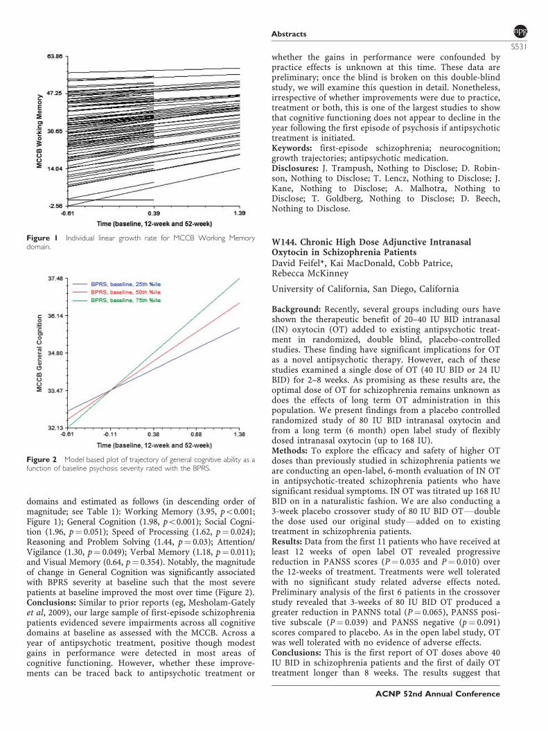
domains and estimated as follows (in descending order ofmagnitude; see Table 1): Working Memory (3.95, po0.001;Figure 1); General Cognition (1.98, po0.001); Social Cogni-tion (1.96, p¼ 0.051); Speed of Processing (1.62, p¼ 0.024);Reasoning and Problem Solving (1.44, p¼ 0.03); Attention/Vigilance (1.30, p¼ 0.049); Verbal Memory (1.18, p¼ 0.011);and Visual Memory (0.64, p¼ 0.354). Notably, the magnitudeof change in General Cognition was significantly associatedwith BPRS severity at baseline such that the most severepatients at baseline improved the most over time (Figure 2).Conclusions: Similar to prior reports (eg, Mesholam-Gatelyet al, 2009), our large sample of first-episode schizophreniapatients evidenced severe impairments across all cognitivedomains at baseline as assessed with the MCCB. Across ayear of antipsychotic treatment, positive though modestgains in performance were detected in most areas ofcognitive functioning. However, whether these improve-ments can be traced back to antipsychotic treatment or
whether the gains in performance were confounded bypractice effects is unknown at this time. These data arepreliminary; once the blind is broken on this double-blindstudy, we will examine this question in detail. Nonetheless,irrespective of whether improvements were due to practice,treatment or both, this is one of the largest studies to showthat cognitive functioning does not appear to decline in theyear following the first episode of psychosis if antipsychotictreatment is initiated.Keywords: first-episode schizophrenia; neurocognition;growth trajectories; antipsychotic medication.Disclosures: J. Trampush, Nothing to Disclose; D. Robin-son, Nothing to Disclose; T. Lencz, Nothing to Disclose; J.Kane, Nothing to Disclose; A. Malhotra, Nothing toDisclose; T. Goldberg, Nothing to Disclose; D. Beech,Nothing to Disclose.
W144. Chronic High Dose Adjunctive IntranasalOxytocin in Schizophrenia PatientsDavid Feifel*, Kai MacDonald, Cobb Patrice,Rebecca McKinney
University of California, San Diego, California
Background: Recently, several groups including ours haveshown the therapeutic benefit of 20–40 IU BID intranasal(IN) oxytocin (OT) added to existing antipsychotic treat-ment in randomized, double blind, placebo-controlledstudies. These finding have significant implications for OTas a novel antipsychotic therapy. However, each of thesestudies examined a single dose of OT (40 IU BID or 24 IUBID) for 2–8 weeks. As promising as these results are, theoptimal dose of OT for schizophrenia remains unknown asdoes the effects of long term OT administration in thispopulation. We present findings from a placebo controlledrandomized study of 80 IU BID intranasal oxytocin andfrom a long term (6 month) open label study of flexiblydosed intranasal oxytocin (up to 168 IU).Methods: To explore the efficacy and safety of higher OTdoses than previously studied in schizophrenia patients weare conducting an open-label, 6-month evaluation of IN OTin antipsychotic-treated schizophrenia patients who havesignificant residual symptoms. IN OT was titrated up 168 IUBID on in a naturalistic fashion. We are also conducting a3-week placebo crossover study of 80 IU BID OTFdoublethe dose used our original studyFadded on to existingtreatment in schizophrenia patients.Results: Data from the first 11 patients who have received atleast 12 weeks of open label OT revealed progressivereduction in PANSS scores (P¼ 0.035 and P¼ 0.010) overthe 12-weeks of treatment. Treatments were well toleratedwith no significant study related adverse effects noted.Preliminary analysis of the first 6 patients in the crossoverstudy revealed that 3-weeks of 80 IU BID OT produced agreater reduction in PANNS total (P¼ 0.065), PANSS posi-tive subscale (P¼ 0.039) and PANSS negative (p¼ 0.091)scores compared to placebo. As in the open label study, OTwas well tolerated with no evidence of adverse effects.Conclusions: This is the first report of OT doses above 40IU BID in schizophrenia patients and the first of daily OTtreatment longer than 8 weeks. The results suggest that
Figure 1 Individual linear growth rate for MCCB Working Memorydomain.
Figure 2 Model based plot of trajectory of general cognitive ability as afunction of baseline psychosis severity rated with the BPRS.
Abstracts
S531
ACNP 52nd Annual Conference
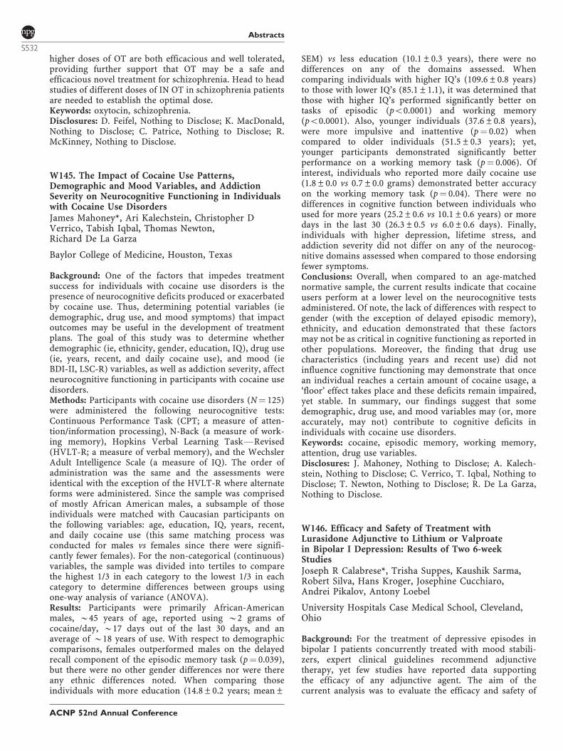
higher doses of OT are both efficacious and well tolerated,providing further support that OT may be a safe andefficacious novel treatment for schizophrenia. Head to headstudies of different doses of IN OT in schizophrenia patientsare needed to establish the optimal dose.Keywords: oxytocin, schizophrenia.Disclosures: D. Feifel, Nothing to Disclose; K. MacDonald,Nothing to Disclose; C. Patrice, Nothing to Disclose; R.McKinney, Nothing to Disclose.
W145. The Impact of Cocaine Use Patterns,Demographic and Mood Variables, and AddictionSeverity on Neurocognitive Functioning in Individualswith Cocaine Use DisordersJames Mahoney*, Ari Kalechstein, Christopher DVerrico, Tabish Iqbal, Thomas Newton,Richard De La Garza
Baylor College of Medicine, Houston, Texas
Background: One of the factors that impedes treatmentsuccess for individuals with cocaine use disorders is thepresence of neurocognitive deficits produced or exacerbatedby cocaine use. Thus, determining potential variables (iedemographic, drug use, and mood symptoms) that impactoutcomes may be useful in the development of treatmentplans. The goal of this study was to determine whetherdemographic (ie, ethnicity, gender, education, IQ), drug use(ie, years, recent, and daily cocaine use), and mood (ieBDI-II, LSC-R) variables, as well as addiction severity, affectneurocognitive functioning in participants with cocaine usedisorders.Methods: Participants with cocaine use disorders (N¼ 125)were administered the following neurocognitive tests:Continuous Performance Task (CPT; a measure of atten-tion/information processing), N-Back (a measure of work-ing memory), Hopkins Verbal Learning TaskFRevised(HVLT-R; a measure of verbal memory), and the WechslerAdult Intelligence Scale (a measure of IQ). The order ofadministration was the same and the assessments wereidentical with the exception of the HVLT-R where alternateforms were administered. Since the sample was comprisedof mostly African American males, a subsample of thoseindividuals were matched with Caucasian participants onthe following variables: age, education, IQ, years, recent,and daily cocaine use (this same matching process wasconducted for males vs females since there were signifi-cantly fewer females). For the non-categorical (continuous)variables, the sample was divided into tertiles to comparethe highest 1/3 in each category to the lowest 1/3 in eachcategory to determine differences between groups usingone-way analysis of variance (ANOVA).Results: Participants were primarily African-Americanmales, B45 years of age, reported using B2 grams ofcocaine/day, B17 days out of the last 30 days, and anaverage of B18 years of use. With respect to demographiccomparisons, females outperformed males on the delayedrecall component of the episodic memory task (p¼ 0.039),but there were no other gender differences nor were thereany ethnic differences noted. When comparing thoseindividuals with more education (14.8±0.2 years; mean±
SEM) vs less education (10.1±0.3 years), there were nodifferences on any of the domains assessed. Whencomparing individuals with higher IQ’s (109.6±0.8 years)to those with lower IQ’s (85.1±1.1), it was determined thatthose with higher IQ’s performed significantly better ontasks of episodic (po0.0001) and working memory(po0.0001). Also, younger individuals (37.6±0.8 years),were more impulsive and inattentive (p¼ 0.02) whencompared to older individuals (51.5±0.3 years); yet,younger participants demonstrated significantly betterperformance on a working memory task (p¼ 0.006). Ofinterest, individuals who reported more daily cocaine use(1.8±0.0 vs 0.7±0.0 grams) demonstrated better accuracyon the working memory task (p¼ 0.04). There were nodifferences in cognitive function between individuals whoused for more years (25.2±0.6 vs 10.1±0.6 years) or moredays in the last 30 (26.3±0.5 vs 6.0±0.6 days). Finally,individuals with higher depression, lifetime stress, andaddiction severity did not differ on any of the neurocog-nitive domains assessed when compared to those endorsingfewer symptoms.Conclusions: Overall, when compared to an age-matchednormative sample, the current results indicate that cocaineusers perform at a lower level on the neurocognitive testsadministered. Of note, the lack of differences with respect togender (with the exception of delayed episodic memory),ethnicity, and education demonstrated that these factorsmay not be as critical in cognitive functioning as reported inother populations. Moreover, the finding that drug usecharacteristics (including years and recent use) did notinfluence cognitive functioning may demonstrate that oncean individual reaches a certain amount of cocaine usage, a‘floor’ effect takes place and these deficits remain impaired,yet stable. In summary, our findings suggest that somedemographic, drug use, and mood variables may (or, moreaccurately, may not) contribute to cognitive deficits inindividuals with cocaine use disorders.Keywords: cocaine, episodic memory, working memory,attention, drug use variables.Disclosures: J. Mahoney, Nothing to Disclose; A. Kalech-stein, Nothing to Disclose; C. Verrico, T. Iqbal, Nothing toDisclose; T. Newton, Nothing to Disclose; R. De La Garza,Nothing to Disclose.
W146. Efficacy and Safety of Treatment withLurasidone Adjunctive to Lithium or Valproatein Bipolar I Depression: Results of Two 6-weekStudiesJoseph R Calabrese*, Trisha Suppes, Kaushik Sarma,Robert Silva, Hans Kroger, Josephine Cucchiaro,Andrei Pikalov, Antony Loebel
University Hospitals Case Medical School, Cleveland,Ohio
Background: For the treatment of depressive episodes inbipolar I patients concurrently treated with mood stabili-zers, expert clinical guidelines recommend adjunctivetherapy, yet few studies have reported data supportingthe efficacy of any adjunctive agent. The aim of thecurrent analysis was to evaluate the efficacy and safety of
Abstracts
S532
ACNP 52nd Annual Conference
lurasidone adjunctive to lithium or valproate in bipolar Idepression.Methods: Data were pooled from two adjunctive therapystudies (D1050235 [Study 1]; D1050292 [Study 2]) withsimilar designs: patients meeting DSM-IV-TR criteria forbipolar I depression, with or without rapid cycling, with aMontgomery Asberg Depression Rating Scale (MADRS)score X20 and a Young Mania Rating Scale score p12were randomized to 6 weeks of once-daily, double-blindtreatment with lurasidone 20–120 mg/day or placebo,adjunctive with either lithium (Li) or valproate (VPA).Therapeutic blood levels of Li or VPA were maintainedfor X28 days prior to randomization in both studies;Study 1 allowed retrospective confirmation of treatmentwith Li or VPA prior to screening, while Study 2 allowedboth retrospective and prospective treatment cohorts.Changes from baseline in MADRS (primary outcome)and Clinical Global Impression Bipolar Severity of Illness(CGI-BP-S; key secondary assessment) were analyzedusing MMRM; secondary efficacy outcomes includedthe Quick Inventory of Depressive SymptomologyFSelfReport (QIDS-SR16), Hamilton Anxiety Rating Scale(HAM-A), the Quality of Life, Enjoyment and Satis-faction Questionnaire (Q-LES-Q-SF), and the SheehanDisability Scale (SDS). These measures were evaluatedusing ANCOVA-LOCF, and logistic regression. Safetyparameters were evaluated based on analysis of the LOCF-endpoint sample.Results: A similar proportion of patients in the adjunctivelurasidone and placebo groups completed Study 1 (N¼ 143;78% vs N¼ 136; 82%) and Study 2 (N¼ 148; 82% vs N¼ 140;80%). Mean MADRS scores at baseline were similar (Study1: 30.7; Study 2: 29.1). For the pooled analysis sample,treatment with lurasidone (vs placebo) was associated withsignificant week 6 improvement in the mean MADRS score(�14.4 vs �11.9; p¼ 0.003; effect size: 0.25). Treatment withlurasidone (vs placebo) was also associated with significantweek 6 improvement in CGI-BP-S scores (�1.7 vs �1.3;p¼ 0.001), QIDS-SR16 scores (�7.4 vs �5.7; pp0.001),HAM-A scores (�7.0 vs �5.0; pp0.001 [LOCF]), andQ-LES-Q-SF scores ( + 18.5 vs + 13.2; pp0.001 [LOCF]),and non-significant improvement in SDS scores (�7.7 vs�6.4; p¼ 0.067 [LOCF]). Responder rates (X50% reductionin MADRS at week 6) were significantly higher for thelurasidone group vs placebo (48 vs 37%; p¼ 0.002; LOCF-endpoint). Remission rates (defined as MADRSp10) weresignificantly higher for the lurasidone group vs placebo (36vs 27%; p¼ 0.008; LOCF-endpoint). In the pooled safetypopulation, minimal LOCF-endpoint changes were ob-served for adjunctive lurasidone vs placebo in mean weight(0.1 vs 0.2 kg), median total cholesterol (�4.0 vs �1.0 mg/dl), LDL (�3.0 vs �1.0 mg/dl), triglycerides ( + 4.0 vs�2.0 mg/dl), and glucose (0.0 vs 0.0 mg/dl). Discontinuationrates due to treatment-emergent adverse events were similar(5.8 vs 4.8%); treatment-emergent adverse events with anincidence X5% (and greater than placebo) were nausea(13.9 vs 10.2%), Parkinsonism (12.8 vs 8.1%), somnolence(11.4 vs 5.1%), and akathisia (10.8 vs 4.8%) in the combinedadjunctive lurasidone and placebo groups, respectively.The incidence of protocol-defined treatment-emergentmania for the lurasidone vs placebo groups was similar(0.8 vs 1.5%).
Conclusions: The pooled results from two similarlydesigned, short-term placebo-controlled studies demon-strated that use of lurasidone as adjunctive therapy withlithium or valproate is an effective treatment for patientswith bipolar I depression. Significant improvement wasobserved in depressive symptoms, and in patient-ratedmeasures of function and quality of life. The low rate ofdiscontinuation due to adverse events, and low incidence ofadverse events, suggest that lurasidone was well-tolerated.Short-term treatment with lurasidone had minimal effect onweight or metabolic parameters.Keywords: bipolar disorder, atypical antipsychotics, majordepressive disorder.Disclosures: J. Calabrese, Part 1: Consultant services toAstraZeneca, Biomedical Dev Corp, Convergent HealthSolutions, Elan, Esai, Forest Labs, Health & Wellness,Hoffman LaRoche, Lundbeck, Merck, Otsuka, Pfizer,Scientia, Spirant Communications Private Limitrex, Suno-vion, Takeda, and Teva., Payment for lectures from theAmerican Foundation for Suicide Prevention, AstraZeneca,Benecke, Cortex Congress, GlaxoSmithKline, Lundbeck,Medwiz Healthcare, Merck, Pfizer, Promedica, Sunovion,Takeda, Ohio Psychiatry Association, Ohio State University,University of Cincinnati, and University of Toronto., Part 4:Cephalon, Elan, NARSAD, Sunovion, and Takeda; T.Suppes, Part 1: Consultant or advisory capacity toOrexigen Therapeutics and Sunonvion Pharmaceuticals.,Has received royalties or honorarium from Medscape, CNSDrug Supplement, and Jones and Bartlett (formerlyCompact Clinicals)., Part 4: Research funding or medicationsupport from Abbott Laboratories, AstraZeneca, JDSPharmaceuticals, NIMH, Pfizer, the Stanley MedicalResearch Institute and Sunovion Pharmaceuticals.; K.Sarma, Part 5: Sunovion Pharmaceuticals, Inc.; R. Silva,Part 5: Sunovion Pharmaceuticals, Inc.; H. Kroger, Part 5:Sunovion Pharmaceuticals, Inc.; J. Cucchiaro, Part 5:Sunovion Pharmaceuticals, Inc.; A. Pikalov, Part 5: Suno-vion Pharmaceuticals, Inc.; A. Loebel, Part 5: SunovionPharmaceuticals, Inc.
W147. Lurasidone Adjunctive Therapy with Lithium orValproate for the Treatment of Bipolar I Depression: ARandomized, Double-blind, Placebo-controlled Study(PREVAIL 3)Trisha Suppes*, Joseph R Calabrese, Robert Silva,Hans Kroger, Josephine Cucchiaro, Andrei Pikalov,Antony Loebel
VA Palo Alto Health Care System, Palo Alto, California
Background: Few studies have been reported that supportthe efficacy of adjunctive therapy for patients with bipolar Idepression who have had an insufficient response tomonotherapy with mood stabilizing agents. In a previousplacebo-controlled trial (1; PREVAIL 1), treatment withlurasidone adjunctive with lithium or valproate significantlyimproved depressive symptoms and was generally well-tolerated. This was the second study to evaluate the short-term efficacy of lurasidone as adjunctive therapy withlithium (Li) or valproate (VPA) for the treatment of bipolarI depression.
Abstracts
S533
ACNP 52nd Annual Conference
Methods: In this multi-regional study, patients wererequired to meet DSM-IV-TR criteria for bipolar I depres-sion, without psychotic features, with or without a rapid-cycling course of illness, and with a Montgomery-AsbergDepression Rating Scale (MADRS) score X20 and a Young-Mania Rating scale (YMRS) score p12 at screening andbaseline. Eligible patients were randomized to 6 weeks ofdouble-blind treatment with either lurasidone 20–120 mg/day or placebo adjunctive with either Li or VPA. Treatmentwith Li or VPA could be ongoing at the time of screening(retrospective assessment cohort), or could be prospectivelyinitiated by the investigator during a lead-in phase(prospective cohort). All patients were required to have aminimum of 28 days of treatment with Li (range, 0.6–1.2 mEq/l) or VPA (range, 50–125 mg/ml). Changes fromdouble-blind baseline in MADRS (primary assessment) andClinical Global Impression Bipolar Severity of Illness (CGI-BP-S; key secondary assessment) were analyzed usingmixed model repeated measures (MMRM) analysis; othersecondary outcomes were analyzed using ANCOVA (LOCF).Results: A total of 180 patients were randomized to theadjunctive lurasidone treatment group and 176 wererandomized to the adjunctive placebo group (treatmentwith Li/VPA was ongoing at the time of initial screening in38.5% of patients, and prospectively initiated in 61.5% ofpatients); 82% in the lurasidone group and 80% in theplacebo group completed the study. Mean MADRS scores atbaseline were 29.1 in both treatment groups, indicative ofmoderate-to-severe depression. Treatment with lurasidonewas associated with significant improvement comparedwith placebo at week 2 to week 5, but a significant differencewas not observed at the primary (week 6) endpoint (�11.8vs �10.4; p¼ 0.18; effect size, 0.16). A larger improvementfor lurasidone vs placebo was observed in patients recruitedinto the study who were already being treated with Li/VPA(retrospective cohort; effect size: 0.31), compared to thosetreated prospectively with Li/VPA (effect size: 0.03). On theCGI-BP-S, endpoint improvement in the lurasidone groupwas not significantly different from the placebo group (�1.4vs �1.1; p¼ 0.095; effect size, 0.20). On the HamiltonAnxiety Rating Scale (HAM-A) treatment with lurasidonewas associated with significantly greater endpoint improve-ment compared with placebo (�5.7 vs �3.8; po0.01).Treatment with lurasidone was associated with significantlygreater endpoint improvement on the Quality of Life,Enjoyment and Satisfaction Questionnaire (Q-LES-Q-SF; +14.3 vs + 10.2; p¼ 0.04), but not on the Sheehan Disabilityscale or other secondary measures. Treatment withlurasidone vs placebo was associated with minimal endpointchanges in mean weight ( + 0.2 vs 0.0 kg), median totalcholesterol (�4.0 vs �2.5 mg/dl), triglycerides ( + 1.0 vs�1.0 mg/dl), glucose (0.0 vs �0.5 mg/dl), and prolactin ( +1.8 vs �0.1 ng/ml). Discontinuation rates due to treatment-emergent adverse events were 5.6% in the lurasidone groupand 2.9% in the placebo group. The most frequentlyreported adverse events in the lurasidone vs placebo groupswere akathisia (14.1 vs 5.3%), somnolence (11.9 vs 4.7%),and Parkinsonism (11.3 vs 7.6%).Conclusions: In this study, which ascertained subjectsutilizing both retrospective and prospective treatmentcohorts (Li or VPA) in patients with bipolar I depression,adjunctive treatment with lurasidone did not result in
significant improvement compared with placebo at studyendpoint. Significant separation from placebo was observedstarting at week 2 through week 5. Lurasidone was alsosignificantly superior to placebo on the HAM-A and Q-LES-Q at endpoint. Lurasidone was generally well-tolerated inthis study with a relatively low overall discontinuation rate,and rate of discontinuation due to adverse events. The lackefficacy in this study appeared in part related to inclusion ofa cohort of patients treated prospectively with moodstabilizers prior to randomization. Use of a prospectivetreatment cohort during lead-in has previously been shownto present methodological challenges in augmentationstudies of mood disorders (2). Minimal changes in weight,lipids and measures of glycemic control were observed.Keywords: bipolar disorder, atypical antipsychotics, majordepressive disorder.Disclosures: T. Suppes, Part 1: Consultant or advisorycapacity to Orexigen Therapeutics and Sunonvion Pharma-ceuticals., Has received royalties or honorarium fromMedscape, CNS Drug Supplement, and Jones and Bartlett(formerly Compact Clinicals)., Part 4: Research funding ormedication support from Abbott Laboratories, AstraZeneca,JDS Pharmaceuticals, NIMH, Pfizer, the Stanley MedicalResearch Institute and Sunovion Pharmaceuticals.; J.Calabrese, Part 1: Consultant services to AstraZeneca,Biomedical Dev Corp, Convergent Health Solutions, Elan,Esai, Forest Labs, Health & Wellness, Hoffman LaRoche,Lundbeck, Merck, Otsuka, Pfizer, Scientia, Spirant Com-munications Private Limitrex, Sunovion, Takeda, and Teva.,Payment for lectures from the American Foundation forSuicide Prevention, AstraZeneca, Benecke, Cortex Congress,GlaxoSmithKline, Lundbeck, Medwiz Healthcare, Merck,Pfizer, Promedica, Sunovion, Takeda, Ohio PsychiatryAssociation, Ohio State University, University of Cincinnati,and University of Toronto., Part 4: Cephalon, Elan,NARSAD, Sunovion, and Takeda; R. Silva, Part 5: SunovionPharmaceuticals, Inc.; H. Kroger, Part 5: Sunovion Phar-maceuticals, Inc.; J. Cucchiaro, Part 5: Sunovion Pharma-ceuticals, Inc.; A. Pikalov, Part 5: SunovionPharmaceuticals, Inc.; A. Loebel, Part 5: Sunovion Pharma-ceuticals, Inc.
W148. Lurasidone in Bipolar I Depression: A 24 Week,Open-label Extension StudyTerence Ketter*, Kaushik Sarma, Robert Silva, Jane Xu,Josephine Cucchiaro, Antony Loebel
Stanford School of Medicine, Stanford, California
Background: Lurasidone has recently been approved by theFDA for the treatment of acute bipolar I depression asmonotherapy, and as adjunctive therapy with lithium orvalproate. The aim of the current 6 month extension studywas to evaluate the longer-term safety and tolerability oflurasidone (20–120 mg/day, flexibly dosed) in patients whorecently completed acute treatment for bipolar I depression.Methods: Patients completing 6 weeks of double-blind,placebo-controlled treatment with either lurasidone mono-therapy (1 study) or lurasidone adjunctive therapy withlithium (Li) or valproate (VPA; 2 studies), were treatedfor 6 months with once-daily flexible doses of lurasidone,
Abstracts
S534
ACNP 52nd Annual Conference
20–120 mg/day in this open-label extension study. Patientswere transitioned directly upon completion of the preceding6 week study to receive 60 mg/day of lurasidone, with weeklydose adjustments to optimize efficacy and tolerability. Inaddition to mood stabilizers (Li, VPA), concomitant treat-ments with benzodiazepines or antidepressants were per-mitted. Safety/tolerability outcomes included the incidence ofadverse events (AEs), discontinuations due to AEs, weight,and laboratory parameters, which were co-primary end-points. Secondary efficacy endpoints included total scores forthe Montgomery-Asberg Depression Rating Scale (MADRS),Clinical Global Impression for Bipolar Disorder, OverallSeverity (CGI-BP-S), Quality of Life Enjoyment and Satisfac-tion Questionnaire (Q-LES-Q), Sheehan Disability Scale(SDS), Hamilton Anxiety Scale (HAM-A), and Young ManiaRating Scale (YMRS). Safety endpoints were analyzed aschange from double-blind baseline for study completers whohad initially been randomized to lurasidone in the initial 6week study (30 weeks of total exposure; monotherapy,n¼ 154; adjunctive, n¼ 104). Treatment-emergent maniawas defined as a YMRS score X16 on 2 consecutive visits, oran AE of mania or hypomania.Results: Of the 817 patients entered into this open-labelextension study, 316 (38.9%) were treated with lurasidoneas monotherapy, and 497 (61.1%) were treated withlurasidone adjunctive with Li or VPA. For the totalpopulation, 47.5% were male, with a mean age of 42.7years, and a mean MADRS score of 15.3 at baseline (ie atentry to this open extension study). A total of 68.4% ofpatients entering the extension study completed it. Treat-ment-emergent AEs with an incidence X5% were akathisia(8.1%), headache (7.7%), nausea (7.6%), insomnia (6.4%),anxiety (5.8%), and Parkinsonism and EPS-related AEs(including akathisia, in aggregate 10.7%). Discontinuationdue to an AE leading occurred in 7.0% of monotherapypatients and 8.7% of adjunctive therapy patients. Meanchange in weight at month 6 was 0.45 kg in the mono-therapy group and 0.90 kg in the adjunctive group.Clinically meaningful change in weight (X7%) for adjunc-tive therapy with lurasidone was: increase, 11.9%; decrease,4.9%; and for monotherapy was: increase, 5.0%; decrease,3.0%). The following median changes were observed atmonth 6 for total cholesterol (monotherapy, 0.0 mg/dl;adjunctive, �1.5 mg/dl); triglycerides (monotherapy, +6.0 mg/dl; adjunctive, + 8.0 mg/dl); glucose (monotherapy,0.0 mg/dl; adjunctive, + 1.0 mg/dl); and prolactin (mono-therapy, + 1.3 ng/dl; adjunctive, + 1.3 ng/dl). The incidenceof treatment-emergent mania was 1.3% in the monotherapytreatment subgroup and 3.8% in the adjunctive subgroup;the incidence of ‘any suicidal ideation or behavior’ onthe Columbia Suicide Severity Rating Scale was 2.1%.The following mean changes were observed at month 6(observed case) for the MADRS (monotherapy: �6.9;adjunctive: �6.5), CGI-BP-S (monotherapy: �0.87; adjunc-tive: �0.85), HAM-A (monotherapy: �2.8; adjunctive:�2.4), YRMS (monotherapy: �0.8; adjunctive: �0.5), SDS(monotherapy: �4.3; adjunctive: �3.3), and Q-LES-Q(monotherapy: + 9.6; adjunctive: + 7.5).Conclusions: Long term treatment with lurasidone 20–120 mg/d for 6 months was safe and well tolerated withminimal effect on weight and metabolic parameters. Therewere minimal differences in tolerability or safety outcomes
with lurasidone monotherapy or as adjunctive therapy withlithium or valproate. Treatment with lurasidone wasassociated with sustained improvement in both the MADRS,and in patient-rated measures of quality of life and function.ClinicalTrials.gov Identifier: NCT00868959 Sponsored bySunovion Pharmaceuticals Inc..Keywords: bipolar disorder, atypical antipsychotics, majordepressive disorder.Disclosures: T. Ketter, Part 1: Consulting fees from AstellasPharmaceuticals, Bristol–Myers Squibb Company, CephalonInc., Johnson & Johnson, Merck & Co., Inc., and SunovionPharmaceuticals, Inc., CME lecture honoraria (not speaker’sbureau honoraria) from Abbott Pharmaceuticals, AstraZe-neca Pharmaceuticals LP, and GlaxoSmithKline. Part 4:AstraZeneca Pharmaceuticals LP, Cephalon Inc., Eli Lillyand Company, Inc., Pfizer Inc., and Sunovion Pharmaceu-ticals, Inc.; K. Sarma, Part 5: Sunovion Pharmaceuticals,Inc.; R. Silva, Part 5: Sunovion Pharmaceuticals, Inc.; J. Xu,Part 5: Sunovion Pharmaceuticals, Inc.; J. Cucchiaro, Part 5:Sunovion Pharmaceuticals, Inc.; A. Loebel, Part 5: SunovionPharmaceuticals, Inc.
W149. Short- and Longer-term Treatment withLurasidone in Patients with Bipolar I Depression: Effecton Metabolic SyndromeSusan McElroy*, Andrei Pikalov, Josephine Cucchiaro,Jay Hsu, Hans Kroger, Debra Phillips, Antony Loebel
Lindner Center of HOPE, Mason, Ohio
Background: Antipsychotics are commonly used in themanagement of bipolar disorder but may lead to adversemetabolic consequences, including weight gain and anincreased risk for diabetes and cardiovascular disease. Theseadverse metabolic effects are in addition to effects associatedwith bipolar illness itself. This pooled analysis evaluated theeffect of short- and longer-term treatment with lurasidone onthe prevalence of metabolic syndrome in bipolar I depression.Methods: The effects of lurasidone, in the dosing range of 20–120 mg/d, was evaluated on the prevalence of meta-bolic syndrome in three short-term and one longer-termextension study. In the short-term studies, patients meetingDSM-IV-TR criteria for bipolar I depression, with or withoutrapid cycling, with a Montgomery Asberg Depression RatingScale (MADRS) score X20, were randomized to 6 weeks ofonce-daily, double-blind, placebo-controlled treatment withlurasidone, either as monotherapy (one study; total N¼ 499)or adjunctive to lithium (Li) or valproate (VPA; two studies;total N¼ 694). Patients completing the 6-week studiescontinued to receive 6 months of additional treatment withlurasidone 20–120 mg/d in an open-label extension study.NCEP criteria (1) for metabolic syndrome were used, withmetabolic syndrome defined as meeting X3 of the following:waist circumference (male, X102 cm; female, X88 cm),triglycerides (X150 mg/dl), HDL-cholesterol (male, o40 mg/dl; female, o50 mg/dl), blood pressure (X130/85 mmHg), orplasma glucose (X110 mg/dl). Change at 6 months (forcompleters) was calculated from double-blind baseline of theacute study.Results: At baseline, the prevalence of metabolic syndromewas similar for lurasidone and placebo, respectively, in the
Abstracts
S535
ACNP 52nd Annual Conference
adjunctive therapy studies (lurasidone, 14.8% and placebo,13.5%) and in the monotherapy study (lurasidone, 14.3%and placebo, 15.5%). After 6 weeks of adjunctive therapy,the prevalence of metabolic syndrome in the lurasidone vsplacebo groups was 17.0 vs 12.4% (LOCF); and after 6 weeksof monotherapy the prevalence was 15.8 vs 17.7% (LOCF).For patients treated with lurasidone in the double-blindstudy and who completed 6 months of extension phasetreatment (30 weeks of total exposure), the prevalence ofmetabolic syndrome, among patients receiving monother-apy and adjunctive therapy, was 16.7 and 15.5%, respec-tively, at double-blind baseline, and 17.9 and 23.8% atmonth 6 of the extension. For the subgroup with metabolicsyndrome at baseline in the monotherapy study, thefollowing median changes were observed (after 30 weeks)in patients (n¼ 30) who completed 6 months of treatmentwith lurasidone in weight (�0.3 kg), cholesterol (�4.0 mg/dl), triglycerides (�22.0 mg/dl), and glucose (�2.0 mg/dl).For the subgroup of lurasidone patients with metabolicsyndrome at double-blind baseline in the adjunctive therapystudies, the following median changes were observed inpatients (n¼ 31) who completed 30 weeks of treatment withlurasidone in weight (0.0 kg), cholesterol (�6.0 mg/dl),triglycerides ( + 11.0 mg/dl), and glucose ( + 2.0 mg/dl).Conclusions: In patients with acute bipolar I depression, upto 7 months of treatment with lurasidone, either asmonotherapy, or as adjunctive therapy when added to Lior VPA, was associated with only minimal metabolicdisturbance. In at-risk patients who met metabolic syn-drome criteria at study entry, treatment with lurasidone wasassociated with no worsening of metabolic parameters.Keywords: bipolar disorder, atypical antipsychotics, meta-bolic syndrome, lipids, glucose.Disclosures: S. McElroy, Part 1: Consultant to, or memberof the scientific advisory boards of Alkermes and Shire.,Part 2: Holds a patent for the use of sulfamate derivativesfor treating impulse control disorders and has receivedpayments from Johnson & Johnson PharmaceuticalResearch & Development, L.L.C., which has exclusive rightsunder the patent., Part 4: Agency for Healthcare Research& Quality (AHRQ); Alkermes, AstraZeneca, Brackett,Cephalon, Corcept, Eli Lilly and Company, MarriottFoundation, NIMH, Orexigen Therapeutics, Inc., Pfizer,Shire, Takeda Pharmaceutical Company Ltd, and TransceptPharmaceutical, Inc.; A. Pikalov, Part 5: Sunovion Pharma-ceuticals, Inc.; J. Cucchiaro, Part 5: Sunovion Pharmaceu-ticals, Inc.; J. Hsu, Part 5: Sunovion Pharmaceuticals, Inc.;H. Kroger; D. Phillips, Part 5: Sunovion Pharmaceuticals,Inc.; A. Loebel, Part 5: Sunovion Pharmaceuticals, Inc.
W150. Early Improvement Predicts Endpoint Responseto Lurasidone in Schizophrenia: Pooled Analysis of FiveDouble-blind TrialsChristoph U Correll*, Andrei Pikalov, Jay Hsu, JosephineCucchiaro, Robert Goldman, Antony Loebel
Hofstra North Shore LIJ School of Medicine and AlbertEinstein College of Medicine, Glen Oaks, New York
Background: Early improvement following initiation ofantipsychotic medication (within 2–3 weeks of initiating
treatment) is a potentially important predictor of subse-quent treatment response that has clinical implications forthe successful management of schizophrenia (1, 2). The goalof this pooled completer analysis was to evaluate the clinicalvalue of early improvement in the PANSS total score andthe CGI-Severity score as predictors of response to 6 weeksof treatment with lurasidone among patients with an acuteexacerbation of schizophrenia.Methods: Data were pooled from 5 similarly designed, six-week, double-blind, placebo-controlled trials of subjectshospitalized with an acute exacerbation of schizophreniawho were randomly assigned to fixed, once-daily doses oflurasidone 40–80 mg (n¼ 404) or 120–160 mg (n¼ 264), orplacebo (n¼ 280), and who completed acute treatment.Endpoint responder rates were calculated using the PANSScriterion (X40% reduction from Baseline). Early improve-ment was separately assessed at weeks 1, 2, and 3 using twocriteria (CGI-Severity X1-point improvement; PANSSimprovement X20%). For each improvement criterion,calculations were made of sensitivity (the probability that apatient who achieved endpoint response was correctlyidentified based on early improvement) and specificity(the probability that a patient who did not achieve endpointresponse was correctly identified based on early improve-ment). Receiver operating characteristic (ROC) curves wereused to determine the optimal cut-scores for prediction ofendpoint response, based on the highest area under thecurve (AUC).Results: Baseline demographic and clinical characteristicswere similar in the group that achieved (vs did not achieve)early improvement, with the exception of duration ofschizophrenia, which was longer in the group that did notachieve early improvement. In the combined lurasidonedosage groups, the proportion of subjects showing earlyimprovement was similar for the PANSSX20% criterionand the CGI-Severity X1 criterion, respectively, at week 1(32.5 and 36.1%), at week 2 (53.8 and 59.8%), and at week 3(70.7 and 88.0%). Endpoint response (week 6) in thelurasidone group was 50.2% using PANSSX40% respondercriteria. For the PANSSX40% responder criterion, at week 1PANSSX20% improvement had a sensitivity¼ 46.6%,specificity¼ 81.6%, and AUCROC¼ 0.660; and CGI-S im-provement X1 had a sensitivity¼ 46.2%, specificity¼ 74.0%,and AUCROC¼ 0.621. At week 2, PANSSX20% improve-ment had a sensitivity¼ 75.2%, specificity¼ 67.9%, andAUCROC¼ 0.733; and CGI-S improvement X1 had asensitivity¼ 74.4%, specificity¼ 54.8%, and AUCROC¼0.650. At week 3, PANSSX20% improvement had a sensi-tivity¼ 91.9%, specificity¼ 50.5%, and AUCROC¼ 0.730; andCGI-S improvement X1 had a sensitivity¼ 87.3%, speci-ficity¼ 43.7%, and AUCROC¼ 0.656. Among patients treatedwith lurasidone who met X40% responder criteria at week 3,57/60 (95.0%) maintained their response at week 6. Amongweek 3 non-responders, 278/607 (45.8%) achieved a response(using the X40% PANSS criterion) by week 6.Conclusions: The ROC area under the curve analysis foundthat the PANSSX20% improvement at week 2 providedadequate prediction of endpoint response. Change in CGI-Severity was less useful as a predictor. Early determinationof the likelihood that a patient will have a favorabletreatment response has important implications for theeffective clinical management of schizophrenia. Further
Abstracts
S536
ACNP 52nd Annual Conference
research is needed to determine whether dose titration is aneffective strategy for increasing responder rates on lurasi-done among patients who do not respond early in thecourse of treatment.References.Leucht et al (2007). J Clin Psychiatry 68: 352–360.Kinon et al (2008). Schizophr Res 102: 230–240.Sponsored by Sunovion Pharmaceuticals Inc.Keywords: schizophrenia, lurasidone, early response, pre-diction, PANSS.Disclosures: C. Correll, Part 1: Actelion, Alexza, AstraZe-neca, Bristol-Myers Squibb, Cephalon, Eli Lilly, GersonLehrman Group, IntraCellular Therapies, Lundbeck, Meda-vante, Medscape, Merck, Novartis, Ortho-McNeill/Janssen/J&J, Otsuka, Pfizer, Roche, ProPhase, Sunovion, Takeda,Teva, and Vanda. Part 2: Bristol-Myers Squibb, Cephalon,Janssen, Lundbeck, Merck, Otsuka, ProPhase. Part 3:Bristol-Myers Squibb, Lundbeck, Merck, Otsuka, ProPhase.Part 4: BMS, Janssen/J&J, Otsuka, Part 5: N/A; A. Pikalov,Part 5: Sunovion Pharmaceuticals, Inc.; J. Hsu, Part 5:Sunovion Pharmaceuticals, Inc.; J. Cucchiaro, Part 5:Sunovion Pharmaceuticals, Inc.; R. Goldman, Part 5:Sunovion Pharmaceuticals, Inc.; A. Loebel, Part 5: SunovionPharmaceuticals, Inc.
W151. Neurocognitive Impairments as PutativePredictors of Neuroleptic-induced Movement Disordersin People with SchizophreniaAnthony Ahmed*
Medical College of Georgia, Augusta, Georgia
Background: Antipsychotic medication management is astandard intervention for schizophrenia symptoms butchronic or prolonged use of both first generation anti-psychotics alone or in combination with atypical antipsy-chotics is associated with the emergence of tardivedyskinesia and other movement disorders. In the absenceof effective treatments for neuroleptic-induced movementdisorders, prevention through the identification of riskfactors is a desirable proposition with potential to improveantipsychotic prescription practice. The current studyexamined neurocognitive phenotypes as putative predictorsof tardive dyskinesia, Parkinsonism, and akathesia inschizophrenia patients.Methods: We recruited 662 patients with a DSM diagnosis ofschizophrenia, schizoaffective, psychotic disorder not other-wise specified, and mood disorder with psychotic featuresreceiving antipsychotic treatments in outpatient and inpatientpsychiatric settings. DSM diagnosis of a psychotic disorderwas ascertained using a Structured Clinical Interview forDSM-IV Axis I (SCID-I). Participants were rated on theMaryland Psychiatric Research Center’s (MPRC) InvoluntaryMovement Scale (MIMS), a 31-item measure of involuntarymovements in several anatomic regions that include tongue;perioral region; eyes and periorbital region; face and jaw;fingers and wrists; elbows and arms; neck, shoulders, andhead; thighs and knees; legs and feet; arm; pelvis; anddiaphragm. We administered the Repeatable Battery for theAssessment of Neuropsychological Status (RBANS); WechslerAdult Intelligence ScaleFThird Edition (WAIS-III); and
Wide-Range Achievement Test (WRAT-3) to assess neuro-cognitive functions and intellectual functioning. We measurepositive symptoms of schizophrenia using the Scale for theAssessment of Positive Symptoms, and negative symptomsusing the Schedule for the Deficit Syndrome. We evaluatedpremorbid social and academic adjustment by administeringthe Premorbid Adjustment Scale (PAS). We measuredpsychosocial functioning with the Level of Functioning Scale,Global Assessment of Functioning (GAF) ratings, and theQuality of Life Inventory (QOLI). The demographic andclinical characteristics of study participants were obtainedusing a standard clinical interview. We used canonicalcorrelational analysis (CCA) to examine the associationbetween neurocognitive domain scores and subtypes ofmovement disorders. Given that distributions of studyvariables were skewed, we used non-parametric correlationsto assess the association with clinical and demographicvariables and anatomic regions.Results: There were no statistically significant differences inglobal severity ratings of movement disorders across sex andethnic groups. Age was however a statistically significantpredictor of dyskinesia with older patients generally receivinghigher severity ratings (rrho¼ 0.338, po0.0001). Level ofeducation was a small but significant predictor of the severityof Parkinsonism (rrho¼ 0.148, po0.001). but not dyskinesia orakathesia. There was no association between the severity ofpositive symptoms and the severity of movement disorders.There was however an association between the severity ofnegative symptoms and Parkinsonism (rrho¼ 0.213, po0.001).With regard to psychosocial outcomes, the severity of dyskinesia(rrho¼�0.320, po0.001) but not Parkinsonism or akathesiawas significantly associated with GAF scores. In contrast, onlyParkinsonism was significantly correlated with Level of Functiontotal score (rrho¼�0.169, po0.001). Dyskinesia, Parkinson-ism, and Akathesia global ratings had significantly negativecorrelations with work status, hours worked per week, earning,and job satisfaction. The severity of dyskinesia had a smallcorrelation with health-related quality of life (rrho¼�0.224,po0.01). The CCA produced a significant association betweenneurocognitive domain scores and 12 individual dyskinesiaitems in the MIMS. Only the first canonical variate pair wassignificant (rc1¼ 0.360, Wilk’s l¼ 0.643, w2 (65), po0.05). Thisvariate pair linked low scores on immediate memory,visuospatial reasoning, attention, delayed memory, and esti-mated IQ to jerking movements of the tongue, and lip smackingand pouting (perioral). The redundancy analysis suggested that6.5% of variance was shared by neurocognition and dyskinesia.The CCA between neurocognition and Parkinsonism didnot achieve significance (rc1¼ 0.337, Wilk’s l¼ 0.669, w2(90), p¼ 0.14).Conclusions: In schizophrenia, neurocognition demonstratea modest association with dyskinesia. There appear to be alink between a neurocognitive profile characterized byimpairments in immediate memory, visuospatial reasoning,attention, delayed memory, and general intelligence anddyskinesia of the tongue and perioral region. The result ofour study is partly consistent with those of prior studies andmay establish neurocognitive phenotypes as possible targetsfor movement disorder risk profiling.Keywords: tardive dyskinesia; parkinsonism; akathesia;schizophrenia; neuroleptic-induced movement disorder.Disclosures: A. Ahmed, Nothing to Disclose.
Abstracts
S537
ACNP 52nd Annual Conference
W152. Loss of Neural Signals Related to CognitiveFlexibility in the Rostral Caudate Following Short-termCocaine Self-administrationBrianna Sleezer*, Benjamin Hayden
University of Rochester, Webster, New York
Background: Drug-induced changes in frontostriatal circuitsare believed to play a pivotal role in the transition fromvoluntary drug use to compulsive drug-associated behaviorsin addiction. While much of this work has focused on howaddictive drugs enhance striatal-mediated habit learningprocesses, little has looked at how drugs might disruptstriatal activity related to flexible, goal-directed decision-making. Given that recent work has implicated the rostralstriatum in goal-directed processing, it is possible thataddictive drugs may promote inflexible behaviors by alteringneural signals in this region. To determine if and howaddictive drugs alter goal-directed processing in the striatum,we examined neural activity in two monkeys performing anovel monkey adaptation of the Wisconsin Card Sorting Test(WCST), a classic and well-studied paradigm for under-standing goal-directed cognition. We recorded the activity ofsingle neurons in the rostral portions of three striatal regions(the caudate, putamen, and ventral striatum) before andduring a period of short-term cocaine self-administration.Methods: On each trial of our task, monkeys are presentedwith a series of three stimuli that differ in shape (circle,triangle, or star) and color (cyan, magenta, or yellow).Monkeys must choose one of these stimuli based on a rule(a specific color or a shape). Once the rule is learned, itmust be maintained for 10, 15, or 20 trials. Correct choicesare followed by positive visual feedback and a reward, whileincorrect choices are followed by negative visual feedbackand no reward. Rule changes are not cued and new rulesmust be learned through trial and error. Following training,we recorded firing rate activity in the rostral regions of thecaudate, putamen, and ventral striatum while two monkeysperformed this task. Our analyses were designed to detectthree types of task-related activity: (1) associative encoding,(2) rule signals, and (3) switching signals. After baselinedata collection was complete, we implemented a cocaineself-administration paradigm (4 mg/kg/day, 5 days/wk), andcollected a second set of physiology data from the caudate,putamen, and ventral striatum. In this experiment, behaviorand/or physiology data was collected in the morning, whileself-administration occurred in the afternoon. Physiologydata were collected from self-administration days 21 to 45.Results: Cocaine Naıve Condition Prior to cocaine exposure,we found that monkeys were able to rapidly learn, maintain,and switch between rule sets. Neurons throughout all threestriatal regions exhibited activity related to associativeencoding, rule maintenance, and behavioral switching. Whilethe ventral striatum and caudate appeared to contributeprimarily to associative encoding and switching, the putamenappeared to contribute less to associative coding andswitching and more to rule maintenance. Cocaine ExposedCondition Although task performance was similar betweenthe cocaine naıve and cocaine exposed conditions, accuracyand motivation were subtly impaired. Changes in neuralresponses were clearer. The most prominent signals were asubstantial reduction in the prevalence of shift signals in the
caudate. We also found a reduction in the strength ofassociative encoding signals in the caudate, but no change inthe number of cells contributing to associative encoding orrule maintenance in the caudate or any of the three measuresin putamen or ventral striatum.Conclusions: Results from our pre-cocaine experimentsindicate that, under normal conditions, all three regions ofthe rostral striatum contribute to cognitive flexibilitythrough their involvement in associative encoding, rulemaintenance, and switching. These findings are inconsistentwith the commonplace notion that striatum is specializedfor habit learning, but are consistent with recent workimplicating the rostral striatum in goal-directed behaviors.Our cocaine experiments suggest that drug-induced changesin striatal activity may occur prior to the development ofany strong behavioral impairments. Specifically, our datasuggest that cocaine exposure reduces neural signals relatedto flexible behavior (shift signals) in the caudate. At firstglance, these findings appear somewhat inconsistent withprevious work, which has suggested that long-termpsychostimulant exposure promotes a shift in the controlof behavior from the ventral striatum to the dorsal striatum(rather than away from the dorsal striatum, as our datawould suggest). However, given that the majority of work inthis area has looked at long-term rather than short-termdrug exposure and the putamen rather than the caudate,our findings are not entirely inconsistent previous work.More specifically, it is possible that, rather than promotinga shift from the ventral striatum to the dorsal striatum (iefrom motivational to habit-related domains), addictivedrugs may instead promote a shift from the rostral caudate,to the ventral striatum, to the putamen (ie from cognitive tomotivational to habit-related domains). These findingsprovide further insight into how the motivational aspectsof drugs are able to initially gain control of behavior priorto the development of drug-related compulsions.Keywords: striatum, macaque, single-unit, addiction, self-administration.Disclosures: B. Sleezer, Nothing to Disclose; B. Hayden,Nothing to Disclose.
W153. Long-term Reduction of Cocaine Seeking in Ratsand Monkeys by Viral Vector-delivered CocaineHydrolase (CocH)Marilyn E Carroll*, Natalie E Zlebnik, Yang Gao,Stephen Brimijoin, PhD
University of Minnesota, Minneapolis, Minnesota
Background: Cocaine dependence is a chronic disorder thatis devastating to individuals and costly to society, and thereare currently no FDA approved treatments. Our researchhas focused on a novel therapeutic approach to cocaineaddiction through protein therapeutics. Human butyryl-cholinesterase, the native enzyme that metabolizes cocainewhen ingested, was modified to create a more catalyticallyefficient enzyme called cocaine hydrolase (CocH) that isencoded in a helper-dependent adenoviral (HdAd) oradeno-associated viral (AAV) vector and delivered by ivinjection. After transduction of the virus in the liver, theorganism produces CocH, which interferes with cocaine-
Abstracts
S538
ACNP 52nd Annual Conference
seeking in a rat model of relapse for up to 6 months (Ankeret al, 2012). In the present study we tested this enzymedelivery system (vs control injections; eg, saline) with ratsand rhesus monkeys using the latest and most potentversions of the HdAd and AAV vector delivery in animalsexposed to cocaine for several hours a day, 7 days per week.We found marked reductions in cocaine self-administrationthat endured for several months. Specificity of CocH wasconfirmed by examining CocH effects (vs saline) onmethamphetamine self-administration.Methods: Rats were trained to self-administer 0.4 mg/kg/infusion cocaine during 2 h daily sessions, 7 days per week.After behavior stabilized for at least 10 days, the vectorgroup received HdAd vector encoding CocH in 1 iv.injection in the tail vein, and a control group received iv.saline. Cocaine self-administration was monitored daily for1–2 months, and subsequently all rats were pre-treated ip.with iso-OMPA (1.5 mg/kg). This esterase inhibitor wasgiven 12 h before the next self-administration session for 8consecutive days to determine whether the decreases incocaine self-administration could be reversed by inactiva-tion of CocH. Following iso-OMPA treatment, rats wereallowed 7 days to re-establish baseline responding. Next, iv.cocaine was replaced with methamphetamine (0.05 mg/kg/infusion) to establish that reduced cocaine self-administra-tion was due to self-administration of the CocH substrate,cocaine, and not a general reduction in motivation for drug.Blood was collected at weekly then bi-weekly intervalsduring cocaine self-administration and daily during iso-OMPA treatment to evaluate the serum enzyme levels inrelation to the amount of cocaine self-administered ortreatment with iso-OMPA. Monkeys were trained to orallyself administer cocaine HCl (0.4 mg/ml) mixed in tap waterunder a fixed-ratio 4 (FR 4) schedule with water alsoavailable during 3-h daily sessions, 7 days per week,according to Meisch et al (1990). When behavior stabilizedfor 2–3 weeks, the monkeys were injected with the HdAd orAAV vector with 1 iv. injection in the femoral vein (undergeneral anesthesia). Behavior was then allowed to stabilize,and daily cocaine intake was monitored for up to 6 months.The monkeys had previously been trained to self-administerphencyclidine and/or ethanol, and they will subsequently betested with these drugs again to establish that reductions incocaine intake are not due to general reductions inmotivation. Food and water intakes were also monitored,and blood was collected weekly, and later bi-weekly andmonthly to analyze the relationship between serum CocHlevels and cocaine intake. The monkeys served as their owncontrols in a repeated measures study with a pre-vectorcocaine baseline. Several cocaine doses will be comparedfrom before to after vector administration.Results: In the rat experiment there was an initial attempt tosurmount the blockade by CocH, and responding nearlydoubled for several days, then it declined to nearly 0 andremained stable for 1–2 months. Iso-OMPA reversed thiseffect and increased responding to the higher levels of saline-treated controls. Vector-treated rats returned to low levels ofcocaine self-administration after iso-OMPA was discontinuedindicating that the esterase inhibitor temporarily suppressedeffects of the CocH vector. Furthermore, methamphetamineself-administration was at levels close to the saline-treatedgroup’s cocaine infusions, and it was unaffected by the CocH
vector. In the monkey study 4 monkeys have been injectedwith low doses of CocH vector to assure a safe and effectivedose for eventual use in humans. This initial group ofmonkeys ranged from low to moderate cocaine responders.Initial results indicated a suppression in cocaine intake (frombaseline) and elevated CocH serum levels that have enduredfor several weeks to 2 months. There were no adverse effectsof either type of vector delivery to the monkeys; thus, thestudy continues with higher HdAd and AAV doses inmonkeys that are moderate to high cocaine responders.Conclusions: Initial results indicate that viral-vector deliv-ery of cocaine hydrolase reduced iv. cocaine self-adminis-tration in rats to near zero levels, and that effect lasted for2–3 months of testing to date. Oral cocaine self-adminis-tration in monkeys was also reduced to about half ofbaseline levels for several weeks to 2 months. Initial datawith monkeys was obtained with low CocH doses, buthigher doses are currently being tested. These resultssuggest that the CocH vector may be a viable option totreat cocaine addiction in humans. Supported by DP1DA031340 (SB) and RO1 DA002486 (MEC).Keywords: effective treatment cocaine abuse viral vector-delivered enzyme CocH rats monkeys.Disclosures: M. Carroll, Nothing to Disclose; N. Zlebnik,Nothing to Disclose; Y. Gao, Nothing to Disclose; S.Brimijoin, Ph.D., Nothing to Disclose.
W154. The Neurokinin-1 Receptor Mediates Stress-induced Reinstatement to Alcohol and Cocaine SeekingJesse R Schank*, Courtney King, Kejun Cheng,Kenner C Rice, David Weinshenker, Jason P Schroeder,Markus Heilig
NIAAA/NIH, Bethesda, Maryland
Background: Substance P (SP) and its preferred neurokinin-1 receptor (NK1R) mediate reward for alcohol and opiatesin rodents, and play an important role in stress and anxiety-related behaviors. We have shown that NK1R antagonisminhibits footshock stress-induced reinstatement of alcoholseeking in non-dependent animals and suppresses escalatedalcohol self-administration in alcohol preferring rats. Todate, no studies have indicated a direct role for NK1R incocaine reward or reinforcement. However, some work hassuggested that intracranial SP infusion can trigger reinstate-ment of cocaine seeking following extinction.Methods: Here, we explored the effect of the NK1Rantagonist L822429 on yohimbine-induced alcohol andcocaine seeking. For these experiments we used operantself-administration followed by extinction and reinstate-ment procedures. Yohimbine is an alpha-2 adrenergicantagonist that is considered to be a pharmacologicalstressor. Yohimbine triggers reinstatement of drug seekingfollowing extinction and induces escalated alcohol self-administration.Results: In our studies, we found that L822429 suppressesthe escalation of alcohol self-administration that is inducedby yohimbine pretreatment and attenuates yohimbine-induced reinstatement of alcohol seeking after extinction.The latter finding parallels the effects of NK1R antagonistson footshock-induced reinstatement of alcohol seeking. We
Abstracts
S539
ACNP 52nd Annual Conference
also observed a suppression of yohimbine-induced rein-statement of cocaine seeking by L822429 at a dose that hadno effect on baseline cocaine self-administration or self-administration of saccharin.Conclusions: To our knowledge, this is the first demonstra-tion that an NK1R antagonist can affect cocaine seekingbehavior. Our findings suggest that the NK1R influencesstress-induced seeking of both alcohol and cocaine. It ishypothesized that this role in stress-elicited reinstatementof drug seeking would extend to other classes of drugs, suchas opiates. Future experiments will explore this, and willattempt to determine the neuroanatomical locus thatmediates the role of NK1R on stress -induced drug seeking.Keywords: stress, reinstatement, neurokinin, cocaine, alcohol.Disclosures: J. Schank, Nothing to Disclose; C. King,Nothing to Disclose; K. Cheng, Nothing to Disclose; K.Rice, Nothing to Disclose; D. Weinshenker, Nothing toDisclose; J. Schroeder, Nothing to Disclose; M. Heilig,Nothing to Disclose.
W155. Chronic Nicotine Treatment Differentially Altersthe Discriminative Stimulus Effects of Nicotine andVarenicline in Rhesus MonkeysColin S Cunningham, Lance McMahon*
UT Health Science Center, San Antonio, Texas
Background: The clinical effectiveness of a low efficacy agonisttreatment for drug dependence is proposed to result frompartial substitution for and antagonism of the effects of thedependence-inducing drug. The nicotinic acetylcholine receptor(nAChR) agonist varenicline (Chantix) has lower efficacy(intrinsic activity) than nicotine at receptor subtypes implicatedin the abuse and dependence liability of nicotine. Underconditions of limited nicotine treatment, ie, rhesus monkeys(n¼ 5) discriminating 1.78 mg/kg of nicotine base, vareniclinewas able to fully mimic the effects of nicotine. Nicotine andvarenicline were equipotent, had a similar duration of action,and the discriminative stimulus effects of both were antagonizedby mecamylamine (Cunningham et al, 2012). Under theseconditions, the desired profile of a low efficacy agonist was notapparent. However, drug history is expected to be an importantdeterminant of the effects of low vs high efficacy agonists.Methods: In the current study, the effects of nicotine andvarenicline were examined under conditions expected to bemore predictive of clinical use, ie, heavy cigarette smoking.A second group of rhesus monkeys (n¼ 5) was trained todiscriminate 1.78 mg/kg of nicotine base; however, thesemonkeys received nicotine base (8.9 mg/kg) daily afterdiscrimination sessions.Results: Nicotine dose-dependently increased drug-appro-priate responding and the potency of nicotine was notsignificantly different from that obtained in monkeysdiscriminating nicotine without chronic nicotine treatment.In contrast, in the chronic nicotine treatment group,varenicline was no longer able to mimic the effects ofnicotine (maximum 28% nicotine-lever responding) up to adose that disrupted behavior. When varenicline andnicotine were combined, the potency of nicotine wasincreased. To examine the extent to which chronic nicotinetreatment resulted in tolerance, the hypothermic effects of
nicotine were assessed. Nicotine base (3.2 mg/kg) signifi-cantly decreased rectal temperature by 11C in monkeys notreceiving chronic nicotine, but did not significantly alterrectal temperature in the chronic nicotine treatment group.Conclusions: Collectively, these results demonstrate thatchronic nicotine treatment results in profound cross-tolerance to the nicotine-like discriminative stimulus effectsof varenicline but not the behaviorally disruptive effects ofvarenicline. That cross-tolerance to varenicline was greaterthan tolerance to nicotine is highly consistent with theprediction of receptor theory that a loss of sensitivity to alow efficacy agonist is greater than loss of sensitivity to ahigh efficacy agonist. However, these data suggest that anypartial nicotine-like effects of varenicline in cigarettesmokers are due to administration of relatively small dosesto avoid side effects, as opposed to low nAChR efficacy.Moreover, there is no evidence from this study thatvarenicline antagonizes the effects of nicotine. Supportedby USPHS grant DA25267.Keywords: nicotine varenicline partial agonist substitutiontherapy Chantix drug discrimination rhesus monkey.Disclosures: C. Cunningham, Nothing to Disclose; L.McMahon, Nothing to Disclose.
W156. Antidepressant-like Effects of Buprenorphineare Primarily Mediated Through the Kappa OpioidReceptorEdgardo Falcon*, Irwin Lucki
University of Pennsylvania, Philadelphia, Pennsylvania
Background: Major Depressive Disorder (MDD) is one of theleading causes of disability worldwide. The limited therapeu-tic effectiveness and long latency to onset of currentantidepressant medications support the need to developnewer and more efficacious drugs. Buprenorphine (BPN) is adrug of mixed pharmacology, with actions as a high affinitypartial agonist at mu-opioid receptors (MORs) and antagonistat kappa-opioid receptors (KORs) (Lufty et al, 2004). In alimited number of clinical studies, BPN has been shown toproduce antidepressant effects in treatment-resistant patients(Bodkin et al, 1995; Nyhuis et al, 2008). Few studies have beencarried out with BPN using antidepressant and anxiolytic-likeresponses in rodents. The goal of this study was to identify themechanisms associated with the antidepressant-like effects ofBPN using the forced swim test (FST) in C57BL/6J mice. Weexamined BPN’s effects in the FST in mice followingpharmacological blockade of kappa opioid receptors and inmice with genetic deletion of MORs or KORs to determine therole of these opioid receptors in the antidepressant-likebehavioral effects of BPN.Methods: Male C57BL/6J mice (8 weeks old) were purchasedfrom Jackson Laboratories. Oprk1�/� mice and Oprm1�/�mice were bred and genotyped in our laboratory. Bupre-norphine (0.125–0.5 mg/kg) was provided by the NIH (RTIInternational). Nor-BNI (10 mg/kg) was purchased fromTocris Biosciences. All drugs were injected i.p. at a volumeof 10 ml/kg. Mice were subjected to the forced swim test(FST), a behavioral test in mice that is sensitive to clinicallyeffective antidepressants, as described previously (Luckiet al, 2000). Briefly, mice were injected with BPN and tested
Abstracts
S540
ACNP 52nd Annual Conference
2 h post-treatment to avoid complications from locomotorhyperactivity. Mice were placed in a cylinder (20 cmdiameter) filled with water (15 cm deep) at room tempera-ture (25±11C). Immobility scores were measured through-out a 6 min session from videotapes. Data is presented asimmobility time during the last 4 min of the swim session.For the antagonist studies, animals were first injected withnor-BNI, followed 24 h later by BPN and the FST session24 h post-BPN.Results: BPN (0.25 and 0.5 mg/kg) significantly reducedimmobility in the FST when tested 24 h post-injection. At thistimepoint, BPN had no discernible effects in locomotor activitythat might account for the behavior observed in the FST. Toevaluate the role of kappa opioid receptors in the antidepres-sant-like response, we tested BPN in the FST after pretreatmentwith the KOR antagonist nor-BNI (10 mg/kg) and also in micewith genetic deletion of KORs (Oprk1�/� mice). Althoughpretreatment of mice with either nor-BNI or BPN alone reducedimmobility, BPN failed to produce a further decrease ofimmobility in mice pretreated with nor-BNI. In addition, BPNfailed to produce a reduction of immobility in the Oprk1�/�mice. In an effort to assess a role for mu opioid receptors in theantidepressant-like response, we tested BPN in Oprm1�/�mice. Interestingly, BPN (0.125 and 0.25 mg/kg) produced agreater reduction of immobility in the Oprm1�/� mice whencompared to their WT littermates. In contrast, the KORantagonist nor-BNI produced a similar reduction of immobilityin the Oprm1�/� mice as that observed in WT mice.Conclusions: Buprenorphine produced a significant reductionof immobility in C57BL/6J mice 24 h post-injection. Pretreat-ment of WT mice with a selective KOR antagonistrecapitulated the antidepressant-like behavioral phenotypeobserved in the Oprk1�/� mice and blocked the behavioraleffects of BPN. The results of this study suggest that a primarycomponent mediating the antidepressant-like response pro-duced by BPN in the FST is the kappa opioid receptor. Incontrast, the antidepressant-like response to BPN wasamplified in the Oprm1�/� mice. The reason for thehypersensitive response of Oprm1�/� mice to BPN is notknown, and will be studied further along with the potentialinteraction of BPN with delta opioid or ORL receptors. Theseresults in the mouse FST agree with previous reports on thepotential of KOR antagonists as antidepressants. Further-more, they support additional clinical testing of BPN fortreatment-resistant depression because BPN is one of the fewagents medically available that is an effective KOR antagonist.Keywords: buprenorphine, depression, kappa receptors,animal models, forced swim test.Disclosures: E. Falcon, Nothing to Disclose; I. Lucki, Part 1:Consultant for Alkermes.
W157. Antidepressant and Anti-inflammatoryProperties in the Action of AgomelatineRaffaella Molteni, Flavia Macchi, Andrea Carlo Rossetti,elisa colombo, Mario Dell’Agli, Marco A Riva,Giorgio Racagni*
University of Milan, Milan, Italy
Background: Agomelatine is a novel antidepressant mole-cule approved by the European Medicines Agency in 2009
and now registered in 88 countries, with a unique receptorprofile different from all other antidepressants used actingas a MT1/MT2 melatonergic agonist and 5-HT2C receptorantagonist. The antidepressant properties of this drug havebeen shown in different animal models and rely on theability to regulate several systems known to be altered indepression. Indeed, through a synergistic activity betweenmelatonergic and 5HT2C receptors agomelatine regulatesneurogenesis and neurotrophic-related mechanisms. Inaddition, agomelatine is able to resynchronize disturbedcircadian rhythms which are modified in depressedpatients. More recently, growing evidence suggests thatthe activation of the inflammatory/immune system con-tributes to depression pathogenesis. Accordingly, aim ofthis work was to evaluate the effect of agomelatine onsystems that at molecular and functional level might berelevant for the relationship and mutual modulation ofdepression and inflammation.Methods: Adult male rats received agomelatine (40 mg/kg,p.o.) or vehicle for 21 days before being challenged with anacute injection of lipopolysaccharide LPS (250 mg/kg; i.p.)16 h after the last drug administration. Rats were sacrificed2, 6, or 24 h after the inflammatory challenge, in order toestablish the ability of agomelatine to interfere with theinitial (2–6 h) or the later phase (24 h) of the inflammatoryresponse. Real time PCR was used for gene expressionanalyses at central levels whereas ELISA was employed forprotein analyses at central and plasma level.Results: Our results demonstrate that agomelatine attenu-ates the inflammatory response induced by LPS injection,showing for the first time in vivo that chronic agomelatinetreatment may affect different components of the immune/inflammatory system. Specifically, we found that agomela-tine significantly reduced the LPS-induced up-regulation ofpro-inflammatory cytokines (IL-1b and IL-6) in the brain aswell as in periphery and was able to regulate microgliaactivation caused by the inflammatory challenge. Indeed,the antidepressant limited the LPS-induced up-regulation ofthe marker for this cellular phenotype CD11b, with aparallel increase of CD68, suggesting the induction of activephagocytosis that might contribute to the anti-inflamma-tory property of agomelatine. The attenuation of microgliaactivation by agomelatine involves neuron-glia cross-talk.Indeed, LPS reduced the expression of neuronal CX3CL1thus leading to increased microglia activation, an effectnormalized by agomelatine. In addition, we found thatagomelatine was also able to alter basal and LPS-inducedchanges of the kynurenine pathway, which represents apoint of convergence between inflammatory and neuro-transmitter defects associated with depression. In particu-lar, agomelatine prevented the LPS-dependent increase ofKMO and was also able to reduce per se the expression ofthis enzyme. Given that KMO converts kynurenine toquinolinic acid, an N-Methyl-D-aspartate agonist, it isfeasible to postulate that diminished KMO expression afteragomelatine reduces NMDA receptor activation, which mayalso relevant for antidepressant properties. Preliminary datashown that agomelatine, acutely, is also able to modulateLPS-induced pro-inflammatory cytokines increase.Conclusions: In summary, our results provide novelevidence on the ability of the antidepressant agomelatineto interfere with molecular systems involved in inflamma-
Abstracts
S541
ACNP 52nd Annual Conference
tory response. Since, in a translational perspective, inflam-mation may contribute to the development of depression ina significant number of patients and may be responsible forresidual symptoms that impair or limit clinical remission,we suggest that the ability of agomelatine to modulate orinterfere with immune/inflammatory system may representan add-on value for its therapeutic activity.Keywords: agomelatine; mood disorders; synergism; in-flammatory response.Disclosures: R. Molteni, Nothing to Disclose; F. Macchi,Nothing to Disclose; A. Rossetti, Nothing to Disclose;e. colombo, Nothing to Disclose; M. Dell’Agli, Nothing toDisclose; M. Riva, Part 1: Servier, Bristol-Myers Squibb, EliLilly, Sunovion and Dainippon Sumitomo Pharma, Part 4:grant from Sunovion; G. Racagni, Part 4: Unrestricted grantfrom Servier.
W158. Agomelatine Treatment Induces Early and Time-dependent Modulation of Rat Hippocampal MiRNomeDaniela Tardito, Mara Seguini, Alessandra Mallei,Maurizio Popoli, Giorgio Racagni*
University of Milan, Milan, Italy
Background: MicroRNAs (miRNAs) are small non-codingRNAs that mediate post-transcriptional silencing of geneexpression via the recognition by base-pairing of specificsequences in target messenger (mRNA). miRNAs regulate keyprocesses in the central nervous system including develop-ment, homeostasis and neuroplasticity McNeill et al, (2012).Recently, dysregulation in miRNA expression was reported indifferent neuropsychiatric disorders, including major depres-sion and a few studies have suggested a role for miRNAs in theaction of mood stabilizers and antidepressants (Tardito et al(2013)). We therefore sought to determine whether treatmentfor 3 and 21 days with agomelatine, an antidepressant with aunique receptor profile as MT1/MT2 receptors agonist and5-HT2C receptor antagonist Racagni et al, (2011) can affect themiRNome expression profile in rat hippocampus.Methods: Adult male Sprague-Dawley rats (9 for group) weretreated with vehicle or agomelatine (40 mg/kg/day i.p, twohours before dark phase) for 3 or 21 days. MiRNA expressionanalysis was carried out in total hippocampus by QuantitativeReal Time PCR reactions by using TaqMan Array rodentMicroRNA A + B Cards Set v3.0 (Life Technologies). Statis-tical analysis was carried out with the Significance Analysis ofMicroarrays software, v.4.0, with FDR for multiple testing ato5%. In order to identify miRNA putative target genes andmolecular pathways potentially involved, bioinformatic ana-lyses were performed. Meta-predictions based on integration,filtering and re-ranking of outputs produced by differenttarget prediction tools was performed and followed byannotation analyses with Gene Ontology subcategories(Biological Processes, Molecular Function and CellularComponent), and KEGG pathways (exact Fisher test withpo0.05; and multiple testing correction).Results: A total of about 400 miRNAs were detected in allsamples (mean Ct value o35). The expression analysisshowed that hippocampal miRNome was significantlymodulated by agomelatine treatment. In particular, 3 daysof treatment induced a marked effect by modifying the
expression levels of 37 miRNAs with 7 up-regulated and 30down-regulated, whereas 21 days of treatment regulated alower number of miRNAs (increasing the expression of 4and reducing that of 2). The bioinformatic analysissuggested that agomelatine treatment for 3 days couldinduce primary regulation of pathways involved in epige-netic mechanisms, inflammation, and morphological andfunctional neuroplasticity among the others. The sameanalysis on miRNAs regulated by 21 days of treatmenthighlighted the possible involvement of downstreammechanisms, mainly related to neuroplasticity and neuro-transmission. Interestingly, several of the effector-codinggenes have been previously associated to both depressionpathophysiology and antidepressant mechanism of action,including agomelatine (ie VEGF, SNARE proteins, Bcl2/6,Erk1/2, glutamatergic receptors, Bmal1, Per2, Interleukins,MAP1b). Experiments are in progress in order to validatesome of the putative identified targets and to compare theeffects of agomelatine on hippocampal miRNome to thoseof classical antidepressants.Conclusions: The results of the present work show for the firsttime that agomelatine induces time-dependent modificationsin rat hippocampal miRNome expression profile. The maineffect was found after 3 days of treatment thus suggesting thatmiRNAs could represent early mediators of agomelatineaction. The bioinformatics analyses revealed that pathwaysinvolved in epigenetic mechanisms, inflammation andneuroplasticity could be affected by the modulated miRNAs.Although further work is needed to get further insight, theseresults suggest that miRNA modulation might be involved inthe early effects of agomelatine.References:McNeill E, Van Vactor D (2012). MicroRNAs shape theneuronal landscape. Neuron 75: 363–379.Tardito D, Mallei A, Popoli M (2013). Lost in translation. Newunexplored avenues for neuropsychopharmacology: epige-netics and microRNAs. Expert Opin Investig Drugs 22: 217–233.Racagni G, Riva MA, Molteni R, Musazzi L, Calabrese F,Popoli M, Tardito D (2011). Mode of action of agomelatine:synergy between melatonergic and 5-HT2C receptors.World J Biol Psychiatry 12: 574–587.Keywords: agomelatine, microRNA, hippocampus.Disclosures: D. Tardito, Nothing to Disclose; M. Seguini,Nothing to Disclose; A. Mallei, Nothing to Disclose; M.Popoli, Part 4: Unrestricted grant from Servier; G. Racagni,Part 4: Unrestricted grant from Servier.
W159. Lurasidone Exerts Antidepressant Properties inthe Chronic Mild Stress Model Through the Regulationof Synaptic and Neuroplastic Mechanisms in thePrefrontal CortexMarco A Riva*, Flavia Macchi, Mariusz Papp, GiorgioRacagni, Raffaella Molteni
University of Milan, Milan, Italy
Background: IIt is well established that stress exposurerepresents the major environmental contributor to thedevelopment of psychiatric conditions. Stress may indeedaffect multiple systems that contribute to the dysfunctionassociated with mood disorders. With this respect, it is
Abstracts
S542
ACNP 52nd Annual Conference
important to establish to what extent long-term treatmentwith psychotropic drugs is able to normalize genes andproteins whose expression and function is altered by stressexposure in key brain regions. In the present study we havetherefore investigated the antidepressant properties of thenovel antipsychotic drug lurasidone in the chronic mildstress (CMS) model of depression and we tried to identifysome of the molecular mechanisms through which lurasi-done may exert its therapeutic activity.Methods: Male Wistar rats were exposed to CMS for 2 weeksand sucrose consumption was used to distinguish betweensusceptible and non-susceptible animals. Control and CMS-susceptible rats were then randomized to receive chronicvehicle or the novel antipsychotic drug lurasidone (3 mg/kg/day) for 5 more weeks, while continuing the stress procedure,in order to evaluate the antidepressant properties andmolecular changes set in motion by chronic drug treatment.Results: After 2 weeks of CMS, we observed decreasedexpression of BDNF only in rats developing anhedonia,suggesting the contribution of the neurotrophin to thebehavioral dysfunction produced by CMS. The down-regula-tion of BDNF expression persisted until the end of the stressprocedure (7 weeks) in vehicle-treated rats, whereas chroniclurasidone treatment was able to revert the depressive-likebehavior and normalized the BDNF mRNA levels in theprefrontal cortex of CMS rats. We also found that stressed ratsdisplay, within the prefrontal cortex, synaptic deficits, asshown by reduced expression of PSD-95, as well as glialpathology, with a significant reduction of GFAP mRNA levels,which may contribute to the overall neuroplastic impairment.Once again, the treatment with lurasidone was able tonormalize the alterations produced by CMS.Conclusions: Our results demonstrate that lurasidone showantidepressant properties in the CMS model and this mayoccur through the modulation synaptic and neuroplasticproteins as well as through the regulation of glial function.The adaptive changes set in motion by repeated treatmentwith lurasidone may contribute to the amelioration offunctional capacities, closely associated with neuronalplasticity, which are deteriorated in patients with majordepression and stress-related disorders.Keywords: brain derived neurotrophic factor; astrocyte;adaptive mechanisms; antipsychotic drug; anhedonia.Disclosures: M. Riva, Part 1: M.A. Riva has receivedhonoraria from Bristol-Myers Squibb, Dainippon Sumito-mo, Eli Lilly and Sunovion. Part 4: M.A. Riva has receivedresearch support from Sunovion.; F. Macchi, Nothing toDisclose; M. Papp, Nothing to Disclose; G. Racagni, Part 1:G. Racagni is member of scientific board for Eli Lilly andServier., Part 4: G. Racagni has received research supportfrom Servier.; R. Molteni, Nothing to Disclose.
W160. Selective Orexin-2 Receptor Antagonism asAdjunctive Therapy for Insomnia in DepressionTimothy Lovenberg*, Jonathan Shelton, SuJin Yun,Pascal Bonaventure, Brock Shireman, Christine Dugovic
Janssen Pharmaceutical R&D, LLC, San Diego, California
Background: Depression is often associated with distur-bances in sleep initiation and continuity that may be the
result of insufficient reduction in activity of the wake-promoting system. Recently developed dual orexin-1 andorexin-2 receptor (OX1/2R) or selective OX2R antagonistshave been shown to promote sleep in various species byinhibiting the output of wake active neurons, as opposed tozolpidem which promotes sleep by stimulating sleep activeneurons via enhanced GABA signaling. While selectiveblockade of OX2R seems to be sufficient to initiate andprolong sleep, the beneficial effect of additional inhibitionof OX1R remains controversial. We investigated the effectsof additional blockade of OX1R in the presence of OX2Rantagonism specifically on REM sleep since REM sleepalterations in depressed patients are characterized by ashortened latency to REM sleep associated with an increasein REM sleep during the first part of the night.Methods: Male Sprague-Dawley rats were implanted withtelemetric devices for EEG/EMG recordings. Two separategroups of animals received the dual OX1/2R antagonist SB-649868 (10–30 mg/kg po) or the OX1R antagonist GSK-1059865(10 mg/kg sc) in combination with the OX2R antagonist JNJ-10397049 (10 mg/kg sc) at dark onset. Sleep-wake parameterswere analyzed during the subsequent 12-h dark phase.Results: As expected, the dual OX1/2R antagonist SB-649868was effective in promoting NREM and REM sleep. However,a disruption of REM sleep was evidenced by a morepronounced reduction in the onset of REM as compared toNREM sleep, a marked enhancement of the REM/total sleepratio and the occurrence of a few episodes of direct wake toREM sleep transitions (REM intrusion). The OX2R antagonistJNJ-10397049 increased NREM duration whereas the OX1Rantagonist GSK-1059865 did not alter sleep. REM sleep wasnot affected either by OX2R or OX1R blockade alone, butadministration of the OX1R antagonist in combination withthe OX2R antagonist induced a significant reduction in REMsleep latency and an increase in REM sleep duration at theexpense of the time spent in NREM sleep.Conclusions: These results indicate that additional blockadeof OX1R to OX2R antagonism elicits a dysregulation ofREM sleep by shifting the balance in favor of REM sleep atthe expense of NREM sleep. This may exacerbate REM sleepdisinhibition (advanced REM sleep latency and increasedREM sleep duration) in depressed patients. Translation ofthis hypothesis remains to be tested in the clinic.Keywords: orexin sleep insomnia.Disclosures: T. Lovenberg, Part 1: Janssen PharmaceuticalR&D, LLC, Part 2: Janssen Pharmaceutical R&D, LLC, Part3: Janssen Pharmaceutical R&D, LLC, Part 4: JanssenPharmaceutical R&D, LLC, Part 5: Janssen PharmaceuticalR&D, LLC; J. Shelton, Part 1: Janssen Pharmaceutical R&D,LLC, Part 2: Janssen Pharmaceutical R&D, LLC, Part 3:Janssen Pharmaceutical R&D, LLC, Part 4: Janssen Phar-maceutical R&D, LLC, Part 5: Janssen Pharmaceutical R&D,LLC; S. Yun, Part 1: Janssen Pharmaceutical R&D, LLC, Part5: Janssen Pharmaceutical R&D, LLC; P. Bonaventure, Part1: Janssen Pharmaceutical R&D, LLC, Part 2: JanssenPharmaceutical R&D, LLC, Part 3: Janssen PharmaceuticalR&D, LLC, Part 4: Janssen Pharmaceutical R&D, LLC, Part5: Janssen Pharmaceutical R&D, LLC; B. Shireman, Part 1:Janssen Pharmaceutical R&D, LLC, Part 2: Janssen Phar-maceutical R&D, LLC, Part 3: Janssen Pharmaceutical R&D,LLC, Part 4: Janssen Pharmaceutical R&D, LLC, Part 5:Janssen Pharmaceutical R&D, LLC; C. Dugovic, Part 1:
Abstracts
S543
ACNP 52nd Annual Conference
Janssen Pharmaceutical R&D, LLC, Part 5: Janssen Phar-maceutical R&D, LLC.
W161. Selective Blockade of 2-arachidonoylglycerolHydrolysis Affects Learning and Memory PerformanceWhile Slowing Down Epileptogenesis in RodentsGuy Griebel*, Philippe Pichat, Sandra Beeske, BrunoBiton, Dominique Francon, Richard Alonso, DmitriWiederschain, Heike Arlt, Bingzhi Zhang, Patrick Avenet,George F Koob, Johanna Escoubet
Sanofi, Chilly-Mazarin, France
Background: Endocannabinoids (eCBs) play a key neuro-modulatory role in the central nervous system, regulatingappetite, cognition, emotion, mood and pain by activationof cannabinoid (CB1) receptors. Alterations in the eCBsystem has been associated with disease in several majortherapeutic areas. In particular, changes in tissue concen-trations of their natural lipid ligands, N-arachidonoyletha-nolamine (anandamide) and 2-arachidonoyglycerol (2-AG)have been observed in neurological and psychiatricdisorders, observations which have fueled considerablepharmaceutical interest in developing eCB-manipulatingdrugs to treat these conditions. These include compoundsthat act at CB1 receptors or inhibit anandamide degradationby fatty acid amide hydrolase (FAAH). Much less attentionfocused on the identification of drugs that modify 2-AGlevels via manipulation of the serine hydrolase monoacyl-glycerol lipase (MAGL), its principal degradative enzyme.Here we report the pharmacological profile of a potent andselective MAGL inhibitor, Compound A.Methods: MAGL selectivity and activity of Compound Awere determined in in vitro and ex vivo biochemical assays,followed by a pharmacokinetic study to measure its brainexposure in mice. The effects of Compound A were thenevaluated using rodent models with a focus on cognitionand epilepsy tests since there is recent evidence that themodulation of 2-AG levels affects cognitive processes andseizure activity.Results: Compound A behaves as a highly selective andcompetitive reversible inhibitor of mouse and human MAGL(IC50¼ 29 and 3.8 nM, respectively). It does not affect theactivities of other human serine hydrolases or interact with apanel of selected kinases, neurotransmitter transporters, ionchannels and receptors, including the binding of a high-affinity ligand to CB1 and CB2 receptors. Ex vivo assaysconfirmed the degree of selectivity across eCBs as CompoundA decreased MAGL activity (ID50¼ 2.7 mg/kg po), whileincreasing levels of 2-AG (MED¼ 3 mg/kg po) in the absenceof effect on other serine hydrolase substrates. Compound Ademonstrated excellent brain permeability in the mouse(brain/plasma 4.3 at 10 mg/kg po). In a preliminary experi-ment, Compound A was found to decrease in vitro long termpotentiation in rat hippocampus. This finding is in line withthose of behavioral experiments showing that Compound Aaffected learning performance in the novel object recognitiontask (NOR), the Y-maze (YM) and the Morris water maze test,suggesting alterations in episodic, working and spatialmemory. The effects of Compound A in the NOR and YMwere antagonized by rimonabant suggesting that they were
mediated by CB1 receptors. It is noteworthy that no toleranceto the effects of the drug was observed in the latter test uponrepeated administration for 5 days. Interestingly, the effects ofCompound A in the NOR were similar to those observedfollowing genetic deletion of MAGL, supporting further therole of 2-AG in the intrinsic modulation of cognitiveprocesses. In acute seizure tests in mice, Compound A wasinactive over a wide dose-range in the 6-Hz model and afterthe administation of the convulsants pentylenetetrazole andkainate. However, in the mouse corneal kindling model ofpartial epilepsy, repeated administration of Compound A for2 weeks delayed the acquisition and decreased the expressionof kindled seizures, suggesting antiepileptogenic and antic-onvulsant activities.Conclusions: These findings demonstrate that selectivepharmacological or genetic blockade of 2-AG hydrolysisaffects memory performance, suggesting that MAGL in-hibitors may be of limited utility as therapeutic agents forCNS disorders. However, it cannot be totally excluded thatthey may serve to treat psychopathologies hallmarked by aninability to extinguish maladaptive behaviors, such as post-traumatic stress syndrome and obsessive-compulsive dis-order. Finally, our study reveals a previously unsuspectedrole of 2-AG in epileptogenesis process, a finding whichdeserves further investigation to determine the therapeuticpotential of MAGL inhibitors as antiepileptic drugs.Keywords: Endocannabinoids, learning, memory, epilepsy,monoacylglycerol lipase.Disclosures: G. Griebel, Part 5: Sanofi; P. Pichat, Part 5:Sanofi; S. Beeske, Part 5: Sanofi; B. Biton, Part 5: Sanofi; D.Francon, Part 5: Sanofi; R. Alonso, Part 5: Sanofi; D.Wiederschain, Part 5: Sanofi; H. Arlt, Part 5: Sanofi; B.Zhang, Part 5: Sanofi; P. Avenet, Part 5: Sanofi; G. Koob,Nothing to Disclose; J. Escoubet, Part 5: Sanofi.
W162. Behavioral Effects of the Cannabinoid CB1Receptor Negative Modulator ORG27569 in RatsYuanyuan Ding, Yanyan Qiu, Yanan Zhang, Jun-Xu Li*
University at Buffalo, Buffalo, New York
Background: Blockade of the cannabinoid CB1 receptorsignaling is implicated in energy homeostasis and psychiatricdisorders and the CB1 receptor antagonist/inverse agonistRimonabants was used clinically for treating obesity. How-ever, its serious side effects (eg, depression) led to forcedwithdrawal from the clinic. Recently, a new CB1 receptormodulating site has been described which may achievefunctional CB1 receptor antagonism without directly inhibit-ing CB1 receptor signaling. Such a strategy might be able toretain similar therapeutic potential as orthosteric CB1 receptorantagonists such as Rimonabants but with better safetyprofile. However, no in vivo functional studies exist tocharacterize the pharmacological effects of CB1 receptormodulators. This study examined the behavioral effects of apurported CB1 receptor negative modulator ORG27569 in rats.Methods: Different groups of rats were used to evaluate thehypothermic and cataleptic effects induced by the syntheticCB1 receptor agonist CP55940 and the endogenous CB1receptor agonist anandamide, alone or in combination withORG27569. Separate groups of rats were used to examine the
Abstracts
S544
ACNP 52nd Annual Conference
observational effects (grooming and scratching behaviors)induced by the CB1 receptor antagonist/inverse agonistrimonabant alone or in combination with ORG27569.Animals used in these studies were maintained in accordancewith the Institutional Animal Care and Use Committee,University at Buffalo, and the Guide for the Care and Use ofLaboratory Animals(8th edition, Institute of LaboratoryAnimal Resources on Life Sciences, National ResearchCouncil, National Academy of Sciences, Washington DC).Results: CP55940 (0.1–1 mg/kg) and anandamide (3.2–32 mg/kg) dose-dependently and markedly decreased therectal temperature in rats, with varied duration of action.When studied alone, ORG27569 had no effect on the rectaltemperature. However, ORG27569 (3.2 and 10 mg/kg)markedly antagonized CP55940- and anandamide-inducedhypothermic effects. CP55940 (0.032–1 mg/kg) producedmarked cataleptic effects. Although rimonabant markedlyantagonized the cataleptic effects of CP55940, ORG27569did not alter such an effect. Rimonabant dose-dependentlyincreased the frequency of grooming and scratchingbehaviors in rats. ORG27569 alone did not increase thefrequency of these observational behaviors and did not alterthe potency of rimonabant in this assay.Conclusions: ORG27569 attenuates the hypothermic but notthe cataleptic effects induced by CB1 receptor agonists.ORG27569 did not alter the observational behaviors inducedby CB1 receptor antagonists. This effect was likely achievedthrough negative allosteric modulation of the CB1 receptorsbecause ORG27569 does not bind to the orthosteric bindingsite but has high affinity at a recently described CB1 receptorallosteric modulating site and has been shown to decrease themaximal effects of CB1 receptor agonists in an in vitrobinding assay. These data extend the preliminary observa-tions by confirming that ORG27569 is a CB1 receptor negativemodulator and can function as a CB1 receptor antagonist invivo. To the extent that ORG27569 has differential antagonismon CB1 receptor agonist-induced behavioral effects, a betterunderstanding of the antagonism profiles is warranted whichmay eventually lead to useful therapeutic actions via biasedCB1 receptor blockade.Keywords: CB1 receptor negative modulators, hypothermia,catalepsy, observational behavior, rats.Disclosures: Y. Ding, Nothing to Disclose; Y. Qiu, Nothingto Disclose; Y. Zhang, Nothing to Disclose; J. Li, Nothing toDisclose.
W163. Discovery and Characterization of a G Protein-biased Agonist That Inhibits b-arrestin Recruitment tothe D2 Dopamine ReceptorDavid R Sibley*, R Benjamin Free, Lani Chun, AmyMoritz, Brittney Miller, Trevor Doyle, Jennie Conroy,Adrian Padron, Julie Meade, Jingbo Xiao, Yang Han,Lihua Duan, Marc Ferrer, Jonathan Javitch,Noel Southall, Juan Marugan
NINDS, Bethesda, Maryland
Background: The D2 dopamine receptor is a highly validateddrug target in neurology and psychiatry. For instance, allreceptor-based antiparkinsonian drugs work via stimulating theD2 receptor, whereas all FDA-approved antipsychotic agents
are antagonists of this receptor. However, most drugs targetingthe D2 receptor are problematic as they are either lessefficacious than desired or possess adverse side effects due toeither cross reactivity with other receptors or activation of non-desired D2 receptor-mediated signaling pathways. A novelapproach for attaining greater selectivity of therapeutic agentstargeting the D2 receptor is to identify and develop ligands thatexhibit functional selectivity, or biased agonism. In contrast tomost agonists, which activate all signaling pathways in parallelwith equal efficacy, functionally biased agonists favor onetransduction pathway over another. Here we report thediscovery of a novel D2 receptor agonist that is fully biasedfor G-protein-linked signaling while having no ability to activateb-arrestin-based signaling. We have also generated preliminarystructure-activity relationship (SAR) data for this compound.Methods: We performed a high throughput-screening(HTS) campaign to interrogate a 380 000 + small moleculelibrary to identify novel D2 dopamine receptor agonists.The primary HTS assay utilized a cell line expressing the D2receptor coupled to a chimeric Gqi5 protein, thereby linkingreceptor activation to robust Ca2 + mobilization. We alsoconducted secondary assays to measure orthogonal D2receptor signaling activities including inhibition of cAMPaccumulation (G protein-linked) and stimulation ofb-arrestin recruitment (non-G protein-linked).Results: The primary HTS screen resulted in the identifica-tion of 2288 compounds with agonist activity. While themajority of the confirmed hit agonist compounds activatedall subsequent signaling pathways tested, some compoundsshowed a diminished ability to stimulate b-arrestin recruit-ment to the D2 receptor. One such compound, MLS1547,was identified as a full agonist at D2 receptor-mediated G-protein-linked signaling (Ca2 + and cAMP assays). Incontrast, the compound demonstrated no ability to activateb-arrestin recruitment (enzyme complementation-based aswell as BRET-based assays), but instead antagonizeddopamine-stimulated b-arrestin recruitment to the D2receptor. Furthermore, the compound failed to stimulateb-arrestin recruitment to any other dopamine subtype (D1-D5). In an effort to characterize this chemical scaffoldfurther, and identify a preliminary SAR that imparts thisunique pharmacological profile, 20 analogs of MLS1547were obtained or synthesized and analyzed in our assays.Interestingly, some of these analogs gained the ability tostimulate b-arrestin recruitment without losing G-proteinsignaling, thereby losing their functional selectivity. Otheranalogs, however, maintained their full G-protein signalingbias. From these data, a preliminary SAR for thefunctionally selective properties of MLS1547 was obtained.Conclusions: MLS1547 is the first example of a G-protein-biased agonist of the D2 receptor. While activating G protein-mediated signaling, the compound fails to promote b-arrestinrecruitment to the receptor. Rather, through occupancy of thereceptor, MLS1547 functions as an antagonist of b-arrestinrecruitment to the D2 receptor. Administration of compoundswith this pharmacological profile to animals should stimulateG protein-based signaling of the D2 receptor while simulta-neously inhibiting signaling through the b-arrestin pathway.Such compounds may also engender less arrestin-mediatedreceptor desensitization or internalization. Importantly,MLS1547 appears to be a full agonist at G protein-mediatedsignaling outputs for the D2 receptor. Functionally selective
Abstracts
S545
ACNP 52nd Annual Conference
probes, such as MLS1547, will help to elucidate the specificroles of D2 receptor signaling pathways in normal physiologyand behavior and also elucidate their involvement in thetherapeutic effects of various pharmaceutical agents.Keywords: dopamine receptor functional-selectivity biased-agonism cAMP b-arrestin D2.Disclosures: D. Sibley, Part 2: American College of Neurop-sychopharmacology, American Society for Pharmacologyand Experimental Therapeutics, Vanguard Mutual Funds,American Century Mutual Funds; R. Free, Nothing toDisclose; L. Chun, Nothing to Disclose; A. Moritz, Nothingto Disclose; B. Miller, Nothing to Disclose; T. Doyle, Nothingto Disclose; J. Conroy, Nothing to Disclose; A. Padron,Nothing to Disclose; J. Meade, Nothing to Disclose; J. Xiao,Nothing to Disclose; Y. Han, Nothing to Disclose; L. Duan,Part 1: Employed at Vaxinnate Corporation, Part 3:Employed at Vaxinnate Corporation, Part 5: Employed atVaxinnate Corporation; M. Ferrer, Nothing to Disclose; J.Javitch, Part 1: I am on the scientific advisory, board of theHope for Depression Research Foundation. I received a,small grant from Intra-Cellular Therapies, Inc. to study D1R,signaling pathways., Part 2: I am on the scientific advisory,board of the Hope for Depression Research Foundation. Ireceived a, small grant from Intra-Cellular Therapies, Inc. tostudy D1R, signaling pathways., Part 4: I received a, smallgrant from Intra-Cellular Therapies, Inc. to study D1R,signaling pathways.; N. Southall, Nothing to Disclose; J.Marugan, Nothing to Disclose.
W164. Morphine-induced Conditioned Place Preferenceand Effects of Morphine Pre-exposure in Adolescentand Adult MiceWouter Koek*
UT Health Science Center, San Antonio, Texas
Background: Given evidence for age-related differences in theeffects of drugs of abuse, surprisingly few preclinical studieshave explored effects of opioids in adolescents (vs adults). Thepresent study compared conditioned rewarding effects ofmorphine, without (experiment 1) and with morphine pre-exposure (experiment 2), in adolescent and adult mice.Methods: Experiment 1: on each of three consecutive days,one of the two conditioning sessions was preceded by aninjection of morphine (0.1–100 mg/kg, i.p.) and the other bysaline; place preference was tested on day 4. Experiment 2:mice received once daily injections of saline or morphine(17.8–56 mg/kg) for 4 days, and 3 days later, placeconditioning with morphine (0.32–10 mg/kg) began.Results: In both experiments, morphine induced condi-tioned place preference along similar inverted U-shapeddose-response curves in adolescent and adult mice, withmaximal effects between 0.32–10 mg/kg. Morphine pre-exposure did not sensitize morphine-induced conditionedplace preference; instead, tolerance occurred, but only inadults. Adolescents were more sensitive than adults tomorphine-induced locomotor stimulation, and were lesssensitive to the body weight-decreasing effects of intermit-tently administered morphine (32–100 mg/kg).Conclusions: The rewarding effects of morphine weresimilar in adolescent and adult mice, but showed differ-
ential tolerance after morphine pre-exposure. In agreementwith previous findings, adolescents were more sensitivethan adults to the acute locomotor stimulating effects ofmorphine, consistent with overactivity of dopamine systemsduring adolescence, and were less sensitive to a withdrawal-related effect of morphine, consistent with evidence thatadolescents exhibit less withdrawal with several drugs ofabuse.Keywords: morphine conditioned place preference with-drawal adolescence mouse.Disclosures: W. Koek, Nothing to Disclose.
W165. Therapeutic Potential of Selective Orexin-1Receptor AntagonistsPascal Bonaventure*, Diane Nepomuceno, Brian Lord,Leah Aluisio, Ian Fraser, James R Shoblock, TamaraBerdyyeva, Brock Shireman, Christine Dugovic,SuJin Yun, Jonathan Shelton, Nicholas Carruthers,Timothy Lovenberg
Janssen Pharmaceutical R&D, LLC, San Diego, California
Background: Over the last 15 years, interest in the orexin(hypocretin) system, which plays a prominent role in themaintenance of wakefulness, has led to at least four dualorexin receptor antagonists entering human trials for thetreatment of sleep related disorders. The orexin-1 receptorand orexin-2 receptors (OX1/2R) are co-located or selec-tively located in unique areas of the CNS suggestingdifferentiated roles. OX1Rs are selectively expressed in thebed nucleus of the stria terminalis, amygdala, cingulatecortex and locus coeruleus. Conversely, OX2Rs are exclu-sively in histaminergic neurons in the tuberomammillarynuclei and play a critical role in wake promotion. Previouslywe have shown that antagonism of the OX2R is sufficient toinitiate and prolong sleep in rodents. Evidence hasaccumulated that demonstrate the involvement of theOX1R in reward pathways associated with drug dependence.The role of OX1Rs in more complex emotional behavior(panic, anxiety) is also emerging. Recent studies havedemonstrated that orexin and glutamate interact at thesynaptic level and that orexin facilitates glutamatergicactions. There is evidence for the overactivation of theOX1R pathway in hyper-excited or hyper-active states, andconsequently a selective OX1R antagonist might normalizeoverexcited networks without inducing sedation. In order tofurther validate the role of the OX1 receptor, characteriza-tion of improved tool compounds in animal models isrequired.Methods: The affinity and potency of OX1R for Compound1 (5-Bromo-N-({1-[(3-fluoro-2-methoxyphenyl)carbonyl]-5-methylpiperidin-2-yl}methyl)pyridin-2-amine) and Com-pound 2(N-({3-[(3-Ethoxy-6-methylpyridin-2-yl)carbonyl]-3-azabicyclo[4.1.0]hept-4-yl}methyl)-5-(trifluoromethyl)-pyrimidin-2-amine) has been determined by radioligandbinding and in vitro functional assays. Receptor occupancyin the rat brain was determined using ex vivo autoradio-graphy. Neurochemistry experiments monitoring levels ofglutamate in prefrontal cortex of freely moving mice wereperformed using enzyme coated biosensors. Effects onsleep/wake stages in rats were measured using EEG/EMG.
Abstracts
S546
ACNP 52nd Annual Conference
Finally, the effect of pharmacological blockade of the OX1Rwas tested in a model of psychological stress (cage exchangeand social interaction).Results: Compound 1 and 2 are high affinity potent selectiveOX1R antagonists. Both compounds are highly selective vsthe OX2R and a panel of over 50 different receptors. Ex vivoreceptor binding studies demonstrate that after systemicadministration of low doses, both compounds cross theblood brain barrier and occupy OX1Rs in rat brain. In wild-type mice, pharmacological blockade of OXR1 (Compound1) significantly attenuated excessive cortical glutamaterelease elicited by the non-selective NMDA antagonistMK-801 (B50%). Mice lacking the OX1R had a bluntedglutamate release response to MK-801 compared to thewild-type control. MK-801 still induced an increase inglutamate release in OX1R knockout mice but this increasein glutamate release was about half of the release observedin wild type animals, consistent with the pharmacologicaldata. In agreement with previous studies, Compounds 1 and2 had no effect on spontaneous sleep. OX1R blockade(Compound 1) attenuated the NREM promoting effect of anOX2R antagonist by increasing REM sleep duration andreducing REM latency. In a model of psychological stress,Compound 2 was found to reverse prolongation of sleeponset (NREM and REM latency) induced by cage exchangewithout affecting the sleep duration. The compound wasshown to reverse the disruption of social interactioninduced by bright light.Conclusions: Compounds 1 and 2 are selective OX1Rantagonists with an excellent selectivity profiles. Bothcompounds engaged the OX1 receptor in rodent brain aftersystemic administration. Our neurochemical data indicatesthat the OX1R interferes with glutamate system function incortex and suggest that selective OX1R antagonist maynormalize overactive networks and thus may represent anovel therapeutic strategy for the treatment of variouspsychiatric disorders. Reversal of the effects of a mildpsychological stress using a selective OX1 antagonist is inagreement with this hypothesis.Keywords: orexin, glutamate, panic.Disclosures: P. Bonaventure, Part 1: Janssen PharmaceuticalR&D, LLC, Part 2: Janssen Pharmaceutical R&D, LLC, Part3: Janssen Pharmaceutical R&D, LLC, Part 4: JanssenPharmaceutical R&D, LLC, Part 5: Janssen PharmaceuticalR&D, LLC; D. Nepomuceno, Part 1: Janssen PharmaceuticalR&D, LLC, Part 2: Janssen Pharmaceutical R&D, LLC, Part3: Janssen Pharmaceutical R&D, LLC, Part 4: JanssenPharmaceutical R&D, LLC, Part 5: Janssen PharmaceuticalR&D, LLC; B. Lord, Part 1: Janssen Pharmaceutical R&D,LLC, Part 2: Janssen Pharmaceutical R&D, LLC, Part 3:Janssen Pharmaceutical R&D, LLC, Part 4: Janssen Phar-maceutical R&D, LLC, Part 5: Janssen Pharmaceutical R&D,LLC; L. Aluisio, Part 1: Janssen Pharmaceutical R&D, LLC,Part 2: Janssen Pharmaceutical R&D, LLC, Part 3: JanssenPharmaceutical R&D, LLC, Part 4: Janssen PharmaceuticalR&D, LLC, Part 5: Janssen Pharmaceutical R&D, LLC; I.Fraser, Part 1: Janssen Pharmaceutical R&D, LLC, Part 2:Janssen Pharmaceutical R&D, LLC, Part 3: Janssen Phar-maceutical R&D, LLC, Part 4 Janssen Pharmaceutical R&D,LLC, Part 5: Janssen Pharmaceutical R&D, LLC; J. Shoblock,Part 1: Janssen Pharmaceutical R&D, LLC, Part 2: JanssenPharmaceutical R&D, LLC, Part 3: Janssen Pharmaceutical
R&D, LLC, Part 4: Janssen Pharmaceutical R&D, LLC, Part5: Janssen Pharmaceutical R&D, LLC; T. Berdyyeva, Part 1:Janssen Pharmaceutical R&D, LLC, Part 2: Janssen Phar-maceutical R&D, LLC, Part 3: Janssen Pharmaceutical R&D,LLC, Part 4: Janssen Pharmaceutical R&D, LLC, Part 5:Janssen Pharmaceutical R&D, LLC; B. Shireman, Part 1:Janssen Pharmaceutical R&D, LLC, Part 2: Janssen Phar-maceutical R&D, LLC, Part 3: Janssen Pharmaceutical R&D,LLC, Part 4: Janssen Pharmaceutical R&D, LLC, Part 5:Janssen Pharmaceutical R&D, LLC; C. Dugovic, Part 1:Janssen Pharmaceutical R&D, LLC, Part 2: Janssen Phar-maceutical R&D, LLC, Part 3: Janssen Pharmaceutical R&D,LLC, Part 4: Janssen Pharmaceutical R&D, LLC, Part 5:Janssen Pharmaceutical R&D, LLC; S. Yun, Part 1: JanssenPharmaceutical R&D, LLC, Part 2 Janssen PharmaceuticalR&D, LLC, Part 3: Janssen Pharmaceutical R&D, LLC, Part4: Janssen Pharmaceutical R&D, LLC, Part 5 JanssenPharmaceutical R&D, LLC; J. Shelton, Part 1: JanssenPharmaceutical R&D, LLC, Part 2: Janssen PharmaceuticalR&D, LLC, Part 3: Janssen Pharmaceutical R&D, LLC, Part 4Janssen Pharmaceutical R&D, LLC, Part 5: Janssen Phar-maceutical R&D, LLC; N. Carruthers, Part 1: JanssenPharmaceutical R&D, LLC, Part 2: Janssen PharmaceuticalR&D, LLC, Part 3: Janssen Pharmaceutical R&D, LLC, Part4: Janssen Pharmaceutical R&D, LLC, Part 5: JanssenPharmaceutical R&D, LLC; T. Lovenberg, Part 1: JanssenPharmaceutical R&D, LLC, Part 2: Janssen PharmaceuticalR&D, LLC, Part 3: Janssen Pharmaceutical R&D, LLC, Part4: Janssen Pharmaceutical R&D, LLC, Part 5: JanssenPharmaceutical R&D, LLC.
W166. Improved Neuritogenesis and MitochondrialDynamics by Levetiracetam Might Explain CognitiveImprovement in Brain Aging and Animal Models ofAlzheimer’s DiseaseWalter E Mueller*, Davide Miano, Carola Schiller,Kristina Leuner
Johann Wolfgang Goethe-University, Frankfurt,Germany
Background: The antiepileptic levetiracetam (Lev) has beenshown to improve hippocampal hyperactivity associatedwith Mild Cognitive Impairment (MCI) in patients andcognitive deficits in an animal model of Alzheimer’s disease(AD). These effects have been explained by improvement ofsynaptic function but the mechanism has not yet beenclarified, also effects on mitochondrial function seem to beinvolved.Methods: Since loss of synapses and neurites asso-ciated with impaired mitochondrial function and dyna-mics (fission and fusion) are typical for brain aging andearly AD, we assessed the effects of Lev on neuriteoutgrowth and mitochondrial parameters. Human neuro-blastoma cells (SY5Y) and PC12 cells were used underconditions imitating aging and SY5Y cells expressingslightly higher beta-amyloid levels typical for very earlystages of AD.Results: These cells exhibit impaired neuritogenesis andmitochondrial dynamics already under baseline conditionsand/or after treatment with rotenone. Lev at a concentration
Abstracts
S547
ACNP 52nd Annual Conference
as low as 20 micromolar improves neuritogenesis andmitochondrial dynamics in both cell lines followingoxidative stress either induced by rotenone treatment(complex I inhibition) or by sodium nitroprusside (SNP).At similar concentrations levetiracetam also had beneficialeffects on both parameters in SY5Y cells overexpressingbeta-amyloid.Conclusions: Improved neuroplasticity followed by im-proved neuronal communication might be associated withthe beneficial effects of Lev on cognitive functions inAlzheimer patients and Alzheimer mice independent of itsanticonvulsant properties.Keywords: Alzheimer disease, mitochondrial dysfunction,neuritogenesis, levetiracetam.Disclosures: W. Mueller, Part 1: Speaker honorarium andresearch grant by UCB; D. Miano, Nothing to Disclose; C.Schiller, Nothing to Disclose; K. Leuner, Part 1: Speakerhonorarium and research grant by UCB.
W167. Fluoxetine Exposure During Adolescence AltersResponses to Aversive Circumstances in AdulthoodSergio Iniguez*, Vincent Vialou, Brandon L Warren,Lace Riggs, Mary Kay Lobo, Raisa Ahmed, Bryan Cruz,Eric Nestler, Carlos A Bolanos-Guzman
California State University, San Bernardino, California
Background: Little is known regarding the mechanismsunderlying the neurobiological consequences of antidepres-sant exposure during adolescence. Here, we assessed forlong-lasting effects of adolescent exposure to Fluoxetine(FLX), a selective serotonin reuptake inhibitor, on beha-vioral reactivity to emotion-eliciting stimuli in adulthood.Methods: We administered FLX (20 mg/kg/day) to maleadolescent C57BL/6 mice (postnatal days 35–49), andassessed their behavioral reactivity to the social defeatprocedure, forced swim test (FST), and the elevated plus-maze (EPM), 21 days after drug administration. We alsoexamined the role of extracellular signal-regulated kinase(ERK1/2)-signaling within the ventral tegmental area (VTA)in mediating the FLX-induced behaviors using a virus-mediated gene transfer approach.Results: Repeated exposure to FLX induced a decrease insensitivity to the negative behavioral consequences resultingafter exposure to the social defeat procedure and the FST(ie, antidepressant-like behavior), along with an increasedsensitivity to the anxiety-eliciting environment of the EPM,in adulthood. FLX exposure during adolescence alsoresulted in decreased ERK2 mRNA expression within theadult VTA. To determine a causal relationship between ERKactivity and behavior, we increased ERK2 levels, using viralvectors, in adult mice pretreated with FLX during adoles-cence. We found that increasing ERK2 levels within theVTA reversed the antidepressant-like effects observed afterFLX exposure.Conclusions: Exposure to FLX during adolescence results inenduring functional outputs indicative of decreasedsensitivity to stress, while enhancing reactivity toanxiety-eliciting environments, in adulthood. This complexbehavioral phenotype is mediated, at least in part, viadecreases in ERK2 signaling within the VTA. Overall, these
findings underscore the need for a better understanding ofthe effects of FLX exposure on the developing nervoussystem.Keywords: Prozac, ERK, ventral tegmental area, juvenile,MAPK1.Disclosures: S. Iniguez, Nothing to Disclose; V. Vialou,Nothing to Disclose; B. Warren, Nothing to Disclose; L.Riggs, Nothing to Disclose; M. Lobo, Nothing to Disclose; R.Ahmed, Nothing to Disclose; B. Cruz, Nothing to Disclose;E. Nestler, Nothing to Disclose; C. Bolanos-Guzman,Nothing to Disclose.
W168. A Role for Innate Immune Signaling in Microgliain Behavioral Changes Induced by Repeated SocialDefeat Stress in MiceXiang Nie, Shiho Kitaoka, Kohei Tanaka,Eri Segi-Nishida, Yuki Imoto, Atsubumi Ogawa,Shuh Narumiya, Tomoyuki Furuyashiki*
Medical Innovation Center, Kyoto, Japan
Background: Previous studies using rodent stress modelshave suggested that repeated stress causes microglialactivation in multiple brain areas. However, a role forstress-induced microglial activation and its mechanismremain to be established. Recent evidence implicates innateimmune molecules, such as Toll-like receptors (TLRs), indetecting endogenous damage-associated molecular patternmolecules (DAMPs) to evoke inflammation upon cellulardamage or stress. Since TLR agonists can activate microgliain vitro and in vivo, we hypothesize that TLR signaling maybe involved in microglial activation and behavioral changesinduced by repeated stress.Methods: We examined mRNA expression of severalendogenous DAMPs in mouse brains before and afterrepeated social defeat stress by quantitative RT-PCR. Wesubjected male wild-type mice and mice lacking TLR of 8-week old to agonistic encounters from aggressor ICR micefor 10 min daily for 10 consecutive days, and examined thelevel of social avoidance from a novel ICR mouse and thelevel of anxiety-like behaviors in the elevated plus maze test.We also examined histological markers associated withmicroglial activation, such as increased Iba-1 immunor-eactivity, and expression of c-Fos, a marker for neuronalresponse, upon stress exposure in wild-type and TLR-deficient mice. To examine microglial expression of innateimmune molecules, we isolated fluorescently labeledmicroglia and non-labeled non-microglial cells fromCX3CR1-EGFP mice before and after repeated stress, andsubjected total RNA extracted from these cells to quanti-tative RT-PCR.Results: Repeated social defeat stress increased mRNA levelsof several, but not all, DAMPs that are known to activateTLR2 and TLR4 in the brain. To examine a role for thesereceptors in stress-induced behavioral changes, we subjectedmice lacking either TLR2 or TLR4 to repeated social defeatstress, but these mice normally developed social avoidanceafter repeated stress. Given that TLR2 and TLR4 partly sharesignaling pathways, we examined mice lacking TLR2 andTLR4 in combination (TLR double knockout; TLR-DKO), andfound that TLR-DKO mice failed to show social avoidance
Abstracts
S548
ACNP 52nd Annual Conference
and elevated anxiety-like behavior after repeated stress.mRNA expression of TLR2 and TLR4 was enriched inmicroglia, and stress-induced microglial activation wasabolished in TLR-DKO mice. As neuronal correlate ofbehavioral changes, social defeat stress induced c-Fosexpression in mPFC neurons, and this c-Fos induction wasdiminished with repetition of stress. TLR-DKO mice showedless attenuation of c-Fos expression in mPFC neurons afterrepeated stress, compared with wild-type mice.Conclusions: Collectively, these results demonstrate that innateimmune signaling through TLR2 and TLR4 is critical formicroglial activation and mPFC dysfunction induced byrepeated stress as well as concomitant behavioral changes.Given enriched expression of TLRs in microglia, our studysuggests that TLR-mediated microglial activation inducesfunctional alteration in the mPFC, thereby leading tobehavioral changes. Our study also highlights innate immunesignaling as a potential pharmaceutical target for stress-relatedpathophysiology in psychiatric disorders. Since repeated stressinduces expression of several DAMPs that can activate TLR2and TLR4 in the brain, it is plausible that TLR in microgliasenses cellular damage through DAMPs released uponrepeated stress. To identify a DAMP involved in stress-inducedbehavioral changes and the mechanism underlying stress-induced release of DAMPs remain for future investigation.Keywords: mouse, stress, depression, microglia, Toll-likereceptor.Disclosures: X. Nie, Nothing to Disclose; S. Kitaoka,Nothing to Disclose; K. Tanaka, Nothing to Disclose; E.Segi-Nishida, Nothing to Disclose; Y. Imoto, Nothing toDisclose; A. Ogawa, Nothing to Disclose; S. Narumiya,Nothing to Disclose; T. Furuyashiki, Nothing to Disclose.
W169. Effects of Ethanol and Antidepressant on thePlatelet BDNF Release Function in the PeripheralBlood: Implication in the Pathogenesis of PsychiatricDiseaseToshikazu Saito*, Eri Hashimoto, Wataru Ukai, TakaoIshii, Yoshiyasu Kigawa, Kengo Furuse, Hanako Tsujino
Sapporo Medical University, School of Medicine,Sapporo, Japan
Background: Recently, it has been elucidated that thealterations of neural circuit including synapse/neuro-gen-esis are the key mechanism of various clinical conditions ofpsychiatric diseases such as schizophrenia, depression, andalcohol related disorders, and the neurogenic and neuro-trophic effects of psychotropics are considered to contributeto the prevention of morphological and functional brainchange induced by those psychiatric diseases. In relation tothese evidences, implication of brain-derived neurotrophicfactor (BDNF) in the pathogenesis of these diseases hasbeen also demonstrated. Previously, we have investigatedthe effect of psychotropic agent such as antidepressant andBDNF on the neurogenetic change induced by alcohol andendoplasmic reticulum (ER) stress, and alterations oftrophic factor signaling and transcription factors, CREBand NRSF/REST play an important role in the neorupro-tective/neurogenetic effects of these psychotropic agents. Inaddition, there are some reports demonstrated that serum
BDNF levels in patients of these disorders are lower thancontrol subjects, and antidepressant and antipsychotictreatment increases its serum levels in responder. However,its mechanism and biological meaning in the pathophysiol-ogy of schizophrenia, depression, and alcohol relatedproblems have not yet understood.Methods: In this study, we investigated the effect ofantidepressant, antipsychotic, and ethanol on the BDNFrelease function of platelet from peripheral blood in vitro.We also analyzed the related cellular signaling usingpharmacological inhibitors including trkB cascade, andalteration of BDNF release function using platelets derivedfrom chronic corticotrophin-treated depression model rats.All experimental protocols in animal studies were approvedby Animal Care and Use Committee of Sapporo MedicalUniversity and were conducted in accordance with theSapporo Medical University Guide for the Care and Use ofLaboratory Animals.Results: In the in vitro study, it was indicated that ethanolsignificantly suppressed the platelet BDNF release whichwas promoted by antidepressant and antidepressant-in-duced BDNF release was inhibited by trkB blockade. In thein vivo study, it was demonstrated that serum BDNF wasincreased by the intravenous treatment of antidepressantand antidepressant-induced platelet BDNF release functionwas dramatically decreased in depression model rat.Conclusions: These results suggested that alterations oftrophic factor functions not only in the brain but also in theperipheral blood possibly underlies the pathogeneticmechanism of neural circuit repair and maintenancedysfunction of the brain damage in these psychiatricdisorders, those might be the key role of psychotropicdrugs for their clinical efficacies.Keywords: ethanol, antidepressant, platelet, BDNF, blood.Disclosures: T. Saito, Part 4: SHIONOGI PHARMA Inc.,MOCHIDA PHARMACEUTICAL CO., LTD., Eli Lilly JapanK. K.; E. Hashimoto, Nothing to Disclose; W. Ukai, Part 4:SHIONOGI PHARMA Inc., MOCHIDA PHARMACEUTI-CAL CO., LTD., Eli Lilly Japan K. K.; T. Ishii, Nothing toDisclose; Y. Kigawa, Nothing to Disclose; K. Furuse,Nothing to Disclose; H. Tsujino, Nothing to Disclose.
W170. The M1 Muscarinic Receptor Subtype Regulatesthe Antidepressant-like Effects of the Rapidly-actingAntidepressant ScopolamineJeffrey M Witkin*, Carl Overshiner, John Catlow,Douglas Schober, Beverly Heinz, Alexander Nikolayev,Tolstikov Vladimir, Wesley Anderson, Richard Higgs,Kuo Ming-Shang, Christian Felder
Lilly Resarch Labs, Indianapolis, Indiana
Background: Scopolamine produces rapid improvement inpatients with depression, in some cases in patients who donot respond to standard of care antidepressants. However,there has been little progress in understanding themechanisms of action of scopolamine that are responsiblefor these effects. Scopolamine is a muscarinic receptorantagonist that displays a relatively similar affinity across allfive muscarinic receptor subtypes (M1-M5). One majorquestion addressed by the present series of studies was to
Abstracts
S549
ACNP 52nd Annual Conference
identify the receptor subtype(s) scopolamine might beblocking to trigger its fast-action and large effect size.Methods: The approach we took to answer this question madeuse of a combination of pharmacological tools and micewithout specific muscarinic receptor subtypes. The behavioralassay utilized in this study to measure an antidepressantphenotype was the mouse force swim test. Mice with geneticdeletion of each muscarinic receptor subtype (M1-M5) wereevaluated in the forced swim assay with scopolamine andseveral other muscarinic receptor antagonists. Behavioraleffects were compared to those obtained in wild-type micewith the full complement of muscarinic receptors. Selectivityof the muscarinic antagonists was established in a bindingassay utilizing [3H]-N-methylscolpolamine. Plasma fromwildtype and M1 receptor �/� mice was also evaluated formultiple analytes to ascertain potential differences afterscopolamine treatment.Results: Scopolamine produced a robust antidepressantresponse in wild-type mice with minimal effective dose of1 mg/kg, the quaternary analog, N-methyl-scopolamine, waswithout effect. Interestingly, mice without M1 or M2 receptorshad a blunted response to scopolamine but not to the non-muscarinic anti-depressant imipramine in the forced-swimassay that detects standard of care antidepressants. Othermuscarinic drugs with a preference for M1 (pirenzepine andVU 0255035) also demonstrated activity in the forced-swim testand these effects were deleted in mice without M1 receptors. Aswith the rapidly-acting anti-depressant ketamine, effects in theforced-swim assay were attenuated by blockade of AMPAreceptors. Metabolomic analysis of plasma from mice givenscopolamine revealed a significant 2-fold difference in ribose-5-phophate levels between M1 receptor �/� and wild-type mice.Conclusions: Taken together, these data establish mus-carinic M1 receptors as the likely target against whichscopolamine interacts to engender antidepressant-likeeffects. The involvement of AMPA receptor facilitation tothese behavioral effects of scopolamine add to the literatureon fast-acting antidepressants (eg, ketamine) that signalthrough this receptor. The effects of scopolamine might berelated to M1 receptor-directed modulation of pyridineneucleotide regulation of nervous system function.Keywords: scopolamine antidepressant M1 muscarinicreceptors forced swim mouse.Disclosures: J. Witkin, Nothing to Disclose; C. Overshiner,Nothing to Disclose; J. Catlow, Nothing to Disclose; D.Schober, Nothing to Disclose; B. Heinz, Nothing to Disclose;A. Nikolayev, Nothing to Disclose; T. Vladimir, Nothing toDisclose; W. Anderson, Nothing to Disclose; R. Higgs,Nothing to Disclose; K. Ming-Shang, Nothing to Disclose; C.Felder, Nothing to Disclose.
W171. Adolescent Cannabis Exposure DifferentiallyAffects Heroin Reinforcement and AccumbensDopamine Transmission in Lewis and Fisher344 RatsGaetano Di Chiara*, Cristina Cadoni, Daniele Lecca,Sandro Fenu
University of Caligari, Cagliari, Italy
Background: Vulnerability to drug addiction depends onacquired as well as genetic factors (Swendsen and Le Moal,
2010). Among acquired factors is previous exposure toother drugs of abuse. Thus, exposure to Cannabis has beensuggested to predispose to heroin abuse and dependence(Gateway Hypothesis) (Kandel et al, 2006). Previous studieshave failed to provide evidence for increase in heroinreinforcement after cannabinoid pre-exposure (Solinaset al, 2004). However, Cannabis use often begins inadolescence, a critical phase of brain development. Indeed,Ellgren et al (2007) did report an increased taking of ivheroin after adolescent THC exposure and suggested areduction of heroin reward. Genetic risk factors also play amajor role in drug addiction. Given these premises, wethought to mimick more closely the human condition byintroducing genetic background as a variable. Therefore westudied the influence of adolescent cannabinoid exposureon heroin reward and reinforcement and on in vivodopamine stimulantion by heroin in two inbred rat strainsdifferentially vulnerable to drugs of abuse, the addictionprone Lewis and the addiction resistant Fisher344 strain.Methods: Male Fischer344 and Lewis inbred rats aged 6weeks (38–42 postnatal day, mid adolescence, Charles River,Calco, Italy) were utilized. One group received for 3 days,twice daily, increasing doses of THC (2, 4 and 8 mg/kg i.p.)while the other received vehicle (3 ml/kg i.p.). All experi-ments were performed at least 30 days after last THCexposure. Microdialysis of DA was performed according toCadoni et al (2009) in rats implanted with probes in then.accumbens (NAc) shell and core. CPP was performed in atwo compartment apparatus according to Fenu et al (2006).On day 1 (pre-conditioning), unconditioned preference wasrecorded for 15 min. On days 2–7 (conditioning) ratsreceived heroin (0.5 mg/kg s.c.) or saline on alternate daysand were confined to a given compartment for 30 min.Testing of place preference was performed for 15 min. Onthe extinction phase, rats were re-exposed to the apparatusfor 6 times. For reinstatement, heroin (0.5 mg/kg) was givenin the home cage and after 40 min rats were tested for placepreference. After 21 days rats were retested for preferencethree times. Behavior was scored as time (sec) spentperforming each behavioral item. The items recorded forextinction were: jumping, sniffing, gnawing, locomotionand rearing.; for reinstatement: stupor, licking, sniffing,gnawing and locomotion. Fixed ratio (FR) heroin iv SA wasperformed according to Lecca et al in 1 h daily sessions(except for weekends). Following acquisition on FR1, SAwas switched to FR3 and then to FR5. After 29 FR sessions,progressive ratio (PR 3–4) was introduced for 13 sessions of4 h each, followed by 8 extinction sessions. Finally,reinstatement was performed by presentation of drug-cuesand, next day, by a priming injection of heroin (0.5 mg/kgsc). Cumulative active and inactive nose-pokes per session,heroin intake and breaking points per session wererecorded and analyzed by ANOVA and post-hoc test.Results: THC (1.0 mg/kg ip) stimulated shell DA in Lewisbut not in Fischer 344 rats. Adolescent THC exposurepotentiated DA stimulant effects of heroin (0.5 mg/kg sc) inthe shell and core of Lewis and only in the core ofFischer344 rats. Control Lewis rats developed stronger CPPto heroin and resistance to extinction compared withFischer344 strain. In Lewis rats, THC exposure did notincrease heroin CPP but potentiated the effect of heroinpriming. In Fischer344 rats, THC exposure increased heroin
Abstracts
S550
ACNP 52nd Annual Conference
CPP and made it resistant to extinction. Lewis rats showedmarked seeking behavior during extinction and hedonicreactions on heroin priming. Fisher344 rats showed noseeking behavior during extinction and stereotypies onheroin priming. THC pre-exposure increased respondingfor iv heroin and heroin intake on FR3 and FR5 and on PRsessions and increased breaking points in Lewis but not inFisher344 rats. After extinction, presentation of discrimi-native stimuli associated with drug availability as well aspriming by passive heroin exposure, reinstated low levels ofresponding on the active lever in Lewis and in Fisher344rats but pre-exposure to THC selectively increased re-instatement in the Lewis strain.Conclusions: Adolescent THC pre-exposure potentiatedheroin stimulatory effects on NAc shell and core DA ofLEW rats and only in the core of F344 rats. In a CPPparadigm, adolescent THC pre-exposure also allowed theinduction of reinstatement of preference for the heroin-paired compartment in Lewis but not in Fisher344 rats.Upon heroin priming, Lewis rats showed marked seekingbehavior while Fisher344 rats showed typical sensitizedstereotyped behavior. In a FR and PR iv SA paradigm,THC pre-exposure increased heroin reinforcement, asindicated by PR responding, only in Lewis rats. Theseobservations suggest that, in genetically predisposedindividuals, adolescent Cannabis exposure can increasevulnerability to heroin addiction by augmenting heroinreinforcing properties.Keywords: THC heroin dopamine reinforcement addiction.Disclosures: G. Di Chiara, Nothing to Disclose; C. Cadoni,Nothing to Disclose; D. Lecca, Nothing to Disclose; S. Fenu,Nothing to Disclose.
W172. Therapeutic Effects of TrkB Ligands on Depression-like Behaviors and Dendritic Changes in the Hippocampusand Nucleus Accumbens After InflammationKenji Hashimoto*, Ji-Chun Zhang, Jin Wu, Qian Ren,Suxia Li, Yukihiko Shirayama
Chiba University Center for Forensic Mental Health,Chiba, Japan
Background: Brain-derived neurotrophic factor (BDNF)-TrkB signaling plays a central role in the therapeuticmechanisms of antidepressants. The purpose of this study isto examine whether 7,8-dihydroxyflavone (7,8-DHF; TrkBagonist) and ANA-12 (TrkB antagonist) could showantidepressant effects in depression-like behaviors andchanges of spine density in mice after inflammation.Methods: Twenty three hours after administration oflipopolysaccharide (LPS) or saline, vehicle, 7,8-DHF, orANA-12 were administered. Behavioral tests, includinglocomotion, tail suspension test (TST) and forced swimmingtest (FST), were performed 1, 4, 6-h after drug administrationof TrkB ligands, respectively. Furthermore, Western blotanalysis of BDNF and Golgi staining were performed.Results: Administration of 7,8-DHF or ANA-12 did not alter thelocomotor activity in mice. In the TST and FST, 7,8-DHFattenuated an increase of the immobility time of LPS-treatedmice, in a dose dependent manner. Antidepressant effects of7,8-DHF was blocked by treatment with ANA-12. Surprisingly,
ANA-12 alone showed antidepressant-like effects in LPS-treatedmice. Furthermore, 7,8-DHF significantly attenuated LPS-induced reduction of BDNF and spines density in CA3 anddentate gyrus (DG), whereas ANA-12 significantly attenuatedLPS-induced increase of BDNF and spine density in nucleusaccumbens (NAc). Moreover, bilateral injection of ANA-12 intoNAc showed antidepressant effects in LPS-treated mice.Conclusions: This study shows that LPS-induced inflamma-tion caused depression-like behavior, as well as alterationsin BDNF protein and spine density within the hippocampusand NAc. The present results suggest that TrkB agonistscould represent effective therapeutic drugs for depressivepatients with decreased BDNF levels in the hippocampus,and that TrkB antagonists may act as therapeutic agents forpatients, who show increased BDNF-TrkB signaling in NAc.Keywords: BDNF; depression; inflammation; spine; TrkB.Disclosures: K. Hashimoto, Nothing to Disclose; J. Zhang,Nothing to Disclose; J. Wu, Nothing to Disclose; Q. Ren,Nothing to Disclose; S. Li, Nothing to Disclose; Y.Shirayama, Nothing to Disclose.
W173. Chronic Methamphetamine-induced RecognitionMemory Deficits are Associated with ImpairedLong-term Depression and Decreased glun2b SurfaceExpression in the Perirhinal CortexMichael D Scofield, Heather Trantham-Davidson,Marek Schwendt, Ronald E See, Carmela M Reichel*
Medical University of South Carolina, Charleston,South Carolina
Background: Methamphetamine (meth) is one of the mostprevalent drugs of abuse worldwide and can result inpronounced cognitive deficits in many users. Of theseimpairments, episodic memory is particularly at risk. Inrats, we have demonstrated pronounced deficits in recogni-tion memory following extended access to self-administeredmeth. Recognition for novelty is dependent on intactperirhinal cortex function. Since repeated presentation offamiliar objects results in long-term depression (LTD)within the perirhinal cortex, we predicted that chronic methmight prevent the induction of LTD as the objects becomerecognizable. Further, we assessed surface expression ofGluN2b receptors, since perirhinal LTD is dependent uponactivation of this receptor.Methods: Male Sprague-Dawley rats self-administered meth(0.02 mg/infusion, i.v.) on an FR1 schedule of reinforcementor received yoked saline infusions. After 7 daily 1-hsessions, rats were switched to 6-h daily access sessionsfor 14 days, and then underwent drug abstinence. Onabstinence day 7, rats were tested for object recognitionmemory using a two-item object recognition task. Imme-diately after the test, rats were decapitated and tissue wasused to evaluate LTD or surface expression of GluN2b in theperirhinal cortex.Results: Rats escalated meth intake over the long accessperiod. Predictably, only control rats spent more timeinteracting with the novel over the familiar object. Incontrast, meth rats spent equal amounts of time with bothobjects, indicating a deficit in object recognition memory.GluN2b surface expression was reduced approximately 40%
Abstracts
S551
ACNP 52nd Annual Conference
in meth rats and LTD was absent in perirhinal slices of methrats only. Specifically, 30 min after LTD induction in slicesfrom control animals, the PS amplitude was reduced to85.68±2.65% of baseline amplitude. In contrast, the PSamplitude in slices from meth rats was similar to baseline(114.12±1.4%), indicating an absence of LTD in this group.Conclusions: Taken together, the consistency of resultsacross the behavioral and cellular levels of analysis suggestsa possibility that chronic self-administered meth impairsrecognition memory by reducing GluN2b receptor mediatedLTD in the perirhinal cortex. Further investigations will testthis notion and determine whether these meth-inducedchanges in synaptic plasticity can be rescued via pharma-cological manipulations in vivo and bath application ofglutamate receptor agonists.Keywords: methamphetamine, memory, glutamate, self-administration, LTD.Disclosures: M. Scofield, Nothing to Disclose; H. Trantham-Davidson, Nothing to Disclose; M. Schwendt, Nothing toDisclose; R. See, Nothing to Disclose; C. Reichel, Nothing toDisclose.
W174. Methylphenidate Enhancement of Early-stageSensory Signal ProcessingBarry Waterhouse*, Rachel Navarra, Gerard Zitnik,Brian Clark
Drexel University College of Medicine, Philadelphia,Pennsylvania
Background: Methylphenidate (MPH) is a prescriptionstimulant used to treat attention deficit hyperactivitydisorder (ADHD). The drug is also rapidly gainingpopularity among healthy individuals for its ability topromote wakefulness, focus attention, and improve con-centration. The off-label use of MPH by non-ADHD patientsfor performance enhancement raises significant health andethical concerns. Extensive work has focused on thereinforcing effects and abuse liability of psychostimulants,but understanding the mechanisms through which theseagents regulate neural circuit functions that governcognitive and sensorimotor processes and result inperformance enhancement has received less attention.Optimal detection of sensory information within complex,dynamic environments is critical for appropriate decisionmaking and executive actions. As such, overall performanceenhancement may significantly rely on more efficientprocessing of incoming sensory stimuli. MPH increasescatecholamine neurotransmission through the blockade ofdopamine and norepinephrine (NE) reuptake transporters.The ascending locus coeruleus (LC)-NE system regulatesbehavioral state and modulates state dependent transmis-sion of sensory signals and low dose MPH has been shownto improve performance in sensory guided behavioral tasks.The rat dorsal lateral geniculate nucleus (dLGN) is theprimary thalamic relay for visual information from theretina to the visual cortex and is densely innervated byfibers from the LC-NE system. The goal of the present studywas to examine the effects of MPH and atomoxetine (ATX),a NE specific reuptake blocker, on visually evokedresponses of single neurons in the rat dLGN.
Methods: Extracellular recordings using bundles of finewires were made from the dLGN of anesthetized rats duringpresentation of variable intensity light stimuli (high,intermediate, low), before and after systemic administrationof vehicle, MPH, or ATX. Peri-event histograms weregenerated from the spike train discharge of single neuronsand used to measure the effects of MPH and ATX on themagnitude and timing of dLGN neuronal responses to lightstimuli.Results: Preliminary experiments in anesthetized animalsidentified the dose range and time course of MPH-mediatedeffects on dLGN neuronal responses to light stimuli. Inthese studies 2.0 mg/kg ip was identified as the optimal doseof MPH for altering the magnitude and timing of stimulusevoked discharges in dLGN neurons. Maximal drug effectswere observed between 15–30 min post-drug. In a majorityof cells tested (n¼ 132, 9 animals) in subsequent studies,both MPH (2.0 mg/kg, i.p.) and ATX (0.5 mg/kg, i.p.)increased the magnitude (90–140% over control) anddecreased the latency (MPH only, avg¼ 1–2 msec shorterthan control) of dLGN unit responses to light stimuli. Inaddition, 54% of cells that were unresponsive to low levellight stimuli prior to drug treatment displayed a ‘gatingeffect,’ wherein prominent responses to these stimuli wereevident following administration of either MPH or ATX.These effects were similar to those observed previously innoradrenergic terminal fields after local application of NEor LC electrical stimulation. Preliminary evidence furthersuggests that the facilitating effects of MPH were due toactivation of a1 adrenergic receptors.Conclusions: Acute administration of MPH or ATXproduces noradrenergic-like modulatory effects in thevisual thalamus of rat brain. Prior studies have shown thatrodent performance in a visually guided sustained attentiontask is enhanced by the same dose of MPH (2.0 mg/kg, ip).Our working hypothesis is that the observed effects onearly-stage visual signal processing are the result ofblockade of NE reuptake and elevated extracellular levelsof NE in the dLGN, and that these actions lead to netenhancement of sensory signal processing. Such alterationsin sensory circuit perations may be a factor in theperformance enhancing effects of MPH and thereforecontribute to its desirability as a performance enhancingdrug (PED).Keywords: methylphenidate atomoxetine performance en-hancement sensory signal processing norepinephrine.Disclosures: B. Waterhouse, Nothing to Disclose; R.Navarra, Nothing to Disclose; G. Zitnik, Nothing toDisclose; B. Clark, Nothing to Disclose.
W175. Optogenetic Control of Central SerotonergicNeurons Affects Anxiety and ImpulsivityYu Ohmura*, Kenji Tanaka, Iku Tsutsui-Kimura, AkihiroYamanaka, Tomomi Tsunematsu, Mitsuhiro Yoshioka
Hokkaido University, Sapporo, Japan
Background: It has generally been thought that serotoninrelease in the forebrain attenuates anxiety and impulsivity.However, there is so far no direct evidence proving thishypothesis because there has been no method that
Abstracts
S552
ACNP 52nd Annual Conference
reversibly, selectively, and temporally-specifically controlsserotonergic activity. Although there is extensive indirectevidence using pharmacological agents or electrolyticlesion, it is mixed. Therefore, in the present study, weaimed to obtaining direct evidence about the effects of acuteserotonin release on anxiety and impulsivity using recentlydeveloped optogenetic tools.Methods: We obtained transgenic mice expressing chan-nelrhodopsin-2 (ChR2) mutant (C128S) only in centralserotonergic neurons by crossing tetO-ChR2(C128S)-EYFPknock-in mice with Tph2-tTA BAC transgenic mice. Weapplied blue light to the median raphe nucleus (MRN) toopen ChR2, and recorded behavioral changes. The elevatedplus maze and dark/bright maze were used to assessanxiety, and the 3-choice serial reaction time task(3-CSRTT) was used to assess impulsivity.Results: Blue light illumination to the MRN increasedanxiety-like behavior in both the elevated plus maze anddark/bright maze tests without affecting locomotor activity.Moreover, the anxiogenic effects were reversed by themicroinjection of 5-HT2C receptor antagonist (SB242084,0.5 mg) to the ventral hippocampus. Optogenetic activationof serotonergic neurons in the MRN suppressed impulsiveaction without affecting other cognitive parameters in the3-CSRTT.Conclusions: Although the present results regarding ser-otonin and anxiety are inconsistent with generally acceptedhypothesis, our findings could account for why increasedanxiety is sometimes observed during the initial phase oftreatment of selective serotonin reuptake inhibitors (SSRIs).Moreover, our findings support a recently suggestedtherapeutic strategy recommending the co-administrationof 5-HT2C receptor antagonists with SSRIs because theblockade of 5-HT2C receptor abolished anxiogenic responseinduced by serotonergic activation. As for impulsivity,serotonergic activation reduced impulsive action, consistentwith generally accepted hypothesis. Thus, serotonergicactivation of the MRN enhanced anxiety but suppressedimpulsivity. Whether the serotonergic neurons involving inanxiety are different from those controlling impulsivityshould be elucidated in future studies.Keywords: optogenetics; impulsivity; anxiety, serotonin;median raphe nucleus.Disclosures: Y. Ohmura, Nothing to Disclose; K. Tanaka,Nothing to Disclose; I. Tsutsui-Kimura, Nothing to Disclose;A. Yamanaka, Nothing to Disclose; T. Tsunematsu, Nothingto Disclose; M. Yoshioka, Nothing to Disclose.
W176. Ketamine Is a Potent Antidepressant in TwoRodent Models of DepressionAleksander Mathe*, Vasco Sousa, Christina WeideFischer, Tiberiu Loredan Stan, Gregers Wegener,Andreas Lennartsson, Per Svenningsson
Karolinska Institutet, Stockholm, Sweden
Background: Mood disorders are the major cause of ‘Yearsof life lived with disability’ and a major cause of ‘Years oflife lost because of premature death’. The problem isgrowing due to the increased life-span and higher depres-sion frequency with increasing age. Methods to prevent the
onset of disease have not been developed and there has beenno major clinical breakthrough since introduction ofmonoaminergic antidepressants in 1950ies, perhaps as aconsequence of the prevailing hypothesis of dysregulationin neurotransmission of biogenic amines. Using currentlyavailable drugs, 30–40% of patients are only partial- or non-responders and additional compounds based on samemechanisms will not solve the problem. Thus there existsa major unmet medical need to develop treatments based ondifferent modes of action. Changes in the monoaminergicsystems may be a sufficient but not a necessary pathophy-siological factor and converging evidence indicates thatother systems, such as the glutamatergic are of paramountimportance. Indeed, work at the Yale University, NIMH,and Icahn School of Medicine at Mount Sinai demonstratedthe marked antidepressant effects of intravenously infusedketamine to treatment resistant patients diagnosed withmajor depressive disorder. In order to better understand themechanisms of ketamine effects we decided to test it onanimal models.Methods: All experiments were approved by the KarolinskaInstitutet’s Committee for Animal Protection. Two ratmodels, bred at the Karolinska Institutet, the FlindersSensitive Line (FSL) and their controls, the FlindersResistant Line (FRL), and the SERT KO (homozygous,heterozygous, and the wild type rats) were used. Maleanimals were injected 10 mg ketamine/kg body weight orvehicle and the Open Field Test (OF) and Forced Swim Test(FST) carried out 30 respectively 40 min later. The tests wererecorded and subsequently scored blindly using theNOLDUS system. One hour after the injection the animalswere euthanized, the brains harvested, immediately deep-frozen, and stored for future analyses. Brains were dissectedinto frontal cortex, hippocampus and striatum and rtPCRand Western blot used for NMDA, AMPA, and mGlu2/3receptors, neuropeptide Y and NPY-Y1 receptor, BDNF andmTor analyses. 2-way ANOVA and Bonferroni post-hoc testwere used for statistical analysis.Results: Behavioral results are presented in this abstrac-t.I.FSL and FRL. (1) OF: FSL moved significantly more(distance/cm) and faster (cm/sec) than FRL in the test(p’so0.01). Ketamine had no significant effect on locomo-tion. (2) FST: FSL showed greater baseline immobilitycompared to FRL (po0.05). Ketamine markedly reducedimmobility time, both total and when assessed minute byminute, in both strains (po0.0001), more so in the FSL.II.SERT KO. (1) OF: no significant differences in eitherdistance covered or speed of movement were found betweenthe homozygous, heterozygous, and the wild type rats in thetest. Ketamine had no significant effect on locomotion. (2)FST: Homozygous rats showed larger mean immobilitytime, though not reaching the statistical level of signifi-cance, compared with the heterozygous and the wild typerats. Ketamine markedly reduced the immobility time, bothtotal and when assessed minute by minute, in all threesubgroups (po0.001).Conclusions: This first study of ketamine in two animalmodels of depression shows that a single injection of thecompound has marked behavioral effects in the FST,generally accepted as a tool to assess antidepressant actionof putative antidepressants. The results are consistent withthe effects of single ketamine injection in treatment
Abstracts
S553
ACNP 52nd Annual Conference
resistant depressed patients. Of note, since the FSL andSERT KO models are phenotypically similar but representdifferent brain pathologies, analysis of ketamine effects onbrain molecular and cellular levels will likely contribute toidentification of the pathways necessary for antidepressanteffects, independent of the underlying pathologies.Keywords: ketamine, depression, animal models, forcedswim test, open field test.Disclosures: A. Mathe, Nothing to Disclose; V. Sousa,Nothing to Disclose; C. Weide Fischer, Nothing to Disclose;T. Stan, Nothing to Disclose; G. Wegener, Nothing toDisclose; A. Lennartsson, Nothing to Disclose; P. Sven-ningsson, Nothing to Disclose.
W177. What’s Serotonin Got to do with It? Studies onthe Actions of SSRIs and Cocaine in SERT M172 MiceLinda D Simmler*, Alexander G Nackenoff, Sonja J Stutz,Noelle Anastasio, Kathryn Cunningham, Randy D Blakely
Vanderbilt University School of Medicine, Nashville,Tennessee
Background: Many drugs with therapeutic or addictiveeffects interact with the presynaptic serotonin transporter(SERT), including SSRIs, cocaine, and MDMA. As theseagents often show activity after chronic administration and/or act at multiple targets in the brain and periphery, thespecific role played by different signaling systems in drugaction continues to be an important point of discussion. Forexample, we and our colleagues (Bonnin et al, Neuropsy-chopharmacology, 2012) have recently shown that the high-affinity SERT antagonist citalopram acts to influence axonaltrajectories via sigma-1 receptors, a serotonin-independentaction. Additionally, we have reported (Prosser et al, SFNabstract, 2012) that the action of cocaine to shift thecircadian rhythm of suprachiasmatic neurons is mediatedby serotonin and not by dopamine. Both of theseobservations have been achieved using a novel mousemodel in which the Slc6a4 gene has been mutated toeliminate high-affinity interactions of multiple drugs withSERT.Methods: A previously described mutation (Ile172Met, ie,I172M) in the Slc6a4 gene leads to loss of high-affinitycocaine recognition at SERT (Henry et al, J Biol Chem,2006). Aiming to optimize performance in behavioralassays, we backcrossed the I172M mutation onto a C57BL/6J background (SERT M172 mice). We assessed SERTprotein expression by western blot and effects of cocaine,citalopram, fluoxetine, and paroxetine in synaptosomaluptake inhibition experiments. In a 2-bottle choice para-digm we compared the preference for cocaine in drinkingwater (0.1 mg/ml) over pure drinking water between WT(SERT I172) and transgenic mice over a period of sevenweeks. All animal studies were performed under approvedprotocols reviewed by the Vanderbilt University orUniversity of Texas Medical Branch Institutional AnimalCare and Use Committee and in accordance with theNational Institutes of Health Guide for the Care and Use ofLaboratory Animals.Results: As with our original mice on the 129S6/S4background (Thompson et al, PNAS, 2011), we find that
the introduced mutation (I172M) does not disturb normalSERT protein expression or functional activity, nor does itinfluence interactions with serotonin or paroxetine, a drugpreviously found to be insensitive to the mutation intransfected cells. In contrast, significant potency shifts areevident for cocaine, citalopram and fluoxetine, affordingopportunities to evaluate the contribution of serotoninsignaling to the actions of these drugs. Moreover, whereasWT animals (SERT I172) display reduced immobility tocitalopram, SERT M172 animals fail to respond to theantidepressant. We then examined the impact of thecocaine-insensitive SERT in cocaine-induced behavior aftereither acute or chronic cocaine administration in the 2-bottle choice paradigm. We find that WT animals display astable preference for cocaine administered in the drinkingwater over several weeks. In contrast, in the first week ofadministration, SERT M172 animals exhibited a blunting ofpreference for cocaine. At week two, this effect reverses,leading to an exaggerated preference for cocaine over WTanimals.Conclusions: These data support the efficacy of the SERTM172 mouse model as a tool to establish the serotonin-dependence of actions elicited by antidepressants andcocaine. Our findings support an essential role for SERTin the acute actions of SSRIs and suggest that SERTantagonism plays a significant, time-dependent role in theplasticity underlying the drive to consume cocaine. Inongoing studies, we are evaluating the actions of cocaine inthese mice in other behavioral tests known to be sensitive toacute and chronic cocaine administration and evaluatingserotonin-dependent transcriptome responses that contri-bute to cocaine action.Keywords: serotonin, cocaine, SSRI, neuroplasticity, trans-genic mouse model.Disclosures: L. Simmler, Nothing to Disclose; A. Nackenoff,Nothing to Disclose; S. Stutz, Nothing to Disclose; N.Anastasio, Nothing to Disclose; K. Cunningham; R. Blakely,Part 1: Lundbeck Inc Scientific Advisory Board, Neuros-cienceDetectives, Inc Research Contract, Part 4: Neuros-cienceDetectives, Inc Research Contract.
W178. Endocannabinoid Elevation Reverses SocialWithdrawal and Normalizes Neuronal ActivationPatterns in the PCP Rat Model of SchizophreniaJulien Matricon, Alexandre Seillier, Andrea Giuffrida*
UT Health Science Center, San Antonio, Texas
Background: The negative symptoms of schizophrenia,which include anhedonia, reduced affect and social with-drawal, are not adequately treated by current antipsychotictherapies. Recent studies suggest a link between negativesymptoms and dysfunctional endocannabinoid transmissionin the brain. In particular, increased levels of the endocan-nabinoid anandamide (AEA) have been reported in thecerebrospinal fluid of drug-naive schizophrenics and in-versely correlated to the severity of negative symptoms(Giuffrida et al, 2004; Leweke and Koethe, 2008). In line withthese observations, we previously showed that URB597-induced elevation of brain AEA in the phencyclidine (PCP)rat model of schizophrenia reverses PCP-induced social
Abstracts
S554
ACNP 52nd Annual Conference
withdrawal via activation of CB1 receptors. The same drug,however, produced a social deficit in control rats via a CB1-independent mechanism (Seillier et al, 2013). In this study weassessed the ability of JZL184, an inhibitor of monoacylglycerol lipase, to selectively elevate the other majorendocannabinoid, 2-arachidonoyl glycerol (2-AG), andreverse PCP-induced social withdrawal. Finally, to identifythe neuronal correlates underlying the effects of PCP andendocannabinoid-enhancing drugs on social behavior, weanalyzed c-Fos expression, a marker of neuronal activity, inseveral cortical, limbic and sub-cortical regions of PCP- andsaline-treated rats undergoing social interaction following aninjection of URB597 (or vehicle).Methods: Dose-response curves of the effects of JZL184 onbrain 2-AG levels and motor activity were constructed toidentify an effective dose for the social interaction test.Quantification of 2-AG was carried out by isotope-dilutionmass spectrometry as previously described (Hardison et al,2006). Motor activity (horizontal and vertical) was mon-itored for 60 min using the ActiMot Activity MeasuringSystem (TSE Systems GmbH, Germany) following an acuteinjection of vehicle (Tween-80/PEG/saline, 10/10/80, 1 ml/kg), or JZL184 (5, 15 and 30 mg/kg, i.p.). For the c-Fosexperiments, brain samples were collected from rats treatedwith PCP (5 mg/kg, i.p., 2 injections/day for 7 days followedby 1-week washout) and saline-treated controls tested forsocial interaction in the presence of URB597 (0.3 mg/kg,i.p.) or vehicle. Rats exploring the arena alone were used ascontrols.Results: Systemic administration of JZL184 (15 and 30 mg/kg,i.p.) produced a 2-fold increase of 2-AG in the hippocampus,striatum and prefrontal cortex without affecting AEA levels.As the highest dose of JZL184 produced significant motorinhibition, all subsequent experiments were carried out usingthe dose of 15 mg/kg. Like URB597, JZL184 reversed the socialdeficit observed in PCP-treated rats. This drug, however, hadno effect on social behavior in control rats. Social interactionproduced neuronal activation in the orbitofrontal cortex ofsaline-treated rats, but not in PCP-treated rats. URB597administration prevented the activation of this brain area insaline-treated rats, whereas it restored neuronal activation inthe PCP-treated group. Interestingly, we found an oppositeactivation pattern in the central amygdala, suggesting criticalcontribution of the prefrontal cortex-amygdala pathway to thedeficits observed in our model. Other brain areas showingdifferent activation patterns between control and PCP ratsincluded the infralimbic cortex, the dorso-medial striatum,the septum, the stria terminalis and the basolateral amygdala.Conclusions: These findings indicate that PCP-inducedsocial withdrawal results from reduced endocannabinoidtransmission and that elevation of endocannabinoid tonecan beneficially affect the negative symptoms of schizo-phrenia. The deleterious effects of URB597, but not JZL184,in control rats may depend on the ability of the former drugto elevate AEA, which in turn may activate non-CB1 targetswhen reaching supra-physiological concentrations. Ourdata also show that PCP-treated rats have altered patternsof neuronal activation in several cortico-limbic regions afterthe social interaction challenge, and that URB597 canreverse these abnormalities. Further investigations areongoing to assess the effects of JZL184 on cFos expression.Supported by NIMH grant RO1MH91130 (AG).
Keywords: cannabinoid, cFos, CB1, anandamide, 2-AG,social interaction.Disclosures: J. Matricon, Nothing to Disclose; A. Seillier,Nothing to Disclose; A. Giuffrida, Nothing to Disclose.
W179. Inhibition of Select Bromodomain ProteinsAttenuates Cocaine RewardGregory C Sartor*, Shaun P Brothers, Claes Wahlestedt
University of Miami Miller School of Medicine, Miami,Florida
Background: Several recent studies have demonstrated thathistone-modifying proteins are involved in behavioral andmolecular responses to cocaine. Representing an unex-plored yet promising target for the treatment of addiction,epigenetic ‘reader’ proteins, such as bromodomain contain-ing proteins, are key regulators of chromatin state.Bromodomain proteins bind acetyl-lysine residues inhistones and recruit histone acetyltransferases, histonedeacetylases and protein complexes leading to chromatinremodeling and transcriptional activation or repression.Given the ability of bromodomain proteins to alterchromatin and influence gene expression, they have longbeen appreciated in cancer research, but have garnered littleattention in the field of neuroscience and especiallyaddiction research. Thus, we investigated the role of aselect family of bromodomain proteins, called bromodo-main with extraterminal bromodomains (BETs: Brd2, Brd3,Brd4, and Brdt), in cocaine reward.Methods: Using the conditioned place preference (CPP)paradigm, male C57bl/6 mice received 3 conditioningsessions where cocaine (15 mg/kg) and saline were pairedwith distinct contexts in the CPP apparatus. Before eachcocaine conditioning injection, the BET bromodomaininhibitor, JQ1 (10, 25, or 50 mg/kg, i.p.), or vehicle wasinjected. As a control, iBET-151 (50 mg/kg), a similar BETinhibitor that does not penetrate the blood brain barrier,was also administered prior to cocaine injections in adifferent group of mice. JQ1-induced (50 mg/kg) CPP wasalso measured to determine the drug’s rewarding oraversive properties. Similar to the cocaine CPP paradigm,JQ1 (50 mg/kg) or vehicle was injected prior to LiCl(150 mg/kg) conditioning in the conditioned place aversion(CPA) paradigm. In additional experiments, male SpragueDawley rats were implanted with guide cannula above thenucleus accumbens and received intra-accumbal injectionsof JQ1 (1 mM) before each cocaine CPP session. Preferencescores were calculated as the time spent in the drug-pairedside during post-test minus the time spent in the drug-paired side during pre-test.Results: In cocaine conditioned mice, systemic administrationof the BET bromodomain inhibitor, JQ1, prior to each cocaineconditioning session dose-dependently reduced cocaine CPP(25 and 50 but not 10 mg/kg, i.p.), whereas a similarbromodomain inhibitor that does not cross the blood brainbarrier (I-BET 151, 50 mg/kg, i.p.) had no affect on cocaineCPP. JQ1 (50 mg/kg, i.p.) alone did not induce a conditionedplace preference or aversion. Additionally, JQ1 (50 mg/kg,i.p.) did not attenuate LiCl-induced (150 mg/kg, i.p.) condi-tioned place aversion, indicating that JQ1 does not affect all
Abstracts
S555
ACNP 52nd Annual Conference
contextual learning paradigms. Lastly, intral-accumbal injec-tions of JQ1 significantly reduced acquisition of cocaine CPP.Conclusions: In the current study, we revealed that systemicand intra-accumbal inhibition of BET bromodomainproteins attenuates cocaine reward-related behaviors, butdoes not affect other types of contextual learning, such asconditioned place aversion. Results from these studies shedlight on novel epigenetic processes involved in cocaine use,which could ultimately lead to new therapeutic avenues fordrug addiction.Keywords: cocaine, conditioned place preference, bromo-domain, JQ1, behavior.Disclosures: G. Sartor, Nothing to Disclose; S. Brothers,Nothing to Disclose; C. Wahlestedt, Part 1: I am aconsultant to Opko Health and a founder of Epigenetix.
W180. Suppression of Drug-evoked NucleusAccumbens Dopamine by Somatic HyperpolarizationJames E McCutcheon*, Samantha M Fortin,Jackson J Cone, Christopher G Sinon, Ilana B Witten,Karl Deisseroth, Garret D Stuber, Mitchell F Roitman
University of Leicester, Leicester, United Kingdom
Background: Dopamine signaling in forebrain regions suchas the nucleus accumbens (NAc) is held under tight controlby several membrane proteins. Importantly, the dopaminetransporter (DAT) clears dopamine from the extracellularspace, and the dopamine D2 autoreceptor (D2R) providesnegative feedback to suppress activity of dopamine cells.Several addictive drugs, for example the psychostimulants,act as DAT blockers; many therapeutic compounds,particularly anti-psychotics, are antagonists of D2Rs.Antagonism of DATs and D2Rs evokes phasic dopaminerelease from terminals in the forebrain (Venton et al, 2007).Both proteins are expressed on dopamine cell bodies in theventral tegmental area (VTA), as well as at the terminals.Therefore, it is unclear whether drug effects are due toinfluencing dopamine release via action at the terminals orif there are additional effects on cell excitability at the cellbody. Indeed, DAT and D2R blockade has been shown toincrease the firing rate of dopamine neurons (Shi et al,2004). Understanding the mechanism of action of thesedifferent drugs relies on determining how DATs and D2Rsthat reside in different cellular compartments contribute toactivity in dopamine cells. In addition, this will give insightinto how dopamine cell firing can be modulated undernormal conditions.Methods: Transgenic rats (Long-Evans background) thatexpress Cre recombinase under the control of the tyrosinehydroxylase promoter (TH-Cre rats; Witten et al, 2011)were used to express halorhodopsin (eNpHR3.0-YFP)specifically in dopamine cells. Halorhodopsin is a light-activated chloride channel that hyperpolarizes cells inresponse to light. This strategy allows manipulation ofactivity with high temporal precision in a specific sub-population of cells within the VTA, ie dopamine cells.Halorhodopsin or control virus (eYFP) was infusedunilaterally into the VTA of TH-Cre or wild-type rats, andsix to ten weeks was allowed for expression and transport.Then, rats were anesthetized with urethane and implanted
with a guide cannula directed towards the nucleusaccumbens shell (from Bregma: AP + 1.7, ML + 0.9), areference electrode in contralateral cortex, and an opticfiber lowered into ipsilateral VTA (AP �5.8, ML + 0.7, DV�8.0 from skull). A carbon fiber electrode was lowered intoNAc shell and phasic dopamine release events (transients)were measured using fast-scan cyclic voltammetry. Follow-ing the experiment, rats were perfused and brains wereprocessed using immunohistochemistry to confirm expres-sion levels of virus.Results: Histological examination of brains revealed robustexpression of virus (halorhodopsin or eYFP) in the VTAand NAc of TH-Cre rats, but not of wild-type controls. Aspreviously shown, occurrence of spontaneous dopaminetransients before drug administration was extremely low.However, intraperitoneal injection of cocaine (DAT blocker;10 mg/kg) and raclopride (D2R antagonist; 1 mg/kg) causeda dramatic increase in dopamine transients. During 30 strials, light (532 nm; 10–15 mW) was applied directly to theVTA for 5 s. In TH-Cre rats expressing halorhodopsin, lightadministration significantly decreased dopamine concen-tration (�17.25±4.63 nM vs baseline) and reduced theprobability of dopamine transients with high temporalprecision only while the light was on. No effect of laser wasobserved in controlsFeg wild-type rats infused withhalorhodopsin or TH-Cre rats infused with eYFPFdemon-strating the specificity of this approach.Conclusions: Here, we show that the increase in dopaminerelease generated by blockade of DATs and D2Rs isprimarily dependent on the drug action at the cell body.As such, somatic hyperpolarization via light-activatedchannels expressed specifically in dopamine cells, stronglysuppressed cocaine/raclopride-evoked dopamine release. Inaddition, we show the utility of this technique for dissectingthe role of specific circuit elements in vivo and fordetermining mechanism of action of drugs. Future experi-ments will use this approach to explore the role ofdopamine suppression in mediating aversive learning.Keywords: dopamine nucleus accumbens cocaine opto-genetics aversion.Disclosures: J. McCutcheon, Nothing to Disclose; S. Fortin,Nothing to Disclose; J. Cone, Nothing to Disclose; C. Sinon,Nothing to Disclose; I. Witten, Nothing to Disclose; K.Deisseroth, Nothing to Disclose; G. Stuber, Nothing toDisclose; M. Roitman, Nothing to Disclose.
W181. Glucagon-like Peptide-1 Receptor ActivationReduces Cocaine RewardGregg Stanwood*, Devon Graham, India Reddy,Lynette Daws, Aurelio Galli
Vanderbilt University, Nashville, Tennessee
Background: The glucagon-like peptide-1 (GLP-1) is bothan incretin hormone and neuropeptide that regulates energyhomeostasis and feeding behavior through stimulation ofhigh affinity GLP-1 receptors (GLP-1R). GLP-1 and GLP-1Rs are expressed within the brain, including within brainreward circuitry. In fact, GLP-1R activation within themesolimbic dopamine (DA) system reduces the hedoniccomponents of food intake and GLP-1R activators are
Abstracts
S556
ACNP 52nd Annual Conference
successful treatments for diabetes mellitus. Given theoverlap between food and drug reward circuitries, we testedthe hypothesis that activation of the GLP-1R would alterdopamine homeostasis and diminish cocaine reward.Methods: Brain slices (300 mm) of male wild-type C57Bl/6Jmice were prepared with a vibratome in an ice-coldoxygenated buffer and used for dopamine uptake, biotiny-lation and immunoblotting studies using standard methods.High-speed chronoamperometry was used to measure DAclearance in vivo. Cocaine reward was assessed using aconditioned place preference (CPP) procedure consisting ofthree phases: preconditioning (Day 1), conditioning (Days2–9), and testing (Day 10). One mouse from each cage wasrandomly assigned a drug treatment (pretreatment/treat-ment) for the CPP procedure: Ex4/SAL, SAL/Cocaine, orEx4/Cocaine (10–100mg/kg Ex4 and 20 mg/kg cocaine).During the conditioning phase of the test, the treatmentswere alternated with SAL/SAL pairings, and treatments werecounter-balanced across cohorts. Statistical tests weretypically ANOVA accompanied by post hoc comparisonsand were conducted using Prism and SAS.Results: GLP-1 and a long-lasting GLP-1R agonist, exendin-4 (Ex-4), dose-dependently increased DA uptake and DATcell surface expression in slices containing the nucleusaccumbens and lateral septum, brain regions which containsignificant levels of GLP-1 receptors. Local application ofGLP-1 increased DA clearance rate as detected by highspeed chronoamperometry. As expected, cocaine alone(20 mg/kg) induced robust CPP. Peripheral Ex-4 signifi-cantly blunted the rewarding properties of cocaine.Importantly, Ex-4 alone did not produce an aversion, asthese mice had no significant change in chamber preferenceover the course of the study.Conclusions: Our data indicate that activation of GLP-1Rsattenuates the rewarding properties of cocaine, likely due toeffects on DA homeostasis. The fact that Ex-4 is alreadyFDA-approved for the treatment of type 2 diabetes (Byettaand Bydureon) demonstrates that there are fundamentalneurobiological circuitries common to drug addiction andfood ‘addiction’, and that the potential utility of Ex-4 as atreatment for psychostimulant abuse and addiction couldimmediately be translated to clinical studies.Keywords: GLP-1, addiction, cocaine, dopamine, transporter.Disclosures: G. Stanwood, Nothing to Disclose; D. Graham,Nothing to Disclose; I. Reddy, Nothing to Disclose; L. Daws,Nothing to Disclose; A. Galli, Nothing to Disclose.
W182. Lithium Ameliorates Rotenone-inducedMethylation and Hydroxymethylation of DNA inCortical Primary NeuronsGustavo Scola, L Trevor Young, Helena Kim,Mirian Salvador, Ana Andreazza*
University of Toronto, Ontario, Canada
Background: Mitochondrial complex I dysfunction is consis-tently reported in bipolar disorder (BD). Alterations inmethylation levels have also been reported in BD, and lithiumwas found to cause modifications in epigenetic factors inpatients and in cells. One of the mechanisms by which lithiummay exert its effects in BD is by improving mitochondrial
function. Therefore, in this study, we aimed to evaluate the roleof mitochondrial complex I dysfunction in methylation andhydroxymethylation of DNA using a cellular model by treatingrat cortical primary neurons with rotenone, which inhibitscomplex I and cause mitochondrial dysfunction. Furthermore,we also examined the ability of lithium to ameliorate rotenone-induced alterations to global 5mc and 5hmc levels.Methods: Rat E18 cortical neurons (PC35102 Neuromics, US)were treated with 0.75 mM of lithium for 7 days and thentreated with different concentrations of rotenone (5, 10, and50 nM) for 30 min. After washout, cells were culturedadditionally for 24 h. Cell viability was verified by levels ofMTT and ATP; complex I activity was measure by standardELISA assay and DNA methylation and hydroxymethylationlevels were assessed by immunocytochemistry.Results: Here, we report decreased complex I activity, ATPproduction, cell viability and increased methylation andhydroxymethylation following rotenone treatment, suggest-ing a relationship between complex I function and levels of5mc and 5hmc. Importantly, lithium was found to preventrotenone-induced alterations to 5mc and 5hmc levels as wellas damage to mitochondrial function produced by rotenone,suggesting that lithium may be ameliorating alterations to5mc and 5hmc levels by preventing rotenone-induceddamage to the mitochondria.Conclusions: The findings of this study demonstrated thatmitochondrial complex I dysfunction induced by rotenonetreatment decreases ATP levels and cell viability whileincreasing methylation and hydroxymethylation of DNA.Notably, lithium pre-treatment was able to prevent complexI dysfunction and, therefore, decrease methylation andhydroxymethylation levels. Decreased expression of com-plex I subunits and its activity are consistent findings in BD,while lithium is one of the most commonly prescribed andeffective drugs to treat this disease. Therefore, our resultdemonstrating that complex I dysfunction induced byrotenone leads to changes in methylation and hydroxy-methylation levels implies a possible interaction betweenthese two systems. Furthermore, our findings suggest thatlithium treatment may benefit patients with mitochondrialdysfunction in part by ameliorating complex I function andpreventing its consequences on DNA methylation andhydroxymethylation.Keywords: lithium, oxidative strees, mitochondrial dysfucn-tion, DNA methylation.Disclosures: G. Scola, Nothing to Disclose; L. Young, Nothingto Disclose; H. Kim, Nothing to Disclose; M. Salvador,Nothing to Disclose; A. Andreazza, Nothing to Disclose.
W183. A Microarray and Proteomics Study of Lithium-treated Mice and Knockout Mice with Lithium-likeBehavior Reveals a Common Effect on MitochondrialFunction and AutophagyGalila Agam*, Lilach Tokery, Yuly Bersudsky,RH Belmaker, Inbar Plaschkes, Vered Chalifa-Caspi,Dieder Moechars, Roberto Buccafusca, Gerard T Berry
Ben-Gurion University of the Negev, Beer-Sheva, Israel
Background: The inositol-depletion hypothesis proposesthat lithium attenuates phosphatidylinositol signaling.
Abstracts
S557
ACNP 52nd Annual Conference
Knockout-mice of two genes (IMPA1, Slc5a3) encoding forproteins related to inositol-metabolism exhibit a lithium-like behavioral phenotype1.1Agam et al, Biochem Soc Trans. 2009.Methods: We performed a DNA-microarray study searchingfor pathways commonly affected by chronic lithium-treatment and by the knockout of each of the genes. Datawas analyzed using three different bioinformatics ap-proaches. Commonly differentially-expressed genes wereverified by real-time PCR. A parallel proteomics study wasalso carried out, confirmed by Western blotting. We soughta potential behavioral correlate of the bioinformatics resultsusing a pharmacological intervention examined in thebipolar-related forced-swim test and amphetamine-in-duced-hyperlocomotion paradigm.Results: All bioinformatics analyses revealed up-regulation ofmitochondrial-function. Three of seven genes commonlydifferentially-expressed in the three paradigms, Cox5a, Ndufs7and Ndufab, all members of the mitochondrial electron-transfer-chain, have been reported as associated with bipolar-disorder and/or lithium treatment. Their differential-expres-sion was verified by real-time PCR analysis. The proteomicsstudy indicated that the mitochondrial function-relatedprocess, autophagy, is enhanced. The result of the bioinfor-matics analysis was consistent with an observed interrelation-ship between treatment with lithium and rotenone, aninhibitor of mitochondrial-function. Lithium and rotenonecounteracted each other’s effects in the forced-swim test andthe amphetamine-induced-hyperlocomotion paradigm.Conclusions: The results support reports of mitochondrialdysfunction in bipolar-disorder and its amelioration bylithium as well as reports of lithium-induced autophagyupregulation, all mediated via lithium’s effect on inositolmetabolism.Keywords: lithium-treatment, knockout-mice, inositol, DNA-microarrays, proteomics, bioinformatics, mitochondrial-func-tion, autophagy, behavior, forced-swim-test, amphetamine-induced hyperlocomotion, beclin1/P62 protein levels.Disclosures: G. Agam, Nothing to Disclose; L. Tokery,Nothing to Disclose; Y. Bersudsky, Nothing to Disclose; R.Belmaker, Nothing to Disclose; I. Plaschkes, Nothing toDisclose; V. Chalifa-Caspi, Nothing to Disclose; D. Moe-chars, Nothing to Disclose; R. Buccafusca, Nothing toDisclose; G. Berry, Nothing to Disclose.
W184. ORM-12741: Receptor Pharmacology of a NovelAlpha2C-Adrenergic Receptor Subtype SelectiveAntagonist with Multi-therapeutic PotentialJukka Sallinen, Juha Rouru, Jyrki Lehtimaki, PaiviMarjamaki, Merja Haaparanta-Solin, Eveliina Arponen,Semi Helin, Olof Solin, Frank Tarazi, MohammedShahid*
Orion Pharma, Nottingham, United Kingdom
Background: The a2-adrenergic receptors (a2A, a2B, a2C)act as inhibitory auto- and hetero- receptors, whichmodulate the neuronal release of monoamines (norepi-nephrine [NE], serotonin [5-HT], and dopamine [DA]) aswell as other neurotransmitters (eg acetylcholine). Blockadeof these receptors has been proposed as a promising
approach for psychiatric therapeutics especially treatmentof affective disorders and cognitive impairment. Howeverthe a2A- and a2B- adrenergic receptor subtypes show awidespread peripheral distribution and raise the potentialfor autonomic system related side effects. In contrast, thea2C-adrenergic receptor shows more brain restrictedexpression, primarily in the striatum and hippocampus.Selective a2C-adrenergic receptor antagonism may there-fore provide an approach to boost central NE activity withlower incidence of peripheral side effects. This studydescribes the receptor pharmacology of ORM-12741, anovel a2C-adrenergic receptor selective antagonist, cur-rently in clinical development.Methods: In vitro binding and functional properties ofORM-12741 were examined in cloned human a2-adrenergicreceptors (a2A, a2B, a2C) expressed in HEK cells. Potentialfor activity at other molecular targets was examined in a 126receptor in vitro screen with 1 mM ORM-12741. In vivoreceptor occupancy of ORM-12741 in rat brain wasdetermined using a proprietary a2C-adrenergic receptorPET tracer (11C-ORM-13070). Brain sections from ratstreated with vehicle or ORM-12741 (2, 10 or 50 mg/kg, s.c.)were collected and the striatal regions were analysed forspecific binding by autoradiography.Results: ORM-12741 shows very high affinity for the humana2C-adrenergic receptor with a Ki value of 0.08 nM. It has amuch lower affinity for the human a2A- and a2B-adrenergic receptors with Ki values of 8.3 and 0.8 nM,respectively. Furthermore, ORM-12741 also showed highselectivity against 126 receptors examined (including a1-adrenergic receptor). In functional assays, ORM-12741blocked NE-induced and a2-adrenergic receptor mediatedCa2 + responses in a concentration-related manner. The Kbvalues for a2A-, a2B- and a2C-adrenergic receptors were 41,5.6 and 0.01 nM, respectively. In rat brain, ORM-12741produced a dose-related displacement of 11C-ORM-13070binding in the striatum with B70% a2C-adrenergicreceptor occupancy at the highest dose tested.Conclusions: The current data show that ORM-12741 is ahighly selective a2C-adrenergic receptor antagonist with a 100fold and 3800 fold selectivity over the a2A-adrenergic receptorin binding and functional assays, respectively, suggesting alow potential for a-adrenergic receptor related cardiovascularside-effects. Consistent with its sub-nanomolar affinity for thea2C-adrenergic receptor, ORM-12741 showed high potencyfor displacing 11C-ORM-13070 with B45% receptor occu-pancy at plasma exposure levels considered to be necessaryfor efficacy in antidepressant, antipsychotic and cognitionassays. The combination of ORM-12741 and 11C-ORM-13070presents a powerful tool to translate and test preclinicalproperties of ORM-12741 and the therapeutic of utility ofdialling in a2C-adrenergic receptor activity towards thetreatment of cognitive and affective dysfunction.Keywords: ORM-12741 a2C-adrenergic receptor antagonistantidepressant cognition.Disclosures: J. Sallinen, Part 5: Orion Pharma; J. Rouru,Part 5: Orion Pharma; J. Lehtimaki, Part 5: Orion Pharma;P. Marjamaki, Nothing to Disclose; M. Haaparanta-Solin,Nothing to Disclose; E. Arponen, Nothing to Disclose; S.Helin, Nothing to Disclose; O. Solin, Nothing to Disclose; F.Tarazi, Nothing to Disclose; M. Shahid, Part 5: OrionPharma.
Abstracts
S558
ACNP 52nd Annual Conference
W185. Differential Effects of Vilazodone vs Citalopramand Paroxetine on Serotonin Transporters and ReceptorsYong Kee Choi, Ronald Oosting, Pradeep Banerjee,Frank Tarazi*
Harvard Medical School, Belmont, Massachusetts
Background: Vilazodone is a novel antidepressant drugapproved for treatment of major depressive disorder inadults. It exhibits an unique mechanism of action as aselective serotonin reuptake inhibitor (SSRI) and a 5-HT1Apartial receptor agonist. Evidence suggests that vilazodonelacks sexual side effects in male rats and in humans, whichis a major adverse event of clinically used SSRIs.Methods: We compared the effects of chronic administra-tion (2 weeks) of therapeutically relevant doses ofvilazodone (3 and 10 mg/kg, IP) vs two other establishedSSRIs; citalopram and paroxetine (both 10 mg/kg, IP) onserotonin transporters (5-HTT), and on serotonin 5-HT1Aand 5-HT2A receptors in different rat forebrain regionsusing autoradiographic assays.Results: Chronic vilazodone administration (3 and 10 mg/kg) reduced 5-HTT levels in medial prefrontal cortex (MPC;by 28 and 49%, respectively), nucleus accumbens (NAc; by48 and 51%), caudate putamen (CPu; by 38 and 42%),hippocampal CA1 (by 42 and 46%) and CA3 (by 51 and 55%)regions. In contrast, citalopram and paroxetine inducedmore profound reductions in 5-HTT levels in MPC (by 82and 91%, respectively), NAc (by 83 and 90%), CPu (by 71and 79%), hippocampal CA1 (by 55 and 72%) and CA3 (by71 and 87%) regions. In addition, vilazodone (3 and 10 mg/kg) treatment decreased 5-HT1A receptor levels in hippo-campal CA1 (by 49 and 50%, respectively) and CA3 (by 49and 42%) regions. In contrast, citalopram and paroxetinetreatment increased 5-HT1A receptors in hippocampal CA1(by 21 and 19%) and CA3 (by 33 and 34%) regions. The threedrugs increased 5-HT2A receptors in cerebral cortex.Conclusions: The observed differential effects of vilazodonevs citalopram and paroxetine on 5-HTT and 5-HT1Areceptors may potentially result in vilazodone’s stimulationof serotonergic neurotransmission in forebrain regions toachieve adequate clinical efficacy, but without certainadverse events, including sexual side effects, commonlyassociated with SSRI treatment.Keywords: citalopram, depression, paroxetine, serotonintransporters, serotonin receptors, vilazodone.Disclosures: Y. Choi, Nothing to Disclose; R. Oosting, Part4: Research Grant: Forest Labs; P. Banerjee, Part 5: ForestLaboratories Inc.; F. Tarazi, Part 4: Research Grants fromForest Labs, Shire Pharmaceuticals, Lundbeck and Novartis.
W186. A Progressive Ratio Determination of the RelativeReinforcing Effect of Methylphenidate vs Cocaine byIntravenous Self-administration Testing in RatsDavid J Heal*, Niki Buckley, Emma L Johnson,Jane Gosden, Sharon L Smith
RenaSci Ltd, Nottingham, United Kingdom
Background: dl-threo-Methylphenidate (methylphenidate)is widely used to treat attention deficit hyperactivity
disorder (ADHD). This drug is highly unusual becausealthough it is a relatively weak catecholamine reuptakeinhibitor, intracerebral microdialysis experiments haverevealed that it has the ability to evoke rapid, largeincreases in the overflow of dopamine and noradrenalinein the brain without a dose-effect ceiling (Heal et al, 2012,Curr Top Behav Neurosci, 9: 361–390). The pharmacody-namics of methylphenidate’s actions on the catecholamineneurotransmitters are similar to those of the b-phenylethy-lamine releasing agents, eg d-amfetamine, and clearlydifferentiated from those of conventional catecholaminereuptake inhibitors, eg bupropion or atomoxetine (Healet al, 2012). In a recent evaluation of the discriminative andreinforcing properties of various stimulants used to manageADHD, methylphenidate was found to cross-generalise tothe d-amfetamine cue in a 2-choice, rat drug-discriminationprocedure and to serve as a positive reinforcer in ratstrained to self-administer low dose cocaine intravenously(Heal et al, 2013, Neuropharmacology, Jun 6; 73C: 348–358).We have now conducted a second study to confirm thepositive reinforcing effect of methylphenidate in ratstrained to self-administer cocaine on a fixed ratio (FR)schedule of drug reinforcement. The relative reinforcingefficacy of methylphenidate was then determined bymeasuring its break-point using a progressive ratio (PR)schedule and comparing it to the break-points of tworeinforcing doses of cocaine.Methods: Eleven, adult, male Sprague-Dawley rats (CharlesRiver UK) were used in the study. They were individuallyhoused on a 12 h/12 h light/dark cycle, at 21±41C and55±20% humidity. The rats were allowed free access towater, but were mildly food restricted. The self-administra-tion protocol is described in detail by Heal et al (2013). Insummary, rats were trained to lever-press for food rewardson a FR2 reinforcement schedule. Once operant respondingfor food pellet rewards was stable under the FR 2 schedule, achronic in-dwelling intravenous (iv) catheter was implantedinto the jugular vein of each rat under gaseous anaesthesia.After implantation of the intravenous catheter, rats wereswitched to self-administer multiple injections of a lowreinforcing dose of cocaine (0.32 or 0.1 mg/kg/injection, iv)on a FR2 reinforcement schedule. Test sessions wereinitiated by a non-contingent injection of drug and ratswere allowed to lever-press for a maximum of 20 injections/1.0 h session/day. After establishing consistent and robustself-administration of cocaine (average X15 cocaine infu-sions over 3 consecutive sessions), it was substituted bysaline (iv) to demonstrate extinction of self-administration(average p8 saline infusions over 3 consecutive sessions).Rats were then given access to the most robustly reinforcingdose of methylphenidate (0.1 mg/kg/injection, iv) in themodel (Heal et al, 2013) or they were re-established oncocaine (0.32 or 0.1 mg/kg/injection, iv). When stablepositive reinforcement had been attained on the FR2schedule, the rats were given access to the reinforcer onan ascending PR schedule (Richardson and Roberts, 1996, JNeurosci Meth, 66: 1–11) in a 2 h session to determine thebreak-point for operant responding.Results: The placebo control, saline (iv), maintained onlylow levels of operant responding (mean total infusions/session±SEM¼ 4.4±0.4 [n¼ 11]). Both doses of cocaineserved as positive reinforcers in this group of rats (mean
Abstracts
S559
ACNP 52nd Annual Conference
total infusions/session±SEM: 0.1 mg/kg/injection¼ 18.2±0.8 [n¼ 6], po0.001 vs saline; 0.32 mg/kg/injection¼16.2±0.6 [n¼ 7], po0.001 vs saline). Methylphenidate(0.1 mg/kg/injection, iv) also served as a positive reinforcerin cocaine-maintained rats (mean total infusions/session±SEM¼ 18.4±0.7 [n¼ 6], po0.001 vs saline). When ratswere given access to cocaine on the PR schedule ofreinforcement, the break-point of operant responding forthe higher dose of cocaine (mean lever-presses±SEM¼43.0±2.8 [n¼ 7]) was significantly greater (po0.01) than forthe lower dose of cocaine (mean lever-presses±SEM¼14.1±1.7 [n¼ 6]). The break-point for methylphenidate (meanlever-presses±SEM¼ 37.8±7.8 [n¼ 6]) was not significantlydifferent from the break-point for the higher dose of cocaine,but it was significantly greater (po0.01) than the lower dose.Conclusions: The findings confirm that methylphenidateserved as a positive reinforcer in rats trained to self-administer low doses of cocaine. The break-point analysisrevealed that the relative reinforcing effect methylphenidatewas equal to that of a highly reinforcing dose of cocaine.These results provide additional evidence to demonstratethat methylphenidate and cocaine possess similar pharma-cological and reinforcing characteristics.Keywords: reinforcing effects, cocaine, methylphenidate,self-administration, progressive ratio.Disclosures: D. Heal, Nothing to Disclose; N. Buckley,Nothing to Disclose; E. Johnson, Nothing to Disclose; J.Gosden, Nothing to Disclose; S. Smith, Nothing to Disclose.
W187. Neonatal SSRI Exposure AltersNeurodevelopment and Risk for Depression in ModelRatsSarah M Clinton*, Matthew E Glover, Phyllis C Pugh,Joshua Cohen, Huda Akil
University of Alabama Birmingham, Alabama
Background: Depression is the most common psychiatricillness afflicting women during their child-bearing years.Pregnant women and new mothers who struggle withdepression need to be treated for their own benefit as wellas for the benefit of their children, and this treatmentfrequently involves administration of selective serotoninreuptake inhibitor (SSRI) antidepressants. SSRIs are generallyconsidered safe as they do not increase risk for miscarriage ormajor birth defects. However, evidence in humans and rodentssuggests that early-life SSRI exposure can have serious long-term behavioral and physiological consequences for offspringexposed in utero or infancy via breast milk. Unfortunately, wehave only limited knowledge of how neonatal SSRI exposureimpacts brain development and ultimately adult behavior,particularly within individuals at high-risk for developingdepression due to biological predisposition. Our studyaddressed two critical questions on this topic: (a) are certainindividuals particularly susceptible to the deleterious effects ofneonatal SSRI exposure; and (b) if so, what neurobiologicalfactors place them at risk.Methods: Our study utilized Sprague-Dawley rats bred forlow vs high behavioral response to noveltyFtraits that, inturn, predict several other aspects of emotional behavior,including propensity to anxiety- and depression-like
behavior. Rats bred for low response to novelty (LRs) alsodisplay high anxiety- and depressive-like behavior com-pared to High Novelty-Responders (HRs), which vigorouslyexplore novelty and show high aggression and impulsivity.The bred LR/HR model offers a unique opportunity to testthe interplay between genetic predisposition for depressionand neonatal SSRI exposure on neurodevelopment andemotional behavior. For the first study, HR and LR femalesreceived the SSRI paroxetine (10 mg/kg/day) via drinkingwater or normal tap water throughout mating, pregnancyand the 3-week postpartum lactation period. HR and LRmale offspring were weaned on postnatal day (P)21, and asadults were tested to assess locomotor response to novelty,anxiety- and depression-like behavior. A second study usedgenome-wide expression profiling to examine the impact ofneonatal SSRI exposure on limbic system (hippocampusand amygdala) development. Another group of LR femaleswas treated with paroxetine (or normal tap water)throughout pregnancy and the postpartum period. LR pupsexposed to neonatal SSRI or vehicle treatment weresacrificed and brains collected at three early developmentaltime points (P7, P14, P21) and adulthood (P75). Theamygdala and hippocampus were dissected and RNA wasextracted for analysis via NimbleGen Rat Gene Expression12� 135 K Arrays.Results: Neonatal SSRI exposure enhanced adult LR off-spring’s already high levels of depression-like behavior(Forced Swim Test immobility), while HRs’ behavior wasunaffected. Neonatal SSRI exposure did not affect LR or HRanxiety-like behavior on the Elevated Plus Maze. Microarrayanalysis revealed dramatic region- and age-specific geneexpression changes in both the hippocampus and amygdala.Thousands of genes were altered by neonatal SSRI exposure inboth brain areas, although the developmental pattern variedby region. There were far greater changes in the P7hippocampus (B9000 genes) compared to later ages (4000–6000 genes each at P14, P21 and P75), whereas in theamygdala, there were fewer changes at P7 (B1500 genes) andmuch greater changes occurring later (B4000 genes each atP14, P21, and P75). Therefore, in addition to the knownneonatal SSRI exposure-induced perturbations of the seroto-nin system itself, these data demonstrate dramatic drug-induced alterations in developing limbic structures that receiveserotonergic input. Ongoing analyses are using bioinformaticsapproaches to hone in on specific molecular pathways that arechanged following neonatal SSRI exposure. Preliminary resultsindicate significant changes in epigenetic mechanisms (such asDNA methylation and histone modification), as well asperturbations in the Nerve Growth Factor system.Conclusions: Prior studies suggest that LR rats represent anovel animal model of comorbid anxiety and depression.Compared to HRs, LR rats show a high level of anxiety- anddepression-like behavior that is reduced by anxiolytic andantidepressant agents, respectively, when administered inadulthood. The LR/HR traits are heritable, with bothbehavioral and neurobiological LR/HR differences emergingvery early in life. The current data show that ‘depression-prone’ LR offspring are especially susceptible to the delete-rious effects of neonatal SSRI exposure (leading to exacer-bated depression-like behavior), while HRs are unaffected.Furthermore, neonatal SSRI exposure induced massive geneexpression differences in the developing and adult hippo-
Abstracts
S560
ACNP 52nd Annual Conference
campus and amygdala of LR rats. Overall our data suggestthat individuals genetically predisposed to depression may beparticularly vulnerable to the negative effects of early-lifeSSRI exposure, which has important clinical implications fortreating depressed pregnant and nursing women.Keywords: SSRI, neurodevelopment, amygdala, hippocam-pus, depression.Disclosures: S. Clinton, Nothing to Disclose; M. Glover,Nothing to Disclose; P. Pugh, Nothing to Disclose; J. Cohen,Nothing to Disclose; H. Akil, Nothing to Disclose.
W188. Effects of Buprenorphine and ALKS 33, Aloneand in Combination, on Monoamine Release within theNucleus Accumbens Shell and Medial Prefrontal Cortexof Male Wistar RatsDaniel R Deaver*, Jacobi I Cunningham,Reginald L Dean, Mark Todtenkopf, David J Eyerman
Alkermes, Inc., Waltham, Massachusetts
Background: ALKS 5461 represents a novel treatment fordepression that combines buprenorphine (BUP), a partial muagonist, with ALKS 33, a potent mu antagonist. ALKS 5461 wasrecently studied as adjunctive therapy in subjects having aninadequate response to antidepressants in a phase 2,sequential parallel comparison design trial, and was found tobe superior to placebo on a range of primary and secondarymeasures of depressive symptoms. These clinical findings areconsistent with a substantial body of non-clinical andpharmacologic research indicating that endogenous opioidsystems regulate mood and are dysregulated in depressiveillness [Belluzzi and Stein, 1977; Garriock, 2010; Gross-Isseroff,1990; Kennedy, 2006]. Antidepressants are known to affectmonoamine release in the mesolimbic system, which maycontribute to their clinical utility. These non-clinical studieswere designed to investigate the effects of BUP and ALKS 33,alone and in combination, on extracellular concentrations ofmonoamines in the nucleus accumbens shell (NAc-sh) and themedial prefrontal cortex (mPFC).Methods: These experiments were approved by AlkermesIACUC and were conducted in accordance with the Guide forthe Care and Use of Laboratory Animals (NRC, 2011). Allstudies utilized male Wistar rats (275–375 g). Rats weremaintained on a 12-h light/dark cycle with unrestricted accessto food and water. Sterile saline was used as the vehicle foreach study. Drugs were administered subcutaneously at adose volume of 1 ml/kg. For the NAc-sh, three sets ofexperiments were conducted to determine the effects of: (a)BUP alone (0.001–1.0 mg/kg; (b) ALKS 33 alone (0.1–10 mg/kg); and (c) a fixed dose of BUP (0.1 mg/kg) with three dosesof ALKS 33 (0, 0.3, 1.0 and 3 mg/kg) on extracellularconcentrations of dopamine (DA), its metabolites (DOPACand HVA) and 5-hydroindole acetic acid (5-HIAA), with theexception that only DA was determined in the BUP dose-response study. For the mPFC, two sets of microdialysisexperiments were conducted to determine the effects of ALKS33 (0.1–10 mg/kg) and in combination as one of three doses(0, 0.3 and 3.0 mg/kg) with a fixed dose of BUP (0.1 mg/kg) onrelease of DA, norepinephrine (NE) and serotonin (5-HT).Microdialysis probes were inserted into the NAc-sh or mPFCusing the following coordinates from bregma: A/P + 1.70; M/l
±0.8; D/V �7.8 or A/P + 3.4; M/l ±0.80; D/V �6.0respectively. Each microdialysis experiment lasted 6 h whichincluded baseline and post-drug treatment periods. Probeplacement was verified in all rats at the end of the study.Bioanalytical analysis of DA, DOPAC, HVA, NE, 5-HT and 5-HIAA was performed by HPLC-EC using either a singlechannel (UHPLC) or a dual channel (HPLC) Antec LeydenALEXYS system. The amount of each analyte was quantifiedas pg per 10ml of sample. Raw data were converted toabsolute and percent change from baseline and analyzedusing a repeated measures 2-way ANOVA using GraphPadPrism 6.0 or SAS 9.2.Results: Within the NAc-sh, BUP resulted in a dose-dependent elevation in extracellular DA with increasesabove baseline of approximately 190, 250 and 375%observed at doses of 0.01, 0.1 and 1.0 mg/kg, respectively.Administration of ALKS 33 had no effect on baselineextracellular concentrations of DA or the monoaminemetabolites DOPAC, HVA or 5-HIAA at any dose. Whenthe lower doses of ALKS 33 (0.3 and 1.0 mg/kg) were co-administered with BUP, average increases in extracellularDA, DOPAC, HVA and 5-HIAA were attenuated, but notentirely blocked. The higher dose of ALKS 33 (3.0 mg/kg)completely blocked BUP-induced increases in extracellularDA, DOPAC, HVA and 5-HIAA. In the mPFC, treatmentwith ALKS 33 had no effect on extracellular DA, NE or 5-HT. BUP (0.1 mg/kg) resulted in maximal increases in DA,NE and 5-HT of approximately 160, 150 and 190%,respectively. ALKS 33 dose-dependently attenuated BUP-induced increases in extracellular DA and 5-HT within thisregion. In contrast, even the high dose ALKS 33 (3.0 mg/kg)only partially attenuated BUP-induced increases in NEwithin this region.Conclusions: Dysregulation of the endogenous opioid systemhas been postulated to play an important role in mooddisorders. Exogenous opioids, including BUP, have beenshown to have beneficial effects in treating depression. In thesestudies, BUP caused increased extracellular concentrations ofmonoamines and their metabolite in the NAc-sh and mPFC. Incontrast ALKS 33, an opioid modulator with potent mureceptor antagonist activity, had no effect on monoaminerelease within these regions of the mesolimbic system.Regional differences in the magnitude of the monoamineresponse to BUP were noted, as well as regional differences inthe effect of BUP when given in combination with ALKS 33 atclinically relevant doses. Differential modulation of mono-amine release within the mesolimbic system may contribute tothe efficacy of ALKS 5461 in the treatment of depression.Endogenous opioids are known to affect other CNS neuro-transmitter systems including acetylcholine, GABA andglutamate. Future non-clinical investigations of ALKS 5461will evaluate effects on these additional neurotransmitters.Keywords: buprenorphine, ALKS 33, depression, micro-dialysis, monoamines.Disclosures: D. Deaver, Part 1: Alkermes, Inc. and ownstock in the company., Part 2: YesPart 5: full-time employeeof Alkermes.; J. Cunningham, Part 1: full time employee ofAlkermes, Plc, Part 2: Yes, Part 3: Yes, Part 5: Alkermes, Plc;R. Dean, Part 5: Alkermes, Inc.; M. Todtenkopf, Part 5:Alkermes, Inc.; D. Eyerman, Part 1: full-time employee ofAlkermes, plc., Part 2: Alkermes plc., Part 3: Alkermes plc.,Part 5: Alkermes, plc.
Abstracts
S561
ACNP 52nd Annual Conference
W189. Investigating the Interaction betweenLisdexamfetamine and S-citalopram on MonoamineNeurochemistry by Dual-Probe Microdialysis in FreelyMoving Rats : Evidence for Synergistic Augmentation ofSerotonin and Dopamine EffluxPete Hutson*, Helen Rowley, Rajiv Kulkarni, David J Heal
Shire Pharmaceuticals, Wayne, Pennsylvania
Background: The SSRIs are the most widely prescribedantidepressants. Although SSRIs are therapeutically benefi-cial, they take several weeks to produce their maximumantidepressant effect, a substantial proportion of patients donot achieve remission, and 30–40% of subjects are un-responsive to SSRIs. To address these shortcomings, variouspharmacological approaches to augment the therapeuticeffects of the SSRIs and/or accelerate their onset of actionhave been tried which include adjunctive administration of 5-HT1A receptor antagonists or atypical antipsychotics.Lisdexamfetamine dimesylate (LDX), a prodrug of d-amphetamine that is pharmacologically inactive until con-verted into d-amphetamine, is currently approved only foruse in patients six years and older with attention-deficit/hyperactivity disorder (ADHD). Intracerebral microdialysishas shown that therapeutically relevant doses of LDXproduce substantial and prolonged increases in extracellularconcentrations of noradrenaline and dopamine in theprefrontal cortex (PFC) and striatum (Rowley et al, 2012, JPsychopharmacol 26 [Abst Suppl], A73). These neurochem-ical effects would be predicted to complement the therapeuticactions of the SSRIs if the compounds were co-administered.We have, therefore, performed dual-probe, intracerebral,microdialysis experiments in rats to investigate the interac-tions between LDX and S-citalopram on extracellular 5-HT(serotonin), noradrenaline and dopamine in hippocampus,nucleus accumbens, prefrontal cortex (PFC) and striatum offreely-moving rats to determine whether neurochemicalmechanisms exist that would support the clinical investiga-tion of co-therapy in depression.Methods: Male Sprague Dawley rats (250–350 g; CharlesRiver UK) were maintained on a 12 h/12 h light/dark cycle,at 21±41C and 55±20% humidity with free access to foodand water. Under gaseous anesthesia, two concentricmicrodialysis probes (CMA, Sweden) were stereotaxicallyimplanted into hippocampus (4 mm tip, AP: �4.8 mm; L: +/�4.8 mm relative to bregma; V: �7.8 mm relative to theskull surface) and nucleus accumbens (2 mm tip, AP: +2.2 mm; L: + /�1.5 mm; V: �8.0 mm) or into PFC (2 mm tip,AP: + 3.2 mm; L: + /�2.5 mm; V: �4.0 mm) and striatum(4 mm tip, AP: + 0.2 mm; L: + /�3.0 mm; V: �7.8 mmaccording to the stereotaxic atlas of Paxinos and Watson(1986). The following day groups of rats (n¼ 7–8) weregiven vehicle (2 ml/kg, po)/vehicle (2 ml/kg, ip), LDX(1.5 mg/kg d-amphetamine base po)/vehicle (ip); vehicle(po)/S-citalopram (5 mg/kg, ip) or LDX (1.5 mg/kg, po)/S-citalopram (5 mg/kg, ip). Following collection of 3 basalsamples, LDX was administered at t¼�20 min followed byS-citalopram (5 mg/kg ip) at t¼ 0 min. Samples werecollected for a further 3 h. Noradrenaline, dopamine and5-HT in the dialysis samples were quantified by reverse-phase, ion-pair, HPLC coupled with electrochemical detec-tion (ALEXYS, Antec, The Netherlands).
Results: S-Citalopram significantly increased the overallefflux of 5-HT in the hippocampus (AUC[0–3.0 h]¼ 427%;po0.01), nucleus accumbens (392%; po0.05) and striatum(288%; po0.01), but it did not significantly enhance 5-HToverflow in PFC. S-Citalopram’s effect was relatively slow inonset taking 60–80 min to reach its peak, but 5-HT effluxremained elevated thereafter. S-Citalopram had no effect onextracellular noradrenaline or dopamine. LDX producedcomplementary monoaminergic changes by increasingextracellular noradrenaline and dopamine in the hippo-campus (AUC [0–3.0 h]¼ 403%; po0.05 and 268%,po0.001, respectively) and PFC (194%; po0.05 and 195%,po0.001, respectively). It also increased dopamine efflux innucleus accumbens (253%; po0.001) and striatum (229%;po0.001), but it had no effect on noradrenaline in theseregions. LDX did not enhance 5-HT efflux. Combining LDXwith S-citalopram significantly increased extracellular con-centrations of noradrenaline and dopamine in the PFC, allthree monoamines in the hippocampus, and dopamine and5-HT in the nucleus accumbens and striatum. Thus, thecomplementary actions of these two compounds onmonoaminergic neurotransmission were realised when theywere administered in combination. There was clearevidence of synergistic augmentation of S-citalopram-induced 5-HT efflux in the hippocampus ((AUC [0–3.0 h]¼ 1089%; greater than additive po0.001), nucleusaccumbens (521%; greater than additive po0.05) andstriatum (561%; greater than additive po0.001). Conver-sely, S-citalopram profoundly augmented the enhancingeffect of LDX on dopamine efflux in the striatum (536%;greater than additive po0.001).Conclusions: The observations that LDX potentiated theeffect of S-citalopram on 5-HT efflux in the hippocampus,nucleus accumbens and striatum, and increased noradre-nergic and dopaminergic neurotransmission to complementSSRI-enhanced serotonergic neurotransmission provideviable neurochemical mechanisms to support the hypoth-esis that adjunctive LDX administration may augmentantidepressant efficacy in SSRI-treated depressed subjects.It must be emphasised that this preclinical study allows forinsight and hypothesis generation, and ongoing Phase IIIclinical trials will help confirm this hypothesis.Keywords: lisdexamfemaine, S -citalopram, serotonin,dopamine microdialysis.Disclosures: P. Hutson, Part 5: shire pharmaceuticals; H.Rowley, Part 5: Renasci Ltd; R. Kulkarni, Part 5: RenasciLtd; D. Heal, Part 5: Renasci Ltd.
W190. GLYX-13, a NMDA Receptor Glycine-siteFunctional Partial Agonist, Produces Long LastingAntidepressant-like Effects through Modulation ofLong-term Synaptic PlasticityJeffrey Burgdorf*, Xiao-lei Zhang, Amanda Gross,Roger Kroes, Patric Stanton, J David Leander,Ronald M Burch, Joseph Moskal
Northwestern University, Evanston, Illinois
Background: GLYX-13, a glycine-site functional partialagonist at the NMDAR, is currently in phase II clinicaldevelopment as an adjunctive therapy for major depressive
Abstracts
S562
ACNP 52nd Annual Conference
disorder (clinicaltrials.gov identifier NCT01684163). GLYX-13 has been shown to: (a) preferentially enhance con-ductance of NR2B-containing NMDARs at rat Schaffercollateral-CA1 synapses in vitro; (b) enhance the magnitudeof long-term potentiation (LTP) of synaptic transmissionwhile simultaneously reducing that of long-term depression(LTD), which differentiates GLYX-13 from other NMDARmodulators such as D-cycloserine. In animal studies, GLYX-13 has been shown to: (a) enhance performance in severalhippocampus-dependent learning tasks in both young adultand learning-impaired aged rats; and (d) produce anantidepressant-like effects in a variety of models withoutketamine-like dissociative, addictive or sedative side effects.Methods: Chronic Unpredictable Stress (CUS) Procedure:Male Sprague-Dawley (SD) rats (2–3 Months old) received 21days of CUS: 9 different CUS stressors were used (2 stressorsper day). Animals in the CUS groups received a single optimaldose of GLYX-13 (3 mg/kg IV; n¼ 10) previously shown toproduce a robust antidepressant-like response or sterile salinevehicle (n¼ 10). Porsolt Test: Animals were placed in a 46 cmtall� 20 cm in diameter clear glass tube filled to 30 cm withtap water (23±11C) for 15 min on the first day (habituation)and 5 min on the subsequent test days (1 h, 24 h, 1 week, 2weeks post-dosing). Sucrose Preference Test: Rats wereexposed to a palatable sucrose solution (1%; Sigma, USA)for 48 h, followed by 4 h of water deprivation and a 1 hexposure to two identical bottles, one filled with sucrosesolution and the other with tap water. Novelty InducedHypophagia (NIH) Test: Animals were food deprived on thenight before testing, and lab chow was placed into the centerchamber of the open field (40� 40� 20 cm) for 10 min underdim-red lighting. Ultrasonic Vocalization (USV) Test: Ani-mals received 3 min of heterospecific rough-and-tumble playconsisting of alternating 15 s blocks of heterospecific play and15 s of no-stimulation. Microarray Analyses: Triplicatemicroarray analyses were performed using the medialprefrontal cortex (MPFC) isolated from individual non-CUStreated animals injected with GLYX- 13 (3 mg/kg IV), orsaline vehicle (1 ml/kg IV). Individual 45-mer oligonucleo-tides complementary to sequences of 1178 cloned rat CNSmRNAs were synthesized on a PolyPlext 96-well oligonu-cleotide synthesizer (GeneMachiness, USA) and spotted intriplicate onto epoxy coated slides (Telechem, USA) using anOmniGridt robotic microarrayer (GeneMachiness). Hippo-campal Slice Electrophysiology: Hippocampal slices wereprepared from adult male SD rats 24 h after a single injectionof GLYX-13 (3 mg/kg IV), ketamine (10 mg/kg IV) or vehicle.LTP at Schaffer collateral-CA1 synapses was measured inresponse to three submaximal bouts of high-frequencySchaffer collateral stimulation (2� 100 Hz/800 ms).Results: A single IV dose of GLYX-13 produced a long-lasting antidepressant-like effect in the Porsolt, sucrosepreference, and NIH tests in rats exposed to CUS. GLYX-13also produced a long-lasting antidepressant-like effect in theUSVs test and increased positive emotional learning in ratsexposed to CUS. The AMPA/kainate receptor antagonistNBQX, 24 h post-dosing, occluded the antidepressant-likeeffect of GLYX-13 in the Porsolt test. GLYX-13, 24 h and 2weeks post-dosing, showed enrichment in LTP- and LTD-associated genes in the medial prefrontal cortex asmeasured by microarray. GLYX-13 metaplasticly enhancesLTP 24 h and 1 week following a single dose, and
persistently enhances LTP following multiple bi-weeklydoses.Conclusions: We have hypothesized that the long-lastingantidepressant effects of GLYX-13 are due to metaplasticenhancement in long-term activity-dependent synapticplasticity. Based on the results reported here we suggestthat the long-lasting antidepressant-like effects of GLYX-13are due at least in part to metaplasticity mechanismsassociated with NMDAR-triggered induction of LTP. Theinduction/upregulation of LTD-associated transcripts at 1 hby GLYX-13 may produce a shift in the threshold for futuresynaptic plasticity processes. This shift may initially favorLTD and be associated with a more sustained rebound thatpersistently favors future LTP since it is well establishedthat the thresholds for LTD and LTP are plastic.Keywords: GLYX-13, NMDA receptor, depression.Disclosures: J. Burgdorf, Part 1: Naurex, Inc, Part 2: Naurex,Inc, Part 3: Naurex, Inc; X. Zhang, Nothing to Disclose; A.Gross, Part 5: Naurex, Inc; R. Kroes, Part 1: Naurex, Inc,Part 2: Naurex, Inc, Part 4: Naurex, Inc; P. Stanton, Part 1:Naurex, Inc, Part 2: Naurex, Inc, Part 4: Naurex, Inc; J.Leander, Part 1: Naurex, Inc, AgeneBio, Nektar, andCoLucid; R. Burch, Part 5: Naurex, Inc; J. Moskal, Part 1:Naurex, Inc, Part 2: Naurex, Inc, Part 3: Naurex, Inc.
W191. Levomilnacipran Inhibits both Norepinephrineand Serotonin Reuptake Across the Clinical Dose RangeJoann O’Connor*, Laishun Chen, Carl Gommoll,Stephen R Zukin
Forest Research Institute, Jersey City, New Jersey
Background: Dysfunction of the norepinephrine (NE) andserotonin (5-HT) systems is thought to play a central role inthe etiology and pathophysiology of major depressivedisorder (MDD). Some symptoms of depression, such asanxiety, agitation, and irritability have been more closelyassociated with 5-HT while other depressive symptoms,such as fatigue, lack of motivation, and decreasedconcentration appear to be more strongly related to NE.Effective treatment options for MDD include the serotonin-norepinephrine reuptake inhibitors (SNRIs) duloxetine,venlafaxine, and desvenlafaxine. These compounds all showgreater potency in vitro for inhibiting reuptake of serotoninrelative to norepinephrine and studies have suggested thatduloxetine and venlafaxine may only inhibit NE reuptake inpatients at higher doses. In contrast, levomilnacipranextended-release (ER), a potent and selective SNRI recentlyapproved for the treatment of MDD in adults, showsapproximately 2-fold greater potency for inhibition of NErelative to 5-HT reuptake, in vitro. The objective of thisstudy was to characterize the pharmacokinetic (PK) profileof levomilnacipran ER 40, 80, and 120 mg/d and to use thePK data to estimate NE and 5-HT reuptake inhibition overthe levomilnacipran ER dose range.Methods: PK data were collected from adult patients withMDD who participated in a positive 8-week placebo-controlled, fixed-dosed trial of levomilnacipran ER 40, 80,and 120 mg/day (Asnis et al, 2013; J Clin Psych;NCT00969709). Approximately 15% of patients in eachlevomilnacipran ER dose group consented to give serial
Abstracts
S563
ACNP 52nd Annual Conference
blood samples after the first 4 weeks of double-blindtreatment. Blood samples were collected predose, and at 2,4, 6, 8, 12, and 24 h postdose. Plasma sample were analyzedusing a validated liquid chromatography-tandem massspectrometry method. PK analysis was performed usingPhoenix WinNonlin (Ver 6.1). Unbound plasma concentra-tions were estimated based on the previously determinedlevomilnacipran plasma protein binding value of 22%. Toevaluate 5-HT and NE inhibition over the levomilnacipranER clinical dose range, unbound plasma concentrationsobtained over a 24-h period from MDD patients receivinglevomilnacipran ER 40, 80, and 120 mg dose were plottedagainst in vitro NE and 5-HT reuptake inhibition datagenerated using human recombinant transporters (Auclairet al, 2013; Neuropharmacology). These raw in vitro datawere curve fitted using SigmaPlot (Ver 12.0) and theequation: y¼min + (max�min)/(1 + [x/IC50]g) (Wherex¼ levomilnacipran concentration, y¼% of remainingactivity, min¼minimal remaining activity; max¼maximalremaining activity, IC50¼ concentration at 50% inhibition,and g¼Hill slope. Concentrations at IC80 and IC90 werecalculated based on parameters of the equation derivedfrom the fitting).Results: Levomilnacipran steady state pharmacokineticswere linear and dose proportional following oral adminis-tration of levomilnacipran ER in the dose range of 40–120 mg/day. The mean maximum plasma concentrations(Cmax) were 92.8, 180.4 ng/ml, and 297.2 ng/ml, for the 40,80, and 120-mg doses, respectively. The mean time tomaximum observed plasma concentration(Tmax) was 6 hfor all 3 doses. The mean areas under the plasmaconcentration-time curves from zero to 24 h (AUC0-t) were1519.8, 2722.2, and 4675.9 h*ng/ml for levomilnacipran ER40, 80, and 120 mg/day, respectively. The mean troughplasma concentrations (Cmin) at steady state for levomil-nacipran ER were 28.8, 52.3, and 90.7 ng/ml for levomilna-cipran ER 40, 80, and 120 mg/day, respectively; these valuesare equivalent to 109.1, 198.1, and 343.6 nM, respectively.The levomilnacipran IC80 and IC90 values were estimatedin vitro to be 39 and 86 nM for the NE reuptake transporter,and 88 and 232 nM for the 5-HT reuptake transporter,respectively. For the entire dosing period of 24 h, theaverage unbound plasma concentrations for levomilnaci-pran that were reached in MDD patients treated withlevomilnacipran ER 40, 80, or 120 mg/day exceeded theconcentration that showed 90 and 80% inhibition of NE and5-HT reuptake, respectively.Conclusions: Administration of once daily levomilnacipranER is expected to inhibit NE and 5-HT reuptake more than90 and 80%, respectively, across the 24-h dosing intervaland approved dose range. These data suggest that theclinical benefits of NE reuptake inhibition could potentiallybe achieved at all 3 clinically approved levomilnacipranER doses.Keywords: levomilnacipran, SNRI, MDD, pharmacokinetic.Disclosures: J. O’Connor, Part 5: Employee of Forest ResearchInstitute, a subsidiary of Forest Laboratories, Inc.; L. Chen,Part 5: Employee of Forest Research Institute, a subsidiary ofForest Laboratories, Inc.; C. Gommoll, Part 5: Employee ofForest Research Institute, a subsidiary of Forest Laboratories,Inc.; S. Zukin, Part 5: Employee of Forest Research Institute, asubsidiary of Forest Laboratories, Inc.
W192. Human Neuronal Precursors: MelatoninAbolishes Cytoskeletal Alterations and PromotesNeuronal Development in Olfactory NeuroepithelialCells Obtained from Schizophrenic SubjectGloria Benıtez-King*, Tania Galvan-Arrieta,Carlos Berlanga, Horacio Zamudio-Meza
National Institute of Psychiatry, Mexico City, Mexico
Background: The availability of human neuronal tissue islimited due ethical reasons, which hampers understandingof the etiology and mechanisms of drug action inpsychiatric disorders. Olfactory neuroepithelial cells (eg,stem and sustentacular cells, immature and matureolfactory neurons) have been used to study neuronaldevelopment in schizophrenic patients (Casella et al, JNeurochem 102:587, 2007; Feron et al, Schizophr Res40:211, 1999). Located in the nasal cavity and amenablefor collection by biopsy, olfactory neuroepithelium cells arecontinuously regenerated by active neurogenic and neuro-developmental processes. Neuroepithelium neuronal pre-cursors can be maintained in culture, and respond tohormones and neurotrophic factors, as shown for centralnervous system neurons. Neuroepithelial biopsies have beenused to validate the neurodevelopment hypothesis ofschizophrenia, however postmortem deterioration, or detri-mental effects of anesthesia cannot be excluded. Melatonin,a molecule synthesized at night in the pineal gland, plays akey role in adult brain neurodevelopment as cytoskeletonmodulator. In rat hippocampus melatonin increasesnew neuron formation and dendritogenesis (Ramırez-Rodrıguez et al, J Pineal Res 50:29, 2011; Domınguez-Alonso et al, J Pineal Res 52:427, 2012), and F-actincontent (Jimenez-Rubio et al, Neurosci Lett 511:47, 2012).Recently, we demonstrated that melatonin-induced differ-entiation of cloned neuronal precursors obtained from ahealthy subject by a non-invasive exfoliation procedure(Benıtez-King et al, J Neurosci Meth 201:35, 2011). Here wereport the effect of melatonin on cytoskeletal organizationand development of neuronal precursors in primarycultures obtained from a schizophrenia patient and from ahealthy subject.Methods: The schizophrenia patient was recruited at theSchizophrenia Clinic at the Instituto Nacional de Psiquia-trıa. All experiments were conducted with the under-standing and written consent of the subjects and approvedby the Institute’s Ethics Committee. Cells were obtained bynasal cavity exfoliation, plated and propagated in a selectionmedia up to 4 passages. Neuronal precursors werecharacterized by IIIb-tubulin and nestin staining, whichare specific neuronal lineage markers. Cells were detachedand replated on glass cover slips at 10 000 cells/cm2 andcultured during 4 days. Cultures were incubated withvehicle, or either 100 nM or 10 mM melatonin during 3, 6or 12 h. Cells were fixed and stained with either an anti-tau1antibody to label axons, or an anti-IIIb tubulin to stainneurites. Microfilaments and nucleus were labeled withrhodamine phalloidin and DAPI, respectively. MT1 andMT2 receptor proteins were stained with an anti-MT1 A/Bantibody.Results: Results showed that olfactory neuronal precursorsin primary cultures from both healthy and schizophrenia
Abstracts
S564
ACNP 52nd Annual Conference
patients express MT1 and MT2 melatonin receptors.Neuronal precursors obtained from the schizophreniapatient showed an aberrant microtubular organization, anirregular microfilament edge, and randomly oriented thickstress fibers. By contrast, in the presence of melatoninenlarged microtubules were observed in neurites andregular microfilament edges; stress fibers were rearrangedand adopted a similar organization as in neuronalprecursors obtained from healthy subjects Also, thethickness of stress fibers was reduced. Associated to thesechanges, melatonin increased axonal formation in bothschizophrenia and healthy subject derived cells.Conclusions: Data indicate that melatonin repairs alteredcytoskeletal organization, and stimulates neurodevelopmentin schizophrenia olfactory neuronal precursors. Thus, wesuggest that melatonin could be a useful agent to reestablishneuronal connectivity in the adult brain of subjects withschizophrenia. Supported by Conacyt/SSA/IMSS/ISSSTEGrant No 86863, and Conacyt/SEP grant No 178075 fromMexico.Keywords: melatonin, cytoskeleton, neuronal precursors,neurodevelopment, schizophrenia.Disclosures: G. Benıtez-King, Nothing to Disclose; T.Galvan-Arrieta, Nothing to Disclose; C. Berlanga, Nothingto Disclose; H. Zamudio-Meza, Nothing to Disclose.
W193. The Effects of Gene Knockout of the VesicularMonoamine Transporter 2 (VMAT2; SLC18A2) and theDopamine Transporter (DAT; SLC3A6) on EthanolConsumption and Escalation in MiceFrank S Hall*, Alexandra Houston-Ludlam,Zhicheng Lin, George Uhl
NIDA, Baltimore, Maryland
Background: Previous research has implicated monoaminesystems in the effects of ethanol and in alcoholism,including both the dopamine transporter (DAT) andvesicular monoamine transporter 2 (VMAT2) genes.Because of the importance of these genes in regulating DAand 5-HT function, and the proposed importance of thesegenes and those neurotransmitter systems in alcoholism,the present studies were undertaken to examine the effectsof transgenic deletion (knockout; KO) of these genes onethanol consumption.Methods: Ethanol consumption was compared in male andfemale wild-type (WT) and knockout (KO) mice with deletionsof the DAT or VMAT2 genes on standard mixed C57BL/6J-129S1 backgrounds (DAT and VMAT strains) and congenicstrains on C57BL/6J genetic backgrounds (DAT BX andVMAT2 BX strains). Voluntary ethanol (2–32% v/v, theconcentration increased every 2 days for 10 days) and waterconsumption were compared in two-bottle, 24 h access, home-cage preference tests in WT ( + / + ) and heterozygous KO ( + /�) mice of each strain. In separate groups of mice theescalation of ethanol intake (8% ethanol vs water, 2 days perweek, M/Th, with only water available on intervening days)was measured over 3 weeks.Results: In the initial concentration study there were noeffects of genotype in the DAT, DAT BX or VMAT strains,but VMAT BX + /� mice consumed slightly less ethanol
than VMAT BX + / + mice. In the escalation studyavailability of ethanol twice per week for 24 h, with noethanol available during intervening periods, producedescalation of ethanol consumption over the course of 3weeks. Escalation of intake was reduced in DAT BX + /�mice of both sexes and in female VMAT + /� and VMATBX + /� mice. Poor escalation, and perhaps floor effects,was observed in male VMAT and VMAT BX mice.Conclusions: These data indicate that reductions in theexpression of both DAT and VMAT may be protec-tive against elevations in ethanol consumption that areproduced by intermittent opportunities to consumeethanol in the escalation paradigm. By contrast, thesegenes seem to have relatively little effect upon initialsensitivity to ethanol even when the consumption of arange of concentrations is assessed. These findings suggestthat differences in DAT and VMAT expression may affectthe predisposition to alcoholism, but that these influencesare only observed in circumstances that model theescalation of ethanol intake associated with initial inter-mittent use that is a part of the addictive process.(Support:NIDA-IRP).Keywords: alcoholism, pharmacogenetics, dopamine trans-porter, vesicular monoamine transporter, escalation.Disclosures: F. Hall, Nothing to Disclose; A. Houston-Ludlam, Nothing to Disclose; Z. Lin, Nothing to Disclose; G.Uhl, Nothing to Disclose.
W194. THC Elicits Temporary or Persistent Changes inExpression of Genes Implicated in Neurodevelopmentin Adolescent Rat Brain RegionsBertha K Madras*, Gregory M Miller, Lisa Ogawa,Josh Zimmer, Eric Vallender, Yasmin Hurd,Susan Westmoreland
Harvard Medical School, Southborough, Massachusetts
Background: Accumulating evidence suggests that theadolescent brain is more vulnerable to the adverse effectsof marijuanaFhigher prevalence of addiction, altered brainmorphology, neural circuitryFcompared with the adultbrain. imaging of human adolescent and adult marijuanausers previously revealed anatomical and/or functionalabnormalities in specific brain regions. Changes in expres-sion of genes implicated in development of neural circuitry,and dopamine signaling are among the possible underlyingmechanisms. Following administration of THC or vehicle toadolescent rats, this study determined whether: (1) THCaffects expression levels of genes encoding axonal guidance/cell adhesion molecules, vascular endothelial growthfactors, and proteins involved in dopamine signaling. (2)the effects of THC on gene expression persist in brains oftreated adolescent rats that have matured into young adults.(3) THC affects markers for dopamine circuitry duringadolescence and whether changes persist in young adult-hood.Methods: We treated adolescent rats (PND 28) with THC(10 rats; 1.5 mg/kg i.p., every 3rd day) or vehicle (10 rats).Five drug-treated and five vehicle control rats wereeuthanized 24 h after the last injection (PND 50) and theremaining 10 rats were euthanized 2 weeks later in early
Abstracts
S565
ACNP 52nd Annual Conference
adulthood (PND 64). Gene expression levels were measuredfollowing mRNA isolation from cerebellum, frontal cortex,hippocampus, and striatum. RNA was isolated from thetissues and cDNA was synthesized with a Reverse Transcrip-tion Kit. Expression of the selected genes were measuredusing real-time PCR on a LightCycler 480 using rat-specific,intron-spanning when possible primers and probe-basedqPCR assays with the Universal Probe Library system. Atleast three technical replicates were performed for each genein every tissue sample. Data were analyzed using the DDCtmethod, normalizing the raw data to beta actin (ACTB) as ahousekeeping gene. The average fold change values withstandard error were analyzed for statistical significance,using a two-tailed, paired t-test in Excel.Results: THC promoted significantly different changes ingene expression in animals euthanized after the last dose,compared with animals euthanized two weeks later, orcompared with vehicle. (1) in cerebellum, Dusp6, DCC,NRP1 and otherswere down-regulated in adult brain ofpreviously treated adolescent- animals; (2) a similar resultwas observed in hippocampus (sema3b. Sema3a, Dusp6)and in striatum (DRD3, Dusp). In rats euthanized right afterthe last dose, gene expression trended above the controlwhile in rats euthanized two weeks after the last dose, geneexpression trended below the control. In the cerebellumhowever, genes appear to be downregulated in all animalsdespite the time of euthanasia.Conclusions: In specific brain regions, THC engendered ashort-term response resulting in upregulation of certaingenes, followed by downregulation of the same genes.However, genes in the cerebellum that were immediatelydownregulated remained downregulated two weeks follow-ing the last dose of THC. Reversal and down-regulation ofTHC-induced increases in gene expression in various brainregions may be interpreted as compensatory decreases afterdrug cessation. In the cerebellum, the effects of THC persistinto adulthood after THC withdrawal. Our results suggestthat THC affects genes critical for neurodevelopment duringprogression from adolescence to adulthood. The findingshave implications for the observed anatomical and/orfunctional abnormalities detected in brain regions of humanadolescent and adult marijuana users.Keywords: marijuana, adolescent brain, axonal guidancemolecules, dopamine, neurodevelopment.Disclosures: B. Madras, Nothing to Disclose; G. Miller,Nothing to Disclose; L. Ogawa, Nothing to Disclose; J.Zimmer, Nothing to Disclose; E. Vallender, Nothing toDisclose; Y. Hurd, Nothing to Disclose; S. Westmoreland,Nothing to Disclose.
W195. The Role of Efficacy on the Interaction BetweenMu Opioid Receptor Agonists and CannabinoidReceptor AgonistsDavid R Maguire*, Charles P France
UT Health Science Center, San Antonio, Texas
Background: Pain continues to be a significant clinicalproblem, and mu opioid receptor agonists (eg, hydro-
codone) are the most effective treatment for moderate tosevere pain. However, the use of opioids is limited byunwanted effects including constipation and sedation, aswell as the development of tolerance and physical depen-dence following repeated use. Preclinical research confirmsthat some therapeutic effects of mu opioid receptor agonists(analgesia) are enhanced by cannabinoid receptor agonists(eg, D9-tetrahydrocannabinol [D9-THC]); however, phar-macological factors that contribute to the interaction arenot fully understood. The purpose of this study was todetermine whether the antinociceptive effects of agoniststhat vary in efficacy at the mu opioid receptor weresimilarly enhanced when combined with cannabinoidreceptor agonists.Methods: The antinociceptive effects of combinations of amu opioid receptor agonist and a cannabinoid receptoragonist were studied in rhesus monkeys (n¼ 4) using awarm water tail withdrawal procedure.Results: When administered alone, the high efficacy muopioid receptor agonist etorphine (0.00056–0.001 mg/kg,s.c.), the moderate efficacy agonist morphine (0.1–10.0 mg/kg, s.c.), and the low efficacy agonist nalbuphine(0.1–32.0 mg/kg, s.c.) dose-dependently increased thelatency for monkeys to remove their tails from warm (50and 54o C) water. Pretreatment with doses of D9-THC(1.0 mg/kg, s.c.) or CP55940 (0.032 mg/kg, s.c.), that had noeffect in 541C water when administered alone, enhanced theantinociceptive effects of etorphine and morphine. Doses ofmorphine and etorphine that were without effect whenadministered alone, became fully effective when combinedwith otherwise ineffective doses of D9-THC and CP55940,shifting the opioid dose-effect curves leftward up to 16-fold.In contrast, the same doses of D9-THC and CP55940 failedto enhance the antinociceptive effects of an equi-effectiverange of doses of nalbuphine.Conclusions: The cannabinoid receptor agonists D9-THCand CP55940 were more effective at enhancing theantinociceptive effects of a high-efficacy mu opioid receptoragonist (etorphine) and a moderate efficacy agonist(morphine) than those of a putatively lower-efficacy agonist(nalbuphine). These results suggest that the capability ofcannabinoids to enhance the antinociceptive effects of muopioid receptor agonists depends upon efficacy at the muopioid receptor and, possibly, that opioids with at leastmoderate efficacy at the mu opioid receptor, such asmorphine, are better suited to maximize the effectiveness ofa combination-treatment strategy. Drug combinations allowfor the possibility that smaller doses of individual drugs canbe combined to maintain or improve the therapeutic effectswhile potentially reducing the likelihood of encounteringthe unwanted effects associated with larger doses of eitherdrug administered alone. Taken together with researchshowing that other (eg, abuse-related) effects of mu opioidreceptor agonists are not similarly enhanced, this researchprovides strong support for combining opioids withcannabinoids to treat pain.Keywords: analgesia, cannabinoids, drug-drug interactions,opioids, pain.Disclosures: D. Maguire, Nothing to Disclose; C. France,Nothing to Disclose.
Abstracts
S566
ACNP 52nd Annual Conference
W196. Dissecting Nucleus Accumbens DynorphinNeurons in Aversion and RewardReam Al-Hasani, Jordan G McCall, Nicole Capik,Blessan Sebastian, Daniel Hong, Audra Foshage,Michael Krashes, Bradford Lowell, Thomas Kash,Michael R Bruchas*
Washington University, St Louis, Missouri
Background: The adverse effects of stress are welldocumented, yet many of the underlying mechanismsremain unclear and controversial. The dynorphin/kappaopioid system is implicated in the mediation of stress andresultant vulnerability to drug abuse. It is thought thatstress causes dynorphin release activating kappa-opioidreceptors (KOR) within both dopaminergic and serotoner-gic nuclei as well as their striatal targets. Consequently,much attention has focused on these systems in themodulation of KOR-mediated responses. Despite ourcurrent knowledge of central dynorphinergic cell bodypopulations, a clear description of the axonal projections ofthese neurons is unknown.Methods: We crossed the Cre-dependent tdTomato (Ai9)reporter mouse to a mouse expressing Cre recombinaseunder the same promoter as dynorphin (Dyn-Cre) soonly dynorphinergic cells express tdTomato. This allowscomplete visualization of dynorphinergic circuitry through-out the brain. We also virally targeted channelrhodopsin-2to striatal dynorphinergic neurons and optogeneticallyactivated neuronal populations in both the dorsal andventral NAc shell to measure aversion and reward beha-viors using place preference, aversion, and operantconditioning.Results: Using our dynorphin-cre-tDtomato cross we showrobust dynorphin expression in cell bodies throughout thebrainstem and forebrain. Clear visualization of intactprojections throughout the brain and dynorphinergicprojections can be seen from and within the cortex,striatum, amygdala, and numerous monoaminergic nuclei.Dynorphinergic neurons within the striatum are particu-larly interesting for the study of stress and drug abuse. Priorstudies have shown that KOR agonists inhibit dopamineand serotonin release in the nucleus accumbens (NAc),which regulates aversive behaviors. Therefore, we investi-gated whether specific modulation of dynorphinergicneuronal firing in the NAc is sufficient to induce aversivebehaviors. This activation significantly increased c-Fosimmunoreactivity in dynorphinergic neurons and inhibitedelectrically-evoked EPSCs which was reversed by norBNIapplication. Furthermore, activation of ventral NAc shellinduced conditioned and real-time aversive behavior, whiledorsal NAc shell stimulation resulted in a place preferencewhich was also shown to be positively reinforcing in anoperant task paradigm.Conclusions: The results presented here for the first timeshow a discrete subregion of dynorphin-containing cells inthe ventral shell of the accumbens are required for aversionmediated by KOR activation. Furthermore, dorsal accum-bens dynorphin cell activity is consistent with reward,perhaps via a classical dopamine D1-mechanism, but thisrequires further study. Understanding the mechanisms bywhich the dynorphin/kappa opioid system regulates nega-
tive affective behaviors will provide valuable insight intopotential treatments for stress disorders and drug abuse.Keywords: optogenetics, dynorphin, nucleaus accumbens,aversion, reward, kappa-opioid.Disclosures: R. Al-Hasani, Nothing to Disclose; J. McCall,Nothing to Disclose; N. Capik, Nothing to Disclose; B.Sebastian, Nothing to Disclose; D. Hong, Nothing toDisclose; A. Foshage, Nothing to Disclose; M. Krashes,Nothing to Disclose; B. Lowell, Nothing to Disclose; T. Kash,Nothing to Disclose; M. Bruchas, Nothing to Disclose.
W197. Rapamycin, an Inhibitor of mTORC1 SignalingActivity, Improved Measures of Sociability in the BTBRT + Itpr3tf/J Mouse Model of Autism Spectrum DisorderJessica Burket, Andrew Benson, Amy Tang,Stephen Deutsch*
Eastern Virginia Medical School, Norfolk, Virginia
Background: mTOR signaling overactivity is a commonpathological point of convergence for several syndromicforms of autism spectrum disorder (ASD), such as TuberousSclerosis Complex and fragile X syndrome, stimulatinginterest in inhibiting mTORC1 activity as a therapeuticstrategy for syndromic and nonsyndromic forms of ASDs.The genetically-inbred BTBR T + Itpr3tf/J (BTBR) mousehas emerged as a model of ASD with upregulated Rassignaling activity, an important driver of mTORC1, infrontal cortex and cerebellum, relative to the C57Bl/6J (B6)mouse strain. Thus, we wondered if inhibiting the mTORC1complex with rapamycin would improve the sociability ofthis strain. Recently, we reported that D-cycloserine, apartial glycineB site agonist of the NMDA receptor, hadprosocial effects in the BTBR mouse. Interestingly, NMDAreceptor activation is an important regulator of mTORC1signaling activity, affecting the cationic amino acid trans-porters that transport arginine into the cell and the durationof signaling by the phosphorylated form of the ExtracellularSignal-Regulated Kinase1/2 (ERK1/2).Methods: Test mice were experimentally-naıve, 4-week oldmale, outbred Swiss Webster (Charles River Laboratories,Wilmington, MA) and genetically-inbred BTBR mice(Jackson Laboratories, Bar Harbor, ME). Stimulus micewere 4-week old male ICR mice (Charles River Laboratories,Wilmington, MA). Test mice were individually weighedprior to drug administration and up to 20 mice were testedin each condition. Rapamycin (10 mg/kg) or its 10% DMSOvehicle was injected intraperitoneally on four consecutivedays in a volume of 0.01 ml/g of body weight and behavioraltesting was conducted 60 min after the last injection. Allanimal procedures were approved by the Eastern VirginiaMedical School Institutional Animal Care and Use Com-mittee and conducted in accordance with the NIH Guide forthe Care and Use of Laboratory Animals. The laboratoryadopted an established mouse behavioral procedure for thequantitative assessment of sociability using a three-com-partment apparatus. A two-way ANOVA was used toexamine effects of strain (BTBR vs Swiss Webster),treatment condition (ie, rapamycin vs vehicle), and theirinteraction on reliably obtained measures of sociability.Post-hoc comparisons were made with the Tukey-Kramer
Abstracts
S567
ACNP 52nd Annual Conference
Multiple Comparison Test. Paired t-tests were used todetermine effects of rapamycin on the salience of theenclosed social stimulus mouse for BTBR and SwissWebster mice.Results: Whereas vehicle-treated Swiss Webster miceshowed a preference for exploring/sniffing the enclosedstimulus mouse (po0.01), the vehicle-treated BTBR micedid not show this same preference. However, BTBR micetreated with rapamycin spent significantly more timeengaged in exploring/sniffing the enclosed stimulus mouse(po0.01). During free interaction between test and stimulusmice, vehicle-treated BTBR and vehicle-treated SwissWebster mice did not differ from each other in terms ofdiscrete episodes of social approach and anogenital sniffing.However, treatment of the BTBR strain with rapamycinsignificantly increased their discrete episodes of socialapproach (po0.05) and anogenital sniffing (po0.001),relative to the vehicle-treated BTBR mice. Rapamycintreatment did not affect episodes of social approach andanogenital sniffing made by the Swiss Webster mice.Conclusions: Treatment with a centrally-effective doseof rapamycin improved the sociability of the BTBRmouse strain on several reliably-obtained measures (ie,exploring/sniffing of the enclosed stimulus mouse andmeasures of social approach and anogenital sniffing duringfree interaction of test and stimulus mice). These datasupport therapeutic targeting of upregulated mTORC1signaling activity to improve sociability. Clearly, viablemedication strategies for the treatment of ASDs must besafe, tolerable and devoid of serious adverse medical sideeffects, especially when administered chronically. Thus,less-toxic alternative strategies for inhibiting mTORC1activity should also be explored in translational clinicaltrials.Keywords: rapamycin, mTOR, sociability, autism, mousemodel.Disclosures: J. Burket, Nothing to Disclose; A. Benson,Nothing to Disclose; A. Tang, Nothing to Disclose; S.Deutsch, Nothing to Disclose.
W198. MDPV Has Potent and Atypical Effects onDopamine Release in Adolescent and Adult RatsCynthia M Kuhn*, Sabrina Ergun, Elizabeth Sears,Quentin Walker
Duke University School of Medicine, Durham, NorthCarolina
Background: ‘Bath salts’ are a group of novel psychostimu-lants that are disguised as bath salts used for externalpurposes only, but are actually snorted, injected or ingestedfor their euphoria-producing effects. A small but emergingliterature suggests that these compounds act mainly asinhibitors of monoamine transporters and/or as mono-amine releasing drugs. Methylenedioxypyrovalerone(MDPV) is the molecule most often present in ‘bath salts’marketed in the United States. Previous data suggests that itacts as an inhibitor of the dopamine and norepinephrinetransporters (DAT and NET). However, its mechanism ofaction is not understood in detail. Furthermore, its actionshave only been described in males. Experience with
previous psychostimulants suggests that adolescents andfemales should exhibit enhanced responses to MDPV. Thepurpose of the present study was to characterize thebehavioral effects of MDPV on adolescent and adult maleand female rats, and to begin a mechanistic study of itsactions on the DAT. We investigated MDPV-inducedlocomotion and conditioned place preference, and studiedits effects on dopamine neurotransmission using fast-scancyclic voltammetry in intact animals.Methods: Adolescent (PN 28) or adult (PN 70 or older) maleand female rats from Charles River Laboratories were usedin all experiments. Locomotor activity was determined byautomated beam crossings and by observational rating aftertreatment of male and female adult and adolescent rats withsaline or by methylenedioxypyrovalerone (MDPV), 0.3 or1 mg/kg s.c. (N¼ 8–12/group). Conditioned place prefer-ence in adolescent and adult males was evaluated using anunbiased design. One Day 1, baseline preference for twosides of a chamber which were differentiated by uniquevisual, tactile and olfactory stimuli. After 3 days of pairingfor 20 min (saline in morning, on one side, MDPV, 1.0 mg/kg, in the afternoon on the other side) animals were allowedto explore the entire chamber on the 4th day, and thechange in preference from baseline was determined. Finally,separate adolescent and adult males were anesthetized withurethane and dopamine release from the nucleus accum-bens shell determined by fast-scan cyclic voltammetry afteradministration of MDPV (1 mg/kg) or cocaine (15 mg/kgi.p.). Statistics on all results were analyzed by 3 way(sex� age� drug) repeated measures ANOVA (for locomo-tion), 2 way (age� drug) repeated measures ANOVA or ttest (CPP) using NCSS. All experiments were approved bythe Duke University IACUC.Results: MDPV caused a robust, dose dependent and longlasting increase in locomotor activity and stereotypy in allgroups (po0.001 for main effect of dose and sex, fordose� time, dose� sex, dose� age, sex� age, do-se� age� interval and dose� sex� interval. Females re-sponded more than males, and adolescent males respondedmore than adult males. MDPV also caused a robust CPP inboth adolescent and adult rats (po0.001 for effect of drug).MDPV had opposite effects on phasic and tonic DA release.MDPV caused a significant increase in the number of DArelease events (phasic) and the increase in adult males was farmore robust following MDPV than cocaine. MDPV increasedtransient duration more in adolescents than adults. A markeddecline in baseline (tonic) DA release was observed from 15 to90 min after administration of MDPV to adults. Surprisingly,this fall in tonic DA concentration was simultaneous withincreased transient frequency (phasic). In adolescents, tonicDA rose for 10 min and then fell slightly by 30 min, but didnot decrease dramatically as observed in adults. Thesefindings contrast markedly with the effects of cocaine, whichreliably increases both tonic and phasic DA release inadolescent and adult rats.Conclusions: These results confirm recent findings thatMDPV is a very potent and efficacious psychostimulant.We show for the first time that MDPV, like otherDAT inhibitors, exerts more locomotor stimulant behaviorand more DA release in adolescent than adult male rats.Finally, the findings with tonic DA indicate that MDPV exertsan effect on DA neurotransmission unlike that of other DAT
Abstracts
S568
ACNP 52nd Annual Conference
inhibitors. The functional significance of this imbalancebetween phasic and tonic release remains to be established,but the increased phasic:tonic ratio could contribute to itshigh reinforcing efficacy. Supported by DA009079.Keywords: adolescence, females, bath salts, MDPV, dopa-mine.Disclosures: C. Kuhn, Nothing to Disclose; S. Ergun,Nothing to Disclose; E. Sears, Nothing to Disclose; Q.Walker, Nothing to Disclose.
W199. Chronic Lithium Treatment Diminishes theAmplitude of Electrically Evoked DopamineConcentration Transients in the Nucleus AccumbensCoreAdem Can*, Roger Cachope, Douglas Frost, JosephCheer, Todd D Gould
University of Maryland, Baltimore, Maryland
Background: Lithium (Li + ) is a prototypical moodstabilizer with efficacy in the treatment of the depressiveand manic phases of bipolar disorder and the reduction ofsuicidal behavior. Increasing evidence indicates that themesolimbic dopaminergic (DAergic) circuitry is crucial forthe expression of depressive and manic behaviors. Li +treatment attenuates mania-like behaviors elicited by agentsthat cause elevated DAergic activity in the nucleusaccumbens (NAc). While the beneficial clinical effects of Li+ as a mood stabilizer are well established, and its effectson intracellular signaling have been extensively character-ized, the physiological mechanism by which Li + exerts itsactions on the mesolimbic DA system are not well under-stood. This gap in knowledge is a likely obstacle to thedevelopment of novel treatments required for mooddisorders. It has previously been shown that chronic Li +treatment lowers extracellular DA concentrations in the ratNAc, as assessed by microdialysis. However, due to thelimited temporal resolution of microdialysis, it is unclearwhether this finding reflects alterations of basal extracel-lular DA levels or changes in the magnitude or duration ofphasic DA transients. In order to address this issue, we usedfast-scanning cyclic voltammetry (FSCV) to record andanalyze changes in extracellular DA concentrations inthe NAc core evoked by electrical stimulation of theventral tegmental area (VTA) with sub-second temporalresolution.Methods: We determined the effects of chronic and acute Li+ treatments on DA release. Mice were 11–12 weeks old atthe start of the experiments. For the chronic Li + treatmentexperiment, mice were fed 0.4% LiCl containing chow orcontrol chow, ad libitum, for 24–40 days resulting in brainLi + concentrations within the human therapeutic range(B1 mM). FSCV recordings were made on the last day oftreatment. For acute Li + treatment experiment, 300 mg/kgLiCl or vehicle was injected i.p. five hours prior to makingFSCV recordings resulting in brain Li + levels similar tothose obtained following chronic administration, and thatwere stable and sustained over the course of the experiment.Mice were anesthetized with 1.5 g/kg urethane. Using astereotaxic frame, a bipolar stimulating electrode wasplaced in the VTA and a glass-encased carbon fiber
electrode was placed in the NAc core. DA transients wereevoked by trains of 60 rectangular, biphasic pulses (2 ms/phase; 1 pulse train/3 min), delivered in the VTA. Whilerecording at the depth of maximum DA release, a series ofeight pulse trains of increasing amplitude (100 mA to 800 mAin 100 mA steps) was applied to the VTA. Transients evokedby 300 mA pulses were used to assess the release andreuptake kinetics. Recording electrodes were calibratedafter the experiment using a DA solution of knownconcentration.Results: Mice treated chronically with Li + manifested asignificantly decreased peak amplitude of stimulation-evoked DA transients in the NAc core (F(1, 16)¼ 5.01,po0.05). There was also a main effect of stimulationamplitude (F(7, 16)¼ 35.24, po0.0001) and a significantinteraction between the stimulation magnitude and treat-ment (F(7, 16)¼ 4.3, po0.001). Post-hoc pairwise compar-isons revealed that chronic Li + treated mice had lowerlevels of DA at all stimulation magnitudes from 300 to700 mA, inclusive, compared to controls (po0.05). ChronicLi + treatment did not significantly alter the rise time ordecay time of the DA transients. Acute Li + treatment didnot significantly alter the peak amplitude or decay constantof stimulus-evoked DA transients.Conclusions: These results demonstrate that chronic (butnot acute) Li + treatment attenuates phasic DA release inthe NAc core evoked by electrical stimulation of the VTAwithout altering the kinetics of DA release or reuptake. Ourdata extend prior findings by showing that the amplitude ofstimulation-evoked DA transients are diminished bychronic Li + treatment. DAergic neurotransmission in theNAc has been associated with both mania-like anddepression-like behaviors in animal models. The amplitudeand timing of DA transients in the NAc are critical inreward prediction, motivational and cognitive control ofbehavior, and impulsivity. Because impulsivity is stronglyassociated with suicide, our findings suggest that reducedDA release in the NAc may be part of the mechanism oflithium’s anti-suicidal effects. These results are alsocongruent with the established clinical finding that Li +treatment is effective only when administered chronically.Keywords: mood disorders, lithium, dopamine, mouse,voltammetry.Disclosures: A. Can, Nothing to Disclose; R. Cachope,Nothing to Disclose; D. Frost, Nothing to Disclose; J. Cheer,Nothing to Disclose; T. Gould, Nothing to Disclose.
W200. Effects of Monoamine Releasers with VaryingSelectivity to Release Dopamine vs Norepinephrine inAssays of Cocaine Discrimination and Cocaine vs FoodChoiceMatthew L Banks*, Clayton Bauer, Bruce Blough, RichardB Rothman, John Partilla, Steve Negus
Virginia Commonwealth University, Richmond, Virginia
Background: There is currently no Food and Drug Admin-istration-approved pharmacotherapy for the treatment ofcocaine dependence. The dopamine (DA)/norepinephrine(NE) vs serotonin (5HT)-selective releaser amphetamine hasdemonstrated efficacy to decrease cocaine-taking behavior in
Abstracts
S569
ACNP 52nd Annual Conference
preclinical and human laboratory studies and clinical trials.However, use of amphetamine as an anti-cocaine addictionpharmacotherapy is hindered by its own high abuse liability.Moreover, the role of DA vs NE release in mediating theabuse potential and anti-cocaine taking effects of monoaminereleasers is not well understood. Previous studies havesuggested a greater role of NE vs DA release in mediating theabuse-related effects. The present study determined effects of5 novel monoamine releasers: 1-(5-chloro-1H-indol-3-yl)-propain-2-amine (PAL-542), 1-(5-fluoro-1H-indol-3-yl)pro-pan-2-amine (PAL-544), 1-(1H-indol-5-yl)propan-2-amine(PAL-571), and (R)-1-(1H-indol-1-yl)propain-2-amine(PAL-569) that varied along a continum in their selectivityfor releasing DA vs NE in an assay of cocaine discriminationin rats and assays of cocaine discrimination and cocaine self-administration in rhesus monkeys. We hypothesized the DA-selective releaser PAL-542 would not produce abuse-relatedeffects in the assay of cocaine discrimination whereas the NE-selective releaser PAL-569 would produce abuse-relatedeffects in both rats and monkeys. We also hypothesizedcontinous treatment with PAL-542 would produce ampheta-mine-like decreases in cocaine choices and reciprocalincreases in food choices in the cocaine vs food choiceprocedure.Methods: Rats were trained to discriminate cocaine (5.6 mg/kg, IP) from saline in a two-key, food-reinforced discrimi-nation procedure. Monkeys were trained to discriminatecocaine (0.32 mg/kg, IM) from saline in a two-key food-reinforced discrimination procedure. Test sessions wereusually conducted on Tuesdays and Fridays, and trainingsessions were conducted on Monday, Wednesday, andThursday. In the assay of cocaine self-administration,rhesus monkeys implanted with a chronic indwellingdouble-lumen venous catheter, initially responded duringdaily 2-h sessions under a concurrent schedule of fooddelivery (1-g pellets, fixed-ratio 100 schedule) and cocaineinjections (0–0.1 mg/kg/injection, fixed-ratio 10 schedule).One lumen of the double-lumen catheter was designated asthe ‘cocaine’ lumen and was always filled with the self-administered cocaine solution. The other lumen wasdesignated as the ‘treatment’ lumen, and saline or PAL-542 (0.032–0.1 mg/kg/h) was continuously infused 23 h/dayduring 7-day treatment blocks.Results: In the assay of cocaine discrimination, only PAL-571 produced full substitution in 1 of 5 rats. All othercompounds (PAL-542, PAL-544, and PAL-569) failed toproduce cocaine-like discriminative stimulus effects up todoses that eliminated rates of responding. In monkeys, theDA-selective releasers PAL-542 and PAL-544 produced fullsubstitution for the discriminative stimulus effects ofcocaine in 2 of 5 monkeys, whereas the NE-selective releaserPAL-569 produced full substitution in 3 of 5 monkeys. PAL-571 produced full substitution in a single monkey.Continuous treatment with the DA-selective releaser PAL-542 produced leftward shifts in the cocaine vs food choicedose effect function and decreased rates of operantresponding.Conclusions: Selectivity to release DA vs NE did not correlatewith cocaine-like discriminative stimulus effects in either ratsor rhesus monkeys. Furthermore, the DA-selective releaserPAL-542 did not produce an amphetamine-like reduction incocaine choice. Overall, these results do not support the
development of the four novel monoamine releasers tested ascandidate anti-cocaine addiction medications.Keywords: rhesus monkey, cocaine, dopamine, norepi-nephrine, serotonin.Disclosures: M. Banks, Part 1: His research has been fundedby NIH. In the past two years he has received compensationas a collaborator with the pharmaceutical companies Abbottand Purdue for projects related to opioid pharmacology andanalgesic drug development. Dr Banks declares that thepresent study was not related to this professional relation-ship and should not be perceived as constituting a potentialconflict of interest.; C. Bauer, Nothing to Disclose; B.Blough, Nothing to Disclose; R. Rothman, Nothing toDisclose; J. Partilla, Nothing to Disclose; S. Negus, Part 1:During the past 2 years, he has received compensation as aconsultant for or collaborator with the pharmaceuticalcompanies Abbott and Limerick Biopharma for projectsrelated to opioid pharmacology, analgesic drug develop-ment, or assessment of abuse liability. Dr Negus declaresthat the present study was not related to this professionalrelationship and should not be perceived as a conflict ofinterest.
W201. NS1738, a Positive Allosteric Modulator ofAlpha7 Nicotinic Receptors, as Adjunctive Treatment inSchizophrenia. An Experimental Study.Monica M Marcus*, Asa Konradsson-Geuken, KristinFeltmann, Vladimir Ivanov, Bjorn Schilstrom, KentJardemark, Torgny H Svensson
Karolinska Institutet, Stockholm, Sweden
Background: The a7 nicotinic acetylcholine receptor(nAChR) is a potential target for treatment of cognitivedeficits in eg schizophrenia. Both preclinical and clinicalstudies with a7nAChR agonists have shown promisingresults. Experimentally, selective a7 agonists have beenshown to normalize defective sensory gating, and adjunctivetreatment with a selective a7 agonist has been found toenhance the antipsychotic-like effect of risperidone in theconditioned avoidance response (CAR) test. Furthermore,add-on treatment with a7 agonists has been found toimprove cognitive impairment and negative symptoms inschizophrenic patients. However, directly acting a7 agonistsseem to require a specific and rather narrow dose-range toimprove cognition and there are concerns about the risk ofunwanted side effects. Therefore, positive allosteric mod-ulators (PAMs) of the a7nAChR may provide a novelstrategy to avoid some of these problems, since a7 PAMsbind to an allosteric binding site and thereby enhances theeffect of the endogenous ligand. Against this background,we have here examined, in rats, the effect of the a7 PAMNS1738 on brain dopaminergic and noradrenergic cell firingin vivo and on a measure of cognitive function. Further-more, adjunctive treatment with NS1738 added to risper-idone was examined for antipsychotic-like efficacy. Finally,we studied the effect of these drugs on glutamatergic NMDAreceptor-mediated transmission in medial prefrontal cortex(mPFC), both when given alone and in combination.Methods: We examined antipsychotic efficacy using theCAR test, the effect on cognition using the novel object
Abstracts
S570
ACNP 52nd Annual Conference
recognition (NOR) test, the effect on cell firing in the ventraltegmental area (VTA) and locus coeruleus (LC), respec-tively, using extracellular single-cell recording in vivo and,finally, the effects on NMDA receptor-mediated currents inpyramidal neurons in the mPFC using intracellular electro-physiological recording in vitro.Results: NS1738 increased both firing rate (0.1 mg/kg i.v.)and in particular burst firing (0.05–0.1 mg/kg i.v.) ofdopaminergic cells in the VTA, whereas no effect was seenon noradrenergic cells in the LC. Furthermore, NS1738(1 mg/kg s.c.) significantly improved recognition memory.Addition of NS1738 (1 mg/kg s.c.) to a low dose ofrisperidone (0.25 mg/kg i.p.) enhanced the antipsychotic-like effect in the CAR model. Whereas neither risperidone(10 nM) nor NS1738 (500 nM) produced any effect onNMDA-induced currents in pyramidal cells in the mPFCwhen administered alone, the combination of the two drugspotentiated these currents (preliminary results).Conclusions: Our results show that the a7 PAM NS1738can enhance cognitive functioning, ie recognition memory,an effect that may be partly related to its stimulatory effecton dopaminergic cells in the VTA, ie increased firing rateand burst firing. NS1738 also enhanced the antipsychotic-like effect of a low dose of risperidone that by itself couldnot generate sufficient antipsychotic activity. These datasuggest that in combination with an a7 PAM, a dosereduction of risperidone with maintained antipsychoticeffect may be achieved. Moreover, a combination of NS1738and risperidone in concentrations that were ineffectivewhen given alone, enhanced cortical glutamatergic NMDAreceptor-mediated transmission, an effect which in princi-ple may contribute to improve working memory. Inconclusion, the present results suggest that, in similaritywith directly acting agonists, a7 PAMs may be used toaugment the therapeutic effect of antipsychotic drugs, suchas risperidone, and in addition to improve cognitiveimpairment and, by inference, also negative symptoms inschizophrenia.Keywords: alpha7 nicotinic receptor PAM antipsychoticdrug schizophrenia cognition dopamine NMDA prefrontalcortex.Disclosures: M. Marcus, Nothing to Disclose; &. Konrads-son-Geuken, Nothing to Disclose; K. Feltmann, Nothing toDisclose; V. Ivanov, Nothing to Disclose; B. Schilstrom,Nothing to Disclose; K. Jardemark, Nothing to Disclose; T.Svensson, Nothing to Disclose.
W202. Acute Vilazodone Administration InducesHypothermia in Mice Through a 5-HT1A MechanismAlvaro Garcia-Garcia*, Pradeep Banerjee, E DavidLeonardo
Columbia University, New York, New York
Background: The serotonin 1A receptor is an inhibitoryG-protein coupled receptor that exists in two major formsin the nervous system. It functions as an autoreceptor onserotonergic neurons in the raphe nuclei, where it regulatesserotonergic tone, and it exists as a heteroreceptor in non-serotonergic neurons where it mediates a hyperpolarizingresponse to released serotonin. Vilazodone is a potent
selective serotonin reuptake inhibitor as well as a highaffinity 5-HT1A partial agonist,.Methods: Here we examined the effects of acute Vilazodoneadministration on body temperature. Previous studies inrats demonstrated that 8-OH-DPAT but not Vilazodoneadministration resulted in a hypothermic response. Thisresponse is thought to be mediated by non-raphe 5-HT1Aheteroreceptors in the rat, suggesting that vilazodone didnot act at these receptors. In this study, we performed asimilar experiment in mice, where the hypothermicresponse to 5-HT1A agonists is known to be mediatedthrough 5-HT1A autoreceptors.Results: Our results demonstrate a robust hypothermicresponse to Vilazodone administration. Such a responsewas not observed with a pure SSRI like Citalopram, and theresponse was blocked when the 5-HT1A antagonist WAY100,635 was co-administered. We further demonstrate thatVilazodone but not Citalopram is able to block thehyperthermic response to stress, and that this effect isblocked by co-administration of WAY 100,635.Conclusions: These data strongly suggest that Vilazodone hassignificant effects at 5-HT1A autoreceptors in the raphe. Inaddition, coupled with the previous experiments conducted inrats, these data confirm an interesting species differencebetween rats and mice in 5-HT1A mediated thermoregulation.Keywords: 5-HT1A, vilazadone, citalopram, depression,anxiety.Disclosures: A. Garcia-Garcia, Nothing to Disclose; P.Banerjee, Part 1: I am a full time employee of ForestLaboratoires and I hold company stocks. Part 3: I am a fulltime employee of Forest Laboratoires and I hold companystocks. Part 5: Forest Laboratories Inc.; E. Leonardo, Part 4:Received a two year grant from Forest Research Labora-tories to investigate the role of 5-HT1A agonism in theefficacy of Vilazodone.
W203. Discovery of Metabotropic Glutamate ReceptorSubtype 5 PAMs that Display Stimulus Bias Revealsthat in Vivo Efficacy in Animal Models can be AchievedWithout Direct Potentiation of NMDAR CurrentsJerri M Rook, Paige N Vinson, Thomas M Bridges,Shaun R Stauffer, Ayan Ghoshal, J Scott Daniels,Colleen M Niswender, Hilde Lavreysen, Claire Mackie,Jose Manuel Bartolome, Gregor J Macdonald,Thomas Steckler, Carrie K Jones, Craig W Lindsley,P Jeffrey Conn*
Vanderbilt University Medical Center, Nashville,Tennessee
Background: Multiple psychiatric and neurological disorders,such as schizophrenia, are associated with disruptions inexcitatory synaptic signaling primarily mediated through theN-methyl-D-aspartate subtype of glutamate receptors(NMDAR). Accumulating evidence indicates that the meta-botropic glutamate receptor subtype 5 (mGlu5) is a closelyassociated signaling partner with the NMDAR and may beimportant in regulating NMDAR function in forebrainregions implicated in the pathology of schizophrenia. Ourgroup and others have shown that selective potentiation ofmGlu5 responses using positive allosteric modulators
Abstracts
S571
ACNP 52nd Annual Conference
(PAMs) potentiates NMDAR function and produces efficacyin animal models of cognition and psychosis. Recently, wehave identified different mGlu5 PAMs that can exhibitdistinct stimulus bias and can selectively modulate couplingto some, but not all signaling pathways. We now report thediscovery and characterization of the selective mGlu5 PAMVU0409551 that does not enhance NMDAR signaling, butdisplays robust efficacy in preclinical models of antipsycho-tic-like activity and enhancement of cognition.Methods: HTS, cheminformatics and medicinal chemistryapproaches were used to develop the new, highly selectiveseries of mGlu5 PAMs as represented by VU0409551. Aniterative analog approach was used in order to generatechemical libraries around initial hits for each receptorsubtype. Calcium mobilization assays were used in parallelto generate functional potency and efficacy data for allcompounds, and potential off target activity was assessed inPanlabs lead profiling screens by percent radioliganddisplacement at 10 mM. Pharmacokinetic parameters weredetermined in a rat plasma-brain-level study using LC/MSdetection of drug levels. Whole cell patching was used tomeasure NMDAR current following drug administration.Finally, in vivo efficacy was determined across severalpreclinical rodent models predictive of antipsychotic-likeactivity or cognitive enhancement in both mice and rats.Results: Here we report discovery of the selective mGlu5PAM VU0409551, which exhibits EC50 and Glumax valuesof 235 nM and 75%, respectively, for potentiation of mGlu5with no activity at the other mGlu subtypes or off targetactivity at other GPCRs. Unlike previously reported mGlu5PAM, VU0409551 does not enhance NMDAR currents inhippocampal neurons or potentiate threshold long termpotentiation (LTP) in the SC-CA1 synapse at a 10 mMconcentration in slice-based physiology experiments. Inter-estingly, VU0409551 produced robust dose-dependenteffects in animal models of antipsychotic-like activity,including reversal of amphetamine-induced hyperlocomo-tion and disruption of prepulse inhibition. Moreover,VU0409551 was highly efficacious in several rodent modelsof cognitive function, including enhancement of theacquisition of the hippocampal-dependent contextual fearconditioning task.Conclusions: Collectively, our studies with VU0409551demonstrate that allosteric modulation of mGlu5 canexhibit stimulus bias and still show in vivo efficacy withoutpotentiating direct coupling of mGlu5 to potentiation ofNMDAR currents. These studies provide further support forthe development of mGlu5 PAMs for the treatment ofsymptoms associated with psychiatric disorders includingschizophrenia.The work is funded by NIH RO1 grant# MH062646 andNS031373 Vanderbilt is a Specialized Chemistry Centerwithin the Molecular Libraries Probe Centers Network.Keywords: mGluR5 schizophrenia animal models physiol-ogy allosteric modulators.Disclosures: J. Rook, Part 4: Sponsored research through theNational Institues of Health; P. Vinson, Part 1: researchfunding through Johnson & Joyhnson, Part 4: Sponsoredresearch with Johnson & Johnson; T. Bridges, Part 1: researchsupport from AZ and Johnson & Johnson, Part 4: Sponsoredresearch with Johnson & Johnson; S. Stauffer, Part 1: researchsupport from Johnson & Johnson, inventor on patents
licensed to Johnson & Johnson. Part 4: Sponsored researchwith Johnson & Johnson; A. Ghoshal, Nothing to Disclose; J.Daniels, Part 1: research support from Bristol Myers Squibb,Johnson & Johnson, and AstraZeneca; Royalties from BristolMyers Squibb, Johnson & Johnson, and AstraZeneca, Part 4:Sponsored research with AZ, Johnson & Johnson, BMS; C.Niswender, Part 1: research support from Bristol MyersSquibb, Johnson & Johnson, and AstraZeneca; receivedroyalties from Bristol Myers Squibb, Johnson & Johnson,and AstraZeneca, Part 4: Sponsored research with AZ,Johnson & Johnson, BMS, and funding through the MichaelJ. Fox Foundation and the National Institues of Health; H.Lavreysen, Part 5: Johnson & Johnson; C. Mackie, Part 5:Johnson & Johnson; J. Bartolome, Part 5: Johnson & Johnson;G. Macdonald, Part 5: Johnson & Johnson; T. Steckler, Part 5:Johnson & Johnson; C. Jones, Part 1: research support fromBristol Myers Squibb, Johnson & Johnson, and Astrazeneca;royalties from Bristol Myers Squibb, Johnson & Johnson, andAstrazeneca, Part 2: licensing and milestone payments fromAZ, BMS and JNJ in excess of 10 000 during 2011–2013, butno equity stake, Part 4: Sponsored research with AZ, Johnson& Johnson, BMS, and funding through the Michael J. FoxFoundation, Barrus Foundation, and the National Institues ofHealth; C. Lindsley, Part 1: research funding from AstraZe-neca, Johnson & Johnson and BMS for the development ofCNS therapeutics. Inventor on patents licensed to AstraZe-neca, Johnson & Johnson and BMS. Dr Lindsley also consultsfor AbbVie Pharmaceuticals, Part 2: licensing and milestonepayments from AZ, BMS and JNJ in excess of 10 000 during2011–2013, but no equity stake, Part 4: Sponsored researchwith AstraZeneca, Johnson & Johnson, BMS, and fundingthrough the Michael J. Fox Foundation and the NationalInstitues of Health; P. Conn, Part 1: research support fromBristol Myers Squibb, Johnson & Johnson, and AstraZeneca;inventor on patents licensed to Bristol Myers Squibb, Johnson& Johnson, and AstraZeneca; Jeff Conn is a consultant forKaruna Pharmaceutical Company, Part 2: licensing andmilestone payments from AZ, BMS and JNJ in excess of10 000 during 2011–2013, but no equity stake, Part 4:Sponsored research with AZ, Johnson & Johnson, BMS, andfunding through the Michael J. Fox Foundation and theNational Institues of Health.
W204. Want It, Need It, and Can’t Control It: TheDynamic Relationship between Impulsivity and thePropensity to Binge EatNoelle Anastasio*, Kathryn Cunningham
University of Texas Medical Branch, Galveston, Texas
Background: Eating is essential for life, but repeatedconsumption of large amounts of food in a brief period(ie, bingeing) can alter the reward value of food and food-related cues and fuel binge-eating cycles. Binge eatingdisorder (BED) is the most prevalent eating disorder in theU.S., and is linked to severe obesity as well as psychologicaland medical consequences. Maladaptive patterns of intakemay be encoded by a preexisting vulnerability coupled withthe influence of environmental variables. Impulsivity, apredisposition toward rapid unplanned reactions to stimuli,is one of the multifaceted determinants underlying the
Abstracts
S572
ACNP 52nd Annual Conference
etiology of dysregulated eating, its pathogenesis, andtreatment outcomes. Impulsivity and dysregulated eatingconverge mechanistically at the level of serotonin (5-HT)neurotransmission at the 5-HT2A receptor (5-HT2AR) and5-HT2CR within an integrated neural network that orches-trates a balance between stimulus-driven and goal-drivenbehaviors. Disturbances in this system may engendermaladaptive eating behaviors [esp., binge eating onpalatable high fat/sugar (‘sweet-fat’) foods] and theresponse to food stimuli. Yet, our understanding of therelationships linking impulsive behavior to binge eatingand/or relapse in the presence of food stimuli, and theirshared mechanisms, is very limited.Methods: Three assays in male Sprague-Dawley rats wereestablished to explore this hypothesis: (1) rats were identifiedas high (HI) or low impulsive action (LI) phenotypes in the 1-choice serial reaction time (1-CSRT) task, (2) the propensity forbinge eating was assessed upon 2-h access to sweet-fat chow(17% sucrose and 45% fat by kCal), and (3) the motivationalefficacy of sweet-fat pellets and associated cues were assessed ina self-administration/forced abstinence paradigm. All experi-ments were carried out in accordance with the NIH Guide forthe Care and Use of Laboratory Animals (2011) and with theUniversity of Texas Medical Branch Institutional Animal Careand Use Committee approval.Results: The upper and lower 25% of rats, respectively, wereidentified as high impulsive (HI) or low impulsive (LI)phenotypes based on premature responses in the 1-CSRTtask. Levels of impulsive action positively correlated withthe ratio of the 5-HT2AR to 5-HT2CR protein expression inthe medial prefrontal cortex (r¼ 0.558, po0.01), suggestinga potentially-causal role for a 5-HT2AR:5-HT2CR imbalancein this phenotype. HI rats consumed significantly morekcal during the 2-h binge vs LI rats (po0.05), experimen-tally linking the propensity for impulsive action to themagnitude of binge eating. Acquisition of sweet-fat chowself-administration was identical in HI and LI rats.However, HI rats exhibited a higher breakpoint and highersweet-fat chow-seeking behavior vs LI rats (po0.05),indicating a greater motivation for sweet-fat chow andreactivity to sweet-fat chow stimuli. Viral-mediated geneticelimination of the 5-HT2CR in the nucleus accumbens, asubregion of the corticostriatal circuitry important inreward and motivation, resulted in high binge eating, highimpulsivity, and a shift in functional tone of the homo-logous 5-HT2AR.Conclusions: We propose that the high sugar/fat food ismore ‘wanted’ by high vs low impulsive rats whichpromotes impulsive behavior and binge eating more so inhigh impulsive rats. Further, these data support thehypothesis that inherent impulsivity and binge eatingreciprocally interact at the level of an imbalance in5-HT2AR:5-HT2CR homeostasis within the corticostriatalcircuit. Through addressing a fundamental gap in ourknowledge of how the neural and behavioral aspects ofimpulsive action relate to binge eating, we hope to developpharmacological strategies to minimize binge eating andenhance clinical practice for disorders of overeating.Keywords: impulsivity, binge-eating, serotonin, corticos-triatal circuit.Disclosures: N. Anastasio, Nothing to Disclose; K. Cunning-ham, Nothing to Disclose.
W206. Electrophysiological Investigation of the Effectsof a Subanesthetic Dose of Ketamine on MonoamineSystemsPierre Blier*, Karim El Iskandarani, Chris Oosterhof,Mostafa El Mansari
University of Ottawa, Ottawa, Ontario, Canada
Background: Considerable attention has been devoted to theglutamatergic system as a possible contributor to theantidepressant response in treatment-resistant patients. Themost striking breakthrough is the rapid antidepressant actionof low doses of ketamine administered intravenously.Ketamine, a derivative of phencyclidine (PCP), is a non-competitive NMDA antagonistthat acts primarily by blockingthe NMDA receptor at the PCP site [1]. The antidepressantqualities of ketamine were also demonstrated preclinically,where a low dose (10 mg/kg) resulted in a decrease in latencyof escapes in the learned helplessness test and a decrease inimmobility times in the forced swim test, both behavioralmodels of depression in rodents [2]. Evidence demonstratestight interactions between the glutamatergic and monoami-nergic systems. It is thus hypothesized that the rapidantidepressant effects of ketamine are due to, at least inpart, its effects on the monoaminergic systems.Methods: Experiments were carried out on male SpragueDawley rats weighing 275–300 g at the time of the experiments.Rats were anesthetized with chloral hydrate (400 mg/kg; ip)and mounted on a stereotaxic frame. Extracellular unitaryrecordings of monoamine neurons were performed usingsingle-barrel glass micropipettes filled with 2 M NaCl solution,with impedances ranging from 2 to 4 MO. Recordings werecarried out in the locus coeruleus (LC) and the ventraltegmental area (VTA). Presumed NE and DA neurons werethen identified using the criteria previously described.Extracellular Recordings were carried out 30 min followingintraperitoneal ketamine administration (10 mg/kg) for acuteexperiments. Extracellular recording and microiontophoresisof CA3 pyramidal neurons in the hippocampus were carriedout with five-barreled glass micropipettes. The central barrelused for the unitary recording was filled with a 2 M NaClsolution, and the impedance of these electrodes ranged from 2to 4 MO. The CA3 region of the hippocampus was identifiedusing the criteria previously described, and the responsivenessof CA3 pyramidal neurons to ionitophoretically appliedAMPA and NMDA was assessed prior to, and up to 30 minfollowing administration of ketamine.Results: Followingacute ketamine administration, an immedi-ate and significant increase in the firing rate of NE neuronswas observed in the LC (control: 1.4±0.06 Hz, ketamine:1.8±0.08 Hz, po0.05). Additionally, there was an enhance-ment in the number neurons exhibiting bursting activity(control: 17%±1.7, ketamine: 30%±3.8, po0.01). A doublingin the number of spontaneously firing DA neurons was alsoobserved in the VTA (control: 1.2±0.08 neurons/track,ketamine: 2.6±0.4 neurons/track, po0.01). The effect ofketamine on these electrophysiological parameters was pre-vented by pre-administration of the AMPA receptor antago-nist NBQX 10 min prior to ketamine administration. DespiteCA3 pyramidal neurons displaying no change in theirresponsiveness to iontophoretically applied NMDA, anincrease in the responsiveness to iontophoretically applied
Abstracts
S573
ACNP 52nd Annual Conference
AMPA was observed 30 min following ketamine administra-tion at a dose of 10 mg/kg (control: 119±12 spikes/nA,ketamine: 191±28 spikes/nA, po0.05). However followingadministration of a higher, yet subanesthetic dose of ketamine(25 mg/kg), pyramidal neurons exhibited a decreased respon-siveness to NMDA (45%±21, po0.05), as well as an enhancedresponsiveness to AMPA (45%±9, Po0.05). The latter wasmore rapid at the higher dose (15 mins).Conclusions: The present results suggest that acuteketamine administration produces an increase in NE andDA activity. This enhancement of firing and burstingactivity of NE neurons in the LC, as well as the increasein number of spontaneously firing DA neurons in the VTAfollowing acute ketamine administration were blocked byNBQX, indicating that these effects are mediated, at least inpart, via AMPA receptors. Furthermore, ketamine admin-istration caused an increase in the responsiveness ofpyramidal neurons to iontophoretically applied AMPA,suggesting an increased responsiveness of these receptors.Collectively, these results suggest that a potentiation ofAMPA receptors responses might play a vital role in therapid, ketamine-induced antidepressant response. Furtherinvestigations are underway in order to determine theactions of ketamine on glutamate and monoaminergicprojection areas. 1. Strayer R.J., Nelson L.S. 2008. Adverseevents associated with ketamine for procedural sedation inadults. Am J Emerg Med 26, 985–1028. 2. Chung C.H. 2012.New perspectives on glutamate receptor antagonists asantidepressants. Arch Pharmacal Res 35, 573–577.Keywords: ketamine; norepinephrine, dopamine; electro-physiology, antidepressant.Disclosures: P. Blier, Part 1: Financial involvements with:Eli Lilly and Co, Forest, Janssen, Lundbeck, Astra-Zeneca,Pfizer, Takeda, Bristol Myers Squibb, Otsuka, Merck,Servier, Part 4: Bristol Myers Squibb, Servier, Lundbeck,Pfizer; K. El Iskandarani, Nothing to Disclose; C. Oosterhof,Nothing to Disclose; M. El Mansari, Nothing to Disclose.
W207. The Effects of Methylphenidate and Atomoxetineon Glutamate in the Prefrontal Cortex of the AwakeSpontaneously Hypertensive Rat Model of ADHDPaul Glaser, Erin Miller, Greg Gerhardt*
University of Kentucky, Lexington, Kentucky
Background: Recent clinical research has implicated glutamatein the etiology of attention-deficit/hyperactivity disorder(ADHD). Using proton magnetic resonance spectroscopy, itwas discovered that children and adults with ADHD exhibitedincreased levels of a marker for glutamine/glutamate in theprefrontal cortex (PFC). Methylphenidate (MPH), a dopaminereuptake inhibitor, and atomoxetine (ATX), a selectivenorepinephrine transporter inhibitor, were both found to lowerthe levels of the glutamate marker in both children and adults.Previous evaluations of the spontaneously hypertensive rat(SHR/NCrl) model of ADHD found that the SHR haveincreased AMPA receptor activity and elevated calcium levelsin the PFC, suggesting that altered glutamatergic neurotrans-mission may exist in the PFC of the SHR. Thus, we sought toinvestigate direct glutamate signaling in the SHR and the effectsof MPH and ATX on these glutamate levels.
Methods: All studies were performed using constant potentialamperometry with the FAST16mkIII system (Quanteon LLC).Glutamate oxidase-coated S2 MEAs (4 Pt sites measuring15� 333mm arranged vertically in dual pairs) were used aspreviously described (see Hinzman et al, 2010). A sizeexclusion layer of 1,3–phenylenediamine (mPD) was electro-plated onto the Pt sites to block large molecule interferents,such as ascorbic acid and dopamine, from reaching therecording sites. Immediately prior to implantation, an in vitrocalibration was performed to ensure that the MEA was sensitiveand selective for glutamate. Seven week old male SHR andWKYs were anesthetized with isoflurane (2–3%) and the MEAwas inserted into the prelimbic prefrontal cortex (from bregma,AP: + 3.2 mm; ML: �0.8 mm; from surface of brain, DV:�3.5 mm). A Ag/AgCl reference electrode was inserted into thecontralateral hemisphere, posterior to bregma. Finally, themodified MEA and Ag/AgCl reference were secured with dentalcement. After a recovery period of 2 days, the animals wereconnected to a miniaturized pre-amplifier that allowed forunrestricted movement around an open-field activity box.Glutamate recordings were obtained at 40 Hz on alternate daysfor 11 consecutive treatment days. Saline was administered s.c.after an hour of baseline recording, followed by MPH (2 mg/kgs.c.) or ATX (1 mg/kg s.c.) administered an hour post-salineinjection. A final 3 h period of glutamate and behavior orbehavior alone was recorded. Behavior was recorded usingDigiscan Animal Monitoring activity boxes (Omnitech Inc.).Resting glutamate levels pre- and post-treatment werecalculated, as well as phasic glutamate during each recordingperiod. The animals’ activity was determined on tethered(glutamate recording) and non-tethered (no glutamate) days,as were the effects of acute and chronic MPH and ATXtreatment between strains. Data were analyzed using two-way(strain x day) repeated-measures ANOVAs followed byBonferroni post-hoc comparisons. All experiments wereapproved by the Institutional Animal Care and Use Committeeand were conducted in accordance with the National Institutesof Health Guide for the Care and Use of Laboratory Animals.Results: Locomotion data was consistent with previousreports using MPH and ATX in these strains. MPHincreased behavior acutely in the SHR and chronically inboth the SHR and WKY and ATX reduced locomotionacutely as well as chronically in the SHR only. On the firstday of the glutamate recordings, the SHR was found tohave higher tonic levels than the WKY in the prelimbiccortex (100 vs 50 mM). Chronic treatment with both MPHand ATX reduced tonic levels to that of control in theprelimbic cortex. Phasic release of glutamate in the PFC wasfound to be similar between the SHR and WKY (averageamplitude of 10 mM and frequency of 1000 phasic events).Interestingly, both chronic MPH and ATX treatmentincreased the amplitude while decreasing the frequency ofthese phasic glutamate events in the SHR (averageamplitude of 15 mM and frequency of 750 phasic events)but not the WKY.Conclusions: We have demonstrated elevated tonic andphasic glutamate in the prelimbic prefrontal cortical regionof the SHR. These data provide evidence that drugs actingon the catecholaminergic systems have a profound effect onglutamate in the SHR model of ADHD, suggesting that noveltherapeutics targeting glutamate may have a beneficialimpact on individuals with ADHD.
Abstracts
S574
ACNP 52nd Annual Conference
Keywords: glutamate ADHD attention atomoxetine methyl-phenidate.Disclosures: P. Glaser, Nothing to Disclose; E. Miller,Nothing to Disclose; G. Gerhardt, Part 1: Owner ofQuanteon, LLC, Part 2: Owner of Quanteon, LLC, Part 4:Contracts from Eli Lilly and Medtronic.
W208. Characterization of the Novel M4 MuscarinicAcetylcholine Receptor Positive Allosteric ModulatorVu0467154 in Animal Models of Antipsychotic-likeActivity, Cognitive Enhancement and Changes in Sleep-wake ArchitectureCarrie K Jones*, Thomas M Bridges, Michael Bubser,Robert W Gould, Ditte Dencker Thorbek,Michael D Grannan, J Scott Daniels, Meredith J Noetzel,Colleen M Niswender, Mark E Duggan,Nicholas J Brandon, John Dunlop, Michael W Wood,Craig W Lindsley, P Jeffrey Conn
Vanderbilt Center of Neuroscience Drug Discovery,Nashville, Tennessee
Background: Accumulating evidence indicates that selec-tive positive allosteric modulators (PAMs) of the M4muscarinic acetylcholine receptor may provide a noveltherapeutic strategy for the treatment of psychotic symp-toms and behavioral disturbances observed in variouspsychiatric and neurologic disorders. Previously, ourgroup reported the discovery of an early generation ofsystemically active M4 PAMs, represented by VU0152100,which displayed central effects in reversing amphetamine-challenge models in rodents. While VU0152100 servedas an important tool compound for initial proof ofconcept studies on the effects of M4 PAMs in vivo,limitations in the physiochemical and pharmacokineticproperties of the ligand restricted its broader use forextensive characterization in preclinical models of psycho-sis and cognitive disruptions. We now report the identifica-tion of a highly optimized series of selective M4 PAMs,represented by VU0467154 that exhibits low nanmolarpotency, enhanced pharmacokinetic properties in mice andrats for in vivo dosing and robust efficacy across severalpreclinical models of antipsychotic-like activity andenhancement of cognition.Methods: HTS and medicinal chemistry approaches wereutilized to identify this highly optimized series of selectiveM4 PAMs, represented by VU0467154. Through an iterativeanalog approach, chemical libraries were generated aroundinitial hits for the M4 receptor subtype. Calcium mobiliza-tion assays were used in parallel to generate functionalpotency and efficacy data for all compounds, and potentialoff target activity was assessed in Recerca lead profilingscreens by percent radioligand displacement at 10 mM.Disposition was determined in mouse and rat pharmaco-kinetic studies using LC/MS/MS quantitation. In vivoefficacy was determined across several mouse and ratmodels of antipsychotic-like activity and in a touchscreenbased pairwise discrimination task alone and after psy-chostimulant challenge. We also assessed the effects ofVU0467154 on sleep-wake architecture using electroence-phalography (EEG) studies in rats.
Results: In vitro, VU0467154 is a highly selective and lownanomolar potency M4 PAM with no activity at the M1,2,3,5up to 30 mM or significant off target activity at other GPRCsat 10 mM. VU0467154 is a low clearance (CLp; 7.8 ml/min/kg), long half-life (t1/2; 5.7 h), and highly brain penetrantligand with excellent pharmacokinetic properties, includinghigh oral bioavailability (%F; 61) following administrationof 3 mg/kg to rats with a maximum concentration in plasma(Cmax) of 700 nM and a time to reach Cmax (Tmax) of 3 h.VU0467154 produced robust dose-dependent reversals ofamphetamine-induced hyperlocomotion in rats and micewithin a dose range that produced no adverse effects ormotor impairments as measured using a modified Irwinneurological test battery and rotarod test. In addition,VU0467154 produced reversals of the N-methyl-D-aspartatesubtype of glutamate receptor (NMDAR) antagonist MK-801-induced hyperlocomotion and disruptions of contex-tual fear conditioning and pairwise discrimination, twohippocampal-mediated memory tasks. Finally, VU0467154selectively increased the latency to paradoxical sleep (acorrelate of REM sleep) and reduced paradoxical sleepwithout affecting sleep onset or total sleep duration similarto clinically available antipsychotics. However, in contrastto some antipsychotic medications, VU0467154 did notdecrease delta power during slow wave sleep, a criticalmeasure of sleep quality.Conclusions: Taken together, VU0467154 represents ahighly optimized M4 PAM tool compound for the evalua-tion of the role of M4 in vivo and provides furthervalidation for the development of selective M4 PAMs as anovel approach for the psychotic symptoms and behavioraldisturbances observed in psychiatric and neurologic dis-orders including schizophrenia and Alzheimer’s disease.The work is funded by NIMH RO1 grants# 2R01 MH73676-06, 1R01 MH086601-04, AstraZeneca Vanderbilt is aSpecialized Chemistry Center within the Molecular LibrariesProbe Centers.Keywords: M4 muscarinic cholinergic receptor schizophre-nia cognition animal models EEG.Disclosures: C. Jones, Part 1: research support from BristolMyers Squibb, Johnson & Johnson, and AstraZeneca;royalties from Bristol Myers Squibb, Johnson & Johnson,and AstraZeneca, Part 2: licensing and milestone paymentsfrom AZ, BMS and JNJ in excess of 10 000 during 2011–2013, but no equity stake, Part 4: Sponsored research withAZ, Johnson & Johnson, BMS, and funding through theMichael J. Fox Foundation, Barrus Foundation and theNational Institues of Health; T. Bridges, Part 1: researchsupport from Johnson & Johnson and AstraZeneca, Part 2:licensing and milestone payments from AZ, but no equitystake, Part 4: Sponsored research with AZ and Johnson &Johnson; M. Bubser, Part 4: Sponsored research with AZ,BMS, and funding through the Michael J. Fox Foundation;R. Gould, Nothing to Disclose; D. Dencker Thorbek,Nothing to Disclose; M. Grannan, Nothing to Disclose; J.Daniels, Part 1: research support from Bristol MyersSquibb, Johnson & Johnson, and AstraZeneca; royaltiesfromBristol Myers Squibb, Johnson & Johnson, andAstraZeneca, Part 2: licensing and milestone paymentsfrom AZ, BMS and JNJ in excess of 10 000 during 2011–2013, but no equity stake, Part 4: Sponsored research withAZ, Johnson & Johnson, BMS, and funding through the
Abstracts
S575
ACNP 52nd Annual Conference
Michael J. Fox Foundation and the National Institues ofHealth; M. Noetzel, Part 4: Sponsored research with AZ andJohnson & Johnson; C. Niswender, Part 1: research supportfrom Bristol Myers Squibb, Johnson & Johnson, andAstraZeneca; royalties from Bristol Myers Squibb, Johnson& Johnson, and AstraZeneca, Part 2: licensing and mile-stone payments from AZ, BMS and JNJ in excess of 10 000during 2011–2013, but no equity stake, Part 4: Sponsoredresearch with AZ, Johnson & Johnson, BMS, and fundingthrough the Michael J. Fox Foundation and the NationalInstitues of Health; M. Duggan, Part 5: AstraZeneca; N.Brandon, Part 5: AstraZeneca; J. Dunlop, Part 5: AstraZeneca;M. Wood, Part 5: AstraZeneca; C. Lindsley, Part 1: researchfunding from AstraZeneca, Johnson & Johnson, and BMS forthe development of CNS therapeutics. Inventor of patentslicensed to AstraZeneca, Johnson & Johnson, and BMS. DrLindsley also consults for AbbVie Pharmaceuticals, Part 4:Sponsored research with AZ, Johnson & Johnson, BMS, andfunding through the Michael J. Fox Foundation and theNational Institues of Health; P. Conn, Part 1 research supportfrom Bristol Myers Squibb, Johnson & Johnson, andAstrazeneca and is an inventor on composition patents,licensed to Bristol Myers Squibb, Johnson & Johnson, andAstraZeneca. Jeff Conn is a consultant for Karuna Pharma-ceutical Company, Part 4: Sponsored research with AZ,Johnson & Johnson, BMS, and funding through the Michael J.Fox Foundation and the National Institues of Health.
W209. Hypnotic and Anxiolytic Properties of theSelective Melatonin MT2 Receptor Partial AgonistUCM765Stefano Comai*, Rafael Ochoa-Sanchez, Quentin Rainer,Gabriella Gobbi
McGIll University Health Center and McGill University,Montreal, Quebec, Canada
Background: Sleep disorders represent a public healthproblem with a prevalence ranging from 11 to 16% and theyare often in co-morbidity with anxiety and depression. Theneurobiology of sleep disorders is still matter of research andcurrently available drugs such as benzodiazepines and non-benzodiazepines are often associated with unwanted sideeffects such as sedation, tolerance, dependence, next-dayimpairments, and abuse liability. Melatonin (MLT) is aneuromodulator derived from serotonin and synthesizedmainly in the pineal gland. MLT controls numerousphysiological processes including circadian rhythms, mood,sleep, and pain. MLT mostly acts through the activation oftwo high-affinity G-protein coupled receptors, MT1 and MT2.At present, it is unknown which MLT receptor subtypemediates hypnotic and anxiolytic properties of MLT. The goalof the present study was to examine whether the selectiveMT2 receptor partial agonist UCM765 N-{2-[(3-methoxyphe-nyl) phenylamino]ethyl}acetamide possess anxiolytic andhypnotic pharmacological properties in rodents.Methods: Experiments were conducted in adult Sprague-Dawley male rats. Anxiety: Rats were acutely injected withUCM765 (5–10–20 mg/kg) and anxiety-related behaviourswere assessed in the elevated plus maze test (EPMT), novelty
suppressed feeding test (NSFT) and open field test (OFT).Sleep: Electroencephalographic (EEG) and electromyographic(EMG) sleep-wake patterns were registered across the 24-hlight-dark cycle. For EEG and EEG monitoring, three stainless-steel epidural electrodes were positioned on the skull over theparietal cortex and three flexible stainless-steel wire electrodeswere implanted into the neck muscles, respectively. UCM765was tested at the different doses of 20, 40, and 60 mg/kg andwas injected subcutaneously every 4 h due to its short half-life.The MT2 antagonist 4-PPDOT was used to antagonize theeffects of UCM765 on anxiety and sleep and MLT was also testto compare its effects with UCM765.Results: UCM765, at the dose of 10 mg/kg, displayedanxiolytic properties by increasing the time spent in theopen arms ( + 208%) of the EPMT and by decreasing thelatency to eat in a new environment (�46%) without affectingthe latency to eat, as well as the amount of food intake peranimal body weight, in the home cage. UCM765 did not altermeasures of anxiety behavior and did not reduce locomotoractivity in the OFT. These effects were blocked by the pre-treatment of 4P-PDOT. In addition, it did not increase thenumber of falls in the rotoroad test, a measure related to thesedative properties of a putative psychotropic compound.UCM765 at the dose of 40 mg/kg significantly reduced thelatency to non rapid eye movement sleep (NREMS) by 59%while increasing the amount of NREMS during the 24-h by38%. The increase in NREMS amount was mainly evidentduring the light/inactive phase of the 24-h light/dark cycle andwas MT2 dependent since was nullified by the pre-treatmentwith 4P-PDOT. Rapid eye movement sleep (REMS) amountand latency were not altered by UCM765. The amount ofwakefulness was instead significantly decreased (�22%) dueto the increase in NREMS amount paralleled by no changes inREMS amount. In comparison, MLT slighly reduced thelatency to the first episode of NREM, without changing thetotal duration of NREMS and REMS. In addition, UCM765significantly increased the number of sleep spindles per minof NREMS ( + 16%), enhanced the power of the delta band ofNREMS ( + 14%), and did not alter REMS theta power.Conclusions: We have provided the first evidence thatdepending on the dosage, the melatonin MT2 receptor partialagonist UCM765 has anxiolytic and hypnotic pharmacologicalproperties. In particular, at the dose of 10 mg/kg UCM765produces anxiolytic effects with no sedation. At the higherdose of 40 mg/kg it increases the amount of NREMS withoutaffecting REMS. UCM765 is more potent than MLT in sleepinduction and total sleep increase. Currently availabletreatments for anxiety and insomnia (benzodiazepines andnon-benzodiazepine) usually produce a similar dose-depen-dent effect on the central nervous system but are endowedwith several side effects such as sedation. These pharmacolo-gical studies have proven that targeting MT2 receptors maybecome a novel pharmacological strategy in anxiety and sleepresearch and therapy, representing a valid alternative tobenzodiazepines and benzodiazepine-derivatives.Keywords: insomnia, anxiety, drug-discovery, melatonin,sleep, REM, NREM, EEG.Disclosures: S. Comai, Nothing to Disclose; R. Ochoa-Sanchez, Nothing to Disclose; Q. Rainer, Nothing toDisclose; G. Gobbi, Part 2: I am the inventor of the patentPub. No. WO/2007/079593.
Abstracts
S576
ACNP 52nd Annual Conference
W210. Forebrain-specific CRF Overexpression DuringEarly Life Increases Vulnerability for PTSD-likeSymptoms in AdulthoodMate Toth*, Maya Gross, Isabelle Mansuy,Emilio Merlo-Pich, Victoria Risbrough
University of California San Diego, La Jolla, California
Background: Compared to the fairly high incidence oflifetime trauma, the prevalence to develop posttraumaticstress disorder (PTSD) is significantly lower suggestingindividual vulnerability factors originating from candi-date genes and previous stressful life events. Accor-dingly, early life stress was repeatedly shown to increasethe risk to develop PTSD, however, the underlyingneurobiological mechanisms need to be clarified. Cortico-tropin releasing factor (CRF) is a key regulator of the stressresponse, showing elevated concentration in the cerebrosp-inal fluid of individuals with PTSD or childhood traumahistory. Hence, CRF may mediate some impacts of early lifestress on stress vulnerability later in life. Additionally,differences in CRFergic signaling may also explainthe higher risk of women for stress-related psychiatricdisorders.Methods: We induced transient CRF over-expression(CRFOE) in double mutant male and female mice duringprepuberty (P2-P23) and tested their susceptibility forsubsequent traumatic stress in adulthood using the predatorstress model of PTSD. Focusing on PTSD-like symptoms, wetested startle reactivity (hyperarousal) and avoidancebehaviors. Using a forebrain-restricted reverse-tetracyclinesystem, CRFOE was limited to forebrain regions to excludedirect alterations of the hypothalamic-pituitary-adrenalaxis. Effects on arousal (acoustic startle), generalizedavoidance (open field, light dark box, social preference)and trauma-specific memory (avoidance of predator odor)were assessed between 1 and 5 weeks after predatorexposure.Results: Prepubertal CRFOE resulted in lasting enhance-ment of startle reactivity compared to mice with no CRFOEhistory as indexed by increased startle magnitude, reducedprepulse inhibition and reduced habituation of startleassessed in adulthood. The impact of prepubertal CRFOEon generalized and trauma-specific avoidance was highlysex-dependent. Whereas females exhibited increased avoid-ance following traumatic stress independently from CRFOEpre-exposure, traumatic stress induced heightened avoid-ance behaviors only in male subjects pre-exposed toprepubertal CRFOE.Conclusions: Our findings suggest that enhanced CRFneurotransmission during early life leads to enduringhyperarousal and increased vulnerability to traumaticstress. Our data suggest that predator stress alone is suffi-cient to induce robust trauma-induced anxiety in femalesregardless of developmental CRF signaling status, while inmales, resiliency is markedly reduced by CRF hypersignal-ing during early life supporting the ‘double-hit‘ hypothesisin the development of heightened anxiety.Keywords: CRF, anxiety, PTSD, development, sex difference.
Disclosures: M. Toth, Nothing to Disclose; M. Gross,Nothing to Disclose; I. Mansuy, Nothing to Disclose; E.Merlo-Pich, Part 5: Dr Merlo-Pich declares that during thepast three year he was full-time employee of GlaxoSmithK-line in 2010–11 and since 2012 he has been a full-timeemployee of F. Hoffmann-La Roche, Basel; V. Risbrough,Part 1: In the last three years Dr Risbrough has receivedfunding from Omeros Pharmaceuticals, Johnson andJohnson, and Sunovion Pharmaceuticals and has consultedfor Clear View Healthcare Partners.
W211. Transdermal Cannabidiol: Long-lastingBeneficial Actions in Animal Models of Drug Seeking,Anxiety, and ImpulsivityFriedbert Weiss*, Remi Martin-Fardon, Dana Hammell,Stan Banks, Rajita Sinha, Audra Stinchcomb,Gustavo Gonzalez-Cuevas
The Scripps Research Institute, La Jolla, California
Background: A major challenge for the successful treatmentof drug and alcohol (EtOH) addiction is long-lastingsusceptibility to relapse. While various medications arecurrently in use for relapse prevention, success ratesachieved with these agents are typically unimpressive.Considering that drug addicts enter vulnerability states forseveral reasons, treatment drugs that concurrently targetmultiple precipitating factors are likely to provide greatlyimproved therapeutic efficacy. Our findings suggest thatcannabidiol (CBD), the main non-psychoactive and non-addictive component of the cannabis sativa plant, mayprovide such a profile of actions.Methods: We examined the effects of a transdermalCBD preparation (tCBD) on both EtOH- and cocaineseeking in reinstatement models of cue- and stress-inducedrelapse. Additionally, we evaluated tCBD actions on twovulnerability states for relapse: negative affect as mea-sured by anxiety-like behavior (elevated plus maze test)and impulsivity associated with a history of EtOHdependence, as measured by a delay discounting task. MaleWistar rats with a history of EtOH or cocaine self-administration were treated at 24 h intervals with tCBD(15 mg/kg) or vehicle for seven days. Reinstatement testswere conducted during treatment as well as a 5-monthpost-treatment phase.Results: tCBD reduced cocaine and EtOH-seeking beha-vior induced by drug-related environmental stimuli andstress in animal models of relapse, attenuated anxiety-likebehavior following cocaine and EtOH withdrawal, andreversed impulsive behavior following chronic EtOHintoxication. tCBD produced these actions without non-specific behavioral suppression or tolerance to thedrug’s ‘therapeutic’ effects with repeated treatment.Of particular significance was the finding that the reduc-tion of drug seeking in animal models of relapse out-lasted a brief 7-day cannabidiol treatment period andremained undiminished after nearly five months oftesting.
Abstracts
S577
ACNP 52nd Annual Conference
Conclusions: Together, these observations suggest thattCBD may restore normal function to neural mechanismsregulating incentive motivation, impulsivity, stress, andanxiety and, thus, may have unique treatment drugpotential beyond mere transient pharmacotherapeuticamelioration of vulnerability states.Keywords: cannabidiol; relapse, impulsivity, anxiety; drugaddiction.Disclosures: F. Weiss, Nothing to Disclose; R. Martin-Fardon, Nothing to Disclose; D. Hammell, Nothing toDisclose; S. Banks, Nothing to Disclose; R. Sinha, Nothing toDisclose; A. Stinchcomb, Nothing to Disclose; G. Gonzalez-Cuevas, Nothing to Disclose.
W212. Intranasal Delivery of an Interfering Peptidewith Antidepressant-like EffectFang Liu*, Virginia Brown
Centre for Addiction and Mental Health, Toronto,Ontario, Canada
Background: Major depressive disorder is an illness associatedwith significant morbidity that may lead to substantial impair-ment in functioning. With current clinical antidepressant treat-ments, only 1/3 of patients achieve full remission of theirsymptoms after a single trial of antidepressant medications. Evenwith multiple antidepressant trials, 10–15% of patients continueto experience persistent depressive symptoms and few alterna-tives have been available for the treatment of resistant symptoms.Methods: Forced swimming test, intranasal delivery.Results: We previously reported that we developed a smallinterfering peptide that is able to disrupt a pathologicalreceptor-receptor interaction between the D1 and D2 dopa-mine receptors present in Major Depressive Disorder andexert anti-depressant-like effects in animal models of depres-sion (Pei et al, 2010). We report here that our interferingpeptide can be delivered to relevant brain areas using a novelintra-nasal delivery system developed by Impel Neuro Pharma.We validate this delivery method by demonstrating that, atsufficient doses, our interfering peptide has an anti-depressanteffect comparable to that of imipramine in the forcedswimming test, a common test for antidepressant efficacy.Conclusions: This study provides further support for the D1-D2 interfering peptide as a potential new treatment optionfor patients suffering from major depression disorder.Keywords: antidepressant, intranasal delivery, dopamineD1-D2 receptor complex, interfering peptide.Disclosures: F. Liu, Nothing to Disclose; V. Brown, Nothingto Disclose.
W213. Adenosine Receptor Involvement inMethamphetamine Conditioned Place Preference,Self-administration and ReinstatementRyan Bachtell*, Kevin Kavanagh, Sophia Levis,Casey O’Neill
University of Colorado, Boulder, Colorado
Background: Methamphetamine is an incredibly addictiveand destructive drug regularly abused by over 35 million
people worldwide. Despite the growing popularity andincreasingly destructive impact of methamphetamine (MA)use on society, the neurobiological alterations resultingfrom MA abuse remain largely unknown. The present studyexplored the alterations in adenosine receptors followingMA self-administration in rats and tested the ability ofadenosine receptor agonists to reduce MA reward, reinfor-cement and relapse.Methods: Male Sprague-Dawley rats were implanted withjugular catheters and were trained to self-administer MA(0.05 mg/kg per infusion) or saline for 14 days. Tissue fromthe nucleus accumbens core and shell was collected 24 hfollowing the last self-administration session and immuno-blotting was used to detect changes in A1 and A2Aadenosine receptor subtypes in dopamine terminal areassuch as the nucleus accumbens, amygdala and prefrontalcortex. In separate sets of rats, we tested whetherstimulating A1 and A2A adenosine receptor subtypes withCPA (0.1 mg/kg, ip) and CGS 21680 (0.03 mg/kg, ip),respectively, would alter the development and expressionof MA-induced place preference using an unbiased design.Finally, we tested the effects of stimulating A1 and A2Aadenosine receptors on MA self-administration on fixed andprogressive ratio schedules and MA-induced reinstatementto MA seeking.Results: MA self-administration significantly decreasedboth A1 and A2A adenosine receptor expression in thenucleus accumbens shell, but not the nucleus accumbenscore. MA self-administration significantly increased A2Aadenosine receptor expression in the amygdala. Stimulatingeither A1 or A2A adenosine receptors significantly reducedMA-induced place preference. Interestingly, stimulating A1,but not A2A adenosine receptors reduced MA self-admin-istration responding and MA-induced reinstatement.Conclusions: These findings demonstrate that MA self-administration produces brain region specific alterations inadenosine receptor expression and that stimulation ofadenosine receptors reduces several behavioral indices ofMA addiction. Together, these studies help to shed light ontothe neurobiological alterations in the adenosine receptorsystems incurred through chronic MA abuse that may aid inthe development of novel treatments for MA addiction.Keywords: psychostimulant, dopamine, relapse, reward,reinforcement.Disclosures: R. Bachtell, Nothing to Disclose; K. Kavanagh,Nothing to Disclose; S. Levis, Nothing to Disclose; C.O’Neill, Nothing to Disclose.
W214. Evaluating a Novel Brain-penetrant HDACInhibitor in Rat Behavioral Models in Relation toTarget Occupancy Assessed by Pet ImagingFrederick A Schroeder*, Changning Wang,Misha M Riley, Surya Reis, Yan-Ling Zhang,Stephen J Haggarty, Jacob M Hooker
Center for Human Genetic Research, Boston,Massachusetts
Background: Evidence increasingly supports that targetingepigenetic mechanisms and chromatin-mediated neuro-plasticity may improve treatments for neuropsychiatric
Abstracts
S578
ACNP 52nd Annual Conference
diseases. Progress toward the goal of identifying such atreatment is currently impeded by: (i) the lack of highlybrain-penetrant compounds; (ii) limited tools to evaluateepigenetic target binding in the living brain; and (iii) a poorunderstanding of the link between HDAC target occupancyand the pharmacological effects of an inhibitor. Given thegrowing interest in targeting histone deacetylases in thebrain, we have recently characterized a hydroxamic acidthat we term ‘Martinostat’as a highly potent inhibitor ofclass I HDAC isoforms 1, 2, and 3, and the class II isoform,HDAC6. Carbon-11 radiolabeling and positron emissiontomography (PET) imaging of this compound revealedrobust brain uptake in rat and non-human primate modelswhich was dose-dependently blocked by pretreatment withthe unlabeled compound. These unique features of Marti-nostat and its [11C]-labeled version provide highly usefultool compounds to advance understanding of the molecularand behavioral effects of CNS-penetrant HDAC inhibitors.Our previous results in rodent behavioral models haveshown that sufficient levels of HDAC inhibition in brain canresult in mood-stabilizing and cognition-enhancing re-sponses. However, little is known about the in vivo leveland duration of HDAC target occupancy required to inducethese changes in behavior. As a number of HDAC inhibitorshave been shown to result in antidepressant-like effects inrodents, here we report on the assessment of Martinostatusing the forced swim test (FST) to begin to investigateeffects following treatment in rats. Additionally we usedopen field (OF) activity tests to assess treatment effects onmotor activity.Methods: Adult, male Sprague-Dawley rats (250–300 g,n¼ 4–6/group) were tested in the FST or OF tests 24 hafter intraperitoneal administration of HDAC inhibitor orvehicle control. FST testing: Rats were exposed to a 15 minsession of forced swimming in 251C water (20 cm diametercylinder, 30 cm water depth). Based on our PET imagingexperiments, we chose to investigate doses of Martinostatrepresenting a range of HDAC occupancy in the brain.Immediately after FST exposure, rats were treated viaintraperitoneal injection with one of three treatments: (i)vehicle (10% DMSO, 10% Tween80, 80% saline), (ii)Martinostat at 1mg/kg (15% HDAC occupancy) or (iii)Martinostat at 1 mg/kg (80% HDAC occupancy). One daylater, rats were tested for immobility time during 5-min FSTsessions which were videotaped and scored by a trainedobserver blinded to treatment groups. OF testing: equivalentrats and treatment groups described above were usedto test for motoric effects using OF test chambers (an60� 60 cm box in which infrared beams track relativeposition, activity and distance traveled). OF was conducted24 h after drug administration. An independent cohort ofrats (n¼ 3/treatment group) was used to measure relativehistone acetylation levels in brain tissue ex vivo via westernblotting with antibodies against acetylated histone H4Lys12,acetylated histone H3Lys9, and total histone H3 or H4 levelsas a loading control. These experiments served as apharmacodynamics measure of HDAC inhibition in braininduced by Martinostat treatment. Analysis of behavioraland biochemical results were relative to response in vehicle-treated controls with effects averaged over treatmentgroups.
Results: We found that HDAC inhibitor treatment sig-nificantly altered forced swimming behavior without aneffect in the open field test indicating a lack of generalmotoric effects. Histone acetylation in the brain wasincreased by Martinostat treatment but had limitedsensitivity to dose. Comparing the behavioral results tothe level of in vivo HDAC target binding indicated by [11C]-CN133 PET experiments, we determined that occupancy ofas little as 15% of HDAC subtypes 1,2,3 and 6 in thebrainFa subset of class I/II HDAC enzymesFis sufficientto reduce immobility in the FST, an antidepressant-likeeffect.Conclusions: These results represent the first study toevaluate behavioral changes induced by the potent, highlybrain penetrant HDAC inhibitor, Martinostat. Furthermore,this work provides a clear path for using [11C]-CN133 tomeasure HDAC target occupancy in the brain followingtreatment with other HDAC inhibitors across doses andtreatment paradigms. Through this mechanism, it is there-fore feasible to clarify understanding of HDAC targetoccupancy in the brain as a function of blocked [11C]-CN133 uptake which can then be interpreted with respect todiverse effects of these compounds on rodent behavioralready in the literature base. The immediate impact of thisstudy is the provision of new tool compounds that willadvance preclinical HDAC inhibitor drug screening efforts.With the long-term goal of imaging [11C]-CN133 inhumans, further optimization these tool compounds couldprovide a non-invasive means to quantify HDAC proteintargets in the brain of patients as well as to monitortreatment effects mediated by binding and inhibiting HDACenzymes in the CNS.Keywords: HDAC, rodent, depression, mood, epigenetic.Disclosures: F. Schroeder, Nothing to Disclose; C. Wang,Nothing to Disclose; M. Riley, Nothing to Disclose; S. Reis,Nothing to Disclose; Y. Zhang, Nothing to Disclose; S.Haggarty, Nothing to Disclose; J. Hooker, Nothing toDisclose.
W215. b-arrestin Dependence of the PutativeAntipsychotics M100907 and LY379268, in AnimalModels of PsychosisCaitlin E McOmish*, James Hanks, Elizabeth LaMarca,Molly Belkin, Elena Y Demireva, Jay A Gingrich
Columbia University Medical Center, New York, NewYork
Background: NMDAR antagonists such as PCP, MK801and ketamine induce or worsen schizophrenia-likesymptomatology. These compounds are considered theforemost pharmacological model for psychosis in animals,and are used as preclinical screens for novel anti-psychotic compounds. Nonetheless, the mechanism bywhich non-competitive NMDAR antagonists mediate theireffects, and the manner in which current and putativeantipsychotic drugs attenuate this response has not beenelucidated. This lack of understanding is further com-pounded by the complexity of factoring in both traditionalG protein signaling as well as non-canonical b-arrestin
Abstracts
S579
ACNP 52nd Annual Conference
dependent signaling. A better understanding of howthese models work may help us to unravel why thetranslation from animal models to human efficacy hasnot achieved high success rates to date. Two classes ofputative antipsychotic compounds that have not livedup to expectation in clinical trials, are serotonin 2Areceptor (5-HT2AR) antagonists, and group II meta-botropic glutamate receptor (mGlu) agonists. 5-HT2ARand mGlu2, both 7TM GPCRs, have been demonstratedto modulate phenotypes induced by MK801 (hyperlocomo-tion and disrupted prepulse inhibition). Understandinghow these compounds mediate their effects and howthey differ from compounds that we know to beeffective in humans may prove to be a useful strategy forimproving success rates in translational neuropharmacol-ogy. Here, we investigate the involvement of non-canonicalb-arrestin dependent signaling in the response to clozapine,M100907 (a 5HT2AR antagonist) and LY379268 (a mGlu2/3agonist).Methods: Wild-type (WT) and b-arrestin 2 knockout(b-arrKO) mice were tested in behavioral assays from12 wks postnatal. Prepulse inhibition was performed inKinder Scientific Startle Chambers. Baseline noise was set at70 dB and startle response was induced by a 115 dB pulseswith prepulses of 2,4,8,12 and 16 dB to measure sensor-imotor gating. Locomotor behavior was performed inKinder Scientific arenas and was recorded for 60–150 min.Mice were treated with 0.3 mg/kg MK801, 2 mg/kg M100907,3 mg/kg LY379 268, clozapine 1.5 mg/kg (low dose), orclozapine 5 mg/kg (high dose). Mice were euthanized 2 hfollowing pharmacological treatment and brains dissected.Striatum was processed and run on 10% reducing gels,transferred to PVDF membrane, and probed for pGSK andGSK, with GAPDH used as a housekeeping gene. A commoninternal control was used to allow for normalization acrossmembranes.Results: MK801 produced significant disruption in openfield and PPI behavior of WT mice. b-arrKO micedemonstrated comparably disrupted PPI, but showed asignificantly reduced locomotor hyperactivity in responseto psychotomimetic doses of MK-801, relative to WTmice. High doses of clozapine produced sedative effectsin both WT and KO mice. The onset of this loco-motor suppression was significantly faster in b-arrKO,suggesting a role for b-arrestin in protecting againstthe negative effects of clozapine. Low dose clozapinewas effective in reducing the effects of MK-801 inboth WT and b-arrKO mice, however M100907 and LY379268, while effective in WT mice, did not reducethe hyperactivity produced by MK801 in the open fieldin b-arrKO mice. Western blotting revealed increasedlevels of pGSKb in the b-arrKO mice, as has been pre-viously described. Interestingly, M100907 and LY379268produced a similar increase in pGSKb in WT mice, but didnot affect expression in KO mice. The ability of clozapine toregulate these signaling pathways is currently underinvestigation.Conclusions: This study reveals a complex involvement ofb-arrestin in animal models of psychosis. In the absence ofb-arrestin2, mice remain responsive to MK801Falbeit at agreatly reduced level. These findings align with the levels ofpGSKb observed in the striatum, a region strongly targeted
by the dopaminergic activity of many antipsychoticcompounds. This study shows that b-arrKO mice showlevels of pGSKb comparable to those observed in WTfollowing AP treatment, and thus, that increasing pGSKb inthe striatum confers reduced risk of displaying thehyperactivity induced by psychotomimetics. However, inthe absence of b-arrestin, the ability to attenuate theresidual hyperactivity with LY379268 or M100907 is lacking.Clozapine retained it’s antipsychotic efficacy. This can beexplained either by the broad binding profile of clozapine(it includes D2 antagonism in it’s repertoire, consistent withfindings showing that haloperidol was also effective in theabsence of these signaling components), or by differentialabilities of clozapine, and M100907, to stimulate andinternalize the receptor, in the absence of b-arrestin.Ongoing studies will be required to unravel these possibi-lities.Keywords: mGlu2, 5-HT2A, clozapine, animal model,schizophrenia.Disclosures: C. McOmish, Nothing to Disclose; J. Hanks,Nothing to Disclose; E. LaMarca, Nothing to Disclose; M.Belkin, Nothing to Disclose; E. Demireva, Nothing toDisclose; J. Gingrich, Nothing to Disclose.
W216. Development of a Novel Class of Antipsychotics:Multifunctional PAT Compounds (5HT2A Antagonists/5HT2C Agonists) Ameliorate the Positive and CognitiveDisrupting Symptoms Associated with PsychosisDrake Morgan*, Clinton Canal, KrishnakanthKondabolu, Myong Kim, Kimberly Robertson, Neil ERowland, Glen M Sizemore, Raymond G Booth
University of Florida, Gainesville, Florida
Background: There are 3 core classes of symptomsassociated with schizophrenia and drug-induced (ie am-phetamine) psychotic disorders: positive symptoms includ-ing hallucinations, negative symptoms such as socialwithdrawal, and cognitive disruption including inattention.A primary problem with currently used antipsychotic drugsis their lack of efficacy for treating the cognitive-disruptingsymptoms. The goals of the current studies were todelineate the potential of novel phenyldimethylaminotetra-lin (PAT) compounds to attenuate the positive symptomsand the cognitive-disruptive symptoms associated withschizophrenia and drug-induced psychoses. Parallel studiesassessed the potential for weight gainFa troubling sideeffect associated with essentially every currently usedantipsychotic.Methods: The (�) and ( + ) enantiomers of trans-4-phenyl-2-N,N-dimethylaminotetralin were synthesized and charac-terized in vitro for binding and function at human 5-HT2A,2B, and 2C receptors. Subsequently, the PAT enantiomerswere assessed in vivo for efficacy after peripheral admin-istration in mouse models related to schizophrenia, drug-induced psychosis, and weight gain: attenuation of thehead-twitch response elicited by the 5HT2 agonist (�)-2,5-dimethoxy-4-iodoamphetamine (DOI), hyperactivity in-duced by the psychostimulant amphetamine, and a modelof binge, compulsive eating. Also, (�)-trans-PAT andamphetamine were assessed for efficacy afer peripheral
Abstracts
S580
ACNP 52nd Annual Conference
administration in a rat model of the cognitive disruptionassociated with psychoses, the observing response proce-dure.Results: In vitro, the ( + )- and (�)-PAT enantiomersfunctioned as 5HT-2A antagonists and 5HT-2C agonists,and did not activate 5HT-2B receptors. Regarding bothbinding and function, the (�)-enantiomer was far morepotent and efficacious. In the mouse models of positivesymptoms of psychoses, the PATs attenuated the number ofDOI-induced head twitches and also negatively modulatedamphetamine-induced hyperactivity, with the (�) enantio-mer being more potent and effective than the ( + )enantiomer. In the compulsive (binge) eating model, (�)-trans-PAT decreased consumption of palatable food,suggesting that weight gain will not be a side effect of thisclass of compounds. In the rat observing response procedure,attention was operationalized as responding on one lever togain access to stimulus conditions that indicate to the ratwhether reinforcers are available via responding on anotherleverFif rats ‘pay attention’, they can respond more effectivelyto receive reinforcers. Amphetamine, the most commonly useddrug for inattention-related disorders, increased respondingon the observing or attending lever. Similarly, (�)-trans-PATalone and/or in combination with amphetamine producedsignificant increases in attention in every rat tested.Conclusions: These and other data indicate the multi-functional serotonin 5HT2 receptor pharmacology ofcertain PAT analogs translates in rodent models to alleviatecognitive dysfunction as well as putative 5HT2A-mediatedpositive symptoms characteristic of the complex andmultifaceted symptomology observed in schizophreniaand drug-induced (eg amphetamine) psychotic disorders.Keywords: serotonin-2 schizophrenia animal models head-twitch response functional selectivity.Disclosures: D. Morgan, Nothing to Disclose; C. Canal,Nothing to Disclose; K. Kondabolu, Nothing to Disclose; M.Kim, Nothing to Disclose; K. Robertson, Nothing toDisclose; N. Rowland, Nothing to Disclose; G. Sizemore,Nothing to Disclose; R. Booth, Nothing to Disclose.
W217. Chronic Ethanol Increases Excitability in theVentral Bed Nucleus of Stria Terminalis viaPostsynaptic Serotonin2c Receptor SignalingCatherine Marcinkiewcz*, Cayce Dorrier, Thomas L Kash
Bowles Center for Alcohol Studies, Chapel Hill, NorthCarolina
Background: Chronic alcohol use can induce persistentchanges in circuit function in the extended amygala whichmay provoke stress responses and subjective feelings ofanxiety during abstinence. Previous results from our labhave shown that chronic intermittent ethanol vaporexposure can elicit robust changes in anxiety-like behaviorin the social interaction test in mice which is at leastpartially mediated by 5HT2c receptors. This was confirmedby the presence of increased FOS-like immunoreactivity(FOS-IR) in the bed nucleus of stria terminalis after 5 daysof ethanol vapor, which was normalized in mice that werepretreated with a 5HT2c-R antagonist. These results suggest
that the BNST may be an important neural substrate in thebehavioral adaptations elicited by alcohol. In this study, weused slice electrophysiology to examine post-ethanolmodifications in neuronal excitability and 5HT2c-R signal-ing within the BNST.Methods: Male DBA/2J mice injected with pyrazole(1 mmol/kg, i.p.) were exposed to ethanol vapor or roomair for 5 days using a 16 h on, 8 h off schedule. Following a24 h withdrawal period, coronal slices of the BNST wereprepared for electrophysiology. All experiments wereconducted in current clamp mode. Membrane excitabilityin air- and ethanol-exposed mice was measured using therheobase (current required to inducing firing) and thevoltage at first spike from resting membrane potential andfrom a holding potential of �70 mV. We also analyzed thenumber of spikes elicited by discrete current steps from 0 to150 pA (VI plots). These experiments were repeated in thepresence of the 5HT2c-R antagonist RS102221 (10 mM).Postsynaptic effects of the 5HT2-R agonist mCPP (20 mM)and RS102221 on membrane potential were also measuredin the presence of TTX. The expression of 5HT2c receptorsin the BNST was also examined by Western blot in air- andethanol-exposed animals.Results: Ethanol vapor exposure increased spiking fre-quency, but not threshold, when current was injectedstarting at rest and at �70 mV, which was normalized bypreapplication of the 5HT2c-R antagonist RS102221. Thenet change in membrane potential induced by bothRS102221 (hyperpolarization) and mCPP (depolarization)in the presence of TTX was also increased following ethanolvapor, suggesting that ethanol upregulates 5HT2c receptorsignaling in the BNST. This was confirmed by Western blotanalysis 5HT2c receptor expression in the BNST followingethanol vapor, which indicated an increase in 5HT2creceptor protein.Conclusions: Taken together, these results suggest that ethanolmodulates neuronal excitability in the BNST via modificationsin 5HT2c receptor signaling, possibly by upregulating expres-sion of postsynaptic 5HT2c receptors.Keywords: alcohol, serotonin, BNST, electrophysiology, 5HT2c.Disclosures: C. Marcinkiewcz, Nothing to Disclose; C.Dorrier, Nothing to Disclose; T. Kash, Nothing to Disclose.
W218. Role of Central Amygdala PACAP in the StressResponseValentina Sabino*, Attilio Iemolo, Riccardo Dore,Xiaofan Wang, Pietro Cottone
Boston University School of Medicine, Boston,Massachusetts
Background: Anxiety disorders are psychiatric conditionscharacterized by feelings of excessive and uncontrollableapprehension and/or fear in the absence of any specificexternal stimuli. They are the most common form of mentaldisorders in the United States, affecting nearly 40 millionadults. Pituitary adenylate cyclase-activating polypeptide(PACAP) is a 38-amino acid peptide belonging to theGHRH/secretin/glucagon/VIP superfamily. PACAP neuronsand fibers are located in the paraventricular (PVN),
Abstracts
S581
ACNP 52nd Annual Conference
ventromedial and supraoptic nuclei of the hypothalamus, aswell as in various extra-hypothalamic regions. PACAP andits receptor PAC1 have been proposed to play a key role inmediating the endocrine and behavioral responses to stress.While PACAP activity in the hypothalamus and thepituitary has been reasonably well-studied, little attentionhas been paid thus far to the investigation of the potentialrole of the extrahypothalamic PACAP system in theresponse to stress; in particular, the central nucleus of theamygdala (CeA), where PACAP and its receptor PAC1 arehighly expressed, may represent a key brain site for theactions of PACAP. The aim of this work was to elucidate therole of the PACAP/PAC1 system of the CeA and the BlA inthe context of anxiety-like behavior and hypothalamuspituitary adrenal (HPA) axis activation.Methods: PACAP was bilaterally administered into the CeAand the BlA of male, adult Wistar rats, and its effects onanxiety-like behavior using the elevated plus maze test wereevaluated; corticosterone levels were also measured in adifferent set of experiments.Results: PACAP administered bilaterally into the CeA andBlA of male, adult Wistar rats (0.03–1 mg/rat) significantlyreduced the percent of time spent in the open arms of anelevated plus maze, demonstrating an anxiogenic profile.Conversely, bilateral injection of PACAP into the BlA hadno effect on anxiety-like behavior in the same test. Inaddition, bilateral injection of PACAP (0.1–1 mg/rat) into theCeA increased the levels of circulating corticosterone,indicating that PACAP, acting locally in the CeA, canactivate the HPA axis. Finally, a single restraint stresssignificantly elevates the levels of PAC1 receptor mRNA andprotein expression in the CeA, and not in the BlA.Conclusions: The present data demonstrate an anxiogenicand HPA activating role for the endogenous PACAP/PAC1system of the CeA, and suggest that this is system isrecruited following exposure to stress.Keywords: anxiety, amygdala, PAC1, CRF, HPA.Disclosures: V. Sabino, Nothing to Disclose; A. Iemolo,Nothing to Disclose; R. Dore, Nothing to Disclose; X. Wang,Nothing to Disclose; P. Cottone, Nothing to Disclose.
W219. NMDA Receptors in the Nucleus AccumbensShell Mediate Compulsive Eating of Palatable FoodPietro Cottone*, Karen Smith, Rahul Rao, Marta Valenza,Clara Velazquez-Sanchez, Valentina Sabino
Boston University School of Medicine, Boston,Massachusetts
Background: Binge-eating disorder affects nearly 15 millionpeople in the U.S.A. and is characterized by excessiveconsumption of palatable food within brief periods of time,and loss of control over eating. The cyclic binge/restrictionpattern of consumption of highly palatable foods has raisedthe question of whether binge eating disorder can beconsidered an addiction-’like’ disorder. TheglutamatergicNMDA receptor system is highly involved in reward-relatedprocesses, but its role in hedonic food intake and bingeeating is poorly understood. This study is aimed atinvestigating the effects of uncompetitive NMDA receptorblockade in a rat model of binge-like eating.
Methods: We trained male Wistar rats to consume either ahighly palatable diet (Palatable group) or a standard chowdiet (Chow group) in operant sessions (1 h/day) on a fixedratio 1 (FR1) schedule of reinforcement. We tested the effectsof the uncompetitive NMDA receptor antagonists, ketamineand memantine on binge-like eating. Moreover, we tested theeffects of memantine on palatablefood seeking behavior usinga second order schedule of reinforcement. In addition, using alight/dark conflict test, we also investigated whether meman-tine could block the time spent and the food eaten in anaversive, open compartment, where the palatable diet wasoffered. Furthermore, we examined the effects of memantineon excessive chow intake induced by food restriction. Finally,we investigated the role of NMDA receptors in binge-likeeating by microinfusing memantine into either the NucleusAccumbens Shell (NAccShell) and Core (NAccCore).Results: The palatable-fed rats quickly developed binge-likeeating behavior and exhibited compulsive eating and risk-taking behavior when palatable food was presented in anaversive environment. Palatable fed rats also showedheightened food seeking behavior compared to control rats.Memantine dose-dependently decreased binge-like eatingwithout affecting water intake, and blocked compulsive eatingof palatable food. Conversely, ketamine did not affect foodand water responding in neither Chow nor Palatable rats.Furthermore, memantine treatment reduced food seekingbehavior, compulsive eating and risk-taking behavior withoutaffecting restriction-induced excessive chow intake. Meman-tine treatment did not affect any of the variables in controlchow-fed rats. Intra-NAccShell injections of memantine dose-dependently decreased binge-like eating selectively in thebingeing rats, without affecting water responding. Intra-NAccShell injections of memantine did not affect food orwater intake in control Chow rats. No effects were observedwhen memantine was microinfused into the NAccCore.Conclusions: Overall, these findings confirm the hypothesisthat dyregulation of glutamatergic NMDA receptor systemwithin the NAccShell is a key component of neuroadaptivemechanisms that lead to the development of binge-likeeating. Our findings propose a novel potential pharmaco-logical strategy to combat binge eating disorder.Keywords: binge eating, palatability, memantine, ketamine,addiction.Disclosures: P. Cottone, Nothing to Disclose; K. Smith,Nothing to Disclose; R. Rao, Nothing to Disclose; M.Valenza, Nothing to Disclose; C. Velazquez-Sanchez,Nothing to Disclose; V. Sabino, Nothing to Disclose.
W220. Panic Disorder and Agoraphobia: NovelGlutamate Mechanisms and Therapeutic Approachesfrom Preclinical ModelAnantha Shekhar*, Philip L Johnson, Andrei Molosh,Stephanie D Fitz, Amy Dietrich, William Truitt,Cris Barnaby, Luc Ver Donck
Indiana University School of Medicine, Indinapolis,Indiana
Background: Panic disorder is a severe anxiety syndromethat is characterized by recurrent spontaneous panic attacksand often leads to rapid development of avoidance
Abstracts
S582
ACNP 52nd Annual Conference
behaviors that are hard to extinguish (phobias). A majorityof these patients also demonstrate panic attacks followingsodium lactate infusions. We have established a preclinicalmodel which demonstrates acute panic-like responsescharacterized by increased acute anxiety, ‘flight’-likelocomotion, respiration, and heart rate responses followinglactate infusions. Here we report on a series of experimentsthat test to see if such panic prone animals: (a) developconditioned fear responses much more readily and/or showdelayed fear-extinction; (b) demonstrate disrupted neuralnetwork properties in the amygdala; (c) show specific genechanges in key brain areas implicated in panic and phobiassuch as the amygdala. compared to controls; and (d) if suchgene changes would be supported by testing noveltherapeutics to ameliorate panic and phobia vulnerabilityin these animals.Methods: Panic-prone animals were prepared by chroni-cally inhibiting GABA synthesis in the perfornical/dor-somedial hypothalamus (PeF/DMH), a key panic generatingregion of rats, with local infusions of active l-allylglycine (l-AG) and inactive d-AG. We tested such panic vulnerablerats in series of experiments: (a) for the induction ofconditioned fear in the standard auditory-cue fear-con-ditioning test; (b) whole-cell patch clamp recordings frombasolateral amygdala projection neurons; (c) for GABA andglutamate related gene expression changes (mRNA levels)in key brain regions; and (d) therapeutic benefits of treatingthem with JNJ-40411813, a positive allosteric modulator ofthe metabotropic glutamate receptor 2 (mGluR2), a targetsupported by the gene change studies. It also displays weak5-HT2A-antagonism.Results: The panic prone rats compared to their inactivecontrols demonstrated normal acquisition of fear condi-tioning. However, during the extinction trials, the panicprone animals showed significant delay in extinction. Next,using whole-cell patch clamp recordings from pyramidalneurons in the basolateral region of amygdala, we demon-strated that the panic prone state induced a significantincrease in excitability compared to control rats. Gene arrayresults demonstrated unique gene expression changes in theamygdala of panic-prone rats that result in enhancedsynaptic excitation, specifically a down regulation of themRNA for mGluR2. Finally pretreatment of these animalswith the mGluR2 positive allosteric modulator JNJ-40411813 dose-dependently blocked the panic prone stateby blocking lactate-induced panic-like responses. Studiesare underway to assess the efficacy of this mechanism inphobia amelioration.Conclusions: These data suggest that panic-prone stateresults in unique gene and electrophysiological changes inthe limbic circuits that are, at least in part, the result ofreduced mGluR2 modulation and enhanced excitability.This appears to result in acute panic-like responses tolactate and other panicogenic challenges, as well as poorability to extinguish conditioned fears, resulting in vulner-ability to phobias. Treatment with mGluR2 allostericmodulators such as JNJ-40411813, a compound that iscurrently in clinical trials for neuropsychiatric disorders,could be an effective approach to treating panic andagoraphobia.Keywords: panic disorder agoraphobia metabotropic gluta-mate receptor 2 gene conditioned fear LTP.
Disclosures: A. Shekhar, Nothing to Disclose; P. Johnson,Nothing to Disclose; A. Molosh, Nothing to Disclose;S. Fitz, Nothing to Disclose; A. Dietrich, Nothing toDisclose; W. Truitt, Nothing to Disclose; C. Barnaby,Nothing to Disclose; L. Ver Donck, Part 1: I am anemployee of JnJ.
W221. Subgrouping Central Serotonin Neurons byTheir NetworksYue Ping Guo, Kathryn Commons*
Boston Children’s Hospital, Boston, Massachusetts
Background: A current hypothesis is that serotonin neuronsare organized into functional subgroups and as a conse-quence, dysfunction of particular subgroups could yielddifferent neuropsychiatric disorders. However, meaningfulfunctional subgroups remain poorly defined, even thoughmany unique morphological features of groups of serotoninneurons have been identified. In order to be functionallydistinct, neurons must be embedded in different circuits.Therefore in this study we sought to define subregions of thelargest group of serotonin neurons, those located within thedorsal raphe nucleus (DRN), by differential afferent input.Methods: Results from anterograde tract tracing experi-ments in mouse available from the Allen Brain ConnectivelyAtlas were searched using the DRN as the target structure(Website: r2012 Allen Institute for Brain Science.Allen Mouse Brain Connectivity Atlas [Internet]. Availablefrom: http://connectivity.brain-map.org/). This was aninformatics analysis that did not involve the new use ofmice or protocol approval. Twenty-three tract-tracing caseswere selected that represented projections from multipleareas. From each case, six image planes spanning therostrocaudal extent of the DRN were identified. Theseimages also included areas of the median raphe nucleus(MRN). Images were manually aligned to cognate referencesections of serotonin neurons, also taken from the AllenBrain Atlas. Using the reference images of serotoninneurons, areas of interest were identified that eitherincluded a cluster of serotonin neurons in the DRN orMRN or areas lateral to these nuclei. With NIH’s Image Jsoftware, the density of the projections to the regions ofinterest was measured for each projection case. Principalcomponent analysis and unsupervised hierarchical cluster-ing using Ward’s method and z-score standardization wereused.Results: Two major subdivisions of the DRN were found.Specifically, there were major differences in afferent inputto the rostral two thirds of the DRN in comparison to thecaudal one third. Brain regions that innervated the rostraltwo thirds of the DRN (in preference to the caudal third ofthe DRN) included zones within the hypothalamus, medialand lateral preoptic areas. The caudal third of the DRN wasmore similar in afferent innervation to the MRN than it wasto the rostral two thirds of the DRN. Afferent projectionsites that targeted the caudal DRN (in preference to therostral DRN) included the anterior cingulate cortex, medialseptum, mammillary nuclei, lateral habenula and inter-peduncular nucleus. While a major division between rostraland caudal DRN was identified, there were additional minor
Abstracts
S583
ACNP 52nd Annual Conference
subdivisions noted as well. For example, the rostral twothirds of the DRN appeared to have distinctive midline andlateral subcomponents. Afferents that provided substantiveinnervation to the lateral DRN arose from the centralnucleus of the amygdala, agranular insular cortex andmedulla.Conclusions: These results indicate the DRN is composed oftwo distinct parts organized along the rostrocaudal axis.While this is the clear primary division, it is possible thatadditional subregions within these areas also exist such asthe rostral-midline and lateral components. Consideringafferent projection patterns together with known patterns ofoutput, the rostral-midline DRN may play a role in motorand motivated behaviors. The lateral DRN may have agreater association with intero- and exteroreceptive func-tions. The role of the caudal DRN may be more closelyaligned with arousal state relevant to mood, particularlyunder aversive conditions.Keywords: depression, serotonin, raphe, informatics, tracttracing.Disclosures: Y. Guo, Nothing to Disclose; K. Commons,Nothing to Disclose.
W222. Kappa Opioid Receptors Inhibit GlutamatergicTransmission to the Extended Amygdala in an InputSpecific MannerThomas L Kash*, Nicole Capik, Michael R Bruchas
University of North Carolina School of Medicine,Chapel Hill, North Carolina
Background: The Bed Nucleus of the Stria Terminalis(BNST) is a component of the extended amygdala, and playsa key role in both stress and addiction. While the BNSTreceives multiple glutamatergic inputs, and expresses bothdynorphin and Kappa Opioid Receptor (KOR), to date therehave been no studies examining the impact of KORsignaling on glutamatergic transmission.Methods: Slice electrophysiology coupled with optogeneticapproaches was used to probe input specific modulation ofkappa opioid receptors in the BNST. In vivo optogeneticapproaches were used to determine the functional inter-action on kappa opioid receptors and amygdala inputs tothe BNST.Results: We found that application of KOR agonists causeda decrease in frequency, but not amplitude of miniatureEPSCs, suggesting the locus of action for this inhibition waspresynaptic. We next explored the signaling involved in thisinhibition. While a previous study found that ERK signalingwas required for KOR inhibition of GABA release in theBNST, we found that KOR inhibition of glutamate releasewas p38map kinase dependent. We then dissected thecircuitry involved in KOR inhibition in the BNST. Wetargeted the prefrontal cortex (PFC) and basolateralamygdala (BLA) with AAV5 CamKiia ChR2 eYFP todemonstrate pathway specific KOR inhibition. KORsinhibited light evoked EPSCs at BLA but not PFC.Conclusions: In combination with previous studies con-ducted by our lab, these results suggest that KORs inhibitGABA and glutamate transmission in the BNST via differentsignaling pathways. Ongoing studies are exploring how
exposure to stress or drugs of abuse can selectively alterspecific pathways and their modulation by KOR signaling.Keywords: amygdala, opioid, stress, anxiety, fear.Disclosures: T. Kash, Nothing to Disclose; N. Capik,Nothing to Disclose; M. Bruchas, Nothing to Disclose.
W223. Suicidal Ideation in Depressed New Mothers:Relationship with Childhood Trauma and SleepDisturbanceDorothy Sit*, James Luther, Jesse Dills, Heather Eng,Dan Buysse, Michele Okun, Stephen Wisniewski,Katherine L Wisner
University of Pittsburgh School of Medicine, Pittsburgh,Pennsylvania
Background: Of new mothers with a positive depressionscreening (Edinburgh Postnatal Depression Scale-FEPDS410) 19.3 percent (n¼ 1396) had thoughts ofself-harm (Wisner et al, JAMA Psychiatry 2013). The highrate of suicidal ideation (SI) in new mothers is a majorconcern. Without proper treatment, mothers with SI are atincreased risk for suicide. From the 1997–1999 UKConfidential Enquiries into Maternal Death suicide wasthe leading cause of maternal death from 42 days to 1 yearpostpartum; 28 percent of maternal deaths (n¼ 242)resulted from suicide (Oates, 2003). In this cross-sectionalstudy, the objective was to characterize potential risk factorsfor postpartum women with SI who were enrolled in theprimary study to screen for postpartum depression(PPD)(Wisner et al, 2013). The aims were to examine therelationship between SI and childhood or adult history oftrauma (physical abuse or sexual abuse) and sleepdisturbances in new mothers within 4–6 weeks afterdelivery. The hypothesis was childhood trauma, currentsleep disturbance and the interaction between childhoodtrauma and current sleep disturbance were associated withSI.Methods: Eligible subjects included new mothers whoreceived an EPDS depression screen within 4–6 weeks afterdelivery and a completed home visit evaluation. Theprimary psychiatric diagnosis was confirmed by theStructured Clinical Interview for DSM-IV (SCID). Patientswith Bipolar Disorders, primary psychotic disorders,alcohol or substance use disorders were excluded. Wecompared the groups with an EPDS item 10 (‘The thought ofharming myself has occurred to me’)¼ 0 (‘never’), 1(‘hardly ever’), 2 or 3 (‘sometimes’ or ‘quite often’). Werecorded baseline demographic characteristics includingage, race, educational level, marital status, health insuranceand parity. We examined comorbid disorders, adult andchildhood abuse history, onset of the current depressiveepisode, total depression scores on the Structured InterviewGuide for the Hamilton Depression Rating Scale, AtypicalDepression Symptoms Version (SIGH-ADS)Fa 29-iteminstrument that incorporates the Hamilton Rating Scale forDepression and a set of questions to assess atypical neuro-vegetative symptoms and the global assessment of function.For sleep disturbance we examined the SIGH-ADS sleepsymptoms (insomniaFitems H6, H7, H8 and wake timeafter sleep onset-WASO greater than 20 min).
Abstracts
S584
ACNP 52nd Annual Conference
Results: Of 648 eligible mothers, 496 (77%) reported that they‘never’ had thoughts of self-harm; 98 (15%) had SI ‘hardlyever’, and 34 (5%) had SI ‘sometimes or quite often’. Youngermothers, African Americans and mothers with public healthinsurance were significantly more likely to have SI. Motherswith the onset of depressive episodes before pregnancy or inthe post-partum, a history of physical abuse in childhood, andlowered global functioning were significantly more likely tohave SI. Cumulative logistic regression models suggested amain effect for history of childhood physical abuse (oddsratio-OR¼ 1.681, 95% confidence interval-CI¼ 1.045, 2.704,p¼ 0.032) and a marginally significant interaction of child-hood physical abuse with WASO420 min (OR¼ 1.576,95%CI 0.950, 2.614, p¼ 0.078).Conclusions: A high percentage of maternal deaths are fromsuicide (Oates, 2003). Our data suggested that amongmothers with a positive depression screening, having a pasthistory of childhood trauma and possibly having recentsleep disturbance contributed to increased risk for thoughtsof suicide. Altered stress responses in patients with early lifeabuse could increase their susceptibility to suicide (Lupienet al, 2009). Patients with childhood abuse who completedsuicide expressed reduced levels of glucocorticoid receptor-GR whereas non-abused patients who completed suicide didnot express altered GR (McGowan, Meaney et al, 2009). Newresearch is imperative to extend our knowledge of therelationship between abnormal responses to stress and thecognitive processes involved with making decisions andexperience of reward (Dombrovski et al, 2013) which couldunderlie suicidal thoughts and precede suicide in post-partum mothers.Keywords: postpartum depression, suicide ideation, child-hood trauma, sleep disturbance.Disclosures: D. Sit, Nothing to Disclose; J. Luther, Nothingto Disclose; J. Dills, Nothing to Disclose; H. Eng, Nothing toDisclose; D. Buysse, Nothing to Disclose; M. Okun, Nothingto Disclose; S. Wisniewski, Nothing to Disclose; K. Wisner,Nothing to Disclose.
W224. Gonadal Hormone Regulation of Stress CircuitryActivity in Healthy Women is Disrupted in MajorDepressive DisorderEmily G Jacobs*, Jill M Goldstein, Laura M Holsen,Katrina Lancaster, Anne Remington, Stephen Buka,Susan Whitfield-Gabrieli, Anne A Klibanski
Harvard Medical School, Cambridge, Massachusetts
Background: As of 2013, major depressive disorder (MDD)is the leading cause of disability worldwide, and womenhave twice the risk of men. The neurobiological mechan-isms that help explain the sex difference in MDD prevalenceare not fully understood. While dysregulation of thehypothalamic-pituitary-adrenal (HPA) stress axis in MDDis well-established, recent work has begun to characterizedeficits within the hypothalamic-pituitary-gonadal (HPG)axis as well as HPA-HPG interactions. Many of the brainregions that regulate the HPA axis, including the amygdala,hippocampus, hypothalamus and medial prefrontal cortex(mPFC), are densely populated with sex steroid andglucocorticoid receptors. Moreover, in a functional MRI
study in healthy women, we found that the activation ofstress circuitry regions in response to a stress challenge wasmodulated across the menstrual cycle, strongly implicatingsex steroids in the regulation of stress response circuitry.Examining whether –and how– this process is disrupted inMDD could improve our understanding of why women areat an increased risk of the disorder. The current study wasdesigned to assess the relationship between sex steroidhormones and stress circuitry dysregulation in a sample ofwomen with recurrent MDD and matched healthy controls.Methods: In a repeated measures design, 11 women withrecurrent MDD, in remission, and 13 matched healthy controlsunderwent behavioral testing, hormonal evaluations and fMRIscanning on two occasions (resulting in a total acquisition of48 datasets). Cases and controls were comparable on age,ethnicity, handedness, SES of origin and verbal IQ. Partici-pants were seen once in the early follicular phase of theirmenstrual cycle and once in the late follicular phase, ordercounter-balanced. Fasting blood samples were acquired at 8AM, immediately prior to the scan, to evaluate steroidhormone concentrations. The current analyses focused onthe impact of 17b-estradiol (E2) given that (i) there are densepopulations of estrogen receptor (ER)-a and b within corticaland subcortical regions that regulate the HPA stress axis (egamygdala, hippocampus, hypothalamus, mPFC) and (ii) E2has the strongest affinity at these receptors compared withestrone and estriol. Progesterone levels were uniformly low atboth visits, but still controlled for in order to isolate the impactof E2 on stress circuitry activation. Subjects performed a mildstress challenge fMRI paradigm in which they viewed negativevalence/high arousal and neutral valence/low arousal images,adapted from the International Affective Picture System.Validity of this task for activating stress response circuitrywas previously demonstrated in healthy and depressedpopulations. Statistical maps comparing negative4neutralimages were generated at the random effects level (FWEpo0.001). Regions of interest (ROIs) were defined as 5 mm(subcortical) and 10 mm (cortical) spheres around peak loci.Mean beta weights were extracted and compared as a functionof hormone state (low, high estradiol) and case status.Results: The two test visits were categorized as ‘low E2’ or‘high E2’ based on the relative change in serum E2concentrations within an individual. Thus, analyses werebased on actual steroid hormone levels and not solely onmenstrual cycle history. Mean hormone levels did not differbetween cases and controls at the low or high E2 visit(p¼ 0.63). Within healthy controls, under low E2 conditionswomen showed robust task-related BOLD activity across allsubcortical ROIs examined, including bilateral amygdala,hippocampus and hypothalamus. Under high E2 conditions,activity in these regions was significantly diminished [pairedt-test (low4high E2); right amygdala, p¼ 0.003; left hippo-campus, p¼ 0.029; right hypothalamus, p¼ 0.011]. Compar-able trend-level differences were observed in correspondingbilateral ROIs. Within MDD women, however, this hormonalregulation was completely absent. There were no significantsignal intensity changes in stress circuitry ROIs between lowand high estradiol sessions (p40.05 for all ROIs).Conclusions: In this study, we found that endogenousfluctuations in 17b-estradiol over the menstrual cycle regulateactivity within stress response circuitry (including amygdala,hippocampus and hypothalamus) during a mild visual stress
Abstracts
S585
ACNP 52nd Annual Conference
paradigm. In healthy women, robust task-related BOLDactivations observed under low E2 conditions were bluntedwhen E2 levels were naturally elevated. These results confirmand extend our previous work in healthy women showing thatBOLD activity in cortical and subcortical stress responseregions is attenuated during late follicular compared to earlyfollicular cycle phase. We now pinpoint this regulation toendogenous fluctuations in 17b-estradiol. This hormonalcapacity to regulate activity in stress circuitry was largelyabsent in remitted MDD women, suggesting that gonadalhormone dysregulation is a potential trait characteristic. Thecase-control difference may be due to interactions betweenthe HPA and HPG axes. Further, a recent study of ours foundan association between hypercortisolemia and amygdalaactivation in response to stress in MDD women. It is possiblethe for the MDD women in our sample an adrenal response tothe stress challenge overrode the ability of gonadal hormonesto regulate regions within stress circuitry.Keywords: depression, sex differences, 17b-estradiol, stresscircuitry.Disclosures: E. Jacobs, Nothing to Disclose; J. Goldstein,Nothing to Disclose; L. Holsen, Nothing to Disclose; K.Lancaster, Nothing to Disclose; A. Remington, Nothing toDisclose; S. Buka, Nothing to Disclose; S. Whitfield-Gabrieli,Nothing to Disclose; A. Klibanski, Nothing to Disclose.
W225. Linked Sex Differences in Cognition andFunctional Connectivity in YouthTheodore D Satterthwaite*, Daniel Wolf, David Roalf, KoshaRuparel, Guray Erus, Simon Vandekar, Efstathios Gennatas,Mark Elliott, Alex Smith, Hakon Hakonarson, RaginiVerma, Christos Davatzikos, Raquel E Gur, Ruben C Gur
University of Pennsylvania Perelman School ofMedicine, Philadelphia, Pennsylvania
Background: Sex differences in human cognition areprominent and well documented. However, it is not knownwhether sex differences in brain structure translate into sexdifferences in functional brain networks. While severalprior studies have presented evidence for sex differences infunctional connectivity, no prior study has attempted tounderstand how sex differences in patterns of cognitionmay relate to sex differences in brain connectivity. Here, ina large sample of children, adolescents, and young adultsstudied as part of the Philadelphia NeurodevelopmentalCohort, we investigated how sex differences in functionalconnectivity relate to sexually divergent patterns of cogni-tion. Our hypothesis was that the extent to which a givensubject demonstrated a stereotypically ‘male’ or ‘female’pattern of brain connectivity would be related to themasculinity or femininity of their cognitive profile.Methods: Subjects included 312 males and 362 females ages 8–22 who were studied as part of the PNC. Groups were matchedon age and in-scanner motion using a greedy matchingalgorithm. Cognition was assessed across major domains usingthe Penn Computerized Neurocognitive Battery (CNB). Allimaging data were acquired on the same scanner (Siemens TimTrio 3 Tesla) using the same imaging sequences. Sequencesincluded resting-state functional connectivity (231 volumes, 3 sTR, 3 mm isotropic voxels), T1-weighted MPRAGE (1 mm
isotropic voxels), and a B0 field map. Functional images wereregistered to template space via co-registration to the anatomicimage using BBR with integrated distortion correction, followedby normalization to template space using ANTS. Subject-leveltimeseries processing utilized an improved framework forminimization of motion artifact. Network nodes were definedusing the system delineated by Power et al (2011), and included264 individual nodes (35 716 edges) belonging to 13 large-scalebrain networks. Graphical measures of network topologyincluding node and network-wise connectivity strength andparticipation coefficient were calculated using the BrainConnectivity Toolbox. Network statistics and edgewise con-nectivity was compared between sexes using two-sample t-tests.Significance thresholds of all mass-univariate analyses of bothcognitive and imaging data were calculated using the falsediscovery rate (Qo0.05). Multivariate patterns of cognition andconnectivity were used to predict an individual subject’s sexwithin a 10-fold cross-validated framework using supportvector machines (SVM) implemented in LIBSVM. Predictivesignificance was assessed using permutation testing (1000permutations). Finally, dimensional summaries of the mascu-linity/femininity of multivariate patterns of cognition andconnectivity were related using Pearson’s correlations.Results: Expected sex differences in cognition were found,including male superiority in spatial and motor tasks, andfemale superiority on non-verbal reasoning and emotionidentification. Males had a significantly higher mean networkpositive participation coefficient than females (t(672)¼ 2.21,p¼ 0.027), reflecting a greater balance of between-moduleconnectivity in males and within-module connectivity infemales. Similarly, on a node-wise level, males demonstrated ahigher positive participation coefficient at several locations inmultiple networks. As suggested by the differences in theparticipation coefficient, edgewise analyses revealed that theproportion of significantly different connections that werewithin-module vs between module varied systematically bysex: connections that were stronger in females were morelikely to be within-module, whereas connections that werestronger in males were more likely to be between modules(w2¼ 10.16, p¼ 0.001). Notably, sex differences in bothcognition and brain connectivity were present across theentire age range studied and neither linear nor nonlinear ageby sex interactions were present. Multivariate patternclassification using SVM demonstrated that while cross-validated sex prediction using patterns of cognition andconnectivity were both highly significant (both po0.001),classification using connectivity was more accurate thanclassification using cognition (71 vs 63%). Critically, thedegree of masculinity or femininity in participants’ cognitiveprofile was significantly correlated with the masculinization(or feminization) of their brain connectivity (p¼ 1.2� 10–7).Conclusions: While sex differences in cognitive ability arewell established, prior accounts have not described howdifferences in brain organization allow such divergentcognitive styles to occur. Our results demonstrate that sexdifferences in patterns of brain connectivity are present atan early age, with males having greater between-moduleconnectivity and females having more within-moduleconnectivity across multiple scales of analysis. Moreimportantly, our results show that sex differences inpatterns of brain connectivity are related to sex-specificprofiles of cognitive performance, for the first time
Abstracts
S586
ACNP 52nd Annual Conference
establishing a link between sex differences in cognition andthe organization of the brain’s functional connectome.Keywords: MRI, functional connectivity, cognition, sexdifferences, multivariate methods, adolescence, development.Disclosures: T. Satterthwaite, Nothing to Disclose; D. Wolf,Nothing to Disclose; D. Roalf, Nothing to Disclose; K.Ruparel, Nothing to Disclose; G. Erus, Nothing to Disclose;S. Vandekar, Nothing to Disclose; E. Gennatas, Nothing toDisclose; M. Elliott, Nothing to Disclose; A. Smith, Nothingto Disclose; H. Hakonarson, Nothing to Disclose; R. Verma,Nothing to Disclose; C. Davatzikos, Nothing to Disclose; R.Gur, Nothing to Disclose; R. Gur, Nothing to Disclose.
W226. Sex Differences in Marijuana’s PositiveSubjective Effects in Daily Marijuana SmokersZiva D Cooper*, Margaret Haney
Columbia University, New York, New York
Background: Marijuana continues to be the most widely usedillicit drug worldwide, with recent epidemiological studiesreporting parallel increases in rates of abuse and number ofpeople seeking treatment for cannabis-use disorders in theUnited States. Sex appears to contribute to the developmentof marijuana-use disorders, with women exhibiting anaccelerated progression from first use to a marijuana-usedisorder relative to men; however, whether there are sex-dependent differences in marijuana’s direct effects that maycontribute to these divergent trajectories is unknown. Thisbetween-groups analysis directly compared the subjectiveeffects of acute marijuana administration in male and femalenon-treatment seeking marijuana smokers matched forcurrent patterns of marijuana use in an effort to control forthe potential impact of tolerance to cannabinoid effects.Methods: Select data from four, double-blind, within-subject,outpatient studies carried out at the New York State PsychiatricInstitute were used for this analysis (N¼ 146). Each of thesestudies measured ratings of drug quality, drug effect, mood, andphysiological effects of a single strength of active marijuana(3.27–5.50% THC, depending on the study) relative to inactivemarijuana (0.00% THC). From each study, data from equalnumbers of male (N¼ 35) and female (N¼ 35) participants,matched for frequency of marijuana use (days/week) andamount of marijuana smoked per day (marijuana cigarettes/day), were pooled. Subjective and cardiovascular data werecollected before (baseline) marijuana smoking and at a range oftime-points after smoking. The time-course for marijuana’ssubjective and cardiovascular effects was analyzed according tomarijuana condition (active and inactive) and sex.Results: Male and female participants did not differ in marijuanasmoking frequency (M¼ 6.9±0.4, F¼ 6.8±0.5 days/week),number of marijuana cigarettes smoked per day (M¼ 4.9±2.9, F¼ 6.3±5.6), or age (M¼ 27±5 years, F¼ 27±6 years).They also did not differ in rates of tobacco cigarette or alcoholuse. However, men weighed significantly more than women(M¼ 72.3±8.7 kg, F¼ 65.4±15.7 kg). Ratings associated withmarijuana quality, ‘Liking,’ ‘Good Effect,’ ‘Take Again,’ and‘Strong,’ and subjective drug effect, ‘High’ and ‘Stimulated’ weresignificantly greater under active relative to inactive marijuanaconditions (pp0.001). Women reported significantly higherratings of ‘Good Effect’ and ‘Take Again’ (pp0.05) relative to
men under the active marijuana condition. Active marijuanaincreased heart rate relative to inactive marijuana; no differencein this effect was observed between men and women.Conclusions: These results indicate that among daily mar-ijuana smokers, women show higher abuse-related subjectiveeffects than men. These sex-dependent differences in mar-ijuana’s direct effects may contribute to marijuana usetrajectories and the development of marijuana-use disorders.Keywords: marijuana, dependence, subjective effects, sexdifferences, abuse liability.Disclosures: Z. Cooper, Nothing to Disclose; M. Haney,Nothing to Disclose.
W227. Sex-specific Behavioral and NeuroanatomicalMarkers of Susceptibility to Failed Fear SuppressionTina Gruene, Elian Roberts, Rebecca Shansky*
Northeastern University, Boston, Massachusetts
Background: A majority of Americans experience a severetrauma in their lifetime, and yet only a small fraction of thosepeople go on to develop Post-Traumatic Stress Disorder(PTSD) as a result. The identification of biological markers thatdistinguish resilience vs susceptibility in this context has been amajor focus in both clinical and basic research in the lastdecade, but a clear picture has yet to emerge. Information isparticularly lacking in female-oriented research, despite reportsthat women are twice as likely as men to develop PTSD after atrauma. In animal models, fear conditioning and extinctionparadigms are used to understand the neural circuits andmechanisms that mediate the processing of a traumatic event.However, less than two percent of this research has been donein female animals, and a consensus on the nature of sexdifferences in these processes has yet to be reached.Methods: To address this problem, we conducted a large-scaleevaluation of behavioral variability in fear conditioning andextinction in gonadally intact male and female rats. Animals thatfail to maintain fear suppression after extinction may be a goodmodel of PTSD susceptibility, and we sought to determinewhether the behavioral and neuroanatomical profile of suscep-tible males differed from that of susceptible females. N¼ 55 maleand n¼ 60 female Sprague Dawley rats underwent stereotaxicsurgery to inject the retrograde tracer Fluorogold (FG) into theBLA, for later identification of the IL-BLA circuit, known to becritical to fear suppression. After recovery from surgery, allanimals were tested on classic 3-day cued fear conditioning,extinction, and extinction retrieval. Animals that reachedcriterion for fear conditioning were sorted by % freezing totone during extinction retrieval, and the top and bottom 25% foreach sex were classified as high fear (HF) or low fear (LF). Toprobe potential neurological bases for these behavioral dis-crepancies, we next asked whether HF and LF animals differedin the morphological characteristics of BLA-projecting ILneurons. FG-labeled IL neurons were filled with Lucifer Yellowand imaged in 3D using confocal microscopy. Neurons weredigitally reconstructed and analyzed for dendritic arborizationand complexity, as well as spine density and morphology.Results: HF and LF males did not differ in freezing duringfear conditioning, but LF males demonstrated facilitatedextinction learning. Conversely, HF females froze signifi-cantly more during fear conditioning than LF females, but
Abstracts
S587
ACNP 52nd Annual Conference
the two groups did not differ during extinction learning.Preliminary neuroanatomical data suggest distinct dispa-rities between HF vs LF males and females.Conclusions: Our data suggest that both the behavioralpredictors and neuroanatomical markers of failed fearsuppression are fundamentally different in males andfemales, and hold implications for the interpretation ofthese measures in future sex differences studies, as well asfor the understanding of the biological substrates ofsusceptibility in males and females.Keywords: PTSD, extinction, sex differences, individualdifferences, vulnerability.Disclosures: T. Gruene, Nothing to Disclose; E. Roberts,Nothing to Disclose; R. Shansky, Nothing to Disclose.
W228. Estrogen Influences C-fos Expression in the FearExtinction Network in Female RatsKara K Cover, Lisa Maeng, Aaron Landau, Daria Turner,Mohammed R Milad, Kelimer Lebron-Milad*
Harvard Medical School, Charlestown, Massachusetts
Background: Previous studies suggest that estrogen plays acritical role during the consolidation of extinction memory.However, the mechanisms of action by which estrogeninfluences fear extinction remain to be investigated. In thisstudy we began to investigate the role of estrogen in theexpression of biological markers of learning and memory,such as c-fos, in the fear extinction network. One of theobjectives of this initial study is to examine how estrogenmay or may not affect baseline functioning of brain regionsinvolved in fear conditioning and extinction.Methods: Experimentally naıve female rats received exo-genous IP injections of estradiol (15 mg/kg) or vehicleduring the metestrus phase of the menstrual cycle. Seventy-five min after injection, the rats were killed and their braintissue was collected. An immunohistochemistry protocolwas applied to analyze c-fos expression in the ventromedialprefrontal cortex, amygdala and hippocampus.Results: C-fos expression was significantly reduced in the centralnucleus of the amygdala, but not in the infralimbic region ofvmPFC in animals receiving estradiol. Further analyses are beingconducted to investigate the influence of estradiol in the hippo-campus and the basolateral nuclei of the amygdala. In animalsthat underwent fear conditioning and failed to extinguish fear,there was further reduction of c-Fos expression with both theamygdala and IL, regardless of estrogen administration.Conclusions: These findings suggest that estradiol mod-ulates c-fos expression in the amygdala but not in vmPFCeven at baseline. Future studies are needed to investigate theinfluence of estrogen in naturally cycling females rat that donot fail extinction. Overall, these findings are necessary tobegin to elucidate the molecular cascade by which estradiolfacilitates extinction memory and could lead to newdevelopments in the treatment of anxiety disorders.Keywords: estrogen, fear extinction, psychiatry disorders,c-fos, PTSD.Disclosures: K. Cover, Nothing to Disclose; L. Maeng,Nothing to Disclose; A. Landau, Nothing to Disclose; D.Turner, Nothing to Disclose; M. Milad, Nothing to Disclose;K. Lebron-Milad, Nothing to Disclose.
W229. Response to Yohimbine and Cocaine Cues inCocaine-dependent IndividualsMegan Moran-Santa Maria*, Aimee McRae-Clark, NateBaker, Viswanathan Ramakrishnan, Kathleen T Brady
Medical University of South Carolina, Charleston, SouthCarolina
Background: Noradrenergic (NA) transmission has beenimplicated in the symptoms of cocaine withdrawal. Preclinicalstudies demonstrate significant differences in NA activitybetween drug naıve animals and animals exposed to cocaine.However, these findings have not been characterized in clinicalpopulations. The objective of this study was to examine thesubjective, endocrine and physiologic responses to the a-2adrenergic receptor antagonist yohimbine and drug-pairedcues between cocaine-dependent individuals and controls.Methods: In a double-blind placebo controlled cross-overstudy, cocaine-dependent men (n¼ 32), cocaine-dependentwomen (n¼ 30), control men (n¼ 32) and control women(n¼ 25) received either yohimbine or placebo prior to twococaine cue reactivity sessions.Results: Anxiety reported in response to yohimbine ascompared with placebo was significantly greater in cocaine-dependent individuals than controls both before (po0.001)and after the cue (po0.001). Anxiety reported in response toyohimbine and the cue as compared with placebo and the cuewas significantly greater in cocaine-dependent women thancocaine-dependent men (p¼ 0.052). Craving in response toyohimbine and the cue as compared with placebo and the cuewas significantly greater in cocaine-dependent women thancocaine-dependent men (p¼ 0.001). Cortisol (po0.001) andDHEA (p¼ 0.003) levels were significantly higher followingyohimbine than placebo. Women had a significantly greaterheart rate response to yohimbine than men (po0.001).Conclusions: These findings suggest a functional link betweenNA transmission and symptoms of withdrawal in cocainedependent individuals. Moreover cocaine-dependent womenreported greater subjective responses to yohimbine thancocaine-dependent men, suggesting that sex differences in NAtone may be an important neurobiologic mechanism under-scoring gender differences in addiction.Keywords: cocaine-dependence, noradrenergic transmis-sion, stress, cues, sex differences.Disclosures: M. Moran-Santa Maria, Nothing to Disclose; A.McRae-Clark, Part 4: Forest Pharmaceuticals: Medicationonly provided for NIH grant; N. Baker, Nothing to Disclose;V. Ramakrishnan, Nothing to Disclose; K. Brady, Part 1:AstraZeneca Pharmaceuticals, Part 4: Teva Pharmaceuticals.
W230. Sex Differences in Progesterone,Allopregnanolone, and ACTH Responses to Metyraponein Men and Women with PTSDSabra Inslicht*, Erin Madden, Anne Richards, EvelynRucker, Aoife O’Donovan, Madhu Rao, Lisa Talbot,Thomas Metzler, Richard Hauger, Thomas Neylan
University of California, San Francisco, California
Background: Epidemiological data suggest that women aretwice as likely as men to develop Posttraumatic Stress
Abstracts
S588
ACNP 52nd Annual Conference
Disorder (PTSD). The biological mechanisms that mayaccount for increased rates of PTSD in women are notknown. The sex hormone, progesterone, through conver-sion to allopregnanolone (ALLO), has potent anxiolyticeffects in the brain via interactions with GABA-A receptors,which may serve to terminate the acute stress response.Several studies using either a direct infusion of CRF, or anindirect CRF stimulation by metyrapone through inhibitionof the 11-b-hydroxysteroiddehydrogenase type-1 enzyme,suggest that CRF, produces a potent increase in progester-one and ALLO. However, the potential effect that proges-terone and ALLO reactivity may account for sex differencesin PTSD has not been previously examined.Methods: Forty-four medically healthy medication-free maleand follicular phase female participants with chronic PTSD(50% mean age 30.63, SD¼ 6.63) and 44 age- and sex- matchedcontrols (50% female; mean age 30.39, SD¼ 8.15), participatedin a study to examine the effects of sex and PTSD status onALLO, progesterone, and ACTH responses to a metyraponechallenge. Participants were admitted into a General ClinicalResearch Center (GCRC) for 3 nights. PTSD status wasdetermined using DSM-IV criteria for PTSD with the ClinicianAdministered PTSD Scale (CAPS) interview. Women partici-pated during the follicular phase of the menstrual cycle andfemale groups were blocked on the presence of menopausalsymptoms. On the second morning on the GCRC, subjectswere given an oral dose of metyrapone 750 mg every 4 hstarting at 12 h before habitual sleep onset, for a total of 3doses, and one dose of 2.5 g at habitual sleep onset along with30ccs of an antacid. Two hours before habitual sleep onset, acatheter was inserted in an antecubital vein for repeatedsampling of blood on nights 2 & 3 (5.5 ccs q 15 min providing32 samples for ACTH and progesterone (16- pre and 16- postmetyrapone) and 6 samples for ALLO (3 pre- and 3 post-metyrapone). Samples were analyzed using radioimmunoassay.Results: Mixed model analyses of log transformeddata revealed a significant sex by PTSD by night interactionin mean progesterone levels, F (1, 2038)¼ 11.7, F(1, 1073)¼2.83, po0.05. Women with PTSD had a greater change inpre- to post-metyrapone progesterone levels than controlwomen, F(1, 974)¼ 3.92, po0.05. In contrast, males withPTSD had a smaller change in progesterone levels thancontrols, F(1, 1064)¼ 8.24, po0.005. We also found asignificant sex by night interaction indicating a greaterincrease in ALLO response to metyrapone in womencompared to men, F (1, 1075)¼ 12.86, po0.0001. Whilethere was no difference between PTSD + women andcontrol women, men with PTSD had a greater ALLOresponse to metyrapone compared to control men, F(1, 565)¼ 7.32, po0.01. Additionally, a significant sex bynight interaction was found for ACTH levels, indicating thatwomen had a greater ACTH response compared to men, F(1, 2043)¼ 80.99, po0.0001. Among women, those withPTSD had a greater change in pre- to post- metyraponeACTH levels compared to female controls, F(1, 976)¼ 15.16,po0.0001, but there was no significant difference amongmale groups. A greater hourly change in ALLO andprogesterone was associated with a greater hourly changein ACTH, r(66)¼ 0.31, po0.05 and r(74)¼ 0.59, po0.0001respectively.Conclusions: Our findings suggest sex differences in the riseof ACTH, progesterone and ALLO in response to mety-
rapone and associations between the hourly changes inprogesterone and ALLO with ACTH. Progesterone hasmultiple actions at regulatory sites throughout the HPAaxis, which may account for the association with ACTH seenin our study. Progesterone modulates CRH promoteractivity through cAMP response elements. Progesteronecan also directly bind to type I and type II GR and interferewith glucocorticoid negative feedback regulation. Proges-terone may also augment the HPA axis response byincreasing chaperone proteins. Our findings of an associa-tions between ALLO and ACTH are consistent with studiesthat have found that ALLO increases following injections ofCRF and ACTH and in response to various stressors,including swim stress, footshock and CO2 exposure. Thefunction of increased ALLO may be to counter-regulateincreasing ACTH and progesterone levels. ALLO binds toGABA receptors and modulates GABAergic inhibition of theHPA axis. Rodent studies have shown that inhibition ofGABA directly inhibits ACTH and corticosterone secretion.ALLO administration blunts cortisol responses to stress,decreases CRF-enhanced startle, and counteracts anxiogenicresponses to CRF during challenging maze tasks. Given thatalterations of the HPA axis have been associated with PTSD,and our findings that among women, those with PTSD havegreater progesterone and ACTH responses to metyrapone,but, in contrast to men, no greater increase in the ALLOresponse, it is possible that a dysregulation of ALLO may beinvolved in the sex bias of PTSD in women.Keywords: PTSD, sex differences, progesterone, allopregna-nolone, ACTH.Disclosures: S. Inslicht, Nothing to Disclose; E. Madden,Nothing to Disclose; A. Richards, Nothing to Disclose; E.Rucker, Nothing to Disclose; A. O’Donovan, Nothing toDisclose; M. Rao, Nothing to Disclose; L. Talbot, Nothing toDisclose; T. Metzler, Nothing to Disclose; R. Hauger,Nothing to Disclose; T. Neylan, Nothing to Disclose.
W231. Diet-induced Obesity Alters Drug RewardDifferentially in Males and FemalesSari Izenwasser*
University of Miami Miller School of Medicine, Miami,Florida
Background: The prevalence of overweight and obese childrenand adolescents has been escalating, and the number ofoverweight adolescents has tripled in the past thirty years. Highfat and/or high sugar diets have been shown to overlap inactivating the same pathways in the brain as drugs of abuse.Adolescence is a vulnerable period associated with a highincidence of drug abuse initiation and an increased risk fordeveloping dependence and addiction, and drug use during thiscritical developmental period has been associated with higherincidence of cocaine abuse later during adulthood. It isimportant to understand factors that modulate the effects ofdrugs of abuse during this critical period of development, as wellas the underlying neurochemical changes mediating these effects.Methods: In this study, the effects of a high fat diet oncocaine reward in male and female adults and adolescentswere examined on cocaine reward and on anxiety using aplus maze.
Abstracts
S589
ACNP 52nd Annual Conference
Results: The data show that a high fat diet produced differenteffects in males and females, both during adolescence andadulthood. In males, a high fat diet led to greater sensitivity tococaine reward in males, but not in females. In contrast,removal of a high fat diet greatly diminished cocaine rewardin males, but made adult females more sensitive to therewarding effects of cocaine. The data also show that levels ofproteins involved in dopaminergic transmission, as well ashormones involved in feeding were differentially affected bydiet in males and females.Conclusions: These data show that a high fat diet duringadolescence differentially affects males and females. Maleson a high fat diet may be more vulnerable to the effects ofdrugs of abuse, whereas females have increased vulner-ability subsequent to a change in diet.Keywords: obesity cocaine conditioned place preference.Disclosures: S. Izenwasser, Nothing to Disclose.
W232. Reduced Motivation to Self-administerMethamphetamine by Oxytocin in a Behavioral-economics Paradigm Predicts Reinstatement ofMethamphetamine SeekingBrittney M Cox*, Brandon Bentzley, Carmela M Reichel,Ronald E See, Gary Aston-Jones
Medical University of South Carolina, Charleston, SouthCarolina
Background: Human and animal studies suggest that femalesdiffer in their motivation to abuse methamphetamine (meth)and show greater propensity to relapse. However, addictionpharmacotherapies have primarily been tested in males,which may not accurately predict treatment outcomes infemales. Evidence suggests that oxytocin, an endogenouspeptide well known for its role in social behaviors andchildbirth, is a promising addiction pharmacotherapy. Wehave shown that oxytocin differentially affects meth seeking inmales vs females on a progressive ratio (PR) schedule ofreinforcement, but shows similar attenuation of reinstatementof meth seeking. To further examine the relationship betweenmotivation and relapse, we used a within-session behavioraleconomics (BE) paradigm, which was modeled after BEprocedures commonly used to assess motivation for reward inhumans and non-human primates. Our paradigm allows forsimultaneous measurement of drug demand at high effort(motivation; a), normalized based on the intake at low effort(baseline consumption; Q0). This approach also allowed us toassess individual variability in meth demand in relation torelapse behaviors, and in response to oxytocin administration.Methods: Male and female Sprague Dawley rats were trainedto self-administer meth for multiple days at each fixed ratio(FR) value used in the BE paradigm (1, 3, 10, 32, 100),followed by a shift to daily BE sessions. During the BEsessions, rats self-administered at each FR value in 5 minbins in descending order, with 20 min timeouts in-betweeneach bin. After responding on the BE paradigm stabilized,rats were tested with oxytocin (1 mg/kg, IP) or saline vehiclein a counterbalanced order with a minimum of 3 stabiliza-tion days between each test. We also assessed the effects ofoxytocin on cue-induced and meth-primed reinstatement ofmeth seeking.
Results: As compared to males, females showed greatermotivation to seek meth (lower a) and higher meth intake(higher Q0). In both sexes, a and Q0 correlated with both cue-induced and meth-primed reinstatement. Oxytocin decreasedmotivation to seek meth in both sexes during BE andreinstatement. Further, a correlation was seen between theeffect of oxytocin during BE (decreased motivation, higher a)and attenuation of cue-induced reinstatement.Conclusions: These sex differences in motivation and methintake are consistent with previous data using similarparadigms. However, these results show that the variables (aand Q0) assessed during BE accurately predict reinstate-ment of meth seeking in both sexes. In addition, the effectsof oxytocin during the BE paradigm predicted attenuationof cue-induced reinstatement. Overall, this novel paradigmwill help to better delineate sex differences observed in themotivation to seek meth and may predict the efficacy ofpharmacotherapies to treat meth addiction.Keywords: self-administration, reinstatement, behavioraleconomics, methamphetamine, oxytocin.Disclosures: B. Cox, Nothing to Disclose; B. Bentzley,Nothing to Disclose; C. Reichel, Nothing to Disclose; R. See,Nothing to Disclose; G. Aston-Jones, Nothing to Disclose.
W233. Sex Differences in Corticotropin ReleaseFactor-evoked Anxiety-related BehaviorDebra Bangasser*, Hannah Simko, Adam Hawkins,Brittany Wicks, Rob Cole, Jeremy Schmidt,Michelle Lerner
Temple University, Philadelphia, Pennsylvania
Background: Stress-related psychiatric disorders, such asanxiety and depression, occur twice as frequently in womenas in men. However, the neurobiological basis for thisdisparity remains largely unknown. Corticotropin-releasingfactor (CRF) orchestrates the stress response and isdysregulated in stress-related disorders. Previously, weidentified sex differences in the CRF1 receptor coupling,signaling, and trafficking that increase sensitivity of locuscoeruleus neurons to CRF in female compared to male rats.Although these CRF-induced physiological sex differenceslikely translate into sex differences in behavior, thispossibility has not been systematically explored. Addition-ally, it is unclear whether sex differences in CRF sensitivityare limited to the locus coeruleus, or whether they occur inother brain regions. In the present study, we explore theseissues by examining whether CRF induces greater anxiety-related behaviors in female compared to male rats.Methods: The CRF-evoked behavior task developed byHoward et al (2008) was used here to examine, for the firsttime, behavioral sex differences in response to CRF. To thisend, adult (460 day old) male and female Sprague-Dawleyrats were surgically implanted with a cannula aimed at thelateral ventricle for CRF administration. Following recovery(1 week), rats were habituated to a behavior chamber thatcontained bedding (7.5 cm deep). The next day following a30 min baseline recording, rats were given the first of threevarying doses of ovine CRF (0.1, 0.3, or 3.0 mg in 3 mlintracerebroventricular infusion) or vehicle (artificial cere-bral spinal fluid). After the infusions, rats were returned to
Abstracts
S590
ACNP 52nd Annual Conference
the chamber and their behavior was videotaped (1 h) forlater evaluation of the following behaviors: burying, adefensive coping strategy indicative of high anxiety;grooming, a displacement behavior associated with arousalreduction; and headshakes, an increased motor responserelated to high arousal. The rats were then tested with theremaining doses using a counterbalanced, repeated-mea-sures design, with 1 week separating each dose.Results: A mixed factor ANOVA revealed a significant inter-action between sex and dose for grooming [F(3, 48)¼ 4.95,po0.05]. Post-hoc tests showed that the lowest dose of CRF(0.1mg) increased grooming similarly in males and females(p40.05). However, females groomed significantly more thanmales at the higher doses (0.3mg and 3.0mg) (pso0.05). CRFincreased burying behavior in a dose-dependent manner[F(3, 48)¼ 5.19, po0.05]. However, there was neither a sexdifference in burying [F(1, 16)o1.0] nor a sex by dose inter-action [F(3, 48)o1.0]. Similarly, there was a dose-dependentincrease in headshakes [F(3, 48)¼ 6.39, po0.05], but no effectof sex [F(1, 16)o1.0] nor an interaction [F(3, 48)o1.0].Conclusions: These studies revealed that high doses of CRFadministered centrally increased grooming in females more thanin males. However, not all anxiety-related behaviors showed agreater response to CRF in females, as sex differences were notobserved in burying or headshakes. Because grooming, burying,and headshaking are mediated by different circuits, these resultssuggest sex differences in CRF sensitivity in the circuits thatmediate grooming. A previous study demonstrated that CRF-evoked grooming was primarily mediated by serotonin, at leastin males (Howard et al, 2008). Thus, the present results suggestthat the serotonin system may be more sensitive to CRF infemales than in males. Further investigation of sex differences inCRF-serotonin interactions may reveal mechanisms underlyingfemale vulnerability to stress-related disorders.Keywords: corticotropin releasing hormone, sex difference,anxiety, stress, depression.Disclosures: D. Bangasser, Nothing to Disclose; H. Simko,Nothing to Disclose; A. Hawkins, Nothing to Disclose; B.Wicks, Nothing to Disclose; R. Cole, Nothing to Disclose; J.Schmidt, Nothing to Disclose; M. Lerner, Nothing to Disclose.
W234. Contributions of Estrogen and OralContraceptive Use to Sex Differences in FunctionalResponding to Conditioned Cues During FearConditioningMoon Jung Hwang, Huijin Song, Rachel Zsido, Edward FPace-Schott, Karen Klahr K Miller, Mohammed R Milad*
Harvard Medical School and Massachusetts GeneralHospital, Charlestown, Massachusetts
Background: Recent data from rodents and human imagingstudies have shown that sex hormones, especially estrogen,modulate the functional reactivity of the fear extinctionnetwork. In parallel, imaging studies in healthy subjects andin PTSD patients have revealed sex differences in the acquisitionof conditioned fear. However, the potential contributions ofestrogens and oral contraceptive use to sex differences duringfear learning have not been specifically examined.Methods: We conducted an fMRI study with simultaneousrecording of skin conductance response (SCR) using a
randomized, between-subjects design, coupled with a stan-dard Pavlovian fear conditioning paradigm and a partialreinforcement schedule. Previously acquired data from ahealthy, naturally cycling group of women were divided intotwo subgroups: high estrogen (HE, n¼ 15) and low estrogen(LE, n¼ 12) groups. An additional group of women using oralcontraceptives (OC, n¼ 13) and a group of men (M, n¼ 33)were included in the analyses. Functional MRI data during thefear conditioning phase were collapsed across all subjects.Using a threshold of po0.01 FDR corrected, functionalregions of interest (ROI) were generated from this activationmap (across all subjects). The beta values of each ROI thatsatisfied this threshold were extracted and analyzed for eachgroup. The total number of subjects used for the generation ofthe functional ROIs was 73.Results: SCR showed no sex difference between men and allwomen combined during fear conditioning. Other than in thedorsal anterior cingulate cortex (dACC), sex differences alsodid not emerge from the functional MRI data when comparingmen with all women combined. However, sex differences, aswell as differences between the different sub-groups of women,in functional brain responses appeared when women wereseparated into the three distinct groups (ie, HE, LE and OC).Although group differences in SCR continued not to be seen,sex differences emerged between the M and LE groups withinthe dorsal anterior cingulate cortex (dACC). Differences inbrain reactivity to conditioned cues were noted between the Mand OC groups in the following brain regions: thalamus,midbrain, amygdala and hippocampus.Conclusions: After establishing that there were no sex differencesin fear conditioning when men were compared to women as awhole, this study is the first to show that fear conditioningdifferences in the brain emerged when women were divided intosubgroups based on their hormonal status. These differences inhormone levels may influence the reactivity of various neuralnodes important for fear conditioning. The specific influence oforal contraceptives on the fear network is important to furtherour understanding of how oral contraceptive use may interactwith natural estrogens during fear learning.Keywords: estrogen, oral contraceptive, sex difference, fearconditioning, fMRI.Disclosures: M. Hwang, Nothing to Disclose; H. Song,Nothing to Disclose; R. Zsido, Nothing to Disclose; E. Pace-Schott, Nothing to Disclose; K. Miller, Nothing to Disclose;M. Milad, Nothing to Disclose.
W235. Sex Differences in the Neural Processing ofEmotions within the Theoretical Framework of theCircumplex Model of AffectJarod Peterson*, James Russell, Yuankai Huo,Angela Tseng, Bradley S Peterson, Zhishun Wang
Columbia University College of Physicians andSurgeons, New York, New York
Background: Although males and females are thought to differin their experience and processing of emotions, little researchsupports this claim. We used functional MRI to assess sex-specific effects on neural activity when processing the valenceand arousal dimensions of facial emotions within thetheoretical framework of the Circumplex Model of Affect.
Abstracts
S591
ACNP 52nd Annual Conference
Methods: We presented monochromatic Ekman facesexpressing a wide range of emotions to 56 healthy childand young adult participants (28 males and 28 individuallyage-matched females, ages 7–34 years, mean 19.5±6.9 years)and asked them to rate the valence and arousal content ofeach face during fMRI. We correlated BOLD signal, an indexof neural activity, with ratings of valence and arousal for eachparticipant, as well as with the absolute value of those ratings(transformational measures intended to identify neuralresponses that occur in response to the extremes of valenceand arousal). We compared those correlations across sexwhile covarying for age using Bayesian inference. We alsoassessed how the sex of the stimulus moderated groupdifferences in the neural processing of valence and arousal.Results: Significant effects, independent of sex of theparticipant or stimulus, included: (a) valence ratingscorrelated inversely with neural activity in inferior temporal,superior temporal, supplemental motor, and dorsal parietalcortices; (b) arousal ratings correlated inversely with neuralactivity in dorsolateral prefrontal, inferior parietal, posteriorparietal, and dorsal anterior cingulate cortices, and caudatenucleus. Findings for differences in correlations across malesand females included: (a) ValenceFinverse correlationswere generally stronger in males, particularly in middlefrontal, medial temporal, lingual, and precuneate cortices; (b)ArousalFsex differences were not significant; (c) AbsoluteValenceFcorrelations with neural activity were inverse inmales but positive in females in superior temporal, middletemporal, and posterior cingulate cortices; (d) AbsoluteArousalFcorrelations with neural activity were positive inmales but inverse in females in middle frontal, superiortemporal, middle temporal, inferior parietal, precuneus, andparahippocampal cortices. Differences in neural activityacross sexes varied significantly according to the sex of thestimulus for ratings of (a) ArousalFinverse correlationswith neural activity were stronger for females viewing malefaces and males viewing female faces in the insula,dorsolateral prefrontal, superior temporal, inferior parietal,and superior parietal cortices; (b) Absolute ValenceFinversecorrelations were stronger for males viewing male faces andpositive correlations were stronger for males viewing femalefaces in inferior frontal, dorsolateral prefrontal, posteriorcingulate, and superior parietal cortices. None of thecorrelations for valence, arousal, or the absolute values cor-related significantly with age of the participants. No inter-actions of age with sex of the participant were significant.Conclusions: Temporal and parietal cortices supported theprocessing of valence independent of sex of the participant.These regions likely support processing of the social contentof emotional faces, activating more with progressively morenegative valence. Stronger inverse correlations in malessuggested that neural response to more negatively valencedfaces is stronger in males than in females. Correlations ofthe absolute value of valence with neural activity also wereinverse in males, significantly more so when viewing malefaces, indicating that greater neural activity with morenegatively valenced faces is nonlinear in males, especially sowhen viewing male faces. These stimulus-dependent effectsmay derive from the subjective alarm males experiencewhen viewing the most negatively valenced emotions, whichinclude anger, fear, and sadness, especially when viewingthem in male faces. Regions supporting the processing of
arousal were dorsolateral prefrontal, parietal, and anteriorcingulate cortices, regions that likely support attentionalprocesses. These regions activated progressively more withdecreasing levels of arousal, which indicates greater neuralactivity in arousal circuits when processing progressivelymore ambiguous facial emotions, which likely requires agreater allocation of attention than when identifying otheremotions, especially as ambiguous stimuli are thought torepresent an unidentified threat. Correlations of neuralactivity with absolute measures of arousal differed acrosssexes, in a direction indicating greater activity in males forthe extremes of arousal, suggesting that neural responses toemotionally ambiguous and highly arousing faces is greaterin males than in females. Inverse correlations of arousalratings with neural activity were stronger for males viewingfemale faces and females viewing male faces, suggesting thatthe neural effects of ambiguity are greater for both sexeswhen viewing faces of the opposite sex.Keywords: circumplex, affect, sex differences, valence, arousal.Disclosures: J. Peterson, Nothing to Disclose; J. Russell,Nothing to Disclose; Y. Huo, Nothing to Disclose; A. Tseng,Nothing to Disclose; B. Peterson, Nothing to Disclose; Z.Wang, Nothing to Disclose.
W236. Adolescent Sex Differences in Fronto-limbicActivity During Selective Attention and EmotionProcessingCrystal E Schiller*, Joshua Bizzell, Sarah Hart,Ayse Belger
University of North Carolina, Chapel Hill, North Carolina
Background: Sex differences in brain maturation emergeduring adolescence, a critical period for the development ofexecutive functioning and emotional processing. Most priorstudies of sex differences in brain maturation have focused onstructural development and have compared adolescents (ages9–17) to adults without accounting for the onset of puberty.The purpose of this investigation is to examine sex differencesin brain function before and after age 12, which is used as aproxy for pubertal development, in asymptomatic adolescentswith and without first-degree family members with schizo-phrenia. If puberty influences brain maturation, then wewould expect older participants show greater sex differences inselective attention and emotion processing. We also exploredresting state connectivity in frontal and limbic regions.Methods: Functional magnetic resonance images (fMRI)were collected from 30 boys and 38 girls ages 9–18. Themajority of participants (n¼ 49) were older than 12, andalthough asymptomatic, 22 participants had a first-degreefamily member with schizophrenia (‘genetic high risk;’GHR). Of the GHR participants, 8 were male and 14 werefemale. Participants performed an emotional oddball taskrequiring both selective attention and suppression of task-irrelevant emotional information. Whole brain analysesexamined sex� age interactions in peak hemodynamicresponses (‘brain activation’) during the presentation oftarget and emotional stimuli for the whole sample.Significant results were followed by t-tests to examine sex,age, and group (GHR vs control) differences. Analyses wereconducted using FMIRB’s Local Analysis of Mixed Effects
Abstracts
S592
ACNP 52nd Annual Conference
(FLAME) within the fMRI Expert Analysis Tool (FEAT).Group-level activation maps were thresholded using a zscore of 2.3 (po0.01) to define contiguous clusters ofactivation. Resting state fMRI data was acquired in 26subjects, and an intrinsic network connectivity analysis wasrun to examine correlations among frontal, striatal, andlimbic regions.Results: During selective attention, there was a significantage� sex interaction in activation of the posterior cingulate.In subjects older than 12, males showed greater activation ofthe lateral occipital cortex and middle temporal gyrus,whereas females showed greater activation of the paracingu-late gyrus and frontal pole. There was not a significant group(GHR vs control) by sex interaction effect in the subjectsolder than 12, and when GHR subjects were excluded fromanalyses, control subjects showed the same pattern ofsignificant sex differences. During emotion processing, therewas a significant age� sex interaction in activation of thehippocampus and lingual gyrus. In subjects older than 12,males showed greater activation of the supramarginal gyrus,hippocampus, precentral gyrus, superior frontal gyrus, andthalamus. Females did not show greater activation than malesin any area. Again, there was not a significant group (GHR vscontrol) by sex interaction effect in the subjects older than12. However, when GHR subjects were excluded fromanalyses, additional sex differences emerged. In the controlgroup, males showed greater activation of the precuneouscortex, hippocampus, anterior cingulate, precentral gyrus,and inferior frontal gyrus, whereas females showed greateractivation of the central opercular cortex during emotionalprocessing. There were no significant sex differences insubjects younger than 12 during selective attention oremotion processing. Resting state analyses revealed thatmales showed greater connectivity than females between theamydala, hippocampus, cingulate and frontal regions.Conclusions: Results suggest that sex differences in activa-tion of the neural circuitry engaged during selective attentionand emotional processing emerge during puberty. Duringselective attention, adolescent males showed increasedactivity in areas associated with object perception andrecognition, whereas females showed increased activity inareas associated with conflict monitoring and error detection,which may signify the emergence of different approaches totask performance at puberty. In contrast to our hypothesis,males showed greater activity than females in severalsubcortical regions during emotion processing. An importantlimitation of this study is the relatively small number ofsubjects younger than 12 (n¼ 19). Thus, the presence of sexdifferences in brain activation in older participants and notin younger participants may reflect either the activationaleffects of puberty on brain maturation or limited power.Resting state results suggest that greater fronto-limbicactivity in older males during executive and emotionprocessing are not task-specific and may reflect genderdifferences in functional connectivity that emerge at puberty.Further research is needed to examine the mechanismscontributing to neural development at puberty, and sub-sequent studies should directly examine the role of pubertaldevelopment and sex steroids in the maturation of the neuralcircuitry engaged during selective attention and emotionalprocessing.
Keywords: sex, gender, neuroimaging, puberty, adolescents.Disclosures: C. Schiller, Nothing to Disclose; J. Bizzell,Nothing to Disclose; S. Hart, Nothing to Disclose; A. Belger,Nothing to Disclose.
W237. Is Alzheimer Disease a Different Disease in Menand Women? Observations from Autopsied Brains andTransgenic StudiesBradley Chaharyn, Paul Pennington, Kelsey Fehr,Zelan Wei, Jennifer Chlan, Darrell Mousseau*
University of Saskatchewan, Saskatoon, Saskatchewan,Canada
Background: A history of depression can increase the risk ofdeveloping Alzheimer disease (AD) in later life, yet how theone leads to the other is unclear. The enzyme monoamineoxidase-A (MAO-A) has been historically associated withdepression, but has also been implicated in the early stages ofAD. The brains of AD patients often have a higher density of‘plaques’ that are composed primarily of the b-amyloidpeptide, which is a product of secretase-mediated cleavage ofthe Amyloid Precursor Protein (APP). Research into causes ofAD have focussed primarily on this peptide and its removalfrom the brain, with very limited clinical success.Methods: Autopsied brain samples from control and ADpatients as well as transgenic mice expressing an AD-relatedAPP were used to compare secretase expression patterns,APP processing, MAO activities and neurochemical corre-lates in males and females.Results: While screening autopsied brain samples fromcontrol and AD patients, we observed a mismatch betweenMAO-A activies and MAO-A protein expression in corre-sponding samples. This mismatch was sex-dependent. Inaddition, we also observed patterns of APP fragments andsecretases that were inconsistent between the male and femalesamples. These inconsistencies were particularly evident in thefemale AD samples. Using transgenic mice expressing an AD-related APP (TgAPP), we observed that older TgAPP micewere significantly more ‘depressed’ (using a test for beha-vioural ‘despair’) than younger TgAPP mice. This ‘depression-like’ phenotype paralleled a shift from a- to b-secretaseprocessing of APP. Overexpression of naturally occurring N-and C-terminal APP fragments in neuronal cell culturesrevealed distinct effects on MAO-A activity that could accountfor a shift in susceptibility for co-morbid depression in AD.Conclusions: Our data support a novel role for APP that couldcontribute directly to a [prodromal] depressed phenotype asAPP is progressively and invariably cleaved by distinct secretases(such as in the aging brain and more so in the brains of patientswith AD). Our data suggest that APP might first and foremost bea depression-related protein and that this unexamined aspect ofAPP function could contribute to the higher prevalence ofdepression as well as AD observed in women.Keywords: secretases; depression; cortex; monoamine;pharmacotherapy.Disclosures: B. Chaharyn, Nothing to Disclose; P. Penning-ton, Nothing to Disclose; K. Fehr, Nothing to Disclose;Z. Wei, Nothing to Disclose; J. Chlan, Nothing to Disclose;D. Mousseau, Nothing to Disclose.
Abstracts
S593
ACNP 52nd Annual Conference


