Embed Size (px)
Citation preview

LAWRENCE
N AT I O N A L
LABORATORY
LIVERMORE
PLEIADES: High Peak Brightness, Subpicosecond Thomson Hard-X-ray Source
Jaroslav Kuba, Scott G. Anderson, C . P. J. Barty, Shawn M. Betts, Rex Booth, Winthrop J. Brown, John K. Crane, Robert R. Cross, David N. Fittinghoff, David J. Gibson, Fred V. Hartemann, Gregory P. Le Sage, James B. Rosenzweig b, Aaron M. Tremaine, and Paul T. Springer.
Proceedings of a talk presented at the Soft X-ray Lasers and Applications V (AM304), Section Hard X-Rays and FELs,
Ernst E. Fill and Szymon Suckewer, Conference chairs, SPIE International Symposium on Optical Science and Technology,
San Diego, California, USA, 3 – 8 August 2003
12/15/2003
UCRL-CONF-201565

Proceedings of a talk submitted to the Soft X-ray Lasers and Applications V (AM304), Section Hard X-Rays and FELs,
Ernst E. Fill and Szymon Suckewer, Conference chairs, SPIE International Symposium on Optical Science and Technology,
San Diego, California, USA, 3 – 8 August 2003
PLEIADES: High Peak Brightness, Subpicosecond Thomson Hard-X-ray Source
Jaroslav Kuba, Scott G. Anderson, C. P. J. Barty, Shawn M. Betts, Rex Booth, Winthrop J. Brown, John K. Crane, Robert R. Cross, David N. Fittinghoff, David J. Gibson, Fred V. Hartemann,
Gregory P. Le Sage, James B. Rosenzweigb, Aaron M. Tremaine, and Paul T. Springer

PLEIADES: High Peak Brightness, Subpicosecond Thomson Hard-X-ray Source
Jaroslav Kuba♣
, Scott G. Anderson, C. P. J. Barty, Shawn M. Betts, Rex Booth, Winthrop J. Brown, John K. Crane, Robert R. Cross, David N. Fittinghoff, David J. Gibson, Fred V. Hartemann,
Gregory P. Le Sage, James B. Rosenzweigb, Aaron M. Tremaine, and Paul T. Springer
Lawrence Livermore National Laboratory, Livermore, CA, USAbUCLA, Department of Physics and Astronomy, Los Angeles, CA, USA
ABSTRACT
The Picosecond Laser-Electron Inter-Action for the Dynamic Evaluation of Structures (PLEIADES) facility, is a unique, novel, tunable (10-200 keV), ultrafast (ps-fs), hard x-ray source that greatly extends the parameter range reached by existing 3rd generation sources, both in terms of x-ray energy range, pulse duration, and peak brightness at high energies. First light was observed at 70 keV early in 2003, and the experimental data agrees with 3D codes developed at LLNL. The x-rays are generated by the interaction of a 50 fs Fourier-transform-limited laser pulse produced by the TW-class FALCON CPA laser and a highly focused, relativistic (20-100 MeV), high brightness (1 nC, 0.3-5 ps, 5 mm.mrad, 0.2% energy spread) photo-electron bunch. The resulting x-ray brightness is expected to exceed 1020 ph/mm2/s/mrad2/0.1% BW. The beam is well-collimated (10 mrad divergence over the full spectrum, 1 mrad for a single color), and the source is a unique tool for time-resolved dynamic measurements in matter, including high-Z materials.
Keywords: Hard x-ray source, Thomson scattering, 4th generation light sources, photon-electron beam interaction
1. INTRODUCTION
The development of ultrashort (subpicosecond), high-brightness hard x-ray sources is an ongoing effort of many groups in recent years with various approaches, including (a) short-pulse laser generated K-alpha sources, (b) electron beam slicing in synchrotrons, (c) free-electron lasers and (d) relativistic Thomson scattering (Fig. 1). With a longer perspective, an enormous effort is also being invested in to a development of so-called 4th generation x-ray sources. For example, free electron lasers operating in the 0.1–1.5nm wavelength range have been proposed for the Stanford Linear Accelerator Center in the USA (operational in 2008) and DESY in Germany (full operation in 2011). For these installations, an unprecedented brightness and associated fluence (up to 30J cm-2) is predicted in pulses <300fs with tunable photon energy between 0.1 and 1 Å.
The high-brightness, hard x-ray sources are vital to reach important frontiers in physics, condensed matter research, chemistry and biology. Namely, their high peak brightness and atomic scale pulse duration will enable single shot diffraction experiments (or so-called “lensless imaging”). Such experiments bring important insights in atomic-motion-driven fundamental processes, such as chemical reactions, phase transitions under shock in materials, surface processes and atomic scale imaging of biological cells on a timescale before radiative damage would degrade the sample. Current studies rely widely on femtosecond optical lasers. The optical pulses are, however, restricted to probing high electronic shells only and hence cannot be successfully used for atomic structure studies in most materials. X-rays that interact with core electrons provide a more suitable tool for atomic structure studies. Current 3rd generation sources, such as synchrotron sources with ~100-ps-long pulses would not allow for fast probing on atomic vibrational period time
♣ [email protected]; phone +1 (925) 424 4390

scales (100 fs). Although several successful diffraction experiments have been carried out recently using K-α sources, e.g. [1]-[4] or early Thomson sources [5], the research is still hindered by the absence of a suitable intense ultrafast source.
With the aim to develop applications in solid state physics, such as shock-wave-induced phase transitions in materials, including high-Z metals, and imaging of biological samples (e.g. proteins) at LLNL, we have developed a tunable (10-200 keV), ultrafast (ps-fs), hard x-ray source that greatly extends the parameter scale reached by existing 3rd generation sources, both in terms of x-ray energy range, pulse duration, and peak brightness at high energies. Our PLEIADES (Picosecond Laser-Electron Inter-Action for the Dynamic Evaluation of Structures) source is based on relativistic Thomson scattering (sometimes referred to also as inverse Compton scattering). In this type of source, the hard x-rays are generated by scattering a high-power, ultra-short laser from a beam of relativistic electrons. The scattered photons are relativistically upshifted in energy into the hard x-ray range as
hνScatt = 2γ2 1−cosϕ( )hν Laser,
where h is the Planck constant, νScatt and νLaser represent the peak frequency of the scattered x-ray photons and original laser photons frequency, respectively, ϕ is the angle between the electron and the laser beams, and γ is the relativistic factor
γ =Ee
mec2 =
1
1− v2
c2
characterizing the ratio of the total electron energy, Ee, and the rest mass electron energy.
Another, perhaps clearer physical picture of the x-ray generation is to consider the laser pulse as being an “undulator” for the propagating electrons. The undulator length and hence the x-ray energy is then given by the laser wavelength as seen by the electrons, i.e. relativisticly contracted. By transforming the wavelength of the emitted x-rays back into the laboratory co-ordinate system, one obtains the same formula for the scattered photon energy as by the scattering description [6].
The x-ray pulses are generated in a narrow cone (with an angle of ~1/γ) in the direction of the electron beam propagation, and the pulse duration is limited by the transit time of the laser pulse through the electron bunch. The x-ray output is defined by the Thomson scattering cross-section
σT =8πr0
2
3≈ 7×10⁻²⁵cm2
,
X-Ray Energy (keV)
10-3 10-2 10-1 100 101 102 103
1033
1031
1029
1027
1025
1023
1021
1019
1017
1015
1035
1013
1011
109
LCLS
TTF
LCL
Ta
LLNL-JLab
NSLS X1
ALS U5.0
Mo - K
ESRF/APS (7 GeV)
He-like Ti LPP
ESRF/APS (6-8 GeV wiggler)-
LCLS (spont.)
Diffraction-Limited XRLs
Saturated XRLs
Harmonic Generation
Ta XRL
Cu- K
-
SPPS
PLEIADES
Pea
k B
right
ness
(N
/s/m
m2 /m
rad2 /0
.1%
ban
dwid
th)
Figure 1. The PLEIADES source is currently one of the brightest hard x-ray sources
(Operational in 2008)

where r0 is the classical electron radius. The number of x-ray photons produced, Nx-rays, is given by
Nx− rays≈ NLaserNe
rLaser2 + re
2σT
τ Laser
τe
where Ne is the number of electrons. The laser beam and electron beam diameters are rLaser and re, respectively, and the electron beam and laser pulse durations are τe, τLaser, respectively.
In practice, two interaction geometries are currently usually employed:
(a) Thomson scattering under 90° interaction (the laser beam is perpendicular to the electron beam) [17]-[19], [21] enables minimization of the interaction interval of the laser pulse across the electron bunch, but poses severe challenges on the timing and spatial overlap between the electron bunch and the laser pulse.
(b) Scattering in a head-on geometry [20], [21], which results typically in a few picosecond pulses with comparatively higher x-ray energy (as 1-cosϕ = 2) and x-ray yields.
Relativistic Thomson scattering has been studied in the astrophysical context since the 1940’s [7]. Following these works, a laboratory Thomson scattering gamma source was first proposed in early 60’s [8], [9], soon after the first demonstration of a laser. In 1989, a Thomson source was proposed again, this time based on considerations on an “electromagnetic undulator” [10] and was theoretically studied by several authors [11]-[16]. The first sub-picosecond hard x-ray production by relativistic Thomson scattering was demonstrated in 1996 by Schoenlein, Leemans et al. at the Lawrence Berkeley National Laboratory [17]-[19]. They demonstrated ~5 x 10⁴ hard x-ray photons (or 240 pJ) at 30 keV in 300 fs using 60-mJ, 100-fs (FWHM) laser pulses interacting with 20-ps (FWHM), ~1.3nC electron bunches in a 90-deg geometry. Subsequently, several groups demonstrated a Thomson source with similar results [20], [21]. The early experiments enabled the demonstration of a pump probe diffraction experiment, where a sample of InSb was heated by a laser and probed by the Thomson source [5]. A thermal expansion of the sample was observed. The limited x-ray flux, however, required averaging over several thousand shots for each time step.
Our experimental effort over the last several years has concentrated on a development of a reliable, high brightness, tunable, hard-x-ray source. The PLEIADES source targeted peak x-ray outputs of up to 109 photons per pulse (i.e. an improvement by 4.5 orders of magnitude over existing Thomson sources) making it an ideal source for single shot diffraction experiments. In this proceeding we report on our first light results with the head-on geometry.
2. EXPERIMENTAL SET UP
100-MEV LINEAR ELECT RON ACCELERATOR
For the Thomson scattering source experiments, we upgraded and customized an existing electron linear accelerator (linac) at LLNL [22]. Specifically, three key elements for the Thomson source were recently implemented:
• A photoinjector that largely improves the beam quality and allows control of the electron bunch production in a linac by a laser
• Electron beam focusing, and • Electron beam alignment with the interacting laser
The radio-frequency (RF) photo-cathode electron injector [23] produces a high-brightness, low-emittance electron beam (where emittance, the product of the width and the transverse velocity spread of the beam, characterizes the beam quality). A pulse of S-band (2.8545 GHz) RF input with 7-MW peak power and 3-µs pulse length produces a peak standing wave electric field of 100 MV/m that accelerates the electrons to 5 MeV in a distance shorter than 10 cm. Control of timing the electron bunch generation, its charge, and its duration is provided by the Photoinjector Laser System (PLS). A 266-nm, 250-µJ laser pulse is focused to a 1-2-mm spot on a copper photo-cathode near the RF field

peak. The electrons are produced with a quantum efficiency of ~2x10⁻⁵ electrons/photon and the electron bunch charge reaches ~1nC with pulse lengths of several picoseconds corresponding to a combined effect of the laser pulse duration, longitudinal emittance and accelerating voltage. Focusing solenoids are employed to preserve the transverse emittance [24] of the electron beam immediately off the cathode and to help match the electron beam into the next accelerating sections, where the pre-accelerated electron bunch from the photoinjector is accelerated in 4 SLAC type linac sections [22]. The linac in this configuration provides variable electron energy output from 40-100 MeV.
Figure 2: Interaction region section of the linac for PLEIADES hard x-ray source.
In the drift space between the final accelerating section and the interaction, the electron beam is focused by a set of focusing quadrupole magnets with the magnetic field gradient of 15 T/m, which corresponds to a focusing equivalent to an f15 lens in optics. Additionally, two cross-oriented dipole magnets help to steer the beam into this magnetic lens and precisely align the beam with the interacting laser beam. After the interaction with photons, the electron beam is deflected by a 30-degree bend dipole magnet and hence separated from the generated x-rays. Finally, the electron beam is absorbed in a Cu electron dump that is calibrated and provides a measure of the electron bunch electric charge. The unwanted parasitic x-rays generated in the dump are properly shielded by a 10-cm-thick lead enclosure.
Initial spot size measurements in the interaction cube (characterized by the emittance of 9 mm-mrad as measured by quad-scan technique,
Focusing quadrupole magnets
Steering magnetOTR e-beam diagnostics
Interaction cube
Figure 3: Focused electron beam spot size (in m2) as calculated from the emittance measurements
Spo
t siz
e (m
2 )

Fig. 3) show that with our set of focusing magnets, the present electron beam can be focused down to ~70µm r.m.s. (i.e. ~160 µm FWHM). Several linac modifications currently being implemented combined with a new pulse shaping of the photo-cathode laser driver are expected to reduce the emittance to ~5 mm-mrad, which should enable the targeted maximum focus of 10 µm r.m.s.
The upgraded PLEIADES linac provides 10Hz, ~5-ps long electron bunches of 250 pC (1.6 x 10⁹ electrons) in the interaction region, with an energy tunable from 20 to 100 MeV. The generated electron bunches can be focused down to ~ 70 µm r.m.s at the interaction point.
TERRAWATT FALCON LAS ER AND LINAC PHOTOIN JECTOR LASER SYSTEMS
Figure 4: A scheme of dual beam Ti:Sapphire laser system for the PLEIADES experiment. The radio-frequency generator acts as a master clock both for the laser oscillator and the Linac klystrons, which enables a precise synchronization between the linac and the laser system. The electron bunches generation in linac photoinjector system is controlled by the Photoinjector Laser System (PLS). The PLS is seeded from Falcon, which enables a precise temporal overlap between the electron bunches and Falcon laser pulses at the interaction point.

We have developed a dual laser system (Fig. 4), comprised of (a) terawatt infrared Falcon laser for the Thomson interaction with the electron beam and (b) a UV Photoinjector Laser System (PLS) that controls the electron bunch generation in the linac. Both lasers are interconnected, which enables mutual synchronization of an electron bunch and a Falcon pulse at the interaction point and hence simplifies their overlap in time.
The Falcon laser driver [25] is a Titanium-doped Sapphire CPA system that produces a 1-Joule uncompressed pulse. The source of the pulses is a commercial Kerr-lens-mode-locked Ti:Sapphire oscillator. The oscillator produces an 81 MHz pulse train with ~40 nm of pulse bandwidth. The pulse repetition frequency is determined by the cavity length and controlled using a mirror with combined picomotor and piezoelectric transducer adjustment. The 25 fs laser oscillator pulse is stretched by a factor of ~20,000 in a compact grating stretcher. The reflective optic stretcher incorporates a large 1480-grooves/mm grating plus a spherical and large flat mirror. At the output of the stretcher, the 600-ps pulse is split: one portion seeds the regenerative amplifier for the Falcon laser system; the remaining portion of the pulse travels through a 50-meter long, single-mode, polarization-maintaining fiber and seeds the regenerative amplifier at the PLS, which ensures synchronization between both lasers.
After amplification in a regenerative and two 4-pass amplifiers, the Falcon laser pulse propagates ~40 meters to a vacuum grating compressor located in the Linac underground cave, where the 600-ps pulse is compressed to ~50 fs and then propagates another 20 meters to the interaction region with the electron beam. In order to ensure a relatively simple and precise alignment and minimize the beam’s spatial jitter we use afocal telescopes to relay the beam from the output of the last amplifier to the compressor. In addition, a set of alignment cameras and remotely controlled crosswire fiducials, as well as automated, closed-loop pointing and centering controls are used to maintain alignment. The total transmission of this transport from the last amplifier output to the interaction region (including the compression in the grating double pass compressor) is 26%. Near the interaction region, the Falcon beam is focused by an off-axis f30 parabola and steered into the interaction cube by a 1/2-inch (12.7-mm) thick fused-silica (Si02) ~40° mirror that is reasonably transparent for the x-rays at the operating energy (60-70 keV; ~40%). This is an important consideration in the head-on interaction geometry, because the laser photons up-shifted in energy to x-rays are back-scattered and can therefore be detected or utilized for applications only after passing through this mirror (Fig. 5). In the interaction chamber, the Falcon laser provides 300 mJ in 50-fs, 800-nm pulses at 10 Hz.
The PLS laser is seeded by the Falcon oscillator. After amplification in regenerative and 4-pass amplifiers, the infrared 90-mJ beam is up-collimated and sent to a grating compressor for pulse compression and frequency tripling. The resulting UV beam is then relayed ~50 meters to the linac’s photo-cathode by 3 transport 1:1 telescopes. The measured transmission for the UV transport is ~70%. The PLS can hence provide ~250-µJ 266-nm Gaussian pulses at 10 Hz with variable duration from ~150 fs – 10 ps, with the standard operation at ~5 ps FWHM. The PLS pulse duration at the linac photo-cathode controls both the repetition rate of electron bunch production and bunch length (duration).
For a successful operation of the Thomson scattering source, it is vital that both the dual laser system and linac klystrons be synchronized. Therefore, in our experiment, an RF crystal oscillator acts as the master clock (phase reference) for the Falcon laser. The self mode-locked Falcon Ti:sapphire oscillator produces an RF signal that is then frequency multiplied to provide the master clock for the RF system. The linac RF is synchronized with the laser oscillator, while the electron production at the photo-cathode is controlled by the PLS laser seeded from the Falcon, synchronizing the sources for both the photon and electron pulses. Moreover, in order to overlap in time the two synchronized beams, three timing adjustment procedures were employed for the Falcon laser.
• For gross adjustments we select a different pulse from the mode-locked oscillator to seed the Falcon and PLS regenerative amplifiers. The precision of this method is limited to ~12 ns by the distance between two consecutive pulses (as defined by the length of the amplifier cavity).
• For adjustments down to ~100 ps, a 2-meter-long manual delay line in the compressor is employed. • A fine adjustment is achieved under vacuum by a small motorized stage equipped with a Newport
850G servo-motor with the precision of ~40 µm (0.13 ps), which is well below the jitter between laser and electron beams.

The procedure enables us to compensate for a large total path difference between the Falcon laser beam and PLS/linac systems, which proved to be equivalent to ~230 ns.
X-RAY DIAGNOSTICS
The generated x-rays are detected by a variety of diagnostics, including an X-ray CCD, CsI(Tl) scintillator coupled to a photodiode, x-ray photodiode and Ge/Li detector. The x-ray CCD is the primary diagnostics for the generated Thomson x-rays. The detector consists of a 145-um thick CsI(Tl) scintillator that is coupled by a fiber optics bundle to an optical Princeton Instruments 16 bit 1340x1300 pixel CCD, with a de-magnification of 4:1. The chip size of 2.54x2.54cm then results in as large a field of view as 10x10 cm. The scintillator design, protected by a 0.5-mm Be filter, provides a photon detection quantum efficiency of 0.4 at 60 keV (i.e., 40 % of these photons interact with the scintillator). The x-ray CCD was calibrated by an Am 241 radioactive source emitting at 59.5 keV, while the source itself was calibrated by a Ge/Li detector. In our application, the CCD can be operated for imaging and photon counting. In the imaging regime, the CCD detects an intense x-ray signal and provides data on beam shape, beam intensity and spatial profile. For small x-ray doses (as achieved by proper filtering), when we can assume that each pixel detects no more than a single event and photon count, we can numerically evaluate this data to obtain spectral information on the x-rays. The pixel value here corresponds to the energy of a photon detected and hence evaluating a histogram of pixel value count results in the spectrum of the beam.
Since the fastest X-ray CCD readout time is 1.7 s, which might make trying to detect the x-rays while temporally scanning the delay between the beams difficult, we also have installed two diodes to aid in the initial detection of the x-rays. The advantage of this method consists in the real-time response of these detectors, which can greatly simplify the fine-tuning of the beams. Both diodes are mounted in an optically tight enclosure on the front surface of the x-ray CCD. One PIN-FD07 optical photodiode, which has a 1.5-ns rise time, is used with a CsI(Tl) scintillator (as is also done in the x-ray CCD). The other diode is an AXUV-100 XUV silicon photodiode. The AXUV diode is originally designed for the energy range of 10-10000 eV, which is on the lower end of the energy expected from the Thomson scattering experiments. The quantum efficiency (in the sense of electrons seen by external circuit per photon) at 10 keV is approximately 2000. To obtain an accurate wavelength measurement of the x-rays, we also plan to field a Ge/Li detector. The Ge/Li detector is a single-photon detector with quantum efficiency for the energies of interest that is close to 100%. The spectral information will also be evaluated by a radiography method using several targets with a range of high-Z materials.
3. X-RAY PRODUCTION
The electron and the Falcon laser beams interact in an interaction cube to produce the Thomson x-rays (Fig. 5). The diameter of the focused laser beam is approximately 30 µm (FWHM) and the electron beam has been focused down to 70 µm r.m.s. The two beams must be overlapped at the interaction point with a precision better than ~60 µm in the x-and y-directions. In the z-direction (direction of the beams) the focused laser Rayleigh length is of the order of one mm, as is the effective “Rayleigh range” (or beta function) of the electron beam. This puts additional constraints both on spatial and temporal overlap of both beams. For typical laser pulses of 50-fs duration and electron bunches variable from 300 fs to several ps, the overlap window is on the order of a few ps. The overlap and correct timing is crucial for the Thomson x-ray generation.
Both the spatial and temporal overlap of the focused electron and laser beams is carried out by means of a pinhole and a 45° polished aluminum cube placed in the interaction area. A series of focusing quadrupoles enables movement of the best focus of the electron beam in ~3-cm range along the z-axis, while the laser beam can easily be fine aligned in the x- and y-directions using the final steering mirror. As a reference point for a rough alignment, the pinhole can be used. For fine alignment and timing, the beams are focused on the corner of the alignment cube (Fig. 6). The laser beam reflects directly to the diagnostics zone, while the electron beam interacts with the aluminum and produces Optical Transition Radiation (OTR). The OTR pulse is generated with a divergence of ~10 mrad and energy of ~20-100pJ in the direction as if reflected by the cube surface. The broadband OTR ranges from 300 nm to ~900 nm. A standard COHU

optical CCD camera then images the tip of the alignment cube and hence guarantees the required spatial overlap of both beams.
Figure 5: The Thomson PLEIADES source scheme in the head-on interaction geometry. The hard x-rays are generated by the interaction of the short pulse Falcon laser with a focused relativistic electron beam. The x-rays are produced by the relativistic Thomson scattering effect in the direction of the electron beam and, after passing through a laser steering mirror, detected by various diagnostics, including an optical CCD coupled to a CsI (Tl) scintillator, a Ge/Li detector and an x-ray diode.
Proper timing of the electron and laser beams is set and monitored by several diagnostics, including an electron beam current pick-off, infrared fast photodiodes for the Falcon laser and OTR, ultraviolet photo-diode, Imacon 500 Series Streak camera, and an LLNL T-REX ultrafast streak camera with sub-ps maximal resolution.
Rough timing (~100 ps) is achieved by a combination the first three diagnostics, while the streak cameras ensure the final tuning down to ~1 ps. To measure the arrival time of the electron bunch for rough overlap with the laser pulse, we use an electron-beam current pickoff. The electron beam propagating through the interaction area generates a short pulse magnetic field, which produces current in the two 100-ohm junctions of the pickoff. The generated signal is then detected by an oscilloscope as ~150 ps FWHM pulses. Similar accuracy is obtained for the arrival time of the laser by using a fast infrared UHS 016 photodiode. In combination with the electron beam pick-off, it provides a rough timing of the experiment. A UV MRD 500 diode is an additional diagnostic that enables the estimation of the electron beam arrival at the interaction zone by detecting the arrival of the UV beam at the photo-cathode and hence timing of the electron beam. For fine timing (~2 ps) a standard Imacon 500 Series streak camera is employed. This camera uses an S20 photo-cathode with a quantum efficiency greater than 5% over the visible wavelengths, which makes simultaneous streaking of the OTR and drive laser light possible. The highest sweep speed achievable with this camera is 18.7 ps/mm. When coupled with the spacing of the channels in the microchannel plate intensifier, and the size of the entrance slit, it gives a peak resolution of 2-5 ps. This makes the camera suitable for the timing between the laser and electron beam.
Optical StreakCamera
Dipole Magnet
FocusingQuadrupolesX-ray
Diagnostics 15 T/m (“f20”)
Off-axisParabola
Interaction Cube
Falcon Laser
Electron Beam
Focusing Chamber
ElectronBeam Dump
Si02 Steering Mirror
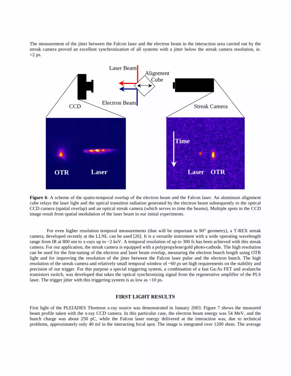
The measurement of the jitter between the Falcon laser and the electron beam in the interaction area carried out by the streak camera proved an excellent synchronization of all systems with a jitter below the streak camera resolution, ie. <2 ps.
Figure 6: A scheme of the spatio-temporal overlap of the electron beam and the Falcon laser. An aluminum alignment cube relays the laser light and the optical transition radiation generated by the electron beam subsequently to the optical CCD camera (spatial overlap) and an optical streak camera (which serves to time the beams). Multiple spots in the CCD image result from spatial modulation of the laser beam in our initial experiments.
For even higher resolution temporal measurements (that will be important in 90° geometry), a T-REX streak camera, developed recently at the LLNL can be used [26]. It is a versatile instrument with a wide operating wavelength range from IR at 800 nm to x-rays up to ~2 keV. A temporal resolution of up to 300 fs has been achieved with this streak camera. For our application, the streak camera is equipped with a polypropylene/gold photo-cathode. The high resolution can be used for the fine-tuning of the electron and laser beam overlap, measuring the electron bunch length using OTR light and for improving the resolution of the jitter between the Falcon laser pulse and the electron bunch. The high resolution of the streak camera and relatively small temporal window of ~60 ps set high requirements on the stability and precision of our trigger. For this purpose a special triggering system, a combination of a fast Ga:As FET and avalanche transistors switch, was developed that takes the optical synchronizing signal from the regenerative amplifier of the PLS laser. The trigger jitter with this triggering system is as low as <10 ps.
FIRST LIGHT RESULTS
First light of the PLEIADES Thomson x-ray source was demonstrated in January 2003. Figure 7 shows the measured beam profile taken with the x-ray CCD camera. In this particular case, the electron beam energy was 54 MeV, and the bunch charge was about 250 pC, while the Falcon laser energy delivered at the interaction was, due to technical problems, approximately only 40 mJ in the interacting focal spot. The image is integrated over 1200 shots. The average
Electron Beam
Laser Beam
CCD Streak Camera
OTR Laser OTRLaser
Time
Alignment Cube

photon count per shot of ~6 x 10⁴ (or 700 pJ/pulse) was observed, with a peak photon energy of 70 keV. The measured FWHM divergence of the beam is 14 mrad.
Figure 7: First PLEIADES light. Generation of hard x-rays at 70 keV with 6 x 10⁴ photons per pulse as observed by the x-ray CCD camera and radiograph of a diode aluminum box. The measured data correspond well to the theoretical model (7b). The intensity profile (taken over the white line in Fig. 7a) is fitted to a theoretical curve (corrected for the mirror absorption)
020406080
100120140160180200
0 20 40 60 80 100
X-ray Energy (keV)
Figure 8: Calculated on-axis spectrum of the Thomson source experiment as fitted from the experimental data.
Our x-ray dose equals the highest photon output demonstrated so far [17], [21]. The theoretical intensity profile (Fig. 7b)
0
300
600
900
1200
1500
-0.015 -0.005 0.005 0.015θθθθ (rad)
Cou
nts

agrees well with the measured profile. The theoretical model accounts for the broadening effects from the measured beam emittance and the narrowing effects derived from the spectral dependence of the transmission coefficient of the laser turning mirror.
In this model, the fitted parameters allow for a calculation of main parameters of the Thomson source. Hence, we determined that the source spectrum (Fig. 8) peaks at 70 keV (0.18 Å) with FWHM of ~8 keV, i.e. the bandwidth ∆λ/λ of ~11%.
5. PERSPECTIVES
Dramatic improvements of the per shot x-ray dose are expected after optimizing the electron beam and Falcon laser parameters; specifically, the electron beam final focus optics, reduction of electron beam emittance through the optimization of both the photo-cathode driver laser uniformity and the electron beam transport, and maximization of the Falcon pump laser energy delivered to the interaction region. These improvements should allow for the realization of final focus spot sizes as small as 10 µm rms, and the production of up to 108-109 x-ray photons per shot (Tab. 1).
Parameter LBL [17] LLNL PLEIADES(projected values)
X-ray output Enhancement
Laser energy 80 mJ 300 mJ 4xElectron current 50 A 1000 A 20xSource radius 50 µm 10 µm 25xSource divergence 10 mrad 5 mrad 4xX-ray bandwidth 15% 4% 4xTotal improvement 3x10⁴
Table 1: Projected key parameters of the PLEIADES source compared to the LBL Thomson scattering source
REFERENCES
1. Christoph Rose-Petruck, Ralph Jimenez, Ting Guo, Andrea Cavalleri, Craig W. Siders, Ferenc Ráksi, Jeff A. Squier, Barry C. Walker, Kent R. Wilson and Christopher P. J. Barty: Picosecond-milliångström lattice dynamics measured by ultrafast X-ray diffraction, Nature 398, 310 (1999).
2. C. W. Siders, A. Cavalleri, K. Sokolowski-Tinten, Cs. Tóth, T. Guo, M. Kammler, M. Horn von Hoegen, K. R. Wilson, D. von der Linde, C. P. J. Barty: Detection of nonthermal melting by ultrafast X-ray diffraction, Science 286, 1340 (1999).
3. A. Rousse, C. Rischel, S. Fourmaux, I. Uschmann, S. Sebban, G. Grillon, Ph. Balcou, E. Forster, J.-P. Geindre, P. Audebert, J.-C. Gauthier, D. Hulin: Non-thermal melting in semiconductors measured at femtosecond resolution, Nature 410, 65 (2001).
4. K. Sokolowski-Tinten, C. Blome, J. Blums, A. Cavalleri, C. Dietrich, A. Tarasevitch, I. Uschmann, E. Förster, M. Kammler, M. Horn-von-Hoegen and D. von der Linde: Femtosecond X-ray measurement of coherent lattice vibrations near the Lindemann stability limit, Nature 422, 287 (2003).
5. A. H. Chin, R. W. Schoenlein, T. E. Glover, P. Balling, W. P. Leemans, and C. V. Shank: Ultrafast structural dynamics in InSb probed by time-resolved x-ray diffraction, Phys. Rev. Lett. 83, 336 (1999).
6. K.-J. Kim, S. Chattopadhyay, C. V. Shank: Generation of femtosecond X-rays by 90° Thomson scattering, Nucl. Instr. and Methods in Phys. Res. A 341, 351-354 (1994).
7. E. Feenberg and H. Primakoff, Phys. Rev. 73, 449 (1948).8. Richard H. Milburn: Electron scattering by an intense polarized photon field, Phys. Rev. Lett. 10, 75 (1963).9. F. R. Arutyunian and V. A. Tumanian: The Compton effect on relativistic electrons and the possibility of obtaining
high energy beams, Phys. Lett. 4, 176 (1963).

10. P. Sprangle, B. Hafizi, and F. Mako: New x-ray source for lithography, Appl. Phys. Lett. 55, 2559 (1989).11. Sally K. Ride, Eric Esarey, and Michael Baine: Thomson scattering of intense lasers from electron beams at
arbitrary interaction angles, Phys. Rev. E 52, 5425 (1995).12. F. V. Hartemann: High-intensity scattering processes of relativistic electrons in vacuum, Phys. Plas. 5, 2037 (1998).13. F. V. Hartemann, J. R. Van Meter, A. L. Troha, E. C. Landahl, N. C. Luhmann, Jr., H. A. Baldis, Atul Gupta, and A.
K. Kermann: Three-dimensional relativistic electron scattering in an ultrahigh-intensity laser focus, Phys. Rev. E 58, 5001 (1998).
14. F. V. Hartemann, H. A. Baldis, A. K. Kerman, A. Le Foll, N. C. Luhmann, Jr., and B. Rupp: Three-dimensional theory of emittance in Compton scattering and x-ray protein crystallography, Phys. Rev. E 64, 016501 (2001).
15. Eric Esarey, Sally K. Ride, Phillip Sprangle: Nonlinear Thomson scattering of intense laser pulses from beams and plasmas, Phys. Rev. E 48, 3003 (1993).
16. F. V. Hartemann, A. K. Kerman: Classical theory of nonlinear Compton scattering, Phys. Rev. Lett. 76, 624 (1996).17. R. W. Schoenlein, W. P. Leemans, A. H. Chin, P. Volfbeyn, T. E. Glover, P. Balling, M. Zolotorev, K.-J. Kim, S.
Chattopadhyay, C. V. Shank: Femtosecond x-ray pulses at 0.4 A generated by 90° Thomson scattering: A tool for probing the structural dynamics of materials, Science 274, 236 (1996).
18. W. P. Leemans, R. W. Schoenlein, P. Volfbeyn, A. H. Chin, T. E. Glover, P. Balling, M. Zolotorev, K.-J. Kim, S. Chattopadhyay, and C. V. Shank: Interaction of relativistic electrons with ultrashort laser pulses: Generation of femtosecond x-rays and microprobing of electron beams, IEEE Journal of Quantum Electronics 33, 1925 (1997).
19. W. P. Leemans, R. W. Schoenlein, P. Volfbeyn, A. H. Chin, T. E. Glover, P. Balling, M. Zolotorev, K. J. Kim, S. Chattopadhyay, and C. V. Shank: X-ray based subpicosecond electron bunch characterization using 90° Thomson Scattering, Phys. Rev. Lett. 77, 4182 (1996).
20. I.V. Pogorelsky,I. Ben-Zvi, T. Hirose,S. Kashiwagi,V. Yakimenko,K. Kusche, P. Siddons,J. Skaritka,T. Kumita,A. Tsunemi,T. Omori,J. Urakawa,M. Washio,K. Yokoya,T. Okugi,Y. Liu, P. He,and D. Cline: Demonstration of 8x10¹⁸ photons/second peaked at 1.8 Å in a relativistic Thomson scattering experiment, Phys. Rev. ST AB 3,090702 (2000)
21. M. Yorozu, J. Yang, Y. Okada, T. Yanagida, F. Sakai, K. Takasago, S. Ito, A. Endo: Fluctuation of femtosecond x-ray pulses generated by a laser-Compton scheme, Appl. Phys. B 74, 327-331 (2002).
22. S. C. Fultz, C. L. Whitten, W. J. Gallagher: The LRL (Livermore) 100 MeV linear electron accelerator and facility, IEEE Trans. on Nucl. Sci. NS-18, 533 (1971).
23. G. P. Le Sage, S. G. Anderson, T. E. Cowan, T. Ditmire, and J. B. Rosenzweig, RF photoinjector development for a short-pulse, hard X-ray Thomson scattering source, presented at Advanced Accelerator Concepts - Ninth Workshop, Santa Fe, NM, USA, P. L. Colestock and S. Kelley Editors, AIP pp. 391-404 (2001).
24. B. E. Carlsten: New photoelectric injector design for the Los Alamos National Laboratory XUV FEL accelerator, Nuclear Instruments & Methods in Physics Research A 285, pp. 313-19 (1989).
25. G. R. Hays, T. Cowan, J. K. Crane, G. P. LeSage, V. Yanovsky, T. Ditmire "Progress toward an integrated 100 TW laser-100 MeV electron linac experiment", Technical digest, Conference on Lasers and Electro-Optics, paper CWK9, p. 293, Baltimore (2000).
26. Jaroslav Kuba, Ronnie Shepherd, Rex Booth, Richard Stewart, Edward C. W. Lee, Robert R. Cross and Paul T. Springer: Sub-picosecond resolution streak camera measurements at LLNL: From IR to x-rays, IN: Ultrafast X-ray Detectors And Applications, Chair: Ali M. Khounsary, SPIE International Symposium on Optical Science and Technology, San Diego, California, USA, 3 – 8 August 2003.
ACKNOWLEDMENT
Work performed under the auspices of the US Department of Energy by the University of California Lawrence Livermore National Laboratory under Contract No. W-7405-Eng-48.


