Embed Size (px)
Citation preview
Pituitary Apoplexy
Claire Briet, Sylvie Salenave, Jean-François Bonneville, Edward R. Laws,and Philippe Chanson
Service d’Endocrinologie et des Maladies de la Reproduction and Centre de Référence des Maladies Endocriniennes Rares dela Croissance (C.B., S.S., P.C.), Hôpital de Bicêtre, Assistance Publique-Hôpitaux de Paris, Le Kremlin-Bicêtre F94275, France;Service d’Endocrinologie (C.B.), Centre Hospitalier Universitaire d’Angers, Angers 49000, France; Service d’Endocrinologie(J.-F.B.), Centre Hospitalier Universitaire de Liège, Liège B4000, Belgium; Unité Mixte de Recherche S1185 (P.C.), UniversitéParis-Saclay, Université Paris-Sud; and Institut National de la Santé et de la Recherche Médicale Unité 1185, Faculté deMédecine Paris-Sud, Le Kremlin-Bicêtre F94276, France; and Neurosurgery, Harvard Medical School, Brigham and Women’sHospital (E.R.L.), Boston, Massachusetts 02115
Pituitary apoplexy, a rare clinical syndrome secondary to abrupt hemorrhage or infarction, complicates 2%–12% ofpituitary adenomas, especially nonfunctioning tumors. Headache of sudden and severe onset is the main symptom,sometimes associated with visual disturbances or ocular palsy. Signs of meningeal irritation or altered consciousnessmay complicate the diagnosis. Precipitating factors (increase in intracranial pressure, arterial hypertension, majorsurgery, anticoagulant therapy or dynamic testing, etc) may be identified. Corticotropic deficiency with adrenalinsufficiency may be life threatening if left untreated. Computed tomography or magnetic resonance imagingconfirms the diagnosis by revealing a pituitary tumor with hemorrhagic and/or necrotic components. Formerlyconsidered a neurosurgical emergency, pituitary apoplexy always used to be treated surgically. Nowadays, conser-vative management is increasingly used in selected patients (those without important visual acuity or field defectsand with normal consciousness), because successive publications give converging evidence that a wait-and-seeapproach may also provide excellent outcomes in terms of oculomotor palsy, pituitary function and subsequenttumor growth. However, it must be kept in mind that studies comparing surgical approach and conservative man-agement were retrospective and not controlled. (Endocrine Reviews 36: 622–645, 2015)
I. IntroductionII. Epidemiology
III. Predisposing and Precipitating FactorsA. Precipitating factorsB. Influence of the adenoma subtype
IV. PathophysiologyV. Clinical Presentation
A. HeadacheB. Visual disturbancesC. Other neurological signsD. Scoring system
VI. Endocrine DysfunctionA. Corticotropic deficiencyB. Other pituitary hormone deficienciesC. Diabetes insipidusD. Pituitary hypersecretion
VII. Diagnostic EvaluationA. Differential diagnosisB. ImagingC. Computed tomographyD. Magnetic resonance imaging
VIII.Management of PA
A. A matter of debateB. Steroid therapy is mandatoryC. Surgical approachD. Conservative approachE. Surgical or conservative management?F. Outcome of ocular palsiesG. Outcome of ocular defectsH. Outcome of pituitary functionI. Outcome of the pituitary tumorJ. Can imaging help to choose between conservative
and surgical treatment?K. United Kingdom guidelines for the management of PA
IX. ConclusionX. Search Strategy and Selection Criteria
I. Introduction
Bailey was the first to describe a case of fatal pituitarytumor-associated hemorrhage in 1898 (1). The sec-
ond description was an autopsy case of hemorrhagic pi-
ISSN Print 0163-769X ISSN Online 1945-7189Printed in USACopyright © 2015 by the Endocrine SocietyReceived April 26, 2015. Accepted September 12, 2015.First Published Online September 28, 2015
Abbreviations: BP, blood pressure; CSF, cerebrospinal fluid; CT, computed tomography;DA, dopamine agonist; DWI, diffusion-weighted imaging; MRI, magnetic resonance im-aging; NFPA, nonfunctioning pituitary adenoma; PA, pituitary apoplexy; PAS, PituitaryApoplexy Score; PRL, prolactin; SAH, subarachnoid hemorrhage; T1, longitudinal relax-ation time; T2, transversal relaxation time; T1W, T1 weighted; T2W, T2 weighted; T2*W,T2-star weighted.
R E V I E W
622 press.endocrine.org/journal/edrv Endocrine Reviews, December 2015, 36(6):622–645 doi: 10.1210/er.2015-1042
Dow
nloaded from https://academ
ic.oup.com/edrv/article/36/6/622/2354744 by guest on 19 July 2022
tuitary infraction in a young acromegalic patient in 1905(2). But the first full description was published in 1950,under the term “pituitary apoplexy” (3). Pituitary apo-plexy (PA) is a clinical syndrome due to abrupt hemor-rhaging and/or infarction of the pituitary gland, generallywithin a pituitary adenoma. Headache of sudden and se-vere onset is the main symptom, sometimes associatedwith visual disturbances or ocular palsy. PA can reveal thepituitary adenoma or occur during its follow-up.
The outcome of acute apoplexy is variable and difficultto predict. Clinical status may deteriorate dramatically(subarachnoid hemorrhage [SAH] from the apoplectic ad-enoma, or cerebral ischemia secondary to cerebral vaso-spasm), or the patient may recover spontaneously, with orwithout sequelae (visual defects, neurological disorders,or pituitary insufficiency). PA sometimes completely de-stroys the adenoma, whereas in other cases, a remnantmay regrow. This explains why the optimal manage-ment of acute PA remains controversial. Indeed, PA wasalmost universally considered a neurosurgical emer-gency in the past (4 –7), but reports of spontaneous clin-ical recovery and/or tumor disappearance have led somespecialists to adopt a conservative approach in selectedcases (8 –21).
II. Epidemiology
PA is a rare event. According to recent epidemiologicalstudies, its prevalence is about 6.2 cases per 100 000 in-
habitants (22) and its incidence 0.17 episodes per 100 000per year (23). Between 2% and 12% of patients with alltypes of adenoma experience apoplexy (4, 6, 10, 12, 24–32), and the diagnosis of pituitary tumor was unknown attime of apoplexy in more than 3 out of 4 cases (Table 1)(12–16, 24, 27, 29, 31, 33–49). If the nonfunctioning pi-tuitary adenomas (NFPAs) (often incidentalomas) werealready known and that a decision was made to managethem conservatively, the risk of PA was calculated to bebetween 0.2 and 0.6 events per 100 person-years in 2metaanalyses (50, 51).
PA can occur at all ages but is most frequent in the fifthor sixth decade, with a male preponderance ranging from1.1 to 2.3/1 (Table 1) (4, 7, 10, 12–16, 24, 27, 29, 31,33–49, 52–54).
Subclinical (asymptomatic) apoplexy is much more fre-quent than acute apoplexy, because up to 25% of all pi-tuitary tumors display hemorrhagic and/or necrotic areas(4–6, 38, 42, 55) either on imaging or at autopsy, but thissubject is beyond the scope of this review.
III. Predisposing and Precipitating Factors
A. Precipitating factors
Precipitating factors are identified in 10%–40% ofcases of PA (Table 1). Many single case-reports of PA havebeen published where the relationship with a potential
Table 1. Demographic, Clinical, Biochemical, and Evolutive Characteristics of Patients With PA as Reported in theMain Retrospective Series Published in the Literature Since 2000 and Including at Least 15 Adults or 8 Children
First Author(Reference)
Year ofPublication n
Mean Age,Years(Range)
SexRatio(M/F)
Length ofDuration ofthe Study
Number (%)of AdenomasPreviouslyKnown
Type of Secretion Defined Clinically and/or Biochemically (%)PrecipitatingFactors (%)GH PRL ACTH NF or GT NA
Biousse (33) 2001 30 51 (21–90) 14/16 1989–2000 6 (20) 0 3 (10) 0 3 (10) 26 (80) 9 (30)Chacko (34) 2002 41 40 (18–65) 28/13 1983–1995 NA 4 (10) 3 (7) 0 34 (83) 0 NASibal (13) 2004 45 49 (16–72) 28/17 1983–2004 8 (18) 1 (2) 14 (31) 3 (7) 2 (4) 25 (56) 18 (40)Ayuk (12) 2004 33 52 (27–79) 20/13 1994–2004 1 (3) 0 0 0 1 (3) 32 (97) 10 (33)Semple (27) 2005 62 51 (18–82) 38/24 1970–2003 12 (19) 1 (2) 7 (11) 0 48 (77) 6 (10) 2 (4)Lubina (40) 2005 40 51 (15–80) 27/13 1985–2002 4 (10) 1 (2.5) 2 (5) 0 1 (2.5) 36 (90) 5 (13)Gruber (14) 2006 30 53 (17–86) 23/7 1988–2004 4 (13) 0 3 (10) 0 1 (3) 26 (87) 37Khaldi (37) 2006 25 54 (20–79) 14/11 1980–2003 3 (12) 1 (4) 5 (20) 0 0 19 (76) 3 (12)Semple (43) 2006 59 51 (NA) 34/25 1970–2003 NA 1 (2) 1 (2) 0 48 (81) 9 (15) 3 (5)Dubuisson (29) 2007 24 56 (23–87) 16/8 1968–2004 6 (25) 1 (4) 3 (13) 1 (4) 1 (4) 18 (75) 12 (50)Zhang (49) 2009 185 38 (16–65) 89/96 1990–2007 NA 14 (8) 117 (63) 6 (3) 0 48 (26) 47 (25)Shou (45) 2009 44 43 (NA) 36/8 2006–2006 NA 2 (5) 3 (7) 0 0 39 (89) NALiu (39) 2010 65 48 (21–87) 30/35 2002–2006 NA 2 (3) 10 (15) 0 0 53 (82) NAPal (41) 2011 32 57 (29–85) 23/9 1985–2008 2 (6) 0 0 0 32 (100) 0 NAZhang (47) 2011 52 52 (18–79) 39/13 2001–2009 NA 3 (6) 12 (23) 1 (2) 1 (2) 35 (67) NASeuk (44) 2011 29 42 (25–68) 21/8 1995–2009 6 (21) 2 (7) 3 (10) 1 (3) 23 (79) 0 NAMoller-Goede (31) 2011 42 53 (21–85) 30/12 1980–2007 1 (2) 1 (2) 7 (17) 2 (5) 29 (69) 3 (7) 32 (76)Leyer (15) 2011 44 53 (12–83) 27/17 1996–2008 12 (27) 0 (0) 4 (9) 2 (4) 6 (14) 32 (73) 23 (52)Chan (35) 2012 17 39 (NA) 6/11 1979–2012 17 (100) 0 0 17 (100) 0 0 2 (12)Sarwar (42) 2013 25 34 (23–41) 3/22 2000–2011 25 (100) 0 25 (100) 0 0 0 8 (32)Kinoshita (38) 2014 58 48 (16–75) 28/30 2006–2012 58 (100) 6 (10) 22 (38) 0 30 (52) 0 5 (9)Vargas (46) 2014 47 51 (NA) 26/21 1999–2013 9 (19) NA NA NA NA 47 (100) NABujawansa (16) 2014 55 58 (14–78) 35/20 1985–2010 4 (7) 4 (7) 6 (11) 0 45 (82) 0 11 (20)Jankowski (36) 2014 9 17 (14–23) 3/6 2008–2013 1 (11) 0 7 (78) 0 0 2 (22) 4 (44)Jho (48) 2014 109 52 (18–87) 69/40 1992–2012 8 (7) 0 1 (1) 0 1 (1) 107 (98) 9 (8)Total 1202 48 y 707/495 187/756 (25) 44/1155 (4) 258/1155 (22) 33/1155 (3) 306/1155 (26) 563/1255 (45) 240/892 (27)
n, number; M, male; F, female; PRL, prolactin; NF, nonfunctioning; GT, gonadotroph; NA, nonavailable; VF, visual field; VA, visual acuity.
doi: 10.1210/er.2015-1042 press.endocrine.org/journal/edrv 623
Dow
nloaded from https://academ
ic.oup.com/edrv/article/36/6/622/2354744 by guest on 19 July 2022
precipitating factor has been suggested. The most relevantclinical situations associated with PA are summarized inTable 2.
Angiographic procedures, particularly cerebral angiog-raphy, have been reported to be associated with PA whichbegan from few minutes to 7 hours after the procedure; PAmay be related either to blood pressure (BP) fluctuationsor to vasospasm (56–59). Among surgical procedures, or-thopedic surgery and cardiac surgery seem more prone tobe complicated by PA than gastrointestinal (60–62) orpulmonary (63) surgery or thyroidectomy (64). PA oc-curred from very early, in the operating theater, to 24–48hours after orthopedic surgery (on the hip or shouldermore often than on the knee) (65–73). Proposed mecha-nisms include intra- or postoperative hypotension, anti-coagulation and/or microemboli leading to infarction.Cardiac surgery, because of BP fluctuations and antico-agulant therapy, is a very “classic” clinical situation lead-ing to intra- or postoperative PA (60, 74–92). Cardiopul-monary bypass is likely to be an important contributor bygenerating important variation in BP. This has led someauthors to recommend, when the presence of pituitaryadenoma is known before the cardiac procedure, to useoff-pump technique which maintains pulsatile flow withan adequate systemic perfusion, as opposed to standardcardiopulmonary bypass (82). The temporal relationshipbetween a head trauma (sometimes quite minor) and theonset of specific signs and symptoms also seems convinc-
ing that cranial trauma may be a cause of PA (93–100).Apoplexy can also occur after dynamic testing (insulin,TRH, GnRH, or GHRH tests and much more rarelyCRH). The period between the test and the beginning ofPA (often few minutes only) is, once again, a good argu-ment supporting that dynamic tests, particularly when dif-ferent agents are combined, are clearly precipitating fac-tors of PA in many instances (101–130). The suspectedmechanism is the imbalance between the increased meta-bolic demand induced by the stimulation and the ability ofincreased blood flow at the level of the pituitary adenoma(see section IV). Reports of PA occurring after stimulationtest are much rarer in the recent past, probably because thevast majority of endocrinologists are now convinced thatmany of these tests (TRH, GnRH) do not bring crucialinformation while they put the patient at risk of PA (121).In this setting, we do not recommend preoperative stim-ulation tests in patients with pituitary macroadenomasparticularly when they present suprasellar extension (131,132) except CRH or Insulin tolerance test when useful forevaluating corticotropic axis. Treatment with GnRH ago-nists for prostate cancer has also been associated with PA,occurring from few minutes to 10 days after the injectionof long-acting formulation (133–143).
PA may be observed in patients under anticoagulationtherapy, sometimes very soon after the initiation of treat-ment, or after a prolonged period of treatment (31, 144–147); new classes of anticoagulant (dabitagran) (148, 149)
Table 1. Continued
Immunocytochemistry (%) Clinical/Biochemical Characteristics at Diagnosis (%)
Surgery (%)
Evolution
GH PRL ACTH FSH/LHNecrosis orNegative NA Headache Vomiting Unconsciousness
AbnormalVF/VA
CranialNervesImpairment Hypopituitarism
NormalPituitaryFunction (%)
Recurrenceof theAdenoma (%)
NA NA NA NA NA 30 (100) 30 (100) NA 9 (30) 14/11 (47/37) 17 (57) 16 (53) 27 (90) 4 (12) 1 (3)3 (7) 1 (2) 1 (2) 0 10 (25) 26 (63) NA NA NA NA NA 28 (68) 41 (100) NA NA2 (4) 2 (4) 3 (7) 2 (4) 21 (47) 15 (33) 43 (96) 35 (78) 10 (22) 20/18 (48/46) 22 (51) 34 (76) 27 (60) 7 (15) 5 (11)1 (3) 1 (3) 0 8 (24) 5 (15) 18 (55) 32 (97) 17 (53) NA 11/27 (36/82) 15 (46) 24 (72) 15 (46) 5 (15) 2 (6)1 (2) 1 (2) 0 0 8 (13) 52 (84) 52 (84) 15 (24) 8 (13) 27/38 (43/61) 26 (43) 45 (73) 58 (93) 7 (12) 46 (74)7 (18) 2 (5) 3 (7) 3 (7) 4 (10) 21 (52) 25 (63) 20 (50) 7 (17) 24/NA (61/NA) 16 (40) 17 (42) 34 (85) 6 (14) 15 (37)4 (13) 2 (7) NA NA NA 24 (80) 27 (90) 14 (47) 3 (10) 10/18 (33/60) 15 (62) 21 (70) 10 (33) 3 (10) 6 (20)NA NA NA NA NA 25 (100) 24 (96) 17 (68) 10 (40) 14/17 (56/68) 20 (80) NA 20 (80) NA NA1 (2) 1 (2) 0 48 (81) 8 (13) 1 (2) 49 (83) NA 9 (15) 39/39 (66/66) 24 (41) 48 (81) 34 (58) 9 (15) NA2 (8) 1 (4) 2 (8) 2 (8) 6 (25) 11 (46) 22 (92) 13 (54) 10 (42) 12/12 (50/50) 13 (54) 17 (71) 21 (87) 2 (8) 4 (17)96 (52) 14 (8) 6 (3) 0 69 (37) 0 70 (38) NA NA 46/96 (25/52) NA 101 (54) 185 (100) 159 (86) 23 (12)2 (5) 0 0 0 12 (27) 30 (68) 42 (96) 29 (66) 1 (2) 33/31 (75/70) 28 (64) NA 44 (100) 14 (32) 014 (22) 2 (3) 1 (2) 7 (11) 11 (17) 30 (46) 30 (46) 11 (17) 4 (6) 50/50 (77/77) 9 (14) 24 (37) 65 (100) 20 (31) 10 (15)NA NA NA NA NA NA 25 (78) 15 (47) 4 (12) 16/22 (50/69) 26 (81) 24 (75) 29 (90) NA 4 (11)NA NA NA NA NA 52 (100) 51 (98) 48 (92) 15 (29) 41/48 (79/92) 39 (75) 17 (33) 52 (100) 6 (11) 5 (10)NA NA NA NA NA 29 (100) 29 (100) NA NA 23/26 (79/90) 21 NA 29 (100) NA NANA NA NA NA NA 42 (100) 5 (12) NA NA 16/NA (38/NA) 32 (76) 19 (45) 39 (93) 12 (29) NA5 (11) 1 (2) 1 (2) 6 (14) 11 (25) 20 (45) 41 (93) 26 (59) 12 (27) 19/24 (43/54) 22 (50) 35 (80) 39 (89) 12 (27) 4 (9)0 0 11 (65) 0 6 (35) 0 12 (70) 10 (59) NA 11/11 (65/65) NA NA 12 (70) 8 (47) 0NA NA NA NA NA 25 (100) 7 (28) NA NA NA NA 3 (13) 2 (8) NA 2 (9)NA NA NA NA NA 58 (100) NA NA NA NA NA NA NA NA NANA NA NA NA NA 47 (100) 35 (75) NA NA 34/34 (73/74) 10 (23) 40 (85) 36 (76) 20 (43) 12 (25)NA NA NA NA NA 55 (100) 48 (87) 14 (25) NA 20/NA (36/NA) 26 (47) NA 33 (55) 8 (14) NA7 (78) 0 0 1 (11) 1 (11) 9 (100) 2 (22) NA 3/0 (33/0) 2 (22) 8 (89) 6 (66) 2 (22)13 (12) 3 (3) 1 (1) 0 81 (74) 11 (10) 95 (87) 36 (33) 14 (13) 43/NA (39/NA) 39 (36) 92 (84) 101 (93) 43 (39) NA
803/1101(73)
322/661(49)
116/691 (17) VF:526/1078(49) VA: 567/832 (68)
420/867 (48) 607/944 (64) 961/1144 (84) (28) 141/707 (20)
624 Briet et al Pituitary Apoplexy Endocrine Reviews, December 2015, 36(6):622–645
Dow
nloaded from https://academ
ic.oup.com/edrv/article/36/6/622/2354744 by guest on 19 July 2022
Table 2. Literature Review on the Main Predisposing or Precipitating Factors of PA
Predisposing/
Precipitating
Factors
First Author, Year
of Publication Reference Age Sex Tumor Type
Drug Used/Type of
Intervention Dose
Duration Before
Apoplexy Weeks/
Days/Hours/Minutes
Angiography Reichenthal, 1980 57 40 M NA Carotid angiography 15 minSuga, 1996 59 29 F PRL Cerebral angiography 6 hLouwerens, 1996 56 32 M GH Cerebral angiography 7 hSkljarevski, 2003 58 66 M NF Coronarography Immediate postprocedure
Orthopedic surgery Lennon, 1998 69 51 M NA Total hip replacement under spinal
anesthesia
6 h
Liu, 2001 70 45 M NA Laparoscopic anterior lumbar
interbody fusion
1 h
Galvin, 2004 65 48 M NA Total hip replacement (�lupus
anticoagulant)
48 h
Khandelwal, 2005 67 65 F NA Total knee replacement 24 hThomason, 2009 73 70 M NA Total hip replacement 4dGoel, 2009 66 76 M Necrosis Total hip replacement 24 hGoel, 2009 66 61 M Necrosis Total knee arthroplasty 24 hKoga, 2010 68 60 M NF Shoulder arthroplasty During the procedureMadhusudhan, 2011 71 62 M No operated Shoulder afthroplasty 48 hPrescott, 2011 72 58 M Not operated Total hip replacement 10 h
Cardiac surgery Peck, 1980 74 68 M Not operated Aortic valve replacement 48 hSlavin, 1984 75 57 M Necrosis CABG �24 hSlavin, 1984 75 55 M NF, hemorrhage,
necrosis
Mitral valve replacement 48 h
Cooper, 1986 78 63 M NF CABG 63 hCooper, 1986 78 55 M PRL,FSH CABG 55 hCooper, 1986 78 62 M NF Mitral and aortic valve
replacement
12 h
Khardori, 1987 81 62 M Necrosis CABG 8 hShapiro, 1990 89 60 ? NA CABG 24 hAbsalom, 1993 76 61 M Hemorrhage CABG 40 hSavage, 1994 88 72 M Necrosis CABG Immediately after
procedureSavage, 1994 88 64 M Necrosis CABG 21 hMeek, 1998 85 56 M Necrosis Mitral valve repair 15 minMattke, 2002 84 64 M NA CABG 20 minAlzetani, 2002 77 72 M Not operated CABG Immediate postopGlass, 2003 79 63 M NA CABG 4 hAbbott, 2004 60 48 M Necrosis Aortic valve replacement 24 hLiberale, 2006 83 73 M NA Aortic abdominal aneurysm
surgery
Immediate postop
Mukhida, 2007 87 71 M Not operate CABG 71 hLevy, 2007 82 58 M NA CABG Postop (time not
provided)Levy, 2007 82 65 M NA CABG Postop (time not
provided)Levy, 2007 82 68 M NA CABG Postop (time not
provided)Levy, 2007 82 65 M NA CABG Postop (time not
provided)Hidiroglu, 2010 80 47 M NA Cardiac surgery 2 hYakupoglu, 2010 91 74 M Necrosis Open cardiac surgery 6 hTansel, 2010 90 60 M NA CABG (anaphylaxis shock due to
allergy to protamine)
During the procedure
Mizuno, 2011 86 73 M Necrosis CABG 73 hKocyigit, 2011 92 66 F NA CABG 66 h
Surgery other Yahagi, 1992 62 47 M NA Cholecystectomy 24 hKato, 1996 64 38 F GH Thyroidectomy 3 hAbbott, 2004 60 66 M NA Laparotomy for resection of
hepatic mass
48 h
Mura, 2014 61 85 M Not operated Laparoscopic/laparotomic resection
of sigmoid colon
0 h
Yoshino, 2014 63 78 M NA Pulmonary lobectomy and lymph
node dissection for squamous
cell carcinoma
128 h
Closed head
trauma
Holness, 1983 95 39 M NF Minor head trauma motor vehicle ImmediateTamasawa, 1988 99 34 M GH Fall from the back of a truck ImmediateItoyama,1990 97 45 M NA Fall 2 meters 3 hUchiyama, 1999 100 66 M NA Fall 15 meters 15 hUchiyama, 1999 100 60 F NA Mild (fall from his height) 1 hHorie, 2002 96 56 F NA Traffic car accident 15 minSmidt, 2007 98 30 M NA Minor head trauma 3 wkDev, 2007 94 40 M necrosis Mild cranial trauma (road traffic) 1 wkBao, 2007 93 79 M Not operated Mild cranial trauma (fall) 1 h
(Continued)
doi: 10.1210/er.2015-1042 press.endocrine.org/journal/edrv 625
Dow
nloaded from https://academ
ic.oup.com/edrv/article/36/6/622/2354744 by guest on 19 July 2022
Table 2. Continued
Predisposing/
Precipitating
Factors
First Author, Year
of Publication Reference Age Sex Tumor Type
Drug Used/Type of
Intervention Dose
Duration Before
Apoplexy Weeks/
Days/Hours/Minutes
Dynamic tests Dunn, 1975 107 22 F GH TRH 400 �g 48 hSilverman, 1978 126 31 M PRL Chlorpromazine 25 mg 1.5 hJordan, 1979 112 21 F ACTH (Nelson) Dexamethasone 8 mg 8 mg The last day of the testCimino, 1981 104 48 M NF GnRH/TRH 100 �g/200 �g 20 minDrury, 1982 106 59 F NF GnRH/TRH 100 �g/200 �g 5 minDrury, 1982 106 66 M GH TRH 200 �g 10 minDrury, 1982 106 39 F PRL GnRH/TRH 100 �g/200 �g 2 minDrury, 1982 106 28 M PRL GnRH/TRH 100 �g/200 �g 15 minKorsic, 1984 114 56 M FSH GnRH 100 �g 2 hBernstein, 1984 102 48 M NF GnRH/TRH/ITT 100 �g/200 �g/0.1 UI/kg
BW
5 min
Chapman, 1985 103 39 F PRL GnRH/TRH/ITT 100/200/0.15 UI/kg BW 0.5 hLever, 1986 116 19 F GH TRH 200 2 minShirataki, 1988 125 50 F GH BRC 2.5 mg 2 minHarvey, 1989 111 50 M NF ITT 0.15 UI/kg 0 minArafah, 1989 101 41 F PRL GnRH 100 �g 1 hMasson, 1993 118 54 F GT GnRH 100 �g 20 minOkuda, 1994 120 60 F NF GnRH/TRH/ITT 100/500/0.1 U/kg 10 minVassallo, 1994 128 81 M NF GnRH/TRH/L-Dopa/ACTH 100 �g/200 �g/500 mg/
250 �g
2 h
Masago, 1995 117 48 M FSH GnRH/TRH/ITT 100 �g/500 �g/0.1 UI/kg
BW
15 min
Masago, 1995 117 54 M NF GnRH/TRH 100 �g/500 �g 10 minFrankart, 1995 109 64 M FSH-LH GnRH/TRH 100 �g/200 �g 48 hGrunenberger, 1996 110 30 M GH GnRH/TRH/glucagon 100 �g/250 �g/2 mg 24 hSzabolcs, 1997 127 54 M NF TRH 200 �g 1 hOtsuka, 1998 121 31 F GH GnRH/TRH/CRH/GHRH 100 �g/200 �g/100 �g/
100 �g
2 min
Dökmetas, 1999 105 28 F GH GnRH/TRH 100 �g/200 �g 88 hSanno, 1999 124 55 M NF GnRH/TRH/CRH/GHRH 100 �g/500 �g/100 �g/
100 �g
30 min
Foppiani, 2000 108 43 F NF GnRH 100 �g Few minutesLee, 2000 115 34 M GH GnRH/TRH/ITT 100 �g/400 �g/0.15 UI/kg
BW
20 min
Riedl, 2000 122 71 F NF GnRH/TRH/CRH/GHRH NA ImmediateMatsuura, 2001 119 63 M NF GnRH/TRH/ITT 100 �g/500 �g/0.1 UI/kg
BW
2 h
Rotman-Pikielny,
2003
123 19 F ACTH CRH 100 �g 48 h
Wang, 2007 129 41 F Necrosis TRH 200 �g 2 hYoshino, 2007 130 36 M Necrosis GnRH/TRH/ITT 100 �g/500 �g/5 IU 24 hYoshino, 2007 130 38 M Necrosis TRH 500 �g 2 hKılıçlı, 2010 113 52 M FSH LH PRL GnRH/TRH 100 �g/200 �g 30 min
GnRH agonists Ando, 1995 133 83 M NA Goserelin 3.6 mg 9 dChanson, 1995 134 78 M FSH Triptorelin 3.75 mg 10 minMorsi, 1996 135 74 M FSH, LH, GH Leuprolide 7.5 mg 15 minReznik, 1997 136 62 M FSH, LH Leuprolin 3.75 mg 4 dEaton, 2001 137 67 M FSH, LH Goserelin Cyproterone 3.6 mg 100 mg/d 4 hHernandez Morin,
2003
138 69 M FSH, LH Leuprolin Bicalutamide 11.25 mg 50 mg/d 30 min
Massoud, 2006 139 70 M FSH Leuprolin 11.25 mg 10 dBłaut, 2006 140 74 M negative Goserelin 3.6 mg 12 hHands, 2007 141 60 M LH NA NA 4 hIto, 2011 142 78 M FSH Goserelin Bicalutamide 3.6 mg 80 mg/d 9 dHuang, 2013 143 77 M necrosis Leuprolin 3.75 mg Few hours
DAs Yamaji, 1981 154 46 M GH BRC 7.5 mg/d 48 wkYamaji, 1981 154 55 F GH BRC NA 24 wkOnesti, 1990 55 34 F PRL BRC NA NAGittelman, 1991 155 19 F NA BRC 5 mg/d 48 wkBiller, 1996 156 26 F PRL CAB 0.5 mg/wk 12 wkPinto, 1998 157 14 F PRL BRC 10 mg/d 24 wkHanna, 1999 158 42 F PRL BRC NA NAVella, 2001 159 30 M PRL CAB 0.5 mg/wk 20 wkKnoepfelmacher,
2004
160 17 M PRL CAB 1.5 mg/wk 52 wk
Balarini Lima, 2008 153 57 M PRL CAB 3.5 mg/wk 7 wkBalarini Lima, 2008 153 27 M PRL CAB 1.5 mg/wk 12 wkBalarini Lima, 2008 153 15 F PRL CAB 1.5 mg/wk 32 wkBalarini Lima, 2008 153 52 F PRL CAB NA 1 wk (1st); 8 wk (2nd)Chng, 2013 161 20 M PRL CAB 0.5 mg/wk 6 wk
wk, weeks; d, days; h, hours; min, minutes; BW, body weight; NA, nonavailable; NF, nonfunctioning; BRC, bromocriptine; CAB, cabergoline; CABG, coronary arterybypass graft; ITT, insulin tolerance test.
626 Briet et al Pituitary Apoplexy Endocrine Reviews, December 2015, 36(6):622–645
Dow
nloaded from https://academ
ic.oup.com/edrv/article/36/6/622/2354744 by guest on 19 July 2022
may also be involved. Bleeding disorders have also been re-ported as associated with PA (150–152). These case reportssuggest that these medications or conditions may favor theoccurrence of PA. However, to our knowledge, there hasbeen no prospective study looking at their specific role on therisk of PA, and it is thus very difficult to propose any rec-ommendation concerning authorization or contraindicationfor anticoagulation prescription in patients with a knownpituitary adenoma.
The role of dopamine agonists (DAs) treatment as pre-cipitating factor is more controversial. As exemplified bycase reports summarized in Table 2, PA may occur duringtreatment with DA, sometimes early after the initiation oftreatment (eg, 1 wk), but this is quite rare (153), suggestinga potential role of DA. However, in most cases, PA isreported to occur after a very prolonged period of treat-ment (55, 153–161), and the responsibility of DA is thusmore questionable. Moreover, in prospective studies an-alyzing the effects of DA on macroprolactinomas, PA werevery rarely or never observed (23, 156, 162–168) evenwhen treatment was initiated at high dose (168), whenhigh doses were used due to DA resistance (165, 169) orwhen pituitary adenomas were already hemorrhagic onmagnetic resonance imaging (MRI) (42). In a review, PAwas found to occur in 1%–6% of macroprolactinomastreated with DA, a rate not higher than that of untreatedmacroprolactinomas (170). Finally, it must be pointed outthat prolactinomas are, by themselves, more prone tobleeding (see section III.B) even if this is generallyasymptomatic. Indeed, in a study analyzing MRI char-acteristics at diagnosis (before any treatment), 16 out of79 macroprolactinomas (20.3%) were identified ashaving pituitary hemorrhage and 3 of them presentedwith “classic apoplexy” (42). Some authors have evensuggested that DA treatment may prevent macropro-lactinomas from PA (31).
Contrary to previous studies, a recent publication sug-gests that diabetes and arterial hypertension do not pre-dispose patients to PA (31).
B. Influence of the adenoma subtypePA is observed more frequently in patients with NFPAs
(Table 1) (7, 12–16, 22, 24, 27, 29, 31, 33–49, 52, 171,172). This may be the result of selection bias, because theseadenomas are generally discovered late and are usuallylarger than functioning adenomas. Indeed, in the vast ma-jority of cases, apoplexy complicates large macroadeno-mas (31). Other tumor types that have been reported toundergo apoplexy include prolactinomas and GH-secret-ing adenomas (12–16, 27, 29, 31, 33–49, 173).
Clinically silent ACTH adenomas may be particularlyprone to necrosis, hemorrhage and cyst transformation.
These complications occur in 30%–64% of cases, com-pared with 6.8%–20% in patients with prolactinomasand 2%–14% in patients with all types of pituitary ade-noma (38, 42, 174–176), and are generally revealed byMRI. Recurrent clinical apoplexy has been described inthese patients (174, 176–179), and may be associated witha brisk and transient increase in ACTH/cortisol produc-tion, leading to hypertension, hyperglycemia, hypokale-mia, and edema.
It must be underlined that in many cases, if not obviousclinically, the potential functioning nature of the adenomathat underwent apoplexy is impossible to assess due to theextent of necrosis.
IV. Pathophysiology
The pathophysiology of PA is not fully understood, but itis noteworthy that most cases involve patients with mac-roadenomas (7, 26, 31, 180, 181).
The understanding of clinical and biochemical pictureof patients with PA is helped by a better knowledge of thecharacteristics of pituitary gland vascularization. Pitu-itary vascularization is supported by a capillary networkcalled hypophysial portal system coming from hypothal-amus via the long portal veins and by direct arterial bloodsupply either from superior hypophysial arteries that de-scends along the pituitary stalk to the anterior pituitarygland or from inferior hypophysial arteries to the posteriorpituitary, both originating from branches of the internalcarotid artery. Moreover, the superior and inferior hypo-physial circulations are anastomised. The venous drainageis directed via hypophysial veins to adjacent venous si-nuses and then to jugular veins. By contrast with normalpituitary, the vascularization of pituitary adenomas arepredominantly supported by a direct arterial blood supplyrather than portal system (43, 182, 183) and contrast-enhanced imaging clearly shows that blood supply to pi-tuitary adenomas is reduced compared with normal pitu-itary (184). Pituitary tumors have reduced angiogenesis asshown by reduced density of microvasculature (185, 186).In addition to the presence of fenestrated endothelial cells,which are characteristic of the normal capillaries of theanterior pituitary, prolactinomas contain arteries, rangingfrom well-formed vessels with multiple layers of smoothmuscle cells to abnormal terminal arterioles, ie, vesselswith fenestrated endothelium surrounded by a variablenumber of smooth muscle cells. Such arteries are not foundin normal anterior pituitary (187).
Nevertheless, pituitary adenomas are prone to bleedand undergo infarction and necrosis, possibly because pi-tuitary gland has this unique rich vascular structure and/or
doi: 10.1210/er.2015-1042 press.endocrine.org/journal/edrv 627
Dow
nloaded from https://academ
ic.oup.com/edrv/article/36/6/622/2354744 by guest on 19 July 2022
because pituitary tumors (which have a high-energy re-quirement) may outgrow their blood supply or becauseischemia (and thus infarction) occurs after compression ofinfundibular or superior hypophyseal vessels against thesellar diaphragm by the expanding tumor mass (180, 188)with intrinsically poor vascularity.
Moreover, as recently demonstrated, pituitary tumorcells are particularly sensitive to glucose deprivation(184).
In this setting, all clinical situations (see section III.A)that acutely decrease systemic BP, such as cardiac, vascu-lar, or orthopedic surgery may decrease blood supply tothe pituitary adenoma and precipitate apoplexy. Dynamictests or hypoglycemia that acutely increase the metabolicneeds of the tumor may also precipitate apoplexy, as wellas severe vomiting/diarrhea with concomitant increasedValsalva pressure.
The inherent fragility of tumoral blood vessels mayalso explain the hemorrhagic tendency (189). Indeed,the vessels of pituitary adenomas show signs of incom-plete maturation and poor fenestration, and their basalmembranes are often ruptured (52, 190, 191). Immu-nohistochemical expression of vascular endothelialgrowth factor was found to correlate positively with therisk of pituitary hemorrhage (192). Pituitary tumor-transforming gene, which is correlated to vasculariza-tion and expression of vascular endothelial growth fac-tor (193), is also overexpressed in pituitary adenomas(194, 195). Fetal liver kinase 1, a vascular marker, isalso expressed, particularly in NFPAs, particularly inolder subjects (196), as is nestin, another vascularmarker (197).
To our knowledge, genetic markers of intracranial vas-cular malformations such as cranial aneurysms (198, 199)have not been studied in patients with PA.
Imaging studies and surgical exploration may revealboth hemorrhage and ischemic necrosis (10, 43, 200).
Whatever the mechanism, the extent of hemorrhageand necrosis will produce an increase in intrasellar pres-sure (26, 52, 201–203), which in turn leads to more orless pronounced compression of neighboring struc-tures, thus explaining the broad clinical spectrum, from“classical” acute PA to totally silent necrotic and/orhemorrhagic adenomas found only on pathologicalexamination.
V. Clinical Presentation
The clinical presentation of PA is highly variable and islargely determined by the extent of hemorrhage, necrosis,and edema (Table 1).
A. HeadacheHeadache is the most prominent symptom of acute ap-
oplexy and is present in more than 80% of patients (12–16, 27, 29, 31, 33–49, 54, 204, 205). Headache is alsogenerally the initial symptom, with sudden and severe on-set described “like a thunderclap in a clear sky” (206). Itis probably due to dural traction or to extravasation ofblood and necrotic material into the subarachnoid space,producing meningeal irritation. Headache is usually ret-roorbital but can be bifrontal or diffuse. It is often asso-ciated with vomiting and nausea and can mimic migraineor meningitis (13, 24, 207). Headache may be subacutewith slow development of symptoms.
B. Visual disturbancesVisual disturbances are present in more than half of
PA patients (12–17, 24, 27, 29, 31, 33– 49, 52, 55, 204).They are due to the sudden hemorrhage-related increasein tumor mass, leading to compression of surroundingstructures (mainly the optic chiasm or optic nerves,from upward expansion of the tumor). Variable degreesof visual-field impairment may be observed, bitemporalhemianopsia being most common. Loss of visual acuityand blindness can occur, but are rare (24, 27, 32, 52, 55,204).
Oculomotor palsies are also frequent, affecting 52% ofpatients in a compilation of studies (208), and are due tofunctional impairment of cranial nerves III, IV, and VI.The third cranial nerve is the most frequently affected (halfof cranial nerve palsies) and is characterized by ptosis,limited eye movements in adduction, and mydriasis (Fig-ure 1) (24, 204, 208). This phenomenon can be due tointracavernous expansion of the tumor mass, a hematomaor, most frequently, to an abrupt pressure increase in thepituitary region (the cranial nerves are exquisitely sensitiveto increased pressure) (203).
C. Other neurological signsSigns or symptoms of meningeal irritation, such as pho-
tophobia (40%), nausea, vomiting (57%), meningismus(25%), and sometimes fever (16%), may be misleading.An initial false diagnosis of meningitis may be made, es-pecially as cerebrospinal fluid (CSF) examination mayshow hyperlymphocytosis (98, 209). Variable degrees ofaltered consciousness may be observed, ranging from leth-argy to stupor or even coma (12–16, 27, 29, 31, 33–49).These signs are due to extravasation of blood or necrotictissue into the subarachnoid space (210).
Cerebral ischemia can occasionally result from me-chanical compression of the carotid artery against the an-terior clinoid, or to cerebral vasospasm, and lead to focalneurological deficits such as hemiparesis or dysphasia, or
628 Briet et al Pituitary Apoplexy Endocrine Reviews, December 2015, 36(6):622–645
Dow
nloaded from https://academ
ic.oup.com/edrv/article/36/6/622/2354744 by guest on 19 July 2022
to a pyramidal syndrome (211, 212). Stroke can be part ofthe differential diagnosis, at presentation or subsequently.
Less frequent manifestations include anosmia (due toolfactory nerve compression), epistaxis or CSF rhinorrhea(due to erosion of the bone of the sella turcica) and facialpain (due to compression of cranial nerve V). Accompa-nying acute adrenal insufficiency may lead to collapse andvital signs changes, suggesting an incorrect diagnosis ofmyocardial infarction.
D. Scoring systemThe UK Pituitary Apoplexy Guidelines Development
Group proposed a Pituitary Apoplexy Score (PAS) basedon the level of consciousness, visual acuity and field de-fects, and ocular palsies (Table 3) in order to enable moreuniform clinical description of PA and, thus, better com-parison of different management options (17).
VI. Endocrine Dysfunction
Acute endocrine dysfunction may also be present, furthercomplicating the clinical picture (Table 1). One or more
anterior pituitary deficiencies arepresent at PA onset (12–17, 24, 25,27, 29, 31, 33–49, 213). In retro-spect, signs and symptoms consistentwith endocrine abnormalities are of-ten present before the apoplectic ep-isode, such as sexual problems, men-strual disturbances, galactorrhea orfatigue (7, 24). These disorders areproduced by mass effect on the nor-mal pituitary. Multiple acute endo-crine insufficiencies can occur, re-lated either to destruction of theanterior pituitary or to increasedintrasellar pressure on the pituitarystalk, impairing the release of hy-pothalamic and/or pituitary hor-mones (201, 204). Details aboutthe various pituitary deficits foundat presentation in the main series ofPA published in the literature since2000 (12, 13, 15, 27, 29, 31, 38,40, 42, 45, 46, 49) are given in Ta-ble 4.
A. Corticotropic deficiencyCorticotropic deficiency is the
most common deficit observed in pa-tients with PA, occurring in 50%–80% of cases (12–15, 17, 24, 25, 27,29, 31, 38, 40, 42, 45, 46, 49, 213).
It is also the most life-threatening hormonal complication,potentially causing severe hemodynamic problems andhyponatremia (214, 215). Because acute secondary adre-nal insufficiency is very frequent in patients with apo-plexy, empiric parenteral corticosteroid supplementation(if possible preceded by blood drawing for subsequentserum cortisol determination) should be given to all pa-tients with signs of PA, without waiting for diagnosticconfirmation.
Severe hypotension occurs when vessels become insen-sitive to endogenous or exogenous catecholamines in theabsence of circulating cortisol (216). Hyponatremia mayalso be observed in patients with corticotropic deficiency,particularly in the acute setting (217–219).
Both hypotension and hyponatremia are not related tohypomineralocorticism as observed in primary adrenal in-sufficiency (Addison’s disease), in which both mineralo-and glucocorticoid levels are insufficient (220). Mineralo-corticoid levels are normal in ACTH-deficient patients.Hyponatremia is a direct consequence of glucocorticoiddeficiency, and is related to inappropriate antidiuresis re-
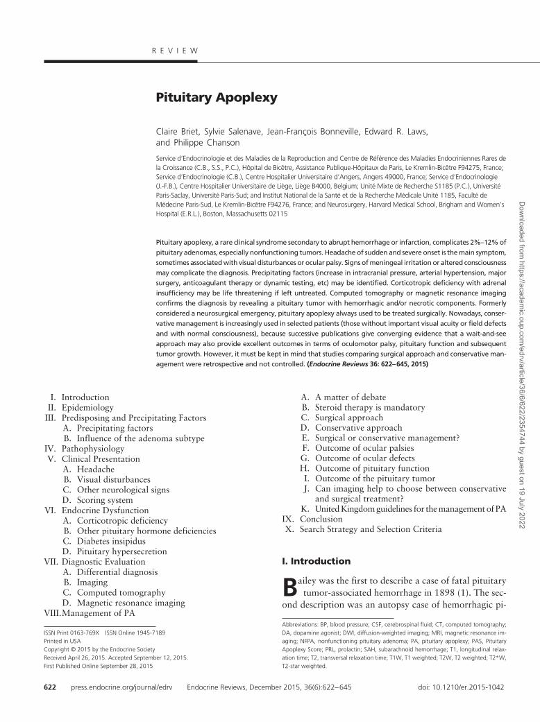
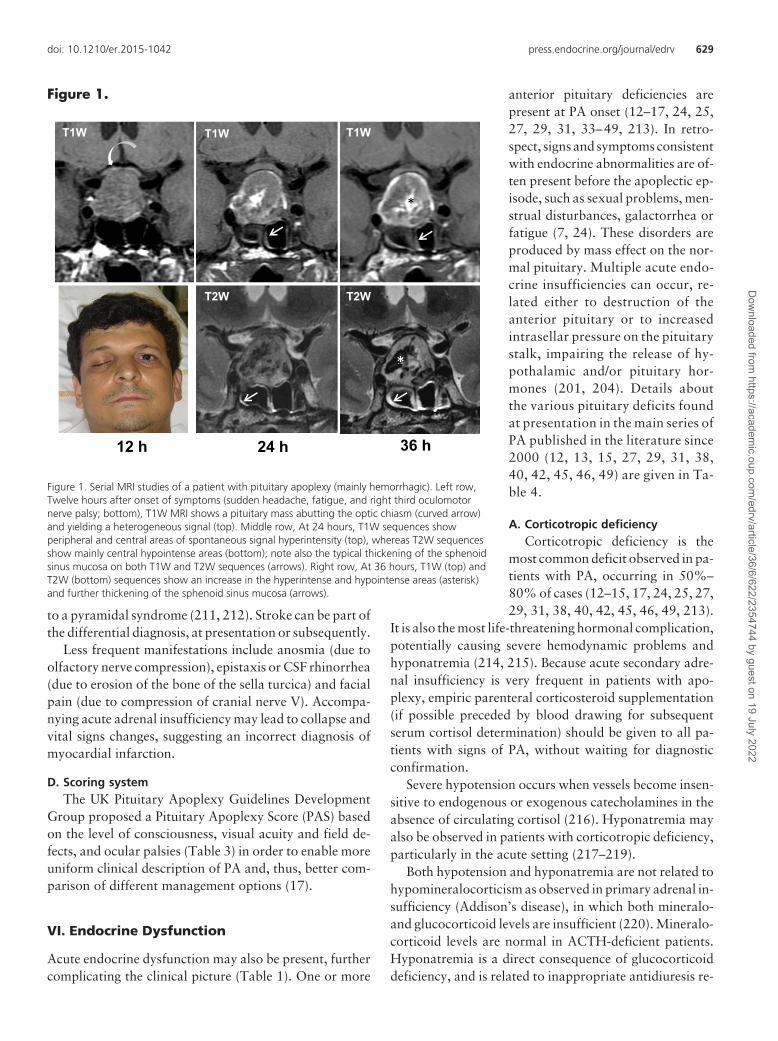
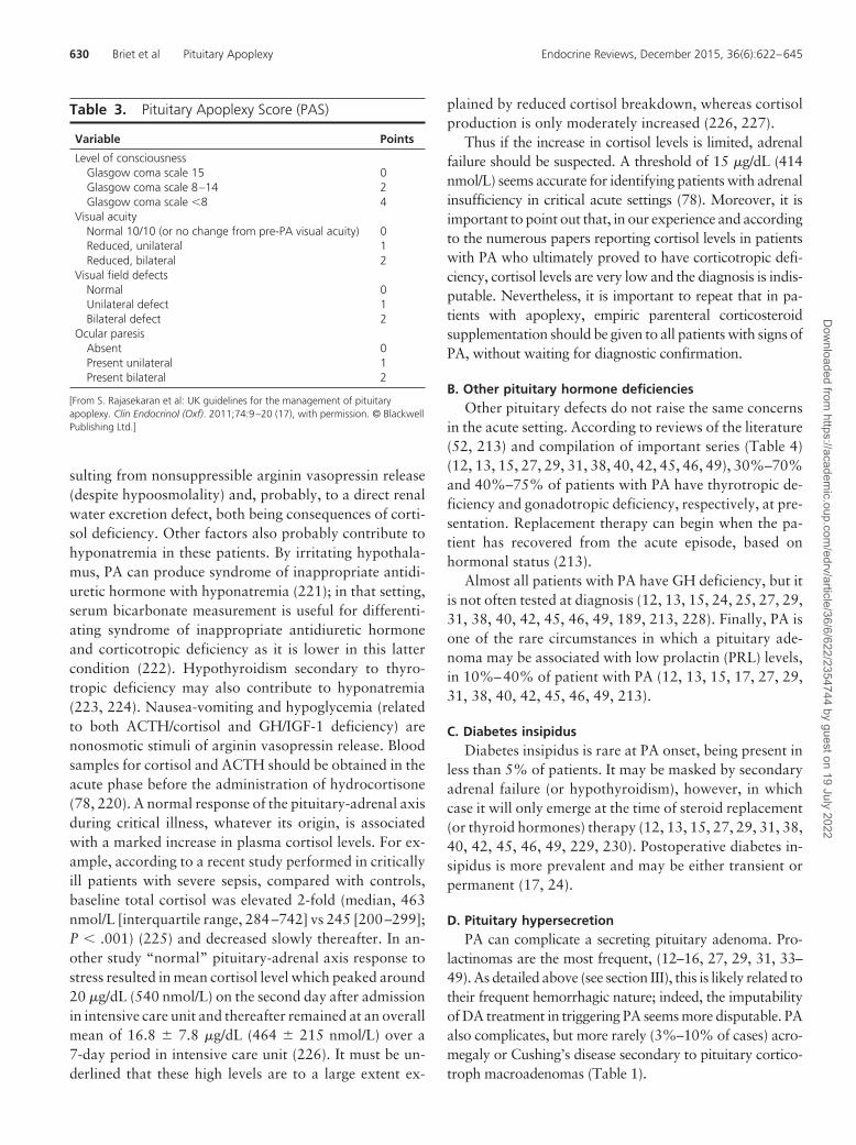
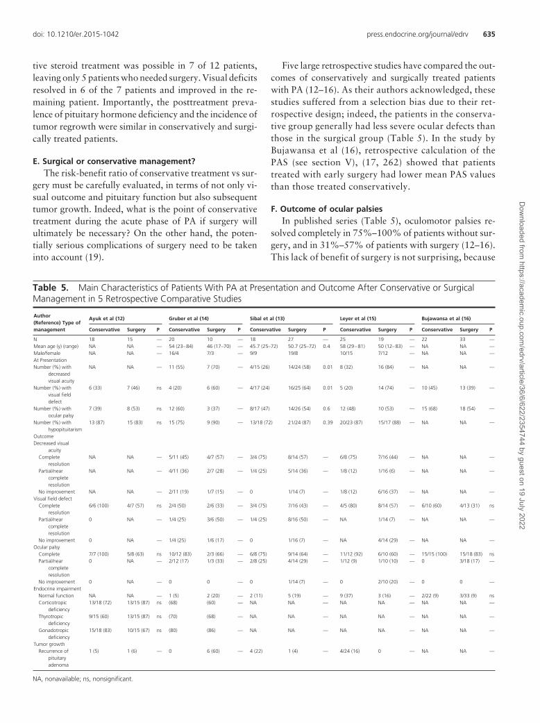
Figure 1.
*
T1W T1W T1W
T2W T2W
12 h 24 h 36 h
*
*
Figure 1. Serial MRI studies of a patient with pituitary apoplexy (mainly hemorrhagic). Left row,Twelve hours after onset of symptoms (sudden headache, fatigue, and right third oculomotornerve palsy; bottom), T1W MRI shows a pituitary mass abutting the optic chiasm (curved arrow)and yielding a heterogeneous signal (top). Middle row, At 24 hours, T1W sequences showperipheral and central areas of spontaneous signal hyperintensity (top), whereas T2W sequencesshow mainly central hypointense areas (bottom); note also the typical thickening of the sphenoidsinus mucosa on both T1W and T2W sequences (arrows). Right row, At 36 hours, T1W (top) andT2W (bottom) sequences show an increase in the hyperintense and hypointense areas (asterisk)and further thickening of the sphenoid sinus mucosa (arrows).
doi: 10.1210/er.2015-1042 press.endocrine.org/journal/edrv 629
Dow
nloaded from https://academ
ic.oup.com/edrv/article/36/6/622/2354744 by guest on 19 July 2022
sulting from nonsuppressible arginin vasopressin release(despite hypoosmolality) and, probably, to a direct renalwater excretion defect, both being consequences of corti-sol deficiency. Other factors also probably contribute tohyponatremia in these patients. By irritating hypothala-mus, PA can produce syndrome of inappropriate antidi-uretic hormone with hyponatremia (221); in that setting,serum bicarbonate measurement is useful for differenti-ating syndrome of inappropriate antidiuretic hormoneand corticotropic deficiency as it is lower in this lattercondition (222). Hypothyroidism secondary to thyro-tropic deficiency may also contribute to hyponatremia(223, 224). Nausea-vomiting and hypoglycemia (relatedto both ACTH/cortisol and GH/IGF-1 deficiency) arenonosmotic stimuli of arginin vasopressin release. Bloodsamples for cortisol and ACTH should be obtained in theacute phase before the administration of hydrocortisone(78, 220). A normal response of the pituitary-adrenal axisduring critical illness, whatever its origin, is associatedwith a marked increase in plasma cortisol levels. For ex-ample, according to a recent study performed in criticallyill patients with severe sepsis, compared with controls,baseline total cortisol was elevated 2-fold (median, 463nmol/L [interquartile range, 284–742] vs 245 [200–299];P � .001) (225) and decreased slowly thereafter. In an-other study “normal” pituitary-adrenal axis response tostress resulted in mean cortisol level which peaked around20 �g/dL (540 nmol/L) on the second day after admissionin intensive care unit and thereafter remained at an overallmean of 16.8 � 7.8 �g/dL (464 � 215 nmol/L) over a7-day period in intensive care unit (226). It must be un-derlined that these high levels are to a large extent ex-
plained by reduced cortisol breakdown, whereas cortisolproduction is only moderately increased (226, 227).
Thus if the increase in cortisol levels is limited, adrenalfailure should be suspected. A threshold of 15 �g/dL (414nmol/L) seems accurate for identifying patients with adrenalinsufficiency in critical acute settings (78). Moreover, it isimportant to point out that, in our experience and accordingto the numerous papers reporting cortisol levels in patientswith PA who ultimately proved to have corticotropic defi-ciency, cortisol levels are very low and the diagnosis is indis-putable. Nevertheless, it is important to repeat that in pa-tients with apoplexy, empiric parenteral corticosteroidsupplementation should be given to all patients with signs ofPA, without waiting for diagnostic confirmation.
B. Other pituitary hormone deficienciesOther pituitary defects do not raise the same concerns
in the acute setting. According to reviews of the literature(52, 213) and compilation of important series (Table 4)(12, 13, 15, 27, 29, 31, 38, 40, 42, 45, 46, 49), 30%–70%and 40%–75% of patients with PA have thyrotropic de-ficiency and gonadotropic deficiency, respectively, at pre-sentation. Replacement therapy can begin when the pa-tient has recovered from the acute episode, based onhormonal status (213).
Almost all patients with PA have GH deficiency, but itis not often tested at diagnosis (12, 13, 15, 24, 25, 27, 29,31, 38, 40, 42, 45, 46, 49, 189, 213, 228). Finally, PA isone of the rare circumstances in which a pituitary ade-noma may be associated with low prolactin (PRL) levels,in 10%–40% of patient with PA (12, 13, 15, 17, 27, 29,31, 38, 40, 42, 45, 46, 49, 213).
C. Diabetes insipidusDiabetes insipidus is rare at PA onset, being present in
less than 5% of patients. It may be masked by secondaryadrenal failure (or hypothyroidism), however, in whichcase it will only emerge at the time of steroid replacement(or thyroid hormones) therapy (12, 13, 15, 27, 29, 31, 38,40, 42, 45, 46, 49, 229, 230). Postoperative diabetes in-sipidus is more prevalent and may be either transient orpermanent (17, 24).
D. Pituitary hypersecretionPA can complicate a secreting pituitary adenoma. Pro-
lactinomas are the most frequent, (12–16, 27, 29, 31, 33–49). As detailed above (see section III), this is likely related totheir frequent hemorrhagic nature; indeed, the imputabilityof DA treatment in triggering PA seems more disputable. PAalso complicates, but more rarely (3%–10% of cases) acro-megaly or Cushing’s disease secondary to pituitary cortico-troph macroadenomas (Table 1).
Table 3. Pituitary Apoplexy Score (PAS)
Variable Points
Level of consciousnessGlasgow coma scale 15 0Glasgow coma scale 8–14 2Glasgow coma scale �8 4
Visual acuityNormal 10/10 (or no change from pre-PA visual acuity) 0Reduced, unilateral 1Reduced, bilateral 2
Visual field defectsNormal 0Unilateral defect 1Bilateral defect 2
Ocular paresisAbsent 0Present unilateral 1Present bilateral 2
[From S. Rajasekaran et al: UK guidelines for the management of pituitaryapoplexy. Clin Endocrinol (Oxf). 2011;74:9–20 (17), with permission. © BlackwellPublishing Ltd.]
630 Briet et al Pituitary Apoplexy Endocrine Reviews, December 2015, 36(6):622–645
Dow
nloaded from https://academ
ic.oup.com/edrv/article/36/6/622/2354744 by guest on 19 July 2022
In some cases, PA leads to resolution of pituitary hy-persecretion by a secreting pituitary adenoma (8, 24, 123,179, 231–239), eg, “fugitive acromegaly.”
VII. Diagnostic Evaluation
A. Differential diagnosis
The clinical presentation of PA may raise 2 major dif-ferential diagnoses, namely SAH and bacterial meningitis.Other neurological events, such as cavernous sinus throm-bosis and midbrain infarction, also need to be eliminated.Lumbar puncture is of little help in differentiating SAHand bacterial meningitis from PA, because the latter maybe accompanied by a high red cell count, xanthochromia,or pleocytosis, and by an increased CSF protein level, par-ticularly when signs of meningeal irritation are present(209, 240, 241). CSF culture will rule out bacterial men-ingitis, and lumbar puncture is thus mandatory if this di-agnosis is suspected. The best tools for diagnosing PA arecomputed tomography (CT) and MRI. By revealing a pi-tuitary tumor, even if no necrosis or hemorrhage isfound, these imaging methods offer confident diagnosticconfirmation.
Diagnosis thus relies on a combination of clinical man-ifestations (eg, sudden headache and visual disturbances)and the detection of a pituitary adenoma, whether beforeor after PA onset.
B. Imaging
Before discussing imaging features it is important tounderstand that the underlying pathophysiological pro-cess in PA can be simple infarction (ie, with little or nohemorrhagic component), hemorrhagic infarction, mixedhemorrhagic infarction and clot, or pure clot (189, 200).This explains why imaging rarely shows pure hemorrhageor infarction but rather mixed features (211).
C. Computed tomography
Given its wide availability, CT is usually the initialemergency examination for patients with severe headacheof sudden onset. It has 2 interests: it rules out SAH and itshows an intrasellar mass in 80% of cases, with hemor-rhagic components in 20%–30% of cases (12, 13, 55,242). After a few days, blood density decreases and may bemore difficult to detect. After administration of contrastmedium, the pituitary tumor shows inhomogeneous en-hancement (243, 244), occasionally with ring enhance-ment (245, 246).
D. Magnetic resonance imaging
MRI is now the imaging procedure of choice (7, 13, 24,55), even in the first days after symptom onset, because itcan detect fresh bleeding (Figure 2). Longitudinal relax-ation time (T1)- and transversal relaxation time (T2)-weighted sequences are both interesting. T1 and T2 havespecific characteristics according to each tissue. T1, T2,and proton density determine the contrast of MR images.The choice of technical parameters as time to repeat andtime to echo allows to obtain images more or less depen-dent on T1 or T2. On T1-weighted (T1W) images, thewater (CSF) is black, the gray matter is darker than thewhite matter; on T2W images, the water (CSF) is hyper-intense, the white matter is darker than gray matter. MRIcan identify hemorrhagic and necrotic areas and show therelationship between the tumor and neighboring struc-tures such as the optic chiasm, cavernous sinuses and hy-pothalamus (247). Because conventional (T1/T2) MRI se-quences may not demonstrate an infarct for 6 hours, andsmall infarcts may be hard to appreciate on CT for days(Figure 3), diffusion-weighted imaging (DWI), which pro-vides information about consistency of macroadenomas,is very useful early in the PA process. Indeed, increasedDWI signal in ischemic tissue is observed within a fewminutes after arterial occlusion. In case of ischemic apo-
Table 4. Percentages of Pituitary Deficiency at Time of PA Presentation in the Main Series Published Since 2000
First Author (Reference)
Year of
Publication
Number of
Patients
Any Pituitary
Deficiency
Gonadotroph
Deficiency
Thyrotroph
Deficiency
Corticotroph
Deficiency
Somatotroph
Deficiency
Lactotroph
Deficiency
Diabetes
Insipidus
Sibal (13) 2004 45 76 76 57 60 NA 40 NAAyuk (12) 2004 33 72 72 37 50 NA 24 NASemple (27) 2005 62 73 40 55 61 6 2 8Lubina (40) 2005 40 42 35 30 50 NA NA 2Dubuisson (29) 2007 24 71 67 67 62.5 58 58 0Zhang (49) 2009 185 54 25 30 NA NA NAShou (45) 2009 44 NA 39 77 73 NA NA NAMöller-Goede (31) 2011 42 45 43 14 7 NA NA 2Leyer (15) 2011 44 89 NA NA 70 NA NA NASarwar (42) 2013 25 13 1 9 13 NA NA NAKinoshita (38) 2014 58 NA 21 13 17 40 6 NAVargas (46) 2014 47 85 49 53 53 35 35 NA
NA, not available.
doi: 10.1210/er.2015-1042 press.endocrine.org/journal/edrv 631
Dow
nloaded from https://academ
ic.oup.com/edrv/article/36/6/622/2354744 by guest on 19 July 2022
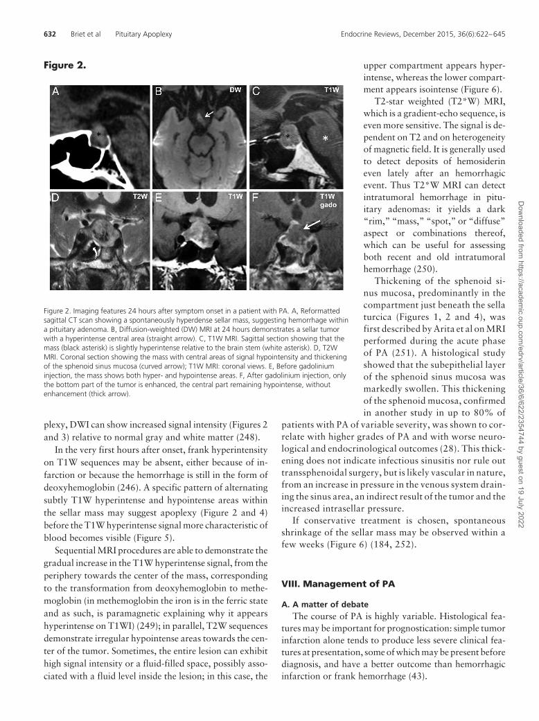
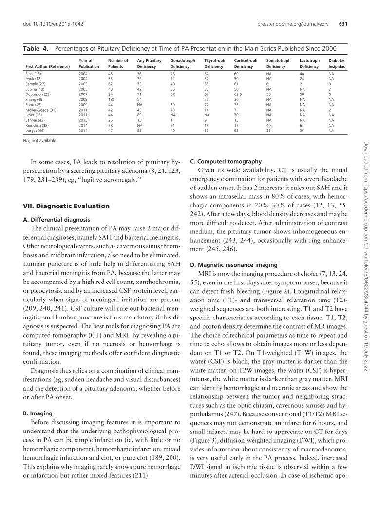
plexy, DWI can show increased signal intensity (Figures 2and 3) relative to normal gray and white matter (248).
In the very first hours after onset, frank hyperintensityon T1W sequences may be absent, either because of in-farction or because the hemorrhage is still in the form ofdeoxyhemoglobin (246). A specific pattern of alternatingsubtly T1W hyperintense and hypointense areas withinthe sellar mass may suggest apoplexy (Figure 2 and 4)before the T1W hyperintense signal more characteristic ofblood becomes visible (Figure 5).
Sequential MRI procedures are able to demonstrate thegradual increase in the T1W hyperintense signal, from theperiphery towards the center of the mass, correspondingto the transformation from deoxyhemoglobin to methe-moglobin (in methemoglobin the iron is in the ferric stateand as such, is paramagnetic explaining why it appearshyperintense on T1WI) (249); in parallel, T2W sequencesdemonstrate irregular hypointense areas towards the cen-ter of the tumor. Sometimes, the entire lesion can exhibithigh signal intensity or a fluid-filled space, possibly asso-ciated with a fluid level inside the lesion; in this case, the
upper compartment appears hyper-intense, whereas the lower compart-ment appears isointense (Figure 6).
T2-star weighted (T2*W) MRI,which is a gradient-echo sequence, iseven more sensitive. The signal is de-pendent on T2 and on heterogeneityof magnetic field. It is generally usedto detect deposits of hemosiderineven lately after an hemorrhagicevent. Thus T2*W MRI can detectintratumoral hemorrhage in pitu-itary adenomas: it yields a dark“rim,” “mass,” “spot,” or “diffuse”aspect or combinations thereof,which can be useful for assessingboth recent and old intratumoralhemorrhage (250).
Thickening of the sphenoid si-nus mucosa, predominantly in thecompartment just beneath the sellaturcica (Figures 1, 2 and 4), wasfirst described by Arita et al on MRIperformed during the acute phaseof PA (251). A histological studyshowed that the subepithelial layerof the sphenoid sinus mucosa wasmarkedly swollen. This thickeningof the sphenoid mucosa, confirmedin another study in up to 80% of
patients with PA of variable severity, was shown to cor-relate with higher grades of PA and with worse neuro-logical and endocrinological outcomes (28). This thick-ening does not indicate infectious sinusitis nor rule outtranssphenoidal surgery, but is likely vascular in nature,from an increase in pressure in the venous system drain-ing the sinus area, an indirect result of the tumor and theincreased intrasellar pressure.
If conservative treatment is chosen, spontaneousshrinkage of the sellar mass may be observed within afew weeks (Figure 6) (184, 252).
VIII. Management of PA
A. A matter of debateThe course of PA is highly variable. Histological fea-
tures may be important for prognostication: simple tumorinfarction alone tends to produce less severe clinical fea-tures at presentation, some of which may be present beforediagnosis, and have a better outcome than hemorrhagicinfarction or frank hemorrhage (43).
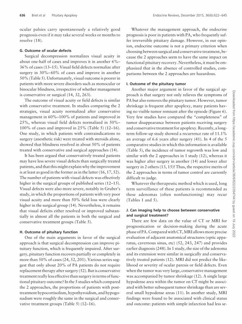
Figure 2.
Figure 2. Imaging features 24 hours after symptom onset in a patient with PA. A, Reformattedsagittal CT scan showing a spontaneously hyperdense sellar mass, suggesting hemorrhage withina pituitary adenoma. B, Diffusion-weighted (DW) MRI at 24 hours demonstrates a sellar tumorwith a hyperintense central area (straight arrow). C, T1W MRI. Sagittal section showing that themass (black asterisk) is slightly hyperintense relative to the brain stem (white asterisk). D, T2WMRI. Coronal section showing the mass with central areas of signal hypointensity and thickeningof the sphenoid sinus mucosa (curved arrow); T1W MRI: coronal views. E, Before gadoliniuminjection, the mass shows both hyper- and hypointense areas. F, After gadolinium injection, onlythe bottom part of the tumor is enhanced, the central part remaining hypointense, withoutenhancement (thick arrow).
632 Briet et al Pituitary Apoplexy Endocrine Reviews, December 2015, 36(6):622–645
Dow
nloaded from https://academ
ic.oup.com/edrv/article/36/6/622/2354744 by guest on 19 July 2022
In mild forms, headache, visual abnormalities and pi-tuitary deficiencies (if not present before onset of PA) de-velop slowly and persist for several days or weeks. In themost acute and severe forms, blindness, coma, neurolog-ical signs and hemodynamic problems may occur withinhours. If the correct diagnosis is not made promptly anddecompression and corticosteroid treatment is not per-formed, death may ensue as a result of adrenal failureand/or neurological complications. Acute PA is thus a truemedical emergency. Most cases, however, fall betweenthese 2 extremes, with headache and visual disturbancesdeveloping over several days.
Recovery of neurological, ophthalmological, and en-docrine function is also highly variable. Altered conscious-
ness improves after decompression;altered visual fields and acuity alsotend to improve after surgery, par-ticularly when they were normal be-fore the acute episode. Permanentsequelae may occur, however, par-ticularly in cases with evidence of op-tic nerve atrophy. Ophtalmoplegiagenerally resolves but this may takeseveral weeks. Endocrine functionoften remains at least slightlyaltered.
The treatment aims are to improvesymptoms and relieve compression oflocal structures, particularly the opticpathways. Surgical decompression isthe most rapid means of achievingthese goals (12–16, 27, 29, 31, 33–49). The dramatic picture presentedby many patients probably explainswhy PA is considered a neurosurgicalemergency and has almost alwaysbeen treated surgically in the past (7,180, 253, 254). However, surgery
may also be harmful, with a risk of postoperative CSF rhi-norrhea, posterior pituitary damage (risk of permanent dia-betes insipidus), and an increased likelihood of hypopituitar-ism due to removal of or damage to normal pituitary tissue.Fortunately, in experienced pituitary centers, these compli-cations are very rare, and this does not prevent to proposesurgery when symptoms are severe and rapidly installedand/or when the tumor is large.
As some patients recover normal visual and endocrinefunction after conservative steroid-based management,the optimal management of acute PA is controversial. Atall events, PA must be managed by an expert multidisci-plinary team including an ophthalmologist, neuroradiolo-
gist, endocrinologist, and neurosur-geon (17).
B. Steroid therapy is mandatoryAs corticotropic deficiency is
present in the vast majority of pa-tients at PA onset and may be lifethreatening, whether treated surgi-cally or conservatively, corticoste-roids should be administered iv assoon as the diagnosis is confirmed; itwill consist of hydrocortisone 50 mgevery 6 hours (52, 255), or a bolus of100–200 mg followed by 50–100mg every 6 hours iv (or im) (220,
Figure 3.
Figure 3. CT scan and MRI images of an ischemic form of PA during the very first hours after thebeginning of symptoms. A, Coronal CT scan. Discrete hypodensity of a pituitary mass and thinning ofthe sellar floor (white arrow). B–D, T1, T2, and contrast-enhanced T1W images. The mass is T1isointense and T2 hyperintense; a rim enhancement is visible after contrast administration, but thecentral part of the mass (asterisk) does not enhance. These images give no indication about thepathologic process. E, Axial DWI shows marked hyperintensity of the lesion (curved arrow), thusconfirming the ischemic origin of the apoplexy (courtesy of Dr C. Magnin).
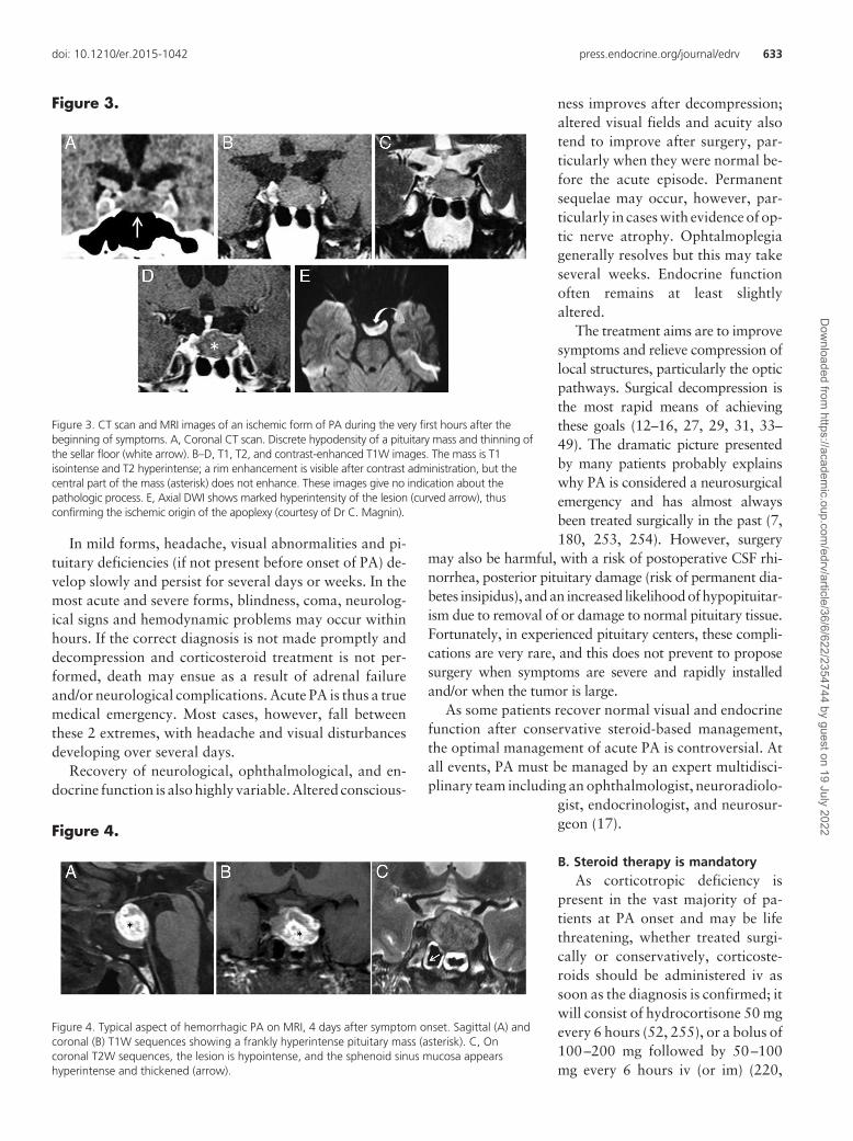
Figure 4.
Figure 4. Typical aspect of hemorrhagic PA on MRI, 4 days after symptom onset. Sagittal (A) andcoronal (B) T1W sequences showing a frankly hyperintense pituitary mass (asterisk). C, Oncoronal T2W sequences, the lesion is hypointense, and the sphenoid sinus mucosa appearshyperintense and thickened (arrow).
doi: 10.1210/er.2015-1042 press.endocrine.org/journal/edrv 633
Dow
nloaded from https://academ
ic.oup.com/edrv/article/36/6/622/2354744 by guest on 19 July 2022

256–258), or 2–4 mg/h by continuous iv administration(17). Patients in shock should initially receive 5% dextrose(to prevent hypoglycemia) in normal saline iv (17, 213, 214).
C. Surgical approachIf surgical management is chosen, the transsphenoidal
approach is almost always recommended, because it al-lows good decompression of the optic pathways and neu-roanatomic structures in contact with the tumor, and be-cause it is associated with low postoperative morbidityand mortality (17).
Transsphenoidal surgery now usually involves trans-nasal septal displacement rather than the classical subla-bial transseptal approach (259). Some neurosurgeons pre-fer to use an operative microscope, others prefer the use ofan endoscope.
Even if surgical complications arerare, particularly in experiencedhands, CSF leakage and diabetes in-sipidus (sometimes permanent) mayoccur (29, 31, 260, 261). Surgical pa-pers dealing with PA rarely mentionthe complication rate. Nevertheless,it seems that endocrine outcome af-ter elective pituitary surgery ispoorer in patients with PA than inpatients without PA. Indeed, in astudy comparing patients operatedfrom pituitary adenomas compli-cated or not with PA, those with PAhad a worse endocrine outcome witha frequency of hypopituitarism in-creasing (from 45% at presentationto 71% during follow-up, odds ra-tio � 4.7, confidence interval �
1.30–25.33; P � .013) in the PA group, whereas it did notchange in the control group (from 48% at presentation to55% during follow-up, odds ratio � 1.5, confidence in-terval � 0.68–3.41; P � .362) (31). Much of this, how-ever, is secondary to damage to the normal gland from theinitial apoplectic event.
Another important point is that, in this acute setting,the operation may be performed by an on-call neurosur-geon rather than by a skilled pituitary neurosurgeon, asunderlined in United Kingdom guidelines (17), and thismay increase the risk of adverse events.
D. Conservative approachReports of spontaneous clinical improvement and
shrinkage (or disappearance) of apoplectic pituitary ade-nomas suggest that a conservativeapproach may be appropriate in se-lected cases. In 1978, Pelkonen et al(8) were among the first to propose aconservative approach, after observ-ing not only spontaneous recoveriesbut also cases in which the apoplexyappeared to cure hormonal hyperse-cretion (GH, ACTH, etc). Other au-thors subsequently also advocated aconservative approach (9, 10).
In 1995, Maccagnan et al re-ported the results of a prospectivestudy in which they treated PA withhigh-dose steroids (11). Only pa-tients whose visual impairmentor altered consciousness failed to im-prove underwent surgery. Conserva-
Figure 5.
Figure 5. MRI in a patient with PA, showing a fluid level inside the pituitary lesion; the uppercompartment is hyperintense, whereas the lower compartment is isointense (T1W sequences,sagittal [A] and axial [B] views).
Figure 6.
Figure 6. Serial imaging studies in a patient with ischemic PA. A, Two days after symptom onset,T1W image shows an heterogeneous pituitary mass (asterisk). Note the ectopic position of theposterior pituitary represented by a T1 hyperintense nodule below the optic chiasm (curvedarrow). B, Forty-eight hours later, T1W image does not show any hyperintense area within in thepituitary mass, suggesting purely necrotic apoplexy. Note the slight thickening of the sphenoidsinus mucosa (straight arrow). C, Four months later, after conservative management, T1WIdemonstrates a spontaneous shrinkage of the tumor.
634 Briet et al Pituitary Apoplexy Endocrine Reviews, December 2015, 36(6):622–645
Dow
nloaded from https://academ
ic.oup.com/edrv/article/36/6/622/2354744 by guest on 19 July 2022
tive steroid treatment was possible in 7 of 12 patients,leaving only 5 patients who needed surgery. Visual deficitsresolved in 6 of the 7 patients and improved in the re-maining patient. Importantly, the posttreatment preva-lence of pituitary hormone deficiency and the incidence oftumor regrowth were similar in conservatively and surgi-cally treated patients.
E. Surgical or conservative management?The risk-benefit ratio of conservative treatment vs sur-
gery must be carefully evaluated, in terms of not only vi-sual outcome and pituitary function but also subsequenttumor growth. Indeed, what is the point of conservativetreatment during the acute phase of PA if surgery willultimately be necessary? On the other hand, the poten-tially serious complications of surgery need to be takeninto account (19).
Five large retrospective studies have compared the out-comes of conservatively and surgically treated patientswith PA (12–16). As their authors acknowledged, thesestudies suffered from a selection bias due to their ret-rospective design; indeed, the patients in the conserva-tive group generally had less severe ocular defects thanthose in the surgical group (Table 5). In the study byBujawansa et al (16), retrospective calculation of thePAS (see section V), (17, 262) showed that patientstreated with early surgery had lower mean PAS valuesthan those treated conservatively.
F. Outcome of ocular palsiesIn published series (Table 5), oculomotor palsies re-
solved completely in 75%–100% of patients without sur-gery, and in 31%–57% of patients with surgery (12–16).This lack of benefit of surgery is not surprising, because
Table 5. Main Characteristics of Patients With PA at Presentation and Outcome After Conservative or SurgicalManagement in 5 Retrospective Comparative Studies
Author(Reference) Type ofmanagement
Ayuk et al (12) Gruber et al (14) Sibal et al (13) Leyer et al (15) Bujawansa et al (16)
Conservative Surgery P Conservative Surgery P Conservative Surgery P Conservative Surgery P Conservative Surgery P
N 18 15 — 20 10 — 18 27 — 25 19 — 22 33 —Mean age (y) (range) NA NA — 54 (23–84) 46 (17–70) — 45.7 (25–72) 50.7 (25–72) 0.4 58 (29–81) 50 (12–83) — NA NA —Male/female NA NA — 16/4 7/3 — 9/9 19/8 10/15 7/12 — NA NA —At PresentationNumber (%) with
decreasedvisual acuity
NA NA — 11 (55) 7 (70) — 4/15 (26) 14/24 (58) 0.01 8 (32) 16 (84) — NA NA —
Number (%) withvisual fielddefect
6 (33) 7 (46) ns 4 (20) 6 (60) — 4/17 (24) 16/25 (64) 0.01 5 (20) 14 (74) — 10 (45) 13 (39) —
Number (%) withocular palsy
7 (39) 8 (53) ns 12 (60) 3 (37) — 8/17 (47) 14/26 (54) 0.6 12 (48) 10 (53) — 15 (68) 18 (54) —
Number (%) withhypopituitarism
13 (87) 15 (83) ns 15 (75) 9 (90) — 13/18 (72) 21/24 (87) 0.39 20/23 (87) 15/17 (88) — NA NA —
OutcomeDecreased visual
acuityComplete
resolutionNA NA — 5/11 (45) 4/7 (57) — 3/4 (75) 8/14 (57) — 6/8 (75) 7/16 (44) — NA NA —
Partial/nearcompleteresolution
NA NA — 4/11 (36) 2/7 (28) — 1/4 (25) 5/14 (36) — 1/8 (12) 1/16 (6) — NA NA —
No improvement NA NA — 2/11 (19) 1/7 (15) — 0 1/14 (7) — 1/8 (12) 6/16 (37) — NA NA —Visual field defect
Completeresolution
6/6 (100) 4/7 (57) ns 2/4 (50) 2/6 (33) — 3/4 (75) 7/16 (43) — 4/5 (80) 8/14 (57) — 6/10 (60) 4/13 (31) ns
Partial/nearcompleteresolution
0 NA — 1/4 (25) 3/6 (50) — 1/4 (25) 8/16 (50) — NA 1/14 (7) — NA NA —
No improvement 0 NA — 1/4 (25) 1/6 (17) — 0 1/16 (7) — NA 4/14 (29) — NA NA —Ocular palsy
Complete 7/7 (100) 5/8 (63) ns 10/12 (83) 2/3 (66) — 6/8 (75) 9/14 (64) — 11/12 (92) 6/10 (60) — 15/15 (100) 15/18 (83) nsPartial/near
completeresolution
0 NA — 2/12 (17) 1/3 (33) — 2/8 (25) 4/14 (29) — 1/12 (9) 1/10 (10) — 0 3/18 (17) —
No improvement 0 NA — 0 0 — 0 1/14 (7) — 0 2/10 (20) — 0 0 —Endocrine impairment
Normal function NA NA — 1 (5) 2 (20) — 2 (11) 5 (19) — 9 (37) 3 (16) — 2/22 (9) 3/33 (9) nsCorticotropic
deficiency13/18 (72) 13/15 (87) ns (68) (60) — NA NA — NA NA — NA NA —
Thyrotropicdeficiency
9/15 (60) 13/15 (87) ns (70) (68) — NA NA — NA NA — NA NA —
Gonadotropicdeficiency
15/18 (83) 10/15 (67) ns (80) (86) — NA NA — NA NA — NA NA —
Tumor growthRecurrence of
pituitaryadenoma
1 (5) 1 (6) — 0 6 (60) — 4 (22) 1 (4) — 4/24 (16) 0 — NA NA —
NA, nonavailable; ns, nonsignificant.
doi: 10.1210/er.2015-1042 press.endocrine.org/journal/edrv 635
Dow
nloaded from https://academ
ic.oup.com/edrv/article/36/6/622/2354744 by guest on 19 July 2022
ocular palsies carry spontaneously a relatively goodprognosis even if it may take several weeks or months toresolve (18).
G. Outcome of ocular defects
Surgical decompression normalizes visual acuity inabout one-half of cases and improves it in another 6%–36% of cases (13–15). Visual field defects normalize aftersurgery in 30%–60% of cases and improve in another50% (Table 5). Unfortunately, visual outcome is poorer inpatients with more severe disorders such as monocular orbinocular blindness, irrespective of whether managementis conservative or surgical (14, 32, 263).
The outcome of visual acuity or field defects is similarwith conservative treatment. In studies comparing the 2strategies, visual acuity normalized after conservativemanagement in 60%–100% of patients and improved in25%, whereas visual field defects normalized in 50%–100% of cases and improved in 25% (Table 5) (12–16).One study, in which patients with contraindications tosurgery (anesthetic risk) were treated with steroids alone,showed that blindness resolved in about 50% of patientstreated with conservative and surgical approaches (14).
It has been argued that conservatively treated patientsmay have less severe visual defects than surgically treatedpatients, and that this might explain why the improvementis at least as good in the former as in the latter (16, 17, 52).The number of patients with visual defects was effectivelyhigher in the surgical groups of published series (12–15).Visual defects were also more severe, notably in Gruber’sstudy, in which the proportions of patients with very poorvisual acuity and more than 50% field loss were clearlyhigher in the surgical group (14). Nevertheless, it remainsthat visual deficits either resolved or improved substan-tially in almost all the patients in both the surgical andconservative treatment groups (Table 5).
H. Outcome of pituitary function
One of the main arguments in favor of the surgicalapproach is that surgical decompression can improve pi-tuitary function, which is frequently impaired. After sur-gery, pituitary function recovers partially or completely inmore than 50% of cases (24, 52, 201). Various series sug-gest that only about 20% of PA patients do not requirereplacement therapy after surgery (52). But is conservativetreatment really less effective than surgery in terms of func-tional pituitary outcome? In the 5 studies which comparedthe 2 approaches, the proportions of patients with post-treatment hypocortisolism, hypothyroidism, and hypogo-nadism were roughly the same in the surgical and conser-vative treatment groups (Table 5) (12–16).
Whatever the management approach, the endocrineprognosis is poor in patients with PA, who frequently suf-fer irreversible pituitary damage. However, in our opin-ion, endocrine outcome is not a primary criterion whenchoosing between surgical and conservative treatment, be-cause the 2 approaches seem to have the same impact onfunctional pituitary recovery. Nevertheless, it must be em-phasized that in the absence of controlled studies, com-parisons between the 2 approaches are hazardous.
I. Outcome of the pituitary tumor
Another major argument in favor of the surgical ap-proach is that surgery not only relieves the symptoms ofPA but also removes the pituitary tumor. However, tumorshrinkage is frequent after apoplexy, many patients hav-ing no visible tumor remnant after the episode (Figure 6).Very few studies have compared the “completeness” oftumor disappearance between patients receiving surgeryandconservative treatment forapoplexy.Recently, a long-term follow-up study showed a recurrence rate of 11.1%an average of 6.6 years after surgery (41). In 4 of the 5comparative studies in which this information is available(Table 5), the incidence of tumor regrowth was low andsimilar with the 2 approaches in 1 study (12), whereas itwas higher after surgery in another (14) and lower aftersurgery in 2 others (13, 15)! Thus, the respective merits ofthe 2 approaches in terms of tumor control are currentlydifficult to judge.
Whatever the therapeutic method which is used, longterm surveillance of these patients is recommended asthese adenomas (often nonfunctioning) may recur(Tables 1 and 5).
J. Can imaging help to choose between conservativeand surgical treatment?
There are few data on the value of CT or MRI forprognostication or decision-making during the acutephase of PA. Compared with CT, MRI allows more preciseevaluation of adjacent anatomical structures (optic appa-ratus, cavernous sinus, etc) (52, 243, 247) and providesearlier diagnosis (248). In 1 study, the size of the adenomaand its extension were similar in surgically and conserva-tively treated patients (12). MRI did not predict the like-lihood or severity of ocular paresis or field defects. Evenwhen the tumor was very large, conservative managementwas accompanied by tumor shrinkage (12). A single largehypodense area within the tumor on CT might be associ-ated with better subsequent tumor shrinkage than are sev-eral small hypodense areas (11). In another study, MRIfindings were found to be associated with clinical statusand outcome: patients with simple infarction had less se-
636 Briet et al Pituitary Apoplexy Endocrine Reviews, December 2015, 36(6):622–645
Dow
nloaded from https://academ
ic.oup.com/edrv/article/36/6/622/2354744 by guest on 19 July 2022
vere clinical features and better outcomes than those withhemorrhagic infarction or hemorrhage (211).
K. United Kingdom guidelines for the managementof PA
Guidelines were recently proposed in the United King-dom for the management of patients with PA (17). Theyrecommend surgical decompression in case of “significantneuro-ophtalmic signs or reduced level of consciousness.”This seems a very reasonable option. A management al-gorithm is proposed in these guidelines. If surgery is cho-sen, then its timing is important. Visual defects used to beconsidered a neurosurgical emergency, but there seems tobe no difference in outcome when surgery is performed inthe first 3 days or during the first week after symptomonset (32, 205, 264). In contrast, the prognosis of visualdefects is less favorable when surgery takes place morethan a week after onset; in 1 study, 86% of cases improvedor resolved when surgery took place within 8 days, vs 46%between 9 and 34 days (24).
The higher number of patients treated conservatively by thesame team nowadays (29.9%) (21) compared as in the past(2.7%)(7)mayberelatedtothe lowerratesofophtalmoparesisand visual field defects in the current series which may be ex-plained by earlier diagnosis enabled by MRI.
IX. Conclusion
PA, due to sudden hemorrhaging and/or infarction of thepituitary gland, generally within a pituitary adenoma, can bedifficult to diagnose. A CT or MRI scan confirms the diag-nosisbyrevealingapituitarytumorwithhemorrhagicand/ornecrotic components. Corticotropic deficiency may be lifethreatening if left untreated, and glucocorticoids must there-fore always be introduced immediately. Owing to the highlyvariable course of this syndrome and the lack of randomizedprospective studies, optimal management of acute PA re-mains controversial. Some authors advocate early transphe-noidal surgical decompression for all patients, whereas oth-ers adopt a conservative approach for selected patients,namely those without visual acuity or field defects and withnormal consciousness. The size of the tumor on MRI is alsoan important part of the clinical decision-making process. Ifconservative treatment is chosen, then careful monitoring ofvisual signs and symptoms is necessary, and surgical decom-pression is recommended if visual disorders do not improveor if they deteriorate. However, clinical deterioration can berapid and patients may not be able to be hospitalized forobservation which may limit this approach.
Reevaluationofpituitaryfunctionandthetumormassinthemonths after the acute apoplectic episode is mandatory to de-
termine whether or not the pituitary defect is permanent, todetermine the possible hypersecretory nature of the adenoma,and to initiate follow-up of a possible tumor remnant.
X. Search Strategy and Selection Criteria
We searched PubMed for articles published from January1970, to December 2014, with the terms “pituitary apo-plexy,” “pituitary hemorrhage,” and “pituitary infarc-tion.” Articles identified by these searches and relevantreferences cited in those articles were reviewed. Only ar-ticles published in English were included. Review articlesand book chapters are also cited to provide readers withmore details and more references than this Review hasroom for. We largely selected those published in the past15 years but did not exclude commonly referenced andseminal older articles.
Acknowledgments
Address all correspondence and requests for reprints to: Philippe Chan-son, MD, Service d’Endocrinologie et des Maladies de la Reproduction,Hôpital de Bicêtre, 78 Rue du Général Leclerc, Le Kremlin-BicêtreF94275, France. E-mail: [email protected].
Disclosure Summary: The authors have nothing to disclose.
References
1. Bailey P. Pathological report of a case of acromegaly, withspecial reference to the lesion in the hypophysis cerebri andin the thyroid gland; and a case of haemorrhage into thepituitary. Phila Med J. 1898;1:789–792.
2. Bleibtreu L. Ein Fall von Akromegalie (Zerstorungder Hy-pophysis durch Blutung). Munch Med Wochenschr. 1905;52:2079–2080.
3. Brougham M, Heusner AP, Adams RD. Acute degenerativechanges in adenomas of the pituitary body–with specialreference to pituitary apoplexy. J Neurosurg. 1950;7:421–439.
4. Wakai S, Fukushima T, Teramoto A, Sano K. Pituitaryapoplexy: its incidence and clinical significance. J Neuro-surg. 1981;55:187–193.
5. Fraioli B, Esposito V, Palma L, Cantore G. Hemorrhagicpituitary adenomas: clinicopathological features and sur-gical treatment. Neurosurgery. 1990;27:741–747; discus-sion 747–748.
6. Bonicki W, Kasperlik-Zaluska A, Koszewski W, Zgliczyn-ski W, Wislawski J. Pituitary apoplexy: endocrine, surgicaland oncological emergency. Incidence, clinical course andtreatment with reference to 799 cases of pituitary adeno-mas. Acta Neurochir (Wien). 1993;120:118–122.
7. Bills DC, Meyer FB, Laws ER Jr, et al. A retrospectiveanalysis of pituitary apoplexy. Neurosurgery. 1993;33:602–608; discussion 608–609.
doi: 10.1210/er.2015-1042 press.endocrine.org/journal/edrv 637
Dow
nloaded from https://academ
ic.oup.com/edrv/article/36/6/622/2354744 by guest on 19 July 2022
8. Pelkonen R, Kuusisto A, Salmi J, et al. Pituitary functionafter pituitary apoplexy. Am J Med. 1978;65:773–778.
9. Jeffcoate WJ, Birch CR. Apoplexy in small pituitary tu-mours. J Neurol Neurosurg Psychiatry. 1986;49:1077–1078.
10. McFadzean RM, Doyle D, Rampling R, Teasdale E, Te-asdale G. Pituitary apoplexy and its effect on vision. Neu-rosurgery. 1991;29:669–675.
11. Maccagnan P, Macedo CL, Kayath MJ, Nogueira RG,Abucham J. Conservative management of pituitary apo-plexy: a prospective study. J Clin Endocrinol Metab. 1995;80:2190–2197.
12. Ayuk J, McGregor EJ, Mitchell RD, Gittoes NJ. Acutemanagement of pituitary apoplexy–surgery or conserva-tive management? Clin Endocrinol (Oxf). 2004;61:747–752.
13. Sibal L, Ball SG, Connolly V, et al. Pituitary apoplexy: areview of clinical presentation, management and outcomein 45 cases. Pituitary. 2004;7:157–163.
14. Gruber A, Clayton J, Kumar S, Robertson I, Howlett TA,Mansell P. Pituitary apoplexy: retrospective review of 30patients–is surgical intervention always necessary? Br JNeurosurg. 2006;20:379–385.
15. Leyer C, Castinetti F, Morange I, et al. A conservative man-agement is preferable in milder forms of pituitary tumorapoplexy. J Endocrinol Invest. 2011;34:502–509.
16. Bujawansa S, Thondam SK, Steele C, et al. Presentation,management and outcomes in acute pituitary apoplexy: alarge single-centre experience from the United Kingdom.Clin Endocrinol (Oxf). 2014;80:419–424.
17. Rajasekaran S, Vanderpump M, Baldeweg S, et al. UKguidelines for the management of pituitary apoplexy. ClinEndocrinol (Oxf). 2011;74:9–20.
18. Santos AB, França MM, Hirosawa RM, Marivo M, ZaniniMA, Nunes VS. Conservative management of pituitary tu-mor apoplexy. Arq Bras Endocrinol Metabol. 2011;55:345–348.
19. Chanson P, Salenave S. Conservative management of pi-tuitary apoplexy. In: Turgut M, Mahapatra AK, Powell M,Muthukumar N, eds. Pituitary Apoplexy. Berlin, Heidel-berg, Germany: Springer-Verlag; 2014:151–156.
20. Capatina C, Inder W, Karavitaki N, Wass JA.Managementof endocrine disease: pituitary tumour apoplexy. Eur J En-docrinol. 2015;172:R179–R190.
21. Singh TD, Valizadeh N, Meyer FB, Atkinson JL, EricksonD, Rabinstein AA. Management and outcomes of pituitaryapoplexy. J Neurosurg. 2015;122:1450–1457.
22. Fernandez A, Karavitaki N, Wass JA. Prevalence of pitu-itary adenomas: a community-based, cross-sectional studyin Banbury (Oxfordshire, UK). Clin Endocrinol (Oxf).2010;72:377–382.
23. Raappana A, Koivukangas J, Ebeling T, Pirilä T. Incidenceof pituitary adenomas in Northern Finland in 1992–2007.J Clin Endocrinol Metab. 2010;95:4268–4275.
24. RandevaHS,Schoebel J,Byrne J,EsiriM,AdamsCB,WassJA. Classical pituitary apoplexy: clinical features, manage-ment and outcome. Clin Endocrinol (Oxf). 1999;51:181–188.
25. da Motta LA, de Mello PA, de Lacerda CM, Neto AP, daMotta LD, Filho MF. Pituitary apoplexy. Clinical course,
endocrine evaluations and treatment analysis. J NeurosurgSci. 1999;43:25–36.
26. Verrees M, Arafah BM, Selman WR. Pituitary tumor ap-oplexy: characteristics, treatment, and outcomes. Neuro-surg Focus. 2004;16:E6.
27. Semple PL, Webb MK, de Villiers JC, Laws ER Jr. Pituitaryapoplexy. Neurosurgery. 2005;56:65–72; discussion 72–63.
28. Liu JK, Couldwell WT. Pituitary apoplexy in the magneticresonance imaging era: clinical significance of sphenoidsinus mucosal thickening. J Neurosurg. 2006;104:892–898.
29. Dubuisson AS, Beckers A, Stevenaert A. Classical pituitarytumour apoplexy: clinical features, management and out-comes in a series of 24 patients. Clin Neurol Neurosurg.2007;109:63–70.
30. Murad-Kejbou S, Eggenberger E. Pituitary apoplexy: eval-uation, management, and prognosis. Curr Opin Ophthal-mol. 2009;20:456–461.
31. Möller-Goede DL, Brändle M, Landau K, Bernays RL,Schmid C. Pituitary apoplexy: re-evaluation of risk factorsfor bleeding into pituitary adenomas and impact on out-come. Eur J Endocrinol. 2011;164:37–43.
32. Turgut M, Ozsunar Y, Basak S, Guney E, Kir E, MeteogluI. Pituitary apoplexy: an overview of 186 cases publishedduring the last century. Acta Neurochir (Wien). 2010;152:749–761.
33. Biousse V, Newman NJ, Oyesiku NM. Precipitating fac-tors in pituitary apoplexy. J Neurol Neurosurg Psychiatry.2001;71:542–545.
34. Chacko AG, Chacko G, Seshadri MS, Chandy MJ. Hem-orrhagic necrosis of pituitary adenomas. Neurol India.2002;50:490–493.
35. Chan D, Rong TC, Dalan R. Cushing’s disease presentingwith pituitary apoplexy. J Clin Neurosci. 2012;19:1586–1589.
36. Jankowski PP, Crawford JR, Khanna P, Malicki DM, Ci-acci JD, Levy ML. Pituitary tumor apoplexy in adolescents.World Neurosurg. 2015;83:644–651.
37. Khaldi M, Ben Hamouda K, Jemel H, Kallel J, Zemmel I.[Pituitary apoplexy. Report of 25 patients]. Neuro-Chiru-rgie. 2006;52:330–338.
38. Kinoshita Y, Tominaga A, Usui S, Arita K, Sugiyama K,Kurisu K. Impact of subclinical haemorrhage on the pitu-itary gland in patients with pituitary adenomas. Clin En-docrinol (Oxf). 2014;80:720–725.
39. Liu ZH, Chang CN, Pai PC, et al. Clinical features andsurgical outcome of clinical and subclinical pituitary apo-plexy. J Clin Neurosci. 2010;17:694–699.
40. Lubina A, Olchovsky D, Berezin M, Ram Z, Hadani M,Shimon I. Management of pituitary apoplexy: clinical ex-perience with 40 patients. Acta Neurochir (Wien). 2005;147:151–157; discussion 157.
41. Pal A, Capatina C, Tenreiro AP, et al. Pituitary apoplexy innon-functioning pituitary adenomas: long term follow upis important because of significant numbers of tumour re-currences. Clin Endocrinol (Oxf). 2011;75:501–504.
42. Sarwar KN, Huda MS, Van de Velde V, et al. The preva-lence and natural history of pituitary hemorrhage in pro-lactinoma. J Clin Endocrinol Metab. 2013;98:2362–2367.
43. Semple PL, De Villiers JC, Bowen RM, Lopes MB, Laws
638 Briet et al Pituitary Apoplexy Endocrine Reviews, December 2015, 36(6):622–645
Dow
nloaded from https://academ
ic.oup.com/edrv/article/36/6/622/2354744 by guest on 19 July 2022
ER Jr. Pituitary apoplexy: do histological features influ-ence the clinical presentation and outcome? J Neurosurg.2006;104:931–937.
44. Seuk JW, Kim CH, Yang MS, Cheong JH, Kim JM. Visualoutcome after transsphenoidal surgery in patients with pi-tuitary apoplexy. J Korean Neurosurg Soc. 2011;49:339–344.
45. Shou XF, Wang YF, Li SQ, et al. Microsurgical treatmentfor typical pituitary apoplexy with 44 patients, accordingto two pathological stages. Minim Invasive Neurosurg.2009;52:207–211.
46. Vargas G, Gonzalez B, Guinto G, et al. Pituitary apoplexyin nonfunctioning pituitary macroadenomas: a case-con-trol study. Endocr Pract. 2014;20:1274–1280.
47. Zhang X, Zhang W, Fu LA, et al. Hemorrhagic pituitarymacroadenoma: characteristics, endoscopic endonasaltranssphenoidal surgery, and outcomes. Ann Surg Oncol.2011;18:246–252.
48. Jho DH, Biller BM, Agarwalla PK, Swearingen B. Pituitaryapoplexy: large surgical series with grading system. WorldNeurosurg. 2014;82:781–790.
49. Zhang F, Chen J, Lu Y, Ding X. Manifestation, manage-ment and outcome of subclinical pituitary adenoma apo-plexy. J Clin Neurosci. 2009;16:1273–1275.
50. Fernández-Balsells MM, Murad MH, Barwise A, et al.Natural history of nonfunctioning pituitary adenomas andincidentalomas: a systematic review and metaanalysis.J Clin Endocrinol Metab. 2011;96:905–912.
51. Sivakumar W, Chamoun R, Nguyen V, Couldwell WT.Incidental pituitary adenomas. Neurosurg Focus. 2011;31:E18.
52. Nawar RN, AbdelMannan D, Selman WR, Arafah BM.Pituitary tumor apoplexy: a review. J Intensive Care Med.2008;23:75–90.
53. Ranabir S, Baruah MP. Pituitary apoplexy. Indian J En-docrinol Metab. 2011;15(suppl 3):S188–S196.
54. Shimon I. Clinical features of pituitary apoplexy. In: Tur-gut M, Mahapatra AK, Powell M, Muthukumar N, eds.Pituitary Apoplexy. Berlin, Heidelberg, Germany: Springer-Verlag; 2014:49–54.
55. Onesti ST, Wisniewski T, Post KD. Clinical versus sub-clinical pituitary apoplexy: presentation, surgical manage-ment, and outcome in 21 patients. Neurosurgery. 1990;26:980–986.
56. Louwerens M, de Herder WW, Postema PT, Tanghe HL,Lamberts SW. Pituitary insufficiency and regression of ac-romegaly caused by pituitary apoplexy following cerebralangiography. Eur J Endocrinol. 1996;134:737–740.
57. Reichenthal E, Manor RS, Shalit MN. Pituitary apoplexyduring carotid angiography. Acta Neurochir (Wien). 1980;54:251–255.
58. Skljarevski V, Khoshyomn S, Fries TJ. Pituitary apoplexyin the setting of coronary angiography. J Neuroimaging.2003;13:276–279.
59. Suga T, Kagawa S, Goto H, Yoshioka K, Hosoya T. [A caseof pituitary adenoma progressing to pituitary apoplexy onthe occasion of cerebral angiography]. No Shinkei Geka.1996;24:475–479.
60. Abbott J, Kirkby GR. Acute visual loss and pituitary ap-oplexy after surgery. BMJ. 2004;329:218–219.
61. Mura P, Cossu AP, Musu M, et al. Pituitary apoplexy after
laparoscopic surgery: a case report. Eur Rev Med Phar-macol Sci. 2014;18:3524–3527.
62. Yahagi N, Nishikawa A, Matsui S, Komoda Y, Sai Y,Amakata Y. Pituitary apoplexy following cholecystec-tomy. Anaesthesia. 1992;47:234–236.
63. Yoshino M, Sekine Y, Koh E, Hata A, Hashimoto N. Pi-tuitary apoplexy after surgical treatment of lung cancer.Ann Thorac Surg. 2014;98:1830–1832.
64. Kato K, Nobori M, Miyauchi Y, et al. Pituitary apoplexyafter subtotal thyroidectomy in an acromegalic patientwith a large goiter. Intern Med. 1996;35:472–477.
65. Galvin JA, Van Stavern GP. Ischemic pituitary apoplexyassociated with the lupus anticoagulant. J Neurol Sci.2004;221:89–90.
66. Goel V, Debnath UK, Singh J, Brydon HL. Pituitary apo-plexy after joint arthroplasty. J Arthroplasty. 2009;24:826.e827–810.
67. Khandelwal M, Chhabra A, Krishnan S. Pituitary apoplexyfollowing bilateral total knee arthroplasty. J PostgradMed. 2005;51:155–156.
68. Koga T, Miyao M, Sato M, et al. Pituitary apoplexy duringgeneral anesthesia inbeachchairposition for shoulder jointarthroplasty. J Anesth. 2010;24:476–478.
69. Lennon M, Seigne P, Cunningham AJ. Pituitary apoplexyafter spinal anaesthesia. Br J Anaesth. 1998;81:616–618.
70. Liu JK, Nwagwu C, Pikus HJ, Couldwell WT. Laparo-scopic anterior lumbar interbody fusion precipitating pi-tuitary apoplexy. Acta Neurochir (Wien). 2001;143:303–306; discussion 306–307.
71. Madhusudhan S, Madhusudhan TR, Haslett RS, Sinha A.Pituitary apoplexy following shoulder arthroplasty: a casereport. J Med Case Rep. 2011;5:284.
72. Prescott H, Ellis E, Soule S. Pituitary infarction: a poten-tially fatal cause of postoperative hyponatraemia and oc-ular palsy. BMJ. 2011;342:d1221.
73. Thomason K, Macleod K, Eyres KS. Hyponatraemia afterorthopaedic surgery - a case of pituitary apoplexy. Ann RColl Surg Engl. 2009;91:W3–W5.
74. Peck V, Lieberman A, Pinto R, Culliford A. Pituitary ap-oplexy following open-heart surgery. NY State J Med.1980;80:641–643.
75. Slavin ML, Budabin M. Pituitary apoplexy associated withcardiac surgery. Am J Ophthalmol. 1984;98:291–296.
76. Absalom M, Rogers KH, Moulton RJ, Mazer CD. Pituitaryapoplexy after coronary artery surgery. Anesth Analg.1993;76:648–649.
77. Alzetani A, Fisher C, Costa R, Ohri SK. Ptosis postcardiacsurgery: a case of pituitary apoplexy. Ann Thorac Surg.2002;73:300–301.
78. Cooper DM, Bazaral MG, Furlan AJ, et al. Pituitary apo-plexy: a complication of cardiac surgery. Ann Thorac Surg.1986;41:547–550.
79. Glass LC. Images in clinical medicine. Pituitary apoplexy.N Engl J Med. 2003;349:2034.
80. Hidiroglu M, Kucuker A, Ucaroglu E, Kucuker SA, SenerE. Pituitary apoplexy after cardiac surgery. Ann ThoracSurg. 2010;89:1635–1637.
81. Khardori R, Bussing RC, Burns GM, Soler NG. Cardiacbypass surgery with haemorrhagic endocrine sequelae.Postgrad Med J. 1987;63:489–492.
82. Levy E, Korach A, Merin G, Feinsod M, Glenville B. Pitu-
doi: 10.1210/er.2015-1042 press.endocrine.org/journal/edrv 639
Dow
nloaded from https://academ
ic.oup.com/edrv/article/36/6/622/2354744 by guest on 19 July 2022
itary apoplexy and CABG: should we change our strategy?Ann Thorac Surg. 2007;84:1388–1390.
83. Liberale G, Bruninx G, Vanderkelen B, Dubois E, Vandu-eren E, Verhelst G. Pituitary apoplexy after aortic abdom-inal aneurysm surgery: a case report. Acta Chir Belg. 2006;106:77–80.
84. Mattke AF, Vender JR, Anstadt MR. Pituitary apoplexypresenting as Addisonian crisis after coronary artery by-pass grafting. Tex Heart Inst J. 2002;29:193–199.
85. Meek EN, Butterworth J, Kon ND, Zvara DA, Ash GE Jr,Martin TJ. Pituitary apoplexy following mitral valve re-pair. Anesthesiology. 1998;89:1580–1582.
86. Mizuno T. Pituitary apoplexy with third cranial nervepalsy after off-pump coronary artery bypass grafting. In-teract Cardiovasc Thorac Surg. 2011;13:240–242.
87. Mukhida K, Kolyvas G. Pituitary apoplexy following car-diac surgery. Can J Neurol Sci. 2007;34:390–393.
88. Savage EB, Gugino L, Starr PA, Black PM, Cohn LH,Aranki SF. Pituitary apoplexy following cardiopulmonarybypass: considerations for a staged cardiac and neurosur-gical procedure. Eur J Cardiothorac Surg. 1994;8:333–336.
89. Shapiro LM. Pituitary apoplexy following coronary arterybypass surgery. J Surg Oncol. 1990;44:66–68.
90. Tansel T, Ugurlucan M, Onursal E. Pituitary apoplexyfollowing coronary artery bypass grafting: report of a case.Acta Chir Belg. 2010;110:484–486.
91. Yakupoglu H, Onal MB, Civelek E, Kircelli A, Celasun B.Pituitary apoplexy after cardiac surgery in a patient withsubclinical pituitary adenoma: case report with review ofliterature. Neurol Neurochir Pol. 2010;44:520–525.
92. Kocyigit O, Kabatas S, Civelek E, et al. A case of pituitaryhemorrhage following cardiopulmonary bypass surgery.Surg Sci. 2011;2:159–162.
93. Bao YJ, Li XG, Jing ZT, Ou SW, Wu AH, Wang YJ. Pi-tuitary apoplexy complicated with subarachnoid hemor-rhage caused by incidentaloma following a head injury:case report. Chin Med J (Engl). 2007;120:2341–2343.
94. Dev R, Singh SK, Sharma MC, Khetan P, Chugh A. Posttraumatic pituitary apoplexy with contiguous intra cere-bral hematoma operated through endonasal route–a casereport. Pituitary. 2007;10:291–294.
95. Holness RO, Ogundimu FA, Langille RA. Pituitary apo-plexy following closed head trauma. Case report. J Neu-rosurg. 1983;59:677–679.
96. Horie N, Tokunaga Y, Takahashi N, Furuichi S, Mori K,Shibata S. [A case of pituitary apoplexy with severe con-sciousness disturbance following mild head trauma]. NoTo Shinkei. 2002;54:697–701.
97. Itoyama Y, Goto S, Miura M, Kuratsu J, Ushio Y, Matsu-moto T. Intracranial arterial vasospasm associated withpituitary apoplexy after head trauma–case report. NeurolMed Chir (Tokyo). 1990;30:350–353.
98. Smidt MH, van der Vliet A, Wesseling P, de Vries J, Twick-ler TB, Vos PE. Pituitary apoplexy after mild head injurymisinterpreted as bacterial meningitis. Eur J Neurol. 2007;14:e7–e8.
99. Tamasawa N, Kurahashi K, Baba T, et al. Spontaneousremission of acromegaly after pituitary apoplexy followinghead trauma. J Endocrinol Invest. 1988;11:429–432.
100. Uchiyama H, Nishizawa S, Satoh A, Yokoyama T, Ue-
mura K. Post-traumatic pituitary apoplexy–two case re-ports. Neurol Med Chir (Tokyo). 1999;39:36–39.
101. Arafah BM, Taylor HC, Salazar R, Saadi H, Selman WR.Apoplexy of a pituitary adenoma after dynamic testingwith gonadotropin-releasing hormone. Am J Med. 1989;87:103–105.
102. Bernstein M, Hegele RA, Gentili F, et al. Pituitary apoplexyassociated with a triple bolus test. Case report. J Neuro-surg. 1984;61:586–590.
103. Chapman AJ, Williams G, Hockley AD, London DR. Pi-tuitary apoplexy after combined test of anterior pituitaryfunction. Br Med J (Clin Res Ed). 1985;291:26.
104. Cimino A, Corsini R, Radaeli E, Bollati A, Giustina G.Transient amaurosis in patient with pituitary macroad-enoma after intravenous gonadotropin and thyrotropin re-leasing hormones. Lancet. 1981;2:95.
105. Dökmetas HS, Selçuklu A, Colak R, Unlühizarci K, Bay-ram F, Kelestimur F. Pituitary apoplexy probably due toTRH and GnRH stimulation tests in a patient with acro-megaly. J Endocrinol Invest. 1999;22:698–700.
106. Drury PL, Belchetz PE, McDonald WI, Thomas DG,Besser GM. Transient amaurosis and headache after thy-rotropin releasing hormone. Lancet. 1982;1:218–219.
107. Dunn PJ, Donald RA, Espiner EA. Regression of acromeg-aly following pituitary apoplexy. Aust NZ J Med. 1975;5:369–372.
108. Foppiani L, Piredda S, Guido R, Spaziante R, Giusti M.Gonadotropin-releasing hormone-induced partial emptysella clinically mimicking pituitary apoplexy in a womanwith a suspected non-secreting macroadenoma. J Endo-crinol Invest. 2000;23:118–121.
109. Frankart L, De Hertogh R, Donckier J, Gilliard C, Buys-schaert M. [Pituitary apoplexy of a gonadotrophinomaand TRH/GnRH tests. Literature review]. Acta Clin Belg.1995;50:163–170.
110. Grunenberger F, Schliengen JL, Vogel T, Orenstein D,Maitrot D, Ruellan A. [Fatal hemorrhagic necrosis of pi-tuitary macro-adenoma after a stimulation test]. PresseMed. 1996;25:1592–1594.
111. Harvey R, Michelagnoli M, McHenry P, Currie DG, Be-wsher PD. Pituitary apoplexy. BMJ. 1989;298:258.
112. Jordan RM, Cook DM, Kendall JW, Kerber CW. Nelson’ssyndrome and spontaneous pituitary tumor infarction.Arch Intern Med. 1979;139:340–342.
113. Kılıçlı F, Dökmetas HS, Gürelik M. Development of pitu-itary apoplexy during TRH/GnRH test in a patient withpituitary macroadenoma. Singapore Med J. 2010;51:e179–e181.
114. Korsic M, Lelas-Bahun N, Surdonja P, Besenski N, HorvatS, Plavsic V. Infarction of FSH-secreting pituitary ade-noma. Acta Endocrinol (Copenh). 1984;107:149–154.
115. Lee DH, Chung MY, Chung DJ, et al. Apoplexy of pitu-itary macroadenoma after combined test of anterior pitu-itary function. Endocr J. 2000;47:329–333.
116. Lever EG, Butler J, Moore P, Cox TC, Maccabe JJ. Infarc-tion of a growth hormone-secreting macroadenoma duringa TRH test. Acta Endocrinol (Copenh). 1986;112:172–179.
117. Masago A, Ueda Y, Kanai H, Nagai H, Umemura S. Pi-tuitary apoplexy after pituitary function test: a report of
640 Briet et al Pituitary Apoplexy Endocrine Reviews, December 2015, 36(6):622–645
Dow
nloaded from https://academ
ic.oup.com/edrv/article/36/6/622/2354744 by guest on 19 July 2022
two cases and review of the literature. Surg Neurol. 1995;43:158–164; discussion 165.
118. Masson EA, Atkin SL, Diver M, White MC. Pituitary ap-oplexy and sudden blindness following the administrationof gonadotrophin releasing hormone. Clin Endocrinol(Oxf). 1993;38:109–110.
119. Matsuura I, Saeki N, Kubota M, Murai H, Yamaura A.Infarction followed by hemorrhage in pituitary adenomadue to endocrine stimulation test. Endocr J. 2001;48:493–498.
120. Okuda O, Umezawa H, Miyaoka M. Pituitary apoplexycaused by endocrine stimulation tests: a case report. SurgNeurol. 1994;42:19–22.
121. Otsuka F, Kageyama J, Ogura T, Makino H. Pituitaryapoplexy induced by a combined anterior pituitary test:case report and literature review. Endocr J. 1998;45:393–398.
122. Riedl M, Clodi M, Kotzmann H, et al. Apoplexy of a pi-tuitary macroadenoma with reversible third, fourth andsixth cranial nerve palsies following administration of hy-pothalamic releasing hormones: MR features. Eur J Ra-diol. 2000;36:1–4.
123. Rotman-Pikielny P, Patronas N, Papanicolaou DA. Pitu-itary apoplexy induced by corticotrophin-releasing hor-mone in a patient with Cushing’s disease. Clin Endocrinol(Oxf). 2003;58:545–549.
124. Sanno N, Ishii Y, Sugiyama M, Takagi R, Node Y, Tera-moto A. Subarachnoid haemorrhage and vasospasm due topituitary apoplexy after pituitary function tests. Acta Neu-rochir (Wien). 1999;141:1009–1010.
125. Shirataki K, Chihara K, Shibata Y, Tamaki N, MatsumotoS, Fujita T. Pituitary apoplexy manifested during a bro-mocriptine test in a patient with a growth hormone- andprolactin-producing pituitary adenoma. Neurosurgery.1988;23:395–398.
126. Silverman VE, Boyd AE 3rd, McCrary JA 3rd, Kohler PO.Pituitary apoplexy following chlorpromazine stimulation.Arch Intern Med. 1978;138:1738–1739.
127. Szabolcs I, Késmárki N, Bor K, et al. Apoplexy of a pitu-itary macroadenoma as a severe complication of preoper-ative thyrotropin-releasing hormone (TRH) testing. ExpClin Endocrinol Diabetes. 1997;105:234–236.
128. Vassallo M, Rana Z, Allen S. Pituitary apoplexy after stim-ulation tests. Postgrad Med J. 1994;70:444–445.
129. Wang HF, Huang CC, Chen YF, Ho DM, Lin HD. Pitu-itary apoplexy after thyrotropin-releasing hormone stim-ulation test in a patient with pituitary macroadenoma.J Chin Med Assoc. 2007;70:392–395.
130. Yoshino A, Katayama Y, Watanabe T, et al. Apoplexyaccompanying pituitary adenoma as a complication of pre-operative anterior pituitary function tests. Acta Neurochir(Wien). 2007;149:557–565; discussion 565.
131. Levy A. Hazards of dynamic testing of pituitary function.Clin Endocrinol (Oxf). 2003;58:543–544.
132. Chanson P, Raverot G, Castinetti F, Cortet-Rudelli C, Gal-land F, Salenave S, French Endocrinology Society Non-Functioning Pituitary Adenoma Work-Group. Manage-ment of clinically non-functioning pituitary adenoma. AnnEndocrinol (Paris). 2015;76:239–247.
133. Ando S, Hoshino T, Mihara S. Pituitary apoplexy aftergoserelin. Lancet. 1995;345:458.
134. Chanson P, Schaison G. Pituitary apoplexy caused byGnRH-agonist treatment revealing gonadotroph ade-noma. J Clin Endocrinol Metab. 1995;80:2267–2268.
135. Morsi A, Jamal S, Silverberg JD. Pituitary apoplexy afterleuprolide administration for carcinoma of the prostate.Clin Endocrinol (Oxf). 1996;44:121–124.
136. Reznik Y, Chapon F, Lahlou N, Deboucher N, Mahoud-eau J. Pituitary apoplexy of a gonadotroph adenoma fol-lowing gonadotrophin releasing hormone agonist therapyfor prostatic cancer. J Endocrinol Invest. 1997;20:566–568.
137. Eaton HJ, Phillips PJ, Hanieh A, Cooper J, Bolt J, TorpyDJ. Rapid onset of pituitary apoplexy after goserelin im-plant for prostate cancer: need for heightened awareness.Intern Med J. 2001;31:313–314.
138. Hernandez Morin N, Huet D, Hautecouverture M. [Twocases of non-functional gonadotroph adenoma pituitaryapoplexy following GnRH-agonist treatment revealing go-nadotroph adenoma and pseudopituitary apoplexy afterGnRH administration]. Ann Endocrinol (Paris). 2003;64:227–231.
139. Massoud W, Paparel P, Lopez JG, Perrin P, Daumont M,Ruffion A. Discovery of a pituitary adenoma following agonadotropin-releasing hormone agonist in a patient withprostate cancer. Int J Urol. 2006;13:303–304.
140. Błaut K, Wisniewski P, Syrenicz A, Sworczak K. Apoplexyof clinically silent pituitary adenoma during prostate can-cer treatment with LHRH analog. Neuro Endocrinol Lett.2006;27:569–572.
141. Hands KE, Alvarez A, Bruder JM. Gonadotropin-releasinghormone agonist-induced pituitary apoplexy in treatmentof prostate cancer: case report and review of literature.Endocr Pract. 2007;13:642–646.
142. Ito Y. Unexpected enlargement of clinically silent pituitarygonadotroph adenoma induced by goserelin acetate givenas treatment for prostate cancer. Int J Urol. 2011;18:83–84.
143. Huang TY, Lin JP, Lieu AS, et al. Pituitary apoplexy in-duced by Gonadotropin-releasing hormone agonists fortreating prostate cancer-report of first Asian case. WorldJ Surg Oncol. 2013;11:254.
144. Nourizadeh AR, Pitts FW. Hemorrhage into pituitary ad-enoma during anticoagulant therapy. JAMA. 1965;193:623–625.
145. Oo MM, Krishna AY, Bonavita GJ, Rutecki GW. Heparintherapy for myocardial infarction: an unusual trigger forpituitary apoplexy. Am J Med Sci. 1997;314:351–353.
146. Willamowicz AS, Houlden RL. Pituitary apoplexy afteranticoagulation for unstable angina. Endocr Pract. 1999;5:273–276.
147. Nagarajan DV, Bird D, Papouchado M. Pituitary apoplexyfollowing anticoagulation for acute coronary syndrome.Heart. 2003;89:10.
148. Doglietto F, Costi E, Villaret AB, Mardighian D, Fonta-nella MM, Giustina A. New oral anticoagulants and pitu-itary apoplexy. Pituitary. Published online ahead of printNovember 23, 2014. PMID: 25417072.
149. Uemura M MF, Shimomura R, Fujinami J, Toyoda K.Pituitary apoplexy during treatment with dabigatran.Neu-rol Clin Neurosci. 2013;1:82–83.
150. Weisberg LA. Pituitary apoplexy. Association of degener-
doi: 10.1210/er.2015-1042 press.endocrine.org/journal/edrv 641
Dow
nloaded from https://academ
ic.oup.com/edrv/article/36/6/622/2354744 by guest on 19 July 2022
ative change in pituitary ademona with radiotherapy anddetection by cerebral computed tomography. Am J Med.1977;63:109–115.
151. Wongpraparut N, Pleanboonlers N, Suwattee P, et al. Pi-tuitary apoplexy in a patient with acute myeloid leukemiaand thrombocytopenia. Pituitary. 2000;3:113–116.
152. Maïza JC, Bennet A, Thorn-Kany M, Lagarrigue J, CaronP. Pituitary apoplexy and idiopathic thrombocytopenicpurpura: a new case and review of the literature. Pituitary.2004;7:189–192.
153. Balarini Lima GA, Machado Ede O, Dos Santos Silva CM,Filho PN, Gadelha MR. Pituitary apoplexy during treat-ment of cystic macroprolactinomas with cabergoline. Pi-tuitary. 2008;11:287–292.
154. Yamaji T, Ishibashi M, Kosaka K, et al. Pituitary apoplexyin acromegaly during bromocriptine therapy. Acta Endo-crinol (Copenh). 1981;98:171–177.
155. Gittelman DK. Bromocriptine associated with postpartumhypertension, seizures, and pituitary hemorrhage. GenHosp Psychiatry. 1991;13:278–280.
156. Biller BM, Molitch ME, Vance ML, et al. Treatment ofprolactin-secreting macroadenomas with the once-weeklydopamine agonist cabergoline. J Clin Endocrinol Metab.1996;81:2338–2343.
157. Pinto G, Zerah M, Trivin C, Brauner R. Pituitary apoplexyin an adolescent with prolactin-secreting adenoma. HormRes. 1998;50:38–41.
158. Hanna FW, Williams OM, Davies JS, Dawson T, Neal J,Scanlon MF. Pituitary apoplexy following metastasis ofbronchogenic adenocarcinoma to a prolactinoma. ClinEndocrinol (Oxf). 1999;51:377–381.
159. Vella A, Young WF Jr. Pituitary apoplexy. Endocrinolo-gist. 2001;11:282–288.
160. Knoepfelmacher M, Gomes MC, Melo ME, MendoncaBB. Pituitary apoplexy during therapy with cabergoline inan adolescent male with prolactin-secreting macroad-enoma. Pituitary. 2004;7:83–87.
161. Chng E, Dalan R. Pituitary apoplexy associated with cab-ergoline therapy. J Clin Neurosci. 2013;20:1637–1643.
162. Bevan JS, Webster J, Burke CW, Scanlon MF. Dopamineagonists and pituitary tumor shrinkage. Endocr Rev. 1992;13:220–240.
163. Colao A, Di Sarno A, Landi ML, et al. Macroprolactinomashrinkage during cabergoline treatment is greater in naivepatients than in patients pretreated with other dopamineagonists: a prospective study in 110 patients. J Clin Endo-crinol Metab. 2000;85:2247–2252.
164. Gillam MP, Molitch ME, Lombardi G, Colao A. Advancesin the treatment of prolactinomas. Endocr Rev. 2006;27:485–534.
165. Delgrange E, Daems T, Verhelst J, Abs R, Maiter D. Char-acterization of resistance to the prolactin-lowering effectsof cabergoline in macroprolactinomas: a study in 122 pa-tients. Eur J Endocrinol. 2009;160:747–752.
166. Di Sarno A, Rota F, Auriemma R, De Martino MC, Lom-bardi G, Colao A. An evaluation of patients with hyperp-rolactinemia: have dynamic tests had their day? J Endo-crinol Invest. 2003;26:39–47.
167. Vroonen L, Jaffrain-Rea ML, Petrossians P, et al. Prolacti-nomas resistant to standard doses of cabergoline: a mul-
ticenter study of 92 patients. Eur J Endocrinol. 2012;167:651–662.
168. Rastogi A, Bhansali A, Dutta P, et al. A comparison be-tween intensive and conventional cabergoline treatment ofnewly diagnosed patients with macroprolactinoma. ClinEndocrinol (Oxf). 2013;79:409–415.
169. Ono M, Miki N, Kawamata T, et al. Prospective study ofhigh-dose cabergoline treatment of prolactinomas in 150patients. J Clin Endocrinol Metab. 2008;93:4721–4727.
170. CarijaR,VucinaD.Frequencyofpituitary tumorapoplexyduring treatment of prolactinomas with dopamine ago-nists: a systematic review. CNS Neurol Disord Drug Tar-gets. 2012;11:1012–1014.
171. Müller-Jensen A, Lüdecke D. Clinical aspects of sponta-neous necrosis of pituitary tumors (pituitary apoplexy).J Neurol. 1981;224:267–271.
172. Theodoraki A, Vanderpump MPJ. Non-functioning pitu-itary tumour apoplexy In: Turgut M, Mahapatra AK, Pow-ell M, Muthukumar N, eds. Pituitary Apoplexy. Berlin,Heidelberg, Germany: Springer-Verlag; 2014:25–33.
173. Salam R, Baruah MP. Apoplexy in previously known tu-mours. In: Turgut M, Mahapatra AK, Powell M, Muthu-kumar N, eds. Pituitary Apoplexy. Berlin, Heidelberg,Germany: Springer-Verlag; 2014:35–40.
174. Scheithauer BW, Jaap AJ, Horvath E, et al. Clinically silentcorticotroph tumors of the pituitary gland. Neurosurgery.2000;47:723–729; discussion 729–730.
175. Sahli R, Christ ER, Seiler R, Kappeler A, Vajtai I. Clini-copathologic correlations of silent corticotroph adenomasof the pituitary: report of four cases and literature review.Pathol Res Pract. 2006;202:457–464.
176. Webb KM, Laurent JJ, Okonkwo DO, Lopes MB, VanceML, Laws ER Jr. Clinical characteristics of silent cortico-trophic adenomas and creation of an internet-accessibledatabase to facilitate their multi-institutional study. Neu-rosurgery. 2003;53:1076–1084; discussion 1084–1075.
177. Lopez JA, Kleinschmidt-Demasters Bk Bk, Sze CI, Wood-mansee WW, Lillehei KO. Silent corticotroph adenomas:further clinical and pathological observations. HumPathol. 2004;35:1137–1147.
178. Choudhry OJ, Choudhry AJ, Nunez EA, et al. Pituitarytumor apoplexy in patients with Cushing’s disease: endo-crinologic and visual outcomes after transsphenoidal sur-gery. Pituitary. 2012;15:428–435.
179. Sahin SB, Cetinkalp S, Erdogan M, et al. Pituitary apoplexyin an adrenocorticotropin-producing pituitary macroad-enoma. Endocrine. 2010;38:143–146.
180. Rolih CA, Ober KP. Pituitary apoplexy. Endocrinol MetabClin North Am. 1993;22:291–302.
181. Turgut M, Seyithanoglu MH, Tüzgen S. Definition, his-tory, frequency, histopathology and pathophysiology ofpituitary apoplexy. In: Turgut M, Mahapatra AK, PowellM, Muthukumar N, eds. Pituitary Apoplexy. Berlin,Heidelberg, Germany: Springer-Verlag; 2014:3–10.
182. Gorczyca W, Hardy J. Microadenomas of the human pi-tuitary and their vascularization. Neurosurgery. 1988;22:1–6.
183. Flerkó B. Fourth Geoffrey Harris Memorial Lecture: thehypophysial portal circulation today. Neuroendocrinol-ogy. 1980;30:56–63.
642 Briet et al Pituitary Apoplexy Endocrine Reviews, December 2015, 36(6):622–645
Dow
nloaded from https://academ
ic.oup.com/edrv/article/36/6/622/2354744 by guest on 19 July 2022
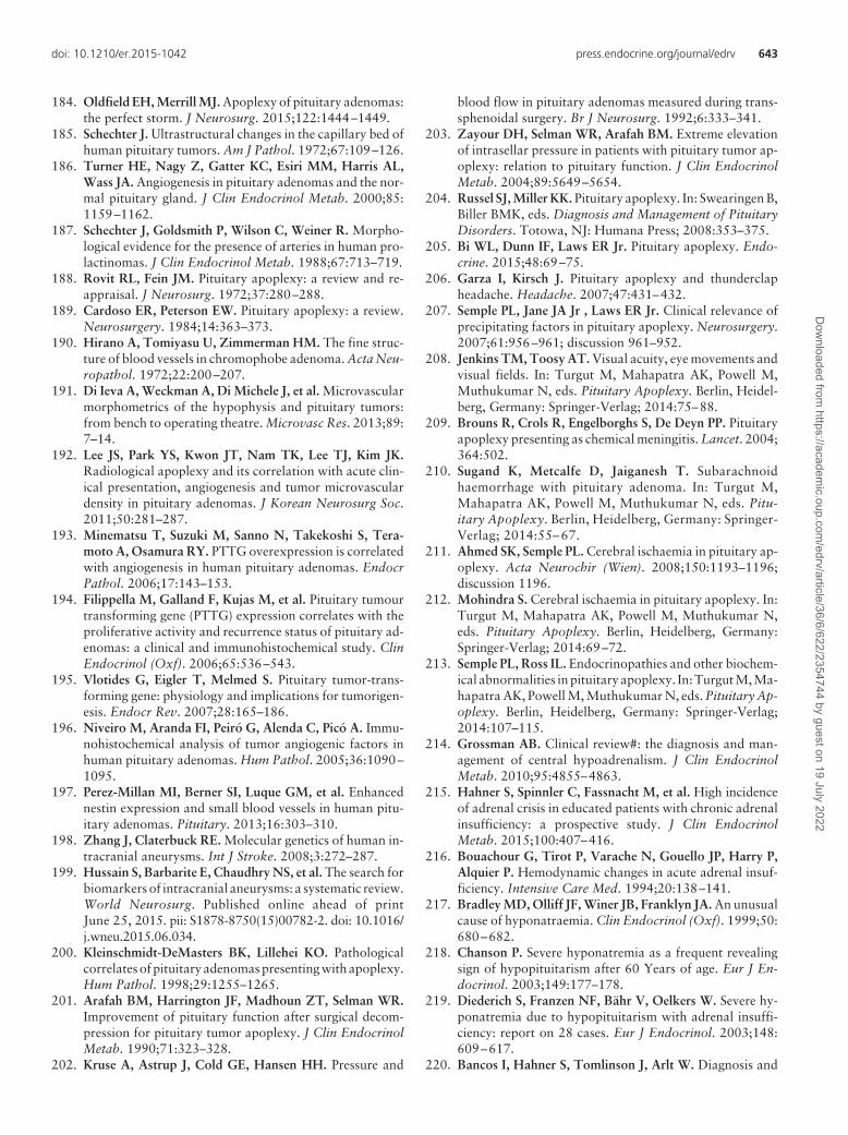
184. Oldfield EH, Merrill MJ. Apoplexy of pituitary adenomas:the perfect storm. J Neurosurg. 2015;122:1444–1449.
185. Schechter J. Ultrastructural changes in the capillary bed ofhuman pituitary tumors. Am J Pathol. 1972;67:109–126.
186. Turner HE, Nagy Z, Gatter KC, Esiri MM, Harris AL,Wass JA. Angiogenesis in pituitary adenomas and the nor-mal pituitary gland. J Clin Endocrinol Metab. 2000;85:1159–1162.
187. Schechter J, Goldsmith P, Wilson C, Weiner R. Morpho-logical evidence for the presence of arteries in human pro-lactinomas. J Clin Endocrinol Metab. 1988;67:713–719.
188. Rovit RL, Fein JM. Pituitary apoplexy: a review and re-appraisal. J Neurosurg. 1972;37:280–288.
189. Cardoso ER, Peterson EW. Pituitary apoplexy: a review.Neurosurgery. 1984;14:363–373.
190. Hirano A, Tomiyasu U, Zimmerman HM. The fine struc-ture of blood vessels in chromophobe adenoma. Acta Neu-ropathol. 1972;22:200–207.
191. Di Ieva A, Weckman A, Di Michele J, et al. Microvascularmorphometrics of the hypophysis and pituitary tumors:from bench to operating theatre. Microvasc Res. 2013;89:7–14.
192. Lee JS, Park YS, Kwon JT, Nam TK, Lee TJ, Kim JK.Radiological apoplexy and its correlation with acute clin-ical presentation, angiogenesis and tumor microvasculardensity in pituitary adenomas. J Korean Neurosurg Soc.2011;50:281–287.
193. Minematsu T, Suzuki M, Sanno N, Takekoshi S, Tera-moto A, Osamura RY. PTTG overexpression is correlatedwith angiogenesis in human pituitary adenomas. EndocrPathol. 2006;17:143–153.
194. Filippella M, Galland F, Kujas M, et al. Pituitary tumourtransforming gene (PTTG) expression correlates with theproliferative activity and recurrence status of pituitary ad-enomas: a clinical and immunohistochemical study. ClinEndocrinol (Oxf). 2006;65:536–543.
195. Vlotides G, Eigler T, Melmed S. Pituitary tumor-trans-forming gene: physiology and implications for tumorigen-esis. Endocr Rev. 2007;28:165–186.
196. Niveiro M, Aranda FI, Peiró G, Alenda C, Picó A. Immu-nohistochemical analysis of tumor angiogenic factors inhuman pituitary adenomas. Hum Pathol. 2005;36:1090–1095.
197. Perez-Millan MI, Berner SI, Luque GM, et al. Enhancednestin expression and small blood vessels in human pitu-itary adenomas. Pituitary. 2013;16:303–310.
198. Zhang J, Claterbuck RE. Molecular genetics of human in-tracranial aneurysms. Int J Stroke. 2008;3:272–287.
199. Hussain S, Barbarite E, Chaudhry NS, et al. The search forbiomarkers of intracranial aneurysms: a systematic review.World Neurosurg. Published online ahead of printJune 25, 2015. pii: S1878-8750(15)00782-2. doi: 10.1016/j.wneu.2015.06.034.
200. Kleinschmidt-DeMasters BK, Lillehei KO. Pathologicalcorrelates of pituitary adenomas presenting with apoplexy.Hum Pathol. 1998;29:1255–1265.
201. Arafah BM, Harrington JF, Madhoun ZT, Selman WR.Improvement of pituitary function after surgical decom-pression for pituitary tumor apoplexy. J Clin EndocrinolMetab. 1990;71:323–328.
202. Kruse A, Astrup J, Cold GE, Hansen HH. Pressure and
blood flow in pituitary adenomas measured during trans-sphenoidal surgery. Br J Neurosurg. 1992;6:333–341.
203. Zayour DH, Selman WR, Arafah BM. Extreme elevationof intrasellar pressure in patients with pituitary tumor ap-oplexy: relation to pituitary function. J Clin EndocrinolMetab. 2004;89:5649–5654.
204. Russel SJ, Miller KK. Pituitary apoplexy. In: Swearingen B,Biller BMK, eds. Diagnosis and Management of PituitaryDisorders. Totowa, NJ: Humana Press; 2008:353–375.
205. Bi WL, Dunn IF, Laws ER Jr. Pituitary apoplexy. Endo-crine. 2015;48:69–75.
206. Garza I, Kirsch J. Pituitary apoplexy and thunderclapheadache. Headache. 2007;47:431–432.
207. Semple PL, Jane JA Jr , Laws ER Jr. Clinical relevance ofprecipitating factors in pituitary apoplexy. Neurosurgery.2007;61:956–961; discussion 961–952.
208. Jenkins TM, Toosy AT. Visual acuity, eye movements andvisual fields. In: Turgut M, Mahapatra AK, Powell M,Muthukumar N, eds. Pituitary Apoplexy. Berlin, Heidel-berg, Germany: Springer-Verlag; 2014:75–88.
209. Brouns R, Crols R, Engelborghs S, De Deyn PP. Pituitaryapoplexy presenting as chemical meningitis. Lancet. 2004;364:502.
210. Sugand K, Metcalfe D, Jaiganesh T. Subarachnoidhaemorrhage with pituitary adenoma. In: Turgut M,Mahapatra AK, Powell M, Muthukumar N, eds. Pitu-itary Apoplexy. Berlin, Heidelberg, Germany: Springer-Verlag; 2014:55– 67.
211. Ahmed SK, Semple PL. Cerebral ischaemia in pituitary ap-oplexy. Acta Neurochir (Wien). 2008;150:1193–1196;discussion 1196.
212. Mohindra S. Cerebral ischaemia in pituitary apoplexy. In:Turgut M, Mahapatra AK, Powell M, Muthukumar N,eds. Pituitary Apoplexy. Berlin, Heidelberg, Germany:Springer-Verlag; 2014:69–72.
213. Semple PL, Ross IL. Endocrinopathies and other biochem-ical abnormalities in pituitary apoplexy. In: Turgut M, Ma-hapatra AK, Powell M, Muthukumar N, eds. Pituitary Ap-oplexy. Berlin, Heidelberg, Germany: Springer-Verlag;2014:107–115.
214. Grossman AB. Clinical review#: the diagnosis and man-agement of central hypoadrenalism. J Clin EndocrinolMetab. 2010;95:4855–4863.
215. Hahner S, Spinnler C, Fassnacht M, et al. High incidenceof adrenal crisis in educated patients with chronic adrenalinsufficiency: a prospective study. J Clin EndocrinolMetab. 2015;100:407–416.
216. Bouachour G, Tirot P, Varache N, Gouello JP, Harry P,Alquier P. Hemodynamic changes in acute adrenal insuf-ficiency. Intensive Care Med. 1994;20:138–141.
217. Bradley MD, Olliff JF, Winer JB, Franklyn JA. An unusualcause of hyponatraemia. Clin Endocrinol (Oxf). 1999;50:680–682.
218. Chanson P. Severe hyponatremia as a frequent revealingsign of hypopituitarism after 60 Years of age. Eur J En-docrinol. 2003;149:177–178.
219. Diederich S, Franzen NF, Bähr V, Oelkers W. Severe hy-ponatremia due to hypopituitarism with adrenal insuffi-ciency: report on 28 cases. Eur J Endocrinol. 2003;148:609–617.
220. Bancos I, Hahner S, Tomlinson J, Arlt W. Diagnosis and
doi: 10.1210/er.2015-1042 press.endocrine.org/journal/edrv 643
Dow
nloaded from https://academ
ic.oup.com/edrv/article/36/6/622/2354744 by guest on 19 July 2022
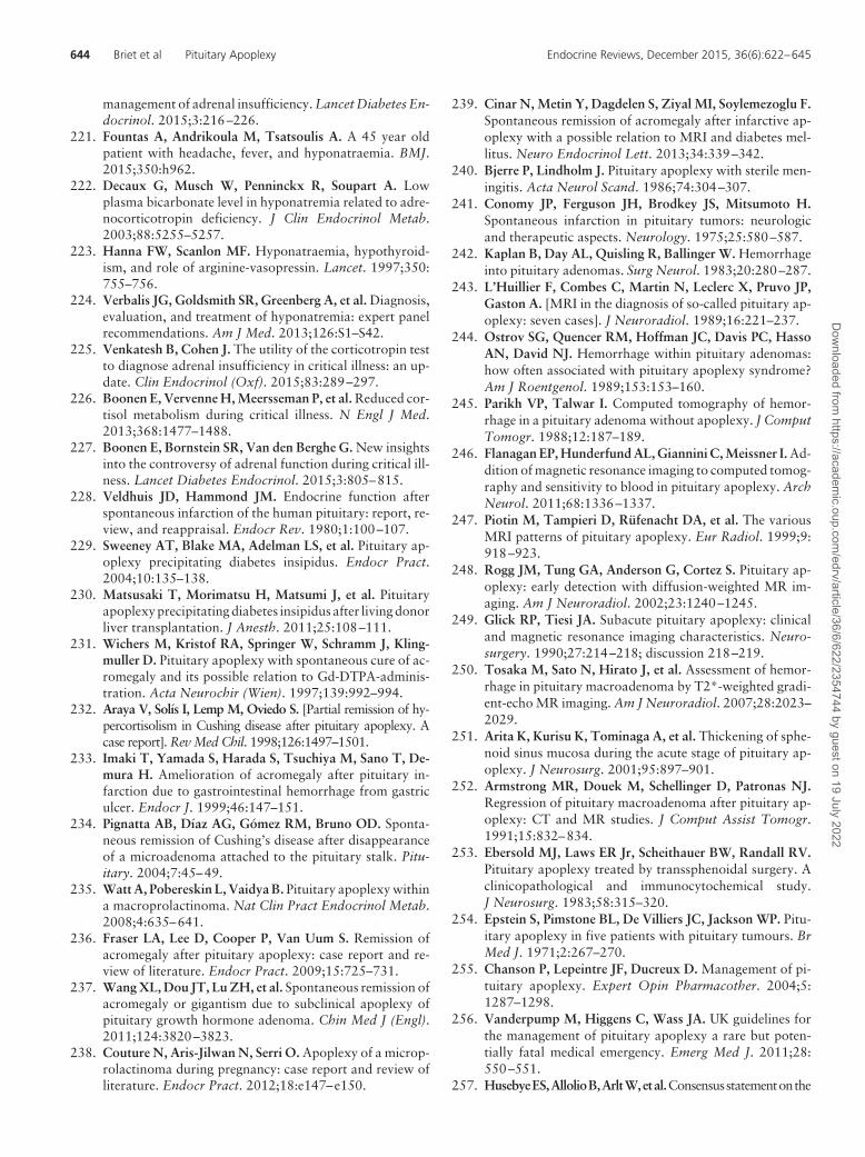
management of adrenal insufficiency. Lancet Diabetes En-docrinol. 2015;3:216–226.
221. Fountas A, Andrikoula M, Tsatsoulis A. A 45 year oldpatient with headache, fever, and hyponatraemia. BMJ.2015;350:h962.
222. Decaux G, Musch W, Penninckx R, Soupart A. Lowplasma bicarbonate level in hyponatremia related to adre-nocorticotropin deficiency. J Clin Endocrinol Metab.2003;88:5255–5257.
223. Hanna FW, Scanlon MF. Hyponatraemia, hypothyroid-ism, and role of arginine-vasopressin. Lancet. 1997;350:755–756.
224. Verbalis JG, Goldsmith SR, Greenberg A, et al. Diagnosis,evaluation, and treatment of hyponatremia: expert panelrecommendations. Am J Med. 2013;126:S1–S42.
225. Venkatesh B, Cohen J. The utility of the corticotropin testto diagnose adrenal insufficiency in critical illness: an up-date. Clin Endocrinol (Oxf). 2015;83:289–297.
226. Boonen E, Vervenne H, Meersseman P, et al. Reduced cor-tisol metabolism during critical illness. N Engl J Med.2013;368:1477–1488.
227. Boonen E, Bornstein SR, Van den Berghe G. New insightsinto the controversy of adrenal function during critical ill-ness. Lancet Diabetes Endocrinol. 2015;3:805–815.
228. Veldhuis JD, Hammond JM. Endocrine function afterspontaneous infarction of the human pituitary: report, re-view, and reappraisal. Endocr Rev. 1980;1:100–107.
229. Sweeney AT, Blake MA, Adelman LS, et al. Pituitary ap-oplexy precipitating diabetes insipidus. Endocr Pract.2004;10:135–138.
230. Matsusaki T, Morimatsu H, Matsumi J, et al. Pituitaryapoplexyprecipitatingdiabetes insipidusafter livingdonorliver transplantation. J Anesth. 2011;25:108–111.
231. Wichers M, Kristof RA, Springer W, Schramm J, Kling-muller D. Pituitary apoplexy with spontaneous cure of ac-romegaly and its possible relation to Gd-DTPA-adminis-tration. Acta Neurochir (Wien). 1997;139:992–994.
232. Araya V, Solís I, Lemp M, Oviedo S. [Partial remission of hy-percortisolism in Cushing disease after pituitary apoplexy. Acase report]. Rev Med Chil. 1998;126:1497–1501.
233. Imaki T, Yamada S, Harada S, Tsuchiya M, Sano T, De-mura H. Amelioration of acromegaly after pituitary in-farction due to gastrointestinal hemorrhage from gastriculcer. Endocr J. 1999;46:147–151.
234. Pignatta AB, Díaz AG, Gómez RM, Bruno OD. Sponta-neous remission of Cushing’s disease after disappearanceof a microadenoma attached to the pituitary stalk. Pitu-itary. 2004;7:45–49.
235. Watt A, Pobereskin L, Vaidya B. Pituitary apoplexy withina macroprolactinoma. Nat Clin Pract Endocrinol Metab.2008;4:635–641.
236. Fraser LA, Lee D, Cooper P, Van Uum S. Remission ofacromegaly after pituitary apoplexy: case report and re-view of literature. Endocr Pract. 2009;15:725–731.
237. Wang XL, Dou JT, Lu ZH, et al. Spontaneous remission ofacromegaly or gigantism due to subclinical apoplexy ofpituitary growth hormone adenoma. Chin Med J (Engl).2011;124:3820–3823.
238. Couture N, Aris-Jilwan N, Serri O. Apoplexy of a microp-rolactinoma during pregnancy: case report and review ofliterature. Endocr Pract. 2012;18:e147–e150.
239. Cinar N, Metin Y, Dagdelen S, Ziyal MI, Soylemezoglu F.Spontaneous remission of acromegaly after infarctive ap-oplexy with a possible relation to MRI and diabetes mel-litus. Neuro Endocrinol Lett. 2013;34:339–342.
240. Bjerre P, Lindholm J. Pituitary apoplexy with sterile men-ingitis. Acta Neurol Scand. 1986;74:304–307.
241. Conomy JP, Ferguson JH, Brodkey JS, Mitsumoto H.Spontaneous infarction in pituitary tumors: neurologicand therapeutic aspects. Neurology. 1975;25:580–587.
242. Kaplan B, Day AL, Quisling R, Ballinger W. Hemorrhageinto pituitary adenomas. Surg Neurol. 1983;20:280–287.
243. L’Huillier F, Combes C, Martin N, Leclerc X, Pruvo JP,Gaston A. [MRI in the diagnosis of so-called pituitary ap-oplexy: seven cases]. J Neuroradiol. 1989;16:221–237.
244. Ostrov SG, Quencer RM, Hoffman JC, Davis PC, HassoAN, David NJ. Hemorrhage within pituitary adenomas:how often associated with pituitary apoplexy syndrome?Am J Roentgenol. 1989;153:153–160.
245. Parikh VP, Talwar I. Computed tomography of hemor-rhage in a pituitary adenoma without apoplexy. J ComputTomogr. 1988;12:187–189.
246. Flanagan EP, Hunderfund AL, Giannini C, Meissner I. Ad-dition of magnetic resonance imaging to computed tomog-raphy and sensitivity to blood in pituitary apoplexy. ArchNeurol. 2011;68:1336–1337.
247. Piotin M, Tampieri D, Rüfenacht DA, et al. The variousMRI patterns of pituitary apoplexy. Eur Radiol. 1999;9:918–923.
248. Rogg JM, Tung GA, Anderson G, Cortez S. Pituitary ap-oplexy: early detection with diffusion-weighted MR im-aging. Am J Neuroradiol. 2002;23:1240–1245.
249. Glick RP, Tiesi JA. Subacute pituitary apoplexy: clinicaland magnetic resonance imaging characteristics. Neuro-surgery. 1990;27:214–218; discussion 218–219.
250. Tosaka M, Sato N, Hirato J, et al. Assessment of hemor-rhage in pituitary macroadenoma by T2*-weighted gradi-ent-echo MR imaging. Am J Neuroradiol. 2007;28:2023–2029.
251. Arita K, Kurisu K, Tominaga A, et al. Thickening of sphe-noid sinus mucosa during the acute stage of pituitary ap-oplexy. J Neurosurg. 2001;95:897–901.
252. Armstrong MR, Douek M, Schellinger D, Patronas NJ.Regression of pituitary macroadenoma after pituitary ap-oplexy: CT and MR studies. J Comput Assist Tomogr.1991;15:832–834.
253. Ebersold MJ, Laws ER Jr, Scheithauer BW, Randall RV.Pituitary apoplexy treated by transsphenoidal surgery. Aclinicopathological and immunocytochemical study.J Neurosurg. 1983;58:315–320.
254. Epstein S, Pimstone BL, De Villiers JC, Jackson WP. Pitu-itary apoplexy in five patients with pituitary tumours. BrMed J. 1971;2:267–270.
255. Chanson P, Lepeintre JF, Ducreux D. Management of pi-tuitary apoplexy. Expert Opin Pharmacother. 2004;5:1287–1298.
256. Vanderpump M, Higgens C, Wass JA. UK guidelines forthe management of pituitary apoplexy a rare but poten-tially fatal medical emergency. Emerg Med J. 2011;28:550–551.
257. HusebyeES,AllolioB,ArltW,etal.Consensusstatementonthe
644 Briet et al Pituitary Apoplexy Endocrine Reviews, December 2015, 36(6):622–645
Dow
nloaded from https://academ
ic.oup.com/edrv/article/36/6/622/2354744 by guest on 19 July 2022
diagnosis, treatmentand follow-upofpatientswithprimaryad-renal insufficiency. J Intern Med. 2014;275:104–115.
258. Arlt W. The approach to the adult with newly diagnosedadrenal insufficiency. J Clin Endocrinol Metab. 2009;94:1059–1067.
259. Kanter AS, Dumont AS, Asthagiri AR, et al. The trans-sphenoidal approach. A historical perspective. NeurosurgFocus. 2005;18:e6.
260. Gondim JA, Almeida JP, Albuquerque LA, et al. Endoscopicendonasal approach for pituitary adenoma: surgical complica-tions in 301 patients. Pituitary. 2011;14:174–183.
261. Berker M, Hazer DB, Yücel T, et al. Complications ofendoscopic surgery of the pituitary adenomas: analysis of
570 patients and review of the literature. Pituitary. 2012;15:288–300.
262. Reddy NL, Rajasekaran S, Han TS, et al. An objectivescoring tool in the management of patients with pituitaryapoplexy. Clin Endocrinol (Oxf). 2011;75:723.
263. Muthukumar N, Rossette D, Soundaram M, SenthilbabuS, Badrinarayanan T. Blindness following pituitary apo-plexy: timing of surgery and neuro-ophthalmic outcome.J Clin Neurosci. 2008;15:873–879.
264. Agrawal D, Mahapatra AK. Visual outcome of blindeyes in pituitary apoplexy after transsphenoidal surgery:a series of 14 eyes. Surg Neurol. 2005;63:42– 46; dis-cussion 46.
doi: 10.1210/er.2015-1042 press.endocrine.org/journal/edrv 645
Dow
nloaded from https://academ
ic.oup.com/edrv/article/36/6/622/2354744 by guest on 19 July 2022


![[The incidence of the pituitary autoantibodies in Graves' disease]](https://img.dokumen.tips/doc/110x75/635915fc68c5ffb42104606f/the-incidence-of-the-pituitary-autoantibodies-in-graves-disease.jpg)


