Embed Size (px)
Citation preview
Research article
Received: 6 May 2014 Revised: 16 July 2014 Accepted: 28 July 2014 Published online in Wiley Online Library
(wileyonlinelibrary.com) DOI 10.1002/jrs.4570
Pigment analysis of Portugueseportraitminiaturesof 17th and 18th centuries by Raman Microscopyand SEM-EDSAlfredina Veiga,a,b,e José Mirão,c,d,e António J. Candeias,a,b,e
Paulo Simões Rodrigues,f Dora Martins Teixeira,a,e Vânia S. F. Muralhag
and Jorge Ginja Teixeiraa,b,e*
Seventeen Portuguese miniature portraits on copper support from the Évora Museum collection (Portugal) were analyzed in situand nondestructively by Ramanmicroscopy (RM), SEM-EDS, and stereomicroscopy. This work constitutes a great breakthrough in
the study ofminiature paintings from the 17th and 18th centuries, since the chemical information known about this unique kind ofpaintings are still scarce, and in particular, this exclusive collectionwas never been subjected to any physicochemical study. In thiswork, each portrait was examined in detail in order to characterize the pigments palette used by the miniaturists. The μ-Ramananalysis, in particular, guaranteed an exceptional visualization and good individual identification of small grains of pigmentsand other constituents of the pictorial layer. Using this technique, 19 compounds were identified, including bluish black covellite,a pigment rarely found in oil paintings. SEM-EDS was used as an important complementary technique to confirm the chemicalnature of some pigments and to identify shell gold (gold dust) in some portraits. Overall, the pigments identified in this largeset of old paintings are broadly consistent with those mentioned in the painting treatises of that time or reported in other moremodern bibliographic sources. Copyright © 2014 John Wiley & Sons, Ltd.Additional supporting information may be found in the online version of this article at the publisher’s web site.
Keywords: pigments; portraits miniatures; copper support; nondestructive analysis; SEM-EDS
* Correspondence to: Jorge Ginja Teixeira, Departamento de Química, Escola deCiências e Tecnologia, Laboratório HERCULES, Centro de Química de Évora,Universidade de Évora, Colégio Luis António Verney, 7000 Évora, Portugal.E-mail: [email protected]
a Chemistry Department, Science and Technology School, University of Évora, 7000Évora, Portugal
b Évora Chemistry Center (CQE), University of Évora, 7000 Évora, Portugal
c Geosciences Department, Science and Technology School, University of Évora,7000 Évora, Portugal
d Évora Geophysics Center (CGE), University of Évora, 7000 Évora, Portugal
e HERCULES Laboratory, University of Évora, 7000 Évora, Portugal
f Center for History of Art and Artistic Research (CHAIA), University of Évora, 7000Évora, Portugal
g Research Unit; VICARTE: Vidro e Cerâmica para as Artes, Faculdade de Ciências eTecnologia, Universidade Nova de Lisboa, 2829-516 Monte de Caparica, Portugal
Introduction
Portrait miniatures painted in oil on copper support, with a typical sizethat does not exceed the palm of the hand, represent a special paint-ing technique dating back to the early 16th century.[1] Supposedlycreated in Italy, but with the credits of its initial practice ascribed toDutchminiaturists, this technique was amply developed and extendedto other continents by those painters and others European painters,during the 17th and 18th centuries andbeginning of the 19th century.[2,3]
Like other techniques in miniature painting that used other sortsof paints and materials (e.g., watercolor on stretched vellum, paperor ivory, or vitreous enamel painted on copper), the idea behindthese art creations was to combine the beauty and realism of theportrait painted in colors of natural tones, with the ability of beingportable, personal, and easily presented to the most diverse audi-ences (private and public), whatever it might be the geographicalplace. In fact, in those times the miniature portrait became animportant and unique visual resource that was used by the eliteswith several purposes, such as an object of religious, familiar, orloving devotions, or an object of sociopolitical distinctiveness, oreven as a diplomatic document or trading currency.[3,4] In someaspects, it can be said that this type of painting had the importancethat the small photographic portrait has nowadays. However in thattime, only the economically wealthy classes had the opportunity toordering and own such exclusive objects. Indeed, in a society wheremembers of royalty, religious orders, and nobility were almostexclusively the main clients of these artworks, the miniaturists tookadvantage of all work opportunities to gain money and reputation.
J. Raman Spectrosc. (2014)
This practice was not an exception in Portugal. During the 16th
century and following centuries, Portuguese and foreigner painterscreated miniature paintings for the elites. In the 17th century, inwhich the most prominent and recognized figures of Portuguesepainting have devoted his time to paint in miniature, the qualityof these works of art seemed to increase significantly. Unfortu-nately, quite a few authors are identifiable, because portrait minia-tures were not, as a rule, signed.[3] Júlio Brandão can be considered
Copyright © 2014 John Wiley & Sons, Ltd.
A. Veiga et al.
a pioneer in the study of the Portuguese miniature, after doing aninventory of the names of known miniaturists, based on the workssigned by them. However, the vast majority of these painters werefrom the 19th century.[5,6]
Concerning the materials used and the pictorial execution, thisparticular painting technique made use of the acquired knowledgeand of the well-known advantages employed in oil paintings oncopper support of larger dimensions. In particular, copper plateprovided a highly rigid and nonabsorbent support with a smoothsurface that after being properly prepared helped to create a lumi-nous painting with extraordinary details and perfect finishing.[7] Af-ter preparation of the copper surface (which involved, among otherprocedures, the increase of its roughness to promote the oil paintadherence), a preparatory/ground layer based on white lead andumber, mixed in an appropriate oil (mainly linseed oil), was appliedfollowed by the application of the pictorial layer. The application ofa preparatory intermediate layer (based on chalk/calcite), betweenthe support and the ground layer (based on lead white), commonlyused in other types of support, was apparently not used in the caseof copper support.[7,8] However, depending on the options of thepainter at the time, its use should not be totally ruled out. As for
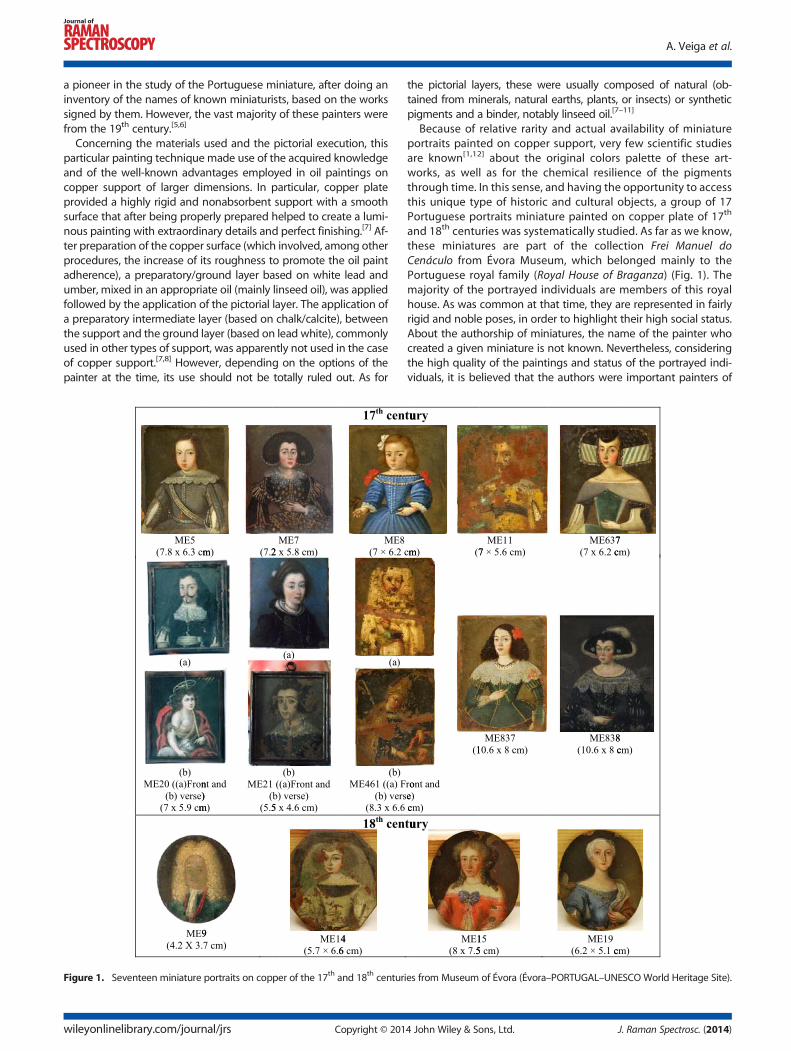
Figure 1. Seventeen miniature portraits on copper of the 17th and 18th centur
wileyonlinelibrary.com/journal/jrs Copyright © 201
the pictorial layers, these were usually composed of natural (ob-tained from minerals, natural earths, plants, or insects) or syntheticpigments and a binder, notably linseed oil.[7–11]
Because of relative rarity and actual availability of miniatureportraits painted on copper support, very few scientific studiesare known[1,12] about the original colors palette of these art-works, as well as for the chemical resilience of the pigmentsthrough time. In this sense, and having the opportunity to accessthis unique type of historic and cultural objects, a group of 17Portuguese portraits miniature painted on copper plate of 17th
and 18th centuries was systematically studied. As far as we know,these miniatures are part of the collection Frei Manuel doCenáculo from Évora Museum, which belonged mainly to thePortuguese royal family (Royal House of Braganza) (Fig. 1). Themajority of the portrayed individuals are members of this royalhouse. As was common at that time, they are represented in fairlyrigid and noble poses, in order to highlight their high social status.About the authorship of miniatures, the name of the painter whocreated a given miniature is not known. Nevertheless, consideringthe high quality of the paintings and status of the portrayed indi-viduals, it is believed that the authors were important painters of
ies from Museum of Évora (Évora–PORTUGAL–UNESCO World Heritage Site).
4 John Wiley & Sons, Ltd. J. Raman Spectrosc. (2014)
Pigment analysis of Portuguese portrait miniatures
that time and, most likely, the same artists who painted the greatpomp portraits of members of the royal family.
Some of the paintings on copper are in perfect condition andthus are exceptionally close to their original appearance. This con-dition, combinedwith the very small dimensions of these paintings,demands the use of nondestructive methods of investigation. Inline with this requirement and with the intention to perform a de-tailed examination about all types of pigments that can be foundin these paintings, giving attention to the particles color, shape, di-mensions, distribution, and elemental and molecular chemicalcomposition, it was decided to use stereomicroscopy, scanningelectron microscopy coupled with energy-dispersive X-ray spec-troscopy (SEM-EDS), and, in special, Raman microscopy (RM).
As is evidenced in numerous works devoted to material study ofartworks, Raman microscopy is an extremely reliable analytical toolin the identification of pigments and other related materials.[13–17]
When RM fails to give an unambiguous response on the chemicalidentification of one material, SEM-EDS analysis can be an excellentalternative to solve this problem.[18] In the analysis of the minia-tures, both techniques allowed the identification of pigments andother materials, without any sampling or causing any damage onthe surface thereof. Here the small dimensions and metallic natureof the copper support constitute a great analytical advantage, espe-cially in SEM-EDS analysis at low vacuum/variable pressure condi-tions, in which it is possible to insert the miniatures inside thespecimen camera, and without running the risk of damaging thepictorial layer of the portraits. Therefore, applying this approachon a large set of miniatures not only will contribute to the knowl-edge of the material identity of each miniature but above all willhelp to build a wider and complete database on the use of pig-ments in miniatures in Portugal (Europe) from the 17th and 18th
centuries.
Experimental
Stereomicroscopy
Themicroscopic observations and the corresponding photographicdocumentation were obtained with a Leica M205C stereomicro-scope eqquiped with a DFC295HD photo camera at different opti-cal magnifications (zoom range of 7.8× to 160×).
Raman Microscopy
The pigments analysis was performed by Ramanmicrospectrometry,using an HORIBA Xplora Raman microscope, with capacity increasedto 100×, and charge coupled device (CCD) detector. Laser wave-lengths of 632.8 nm (red He–Ne laser line) and 785.0 nm (NIR laser)were used for excitation in order to choose the better spectrum interms of signal to background fluorescence ratio. Wavenumbercalibration was performed with the Raman peak of a Silicon crystalat 520 cm�1. The laser beam was focused on the grains of pigmenteither with 50× or 100× objective lens. The laser power at the surfaceof the sample was held to ≤ 1.0 mW (632.8 nm) and ≤ 2.8 mW(785.0 nm), to avoid any possible destruction of the paintings.Raman spectra were obtained in scanning mode, after five scans,with acquisition time of 10–20 s and spectral resolution of 2 cm�1.
In order to know the molecular composition of the pigments,which appear in the form of grains of micrometric dimensions ina quite complex matrix, a systematic analysis point-by-point wasimplemented. Despite the strong background fluorescence for
J. Raman Spectrosc. (2014) Copyright © 2014 John Wiley
most paintings, no treatment of baseline adjustment or smoothingwas performed.
SEM-EDS
The analysis of pigments and other products present in the minia-ture portraits were also performed by scanning electron micros-copy (using an Hitachi S3700N, with a specimen chamber thataccomodates samples up to 300 mm in diameter and 110 mm inheight) coupled with energy-dispersive X-ray spectrometry(BRUKER Xflash 5010SDD), in situ, without any sampling or prepara-tion (Fig. S1 in the supporting information).
In order to evaluate in what conditions the pictorial layer of thesepaintings withstand the low vacuum/variable pressure environ-ment within the SEM chamber, without suffer any kind of damage,it was decided to start the SEM analysis with the miniatures thathave portraits in advanced state of degradation (namely, minia-tures ME11, ME461, and ME14). Using a low vacuum of 40 Pa, anaccelerating voltage of 10–20 kV, and an exposition time up to 90min, it was possible to ascertain that the pictorial layer of thoseminiatures was sufficiently robust, remaining strongly linked tothe copper support. In fact, a systematic visual inspection of severalselected areas of the paintings (made by observation with the na-ked eye, optical microscopy, or SEM) disclosed no change in thephysical characteristics of the pictorial layers. On the basis of thesefindings and on the strong possibility that the damaging influenceof microair bubbles, inside or under the paint layer, is very reduced(due to the small amount of air accumulated therein, since thevolume of the paint layer in the portraits is very small, the distribu-tion of microcracks developed along its surface is large and thesupport has a nonporous nature), the SEM-EDS analysis of allminiatures was performed using the developed protocol, withoutexceeding unnecessarily the useful time of analysis.
Results
Table 1 contains a summary of the pigments found on the minia-tures analyzed by RM and SEM-EDS. The analysis of Raman spectraand their assignment to a specific pigment or other colored constit-uent was made using databases of reference materials reported inthe literature[15,17,19–21] except when noted. To depict the relativeintensity of the Raman bands of each spectrum, the following sym-bolismwas adopted: vs, very strong; s, strong; m, medium; w, weak;vw, very weak; sh, shoulder; br, broad.[19]
White compounds
The analysis of the white grains at the surface of paint layers led tothe identification of three white compounds: lead carbonate, PbCO3;lead white also known as basic lead carbonate, [2PbCO3·Pb(OH)2];and calcium carbonate, CaCO3. The twowhite lead compounds wereidentified by the strongest feature of their Raman spectrum (Fig. S2in the supporting information).[22] The doublet of bands for leadwhite (Fig. S3 in the supporting information) is regularly assignedto the existence of carbonate ions in, at least, two different sites.
As it is well known, one of those white compounds of lead(namely, lead white) was used extensively in the past as the mainwhite pigment to provide the white primary coloration of thepainted areas or to lighten some colors and highlight others. Atthe same time, it was also used to prepare a base coat over thecanvas material (in the present case, a copper plate), upon whichthe pictorial layer was finally created.[7–12] However, as much as it
& Sons, Ltd. wileyonlinelibrary.com/journal/jrs
Table
1.Synop
sisof
allcom
pou
ndsiden
tifiedin
thisstud
y
Miniature
portraitson
copper
support
Pigm
ents
Lead
Carbon
ate
Lead
White
Calcium
Carbon
ate
Verm
ilion
Hem
atite
Red
Lead
Goe
thite
Lead tin
yellow
typeI
Orpim
entMassicotNap
lesYe
llow
pyrochlores
Shell
Gold*
Azurite
Lapis
lazuli
Prussian
Blue
Indigo
Covellite
Carbon
black
17th
century
ME5
√√
√√
√√
√√
√√
ME7
√√
√√
√√
√√
√√
√ME8
√√
√√
√√
√√
√√
√ME11
√√
√√
√√
√√
√ME20Fron
t√
√√
√√
√√
√√
√√
√ME20Verse
√√
√√
√√
√√
√√
ME21Fron
t√
√√
√√
√√
√√
ME21Verse
√√
√√
√√
√√
√ME461
Fron
t√
√√
√√
√√
√ME461
Verse
√√
√√
√√
ME637
√√
√√
√√
√ME837
√√
√√
√√
√√
ME838
√√
√√
√√
√√
√√
√√
√18th
century
ME9
√√
√√
√√
√√
√ME14
√√
√√
√√
√ME15
√√
√√
√√
√ME19
√√
√√
√√
√√
*SEM-EDS.
A. Veiga et al.
wileyonlinelibrary.com/journal/jrs Copyright © 2014 John Wiley & Sons, Ltd. J. Raman Spectrosc. (2014)
100 300 500 700 900 1100
Ram
an I
nten
sity
142
284381
252 292
310 354381
155202
(b)
(c)
(d)(e)
205245
300
390480
129
196290
454 (a)140
298333
448 510
976
Wavenumber / cm-1
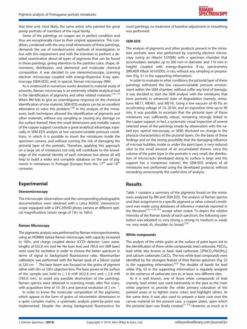
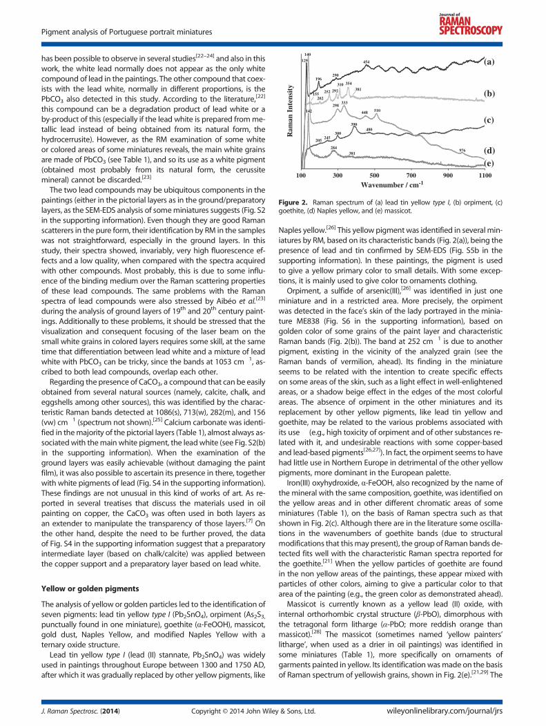
Figure 2. Raman spectrum of (a) lead tin yellow type I, (b) orpiment, (c)goethite, (d) Naples yellow, and (e) massicot.
Pigment analysis of Portuguese portrait miniatures
has been possible to observe in several studies[22–24] and also in thiswork, the white lead normally does not appear as the only whitecompound of lead in the paintings. The other compound that coex-ists with the lead white, normally in different proportions, is thePbCO3 also detected in this study. According to the literature,[22]
this compound can be a degradation product of lead white or aby-product of this (especially if the lead white is prepared fromme-tallic lead instead of being obtained from its natural form, thehydrocerrusite). However, as the RM examination of some whiteor colored areas of some miniatures reveals, the main white grainsare made of PbCO3 (see Table 1), and so its use as a white pigment(obtained most probably from its natural form, the cerussitemineral) cannot be discarded.[23]
The two lead compounds may be ubiquitous components in thepaintings (either in the pictorial layers as in the ground/preparatorylayers, as the SEM-EDS analysis of someminiatures suggests (Fig. S2in the supporting information). Even though they are good Ramanscatterers in the pure form, their identification by RM in the sampleswas not straightforward, especially in the ground layers. In thisstudy, their spectra showed, invariably, very high fluorescence ef-fects and a low quality, when compared with the spectra acquiredwith other compounds. Most probably, this is due to some influ-ence of the binding medium over the Raman scattering propertiesof these lead compounds. The same problems with the Ramanspectra of lead compounds were also stressed by Aibéo et al.[23]
during the analysis of ground layers of 19th and 20th century paint-ings. Additionally to these problems, it should be stressed that thevisualization and consequent focusing of the laser beam on thesmall white grains in colored layers requires some skill, at the sametime that differentiation between lead white and a mixture of leadwhite with PbCO3 can be tricky, since the bands at 1053 cm�1, as-cribed to both lead compounds, overlap each other.
Regarding the presence of CaCO3, a compound that can be easilyobtained from several natural sources (namely, calcite, chalk, andeggshells among other sources), this was identified by the charac-teristic Raman bands detected at 1086(s), 713(w), 282(m), and 156(vw) cm�1 (spectrum not shown).[25] Calcium carbonate was identi-fied in themajority of the pictorial layers (Table 1), almost always as-sociatedwith themain white pigment, the leadwhite (see Fig. S2(b)in the supporting information). When the examination of theground layers was easily achievable (without damaging the paintfilm), it was also possible to ascertain its presence in there, togetherwith white pigments of lead (Fig. S4 in the supporting information).These findings are not unusual in this kind of works of art. As re-ported in several treatises that discuss the materials used in oilpainting on copper, the CaCO3 was often used in both layers asan extender to manipulate the transparency of those layers.[7] Onthe other hand, despite the need to be further proved, the dataof Fig. S4 in the supporting information suggest that a preparatoryintermediate layer (based on chalk/calcite) was applied betweenthe copper support and a preparatory layer based on lead white.
Yellow or golden pigments
The analysis of yellow or golden particles led to the identification ofseven pigments: lead tin yellow type I (Pb2SnO4), orpiment (As2S3,punctually found in one miniature), goethite (α-FeOOH), massicot,gold dust, Naples Yellow, and modified Naples Yellow with aternary oxide structure.
Lead tin yellow type I (lead (II) stannate, Pb2SnO4) was widelyused in paintings throughout Europe between 1300 and 1750 AD,after which it was gradually replaced by other yellow pigments, like
J. Raman Spectrosc. (2014) Copyright © 2014 John Wiley
Naples yellow.[26] This yellow pigment was identified in several min-iatures by RM, based on its characteristic bands (Fig. 2(a)), being thepresence of lead and tin confirmed by SEM-EDS (Fig. S5b in thesupporting information). In these paintings, the pigment is usedto give a yellow primary color to small details. With some excep-tions, it is mainly used to give color to ornaments clothing.
Orpiment, a sulfide of arsenic(III),[26] was identified in just oneminiature and in a restricted area. More precisely, the orpimentwas detected in the face’s skin of the lady portrayed in the minia-ture ME838 (Fig. S6 in the supporting information), based ongolden color of some grains of the paint layer and characteristicRaman bands (Fig. 2(b)). The band at 252 cm�1 is due to anotherpigment, existing in the vicinity of the analyzed grain (see theRaman bands of vermilion, ahead). Its finding in the miniatureseems to be related with the intention to create specific effectson some areas of the skin, such as a light effect in well-enlightenedareas, or a shadow beige effect in the edges of the most colorfulareas. The absence of orpiment in the other miniatures and itsreplacement by other yellow pigments, like lead tin yellow andgoethite, may be related to the various problems associated withits use (e.g., high toxicity of orpiment and of other substances re-lated with it, and undesirable reactions with some copper-basedand lead-based pigments[26,27]). In fact, the orpiment seems to havehad little use in Northern Europe in detrimental of the other yellowpigments, more dominant in the European palette.
Iron(III) oxyhydroxide, α-FeOOH, also recognized by the name ofthe mineral with the same composition, goethite, was identified onthe yellow areas and in other different chromatic areas of someminiatures (Table 1), on the basis of Raman spectra such as thatshown in Fig. 2(c). Although there are in the literature some oscilla-tions in the wavenumbers of goethite bands (due to structuralmodifications that thismay present), the group of Raman bands de-tected fits well with the characteristic Raman spectra reported forthe goethite.[21] When the yellow particles of goethite are foundin the non yellow areas of the paintings, these appear mixed withparticles of other colors, aiming to give a particular color to thatarea of the painting (e.g., the green color as demonstrated ahead).
Massicot is currently known as a yellow lead (II) oxide, withinternal orthorhombic crystal structure (β-PbO), dimorphous withthe tetragonal form litharge (α-PbO; more reddish orange thanmassicot).[28] The massicot (sometimes named ‘yellow painters’litharge’, when used as a drier in oil paintings) was identified insome miniatures (Table 1), more specifically on ornaments ofgarments painted in yellow. Its identification wasmade on the basisof Raman spectrum of yellowish grains, shown in Fig. 2(e).[21,29] The
& Sons, Ltd. wileyonlinelibrary.com/journal/jrs
A. Veiga et al.
massicot is not confused here with the dimorphic litharge, becausethis has a slightly different Raman spectrum, with characteristicbands at 149(vs), 289(vw), ca 342(w).[19,21,29]
Massicot exists as a native mineral, as well as a product made byheating lead white to a high temperature (the litharge can be alsofound in the mineral form and is obtained at lower temperatures).In Raman microanalysis, intentionally or not, it can be also pro-duced during the laser heating-induced degradation of other leadoxides (e.g., PbO2 (black) and litharge (reddish orange)[30] or leadII
sulfide (black), present as degradation products of the lead whiteused in the paintings. In the present case, and taking into accountthe energy and power of lasers used, it was not found whatever ev-idence that might suggest the development of such effect. Thespectrum obtained does not show the presence of broad bandswith a red shift (lower wavenumbers), which are characteristic ofmassicot formed by laser degradation of the lead oxides abovementioned (including the red oxide Pb3O4).
[30] Additionally, thesecompounds darker in color (including PbS) were not identified inthe same area where occur the yellowish grains of massicot.Accordingly, we believe that the massicot found was used inten-tionally as a yellow pigment in the paintings.Gold dust was a singular pigment used to give a strong yellowish
color to some ornaments. This pigment, mainly known by shellgold,[26] was identified in a very restricted number of miniaturesby SEM-EDS analysis (Fig. S7 in the supporting information). Asthe micrograph of this figure proves, the shell gold was appliedover the paint layers (the clear flakes, made of gold, are disposedabove the grayer layer made of paint).In addition to the five yellowish pigments listed above, it was
possible to identify the unmodified Naples yellow (a yellow lead(II) antimonate (Pb2Sb207), which can appear with different Pb:Sbmolar ratios) and the modified Naples yellow with a ternary oxidestructure (a yellow lead (II) antimonate, with the antimony partiallyreplaced by other elements like tin, iron, and zinc).[22,31–36]
In some of the oil paintings under study (Table 1), the presence ofNaples yellow and/or of its chemical variations was judged on thebasis of a series of Raman spectra, more or less similar to the repre-sentative spectrum shown in Fig. 2(d). The first five bands arecharacteristic of Naples yellow but do not allow us to rule out un-equivocally the presence of its chemical variations.[34,37] As it is wellestablished, the development of a very strong band varying from125 cm�1 to 135–139 cm�1 or up to 147 cm�1[22,35] is ascribed tothe lattice Pb–O stretching mode in these lead–antimony-basedcompounds (and other lead-based yellows), being its position de-pendent on the time, temperature, Pb:Sb molar ratio, and foreignmetallic atoms prevailing in their synthesis. In all spectra acquired,a very strong band around 140 cm�1 always stood out. On theother hand, the three bands that follow, which are related with vi-brational modes of both Sb–O and Pb–O bonds,[35] have more ran-dom characteristics. In most of the spectra, they appear welldefined, but some of them, in a few spectra, are very weak or barelyperceptible. In particular, the absence of bands at about 333 and448 cm�1 is assumed by some authors[35,38] as being indicative ofa lead pyroantimonate original structure, whereas for others[36]
the presence of these bands (along with the bands at 140 and510 cm�1) may be indicative of the same. Meanwhile, in most ofthe spectra, the band that develops around 510 cm�1 (ascribed tothe totally symmetric elongation of the SbO6 octahedra present inthe cubic pyrochlore structure of Naples yellow pyrochlores)[35,36]
exhibits a mean intensity, being slightly more intense in thosefew spectra, wherein the bands around 333 and 448 cm�1 arebarely perceptible.
wileyonlinelibrary.com/journal/jrs Copyright © 201
Bearing in mind the published findings about the different formsof these Naples yellow pyrochlores and some different interpreta-tions presented therein, the data depicted above suggest that thereis a great chance that the main forms of lead pyroantimonateyellow used in these paintings have a partially modified structureor greatly modified by the insertion of a third oxide.[35,37] This isnot uncommon, since several scientific studies focused on Europeanpaintings (mainly from the period 1500–1850)[31–33,38] revealed ajoint occurrence of Pb, Sb, and Sn (or Fe) in the pictorial layers.In turn, considering the limited number of spectra obtained withwell-defined bands at 140 and 510 cm�1, but without percepti-ble bands at 333 and 448 cm�1, the unmodified leadantimonate, the stricto sensu Naples yellow,[34] seems to bepresent in a much lesser extension.
To confirm these assumptions, and to know what are theelements that eventually can make part of the modified leadpyroantimonate compounds, the areas where such compoundswere found were analyzed by SEM-EDS. These analysis corrobo-rated that the dominant form of Naples yellow pyrochlores is theone in which there is substitution of antimony by other elementsand that the dominant element is tin (other elements, like zincand ironwere also found, but in minor proportions; see for exampleFig. S8 in the supporting information). The positive identification ofgrains of Naples yellow in its original form was also possible butmuch less frequently (the intense emission lines of Pb and Sb areobserved in these few SEM-EDS spectra, without any signal of Sn,Zn, or Fe; results not shown). As it is assumed in other works,[33]
here it is not also clear what caused simultaneous usage ofPb–Sb–Sn yellows and Naples yellow, the latter apparently in minorproportions.
In addition it is worth mentioning that the small Raman bandidentified at about 976 cm�1 (Fig. 2(d)) is due, most probably, tothe presence of lead sulfate (PbSO4, anglesite). As reported inprevious works,[22,33] this is a compound that occurs sometimesassociated with Naples yellow and its ternary compounds Pb–Sb–Sn as a by-product resulting from their synthesis.
Red pigments
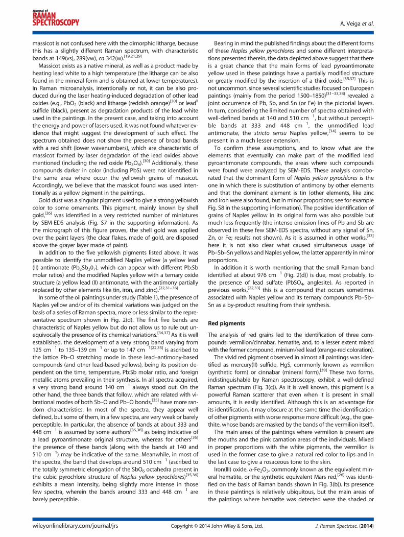
The analysis of red grains led to the identification of three com-pounds: vermilion/cinnabar, hematite, and, to a lesser extent mixedwith the former compound, minium/red lead (orange-red coloration).
The vivid red pigment observed in almost all paintings was iden-tified as mercury(II) sulfide, HgS, commonly known as vermilion(synthetic form) or cinnabar (mineral form).[39] These two forms,indistinguishable by Raman spectroscopy, exhibit a well-definedRaman spectrum (Fig. 3(c)). As it is well known, this pigment is apowerful Raman scatterer that even when it is present in smallamounts, it is easily identified. Although this is an advantage forits identification, it may obscure at the same time the identificationof other pigments with worse responsemore difficult (e.g., the goe-thite, whose bands aremasked by the bands of the vermilion itself).
The main areas of the paintings where vermilion is present arethe mouths and the pink carnation areas of the individuals. Mixedin proper proportions with the white pigments, the vermilion isused in the former case to give a natural red color to lips and inthe last case to give a rosaceous tone to the skin.
Iron(III) oxide, α-Fe2O3, commonly known as the equivalent min-eral hematite, or the synthetic equivalent Mars red,[26] was identi-fied on the basis of Raman bands shown in Fig. 3(b)). Its presencein these paintings is relatively ubiquitous, but the main areas ofthe paintings where hematite was detected were the shaded or
4 John Wiley & Sons, Ltd. J. Raman Spectrosc. (2014)
100 300 500 700 900 1100 1300 1500
Ram
an I
nten
sity
252
283342
225292
410
495
612
1317
551149
122
391 481343
(a)
(b)
(c)
Wavenumber / cm-1
Figure 3. Raman spectrum of (a) minium/red lead (Pb3O4) mixed mostprobably with litharge (α-PbO, with Raman bands at 149 and 343 cm�1).(Note: The spectrum ascribed to red lead is representative of several ana-lyzed grains, but in the analysis of other similar grains, the band at 149cm�1 is considerably less intense and the band at 343 cm�1 is not detected),(b) hematite(α-Fe2O3), and (c) vermilion/cinnabar (HgS).
100 300 500 700 900 1100 1300 1500 1700 1900 2100 2300
Ram
an I
nten
sity
259
548
1097
280 525 21022154
248
400
8381095
767 1431
(a)
(b)
(c)
290
584
Wavenumber / cm-1
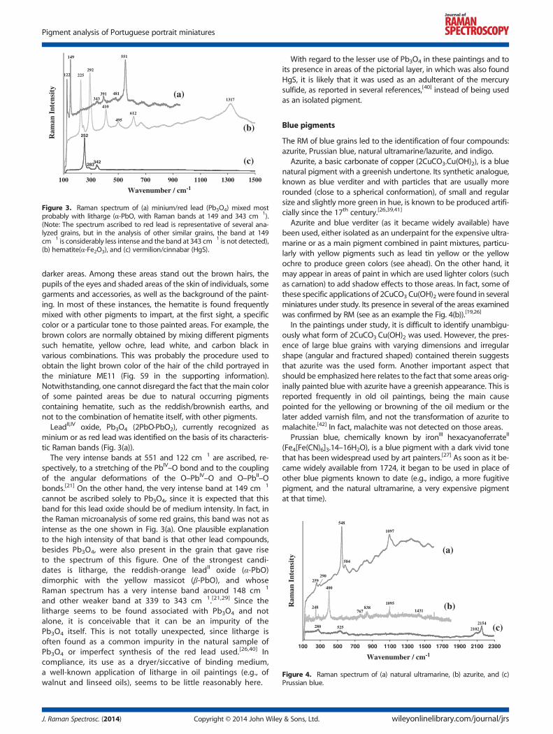
Figure 4. Raman spectrum of (a) natural ultramarine, (b) azurite, and (c)Prussian blue.
Pigment analysis of Portuguese portrait miniatures
darker areas. Among these areas stand out the brown hairs, thepupils of the eyes and shaded areas of the skin of individuals, somegarments and accessories, as well as the background of the paint-ing. In most of these instances, the hematite is found frequentlymixed with other pigments to impart, at the first sight, a specificcolor or a particular tone to those painted areas. For example, thebrown colors are normally obtained by mixing different pigmentssuch hematite, yellow ochre, lead white, and carbon black invarious combinations. This was probably the procedure used toobtain the light brown color of the hair of the child portrayed inthe miniature ME11 (Fig. S9 in the supporting information).Notwithstanding, one cannot disregard the fact that the main colorof some painted areas be due to natural occurring pigmentscontaining hematite, such as the reddish/brownish earths, andnot to the combination of hematite itself, with other pigments.
LeadII,IV oxide, Pb3O4 (2PbO·PbO2), currently recognized asminium or as red lead was identified on the basis of its characteris-tic Raman bands (Fig. 3(a)).
The very intense bands at 551 and 122 cm�1 are ascribed, re-spectively, to a stretching of the PbIV–O bond and to the couplingof the angular deformations of the O–PbIV–O and O–PbII–Obonds.[21] On the other hand, the very intense band at 149 cm�1
cannot be ascribed solely to Pb3O4, since it is expected that thisband for this lead oxide should be of medium intensity. In fact, inthe Raman microanalysis of some red grains, this band was not asintense as the one shown in Fig. 3(a). One plausible explanationto the high intensity of that band is that other lead compounds,besides Pb3O4, were also present in the grain that gave riseto the spectrum of this figure. One of the strongest candi-dates is litharge, the reddish-orange leadII oxide (α-PbO)dimorphic with the yellow massicot (β-PbO), and whoseRaman spectrum has a very intense band around 148 cm�1
and other weaker band at 339 to 343 cm�1.[21,29] Since thelitharge seems to be found associated with Pb3O4 and notalone, it is conceivable that it can be an impurity of thePb3O4 itself. This is not totally unexpected, since litharge isoften found as a common impurity in the natural sample ofPb3O4 or imperfect synthesis of the red lead used.[26,40] Incompliance, its use as a dryer/siccative of binding medium,a well-known application of litharge in oil paintings (e.g., ofwalnut and linseed oils), seems to be little reasonably here.
J. Raman Spectrosc. (2014) Copyright © 2014 John Wiley
With regard to the lesser use of Pb3O4 in these paintings and toits presence in areas of the pictorial layer, in which was also foundHgS, it is likely that it was used as an adulterant of the mercurysulfide, as reported in several references,[40] instead of being usedas an isolated pigment.
Blue pigments
The RM of blue grains led to the identification of four compounds:azurite, Prussian blue, natural ultramarine/lazurite, and indigo.
Azurite, a basic carbonate of copper (2CuCO3.Cu(OH)2), is a bluenatural pigment with a greenish undertone. Its synthetic analogue,known as blue verditer and with particles that are usually morerounded (close to a spherical conformation), of small and regularsize and slightly more green in hue, is known to be produced artifi-cially since the 17th century.[26,39,41]
Azurite and blue verditer (as it became widely available) havebeen used, either isolated as an underpaint for the expensive ultra-marine or as a main pigment combined in paint mixtures, particu-larly with yellow pigments such as lead tin yellow or the yellowochre to produce green colors (see ahead). On the other hand, itmay appear in areas of paint in which are used lighter colors (suchas carnation) to add shadow effects to those areas. In fact, some ofthese specific applications of 2CuCO3 Cu(OH)2 were found in severalminiatures under study. Its presence in several of the areas examinedwas confirmed by RM (see as an example the Fig. 4(b)).[19,26]
In the paintings under study, it is difficult to identify unambigu-ously what form of 2CuCO3 Cu(OH)2 was used. However, the pres-ence of large blue grains with varying dimensions and irregularshape (angular and fractured shaped) contained therein suggeststhat azurite was the used form. Another important aspect thatshould be emphasized here relates to the fact that some areas orig-inally painted blue with azurite have a greenish appearance. This isreported frequently in old oil paintings, being the main causepointed for the yellowing or browning of the oil medium or thelater added varnish film, and not the transformation of azurite tomalachite.[42] In fact, malachite was not detected on those areas.
Prussian blue, chemically known by ironIII hexacyanoferrateII
(Fe4[Fe(CN)6]3.14–16H2O), is a blue pigment with a dark vivid tonethat has been widespread used by art painters.[27] As soon as it be-came widely available from 1724, it began to be used in place ofother blue pigments known to date (e.g., indigo, a more fugitivepigment, and the natural ultramarine, a very expensive pigmentat that time).
& Sons, Ltd. wileyonlinelibrary.com/journal/jrs
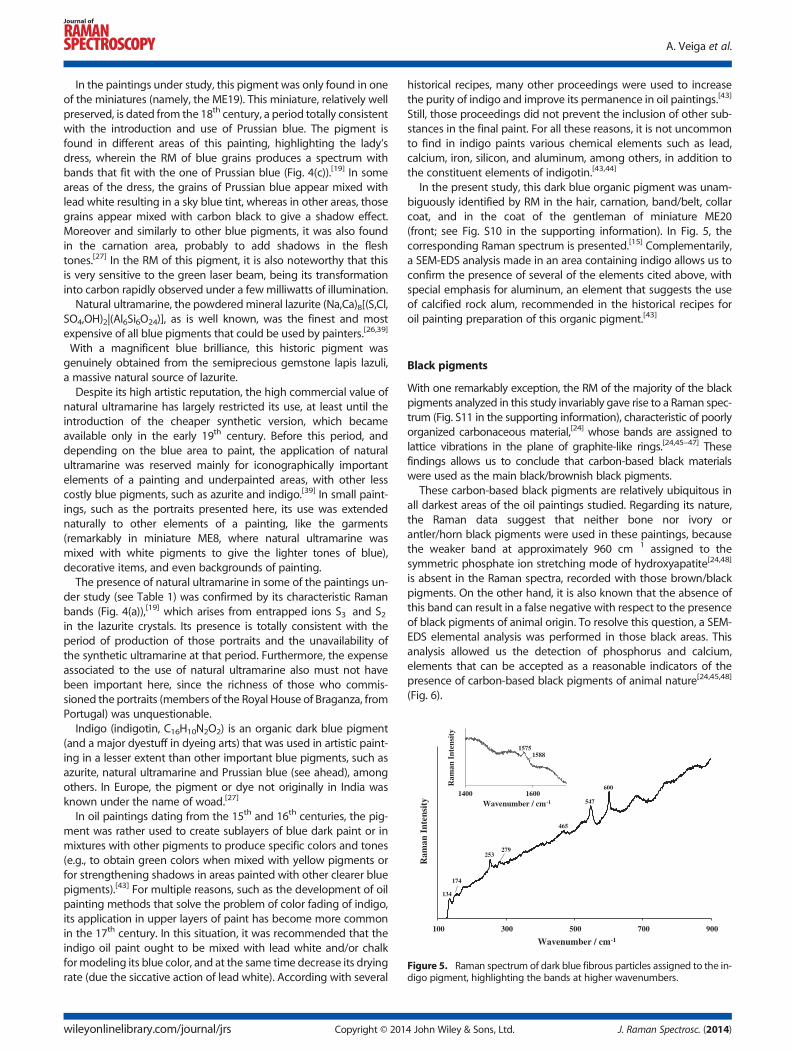
Figure 5. Raman spectrum of dark blue fibrous particles assigned to the in-digo pigment, highlighting the bands at higher wavenumbers.
A. Veiga et al.
In the paintings under study, this pigment was only found in oneof the miniatures (namely, the ME19). This miniature, relatively wellpreserved, is dated from the 18th century, a period totally consistentwith the introduction and use of Prussian blue. The pigment isfound in different areas of this painting, highlighting the lady’sdress, wherein the RM of blue grains produces a spectrum withbands that fit with the one of Prussian blue (Fig. 4(c)).[19] In someareas of the dress, the grains of Prussian blue appear mixed withlead white resulting in a sky blue tint, whereas in other areas, thosegrains appear mixed with carbon black to give a shadow effect.Moreover and similarly to other blue pigments, it was also foundin the carnation area, probably to add shadows in the fleshtones.[27] In the RM of this pigment, it is also noteworthy that thisis very sensitive to the green laser beam, being its transformationinto carbon rapidly observed under a fewmilliwatts of illumination.Natural ultramarine, the powderedmineral lazurite (Na,Ca)8[(S,Cl,
SO4,OH)2|(Al6Si6O24)], as is well known, was the finest and mostexpensive of all blue pigments that could be used by painters.[26,39]
With a magnificent blue brilliance, this historic pigment wasgenuinely obtained from the semiprecious gemstone lapis lazuli,a massive natural source of lazurite.Despite its high artistic reputation, the high commercial value of
natural ultramarine has largely restricted its use, at least until theintroduction of the cheaper synthetic version, which becameavailable only in the early 19th century. Before this period, anddepending on the blue area to paint, the application of naturalultramarine was reserved mainly for iconographically importantelements of a painting and underpainted areas, with other lesscostly blue pigments, such as azurite and indigo.[39] In small paint-ings, such as the portraits presented here, its use was extendednaturally to other elements of a painting, like the garments(remarkably in miniature ME8, where natural ultramarine wasmixed with white pigments to give the lighter tones of blue),decorative items, and even backgrounds of painting.The presence of natural ultramarine in some of the paintings un-
der study (see Table 1) was confirmed by its characteristic Ramanbands (Fig. 4(a)),[19] which arises from entrapped ions S3
� and S2�
in the lazurite crystals. Its presence is totally consistent with theperiod of production of those portraits and the unavailability ofthe synthetic ultramarine at that period. Furthermore, the expenseassociated to the use of natural ultramarine also must not havebeen important here, since the richness of those who commis-sioned the portraits (members of the Royal House of Braganza, fromPortugal) was unquestionable.Indigo (indigotin, C16H10N2O2) is an organic dark blue pigment
(and a major dyestuff in dyeing arts) that was used in artistic paint-ing in a lesser extent than other important blue pigments, such asazurite, natural ultramarine and Prussian blue (see ahead), amongothers. In Europe, the pigment or dye not originally in India wasknown under the name of woad.[27]
In oil paintings dating from the 15th and 16th centuries, the pig-ment was rather used to create sublayers of blue dark paint or inmixtures with other pigments to produce specific colors and tones(e.g., to obtain green colors when mixed with yellow pigments orfor strengthening shadows in areas painted with other clearer bluepigments).[43] For multiple reasons, such as the development of oilpainting methods that solve the problem of color fading of indigo,its application in upper layers of paint has become more commonin the 17th century. In this situation, it was recommended that theindigo oil paint ought to be mixed with lead white and/or chalkformodeling its blue color, and at the same time decrease its dryingrate (due the siccative action of lead white). According with several
wileyonlinelibrary.com/journal/jrs Copyright © 201
historical recipes, many other proceedings were used to increasethe purity of indigo and improve its permanence in oil paintings.[43]
Still, those proceedings did not prevent the inclusion of other sub-stances in the final paint. For all these reasons, it is not uncommonto find in indigo paints various chemical elements such as lead,calcium, iron, silicon, and aluminum, among others, in addition tothe constituent elements of indigotin.[43,44]
In the present study, this dark blue organic pigment was unam-biguously identified by RM in the hair, carnation, band/belt, collarcoat, and in the coat of the gentleman of miniature ME20(front; see Fig. S10 in the supporting information). In Fig. 5, thecorresponding Raman spectrum is presented.[15] Complementarily,a SEM-EDS analysis made in an area containing indigo allows us toconfirm the presence of several of the elements cited above, withspecial emphasis for aluminum, an element that suggests the useof calcified rock alum, recommended in the historical recipes foroil painting preparation of this organic pigment.[43]
Black pigments
With one remarkably exception, the RM of the majority of the blackpigments analyzed in this study invariably gave rise to a Raman spec-trum (Fig. S11 in the supporting information), characteristic of poorlyorganized carbonaceous material,[24] whose bands are assigned tolattice vibrations in the plane of graphite-like rings.[24,45–47] Thesefindings allows us to conclude that carbon-based black materialswere used as the main black/brownish black pigments.
These carbon-based black pigments are relatively ubiquitous inall darkest areas of the oil paintings studied. Regarding its nature,the Raman data suggest that neither bone nor ivory orantler/horn black pigments were used in these paintings, becausethe weaker band at approximately 960 cm�1 assigned to thesymmetric phosphate ion stretching mode of hydroxyapatite[24,48]
is absent in the Raman spectra, recorded with those brown/blackpigments. On the other hand, it is also known that the absence ofthis band can result in a false negative with respect to the presenceof black pigments of animal origin. To resolve this question, a SEM-EDS elemental analysis was performed in those black areas. Thisanalysis allowed us the detection of phosphorus and calcium,elements that can be accepted as a reasonable indicators of thepresence of carbon-based black pigments of animal nature[24,45,48]
(Fig. 6).
4 John Wiley & Sons, Ltd. J. Raman Spectrosc. (2014)
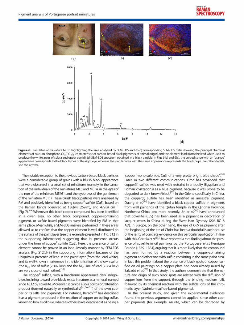
Figure 6. (a) Detail of miniature ME15 highlighting the area analyzed by SEM-EDS and (b–c) corresponding SEM-EDS data, showing the principal chemicalelements of calcium phosphate, Ca3(PO4)2 (characteristic of carbon-based black pigments of animal origin) and the element lead (from the lead white used toproduce the white areas of sclera and upper eyelid); (d) SEM-EDS spectrum obtained in a black particle. In Figs 6(b) and 6(c), the curved stripe with an ‘orange’appearance corresponds to the black lashes of the right eye, whereas the circular area with the same appearance represents the black pupil. For other details,see the arrows.
Pigment analysis of Portuguese portrait miniatures
The notable exception to the previous carbon-based black particleswere a considerable group of grains with a bluish black appearancethat were observed in a small set of miniatures (namely, in the carna-tion of the individuals of the miniatures ME5 andME14, in the eyes ofthe nun of the miniature ME461, and the eyebrows of the gentlemanof the miniature ME11). These bluish black particles were analyzed byRM and positively identified as being copperII sulfide (CuS), based onthe Raman bands observed at 136(w), 262(m), and 472(s) cm�1
(Fig. 7).[49] Whenever this black copper compound has been identifiedin a given area, no other black compound, copper-containingpigment, or sulfide-based pigments were identified by RM in thatsame place. Meanwhile, a SEM-EDS analysis performed in these areasallowed us to confirm that the copper element is well distributed onthe surface of the paint layer (see the example presented in Fig. S12 inthe supporting information) suggesting that its presence occursunder the form of copperII sulfide (CuS). Here, the presence of sulfurelement cannot be proved in an inequivocally manner by SEM-EDSanalysis (Fig. S12(d) in the supporting information) because of theubiquitous presence of lead in the paint layer (from the lead white),and its well-known interference in the identification of the own sulfur(the Kα1 line of sulfur (2.307 keV) and the Mα1 line of lead (2.364 keV)are very close of each other).[18]
The copperII sulfide, with a handsome appearance dark indigo–blue, inclining toward blue-black, exists in nature as amineral, namedsince 1832 by covellite. Moreover, it can be also a corrosion/alterationproduct (formed naturally or synthetically)[41,50–52]) of the own cop-per or its salts and pigments. For instance, Bersch[50] has describedit as a pigment produced in the reaction of copper on boiling sulfur,known to him as oil blue, whereas others have described it as being a
J. Raman Spectrosc. (2014) Copyright © 2014 John Wiley
‘copper mono-sulphide, CuS, of a very pretty bright blue shade’.[26]
Later, in two different communications, Orna has advanced thatcopper(II) sulfide was used with restraint in antiquity (Egyptian andRoman civilizations) as a blue pigment, because it was prone to bedegraded to dark brown/black.[53] In the Orient, specifically in China,the copper(II) sulfide has been identified as ancestral pigment.Duang et al.[54] have identified a black copper sulfide in pigmentsfrom wall paintings of the Qutan temple in the Qinghai Province,Northwest China, and more recently, Jin et al.[55] have announcedthat covellite (CuS) has been used as a pigment in decoration oflacquer wares in China during the West Han Dynasty (206 BC–8AD). In Europe, on the other hand, the use of CuS as pigment afterthe beginning of the era of Christ has been a doubtful issue becauseof the rarity of concrete evidence on this particular application. In linewith this, Correia et al.[22] have reported a rare finding about the pres-ence of covellite in oil paintings by the Portuguese artist HenriquePousão (1859–1884), arguing that it ismore likely that the compoundhas been formed by a reaction between a copper-containingpigment and other one with sulfur, coexisting in the same paint area.In fact, this problem about the presence of black spots of copper sul-fide on oil paintings on a copper plate had been already raised bySalvadó et al.[52] In that study, the authors demonstrate that the na-ture and origin of such black spots are related with the diffusion ofcopper ions from the support, through the binding medium (oil),followed by its chemical reaction with the sulfide ions of the chro-matic layer (cadmium sulfide-based pigments).
In the present study, and given the experimental evidencesfound, the previous argument cannot be applied, since other cop-per pigments (for example, azurite, which can be degraded by
& Sons, Ltd. wileyonlinelibrary.com/journal/jrs
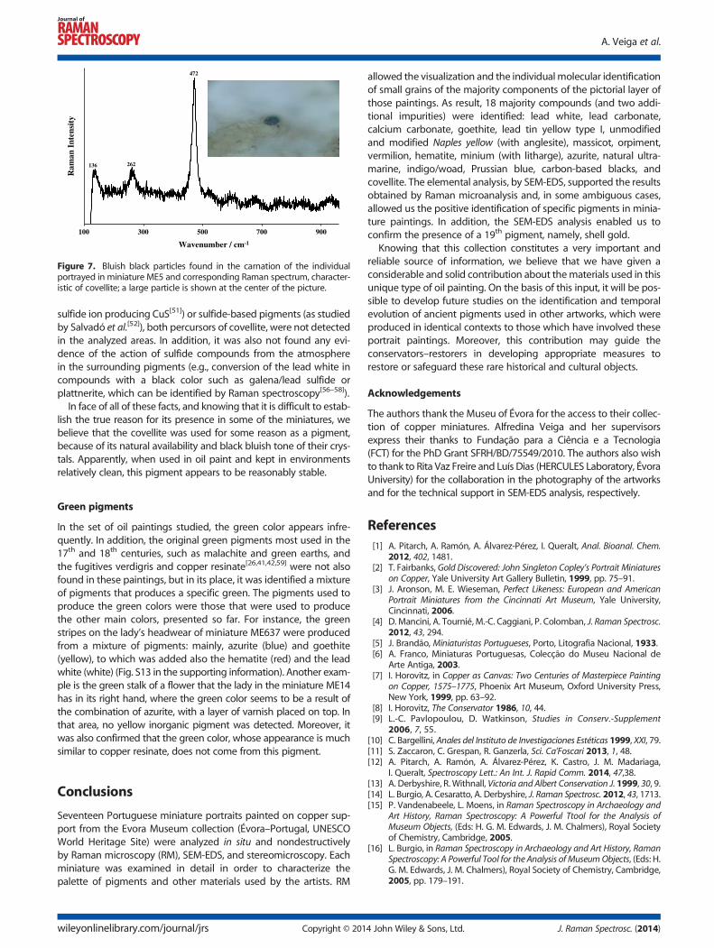
Figure 7. Bluish black particles found in the carnation of the individualportrayed in miniature ME5 and corresponding Raman spectrum, character-istic of covellite; a large particle is shown at the center of the picture.
A. Veiga et al.
sulfide ion producing CuS[51]) or sulfide-based pigments (as studiedby Salvadó et al.[52]), both percursors of covellite, were not detectedin the analyzed areas. In addition, it was also not found any evi-dence of the action of sulfide compounds from the atmospherein the surrounding pigments (e.g., conversion of the lead white incompounds with a black color such as galena/lead sulfide orplattnerite, which can be identified by Raman spectroscopy[56–58]).In face of all of these facts, and knowing that it is difficult to estab-
lish the true reason for its presence in some of the miniatures, webelieve that the covellite was used for some reason as a pigment,because of its natural availability and black bluish tone of their crys-tals. Apparently, when used in oil paint and kept in environmentsrelatively clean, this pigment appears to be reasonably stable.
Green pigments
In the set of oil paintings studied, the green color appears infre-quently. In addition, the original green pigments most used in the17th and 18th centuries, such as malachite and green earths, andthe fugitives verdigris and copper resinate[26,41,42,59] were not alsofound in these paintings, but in its place, it was identified a mixtureof pigments that produces a specific green. The pigments used toproduce the green colors were those that were used to producethe other main colors, presented so far. For instance, the greenstripes on the lady’s headwear of miniature ME637 were producedfrom a mixture of pigments: mainly, azurite (blue) and goethite(yellow), to which was added also the hematite (red) and the leadwhite (white) (Fig. S13 in the supporting information). Another exam-ple is the green stalk of a flower that the lady in the miniature ME14has in its right hand, where the green color seems to be a result ofthe combination of azurite, with a layer of varnish placed on top. Inthat area, no yellow inorganic pigment was detected. Moreover, itwas also confirmed that the green color, whose appearance is muchsimilar to copper resinate, does not come from this pigment.
Conclusions
Seventeen Portuguese miniature portraits painted on copper sup-port from the Evora Museum collection (Évora–Portugal, UNESCOWorld Heritage Site) were analyzed in situ and nondestructivelyby Raman microscopy (RM), SEM-EDS, and stereomicroscopy. Eachminiature was examined in detail in order to characterize thepalette of pigments and other materials used by the artists. RM
wileyonlinelibrary.com/journal/jrs Copyright © 201
allowed the visualization and the individual molecular identificationof small grains of the majority components of the pictorial layer ofthose paintings. As result, 18 majority compounds (and two addi-tional impurities) were identified: lead white, lead carbonate,calcium carbonate, goethite, lead tin yellow type I, unmodifiedand modified Naples yellow (with anglesite), massicot, orpiment,vermilion, hematite, minium (with litharge), azurite, natural ultra-marine, indigo/woad, Prussian blue, carbon-based blacks, andcovellite. The elemental analysis, by SEM-EDS, supported the resultsobtained by Raman microanalysis and, in some ambiguous cases,allowed us the positive identification of specific pigments in minia-ture paintings. In addition, the SEM-EDS analysis enabled us toconfirm the presence of a 19th pigment, namely, shell gold.
Knowing that this collection constitutes a very important andreliable source of information, we believe that we have given aconsiderable and solid contribution about thematerials used in thisunique type of oil painting. On the basis of this input, it will be pos-sible to develop future studies on the identification and temporalevolution of ancient pigments used in other artworks, which wereproduced in identical contexts to those which have involved theseportrait paintings. Moreover, this contribution may guide theconservators–restorers in developing appropriate measures torestore or safeguard these rare historical and cultural objects.
Acknowledgements
The authors thank the Museu of Évora for the access to their collec-tion of copper miniatures. Alfredina Veiga and her supervisorsexpress their thanks to Fundação para a Ciência e a Tecnologia(FCT) for the PhD Grant SFRH/BD/75549/2010. The authors also wishto thank to Rita Vaz Freire and Luís Dias (HERCULES Laboratory, ÉvoraUniversity) for the collaboration in the photography of the artworksand for the technical support in SEM-EDS analysis, respectively.
References[1] A. Pitarch, A. Ramón, A. Álvarez-Pérez, I. Queralt, Anal. Bioanal. Chem.
2012, 402, 1481.[2] T. Fairbanks, Gold Discovered: John Singleton Copley’s Portrait Miniatures
on Copper, Yale University Art Gallery Bulletin, 1999, pp. 75–91.[3] J. Aronson, M. E. Wieseman, Perfect Likeness: European and American
Portrait Miniatures from the Cincinnati Art Museum, Yale University,Cincinnati, 2006.
[4] D. Mancini, A. Tournié, M.-C. Caggiani, P. Colomban, J. Raman Spectrosc.2012, 43, 294.
[5] J. Brandão, Miniaturistas Portugueses, Porto, Litografia Nacional, 1933.[6] A. Franco, Miniaturas Portuguesas, Colecção do Museu Nacional de
Arte Antiga, 2003.[7] I. Horovitz, in Copper as Canvas: Two Centuries of Masterpiece Painting
on Copper, 1575–1775, Phoenix Art Museum, Oxford University Press,New York, 1999, pp. 63–92.
[8] I. Horovitz, The Conservator 1986, 10, 44.[9] L.-C. Pavlopoulou, D. Watkinson, Studies in Conserv.-Supplement
2006, 7, 55.[10] C. Bargellini, Anales del Instituto de Investigaciones Estéticas 1999, XXI, 79.[11] S. Zaccaron, C. Grespan, R. Ganzerla, Sci. Ca’Foscari 2013, 1, 48.[12] A. Pitarch, A. Ramón, A. Álvarez-Pérez, K. Castro, J. M. Madariaga,
I. Queralt, Spectroscopy Lett.: An Int. J. Rapid Comm. 2014, 47,38.[13] A. Derbyshire, R. Withnall, Victoria and Albert Conservation J. 1999, 30, 9.[14] L. Burgio, A. Cesaratto, A. Derbyshire, J. Raman Spectrosc. 2012, 43, 1713.[15] P. Vandenabeele, L. Moens, in Raman Spectroscopy in Archaeology and
Art History, Raman Spectroscopy: A Powerful Ttool for the Analysis ofMuseum Objects, (Eds: H. G. M. Edwards, J. M. Chalmers), Royal Societyof Chemistry, Cambridge, 2005.
[16] L. Burgio, in Raman Spectroscopy in Archaeology and Art History, RamanSpectroscopy: A Powerful Tool for the Analysis of MuseumObjects, (Eds: H.G. M. Edwards, J. M. Chalmers), Royal Society of Chemistry, Cambridge,2005, pp. 179–191.
4 John Wiley & Sons, Ltd. J. Raman Spectrosc. (2014)
Pigment analysis of Portuguese portrait miniatures
[17] R. J. H. Clark, in Handbook of Vibrational Spectroscopy, (Eds: J. M.Chalmers, P. R. Griffiths), Wiley, Chichester, 2002, pp. 2977–2992.
[18] J. I. Goldstein, D. E. Newbury, D. C. Joy, C. E. Lyman, P. Echlin, E. Lifshin,L. Sawyer, J. R. Michael, Scanning Electron Microscopy and X-RayMicroanalysis, 3rd Ed., Kluwer Academic/Plenum Publishers, NewYork, 2005.
[19] I. M. Bell, R. J. H. Clark, P. J. Gibbs, Spectrochim. Acta A 1997, 53, 2159.[20] L. Burgio, R. J. H. Clark, Spectrochim. Acta Part A 2001, 57, 1491.[21] M. Bouchard, D.C. Smith, Spectrochimica Acta Part A 2003, 59, 2247.[22] A. M. Correia, R. J. H. Clark, M. I. M. Ribeiro, M. L. T. S. Duarte, J. Raman
Spectrosc. 2007, 38, 1390.[23] C. L. Aibéo, S. Goffin, O. Schalm, G. van der Snickt, N. Laquiére,
P. Eyskens, K. Janssens, J. Raman Spectrosc. 2008, 39, 1091.[24] J. L. Perez-Rodriguez, A. Duran, Spectrosc. Lett.: An Int. J. Rapid
Communication 2014, 47, 223.[25] S. Gunasekaran, G. Anbalagan, Spectrochim Acta Part A 2007, 68, 656.[26] N. Eastaugh, V. Walsh, T. Chaplin, R. Siddall, Pigment Compendium, A
Dictionary of Historical Pigments, Oxford: Elsevier Butterworth-Heinemann, Amsterdam, 2004.
[27] E. W. Fitzhugh (Ed.), Artists’ Pigments. A Handbook of Their History andCharacteristics, Vol. 3, Oxford University Press, New York, 1997.
[28] G. Rapp, Archaeomineralogy, 2nd Ed., Springer-Verlag, Berlin,Heidelberg 2009, pp. 213-214.
[29] G.D. Smith, R.J.H. Clark, Appl. Spectrosc. 2002, 56, 804.[30] L. Burgio, R.J. H. Clark, S. Firth, Analyst 2001, 126, 222.[31] J. Dik, F. Tichelaar, K. Goubitz, R. Peschar, H. Schenk, Zeitschrift fuer
Kunsttechn u. Konservierung 2002 16, 291 (or, J. Dik, ‘19th CenturyNaples Yellow Re-Examined’, in Scientific analysis of historical paintand the implications for art history and art conservation. The casestudies of naples yellow and discoloured smalt, Ph.D. thesis, Faculty ofScience, 2003, pp. 69-99 (Available from: http://dare.uva.nl/document/196004 in March 15, 2014).
[32] C. Seccaroni, Giallorino, Storia dei pigmenti gialli di natura sintetica, DeLuca Editori d’Arte, Roma, 2006.
[33] D. Hradil, T. Grygar, J. Hradilová, P. Bezdicka, V. Grunwaldová, I. Fogas,C. Miliani, J. Cult. Heritage 2007, 8, 377.
[34] B. Kirmizi, P. Colomban, B. Quette, J. Raman Spectrosc. 2010, 41, 780.[35] F. Rosi, V. Manuali, T. Grygar, P. Bezdicka, B.G. Brunetti, A. Sgamellotti,
L. Burgio, C. Seccaronif, C. Miliani, J. Raman Spectrosc. 2011, 42, 407.[36] M. T. Doménech-Carbó, H. G. M. Edwards, A. Doménech-Carbó,
J. M. del Hoyo-Meléndeza, J. de la Cruz-Cañizares, J. Raman Spectrosc.2012, 43, 1250.
[37] C. Pelosi, G. Agresti, U. Santamaria, E. Mattei, e-PreservationScience2010, 7, 108.
[38] L. Cartechini, F. Rosi, C. Miliani, F. D’Acapito, B. G. Brunetti,A. Sgamellottic, J. Anal. At. Spectrom. 2011, 26, 2500.
[39] A. Roy (Ed.), Artists’ Pigments. A Handbook of Their History and Character-istics, Vol. 2, Oxford University Press, New York, 1997.
J. Raman Spectrosc. (2014) Copyright © 2014 John Wiley
[40] E. W. Fitzhugh, in Artists’ Pigments. A Handbook of Their History andCharacteristics, Vol. 1 (Ed: R. E. Feller), Oxford University Press, NewYork, 1986, pp. 109–139.
[41] D. A. Scott, Copper and Bronze in Art: Corrosion, Colorants,Conservation, Getty Trust Publications: Getty ConservationInstitute, Los Angeles, 2002.
[42] R. J. Gettens, E. W. Fitzhugh, Studies in Conserv. 1966 11, 54.[43] M. E. van Hommes, Indigo as a Pigment inOil Painting and the Problem
of Its Fading, in Discoloration in Renaissance and Baroque Oil Paintings.Instructions for Painters, Theoretical Concepts, and Scientific Data, Ph.D.thesis, Universiteit van Amsterdam, 2002, pp. 109–166 (Availablefrom: http://dare.uva.nl/document/62357 in March 17, 2014).
[44] J. Zagora, SEM-EDX Pigment Analysis and Multi-analytical Study of theGround and Paint Layers of Francesco Fedrigazzi’s Painting fromKostanje, CeROArt, 2013, Available from: http://ceroart.revues.org/3248 in February 18, 2014.)
[45] S. Potgieter-Vermaak, N. Maledi, N. Wagner, J.H.P. Van Heerden,R. Van Grieken, J.H. Potgieter, J. Raman Spectrosc. 2011, 42, 123.
[46] Y. Wang, D. C. Alsmeyer, R. L. McCreery, Chem. Mater. 1990, 2, 557.[47] O. Beyssac, B. Goffé, J. Petitet, E. Froigneux, M. Moreau, J. Rouzaud,
Spectrochim Acta Part A. 2003, 59, 2267.[48] E. P. Tomasini, E. B. Halac, M. Reinoso, E. J. Di Liscia, M. S. Maier, J. Raman
Spectrosc. 2012, 43, 1671.[49] T. P. Mernagh, A. G. Trudu, Chem. Geol. 1993, 103, 113.[50] J. Bersch, The Manufacture of Mineral and Lake Pigments, Containing
Directions for the Manufacture of All Artificial Artists’ and Painters’Colours, Enamel Colours, Soot and Metallic Pigments, Scott,Greenwood & CO., London, 1901 (Translation from the Second,Revised Edition, by A.C. Wright).
[51] G. D. Smith, R. J. H. Clark, J. of Cult. Heritage 2002, 3, 101.[52] N. Salvadó, J. Molera, M. Vendrell-Saz, Anal. Chim. Acta 2003, 479, 255.[53] M. V. Orna, M. J. D. Low, M. M. Julian, Studies in Conserv. 1980, 25, 53.[54] S. Duang, J.-I. Miyata, N. Kumagai, R. Sugishita, Kobunkazai no kagaku
1987, 32, 13.[55] P.-J. Jin, Z.-Q. Yao, M.-L. Zhang, Y.-H. Lia, H.-P. Xinga, J. Raman Spectrosc.
2010, 41, 222.[56] C. Miguel, A. Claro, A. P. Gonçalves, V. S. F. Muralha, M. J. Melo, J. Raman
Spectrosc. 2009, 40, 1966.[57] V. S. F. Muralha, C. Miguel, M. J. Melo, J. Raman Spectrosc. 2012, 43, 1737.[58] M. Aceto, G. Gatti, A. Agostino, G. Fenoglio, V. Giordano, M. Varetto,
G. Castagneri, J. Raman Spectrosc. 2012, 43, 1754.[59] C. Conti, J. Striova, I. Aliatis, E. Possenti, G. Massonnet, C. Muehlethaler,
T. Poli, M. Positano, J. Raman Spectrosc. 2014, DOI: 10.1002/jrs.4455.
Supporting information
Additional supporting information may be found in the onlineversion of this article at the publisher’s web site.
& Sons, Ltd. wileyonlinelibrary.com/journal/jrs


![[ONDER]TROUWEN AMSTERDAM - 17th Century Hollanders](https://img.dokumen.tips/doc/110x75/63227d83ae0f5e819105eae4/ondertrouwen-amsterdam-17th-century-hollanders.jpg)


