Embed Size (px)
Citation preview
�����������������
Citation: Giannelli, G.; Bisceglie, F.;
Pelosi, G.; Bonati, B.; Cardarelli, M.;
Antenozio, M.L.; Degola, F.; Visioli, G.
Phyto-Beneficial Traits of
Rhizosphere Bacteria: In Vitro
Exploration of Plant Growth
Promoting and Phytopathogen
Biocontrol Ability of Selected Strains
Isolated from Harsh Environments.
Plants 2022, 11, 230. https://doi.org/
10.3390/plants11020230
Academic Editor: Mariana Amato
Received: 21 December 2021
Accepted: 14 January 2022
Published: 17 January 2022
Publisher’s Note: MDPI stays neutral
with regard to jurisdictional claims in
published maps and institutional affil-
iations.
Copyright: © 2022 by the authors.
Licensee MDPI, Basel, Switzerland.
This article is an open access article
distributed under the terms and
conditions of the Creative Commons
Attribution (CC BY) license (https://
creativecommons.org/licenses/by/
4.0/).
plants
Article
Phyto-Beneficial Traits of Rhizosphere Bacteria: In VitroExploration of Plant Growth Promoting and PhytopathogenBiocontrol Ability of Selected Strains Isolated fromHarsh EnvironmentsGianluigi Giannelli 1, Franco Bisceglie 1,2 , Giorgio Pelosi 1,2, Beatrice Bonati 1, Maura Cardarelli 3,Maria Luisa Antenozio 3,4, Francesca Degola 1,*,† and Giovanna Visioli 1,*,†
1 Dipartimento di Scienze Chimiche, della Vita e della Sostenibilità Ambientale, Università di Parma,Parco Area delle Scienze 11/a, 43124 Parma, Italy; [email protected] (G.G.);[email protected] (F.B.); [email protected] (G.P.); [email protected] (B.B.)
2 C.I.R.C.M.S.B.—Consorzio Interuniversitario di Ricerca in Chimica dei Metalli nei Sistemi Biologici,Parma Local Unit, 43124 Parma, Italy
3 IBPM-CNR, P.le A. Moro 5, 00185 Roma, Italy; [email protected] (M.C.);[email protected] (M.L.A.)
4 Dipartimento di Biologia e Biotecnologie, Università Sapienza di Roma, 00185 Roma, Italy* Correspondence: [email protected] (F.D.); [email protected] (G.V.)† These authors contributed equally to this work.
Abstract: Beneficial interactions between plants and some bacterial species have been long recognized,as they proved to exert various growth-promoting and health-protective activities on economicallyrelevant crops. In this study, the growth promoting and antifungal activity of six bacterial strains,Paenarthrobacter ureafaciens, Beijerinckia fluminensis, Pseudomonas protegens, Arthrobacter sp., Arthrobac-ter defluii, and Arthrobacter nicotinovorans, were investigated. The tested strains resulted positive forsome plant growth promoting (PGP) traits, such as indole-3-acetic acid (IAA), 1-aminocyclopropane-1-carboxylate-deaminase (ACC-deaminase), siderophore production, and solubilization of phosphates.The effect of the selected bacteria on Arabidopsis thaliana seedlings growth was assessed using dif-ferent morphological parameters. Bacterial activity against the phytopathogenic fungal speciesAspergillus flavus, Fusarium proliferatum, and Fusarium verticillioides was also assessed, since thesecause major yield losses in cereal crops and are well-known mycotoxin producers. Strains Pvr_9(B. fluminensis) and PHA_1 (P. protegens) showed an important growth-promoting effect on A. thalianacoupled with a high antifungal activity on all the three fungal species. The analysis of bacterialbroths through ultra performance liquid chromatography–mass spectrometry (UPLC–MS) and liquidchromatography–electrospray ionization–mass spectrometry (LC–ESI–MS/MS) confirmed the pres-ence of potential PGP-compounds, among these are desferrioxamine B, aminochelin, asperchrome B,quinolobactin siderophores, and salicylic acid.
Keywords: antifungal metabolites; biocontrol agents; plant growth promoting rhizobacteria; phy-topathogen antagonists; siderophore production; stressful soils
1. Introduction
The rhizosphere is a complex ecosystem in which many relationships are establishedbetween bacteria, fungi, and plant root apparatus, and represents the main source ofnutrients for plant growth [1]. In particular, many soil microbes have established goodrelationships with plants, supporting their growth and health, for example helping plantsto manage both biotic and abiotic stress [2–4]. In particular, plant growth promotingrhizobacteria (PGPR) are microorganisms, which form symbiotic interactions with plantroots, promoting plant health and productivity through different mechanisms such as
Plants 2022, 11, 230. https://doi.org/10.3390/plants11020230 https://www.mdpi.com/journal/plants
Plants 2022, 11, 230 2 of 15
production of plant hormones (auxins, cytokinin, and gibberellins); inhibition of plantsenescence; N2 fixation; phosphate solubilization and mineralization of other nutrients;and siderophores production [5]. In addition, being present in the rhizosphere, PGPR mayalso be endophytic (PGPE) (for example, by colonizing the plant’s tissues), symbiotic (forexample, by colonizing the interior of the roots of specific plants by forming nodules), orphyllospheric (i.e., they can be found on the surfaces of plant leaves and stems) [6].
The majority of the most known PGPR belong to the genera Alcaligenes, Arthrobacter,Azospirillum, Azotobacter, Bacillus, Burkholderia, Enterobacter, Klebsiella, Pseudomonas, Rhizo-bium, and Serratia [7]. PGPR beneficial effects on plants include an increase in root growthand shoot biomass, chlorophyll content, nutrient uptake, total protein content, hydraulicactivity, abiotic stress tolerance, shoot and root weights, and a delayed senescence. PGPRare, thus, often employed as biofertilizers [8].
Besides being determinant for plant health and soil fertility, the interactions betweenbeneficial microbes and plant rhizosphere can also exert direct, positive effects againstphytopathies. PGPR can suppress diseases by directly synthesizing pathogen-antagonizingcompounds, as well as by triggering plant immune responses [9]. Some PGPR have beenfound to possess several chemotypical traits that make them potential antifungal agentsfor biocontrol purposes. They can produce siderophores, antimicrobials, lytic enzymes,and various extracellular metabolites which can interfere with, if not completely inhibit,the growth of different, devastating phytopathogenic fungal species with a broad hostrange [10]. For example, Pseudomonas spp. strains isolated from the rhizosphere of alfalfaand clover plants growing on extremely poor pseudogley soil showed interesting antifungalactivity against Trichoderma viride, Aspergillus fumigatus, and Aspergillus niger [11], whileplant-promoting Pseudomonas fluorescens and Bacillus spp. strains from a PGPR collectionwere found to effectively inhibit three spore-forming genera (Alternaria spp., Fusariumspp., Bipolaris spp.) [12]. Again, Phytophthora capsici, a cucumber pathogen, was success-fully suppressed by specific isolates of Pseudomonas stutzeri and B. amyloliquefaciens [13].Recently, a battery of bacteria isolated from the rhizosphere of crops cultivated in dif-ferent agroecosystems of Pakistan was screened for their biocontrol potential against arange of fungal phytopathogens, showing antagonistic activity against Fusarium oxysporum,F. moniliforme, Rhizoctonia solani, Colletotrichum gloeosporioides, C. falcatum, Aspergillus niger,and A. flavus [14]; the antimicrobial effect, which was ascribed to the individuation ofantifungal metabolites such as specific antibiotics and cell wall degrading enzymes, wasaccompanied by the production of a number of compounds recognized as plant growthpromoters (hormones and siderophores), suggesting that these PGPR can be exploited fordual-purpose strategies based on the application of a single formulation acting as biopesti-cide and biofertilizer [15]. It is worthy of consideration that specific bacterial siderophoreshave been demonstrated to possess direct antifungal activity (often affecting spore ger-mination) against phytopathogens such as F. oxysporum, F. udum, A. niger, A. flavus, andSclerotium rolfsii [16–18]; pyoverdine and pyocheline in particular, produced by P. aeruginosaand Burkholderia spp., have been attributed the most relevant antifungal activities of thesebacterial species [19].
Interestingly, other molecules produced by some rhizosphere bacteria and also in-volved in the plant disease resistance show antifungal properties, as it is the case of salicylicacid (SA) and its derivatives [20–24].
In this panorama, the aim of this work was (i) to characterize bacterial isolates—derived from different soil and rhizosphere environments—for their capacity to improveArabidopsis thaliana growth, (ii) to test their potential activity as biocontrol agents againstphytopathogenic fungi species, and (iii) to identify possible molecules involved in plantmineral nutrition or with antimicrobial activity.
Plants 2022, 11, 230 3 of 15
2. Results2.1. Evaluation of the Bacterial Strains Properties
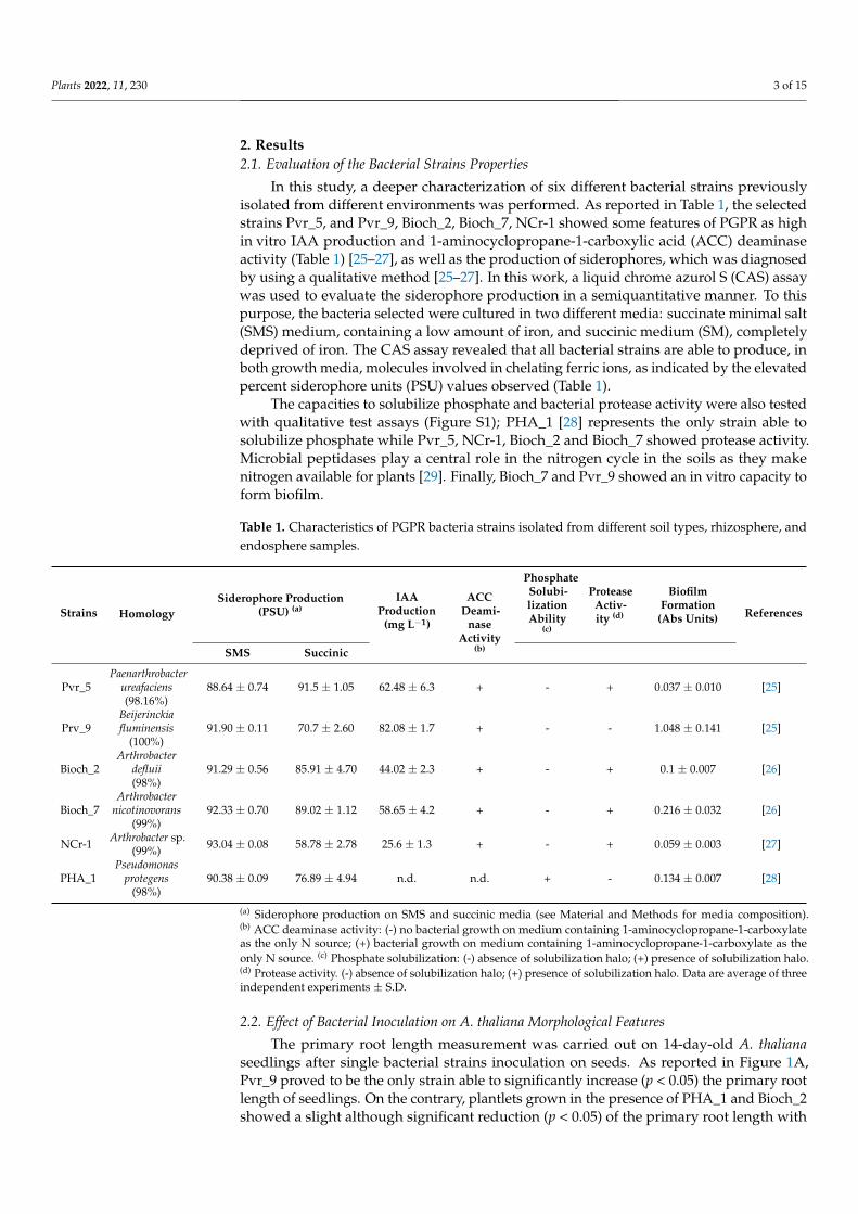
In this study, a deeper characterization of six different bacterial strains previouslyisolated from different environments was performed. As reported in Table 1, the selectedstrains Pvr_5, and Pvr_9, Bioch_2, Bioch_7, NCr-1 showed some features of PGPR as highin vitro IAA production and 1-aminocyclopropane-1-carboxylic acid (ACC) deaminaseactivity (Table 1) [25–27], as well as the production of siderophores, which was diagnosedby using a qualitative method [25–27]. In this work, a liquid chrome azurol S (CAS) assaywas used to evaluate the siderophore production in a semiquantitative manner. To thispurpose, the bacteria selected were cultured in two different media: succinate minimal salt(SMS) medium, containing a low amount of iron, and succinic medium (SM), completelydeprived of iron. The CAS assay revealed that all bacterial strains are able to produce, inboth growth media, molecules involved in chelating ferric ions, as indicated by the elevatedpercent siderophore units (PSU) values observed (Table 1).
The capacities to solubilize phosphate and bacterial protease activity were also testedwith qualitative test assays (Figure S1); PHA_1 [28] represents the only strain able tosolubilize phosphate while Pvr_5, NCr-1, Bioch_2 and Bioch_7 showed protease activity.Microbial peptidases play a central role in the nitrogen cycle in the soils as they makenitrogen available for plants [29]. Finally, Bioch_7 and Pvr_9 showed an in vitro capacity toform biofilm.
Table 1. Characteristics of PGPR bacteria strains isolated from different soil types, rhizosphere, andendosphere samples.
Strains HomologySiderophore Production
(PSU) (a)IAA
Production(mg L−1)
ACCDeami-
naseActivity
(b)
PhosphateSolubi-lizationAbility
(c)
ProteaseActiv-ity (d)
BiofilmFormation
(Abs Units) References
SMS Succinic
Pvr_5Paenarthrobacter
ureafaciens(98.16%)
88.64 ± 0.74 91.5 ± 1.05 62.48 ± 6.3 + - + 0.037 ± 0.010 [25]
Prv_9Beijerinckiafluminensis
(100%)91.90 ± 0.11 70.7 ± 2.60 82.08 ± 1.7 + - - 1.048 ± 0.141 [25]
Bioch_2Arthrobacter
defluii(98%)
91.29 ± 0.56 85.91 ± 4.70 44.02 ± 2.3 + - + 0.1 ± 0.007 [26]
Bioch_7Arthrobacter
nicotinovorans(99%)
92.33 ± 0.70 89.02 ± 1.12 58.65 ± 4.2 + - + 0.216 ± 0.032 [26]
NCr-1 Arthrobacter sp.(99%) 93.04 ± 0.08 58.78 ± 2.78 25.6 ± 1.3 + - + 0.059 ± 0.003 [27]
PHA_1Pseudomonas
protegens(98%)
90.38 ± 0.09 76.89 ± 4.94 n.d. n.d. + - 0.134 ± 0.007 [28]
(a) Siderophore production on SMS and succinic media (see Material and Methods for media composition).(b) ACC deaminase activity: (-) no bacterial growth on medium containing 1-aminocyclopropane-1-carboxylateas the only N source; (+) bacterial growth on medium containing 1-aminocyclopropane-1-carboxylate as theonly N source. (c) Phosphate solubilization: (-) absence of solubilization halo; (+) presence of solubilization halo.(d) Protease activity. (-) absence of solubilization halo; (+) presence of solubilization halo. Data are average of threeindependent experiments ± S.D.
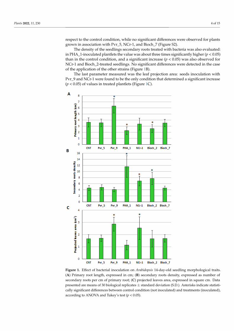
2.2. Effect of Bacterial Inoculation on A. thaliana Morphological Features
The primary root length measurement was carried out on 14-day-old A. thalianaseedlings after single bacterial strains inoculation on seeds. As reported in Figure 1A,Pvr_9 proved to be the only strain able to significantly increase (p < 0.05) the primary rootlength of seedlings. On the contrary, plantlets grown in the presence of PHA_1 and Bioch_2showed a slight although significant reduction (p < 0.05) of the primary root length with
Plants 2022, 11, 230 4 of 15
respect to the control condition, while no significant differences were observed for plantsgrown in association with Pvr_5, NCr-1, and Bioch_7 (Figure S2).
The density of the seedlings secondary roots treated with bacteria was also evaluated:in PHA_1-inoculated plantlets the value was about three times significantly higher (p < 0.05)than in the control condition, and a significant increase (p < 0.05) was also observed forNCr-1 and Bioch_2-treated seedlings. No significant differences were detected in the caseof the application of the other strains (Figure 1B).
The last parameter measured was the leaf projection area: seeds inoculation withPvr_9 and NCr-1 were found to be the only condition that determined a significant increase(p < 0.05) of values in treated plantlets (Figure 1C).
Plants 2022, 11, x FOR PEER REVIEW 4 of 16
proved to be the only strain able to significantly increase (p < 0.05) the primary root length
of seedlings. On the contrary, plantlets grown in the presence of PHA_1 and Bioch_2
showed a slight although significant reduction (p < 0.05) of the primary root length with
respect to the control condition, while no significant differences were observed for plants
grown in association with Pvr_5, NCr-1, and Bioch_7 (Figure S2).
The density of the seedlings secondary roots treated with bacteria was also evaluated:
in PHA_1-inoculated plantlets the value was about three times significantly higher (p <
0.05) than in the control condition, and a significant increase (p < 0.05) was also observed
for NCr-1 and Bioch_2-treated seedlings. No significant differences were detected in the
case of the application of the other strains (Figure 1B).
The last parameter measured was the leaf projection area: seeds inoculation with
Pvr_9 and NCr-1 were found to be the only condition that determined a significant in-
crease (p < 0.05) of values in treated plantlets (Figure 1C).
Figure 1. Effect of bacterial inoculation on Arabidopsis 14-day-old seedling morphological traits. (A)
Primary root length, expressed in cm; (B) secondary roots density, expressed as number of second-
ary roots per cm of primary root; (C) projected leaves area, expressed in square cm. Data presented
are means of 30 biological replicates ± standard deviation (S.D.). Asterisks indicate statistically sig-
nificant differences between control condition (not inoculated) and treatments (inoculated), accord-
ing to ANOVA and Tukey’s test (p < 0.05).
2.3. Antifungal Activity against Selected Phytopathogenic Fungi
Bacterial strains were tested for their direct activity against the phytopathogenic spe-
cies A. flavus, F. verticillioides, and F. proliferatum: the antifungal potential was assayed by
Figure 1. Effect of bacterial inoculation on Arabidopsis 14-day-old seedling morphological traits.(A) Primary root length, expressed in cm; (B) secondary roots density, expressed as number ofsecondary roots per cm of primary root; (C) projected leaves area, expressed in square cm. Datapresented are means of 30 biological replicates ± standard deviation (S.D.). Asterisks indicate statisti-cally significant differences between control condition (not inoculated) and treatments (inoculated),according to ANOVA and Tukey’s test (p < 0.05).
Plants 2022, 11, 230 5 of 15
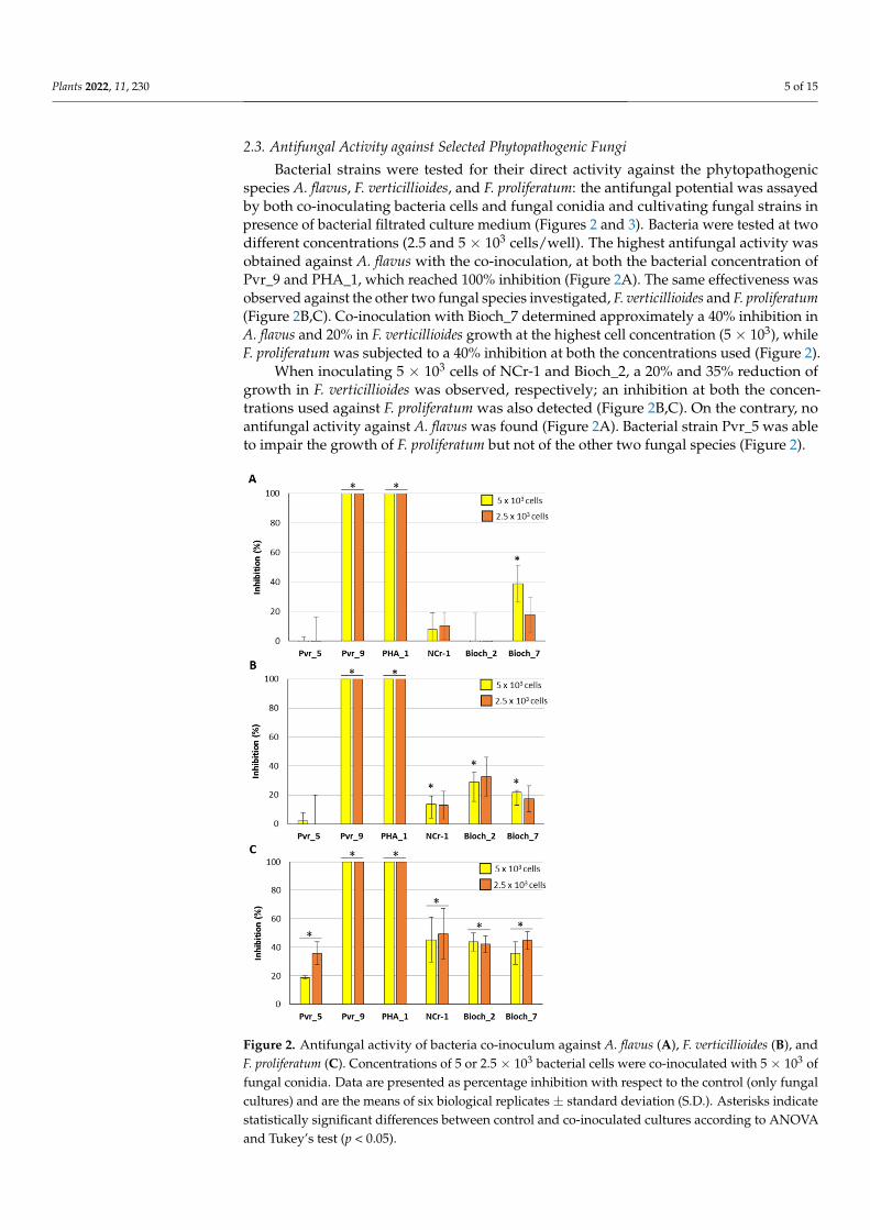
2.3. Antifungal Activity against Selected Phytopathogenic Fungi
Bacterial strains were tested for their direct activity against the phytopathogenicspecies A. flavus, F. verticillioides, and F. proliferatum: the antifungal potential was assayedby both co-inoculating bacteria cells and fungal conidia and cultivating fungal strains inpresence of bacterial filtrated culture medium (Figures 2 and 3). Bacteria were tested at twodifferent concentrations (2.5 and 5 × 10³ cells/well). The highest antifungal activity wasobtained against A. flavus with the co-inoculation, at both the bacterial concentration ofPvr_9 and PHA_1, which reached 100% inhibition (Figure 2A). The same effectiveness wasobserved against the other two fungal species investigated, F. verticillioides and F. proliferatum(Figure 2B,C). Co-inoculation with Bioch_7 determined approximately a 40% inhibition inA. flavus and 20% in F. verticillioides growth at the highest cell concentration (5 × 10³), whileF. proliferatum was subjected to a 40% inhibition at both the concentrations used (Figure 2).
When inoculating 5 × 10³ cells of NCr-1 and Bioch_2, a 20% and 35% reduction ofgrowth in F. verticillioides was observed, respectively; an inhibition at both the concen-trations used against F. proliferatum was also detected (Figure 2B,C). On the contrary, noantifungal activity against A. flavus was found (Figure 2A). Bacterial strain Pvr_5 was ableto impair the growth of F. proliferatum but not of the other two fungal species (Figure 2).
Plants 2022, 11, x FOR PEER REVIEW 5 of 15
was observed against the other two fungal species investigated, F. verticillioides and F. proliferatum (Figure 2 B,C). Co-inoculation with Bioch_7 determined approximately a 40% inhibition in A. flavus and 20% in F. verticillioides growth at the highest cell concentration (5 × 10³), while F. proliferatum was subjected to a 40% inhibition at both the concentrations used (Figure 2).
When inoculating 5 × 10³ cells of NCr-1 and Bioch_2, a 20% and 35% reduction of growth in F. verticillioides was observed, respectively; an inhibition at both the concentrations used against F. proliferatum was also detected (Figure 2B,C). On the contrary, no antifungal activity against A. flavus was found (Figure 2A). Bacterial strain Pvr_5 was able to impair the growth of F. proliferatum but not of the other two fungal species (Figure 2).
Figure 2. Antifungal activity of bacteria co-inoculum against A. flavus (A), F. verticillioides (B), and F. proliferatum (C). Concentrations of 5 or 2.5 × 10³ bacterial cells were co-inoculated with 5 × 10³ of
fungal conidia. Data are presented as percentage inhibition with respect to the control (only fungal cultures) and are the means of six biological replicates ± standard deviation (S.D.). Asterisks indicate statistically significant differences between control and co-inoculated cultures according to ANOVA and Tukey’s test (p < 0.05).
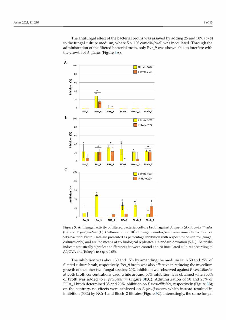
The antifungal effect of the bacterial broths was assayed by adding 25 and 50% (v/v) to the fungal culture medium, where 5 × 10³ conidia/well was inoculated. Through the administration of the filtered bacterial broth, only Pvr_9 was shown able to interfere with the growth of A. flavus (Figure 3A).
Figure 2. Antifungal activity of bacteria co-inoculum against A. flavus (A), F. verticillioides (B), andF. proliferatum (C). Concentrations of 5 or 2.5 × 10³ bacterial cells were co-inoculated with 5 × 10³ offungal conidia. Data are presented as percentage inhibition with respect to the control (only fungalcultures) and are the means of six biological replicates ± standard deviation (S.D.). Asterisks indicatestatistically significant differences between control and co-inoculated cultures according to ANOVAand Tukey’s test (p < 0.05).
Plants 2022, 11, 230 6 of 15
The antifungal effect of the bacterial broths was assayed by adding 25 and 50% (v/v)to the fungal culture medium, where 5 × 10³ conidia/well was inoculated. Through theadministration of the filtered bacterial broth, only Pvr_9 was shown able to interfere withthe growth of A. flavus (Figure 3A).
Plants 2022, 11, x FOR PEER REVIEW 6 of 15
The inhibition was about 30 and 15% by amending the medium with 50 and 25% of filtered culture broth, respectively. Pvr_9 broth was also effective in reducing the mycelium growth of the other two fungal species: 20% inhibition was observed against F. verticilliodes at both broth concentrations used while around 50% inhibition was obtained when 50% of broth was added to F. proliferatum (Figure 3B,C). Administration of 50 and 25% of PHA_1 broth determined 35 and 20% inhibition on F. verticillioides, respectively (FIure 3B); on the contrary, no effects were achieved on F. proliferatum, which instead resulted in inhibition (50%) by NCr-1 and Bioch_2 filtrates (Figure 3C). Interestingly, the same fungal species resulted in more effect by 25 than 50% of Bioch_7 broth, a peculiarity that might be attributed to a combined effect of specific and nonspecific inhibitors that differentially act on mycelium development (Figure 3C).
Finally, 20% inhibition of F. verticilliodes growth was recorded when using Bioch_7 filtered culture broth at every percentage, and 50% of Pvr_5, NCr-1, and Bioch_2.
Figure 3. Antifungal activity of filtered bacterial culture broth against A. flavus (A), F. verticillioides (B), and F. proliferatum (C). Cultures of 5 × 10³ of fungal conidia/well were amended with 25 or 50% bacterial broth. Data are presented as percentage inhibition with respect to the control (fungal cultures only) and are the means of six biological replicates ± standard deviation (S.D.). Asterisks indicate statistically significant differences between control and co-inoculated cultures according to ANOVA and Tukey’s test (p < 0.05).
2.4. Identification of Potentially Beneficial Molecules for Plant in Bacterial Broths
Figure 3. Antifungal activity of filtered bacterial culture broth against A. flavus (A), F. verticillioides(B), and F. proliferatum (C). Cultures of 5 × 10³ of fungal conidia/well were amended with 25 or50% bacterial broth. Data are presented as percentage inhibition with respect to the control (fungalcultures only) and are the means of six biological replicates ± standard deviation (S.D.). Asterisksindicate statistically significant differences between control and co-inoculated cultures according toANOVA and Tukey’s test (p < 0.05).
The inhibition was about 30 and 15% by amending the medium with 50 and 25% offiltered culture broth, respectively. Pvr_9 broth was also effective in reducing the myceliumgrowth of the other two fungal species: 20% inhibition was observed against F. verticilliodesat both broth concentrations used while around 50% inhibition was obtained when 50%of broth was added to F. proliferatum (Figure 3B,C). Administration of 50 and 25% ofPHA_1 broth determined 35 and 20% inhibition on F. verticillioides, respectively (Figure 3B);on the contrary, no effects were achieved on F. proliferatum, which instead resulted ininhibition (50%) by NCr-1 and Bioch_2 filtrates (Figure 3C). Interestingly, the same fungal
Plants 2022, 11, 230 7 of 15
species resulted in more effect by 25 than 50% of Bioch_7 broth, a peculiarity that might beattributed to a combined effect of specific and nonspecific inhibitors that differentially acton mycelium development (Figure 3C).
Finally, 20% inhibition of F. verticilliodes growth was recorded when using Bioch_7filtered culture broth at every percentage, and 50% of Pvr_5, NCr-1, and Bioch_2.
2.4. Identification of Potentially Beneficial Molecules for Plant in Bacterial Broths
UPLC–MS and LC–ESI–MS analyses of SMS and SM culture broths from bacteria wereconducted in order to identify compounds possibly linked to the plant-promoting and/orfungal-inhibitory activities observed. Salicylic acid was found in the culture broth of Pvr_5and Bioch_7 grown in SMS medium, and in Pvr_9, PHA_1, and NCr-1 grown in SMS andSM media. Among the molecules identified, UPLC–MS analysis revealed the presenceof the hydroxamate desferrioxamine B in the SMS broth of Pvr_5, while LC–ESI–MS/MSanalysis was able to detect the presence of the catecholate aminochelin in the SM brothof Pvr_9. In SM medium, NCr-1 was found to produce the hydroxamate siderophoreasperchrome B, and Bioch_2 the carboxylate quinolobactin (Table 2).
Table 2. Identified molecules produced by bacteria and their relative functional groups, along withthe growth medium and the technique used for the analysis (n.d., not detected).
Isolates Functional Group SMS Medium Succinic Medium
Pvr_5Carboxylate Salicylic Acid (UPLC–MS) n.d.
Hydroxamate Desferrioxamine B (UPLC–MS) n.d.
Pvr_9Carboxylate Salicylic Acid (UPLC–MS; LC–ESI–MS/MS) Salicylic Acid (UPLC–MS; LC–ESI–MS/MS)Catecholate n.d. Aminochelin (LC–ESI–MS/MS)
PHA_1 Carboxylate Salicylic Acid (UPLC–MS; LC–ESI–MS/MS) Salicylic Acid (UPLC–MS; LC–ESI–MS/MS)
NCr-1Carboxylate Salicylic Acid (UPLC–MS; LC–ESI–MS/MS) Salicylic Acid (UPLC–MS; LC–ESI–MS/MS)
Hydroxamate n.d. Asperchrome B (UPLC–MS)
Bioch_2 Carboxylate Quinolobactin (UPLC–MS) n.d.
Bioch_7 Carboxylate Salicylic Acid (UPLC–MS) n.d.
3. Discussion
The beneficial interaction between plants and some rhizobacteria has been long recog-nized, as they proved to exert various growth-promoting and health-protective activitieson economically relevant crops. However, although many of them express similar PGPRactivity, some typically possess more than one beneficial trait, facilitating in different waysthe interfacing of their plant symbionts with the environment. Thus, since the nature andthe mechanism of such positive biological interactions have still to be completely clarified,and because PGPR species from same genus often exhibit different interactions with thephytosphere [30,31], the exploration and characterization of new, potentially beneficialstrains is highly desirable as well. With this purpose, the bacterial strains analyzed in thisstudy were chosen amongst a previously described panel, containing isolates recoveredfrom the rhizosphere—or the surrounding soil—of plants grown in stressful environments;the mining of harsh ecosystems is in fact considered particularly promising for seekingplant-beneficial bacteria, having the microbiota from these areas subjected to evolutionarypressures that have, in turn, led to adaptive features related to a more effective stress re-sponse of their hosts than plants (and the relevant rhizosphere) found in cultivated land [32].Recently, the screening of rhizobacteria associated with halophytes and drought-tolerantplants inhabiting salty and arid areas of the Mediterranean basin successfully allowed forindividualizing isolates that showed interesting abiotic stress-contrasting and biocontroltraits [33], validating the exploration of similar, extreme environments as a rewardingstrategy for the individuation of PGP strains.
Plants 2022, 11, 230 8 of 15
Identified at the genera and species level by 16S rDNA sequencing, and only partiallycharacterized for their putative PGPR properties, bacteria strains elected for this workbelonged to differently demanding environments: Pvr_5 and Pvr_9 were isolated from therhizosphere of the As-hyperaccumulating fern Pteris vittata [27], PHA_1 from a soil richin hydrocarbons [28], NCr-1 was found to be an endophyte of the Ni-hyperaccumulatorNoccaea caerulescens [25], and Bioch_2 and Bioch_7 were isolated from a third-year biochar-amended soil [26]. In this work, for this purpose, a deeper characterization of the selectedbacterial strains was performed. In particular, a study on their plant-protective/promotingcharacteristics and potentials was carried out, performing observations of the direct ef-fects on the growth parameters of the model plant species A. thaliana and on selectedphytopathogenic fungi.
Among the tested strains, Pvr_9 was considered the most interesting, due to theimportant effects shown as both plant growth promoter and biocontrol agent against somephytopathogenic fungi. The molecular characterization previously conducted showed ahomology with the bacterial species Beijerinckia fluminensis [27], belonging to a genus thatis still poorly characterized for its putative PGPR properties.
On the contrary, strain PHA_1, which shows a significant increase in Arabidopsissecondary root formation and interesting features as a biocontrol agent against phy-topathogenic fungi tested, belong to the well-known Pseudomonas genus, which groupincludes various interesting species that show microbial biocontrol features and PGP traits,and that has proven to be very versatile, with great potential from an agronomic point ofview. Many works described P. protegens as an effective antimicrobial agent. Cesa-Lunaand collaborators [34] evaluated the ability of P. protegens strain EMM-1 against differentfungal species, reporting significant activity against Aspergillus spp. and Fusarium spp.P. protegens strain AS15 was shown to be an effective biocontrol agent against A. flavus,whose growth and aflatoxin production were lowered on rice grains after the bacterialco-inoculation [35]. The powerful antifungal activity of this species was confirmed by ourresults; in fact, PHA_1 proved to be highly inhibitory on the fungal growth, especially whenthe conidia were forced to germinate in the presence of the bacterial cells in co-inoculationassays; in fact, the inhibition reached 100%, independent of the bacterial cell concentra-tion. In addition to the production of antimicrobial compounds, which have also beensuggested by the presence, in the genome of the strain FD6, of 12 putative gene clustersfor secondary metabolites production, including the antibiotics 2,4-diacetylphloroglucinol(2,4-DAPG), pyoluteorin (PLT), and pyrrolnitrin (PRN) [21], various PGP traits were alsoreported for some P. protegens strains, as the production of siderophores, ammonia, and IAA,the phosphate solubilization [36]. Here, the evaluation of the association of PHA_1 withA. thaliana showed a significant increase in the number of secondary roots per cm of primaryroot, in accordance with what has been recently observed on maize roots inoculated withPseudomonas PS01 strain [37].
Bioch_2, Bioch_7, and NCr-1 belong to the Arthrobacter genus and Pvr_5 to the Pae-narthrobacter genus, in which many plant endophytes are grouped. The plant growthpromoting traits of the genus Arthrobacter is well documented; their capabilities to produceauxins, siderophores, and ACC deaminase, as well as to exert a P-solubilizing activity, arewidely reported, and often associated with a reduction of plant stress when Arthrobacter isinoculated. Safdarian et al. [38] showed that A. nitroguajacolicus was able to act as a plantgrowth promoting bacterium on maize under salt stress condition; Tchuisseu Tchakountéet al. [39] recovered, from the maize rhizosphere, 29 isolates belonging to the Arthrobactergenus and showed that many possessed at least one of the tested PGP traits. The presenceof PGP trait within the Arthrobacter genus was also confirmed by Kumar et al. [40]. Inhis work A. chlorophenolicus showed NH3 production, HCN production, N2 fixation, IAAproduction, and P-solubilizing capabilities.
All the bacteria tested were siderophore producers and, with the exclusion of Pvr_5,all the strains were more or less able to interfere with the mycelium growth of Fusarium. As
Plants 2022, 11, 230 9 of 15
previously reported, siderophores can mitigate the toxic effect of fusaric acid produced bythe genus Fusarium on Pseudomonas protegens Pf-5 [41].
In addition, all the bacterial strains selected showed high siderophore activity. Thereis increasing interest on siderophore-producing bacteria and siderophore molecules, notonly for their possible role in iron bioavailability for plant nutrition, but also to theirsuppressive activity against fungal phytopathogens. Jin et al. showed that IAA and soilmicrobial siderophores are both important for Fe uptake by plants [42]. The siderophorepyoverdine produced by P. fluorescens was shown to have an important role in the ironuptake of A. thaliana [43]. Masalha et al. [44] showed the importance of microbial activityfor the iron acquisition in Zea mays and in Helianthus annuus. Siderophores produced byPseudomonas syringae are biologically active against Fusarium oxysporum and other plantpathogenic fungi, through suppression of sporulation and of fungal growth [45].
For this purpose, as an objective of this study, the identification of the siderophoresproduced by bacterial strains could help to better investigate possible molecules involvednot only in plant nutrition, but also in bacterial antimicrobial activity against the phy-topathogenic fungi tested. Among the molecules with hydroxamate functional group,asperchrome B and desferrioxamine B are well-known siderophores, which are producedby various species of bacteria and fungi. Desferrioxamine B in particular is a linear tril-hydroxamic acid siderophore [46]. In addition to chelating Fe (III), desferrioxamine B isalso able to bind, for instance, Cu (II), Se (II), Pb (II), Co (III), Mn (III), and Bi (III) [47].Desferrioxamine B and its chemical derivatives have received much attention because oftheir particular biological activity. The applications in the medical field of this moleculeconcerns its use in antimalarial prophylaxis, in a strategy based on the use of antibioticslinked to siderophores to facilitate their entry into cells (Trojan horse strategy), its use as afluorescent sensor, and in treatment in cases of patients suffering from metal poisoning andiron overload [48].
Among the molecules with catecholate functional groups, we find aminochelin pro-duced by Pvr_9. A characterization of the chemical properties of aminochelin was carriedout by [49]. Aminochelin is a triprotic acid with two chatecol protons and one amineproton, with a simple bidentate structure and a high hydrophobicity. This structure en-ables Fe (III) chelation and to solubilize ferric hydroxides. The carboxylate quinolobactin,an 8-hydroxy-4-methoxy-2-quinoline carboxylic acid, was identified as a siderophore forPseudomonas fluorescens ATCC 17400 [50].
Finally, the carboxylate containing salicylic acid (SA) was found to be produced bymost of the bacterial strains tested. In addition to its use by bacteria to maintain iron-limitinggrowth conditions [20], SA production was reported to also exert an inhibitory potentialagainst several postharvest pathogens, including Botrytis cinerea [21], F. oxysporum [22],Penicillium expansum [23], and Rhizopus stolonifer [24].
4. Materials and Methods4.1. Microorganisms Used in This Study and Growth Conditions
Six bacterial strains isolated from different sources were selected for this work froma collection of PGPR present in our laboratory: Bioch_2 (homologous to Arthrobacter de-fluvii) and Bioch_7 (homologous to Arthrobacter nicotinovorans) strains were previouslyisolated by a maize-derived biochar utilized as amendment in a three year poplar shortrotation coppice plantation [26]; Ncr-1 (homologous to Arthrobacter sp.) is an endophytestrain isolated from the roots of the Ni-hyperaccumulator Noccaea caerulescens [27]; PHA_1(homologous to Pseudomonas protegens) was isolated from a soil contaminated with hydro-carbons [28]; Pvr_5 (homologous to Paenarthrobacter ureafaciens); and Pvr_9 (homologousto Beijerinckia fluminensis) were isolated from the rhizosphere of the As-hyperaccumulatorPteris vittata fern [25]. PGPR characteristics were reported in Table 1.
The aflatoxigenic A. flavus strain CR10 and two strains of F. verticilloides and F. pro-liferatum were used to assess the antifungal activity of bacteria. All the fungal strainswere maintained on potato dextrose agar medium (PDA; Oxoid, Thermo Fisher Scientific
Plants 2022, 11, 230 10 of 15
Waltham, MA, USA). For conidia production, A. flavus was cultured on PDA for 14 days at28 ◦C, while Fusarium strains were cultured on nutrient synthetic medium (SNA; KH2PO41.0 g L−1, KNO3 1.0 g L−1, MgSO4·7H2O 0.5 g L−1, KCl 0.5 g L−1 Glucose 0.2 g L−1, Sucrose0.2 g L−1, Agar 15.0 g L−1) for 20 days.
4.2. Assessment of PGP Traits of Bacterial Strains
Inorganic phosphate solubilization activity of the selected bacteria was assessed usingPikovskaya (PVK) medium (dextrose 10 g L−1, yeast extract 0.5 g L−1, calcium phosphate5 g L−1, ammonium sulfate 0.5 g L−1, potassium chloride 0.2 g L−1, magnesium sulphate0.1 g L−1, manganese sulfate 0.0001 g L−1, ferrous sulfate 0.0001 g L−1, agar 10 g L−1) [51].Bacterial strains were streaked on PVK agar medium and incubated for 5 days at 28 ◦C.The phosphate solubilization was assessed by the visualization of a clear halo around thebacterial colony.
Protease activity was evaluated in skim milk agar plate medium (casein hydrolysate10 g L−1, yeast extract 5 g L−1, NaCl 4 g L−1, skim milk powder 20 g L−1, agar 10 g L−1).Bacterial strains were streaked and incubated for 5 days at 28 ◦C. Protease production wasdetermined by the presence of a clear halo surrounding the bacterial colony [52].
Biofilm formation was assessed following the protocol described by O’Toole, withsome modifications [53]. An overnight bacterial culture in plate count agar (PCA) mediumwas diluted 1:100 in fresh PCA liquid medium and 100 µL was inoculated in a well of a96-well plate and then placed in static growth for 5 days at 28 ◦C. After incubation, themedium was discarded and the plate submerged in water two times. Then, 125 µL of a0.1% solution of crystal violet for each well was added and the plate incubated for 15 minat room temperature. The plate was rinsed 3 times with water and, after water removal,dried for 2 h. A volume of 125 µL of 30% acetic acid solution was added; after 15 min ofincubation, absorbance was quantified at 595 nm wavelength.
To measure siderophore activity, bacteria were grown in either SMS (sucrose 1% (w/v),(NH4)2SO4 0.1%, K2HPO4 0.2%, MgSO4 0.05%, NaCl 0.01%, yeast extract 0.05%, CaCO30.05%, tryptophan 0.5 mg mL−1) or SM (succinic acid 4%, (NH4)2SO4 1%, KH2PO4 3%,K2HPO4 0.1%, MgSO4 0.2%) for three days; cultures were then centrifuged to removethe cells and 500 µL of supernatant was added to the same volume of CAS solution,then incubated for 20 min at RT. The CAS assay solution contained 6 mL of 10 mMhexadecyltrimethylammonium bromide (HDTMA), 1.5 mL of 1 mM FeCl3, 7.5 mL of2 mM CAS, 4.307 g of piperazine, and 6.25 mL of 12 M HCl, then diluted to 100 mL withdouble-distilled water according to Jeong et al. 2014 [54]. To quantify the activity ofsiderophores produced by each strain, absorbance at 630 nm was determined, and theresult was expressed as siderophore unit (percentage) [55]. Three replicates per bacterialcolony were analyzed. The results are expressed as mean ± S.D.
4.3. Seed Bacterial Inoculation and Plants Growth Parameters
Arabidopsis thaliana (L.) Heynh. Columbia-0 seeds were used. Seeds were surfacesterilized for 5 min with 40% NaClO solution, then washed four times with double-distilledsterile water. After washing, seeds were kept three days in the dark at 4 ◦C to allowthe synchronization of germination. Bacterial strains were grown in 3 mL of Luria andBertani medium on shaking (130 rpm) at 28 ◦C for 24 h. Seed inoculation with the dif-ferent strains was performed as follows: seeds were kept for 1.5 h in a bacterial solution(1 × 10⁸ cells mL−1) on shaking, then recovered and plated on half strength MS [56] + 1%w/v sucrose agar medium. Plates were incubated in a vertical position in an environmen-tally controlled chamber growth (24 ◦C; 16/8 h light/dark photoperiod; 120 µmol m−2 s−1
photosynthetically active radiation, 75% relative humidity (RH)) for germination and rootelongation. Plantlets were collected after 14 days for growth measurements. Primaryroot length, rosette area, and number of lateral roots were measured on 14-day-old plantsinoculated or not with bacteria isolates. The number of total lateral roots was normalizedfor the total length of the primary root. All the measures were performed using ImageJ
Plants 2022, 11, 230 11 of 15
software (available at http://rsb.info.nih.gov/ij/ accessed on 20 September 2021; devel-oped by Wayne Rasband, National Institutes of Health, Bethesda, MD, USA). The resultsare expressed as mean ± S.D. A total of 30 plants per treatment were analyzed.
4.4. Direct Antifungal Activity Assay
Antifungal activity of bacteria was assessed through a 96-multiwell plate cultivationsystem. In the first assay, bacteria were grown for three days in PCA (enzymatic digest ofcasein 10.0 g L−1, yeast extract 2.5 g L−1, dextrose 1.0 g L−1) liquid medium on shakingat 28 ◦C, then aliquots of cells were recovered and washed twice in bidistilled water;bacterial cells were then properly diluted and co-inoculated in 96-multiwell plates, in afinal volume of 200 µL of PCA liquid medium, with fungal conidia suspensions at the sameconcentration. Plates were incubated in the dark in static growth at 28 ◦C.
A second assay was performed to assess the antifungal activity of bacteria broth:bacteria were grown for three days in PCA liquid medium on shaking at 28 ◦C; cultureswere then centrifuged at 4000 rpm for 20 min and the cells discarded. Each broth wasfiltered with a 0.22 µm filter. Then, spores of each fungal species (5 × 10³) were inoculatedin 96-multiwell plates with 50 or 100 µL of filtered broth to a final volume of 200 µL/wellof PCA medium, corresponding to the 25 and 50% (v/v) of the culture, respectively.
In both assays, biomass production was assessed after ten days of incubation forA. flavus, while F. verticilloides and F. proliferatum were evaluated after 14 days; myceliafrom single wells were recovered, slightly dried on paper, and weighted. Values wereexpressed as percentage of inhibition with respect to the control. Inocula were performedin quadruplicate, and experiments were performed in triplicate.
4.5. Identification of Potential Plant Growth Beneficial Molecules by Bacterial Strains
Bacterial broths obtained from a three-day culture were centrifuged and the super-natant was recovered and added with methanol at a 3:1 volume ratio. Then, four volumesof ethanol were added and the samples were left undisturbed overnight at 4 ◦C [44]. Thesupernatant was recovered and concentrated at 45 ◦C with a vacuum rotary evaporatorand utilized for the following analyses.
4.5.1. Detection of Functional Groups
Each sample was subjected to two different tests for the detection of the iron-chelatingfunctional groups. The tetrazolium test was employed to verify the presence of hydroxam-ate type of siderophore [57]. Briefly, a pinch of tetrazolium salt was added in a test tube towhich 1–2 drops of 2 N NaOH was added and subsequently 1 mL of test sample. Immediatedevelopment of a deep red color was taken as a positive reaction by hydroxamate-typesiderophore. Moreover, Arnow’s test was used to determine functional groups belongingto the catecholate type of siderophores [58]. This method is based on the reaction betweencatechol and nitrite–molybdate reagent, in acidic conditions, producing a yellow color. Thecolor changes to an intense orange-red in alkaline conditions. For this purpose, 1.0 mL ofculture filtrate was combined with 1.0 mL of HCl 0.5 mol·L−1. Subsequently, 1.0 mL ofnitrite–molybdate reagent was added and then 1.0 mL of NaOH 1.0 mol·L−1. The assay wasincubated at room temperature for approximately 5 min to allow full color development.As a blank control sample, 1.0 mL of deionized water was used. Nitrite–molybdate reagentwas prepared by dissolving 10 g of sodium nitrite and 10 g of sodium molybdate in 100 mLof deionized water. The presence of an orange-red color solution detects the catecholatetype siderophore. The color intensity depends on the amount of catechol present [58,59].
4.5.2. UPLC Determination
To better identify the siderophore, the solutions were also tested by means of ultra-performance liquid chromatography (UPLC) (Waters S.p.A. Sesto San Giovanni (MI), Italy)associated with electrospray ionization mass spectrometry (Waters Acquity UPLC/ESI–MS,single quadrupoles detector) (Waters S.p.A. Sesto San Giovanni (MI), Italy). To separate
Plants 2022, 11, 230 12 of 15
active components, each sample was injected and separated on a C18 column (WatersAcquity UPLC BEH300 C18 1.7 µm, 2.1·50 mm) using a gradient of 0.1% aqueous formicacid (A) and acetonitrile (B) as mobile phase (0–5 min 1.5–45% B, 5–16 min 45–100% B andthen 16–19 min 100% B; flow rate 0.2 mL·min−1; temperature 30 ◦C). The capillary and conevoltages in ESI mode were 3.8 kV and 25 V, respectively [59,60]. Ion transfer capillary washeated at 300 ◦C. Cone and desolvation gas flow was, respectively, at 100 and 480 L·h−1.Positive-ion full-scan mass spectra were recorded from m/z 50 to 2000.
4.5.3. LC–ESI–MS/MS Determination
High resolution mass spectrometry was performed on the samples using a HPLCDIONEX Ultimate3000 interfaced with a LTQ-Orbitrap XL Thermo Fisher Scientific (Waltham,MA, USA). Samples were injected on an Aeris Peptide 3.6u XB-C18 2.1 mm × 15 cm (Phe-nomenex; Via M. Serenari, 15/D, 40013 Castel Maggiore (BO), Italy). The mobile phaseconsisted of water with 0.1% formic acid (solvent A) and methanol with 0.1% formic acid(solvent B); gradient: 0–5 min 99% A, 5–35 min from 99% A to 5% A, 35–40 min 5% A,40–41 min from 5% A to 99% A, 41–50 min 99% A; flow rate was 0.2 mL/min; columntemperature 35 ◦C; injection volume 5 µL. Samples were acquired in positive and negativemode. Electrospray ionization at positive (spray voltage 3 kV; capillary voltage 13 V; sourcetemperature 275 ◦C; tube lens 100 V; sheath gas flow rate 40; aux gas flow rate 10; and sweepgas flow rate 5) and negative (spray voltage 3.2 kV; capillary voltage −35 V; source temper-ature 275 ◦C; tube lens −110 V; sheath gas flow rate 40; aux gas flow rate 10; and sweep gasflow rate 5) ion modes. The mass data acquisition was performed by four scan events. Datawere analyzed using a database dedicated to microbial siderophores and created by Prof.Samuel Bertrand (http://bertrandsamuel.free.fr/siderophore_base/index.php released on8 June 2011, accessed on 14 December 2021); compounds were identified through the mainadduct encountered using LC–ESI–MS, namely, [M + H]+, [M − 2H + Fe]+, and [M − H]−.
4.6. Statistical Analyses
For statistical analyses, one-way analysis of variance (ANOVA) was used in the Past4.06b software [61]. Results of plant growth measures and antifungal activity were analysedby Tukey’s test; differences were considered significant at p < 0.05.
5. Conclusions
Amongst the bacterial strains evaluated, Pvr_9 was found to possess the best char-acteristics for both promoting the plant growth and acting as biocontrol agent againstphytopathogens. The preliminary results achieved not only confirmed the mining of harshenvironments as a promising tool for the individuation of potential PGPR, but also provideimportant clues about the direct antagonistic effect of these strains on Aspergillus and Fusar-ium species relevant to crops. Future investigations devoted to deepening and clarifyingthe mechanism ruling the positive effects on the growth of plants—and in particular ofeconomically important crops—are needed before any possible application in agriculturalsystems can be proposed. In particular, more research is desirable to elucidate the directantimicrobial potential of the siderophores identified, which would support the possibleuse of such bacteria as biocompetitors able to act against phytopathogenic fungal species indifferent synergistic ways.
Supplementary Materials: The following supporting information can be downloaded at: https://www.mdpi.com/article/10.3390/plants11020230/s1, Figure S1: Phosphate solubilization assay;Figure S2: Primary root elongation.
Author Contributions: Conceptualization, F.D. and G.V.; investigation, G.G. and B.B.; resources,G.V., G.P. and F.B.; writing—original draft preparation, G.G., G.V. and F.D.; writing—review andediting, G.P., F.B., M.L.A. and M.C.; visualization, M.L.A. and G.G.; supervision, F.D. and G.V.; projectadministration, G.V.; funding acquisition, G.P., G.V. and M.C. All authors have read and agreed tothe published version of the manuscript.
Plants 2022, 11, 230 13 of 15
Funding: This work has been carried out in the frame of the activities of the “COMP-HUB” Initiative,funded by the “Department of Excellence” Program of the Italian Ministry of Education, Universityand Research (MIUR, 2018–2022).
Institutional Review Board Statement: Not applicable.
Informed Consent Statement: Not applicable.
Data Availability Statement: Data is contained within the article and Supplementary Material.
Acknowledgments: We are indebted with Antonietta Cirasolo and Caterina Agrimonti for theirtechnical support during the realization of this research.
Conflicts of Interest: The authors declare no conflict of interest.
References1. Müller, D.B.; Vogel, C.; Bai, Y.; Vorholt, J.A. The plant microbiota: Systems-level insights and perspectives. Ann. Rev. Genet. 2016,
50, 211–234. [CrossRef] [PubMed]2. Lyu, D.; Backer, R.; Subramanian, S.; Smith, D. Phytomicrobiome coordination signals hold potential for climate change-resilient
agriculture. Front. Plant Sci. 2020, 11, 634. [CrossRef]3. Bertola, M.; Ferrarini, A.; Visioli, G. Improvement of Soil Microbial Biodiversity through Sustainable Agricultural Practices and
Its Evaluation by -Omics Approaches: A Perspective for the Environment, Food Quality and Human Safety. Microorganisms 2021,9, 1400. [CrossRef]
4. Berg, G.; Rybakova, D.; Grube, M.; Koberl, M. The plant microbiome explored: Implications for experimental botany. J. Exp. Bot.2016, 67, 995–1002. [CrossRef]
5. Backer, R.; Rokem, J.S.; Ilangumaran, G.; Lamont, J.; Praslickova, D.; Ricci, E.; Subramanian, S.; Smith, D.L. Plant growth-promoting rhizobacteria: Context, mechanisms of action, and roadmap to commercialization of biostimulants for sustainableagriculture. Front. Plant Sci. 2018, 9, 1473. [CrossRef] [PubMed]
6. Glick, B.R.; Gamalero, E. Recent Developments in the Study of Plant Microbiomes. Microorganisms 2021, 9, 1533. [CrossRef]7. Pirttilä, A.M.; Mohammad Parast Tabas, H.; Baruah, N.; Koskimäki, J.J. Biofertilizers and Biocontrol Agents for Agriculture: How
to Identify and Develop New Potent Microbial Strains and Traits. Microorganisms 2021, 9, 817. [CrossRef]8. Adesemoye, A.O.; Kloepper, J.W. Plant–microbes interactions in enhanced fertilizer-use efficiency. Appl. Microbiol. Biotechnol.
2009, 85, 1–12. [CrossRef] [PubMed]9. Jiao, X.; Takishita, Y.; Guisheng, Z.; Smith, D.L. Plant Associated Rhizobacteria for Biocontrol and Plant Growth Enhancement.
Front. Plant Sci. 2021, 12, 420. [CrossRef]10. Compant, S.; Clément, C.; Sessitsch, A. Plant growth-promoting bacteria in the rhizo- and endosphere of plants: Their role,
colonization, mechanisms involved and prospects for utilization. Soil Biol. Biochem. 2010, 42, 669–678. [CrossRef]11. Jošic, D.; Ciric, A.; Sokovic, M.; Stanojkovic-Sebic, A.; Pivic, R.; Lepšanovic, Z.; Glamoclija, J. Antifungal activities of indigenous
plant growth promoting Pseudomonas spp. from alfalfa and clover rhizosphere. Front. Life Sci. 2015, 8, 131–138. [CrossRef]12. Singh, A.; Singh, K.P.; Singh, M.; Bhareti, P.; Singh, U.P. Antifungal activity of some strains of plant growth-promoting rhizobacte-
ria. J. Pharmacogn. Phytochem. 2017, 6, 577–582.13. Islam, S.; Akanda, A.M.; Prova, A.; Islam, M.T.; Hossain, M.M. Isolation and identification of plant growth promoting rhizobacteria
from cucumber rhizosphere and their effect on plant growth promotion and disease suppression. Front. Microbiol. 2016, 6, 1360.[CrossRef] [PubMed]
14. Ali, S.; Hameed, S.; Shahid, M.; Iqbal, M.; Lazarovits, G.; Imran, A. Functional characterization of potential PGPR exhibitingbroad-spectrum antifungal activity. Microbiol. Res. 2020, 232, 126389. [CrossRef] [PubMed]
15. Pellegrini, M.; Pagnani, G.; Bernardi, M.; Mattedi, A.; Spera, D.M.; Gallo, M.D. Cell-Free Supernatants of Plant Growth-PromotingBacteria: A Review of Their Use as Biostimulant and Microbial Biocontrol Agents in Sustainable Agriculture. Sustainability 2020,12, 9917. [CrossRef]
16. Manwar, A.V.; Khandelwal, S.R.; Chaudhari, B.L.; Meyer, J.M.; Chincholkar, S.B. Siderophore production by a marine Pseudomonasaeruginosa and its antagonistic action against phytopathogenic fungi. Appl. Biochem. Biotechnol. 2004, 118, 243–251. [CrossRef]
17. Sulochana, M.B.; Jayachandra, S.Y.; Kumar, S.K.; Dayanand, A. Antifungal attributes of siderophore produced by the Pseu-domonas aeruginosa JAS-25. J. Basic. Microbiol. 2014, 54, 418–424. [CrossRef]
18. Maindad, D.V.; Kasture, V.M.; Chaudhari, H.; Dhavale, D.D.; Chopade, B.A.; Sachdev, D.P. Characterization and Fungal InhibitionActivity of Siderophore from Wheat Rhizosphere Associated Acinetobacter calcoaceticus Strain HIRFA32. Indian J. Microbiol. 2014,54, 315–322. [CrossRef]
19. Sass, G.; Nazik, H.; Penner, J.; Shah, H.; Ansari, S.R.; Clemons, K.V.; Groleau, M.C.; Dietl, A.M.; Visca, P.; Haas, H.; et al. Studies ofPseudomonas aeruginosa mutants indicate pyoverdine as the central factor in inhibition of Aspergillus fumigatus biofilm. J. Bacteriol.2017, 200, e00345-17. [CrossRef]
20. Bakker, P.A.H.M.; Ran, L.; Mercado-Blanco, J. Rhizobacterial salicylate production provokes headaches! Plant Soil 2014, 382, 1–16.[CrossRef]
Plants 2022, 11, 230 14 of 15
21. Zhang, Q.X.; Kong, X.W.; Li, S.Y.; Chen, X.J.; Chen, X.J. Antibiotics of Pseudomonas protegens FD6 are essential for biocontrolactivity. Australas. Plant Pathol. 2020, 49, 307–317. [CrossRef]
22. Mandal, S.; Mallick, N.; Mitra, A. Salicylic acid-induced resistance to Fusarium oxysporum f. sp. lycopersici in tomato. Plant Physiol.Biochem. 2009, 47, 642–649. [CrossRef]
23. Da Rocha Neto, A.C.; Maraschin, M.; Di Piero, R.M. Antifungal activity of salicylic acid against Penicillium expansum and itspossible mechanisms of action. Internat. J. Food Microbiol. 2015, 215, 64–70. [CrossRef] [PubMed]
24. Panahirad, S.; Zaare-Nahandi, F.; Safaralizadeh, R.; Alizadeh-Salteh, S. Postharvest Control of Rhizopus stolonifer. J. Food Saf.2012, 32, 502–507. [CrossRef]
25. Antenozio, M.L.; Giannelli, G.; Marabottini, R.; Brunetti, P.; Allevato, E.; Marzi, D.; Capobianco, G.; Bonifazi, G.; Serranti, S.;Visioli, G.; et al. Phytoextraction efficiency of Pteris vittata grown on a naturally As-rich soil and characterization of As-restistantrhizopshere bacteria. Sci. Rep. 2021, 11, 6794. [CrossRef]
26. Visioli, G.; D’Egidio, S.; Vamerali, T.; Mattarozzi, M.; Sanangelantoni, A.M. Culturable endophytic bacteria enhance Ni transloca-tion in the hyperaccumulator Noccaea caerulescens. Chemosphere 2014, 117, 538–544. [CrossRef] [PubMed]
27. Bertola, M.; Mattarozzi, M.; Careri, M.; Sanangelantoni, A.M.; Visioli, G. PGPB colonizing three-year biochar amended soil:Towards biochar-mediated biofertilization. J. Soil Sci. Plant Nutr. 2019, 19, 841–850. [CrossRef]
28. Rizzo, P.; Malerba, M.; Bucci, A.; Sanangelantoni, A.M.; Remelli, S.; Celico, F. Potential Enhancement of the In-Situ Bioremediationof Contaminated Sites through the Isolation and Screening of Bacterial Strains in Natural Hydrocarbon Springs. Water 2020,12, 2090. [CrossRef]
29. Bach, H.J.; Munch, J. Identification of bacterial sources of soil peptidases. Biol. Fertil. Soils 2000, 31, 219–224. [CrossRef]30. Sessitsch, A.; Pfaffenbichler, N.; Mitter, B. Microbiome Applications from Lab to Field: Facing Complexity. Trends Plant Sci. 2019,
24, 194–198. [CrossRef]31. Olanrewaju, O.S.; Glick, B.R.; Babalola, O.O. Mechanisms of action of plant growth promoting bacteria. World J. Microbiol.
Biotechnol. 2017, 33, 197. [CrossRef]32. Fierer, N. Embracing the unknown: Disentangling the complexities of the soil microbiome. Nat. Rev. Microbiol. 2017, 15, 579–590.
[CrossRef] [PubMed]33. Leontidou, K.; Genitsaris, S.; Papadopoulou, A.; Kamou, N.; Bosmali, I.; Matsi, T.; Madesis, P.; Vokou, D.; Karamanoli, K.;
Mellidou, I. Plant growth promoting rhizobacteria isolated from halophytes and drought-tolerant plants: Genomic characterisa-tion and exploration of phyto-beneficial traits. Sci. Rep. 2020, 10, 14857. [CrossRef]
34. Cesa-Luna, C.; Baez, A.; Aguayo-Acosta, A.; Llano-Villarreal, R.C.; Juárez-González, V.R.; Gaytán, P.; del Rocío Bustillos-Cristales, M.; Rivera-Urbalejo, A.; Muñoz-Rojas, J.; Quintero-Hernández, V. Growth inhibition of pathogenic microorganisms byPseudomonas protegens EMM-1 and partial characterization of inhibitory substances. PLoS ONE 2020, 15, e0240545. [CrossRef]
35. Mannaa, M.; Oh, J.; Kim, K. Microbe-mediated control of Aspergillus flavus in stored rice grains with a focus on aflatoxin inhibitionand biodegradation. Ann. Appl. Biol. 2017, 171, 376–392. [CrossRef]
36. Andreolli, M.; Zapparoli, G.; Angelini, E.; Lucchetta, G.; Lampis, S.; Vallini, G. Pseudomonas protegens MP12: A plant growth-promoting endophytic bacterium with broad-spectrum antifungal activity against grapevine phytopathogens. Microbiol. Res.2019, 219, 123–131. [CrossRef]
37. Chu, T.N.; Bui, L.V.; Hoang, M.T.T. Pseudomonas PS01 Isolated from Maize Rhizosphere Alters Root System Architecture andPromotes Plant Growth. Microorganisms 2020, 8, 471. [CrossRef]
38. Safdarian, M.; Askari, H.; Shariati, J.V.; Nematzadeh, G. Transcriptional responses of wheat roots inoculated with Arthrobacternitroguajacolicus to salt stress. Sci. Rep. 2019, 9, 1792. [CrossRef]
39. Tchuisseu Tchakounté, G.V.; Berger, B.; Patz, S.; Fankem, H.; Ruppel, S. Community structure and plant growth-promotingpotential of cultivable bacteria isolated from Cameroon soil. Microbiol. Res. 2018, 214, 47–59. [CrossRef] [PubMed]
40. Kumar, A.; Maurya, B.R.; Raghuwanshi, R. Isolation and characterization of PGPR and their effect on growth, yield and nutrientcontent in wheat (Triticum aestivum L.). Biocatal. Agric. Biotechnol. 2014, 3, 121–128. [CrossRef]
41. Ruiz, J.A.; Bernar, E.M.; Jung, K. Production of siderophores increases resistance to fusaric acid in Pseudomonas protegens Pf-5.PLoS ONE 2015, 10, e0117040. [CrossRef] [PubMed]
42. Jin, C.W.; He, Y.F.; Tang, C.X.; Wu, P.; Zheng, S.J. Mechanisms of microbially enhanced Fe acquisition in red clover (Tri-folium pratense L.). Plant Cell Environ. 2006, 29, 888–897. [CrossRef]
43. Vansuyt, G.; Robin, A.; Briat, J.; Catherine Curie, C.; Lemanceau, P. Iron Acquisition from Fe-Pyoverdine by Arabidopsis thaliana.Mol. Plant Microbe Interact. 2007, 20, 441–447. [CrossRef]
44. Masalha, J.; Kosegarten, H.; Elmaci, Ö.; Mengel, K. The central role of microbial activity for iron acquisition in maize andsunflower. Biol. Fertil. Soils 2000, 30, 433–439. [CrossRef]
45. Yu, S.; Teng, C.; Liang, J.; Song, T.; Dong, L.; Bai, X.; Jin, Y.; Qu, J. Characterization of siderophore produced by Pseudomonassyringae BAF.1 and its inhibitory effects on spore germination and mycelium morphology of Fusarium oxysporum. J. Microbiol.2017, 55, 877–884. [CrossRef]
46. Codd, R.; Richardson-Sanchez, T.; Telfer, T.J.; Gotsbacher, M.P. Advances in the Chemical Biology of Desferrioxamine B. ACSChem. Biol. 2018, 13, 11–25. [CrossRef] [PubMed]
47. Bellotti, D.; Remelli, M. Deferoxamine B: A Natural, Excellent and Versatile Metal Chelator. Molecules 2021, 26, 3255. [CrossRef]48. Nagoba, B.; Vedpathak, D. Medical Applications of Siderophores. Eur. J. Gen. Med. 2011, 8, 229–235. [CrossRef]
Plants 2022, 11, 230 15 of 15
49. Khodr, H.H.; Hider, R.C.; Duhme-Klair, A.K. The iron-binding properties of aminochelin, the mono(catecholamide) siderophoreof Azotobacter vinelandii. J. Biol. Inorg. Chem. 2002, 7, 891–896. [CrossRef]
50. Mossialos, D.; Meyer, J.M.; Budzikiewicz, H.; Wolff, U.; Koedam, N.; Baysse, C.; Anjaiah, V.; Cornelis, P. Quinolobactin, a newsiderophore of Pseudomonas fluorescens ATCC 17400, the production of which is repressed by the cognate pyoverdine. Appl.Environ. Microbiol. 2000, 66, 487–492. [CrossRef]
51. Nautiyal, C.S. An efficient microbiological growth medium for screening phosphate solubilizing microorganisms. FEMS Microbiol.Lett. 1999, 170, 265–270. [CrossRef] [PubMed]
52. Kavitha, T.; Nelson, R.; Jesi, S.J. Screening of rhizobacteria for plant growth promoting traits and antifungal activity againstcharcoal rot pathogen Macrophomina phaseolina. Int. J. Pharm. Bio Sci. 2013, 4, 177–186.
53. O’Toole, G.A. Microtiter Dish Biofilm Formation Assay. J. Vis. Exp. 2011, e2437. [CrossRef] [PubMed]54. Jeong, S.; Sun Moon, H.; Nam, K. Enhanced uptake and translocation of arsenic in cretan brake fern (Pteris cretica L.) through
siderophorearsenic complex formation with an aid of rhizospheric bacterial activity. J. Hazard. Mat. 2014, 280, 536–543. [CrossRef]55. Payne, S.M. Detection, isolation, and characterization of siderophores. Methods Enzymol. 1994, 235, 329–344. [CrossRef] [PubMed]56. Murashige, T.; Skoog, F. A revised medium for rapid growth and bioassays with tobacco tissue cultures. Physiol. Plant. 1962,
15, 473–497. [CrossRef]57. Snow, G.A. Mycobactin, a growth factor for Mycobacterium johnei: II. Degradation and identification of fragments. J. Chem. Soc.
1954, 11, 2588–2596. [CrossRef]58. Arnow, L.E. Colorimetric determination of the components of 3,4-dihydroxyphenylalanine tyrosine mixtures. J. Biol. Chem. 1937,
118, 531–537. [CrossRef]59. Ferreira, C.M.H.; Vilas-Boas, Â.; Sousa, C.A.; Soares, H.M.V.; Soares, E.V. Comparison of five bacterial strains producing
siderophores with ability to chelate iron under alkaline conditions. AMB Express 2019, 9, 78. [CrossRef]60. Dimkpa, C.; Svatos, A.; Merten, D.; Büchel, G.; Kothe, E. Hydroxamate siderophores produced by Streptomyces acidiscabies
E13 bind nickel and promote growth in cowpea (Vigna unguiculata L.) under nickel stress. Can. J. Microbiol. 2008, 54, 163–172.[CrossRef]
61. Hammer, Ø.; Harper, D.A.T.; Ryan, P.D. PAST: Paleontological Statistics Software Package for Education and Data Analysis. Palaeontol.Electron. 2001, 4, 9. Available online: http://palaeo-electronica.org/2001_1/past/issue1_01.htm (accessed on 13 May 2011).


