Embed Size (px)
Citation preview
Phenolics Impart Au3+-Stress Tolerance to Cowpea byGenerating NanoparticlesNisha Shabnam1, P. Pardha-Saradhi1, P. Sharmila2*
1 Department of Environmental Studies, University of Delhi, Delhi, Delhi, India, 2 Department of Chemistry, Indian Institute of Technology, New Delhi, Delhi, India
Abstract
While evaluating impact of Au nanoparticles on seed germination and early seedling growth of cowpea, HAuCl4 was used ascontrol. Seedlings of cowpea raised in HAuCl4, even at concentration as high as 1 mM, did not show any suppression ingrowth. Accordingly, Au3+, despite being a heavy metal, did not alter levels of stress markers (viz. proline andmalondialdehyde) in cowpea. Interestingly, cowpea turned clear pale yellow HAuCl4 solutions colloidal purple during thecourse of seed germination and seedling growth. These purple colloidal suspensions showed Au-nanoparticle specificsurface plasmon resonance band in absorption spectra. Transmission electron microscopic and powder X-ray diffractioninvestigations confirmed presence of crystalline Au-nanoparticles in these purple suspensions. Each germinating seed ofcowpea released ,35 nmoles of GAE of phenolics and since phenolics promote generation of Au-nanoparticles, which areless/non toxic compared to Au3+, it was contemplated that potential of cowpea to withstand Au3+ is linked to phenolics. Ofthe different components of germinating seed of cowpea tested, seed coat possessed immense power to generate Au-nanoparticles, as it was the key source of phenolics. To establish role of phenolics in generation of Au-nanoparticles (i) seedcoat and (ii) the incubation medium in which phenolics were released by germinating seeds, were tested for their efficacy togenerate Au-nanoparticles. Interestingly, incubation of either of these components with Au3+ triggered increase ingeneration of Au-nanoparticles with concomitant decrease in phenolics. Accordingly, with increase in concentration ofAu3+, a proportionate increase in generation of Au-nanoparticles and decrease in phenolics was recorded. In summary, ourfindings clearly established that cowpea possessed potential to withstand Au3+-stress as the phenolics released by seedcoat of germinating seeds possess potential to reduce toxic Au3+ to form non/less toxic Au-nanoparticles. Our investigationsalso pave a novel, simple, green and economically viable protocol for generation of Au-nanoparticles.
Citation: Shabnam N, Pardha-Saradhi P, Sharmila P (2014) Phenolics Impart Au3+-Stress Tolerance to Cowpea by Generating Nanoparticles. PLoS ONE 9(1):e85242. doi:10.1371/journal.pone.0085242
Editor: Stephen J. Johnson, University of Kansas, United States of America
Received June 5, 2013; Accepted November 25, 2013; Published January 9, 2014
Copyright: � 2014 Shabnam et al. This is an open-access article distributed under the terms of the Creative Commons Attribution License, which permitsunrestricted use, distribution, and reproduction in any medium, provided the original author and source are credited.
Funding: Financial assistance provided to (i) Nisha Shabnam by University Grants Commission (Govt. of India); and (ii) P. Sharmila by Department ofBiotechnology (Govt. of India) under Bio-CARe Women Scientist Scheme is duly acknowledged. The funders had no role in study design, data collection andanalysis, decision to publish, or preparation of the manuscript.
Competing Interests: The authors have declared that no competing interests exist.
* E-mail: [email protected]
Introduction
With the rapid expansion of electronic industry, the demand
and cost of gold has increased markedly over past four decades. In
general, gold comes into the environment from primary (i.e. ores)
and secondary sources such as electronic scrap and waste
electroplating solutions [1–3]. Au, whose density is 19.32 g
cm23, like other heavy metals, has a negative impact on
physiology and biochemistry of microorganisms and animal
systems including humans [4,5]. Researchers working with animal
systems could trace Au in various organs including ovaries,
hypothalamus, liver, adrenals, kidneys, testes, lymph nodes and
pituitary glands [6]. In fact, Au has been shown to be transported
even over placental barrier into human embryo. In majority of
cases, Au was located in lysosomes and whenever the concentra-
tion of Au exceeded a certain level, the lysosomal membrane
ruptured releasing its contents into the cytosol [6]. In addition, Au
also interferes with functioning of energy transducing system (i.e.
mitochondria), nuclei and vacuoles [4]. To the best of our
knowledge, no significant studies have been carried to investigate
the impact of Au on plant growth and development. However,
there are reports on accumulation of Au, synthesis and accumu-
lation of Au-nanoparticles in cells of plants exposed to Au salts [7–
10].
The degree and mechanism of tolerance to heavy metals vary
significantly amongst plant species [11]. The basic mechanisms
evolved by plants to counter heavy metal tolerance include (i)
formation of exogenous non-toxic metal-chelates with organic
acids, polyphosphates and siderphores, which restrict metal uptake
[12–14]; (ii) interaction of toxic metal species with ligands located
on cell surface/wall [15]; (iii) active (i.e. energy demanding) efflux
of metal ions [16]; and (iv) endogenous chelation/sequestration
involving various biomolecules including citrate, oxalate, malate
[15], phytochelatins, metallothioneins [17] and phenolic com-
pounds [18,19]. Many phenolic compounds have been reported to
have superior tendency to form stable complexes with most
widespread toxic metals such as Ni, Cu, Co and Mn than many
organic acids [19]. Phenolics, characterized by at least one
aromatic ring (C6) bearing one or more hydroxyl groups [20], are
a group of low molecular weight secondary metabolites that are
known to impart heavy metal stress tolerance either by chelating
metal ions or by scavenging heavy metal stress induced reactive
oxygen species [18,19,21].
PLOS ONE | www.plosone.org 1 January 2014 | Volume 9 | Issue 1 | e85242
Owing to lack of any detailed studies, present investigations
were initiated with an aim to evaluate impact of Au3+ on growth
and development on a leguminous crop, cowpea (Vigna unguiculata).
Our results showed for the first time that (i) cowpea posesses
excellent potential to withstand Au3+-stress due to the presence of
phenolics; and (ii) phenolics released during the course of seed
germination and early seedling growth of cowpea play a vital role
in detoxification of Au3+ by forming Au-nanoparticles.
Materials and Methods
Seeds of cowpea [Vigna unguiculata (L.) Walp., Fabaceae] were
obtained from the local farmers of Haldwani (Uttarakhand, India).
Impact of Au3+
Impact of different concentrations (viz. 0, 0.05, 0.1, 0.25, 0.5
and 1 mM) of Au3+ on seed germination and early seedling growth
of cowpea was evaluated using HAuCl4. After washing with 0.1%
cetrimide and distilled water, seeds were surface sterilized with
0.1% mercuric chloride for 2 min, rinsed with sterile distilled
water and inoculated in autoclaved bottles containing 75 g of
uniform sized glass beads with 20 ml test solution under sterile
conditions. These bottles were incubated at 2562uC under a 16/
8 h light/dark cycle at a light intensity of 60 mmol m22 s21.
Growth of seedlings was measured in terms of length and fresh
weight of root and shoot. Levels of malondialdehyde (MDA) and
proline in root and shoot were measured in 4 d old seedlings.
Estimation of Proline and MalondialdehydeFor determining levels of proline and malondialdehyde (MDA),
root and shoot of seedlings were homogenized in 5% TCA and
centrifuged at 15,000 xg for 15 min. Proline levels were measured
according to Bates et al. [22]. MDA levels were determined
following the protocol of Heath and Packer [23]. MDA and
proline levels were expressed in nmoles or mmoles g21 fresh
weight.
Estimation of Total Phenolic ContentTotal phenolics were measured as per Ainsworth and Gillespie
[24] using Folin-Ciocalteau reagent and expressed in terms of
nmoles of gallic acid equivalents (GAE).
Contribution of Different Components of Cowpea toGenerate Au-nanoparticles
Different components namely seed coat and cotyledons were
excised carefully from 4 d old cowpea seedlings raised in distilled
water under sterile conditions. Seed coat, cotyledons and rest of
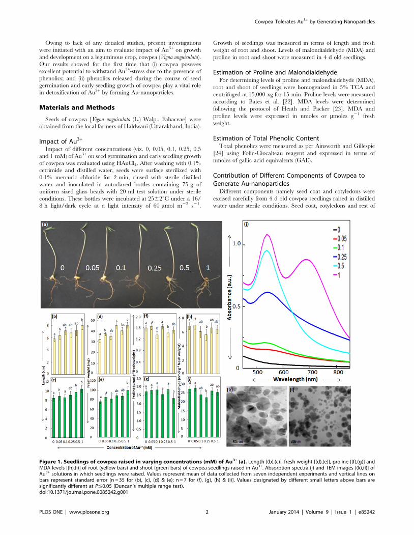
Figure 1. Seedlings of cowpea raised in varying concentrations (mM) of Au3+ (a). Length [(b),(c)], fresh weight [(d),(e)], proline [(f),(g)] andMDA levels [(h),(i)] of root (yellow bars) and shoot (green bars) of cowpea seedlings raised in Au3+. Absorption spectra (j) and TEM images [(k),(l)] ofAu3+ solutions in which seedlings were raised. Values represent mean of data collected from seven independent experiments and vertical lines onbars represent standard error [n = 35 for (b), (c), (d) & (e); n = 7 for (f), (g), (h) & (i)]. Values designated by different small letters above bars aresignificantly different at P#0.05 (Duncan’s multiple range test).doi:10.1371/journal.pone.0085242.g001
Cowpea Tolerates Au3+ by Generating Nanoparticles
PLOS ONE | www.plosone.org 2 January 2014 | Volume 9 | Issue 1 | e85242
the seedlings (i.e. seedlings devoid of cotyledons+seed coat) were
treated with 10 ml of different concentrations of sterile HAuCl4 for
24 h to test their potential to generate Au-nanoparticles. Seedlings
devoid of cotyledons+seed coat were treated by immersing their
roots in HAuCl4 solutions. Contol incubation medium (i.e. distilled
water), in which seedlings of cowpea were raised for 4 d, was also
tested for its efficacy to form Au-nanoparticles by incubating
500 ml of this medium with 10 ml HAuCl4.
Characterization of Au-nanoparticlesUV-Vis spectra of Au3+ solutions (i) in which seedlings of
cowpea were raised; and (ii) incubated independently with various
components of seedlings of cowpea and distilled water in which
seedlings were raised as detailed above, were recorded from 190 to
1100 nm using Specord 200 Analytikjena UV-Vis spectropho-
tometer. For transmission electron microscopic (TEM) studies,
10 ml of colloidal suspension was drop-coated on 200 mesh copper
grid with an ultrathin continuous carbon film and allowed to dry
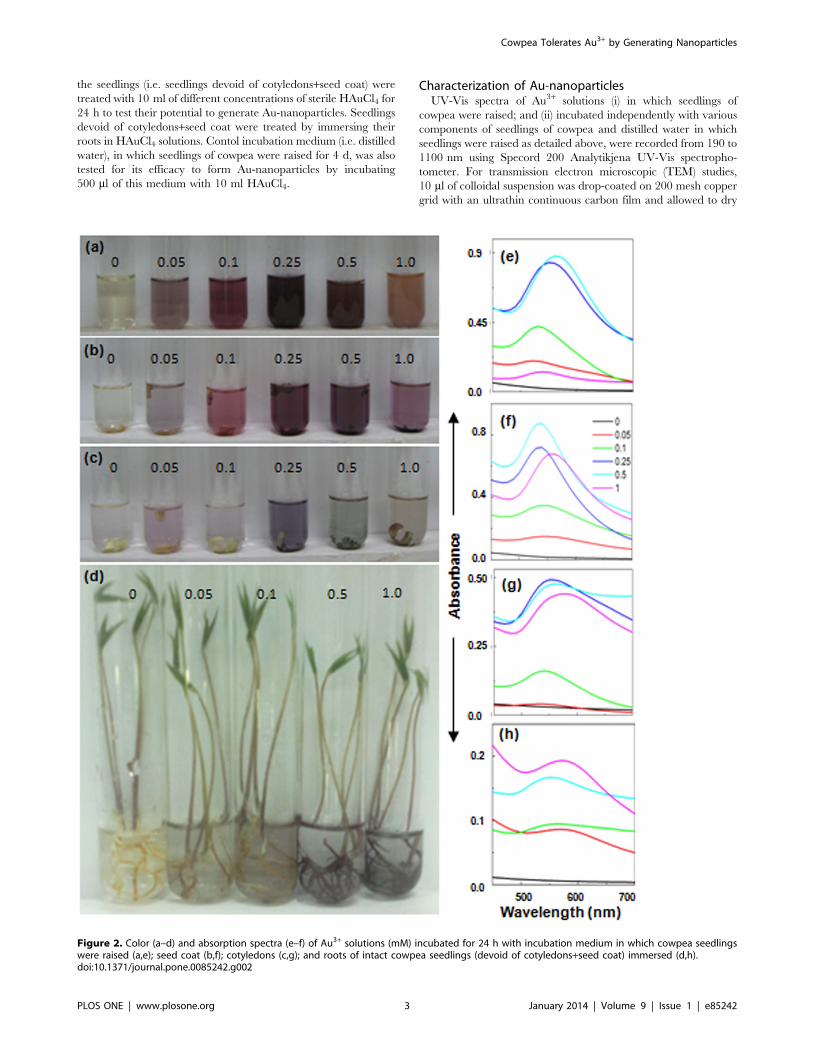
Figure 2. Color (a–d) and absorption spectra (e–f) of Au3+ solutions (mM) incubated for 24 h with incubation medium in which cowpea seedlingswere raised (a,e); seed coat (b,f); cotyledons (c,g); and roots of intact cowpea seedlings (devoid of cotyledons+seed coat) immersed (d,h).doi:10.1371/journal.pone.0085242.g002
Cowpea Tolerates Au3+ by Generating Nanoparticles
PLOS ONE | www.plosone.org 3 January 2014 | Volume 9 | Issue 1 | e85242
in a desiccator at room temperature. Grids were viewed in the
transmission electron microscope (Technai G2 T30) at a voltage of
300 kV. The hardware associated with the machine also allowed
(i) energy dispersive X-ray (EDX) analysis to measure the
elemental composition; and (ii) selected area electron diffraction
(SAED) analysis to determine crystalline/amorphous nature, of
nanoparticles.
Powder X-ray Diffraction StudiesFor powder X-ray diffraction (PXRD) studies, colloidal
suspensions obtained by incubating different components with
Au3+ solutions were centrifuged. The pellet obtained was re-
suspended in distilled water, drop coated on silica surface, dried in
desiccator and used for collecting PXRD pattern using Rigaku
Rotaflex RAD-B with copper target CuK(a)1 radiation with tube
voltage 40 kV and 60 mA in 2 theta (h) range of 30–80u.
Stasistical AnalysisAll experiments were carried out independently at least seven
times. Duncan’s multiple range test was used to to determine the
level of significance in physiological and biochemical data [25].
Results and Discussion
Impact of Au3+ on Seedling GrowthWhile evaluating impact of Au-nanoparticles on seed germina-
tion and early seedling growth of cowpea, HAuCl4 was used as
control. To our surprise, in spite of being a heavy metal ion, Au3+
was not associated with any significant alteration in seedling
growth (measured in terms of length and fresh weight of root and
shoot) of cowpea even when present at a concentration of 1 mM
(Figure 1). It is well documented that heavy metal ions such as
Cd+, Zn2+, Co2+ and Pb2+ inhibit plant growth and development
[26]. In general, crop plants exposed to heavy metal stress show
enhanced levels of stress markers, proline (an imino acid) and
MDA (a cytotoxic byproduct of lipid peroxidation), concomitant
with suppression in growth [27,28]. Heavy metal stress induced
enhancement in MDA levels is due to lipid peroxidation by
reactive oxygen species (ROS) that are generated due to
suppression in electron transport system and/or promotion of
Fenton’s reaction [29]. Increase in level of proline under heavy
metal stress is linked to (i) its synthesis to ensure appropriate
recycling of NAD(P)+ for cellular metabolism including its role as
terminal acceptor of light mediated photosynthetic electron
transport [29,30]; (ii) its role in scavenging ROS [31]; and (iii)
protection of enzymes and other macromolecular structures/
complexes [32]. Astonishingly, seedlings of cowpea raised in Au3+,
even at concentration as high as 1 mM, did not show any
enhancement in levels of proline and MDA, which is in
synchronization with unaltered growth (Figure 1). These findings
depicted that cowpea possessesremarkable potential to tolerate
Au3+ by some unique mechanism.
Unexpectedly, clear pale yellow Au3+ solutions in which
seedlings of cowpea were raised, turned colloidal purple. Such
an alteration in color of Au3+ solution (from clear pale yellow to
colloidal purple) is due to generation of Au-nanoparticles [33,34].
Absorption spectra of these colloidal purple suspensions showed
distinct peak at ,550 nm which is well documented to arise due to
surface plasmon resonance of Au-nanoparticles [33,34]. In
general, intensity of the purple color and Au-nanoparticle specific
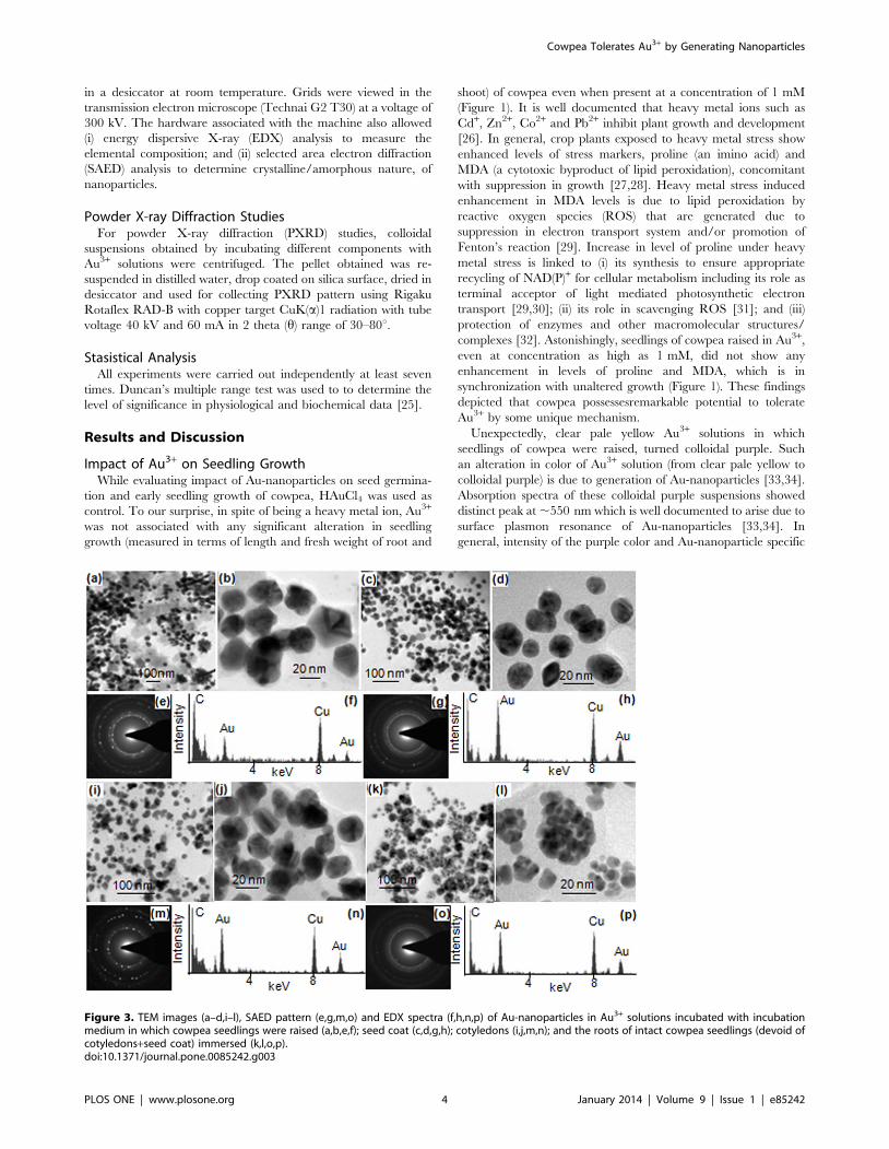
Figure 3. TEM images (a–d,i–l), SAED pattern (e,g,m,o) and EDX spectra (f,h,n,p) of Au-nanoparticles in Au3+ solutions incubated with incubationmedium in which cowpea seedlings were raised (a,b,e,f); seed coat (c,d,g,h); cotyledons (i,j,m,n); and the roots of intact cowpea seedlings (devoid ofcotyledons+seed coat) immersed (k,l,o,p).doi:10.1371/journal.pone.0085242.g003
Cowpea Tolerates Au3+ by Generating Nanoparticles
PLOS ONE | www.plosone.org 4 January 2014 | Volume 9 | Issue 1 | e85242
absorbance peak of these suspensions increased with increase in
concentration of Au3+ (Figure 1). Occasionally, an additional peak
in infra red region was recorded in absorption spectra of 1 mM
Au3+ solution in which seedlings of cowpea were raised. At
present, it is difficult to presume reasons behind the appearance of
this IR peak in only 1 mM Au3+ and not other concentrations of
Au3+ in which seedlings of cowpea were raised. Transmission
electron microscopic investigations confirmed presence of distinct
crystalline nanoparticles in range of 20–50 nm in these purple
colloidal suspensions (Figure 1). It is known that ionic speciation
state of metals is more toxic compared to nanoparticle speciation
state [35]. Therefore, we hypothesize that the potential of cowpea
to withstand Au3+-stress is linked to its inbuilt potential to generate
Au-nanoparticles.
Hypothesizing the Involvemenmt of Phenolics inImparting Au3+-Stress Tolerance
Control incubation medium (i.e. distilled water) turned brown
during the course of seed germination and early seedling growth. It
is known that germinating legume seeds release phenolics [36],
which impart brown coloration to the incubation medium.
Interestingly, each seedling of cowpea released ,35 nmoles
GAE of phenolics during the course of seed germination and early
seedling growth. It is well documented that phenolics such as gallic
acid, catechin promote generation of Au-nanoparticles [37,38].
This prompted us to believe that phenolics released by germinat-
ing seeds of cowpea could be responsible for generation of Au-
nanoparticles. Owing to electron donating capacity of phenolics
[21], we believe that phenolics released in large quantities during
seed germination and early seedling growth must have reduced
Au3+ and promoted formation of Au-nanoparticles. This seems to
be an ideal Au3+-tolerance mechanism exhibited by cowpea,
wherein toxic Au3+ is converted to less/non-toxic Au-nanoparti-
cles by phenolics released during seed germination and early
seedling growth.
Identifying the Key Component(s) Involved in Generationof Au-nanoparticles
To identify the key component(s) responsible for formation of
Au-nanoparticles and imparting Au3+-stress tolerance to cowpea
seedlings, four different components namely (i) control incubation
medium (i.e. distilled water) in which seedlings were raised; (ii) seed
coat; (iii) cotyledons; and (iv) seedlings devoid of cotyledons+seed
coat, were tested for their efficacy to generate Au-nanoparticles by
independently incubating them in different levels of Au3+. As
anticipated, brown colored incubation medium possessed abun-
dant potential to turn pale yellow Au3+ solutions purple indicating
the generation of Au-nanoparticles (Figure 2). Of the three
components of 4 d old seedlings (viz. seed coat, cotyledons and
seedlings devoid of cotyledons+seed coat), seed coat possessed
maximum potential to turn pale yellow Au3+ solutions purple
(Figure 2). Accordingly, Au-nanoparticle specific plasmon reso-
nance band in absorprion spectra of purple suspensions formed by
(i) incubation medium in which seedlings were raised and (ii) seed
coat, was more intense compared to those formed by cotyledons
and seedlings devoid of cotyledons+seed coat.
Transmission electron microscopic investigations confirmed
presence of nanoparticles in purple colloidal suspensions formed
independently by all four components when incubated with Au3+
solutions. However, the size and morphology of nanoparticles
varied depending on the component. Seed coat and incubation
medium generated nanoparticles in range of 10–30 nm, while
cotyledons and seedlings devoid of cotyledons+seed coat generated
nanoparticles in the range of 10–25 and 5–10 nm, repectively
(Figure 3). These results indicated that the mechanism of
generation of nanoparticles in the former two cases is similar,
but, vary distinctly from the latter two cases. However, irrespective
of the component responsible for generation of Au-nanoparticles,
EDX spectra showed two prominent peaks of Au confirming that
these nanoparticles were composed of Au (Figure 3). The
prominent peaks of Cu and C seen in these EDX spectra arose
from carbon coated copper grids, on which samples were loaded.
Irrespective of the component responsible for generation of Au-
nanoparticles, the nanoparticles were crystalline as revealed by
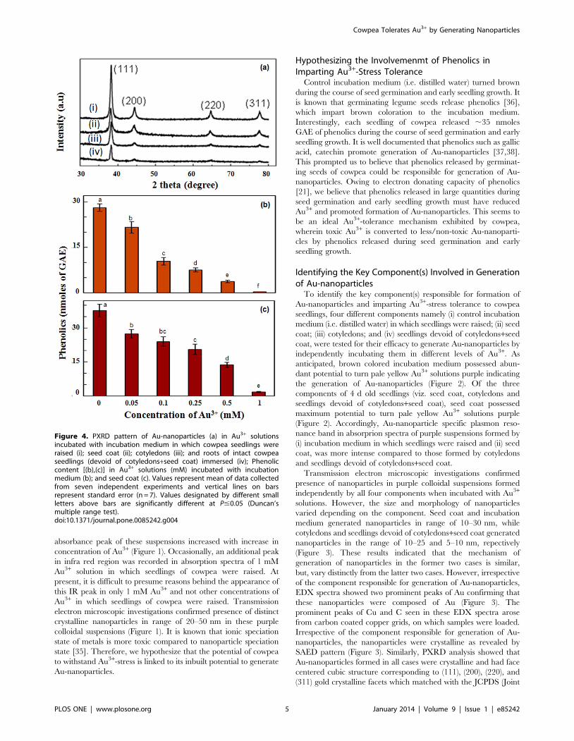
SAED pattern (Figure 3). Similarly, PXRD analysis showed that
Au-nanoparticles formed in all cases were crystalline and had face
centered cubic structure corresponding to (111), (200), (220), and
(311) gold crystalline facets which matched with the JCPDS (Joint
Figure 4. PXRD pattern of Au-nanoparticles (a) in Au3+ solutionsincubated with incubation medium in which cowpea seedlings wereraised (i); seed coat (ii); cotyledons (iii); and roots of intact cowpeaseedlings (devoid of cotyledons+seed coat) immersed (iv); Phenoliccontent [(b),(c)] in Au3+ solutions (mM) incubated with incubationmedium (b); and seed coat (c). Values represent mean of data collectedfrom seven independent experiments and vertical lines on barsrepresent standard error (n = 7). Values designated by different smallletters above bars are significantly different at P#0.05 (Duncan’smultiple range test).doi:10.1371/journal.pone.0085242.g004
Cowpea Tolerates Au3+ by Generating Nanoparticles
PLOS ONE | www.plosone.org 5 January 2014 | Volume 9 | Issue 1 | e85242
Committee on Powder Diffraction Studies) File No. 04–0784
(Figure 4).
Establishing Role of Phenolics in Imparting Au3+-stressTolerance
Interestingly, as evident from figure 2d, seedlings devoid of
cotyledons+seed coat showed significant suppression in growth
upon exposure to Au3+ solutions at concentration above 0.1 mM.
This is in contrast to the unperturbed growth response shown by
seedlings raised in presence of different levels of Au3+ (Figure 1)
and seedlings with cotyledons+seed coat incubated in different
levels of Au3+ (data not shown). Potential of cowpea seedlings with
seed coat to generate 5–6 fold higher level of Au-nanoparticles
compared to the seedlings devoid of seed coat is evident from the
intensity of Au-nanoparticle specific absorption peak of purple
colloidal suspensions formed by them (Figures 1 and 2). This
clearly established that seed coat, which is the key source of
phenolics play a pivotal role in imparting Au3+ tolerance to
cowpea.
Close correlation between potential of seed coat to release large
quantity of phenolics with its potential to generate large
proportion of Au-nanoparticles, as evident from figure 2, strength-
ens our hypothesis that phenolics released during seed germination
play a vital role in imparting Au3+-stress tolerance through
formation of Au-nanopartoicles. This hypothesis was further
strengthened by the potential of control incubation medium
(distilled water in which seeds were germinated and seedlings
raised) that contained large quantity of phenolics to generate large
proportion of Au-nanoparticles (Figure 2).
To establish direct role of phenolics in generating Au-
nanoparticles, the level of phenolics in Au3+ solutions incubated
with seed coat and incubation medium (in which seeds were
germinated and seedlings raised) were determined. As evident
from figure 4, level of phenolics decreased progressively with
increase in concentration of Au3+. This clearly established that
phenolics played a critical role in reduction of Au3+ and generation
of Au-nanoparticles. As stated earlier, phenolics possess potential
to donate electrons to metal ions such as Au3+ and promote
synthesis of Au-nanoparticles.
In nutshell, our results convincingly demonstrated that seed
coat, being key source of phenolics, is the most powerful
component of developing seedling responsible for generation of
Au-nanoparticles. In light of above elaborated experimental
evidences, we believe that seed coat plays most vital role in
imparting Au3+-stress tolerance to cowpea. Earlier researchers had
reported that seed coat plays important role in chemical protection
from oxidative damage as it possesses phenolics which act as
antioxidants [39,40]. Although, we do not rule out the possible
role of phenolics in acting as antioxidants as has been established
by these researchers, our results convincingly demonstrated for the
first time that phenolics released by seed coat impart Au3+-stress
tolerance by rapidly converting toxic ionic speciation state into
less/non-toxic nanoparticle speciation state.
Conclusions
Our above findings demonstrated for the first time that cowpea
has substantial potential to withstand Au3+-stress during seed
germination and early seedling growth. Our results established
that (i) the potential of cowpea to withstand Au3+-stress is linked to
phenolics; (ii) seed coat is the key source of phenolics during seed
germination and early seedling growth and hence is important for
imparting Au3+-stress tolerance; and (iii) phenolics impart Au3+-
stress tolerance by converting toxic ionic speciation state of Au to
less/non-toxic nanoparticle speciation state. Owing to the proven
role of phenolics in imparting metal ion stresss tolerance, we
believe that it is important to understand molecular and genetic
basis of modulating the synthesis of phenolics for improving
tolerance of agricultural/forestry plant species against metal ion
stress. Our findings also furnish a novel, simple, green and
economically viable protocol for using seed/seed coat of legume
seeds for synthesis of metal nanoparticles.
Acknowledgments
Support rendered by University Science Instrumentation Facility,
University of Delhi is duly acknowledged. We are thankful to Mr. Rahul
Bhardwaj for providing assistance during TEM analysis and Mr. Harsh
Kumar for PXRD studies.
Author Contributions
Conceived and designed the experiments: PS PPS. Performed the
experiments: NS PPS PS. Analyzed the data: NS PPS PS. Contributed
reagents/materials/analysis tools: PS PPS. Wrote the paper: NS PPS PS.
References
1. Ishikawa S, Suyama K, Arihara K, Itoh M (2002) Uptake and recovery of gold
ions from electroplating wastes by using egg shell membrane. Biores Technol 81:
201–206.
2. Baba AA, Adekola FA, Ojutemieden DO, Dada FK (2011) Solvent extraction of
gold from hydrochloric acid-leached Nigerian gold ore by tributylphosphate.
Chem Bull 1: 1–9.
3. Syed S (2012) Recovery of gold from secondary sources-A review. Hydromet-
allurgy 115–116: 30–51.
4. Ainsworth SK, Swain RP, Watabe N, Brackett NC Jr, Pilia P, et al. (1981) Gold
nephropathy, ultrastructural fluorescent and energy-dispersive x-ray microanal-
ysis study. Arch Pathol Lab Med 105: 73–78.
5. Lengke MF, Southam G (2005) The effect of thiosulfate-oxidizing bacteria on
the stability of the gold-thiosulfate complex. Geochim Cosmochim Acta 69:
3759–3772.
6. Danscher G, Stoltenberg M (2006) Silver enhancement of quantum dots
resulting from (1) metabolism of toxic metals in animals and humans, (2) in vivo,
in vitro and immersion created zinc–sulphur/zinc–selenium nanocrystals, (3)
metal ions liberated from metal implants and particles. Prog Histochem
Cytochem 41: 57–139.
7. Anderson CWN, Brooks RR, Stewart RB, Simcock R (1998) Harvesting a crop
of gold in plants. Nature 395: 553–554.
8. Beattie IR, Haverkamp RG (2011) Silver and gold nanoparticles in plants: sites
for the reduction to metal. Metallomics 3: 628–632.
9. Sharma NC, Sahi SV, Nath S, Parsons JG, Gardea-Torresday JL, et al. (2007)
Synthesis of plant-mediated gold nanoparticles and catalytic role of biomatrix-
embedded nanomaterials. Environ Sci Technol 41: 5137–5142.
10. Bali R, Harris AT (2010) Biogenic synthesis of Au nanoparticles using vascular
plants. Ind Eng Chem Res 49: 12762–12772.
11. Maestri E, Marmiroli M, Visioli G, Marmiroli N (2010) Metal tolerance and
hyperaccumulation: Costs and trade-offs between traits and environment.
Environ Expt Bot 68: 1–13.
12. Neilands JB (1995) Siderophores: structure and function of microbial iron
transport compounds. J Biol Chem 270: 26723–26726.
13. Ma JF, Ryan PR, Delhaize E (2001) Aluminium tolerance in plants and the
complexing role of organic acids. Trends Plant Sci 6: 273–271.
14. Wenzl P, Patino GM, Chaves AL, Mayer JE, Rao IM (2001) The high level of
aluminum resistance in signalgrass is not associated with known mechanisms of
external aluminum detoxification in root apices. Plant Physiol 125: 1473–1484.
15. Bringezu K, Litchenberger O, Leopold I, Neumann D (1999) Heavy metal
tolerance of Silene vulgaris. J Plant Physiol 154: 536–546.
16. Migocka M, Papiernia A, Kosatka E, Klobus G (2011) Comparative study of the
active cadmium efflux systems operating at the plasma membrane and tonoplast
of cucumber root cells. J Exp Bot 62: 4903–4916.
17. Cobbett C, Goldsbrough PB (2002) Phytochelatins and metallothioneins: roles in
heavy metal detoxification and homeostasis. Annu Rev Plant Biol 53: 159–182.
18. Lavid N, Schwartz A, Yarden O, Tel-Or E (2001) The involvement of
polyphenols and peroxidase activities in epidermal glands of water lily
(Nymphaeceae). Planta 212: 323–333.
Cowpea Tolerates Au3+ by Generating Nanoparticles
PLOS ONE | www.plosone.org 6 January 2014 | Volume 9 | Issue 1 | e85242
19. Jung C, Maeder V, Funk F, Frey B, Sticher H, et al. (2003) Release of phenols
from Lupinus albus L. roots exposed to Cu and their possible role in Cudetoxification. Plant Soil 252: 301–312.
20. Michalak A (2006) Phenolic compounds and their antioxidant activity in plants
growing under heavy metal stress. Polish J Environ Stud 15: 523–530.21. Rice-Evans CA, Miller NJ, Paganga G (1996) Structures-antioxidant activity
relationships of flavonoids and phenolic acids. Free Rad Biol Med 20: 933–956.22. Bates LS, Walderen RD, Taere ID (1973) Rapid determination of free proline
for water stress studies. Plant Soil 39: 205–207.
23. Heath RL, Packer L (1968) Photoperoxidation in isolated chloroplast. I. Kineticsand stoichiometry of fatty acid peroxidation. Arch Biochem Biophys 125: 189–
198.24. Ainsworth EA, Gillespie KM (2007) Estimation of total phenolic content and
other oxidation substances in plant tissues using Folin- Ciocalteu reagent. NatProtoc 2: 875–877.
25. Duncan DB (1955) Multiple range and multiple F tests. Biometrics 39: 205–207.
26. Alia, Pardha Saradhi P (1991) Proline accumulation under heavy metal stress.J Plant Physiol 13: 554–558.
27. Alia, Prasad KVSK, Pardha Saradhi P (1995) Effect of zinc on free radicals andproline in Brassica and Cajanus. Phytochemistry 39: 45–47.
28. Pardha Saradhi P, Alia, Arora S, Prasad KVSK (1995) Proline accumulates in
plants exposed to UV radiation and protect them against UV inducedperoxidation. Biochem Biophys Res Com 209: 1–5.
29. Alia, Pardha Saradhi P (1993) Suppression in mitochondrial electron transport isthe prime cause behind stress induced proline accumulation. Biochem Biophys
Res Com 193: 54–58.30. Sharmila P, Anwar F, Sharma KR, Pardha Saradhi P (2008) Management of
abiotic stresses in grain legumes through manipulation of genes for compatible
solutes. In: Kirti PB (ed) Handbook of New Technologies for GeneticImprovement of Legumes, CRC Press, USA, pp 577–603.
31. Alia, Pardha Saradhi P, Mohanty P (1997) Involvement of proline in protecting
thylakoid membranes against free radical-induced photodamage. J Photochem
Photobiol B: Biology 38:253–257.
32. Sivakumar P, Sharmila P, Pardha Saradhi P (1998) Proline suppresses Rubisco
activity in higher plants. Biochem Biophys Res Commun. 252: 428–432.
33. Shabnam N, Pardha-Saradhi P (2013) Photosynthetic Electron Transport
System Promotes Synthesis of Au-Nanoparticles. PLoS ONE 8(8): e71123.
34. Yamal G, Sharmila P, Rao KS, Pardha-Saradhi P (2013) Yeast Extract
Mannitol medium and its constituents promote synthesis of Au nanoparticles.
Process Biochem 48: 532–538.
35. Klaus-Joerger T, Joerger R, Olsson E, Granqvist C-G (2001) Bacteria as workers
in the living factory: metal-accumulating bacteria and their potential for
materials science. Trends Biotechnol 9: 15–20.
36. Bekkara F, Jay M, Viricel MR, Rome S (1998) Distribution of phenolic
compounds within seed and seedlings of two Vicia faba cvs differing in their seed
tannin content, and study of their seed and root phenolic exudations. Plant Soil
203: 27–36.
37. Wang W, Chen Q, Jiang C, Yang D, Liu X, et al. (2007) One-step synthesis of
biocompatible gold nanoparticles using gallic acid in the presence of poly-(N-
vinyl-2-pyrrolidone). Coll. Surf. Physicochem Eng Asp 301: 73–79.
38. Nune SK, Chanda N, Shukla R, Katti K, Kulkarni RR, et al. (2009) Green
nanotechnology from tea: phytochemicals in tea as building blocks for
production of biocompatible gold nanoparticles. J Mat Chem 19: 2912–2920.
39. Duenas M, Hernandez T, Estrella I (2006) Assessment of in vitro antioxidant
capacity and seed coat and the cotyledon of legumes in relation to their phenolic
contents. Food Chem 98: 95–103.
40. Siddhuraju P, Becker K (2007) The antioxidant and free radical scavenging
activities of processed cowpea (Vigna unguiculata (L.) Walp) seed extracts. Food
Chem 101: 10–19.
Cowpea Tolerates Au3+ by Generating Nanoparticles
PLOS ONE | www.plosone.org 7 January 2014 | Volume 9 | Issue 1 | e85242


![Cowpea (Vigna unguiculata [L.] Walp.) genotypes response to multiple abiotic stresses](https://img.dokumen.tips/doc/110x75/63442c5a03a48733920ac48a/cowpea-vigna-unguiculata-l-walp-genotypes-response-to-multiple-abiotic-stresses.jpg)


