Embed Size (px)
Citation preview

421
Jun Hirabayashi (ed.), Lectins: Methods and Protocols, Methods in Molecular Biology, vol. 1200,DOI 10.1007/978-1-4939-1292-6_37, © Springer Science+Business Media New York 2014
Chapter 37
Perspectives in Glycomics and Lectin Engineering
Jan Tkac , Tomas Bertok , Jozef Nahalka , and Peter Gemeiner
Abstract
This chapter would like to provide a short survey of the most promising concepts applied recently in analysis of glycoproteins based on lectins. The fi rst part describes the most exciting analytical approaches used in the fi eld of glycoprofi ling based on integration of nanoparticles, nanowires, nanotubes, or nanochannels or using novel transducing platforms allowing to detect very low levels of glycoproteins in a label-free mode of operation. The second part describes application of recombinant lectins containing several tags applied for oriented and ordered immobilization of lectins. Besides already established concepts of glyco-profi ling several novel aspects, which we think will be taken into account for future, more robust glycan analysis, are described including modifi ed lectins, peptide lectin aptamers, and DNA aptamers with lectin- like specifi city introduced by modifi ed nucleotides. The last part of the chapter describes a novel concept of a glycocodon, which can lead to a better understanding of glycan–lectin interaction and for design of novel lectins with unknown specifi cities and/or better affi nities toward glycan target or for rational design of peptide lectin aptamers or DNA aptamers.
Key words Biosensors , Glycomics , Lectins , Nanoparticles , DNA aptamers , Lectin peptide aptamers , Recombinant lectins
1 Introduction
Since the introduction of DNA biochips in 1995 [ 1 ], the technology has been intensively applied in assays of genome-wide expression to seek information about possible functions of novel or poorly characterized genes [ 2 ] and for diagnostic purposes, as well [ 3 ]. Even though DNA microarray technology has shed light on many physiological functions of genes by determination of expression of gene clusters, there is quite often only a very low correlation between RNA and protein abundance detected in single-cell organisms [ 4 ] and in higher ones, including humans [ 5 ]. Since quantitative analysis of proteins is central to proteomics with a focus on design of novel drugs, diagnostics of diseases, and their therapeutic applications, protein microarrays were successfully launched to address these issues [ 6 ].

422
Analysis of fi nely tuned posttranslational modifi cations (PTMs) of proteins is an additional challenge for current analytical technol-ogy. Glycosylation is a highly abundant form of PTM of proteins and it is estimated that 70–80 % of human proteins are glycosylated [ 7 ]. Importance of glycans can be further highlighted by the fact that 70 % of all therapeutic proteins are glycosylated [ 8 ]. Glycan- mediated recognition plays an important role in many different cell’s processes such as fertilization, immune response, differentiation of cells, cell–matrix interaction, cell–cell adhesion, etc. [ 9 , 10 ]. Glycans present on the surface of cells are naturally involved in pathological processes including viral and bacterial infections, in neurological disorder and in tumor growth and metastasis [ 3 , 11 – 16 ]. Thus, better understanding of glycan-mediated pathogenesis is essential in order to establish a “policy” to develop effi cient routes for disease treatment with several recent studies as good examples, e.g., “neutralization” of various forms of viruses [ 17 , 18 ] or more effi cient vaccines against various diseases [ 19 , 20 ]. A changed glycosylation on a protein backbone can be effectively applied in early stage diagnostics of several diseases, including different forms of cancer with known glycan-based biomarkers [ 21 – 23 ]. Moreover, many previously established and even commercially successful strategies used to treat diseases are currently being revisited in light of glycan recognition in order to lower side effects, enhance serum half-life, or decrease cellular toxicity [ 3 , 24 , 25 ]. Recently, the fi rst glyco-engineered antibody was approved to the market, which was called by the authors “a triumph for glyco- engineering” [ 26 ].
Glycomics focuses on revealing fi nely tuned reading mechanisms in the cell orchestra based on graded affi nity, avidity, and multiva-lency of glycans (i.e., sugar chains covalently attached to proteins and lipids) [ 27 ]. Glycans are information-rich molecules applicable in coding tools of the cell since they can form enormous number of possible unique sequences from basic building units [ 28 ]. It is estimated that the size of the cellular glycome can be up to 500,000 glycan modifi ed biomolecules (proteins and lipids) formed from 7,000 unique glycan sequences [ 29 ]. Thus, it is not a surprise the glycome is sometimes referred to as the “third alphabet” in biology, after genetics and proteomics [ 30 ]. A huge glycan variation can explain human complexity in light of a paradoxically small genome. This glycan complexity together with similar physicochemical properties of glycans is the main reason why the progress in the fi eld of glycomics has been behind advances in genomics and proteomics [ 31 ].
Traditional glycoprofi ling protocols rely on glycan release from a biomolecule with a subsequent quantifi cation by an array of tech-niques including capillary electrophoresis, liquid chromatography, and mass spectrometry [ 30 , 32 – 37 ]. There is an alternative way for glycoprofi ling by application of lectins, natural glycan recognizing proteins [ 28 , 38 , 39 ] in combination with various transducing pro-tocols [ 12 , 40 , 41 ]. The most powerful glycoprofi ling tool relies
Jan Tkac et al.

423
on lectins arrayed on solid surfaces for direct analysis of glycoproteins, glycolipids, membranes, and even glycans on the surface of intact cells [ 11 , 12 , 42 , 43 ]. Even though lectin microarrays offer high-throughput assay protocols with a minute consumption of samples and reagents, there are some drawbacks such as a need to fl uores-cently label the sample or the lectin, which negatively affects the performance of detection [ 11 , 12 ], relatively high detection limits, and quite narrow working concentration ranges. Thus, the ideal detection platform should be based on protocols without a need to label a glycoprotein or a lectin, in a way similar to natural processes occurring within a cell [ 30 ].
Lectins (lat. legere = to choose) are proteins able to recognize and reversibly bind to free or bound mono- and oligosaccharides [ 44 ]. They are not usually catalytically active, do not participate in the immune response of higher organisms, and can be found in viruses, bacteria, fungi, plants, and animals. They are therefore a relatively heterogeneous group of oligomeric proteins belonging to distinct families with similar sequences and are considered as natural glycocode decipherers [ 28 ]. Lectins, unlike antibodies, have a low specifi city and affi nity with K d ranged from 10 −3 to 10 −7 M [ 30 ] and lectins with a new specifi city cannot be raised in a way similar to antibodies.
In the following sections we will focus on ways how to improve glycan detection either by application of novel nanoscale- controlled patterning protocols, nanoengineered devices or by application of novel, recombinant lectins, lectin-like aptamers, or lectin peptide aptamers. The fi nal part of this book chapter will focus on a com-pletely novel area in the glycomics—the idea of a glycocodon.
2 Perspectives of Novel Formats of Analysis Applicable in Glycomics
The use of nanotechnology, sophisticated nanoscale patterning protocols, and advanced detection platforms can help to overcome the drawbacks of lectin microarray technology allowing it to work in a label-free mode of operation, with a high sensitivity, low detection limits, a wide concentration window and in some cases, real- time analysis of a binding event is possible [ 9 , 30 , 45 – 51 ]. These devices can differ in their mode of signal transduction compared to tradi-tional methods and will be divided into three categories according to their mode of action. Various traditional analytical techniques based on lectins (i.e., surface plasmon resonance and quartz crystal microbalance) are covered by different chapters accompanied this one within this book and are not discussed here.
Microcantilever biochips offer a novel approach for detection of a molecular binding based on a change in mass accumulated on the surface of a cantilever during biorecognition. It is a label-free tech-nique allowing to monitor biospecifi c interaction in a real time,
2.1 Mechanical Platforms
Perspectives in Glycomics and Lectin Engineering
424
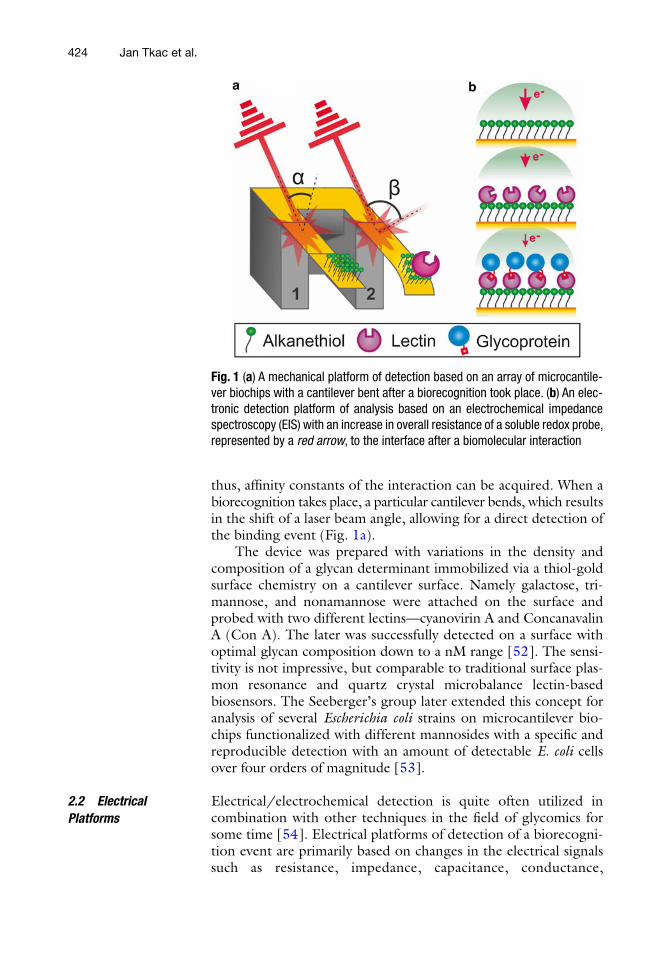
thus, affi nity constants of the interaction can be acquired. When a biorecognition takes place, a particular cantilever bends, which results in the shift of a laser beam angle, allowing for a direct detection of the binding event ( Fig. 1a ).
The device was prepared with variations in the density and composition of a glycan determinant immobilized via a thiol-gold surface chemistry on a cantilever surface. Namely galactose, tri-mannose, and nonamannose were attached on the surface and probed with two different lectins—cyanovirin A and Concanavalin A (Con A). The later was successfully detected on a surface with optimal glycan composition down to a nM range [ 52 ]. The sensi-tivity is not impressive, but comparable to traditional surface plas-mon resonance and quartz crystal microbalance lectin-based biosensors. The Seeberger’s group later extended this concept for analysis of several Escherichia coli strains on microcantilever bio-chips functionalized with different mannosides with a specifi c and reproducible detection with an amount of detectable E. coli cells over four orders of magnitude [ 53 ].
Electrical/electrochemical detection is quite often utilized in combination with other techniques in the fi eld of glycomics for some time [ 54 ]. Electrical platforms of detection of a biorecogni-tion event are primarily based on changes in the electrical signals such as resistance, impedance, capacitance, conductance,
2.2 Electrical Platforms
Fig. 1 ( a ) A mechanical platform of detection based on an array of microcantile-ver biochips with a cantilever bent after a biorecognition took place. ( b ) An elec-tronic detection platform of analysis based on an electrochemical impedance spectroscopy (EIS) with an increase in overall resistance of a soluble redox probe, represented by a red arrow , to the interface after a biomolecular interaction
Jan Tkac et al.
425
potential, and current [ 55 ]. These analytical techniques are usually nondestructive and extremely sensitive, offering quite a wide con-centration working range with a possibility to work in an array format of analysis [ 30 ].
The most frequently used label-free electrochemical technique is EIS, which is based on an electric perturbation of a thin layer on the conductive surface by small alternating current amplitude with ability to provide characteristics of this interface utilizable in sensing. EIS results are typically transformed into a complex plane Nyquist plot vectors, which by application of an equivalent circuit can provide information about electron transfer resistance of a sol-uble redox probe in a direct way (Fig. 1b ). When a biorecognition took place, an electrode interface is modifi ed and a subtle change in interfacial layer characteristics can be used for detection. EIS investigation is most frequently performed in the presence of a redox probe with detection of a change of resistance of the inter-face used for a signal generation. EIS is extensively used as a non-destructive technique for reliable analysis of surface conditions and allows complex biorecognition events to be probed in a simple, sensitive, and label-free manner and is being increasingly popular to develop electrochemical lectin-based biosensors for glycan determination [ 9 , 30 ].
Initial efforts to detect glycoproteins by EIS were launched by the group of Prof. Joshi with sialic acid binding Sambucus nigra agglutinin (SNA) and a galactose binding peanut agglutinin cova-lently immobilized on printed circuit board electrodes [ 56 ]. The assays were really quick with a response time of 80 s and with sensi-tivity of glycoprotein detection down to 10 pg/mL (e.g., 150 fM), while using a cost-effective electrode material [ 56 ]. A group of Prof. Oliveira put a substantial effort to use lectin modifi ed surfaces with EIS detection for discrimination between healthy human sam-ples and samples from patients infected by a mosquito- borne Dengue virus (breakbone fever) with a high mortality rate [ 9 ]. Their device with two different lectins immobilized on gold nanoparticles offered a detection limit in the low nM range [ 57 ]. Another EIS-based biosensor was built on a surface of the silicon chip with an array of gold electrodes interfaced with nanoporous alumina membrane with high density of nanowells [ 58 ]. The bio-sensor offered a high reliability of assays and a good agreement with enzyme-linked lectin assays (ELLA). The detection limit of a biosen-sor for its analyte was fi ve orders of magnitude lower compared to ELLA (i.e., 20 fM vs. 4.6 nM). An assay time for the biosensor of 15 min was much shorter compared to 4 h needed for ELLA. Moreover, a minute amount of sample (10 μL) was suffi cient for the analysis by the biosensor [ 58 ].
In our recent work we focused on the development of ultra-sensitive impedimetric lectin biosensors with detection limits down
2.2.1 Electrochemical Impedance Spectroscopy (EIS)
Perspectives in Glycomics and Lectin Engineering

426
to a single-molecule level based on controlled architecture at the nanoscale [ 59 – 61 ]. In the fi rst study the biosensor was able to detect a glycoprotein in a concentration window spanning seven orders of magnitude with a detection limit for the glycoprotein down to 0.3 fM, which was the lowest glycoprotein concentration detected [ 59 ]. In the following study an incorporation of gold nanoparticles offered even lower and unprecedented detection limit of 0.5 aM with quite a wide dynamic concentration range covered [ 61 ]. In our last study the EIS-biosensors were constructed with three different lectins to be able to detect changes on immu-noglobulins with progression of a rheumatoid arthritis in humans. The biosensor with improved antifouling properties offered a detection limit in the fM range and worked properly even with 1,000× diluted human plasma. The biosensor performance was directly compared to the state-of-the-art glycoprofi ling tool based on fl uorescent lectin microarrays with a detection limit in the nM level [ 60 ]. Moreover, a sandwich confi guration offered a detection limit down to aM concentration [ 60 ]. A detection limit down to fM range for analysis of alpha-fetoprotein (a biomarker for hepato-cellular carcinoma) was recently observed on a device modifi ed by arrays of single-walled carbon nanotubes and wheat- germ aggluti-nin with EIS as a transducing mechanism [ 62 ].
In NTFETs, semiconducting nanotubes or nanowires act as a channel between two metal electrodes (source and drain) while the two electrodes are held at a constant bias voltage using a so-called gate electrode (Fig. 2a ) [ 30 , 49 ]. When the device with an
2.2.2 Nanotube Field Effect Transistor (NTFET) Sensors
Fig. 2 ( a ) A fi eld-effect transistor (FET) sensing based on a changed conductivity of a single-walled carbon nanotube (SWCNT) positioned in between a source and a drain. ( b ) An optical detection platform based on quenching of an intrinsic fl uorescence of a SWCNT by Ni-tether employed for lectin immobilization via His 6 tag. A fl uorescence of a SWCNT is partly restored after biorecognition since Ni-tether is pushed away from the SWCNT surface
Jan Tkac et al.
427
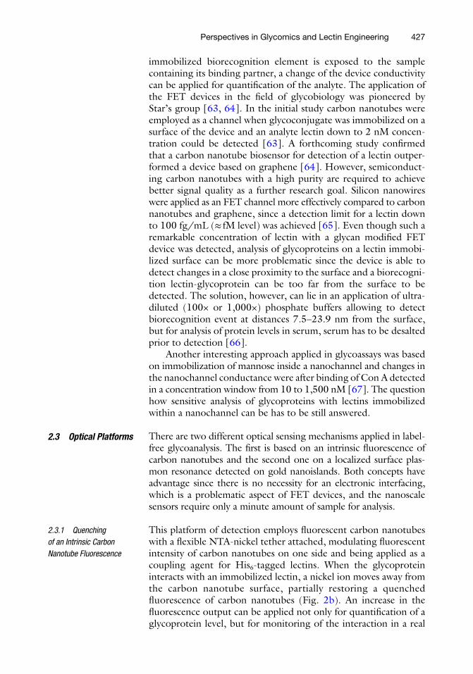
immobilized biorecognition element is exposed to the sample containing its binding partner, a change of the device conductivity can be applied for quantifi cation of the analyte. The application of the FET devices in the fi eld of glycobiology was pioneered by Star’s group [ 63 , 64 ]. In the initial study carbon nanotubes were employed as a channel when glycoconjugate was immobilized on a surface of the device and an analyte lectin down to 2 nM concen-tration could be detected [ 63 ]. A forthcoming study confi rmed that a carbon nanotube biosensor for detection of a lectin outper-formed a device based on graphene [ 64 ]. However, semiconduct-ing carbon nanotubes with a high purity are required to achieve better signal quality as a further research goal. Silicon nanowires were applied as an FET channel more effectively compared to carbon nanotubes and graphene, since a detection limit for a lectin down to 100 fg/mL (≈ fM level) was achieved [ 65 ]. Even though such a remarkable concentration of lectin with a glycan modifi ed FET device was detected, analysis of glycoproteins on a lectin immobi-lized surface can be more problematic since the device is able to detect changes in a close proximity to the surface and a biorecogni-tion lectin-glycoprotein can be too far from the surface to be detected. The solution, however, can lie in an application of ultra-diluted (100× or 1,000×) phosphate buffers allowing to detect biorecognition event at distances 7.5–23.9 nm from the surface, but for analysis of protein levels in serum, serum has to be desalted prior to detection [ 66 ].
Another interesting approach applied in glycoassays was based on immobilization of mannose inside a nanochannel and changes in the nanochannel conductance were after binding of Con A detected in a concentration window from 10 to 1,500 nM [ 67 ]. The question how sensitive analysis of glycoproteins with lectins immobilized within a nanochannel can be has to be still answered.
There are two different optical sensing mechanisms applied in label-free glycoanalysis. The fi rst is based on an intrinsic fl uorescence of carbon nanotubes and the second one on a localized surface plas-mon resonance detected on gold nanoislands. Both concepts have advantage since there is no necessity for an electronic interfacing, which is a problematic aspect of FET devices, and the nanoscale sensors require only a minute amount of sample for analysis.
This platform of detection employs fl uorescent carbon nanotubes with a fl exible NTA-nickel tether attached, modulating fl uorescent intensity of carbon nanotubes on one side and being applied as a coupling agent for His 6 -tagged lectins. When the glycoprotein interacts with an immobilized lectin, a nickel ion moves away from the carbon nanotube surface, partially restoring a quenched fl uorescence of carbon nanotubes (Fig. 2b ). An increase in the fl uorescence output can be applied not only for quantifi cation of a glycoprotein level, but for monitoring of the interaction in a real
2.3 Optical Platforms
2.3.1 Quenching of an Intrinsic Carbon Nanotube Fluorescence
Perspectives in Glycomics and Lectin Engineering
428
time, providing kinetic and affi nity constants, as well. The absolute detection limit of the device for the glycoprotein was not that impressive (2 μg, i.e., 670 nM), but authors believe the device has a room for improvement (i.e., by using high quality nanotube sensors) [ 47 , 68 ]. In a recent study authors extended this initial study for glycoprofi ling of different forms of IgGs [ 69 ].
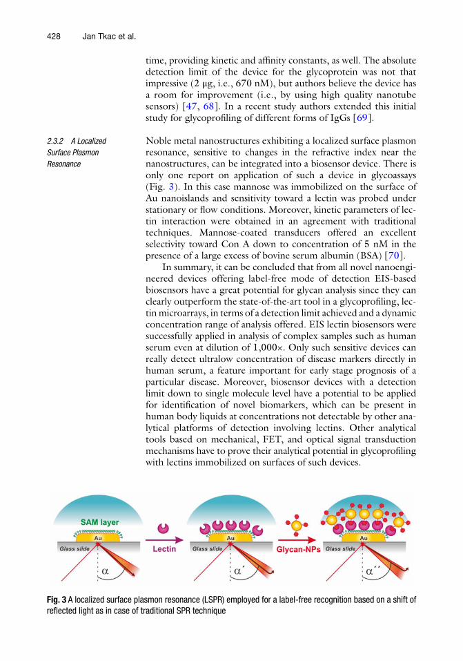
Noble metal nanostructures exhibiting a localized surface plasmon resonance, sensitive to changes in the refractive index near the nanostructures, can be integrated into a biosensor device. There is only one report on application of such a device in glycoassays (Fig. 3 ). In this case mannose was immobilized on the surface of Au nanoislands and sensitivity toward a lectin was probed under stationary or fl ow conditions. Moreover, kinetic parameters of lec-tin interaction were obtained in an agreement with traditional techniques. Mannose-coated transducers offered an excellent selectivity toward Con A down to concentration of 5 nM in the presence of a large excess of bovine serum albumin (BSA) [ 70 ].
In summary, it can be concluded that from all novel nanoengi-neered devices offering label-free mode of detection EIS-based biosensors have a great potential for glycan analysis since they can clearly outperform the state-of-the-art tool in a glycoprofi ling, lec-tin microarrays, in terms of a detection limit achieved and a dynamic concentration range of analysis offered. EIS lectin biosensors were successfully applied in analysis of complex samples such as human serum even at dilution of 1,000×. Only such sensitive devices can really detect ultralow concentration of disease markers directly in human serum, a feature important for early stage prognosis of a particular disease. Moreover, biosensor devices with a detection limit down to single molecule level have a potential to be applied for identifi cation of novel biomarkers, which can be present in human body liquids at concentrations not detectable by other ana-lytical platforms of detection involving lectins. Other analytical tools based on mechanical, FET, and optical signal transduction mechanisms have to prove their analytical potential in glycoprofi ling with lectins immobilized on surfaces of such devices.
2.3.2 A Localized Surface Plasmon Resonance
Fig. 3 A localized surface plasmon resonance (LSPR) employed for a label-free recognition based on a shift of refl ected light as in case of traditional SPR technique
Jan Tkac et al.

429
3 Perspectives in Lectin Engineering
The glycan binding sites of lectins are usually a shallow groove or a pocket present at the protein surface, or at the interface of oligo-mers [ 7 ]. Four main amino acids are part of an affi nity site including asparagine, aspartic acid, glycine (arginine in Con A), and an aro-matic residue for interaction with glycan via hydrogen bonds and hydrophobic interactions [ 71 ]. Ionic interactions are especially involved in recognition of negatively charged glycans containing sialic acids. Lectin-monosaccharide binding is relatively weak, which is why several approaches were applied to enhance practical utility of lectins in glycoprofi ling [ 7 ].
Recombinant DNA technology for producing lectins was tradi-tionally applied to establish primary structure; to study genetics, evolution, and biosynthesis; to elucidate the role of amino acids in recognition; to produce lectins with altered specifi city and/or affi n-ity; and to study their function in the organism of origin [ 72 ]. Novel trend is to apply this technology for producing lectins to be utilized for construction of various lectin-based biodevices. Recombinant lectin technology can signifi cantly reduce drawbacks of traditional lectin isolation such as a long processing time, often quite a low yield, and batch-to-batch variation of the product quality depending on the source, with presence of various contaminants or different lectin isoforms [ 11 , 73 ]. Moreover, recombinant technology offers to produce lectins either without any glycosylation, which can in many cases complicate glycoprofi ling, by expression in prokaryotic hosts and to introduce various tags (His 6 - tag, glutathione transfer-ase), which can be effectively utilized not only for one-step purifi ca-tion process, but more importantly for an oriented immobilization of lectins on various surfaces [ 38 ]. Although lectin peptide aptamers have not been produced yet, it is a question of time, when such arti-fi cial glycan binding proteins emerge as an effi cient tool in the area of glycobiology. It is estimated that another player in the area of glycoprofi ling will make a substantial fi ngerprint i.e. lectin aptamers based on expanded genetic alphabet by introduction of modifi ed nucleotides. Other concepts based on modifi ed lectins in glycopro-fi ling with added value are fi nally described.
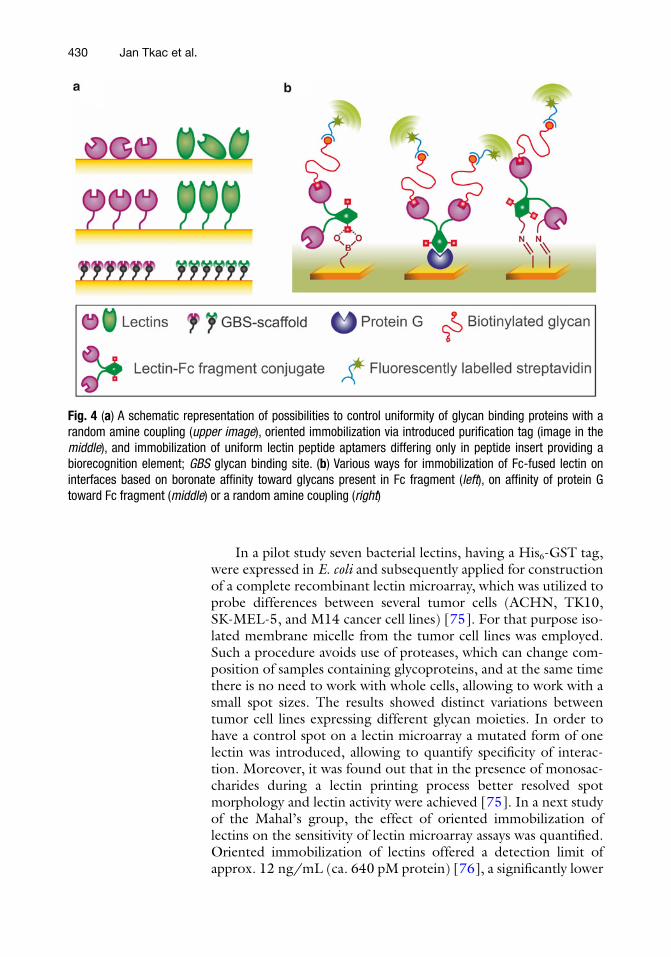
A controlled immobilization of lectins on a diverse range of surfaces can have a detrimental effect on the sensitivity of assays, since lectins can be attached in a way a biorecognition site is directly exposed to the solution phase for an effi cient biorecognition. As a result almost 100 % of immobilized lectin molecules can have a proper orientation with an increased chance for catching its analyte (Fig. 4a ). Moreover the presence of a linker, which attaches tag to the protein backbone, can signifi cantly lower possible interaction of the protein with the surface, which can eventually lead to a denaturation of a protein [ 74 ].
3.1 Oriented Immobilization of Recombinant Lectins
Perspectives in Glycomics and Lectin Engineering
430
In a pilot study seven bacterial lectins, having a His 6 -GST tag, were expressed in E. coli and subsequently applied for construction of a complete recombinant lectin microarray, which was utilized to probe differences between several tumor cells (ACHN, TK10, SK-MEL-5, and M14 cancer cell lines) [ 75 ]. For that purpose iso-lated membrane micelle from the tumor cell lines was employed. Such a procedure avoids use of proteases, which can change com-position of samples containing glycoproteins, and at the same time there is no need to work with whole cells, allowing to work with a small spot sizes. The results showed distinct variations between tumor cell lines expressing different glycan moieties. In order to have a control spot on a lectin microarray a mutated form of one lectin was introduced, allowing to quantify specifi city of interac-tion. Moreover, it was found out that in the presence of monosac-charides during a lectin printing process better resolved spot morphology and lectin activity were achieved [ 75 ]. In a next study of the Mahal’s group, the effect of oriented immobilization of lectins on the sensitivity of lectin microarray assays was quantifi ed. Oriented immobilization of lectins offered a detection limit of approx. 12 ng/mL (ca. 640 pM protein) [ 76 ], a signifi cantly lower
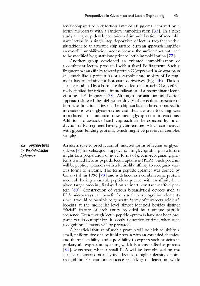
Fig. 4 ( a ) A schematic representation of possibilities to control uniformity of glycan binding proteins with a random amine coupling ( upper image ), oriented immobilization via introduced purifi cation tag (image in the middle ), and immobilization of uniform lectin peptide aptamers differing only in peptide insert providing a biorecognition element; GBS glycan binding site. ( b ) Various ways for immobilization of Fc-fused lectin on interfaces based on boronate affi nity toward glycans present in Fc fragment ( left ), on affi nity of protein G toward Fc fragment ( middle ) or a random amine coupling ( right )
Jan Tkac et al.
431
level compared to a detection limit of 10 μg/mL achieved on a lectin microarray with a random immobilization [ 33 ]. In a next study the group developed oriented immobilization of recombi-nant lectins in a single step deposition of lectins together with a glutathione to an activated chip surface. Such an approach simplifi es an overall immobilization process because the surface does not need to be modifi ed by glutathione prior to lectin immobilization [ 77 ].
Another group developed an oriented immobilization of recombinant lectins produced with a fused Fc-fragment. Such a fragment has an affi nity toward protein G (expressed in Streptococcus sp., much like a protein A) or a carbohydrate moiety of Fc frag-ment has an affi nity for boronate derivatives (Fig. 4b ). Thus, a surface modifi ed by a boronate derivatives or a protein G was effec-tively applied for oriented immobilization of a recombinant lectin via a fused Fc fragment [ 78 ]. Although boronate immobilization approach showed the highest sensitivity of detection, presence of boronate functionalities on the chip surface induced nonspecifi c interactions with glycoproteins and thus dextran blocking was introduced to minimize unwanted glycoprotein interactions. Additional drawback of such approach can be expected by intro-duction of Fc fragment having glycan entities, which can interact with glycan-binding proteins, which might be present in complex samples.
An alternative to production of mutated forms of lectins or glyco-sidases [ 7 ] for subsequent application in glycoprofi ling in a future might be a preparation of novel forms of glycan-recognizing pro-teins termed here as peptide lectin aptamers (PLA). Such proteins will be peptide aptamers with a lectin-like affi nity to recognize vari-ous forms of glycans. The term peptide aptamer was coined by Colas et al. in 1996 [ 79 ] and is defi ned as a combinatorial protein molecule having a variable peptide sequence, with an affi nity for a given target protein, displayed on an inert, constant scaffold pro-tein [ 80 ]. Construction of various bioanalytical devices such as PLA microarrays can benefi t from such biorecognition elements since it would be possible to generate “army of terracotta soldiers” looking at the molecular level almost identical besides distinct “facial” feature of each entity provided by a unique peptide sequence. Even though lectin peptide aptamers have not been pre-pared yet, in our opinion, it is only a question of time, when such recognition elements will be prepared.
A benefi cial feature of such a protein will be high solubility, a small, uniform size of a scaffold protein with an extended chemical and thermal stability, and a possibility to express such proteins in prokaryotic expression systems, which is a cost-effective process [ 81 ]. Moreover, when a small PLA will be immobilized on the surface of various bioanalytical devices, a higher density of bio-recognition element can enhance sensitivity of detection, while
3.2 Perspectives for Peptide Lectin Aptamers
Perspectives in Glycomics and Lectin Engineering
432
suppressing nonspecifi c interactions and lowering background signals [ 80 ]. Peptide aptamers are produced by protein engineer-ing from high-complexity combinatorial libraries with appropriate isolation/selection methods [ 80 – 82 ]. Thus, a need to have knowl-edge of the protein structure and the mechanism behind binding is not necessary. There are however some requirements for the scaf-fold protein to posses such as lack of a biological activity and ability to accommodate a wide range of peptides without changing a 3-D structure [ 83 ].
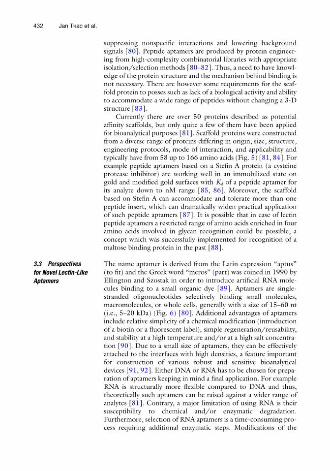
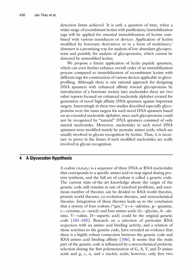
Currently there are over 50 proteins described as potential affi nity scaffolds, but only quite a few of them have been applied for bioanalytical purposes [ 81 ]. Scaffold proteins were constructed from a diverse range of proteins differing in origin, size, structure, engineering protocols, mode of interaction, and applicability and typically have from 58 up to 166 amino acids (Fig. 5 ) [ 81 , 84 ]. For example peptide aptamers based on a Stefi n A protein (a cysteine protease inhibitor) are working well in an immobilized state on gold and modifi ed gold surfaces with K d of a peptide aptamer for its analyte down to nM range [ 85 , 86 ]. Moreover, the scaffold based on Stefi n A can accommodate and tolerate more than one peptide insert, which can dramatically widen practical application of such peptide aptamers [ 87 ]. It is possible that in case of lectin peptide aptamers a restricted range of amino acids enriched in four amino acids involved in glycan recognition could be possible, a concept which was successfully implemented for recognition of a maltose binding protein in the past [ 88 ].
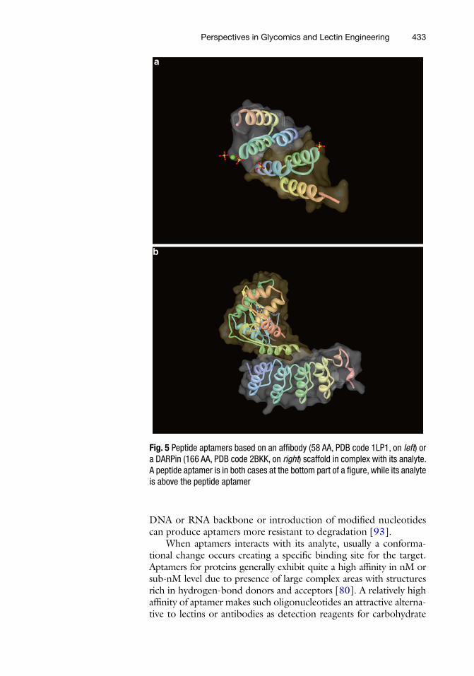
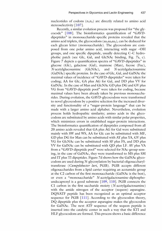
The name aptamer is derived from the Latin expression “aptus” (to fi t) and the Greek word “meros” (part) was coined in 1990 by Ellington and Szostak in order to introduce artifi cial RNA mole-cules binding to a small organic dye [ 89 ]. Aptamers are single- stranded oligonucleotides selectively binding small molecules, macromolecules, or whole cells, generally with a size of 15–60 nt (i.e., 5–20 kDa) (Fig. 6 ) [ 80 ]. Additional advantages of aptamers include relative simplicity of a chemical modifi cation (introduction of a biotin or a fl uorescent label), simple regeneration/reusability, and stability at a high temperature and/or at a high salt concentra-tion [ 90 ]. Due to a small size of aptamers, they can be effectively attached to the interfaces with high densities, a feature important for construction of various robust and sensitive bioanalytical devices [ 91 , 92 ]. Either DNA or RNA has to be chosen for prepa-ration of aptamers keeping in mind a fi nal application. For example RNA is structurally more fl exible compared to DNA and thus, theoretically such aptamers can be raised against a wider range of analytes [ 81 ]. Contrary, a major limitation of using RNA is their susceptibility to chemical and/or enzymatic degradation. Furthermore, selection of RNA aptamers is a time-consuming pro-cess requiring additional enzymatic steps. Modifi cations of the
3.3 Perspectives for Novel Lectin-Like Aptamers
Jan Tkac et al.
433
DNA or RNA backbone or introduction of modifi ed nucleotides can produce aptamers more resistant to degradation [ 93 ].
When aptamers interacts with its analyte, usually a conforma-tional change occurs creating a specifi c binding site for the target. Aptamers for proteins generally exhibit quite a high affi nity in nM or sub-nM level due to presence of large complex areas with structures rich in hydrogen-bond donors and acceptors [ 80 ]. A relatively high affi nity of aptamer makes such oligonucleotides an attractive alterna-tive to lectins or antibodies as detection reagents for carbohydrate
Fig. 5 Peptide aptamers based on an affi body (58 AA, PDB code 1LP1, on left ) or a DARPin (166 AA, PDB code 2BKK, on right ) scaffold in complex with its analyte. A peptide aptamer is in both cases at the bottom part of a fi gure, while its analyte is above the peptide aptamer
Perspectives in Glycomics and Lectin Engineering

434
antigens. In order to increase palette of analytes being recognized by aptamers, modifi ed nucleotides were introduced.
A rational approach for preparation of aptamers with a high affi nity binding of glycoproteins by an extending library of nucleo-tides modifi ed by incorporation of a boronic acid moiety was recently introduced by Wang’s lab [ 94 ]. The study showed that affi nity with K d of 6–17 nM for fi brinogen using boronate modi-fi ed DNA aptamers was higher compared to the affi nity with K d of 64–122 nM for the same analyte using DNA aptamers with natural pool of nucleotides. The fact that for the interaction between a glycoprotein and boronate modifi ed DNA aptamer it is important an interaction between boronate moiety and glycan of fi brinogen was confi rmed by analysis of a deglycosylated fi brinogen with a decreased affi nity ( K d of 87–390 nM) [ 94 ].
An interesting approach for preparation of a wider library of nucleotides was recently introduced by incorporation of six new 5-position modifi ed dUTP derivatives with fi ve derivatives con-taining an aromatic ring [ 95 ]. DNA aptamers based on an extended pool of nucleotides were able to bind a necrosis factor receptor superfamily member 9 (TNFRSF9) with a high affi nity of K d = 4–6 nM for the fi rst time. Interestingly two new derivatives containing either indole derivative or a benzene ring were the best TNFRSF9 binders. This fact is quite interesting since TNFRSF9 is a glycoprotein [ 96 ] and we can only speculate that these two aromatic derivatives of dUTP were involved in recognition of
Fig. 6 An RNA aptamer ( purple ) bound to its analyte peptide ( white-magenta chain) (a PDB code 1EXY)
Jan Tkac et al.
435
TNFRSF9 via a glycan interaction. In a recent and similar study a derivative of imidazole (7-(2-thienyl)imidazo[4,5- b ]pyridine, Ds) containing nucleotides was applied for generation of novel DNA aptamers binding to two glycoproteins vascular endothelial cell growth factor-165 (VEGF-165) and interferon-γ (IFN-γ) with an enhanced affi nity [ 97 ]. The study revealed K d down to 0.65 pM with DNA aptamers based on Ds nucleotides, while the best K d of 57 pM for the DNA aptamers containing natural nucleotides was found for VEGF-165 [ 97 ]. Similarly, DNA aptamers based on Ds nucleotides offered much lower K d of 0.038 nM compared to DNA aptamers based on natural nucleotides with K d of 9.1 nM for IFN-γ [ 97 ]. Here we can again only speculate if the role of Ds nucleotides in enhanced affi nity for two glycoproteins is in interaction of Ds modifi ed nucleotides with the glycan moiety of glycoproteins.
There are several very interesting strategies as to how to enhance analytical applicability of lectins by their simple modifi cations, which can dramatically infl uence the fi eld of glycoprofi ling in a future.
The fi rst study focused on application of multimers of eight different lectins prepared by incubation of biotinylated lectins with streptavidin. A wheat germ agglutinin (WGA) multimers inte-grated into lectin microarrays showed 4–40 times better sensitivity in analysis of glycans in human plasma and much better perfor-mance in glycoprofi ling of samples from people having pancreatic cancer compared to utilization of WGA lectin monomer [ 98 ]. Authors of the study suggested that such lectin multimers with an enhanced affi nity toward glycans can broaden the range of glycans, which can be detected. Moreover, according to authors lectin mul-timers might provide a fundamentally new biorecognition infor-mation not achievable by lectin monomers [ 98 ]. The second study described attachment of a boronate functionality to two different lectins in order to enhance affi nity 2- to 60-fold for a particular glycan binding [ 99 ]. Such modifi ed lectins were tested in a whole cell lysate with an excellent specifi city for analysis of 295 N-linked glycopeptides. These results revealed that application of boronate modifi ed lectins can facilitate identifi cation of glycans present on the surface of low-abundant glycoproteins [ 99 ]. The third study indicated that by preparation of a lectin mutant with artifi cially introduced cysteine into lectin Galanthus nivalis agglutinin it was possible to prepare lectin dimmers via a disulfi de linkage between two lectin mutants [ 100 ]. Agglutination activity of a lectin dimmer increased 16-fold compared to a lectin monomer and interestingly a transformation monomer/dimmer can be redox-switchable by addition of mild oxidation or reducing agents [ 100 ].
It can be summed up there are very exciting concepts already introduced in the fi eld of lectin glycoengineering such as integra-tion of deglycosylated forms of recombinant lectins into lectin microarrays with enhanced sensitivity of analysis and with lower
3.4 Other Novel Forms of Lectins
Perspectives in Glycomics and Lectin Engineering
436
detection limits achieved. It is only a question of time, when a wider range of recombinant lectins with purifi cation/immobilization tags will be applied for oriented immobilization of lectins com-bined with various transducers or devices. Application of lectins modifi ed by boronate derivatives or in a form of multimers/dimmers is a promising way for analysis of low abundant glycopro-teins and possibly for analysis of glycoproteins, which cannot be detected by unmodifi ed lectins.
We propose a future application of lectin peptide aptamers, which can even further enhance overall order of an immobilization process compared to immobilization of recombinant lectins with different tags for construction of various devices applicable in glyco-profi ling. Although there is one rational approach for designing DNA aptamers with enhanced affi nity toward glycoproteins by introduction of a boronate moiety into nucleotides there are two other reports focused on enhanced nucleotide alphabet created for generation of novel high affi nity DNA aptamers against important targets. Interestingly in these two studies described especially glyco-proteins were the main targets for such novel DNA aptamers based on an extended nucleotide alphabet, since such glycoproteins could not be recognized by “natural” DNA aptamers consisted of only natural nucleotides. Moreover, nucleotides in such novel DNA aptamers were modifi ed mainly by aromatic amino acids, which are usually involved in glycan recognition by lectins. Thus, it is neces-sary to prove in the future if such modifi ed nucleotides are really involved in glycan recognition.
4 A Glycocodon Hypothesis
A codon (n 1 n 2 n 3 ) is a sequence of three DNA or RNA nucleotides that corresponds to a specifi c amino acid or stop signal during pro-tein synthesis, and the full set of codons is called a genetic code. The current state-of-the-art knowledge about the origin of the genetic code still remains as one of unsolved problems, and enor-mous number of theories can be divided to RNA world theories, protein world theories, co-evolution theories, and stereochemical theories. Integration of these theories leads us to the conclusion that a system of four codons (“gnc,” n = a—adenine, g—guanine, c—cytosine, u—uracil) and four amino acids (G—glycine, A—ala-nine, V—valine, D—aspartic acid) could be the original genetic code [ 101 – 105 ]. Research on a selection of particular RNA sequences with an amino acid binding activity, and a relation of those activities to the genetic code, have revealed an evidence that there is a highly robust connection between the genetic code and RNA-amino acid binding affi nity [ 106 ]. It seems that the main part of the genetic code is infl uenced by a stereochemical prebiotic selection during the fi rst polymerization of G, A, V, and D amino acids and g, c, u, and a nucleic acids; however, only fi rst two
Jan Tkac et al.
437
nucleotides of codons (n 1 n 2 ) are directly related to amino acid stereoselectivity [ 107 ].
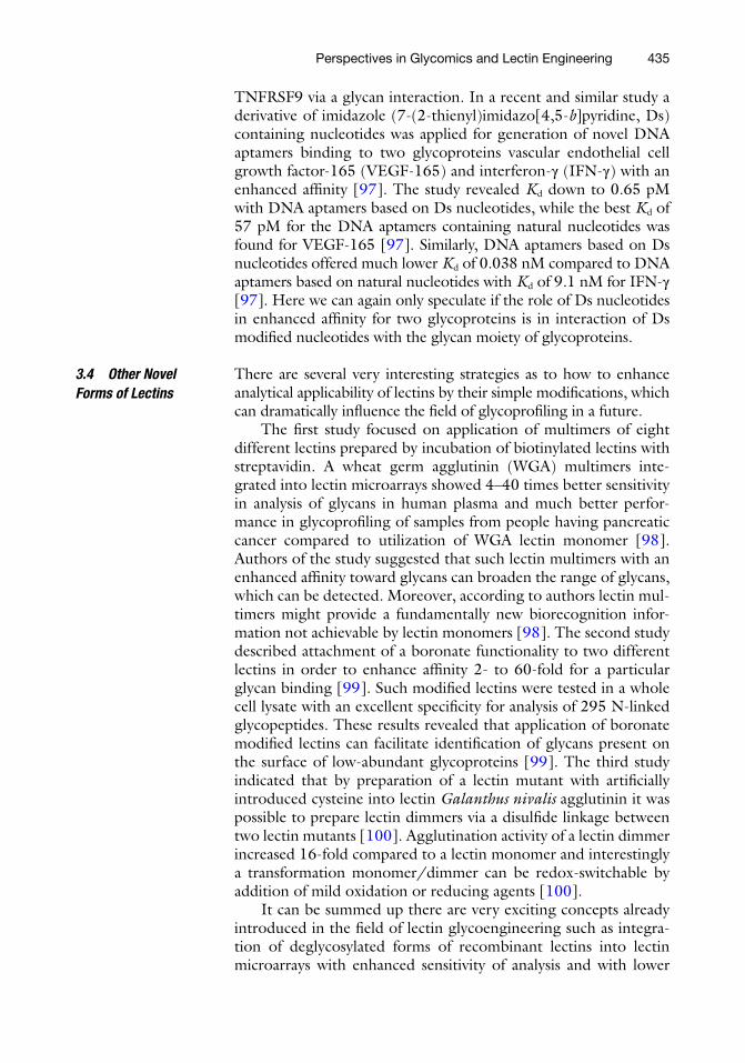
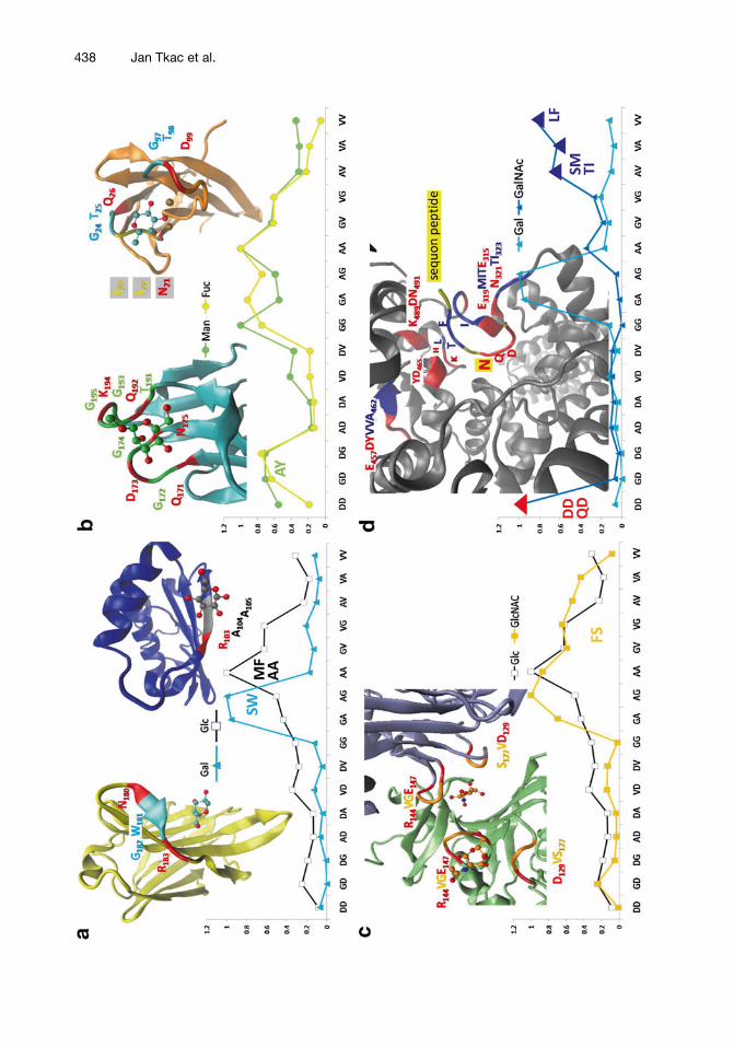
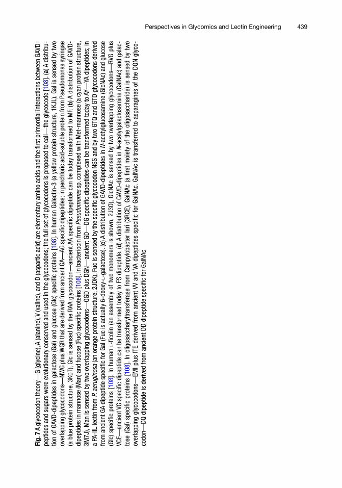
Recently, a similar evolution process was proposed for “the gly-cocode” [ 108 ]. The bioinformatics quantifi cation of “GAVD- dipeptides” in monosaccharide-specifi c proteins revealed that the amino acid triplets, the glycocodons (aa 1 aa 2 aa 3 ), can be deduced for each glycan letter (monosaccharide). The glycocodons are com-posed from one polar amino acid, interacting with sugar –OH groups, and one specifi c dipeptide, usually detecting C–C hydro-phobic patch (see Glc, Gal, and GlcNAc binding, Fig. 7a, c ). Figure 7 depicts a quantifi cation spectra of “GAVD-dipeptides” in glucose (Glc), galactose (Gal), mannose (Man), fucose (Fuc), N -acetylglucosamine (GlcNAc), and N -acetylgalactosamine (GalNAc) specifi c proteins. In the case of Glc, Gal, and GalNAc the maximal values of incidence of “GAVD-dipeptides” were taken for coding; AA for Glc, GA plus AG for Gal, and DD plus VV for GalNAc. In the case of Man and GlcNAc GD plus DG and GV plus VG from “GAVD-dipeptide pool” were taken for coding, because maximal values have been already taken by previous monosaccha-rides. During evolution, the GAVD-glycocodons were transformed to novel glycocodons by a positive selection for the increased diver-sity and functionality of a “sugar–protein language” that can be made with a larger amino acid alphabet. Nevertheless, evolution process holds hydropathic similarity; amino acids in the glyco-codons are substituted by amino acids with similar polar properties, which minimizes errors in established sugar–protein interactions. The bioinformatics quantifi cation of dipeptides composed from all 20 amino acids revealed that GA plus AG for Gal were substituted mainly with SW and WS, AA for Glc can be substituted with MF, GD plus DG for Man can be substituted with AY plus YA, GV plus VG for GlcNAc can be substituted with SF plus FS, and DD plus VV for GalNAc can be substituted with QD plus LF. AV plus VA from a “GAVD-dipeptide pool” were selected for NAc-group sens-ing, in the case of GalNAc, they were transformed to MS plus SM and IT plus TI dipeptides. Figure 7d shows how the GalNAc glyco-codons are used during N-glycosylation by bacterial oligosaccharyl-transferase ( Campylobacter lari , PGlB). PGlB accepts different oligosaccharides from a lipid carrier requiring an acetamido group at the C2 carbon of the fi rst monosaccharide (GalNAc is the best), or even a “monosaccharide” N -acetylgalactosamine-diphospho-undecaprenyl is a good substrate [ 109 , 110 ]. PGlB connects the C1 carbon in the fi rst saccharide moiety ( N -acetylgalactosamine) with the amide nitrogen of the acceptor (sequon) asparagine. DQNATF peptide has been recognized as an optimal acceptor sequence for PGlB [ 111 ]. According to the glycocodon theory, DQ dipeptide plus the acceptor asparagine makes the glycocodon for GalNAc. The next ATF sequence of the sequon peptide is inserted into the catalytic center in such a way that the KTI and HLF glycocodons are formed. This process shows a basic difference
Perspectives in Glycomics and Lectin Engineering
438 Jan Tkac et al.
439
Fig.
7 A
gly
coco
don
theo
ry—
G (g
lyci
ne),
A (a
lani
ne),
V (v
alin
e), a
nd D
(asp
artic
aci
d) a
re e
lem
enta
ry a
min
o ac
ids
and
the
fi rst
prim
ordi
al in
tera
ctio
ns b
etw
een
GAVD
-pe
ptid
es a
nd s
ugar
s w
ere
evol
utio
nary
con
serv
ed a
nd u
sed
in th
e gl
ycoc
odon
s; th
e fu
ll se
t of g
lyco
codo
ns is
pro
pose
d to
cal
l—th
e gl
ycoc
ode
[ 108
]. ( a
) A d
istri
bu-
tion
of G
AVD-
dipe
ptid
es in
gal
acto
se (G
al) a
nd g
luco
se (G
lc) s
peci
fi c p
rote
ins
[ 108
]. In
hum
an G
alec
tin-3
(a y
ello
w p
rote
in s
truct
ure,
1KJ
L), G
al is
sen
sed
by tw
o ov
erla
ppin
g gl
ycoc
odon
s—NW
G pl
us W
GR th
at a
re d
eriv
ed fr
om a
ncie
nt G
A—AG
spe
cifi c
dip
eptid
es; i
n pe
rchl
oric
aci
d-so
lubl
e pr
otei
n fro
m P
seud
omon
as s
yrin
gae
(a b
lue
prot
ein
stru
ctur
e, 3
K0T)
, Glc
is s
ense
d by
the
RAA
glyc
ocod
on—
anci
ent A
A sp
ecifi
c di
pept
ide
can
be to
day
trans
form
ed to
MF.
( b ) A
dis
tribu
tion
of G
AVD-
dipe
ptid
es in
man
nose
(Man
) and
fuco
se (F
uc) s
peci
fi c p
rote
ins
[ 108
]. In
bac
terio
cin
from
Pse
udom
onas
sp.
com
plex
ed w
ith M
et-m
anno
se (a
cya
n pr
otei
n st
ruct
ure,
3M
7J),
Man
is s
ense
d by
two
over
lapp
ing
glyc
ocod
ons—
QGD
plus
DGN
—an
cien
t GD—
DG s
peci
fi c d
ipep
tides
can
be
trans
form
ed to
day
to A
Y—YA
dip
eptid
es; i
n a
PA-II
L le
ctin
from
P. a
erug
inos
a (a
n or
ange
pro
tein
stru
ctur
e, 2
JDK)
, Fuc
is s
ense
d by
the
spec
ifi c
glyc
ocod
on N
SS a
nd b
y tw
o GT
Q an
d GT
D gl
ycoc
odon
s de
rived
fro
m a
ncie
nt G
A di
pept
ide
spec
ifi c
for G
al (F
uc is
act
ually
6-d
eoxy
- L -g
alac
tose
). ( c
) A d
istri
butio
n of
GAV
D-di
pept
ides
in N
-ace
tylg
luco
sam
ine
(Glc
NAc)
and
glu
cose
(G
lc) s
peci
fi c p
rote
ins
[ 108
]. In
hum
an L
-fi c
olin
(an
asse
mbl
y of
two
mon
omer
s is
sho
wn,
2J3
O), G
lcNA
c is
sen
sed
by tw
o ov
erla
ppin
g gl
ycoc
odon
s—RV
G pl
us
VGE—
anci
ent V
G sp
ecifi
c di
pept
ide
can
be tr
ansf
orm
ed to
day
to F
S di
pept
ide.
( d ) A
dis
tribu
tion
of G
AVD-
dipe
ptid
es in
N -a
cety
lgal
acto
sam
ine
(Gal
NAc)
and
gal
ac-
tose
(Gal
) spe
cifi c
pro
tein
s [ 1
08 ].
In o
ligos
acch
aryl
trans
fera
se f
rom
Cam
pylo
bact
er la
ri (3
RCE)
, Gal
NAc
(a fi
rst
moi
ety
of t
he o
ligos
acch
arid
e) is
sen
sed
by t
wo
over
lapp
ing
glyc
ocod
ons—
EMI p
lus
ITE
deriv
ed fr
om a
ncie
nt V
V an
d VA
dip
eptid
es s
peci
fi c fo
r Ga
lNAc
. Gal
NAc
is tr
ansf
erre
d to
asp
arag
ines
of t
he D
QN g
lyco
-co
don—
DQ d
ipep
tide
is d
eriv
ed fr
om a
ncie
nt D
D di
pept
ide
spec
ifi c
for G
alNA
c
Perspectives in Glycomics and Lectin Engineering

440
between the codons and the glycocodons. When the codons are read from the nucleotide sequence, they are read in succession and do not overlap with one another. On the contrary, the glycocodons are used in a way of a “key-lock” principle—three different protein chains can make the glycocodon in 3D space and the glycocodons frequently overlap (Fig. 7 ).
It should be emphasized that the glycocodons were theoreti-cally deduced by a bioinformatics study and it will be necessary to perform a study in the laboratory to establish the strongest correla-tion between the monosaccharides and the glycocodons and to determine the shortest peptides for the recognition of the specifi c monosaccharide. However, the glycocodon theory represents a tool showing how the PLA or novel DNA aptamers based on nucleotide derivatives should be organized and programmed.
5 Conclusions
This chapter described various tools, which have been recently applied in order to extend analytical usefulness of lectin-based devices in glycoprofi ling. A positive aspect of recent effort in the fi eld is utilization of a great potential nanotechnology can bring into quite a complex and challenging analysis of glycans. Such approaches proved analysis of glycans by different biosensors can be extremely sensitive with a concentration range spanning few orders of magni-tude, which are features essential in analysis of low- abundant glyco-proteins. Control of immobilization of lectins is other important issue, which was successfully addressed in a pilot study showing that attachment of recombinant lectins on surfaces via various tags pres-ent in recombinant lectin improved sensitivity of glycan analysis. The book chapter described also future prospect of PLA in order to increase sensitivity and stability of analysis, while suppressing non-specifi c interactions. Additional issue to focus on in the future is investigation if modifi ed nucleotides can be successfully applied in preparation of novel DNA aptamers targeting glycoproteins with high affi nity and selectivity. The fi nal part of the chapter describes the concept of a glycocodon, which can lead to a better understand-ing of glycan–lectin interaction and for design of novel lectins with unknown specifi cities and/or better affi nities toward glycan target or for rational design of PLA or DNA aptamers.
Acknowledgement
The fi nancial support from the Slovak research and development agency APVV 0282-11, from VEGA 2/0127/10 and 2/0162/14 is acknowledged. The research leading to these results has received partly funding from the European Research Council under the
Jan Tkac et al.
441
European Union’s Seventh Framework Programme (FP/2007-2013)/ERC Grant Agreement n. 311532, from the European Union’s Seventh Framework Programme for research, technologi-cal development and demonstration under Grant agreement n. 317420 and from Qatar Foundation under Project n. 6-381-1-078. This contribution was partly supported by the project: Centre of excellence for white-green biotechnology, ITMS 26220120054, supported by the Research & Development Operational Programme funded by the ERDF.
References
1. Schena M, Shalon D, Davis RW, Brown PO (1995) Quantitative monitoring of gene expression patterns with a complementary DNA microarray. Science 270:467–470
2. Eisen MB, Spellman PT, Brown PO, Botstein D (1998) Cluster analysis and display of genome-wide expression patterns. Proc Natl Acad Sci 95:14863–14868
3. Alizadeh AA, Eisen MB, Davis RE, Ma C, Lossos IS, Rosenwald A, Boldrick JC, Sabet H, Tran T, Yu X, Powell JI, Yang L, Marti GE, Moore T, Hudson J, Lu L, Lewis DB, Tibshirani R, Sherlock G, Chan WC, Greiner TC, Weisenburger DD, Armitage JO, Warnke R, Levy R, Wilson W, Grever MR, Byrd JC, Botstein D, Brown PO, Staudt LM (2000) Distinct types of diffuse large b-cell lym-phoma identifi ed by gene expression profi l-ing. Nature 403:503–511
4. Gygi SP, Rochon Y, Franza BR, Aebersold R (1999) Correlation between protein and mRNA abundance in yeast. Mol Cell Biol 19:1720–1730
5. Gry M, Rimini R, Stromberg S, Asplund A, Ponten F, Uhlen M, Nilsson P (2009) Correlations between RNA and protein expression profi les in 23 human cell lines. BMC Genomics 10:365
6. Lee J-R, Magee DM, Gaster RS, LaBaer J, Wang SX (2013) Emerging protein array technologies for proteomics. Expert Rev Proteome 10:65–75
7. Arnaud J, Audfray A, Imberty A (2013) Binding sugars: from natural lectins to syn-thetic receptors and engineered neolectins. Chem Soc Rev 42:4798–4813
8. Baker JL, Çelik E, DeLisa MP (2013) Expanding the glycoengineering toolbox: the rise of bacterial N -linked protein glycosyl-ation. Trends Biotechnol 31:313–323
9. Bertók T, Katrlík J, Gemeiner P, Tkac J (2013) Electrochemical lectin based biosen-sors as a label-free tool in glycomics. Microchim Acta 180:1–13
10. Varki A et al (2009) Essentials of glycobiol-ogy, 2nd edn. Cold Spring Harbor Laboratory Press, Cold Spring Harbor, NY
11. Gemeiner P, Mislovicová D, Tkác J, Svitel J, Pätoprsty V, Hrabárová E, Kogan G, Kozár T (2009) Lectinomics II: a highway to biomed-ical/clinical diagnostics. Biotechnol Adv 27:1–15
12. Katrlík J, Švitel J, Gemeiner P, Kožár T, Tkac J (2010) Glycan and lectin microarrays for glycomics and medicinal applications. Med Res Rev 30:394–418
13. Krishnamoorthy L, Bess JW, Preston AB, Nagashima K, Mahal LK (2009) HIV-1 and microvesicles from T cells share a common glycome, arguing for a common origin. Nat Chem Biol 5:244–250
14. Schauer R, Kamerling JP (2011) The chemistry and biology of trypanosomal trans- sialidases: virulence factors in chagas disease and sleeping sickness. ChemBioChem 12:2246–2264
15. Song X, Lasanajak Y, Xia B, Heimburg- Molinaro J, Rhea JM, Ju H, Zhao C, Molinaro RJ, Cummings RD, Smith DF (2011) Shotgun glycomics: a microarray strategy for functional glycomics. Nat Methods 8:85–90
16. Vaishnava S, Yamamoto M, Severson KM, Ruhn KA, Yu X, Koren O, Ley R, Wakeland EK, Hooper LV (2011) The antibacterial lec-tin regIIIγ promotes the spatial segregation of microbiota and host in the intestine. Science 334:255–258
17. Burton DR, Poignard P, Stanfi eld RL, Wilson IA (2012) Broadly neutralizing antibodies present new prospects to counter highly anti-genically diverse viruses. Science 337:183–186
18. Pejchal R, Doores KJ, Walker LM, Khayat R, Huang P-S, Wang S-K, Stanfi eld RL, Julien J-P, Ramos A, Crispin M, Depetris R, Katpally U, Marozsan A, Cupo A, Maloveste S, Liu Y, McBride R, Ito Y, Sanders RW, Ogohara C, Paulson JC, Feizi T, Scanlan CN, Wong C-H, Moore JP, Olson WC, Ward AB, Poignard P,
Perspectives in Glycomics and Lectin Engineering
442
Schief WR, Burton DR, Wilson IA (2011) A potent and broad neutralizing antibody rec-ognizes and penetrates the HIV glycan shield. Science 334:1097–1103
19. Kim J-H, Resende R, Wennekes T, Chen H-M, Bance N, Buchini S, Watts AG, Pilling P, Streltsov VA, Petric M, Liggins R, Barrett S, McKimm-Breschkin JL, Niikura M, Withers SG (2013) Mechanism-based cova-lent neuraminidase inhibitors with broad- spectrum infl uenza antiviral activity. Science 340:71–75
20. Klein F, Halper-Stromberg A, Horwitz JA, Gruell H, Scheid JF, Bournazos S, Mouquet H, Spatz LA, Diskin R, Abadir A, Zang T, Dorner M, Billerbeck E, Labitt RN, Gaebler C, Marcovecchio PM, Incesu R-B, Eisenreich TR, Bieniasz PD, Seaman MS, Bjorkman PJ, Ravetch JV, Ploss A, Nussenzweig MC (2012) HIV therapy by a combination of broadly neutralizing antibodies in humanized mice. Nature 492:118–122
21. Chandler KB, Goldman R (2013) Glycoprotein disease markers and single protein- omics. Mol Cell Proteomics 12:836–845
22. Ferens-Sieczkowska M, Kowalska B, Kratz EM (2013) Seminal plasma glycoproteins in male infertility and prostate diseases: is there a chance for glyco-biomarkers? Biomarkers 18:10–22
23. Gilgunn S, Conroy PJ, Saldova R, Rudd PM, O'Kennedy RJ (2013) Aberrant PSA glyco-sylation – a sweet predictor of prostate cancer. Nat Rev Urol 10:99–107
24. Schmaltz RM, Hanson SR, Wong C-H (2011) Enzymes in the synthesis of glycocon-jugates. Chem Rev 111:4259–4307
25. van Bueren JJL, Rispens T, Verploegen S, van der Palen-Merkus T, Stapel S, Workman LJ, James H, van Berkel PHC, van de Winkel JGJ, Platts-Mills TAE, Parren PWHI (2011) Anti-galactose-[alpha]-1,3-galactose ige from aller-gic patients does not bind [alpha]-galactosylated glycans on intact therapeutic antibody fc domains. Nat Biotechnol 29:574–576
26. Beck A, Reichert JM (2012) Marketing approval of mogamulizumab: a triumph for glyco-engineering. MAbs 4:419–425
27. Raman R, Raguram S, Venkataraman G, Paulson JC, Sasisekharan R (2005) Glycomics: an integrated systems approach to structure–function relationships of glycans. Nat Methods 2:817–824
28. Gabius H-J, André S, Jiménez-Barbero J, Romero A, Solís D (2011) From lectin struc-ture to functional glycomics: principles of the sugar code. Trends Biochem Sci 36:298–313
29. Cummings RD (2009) The repertoire of gly-can determinants in the human glycome. Mol Biosyst 5:1087–1104
30. Reuel NF, Mu B, Zhang J, Hinckley A, Strano MS (2012) Nanoengineered glycan sensors enabling native glycoprofi ling for medicinal applications: towards profi ling glycoproteins without labeling or liberation steps. Chem Soc Rev 41:5744–5779
31. Bertozzi CR, Kiessling LL (2001) Chemical glycobiology. Science 291:2357–2364
32. Furukawa JI, Fujitani N, Shinohara Y (2013) Recent advances in cellular glycomic analyses. Biomolecules 3:198–225
33. Rakus JF, Mahal LK (2011) New technolo-gies for glycomic analysis: toward a systematic understanding of the glycome. Annu Rev Anal Chem 4:367–392
34. Smith DF, Cummings RD (2013) Application of microarrays to deciphering the structure and function of the human glycome. Mol Cell Proteomics 12:902–912
35. Alley WR, Mann BF, Novotny MV (2013) High-sensitivity analytical approaches for the structural characterization of glycoproteins. Chem Rev 113:2668–2732
36. Lazar IM, Lee W, Lazar AC (2013) Glycoproteomics on the rise: established methods, advanced techniques, sophisticated biological applications. Electrophoresis 34:113–125
37. Novotny M, Alley W Jr, Mann B (2013) Analytical glycobiology at high sensitivity: current approaches and directions. Glycoconj J 30:89–117
38. Oliveira C, Teixeira JA, Domingues L (2013) Recombinant lectins: an array of tailor-made glycan-interaction biosynthetic tools. Crit Rev Biotechnol 33:66–80
39. Murphy P, André S, Gabius H-J (2013) The third dimension of reading the sugar code by lectins: design of glycoclusters with cyclic scaffolds as tools with the aim to defi ne cor-relations between spatial presentation and activity. Molecules 18:4026–4053
40. Mislovičová D, Katrlík J, Paulovičová E, Gemeiner P, Tkac J (2012) Comparison of three distinct ella protocols for determination of apparent affi nity constants between con a and glycoproteins. Colloids Surf B Biointerfaces 94:163–169
41. Mislovičová D, Gemeiner P, Kozarova A, Kožár T (2009) Lectinomics i. Relevance of exogenous plant lectins in biomedical diag-nostics. Biologia 64:1–19
42. Hirabayashi J, Yamada M, Kuno A, Tateno H (2013) Lectin microarrays: concept, principle and applications. Chem Soc Rev 42:4443–4458
43. Krishnamoorthy L, Mahal LK (2009) Glycomic analysis: an array of technologies. ACS Chem Biol 4:715–732
Jan Tkac et al.
443
44. Lis H, Sharon N (1998) Lectins: carbohydrate- specifi c proteins that mediate cellular recogni-tion. Chem Rev 98:637–674
45. Cunningham S, Gerlach JQ, Kane M, Joshi L (2010) Glyco-biosensors: recent advances and applications for the detection of free and bound carbohydrates. Analyst 135:2471–2480
46. Gerlach JQ, Cunningham S, Kane M, Joshi L (2010) Glycobiomimics and glycobiosensors. Biochem Soc Trans 38:1333–1336
47. Reuel NF, Ahn J-H, Kim J-H, Zhang J, Boghossian AA, Mahal LK, Strano MS (2011) Transduction of glycan–lectin binding using near-infrared fl uorescent single-walled carbon nanotubes for glycan profi ling. J Am Chem Soc 133:17923–17933
48. Sanchez-Pomales G, Zangmeister RA (2011) Recent advances in electrochemical glycobio-sensing. Int J Electrochem 2011
49. Tkac J, Davis JJ (2009) Label-free fi eld effect protein sensing. In: Davis JJ (ed) Engineering the bioelectronic interface: applications to analyte biosensing and protein detection. Royal Society of Chemistry, Cambridge, pp 193–224, doi: 10.1039/9781847559777
50. Reichardt NC, Martin-Lomas M, Penades S (2013) Glyconanotechnology. Chem Soc Rev 42:4358–4376
51. Zeng X, Andrade CAS, Oliveira MDL, Sun X-L (2012) Carbohydrate–protein interac-tions and their biosensing applications. Anal Bioanal Chem 402:3161–3176
52. Gruber K, Horlacher T, Castelli R, Mader A, Seeberger PH, Hermann BA (2011) Cantilever array sensors detect specifi c carbohydrate- protein interactions with pico-molar sensitivity. ACS Nano 5:3670–3678
53. Mader A, Gruber K, Castelli R, Hermann BA, Seeberger PH, Radler JO, Leisner M (2012) Discrimination of escherichia coli strains using glycan cantilever array sensors. Nano Lett 12:420–423
54. Jelinek R, Kolusheva S (2004) Carbohydrate biosensors. Chem Rev 104:5987–6015
55. Luo X, Davis JJ (2013) Electrical biosensors and the label free detection of protein disease biomarkers. Chem Soc Rev 42:5944–5962
56. La Belle JT, Gerlach JQ, Svarovsky S, Joshi L (2007) Label-free impedimetric detection of glycan–lectin interactions. Anal Chem 79:6959–6964
57. Oliveira MDL, Correia MTS, Coelho LCBB, Diniz FB (2008) Electrochemical evaluation of lectin–sugar interaction on gold electrode mod-ifi ed with colloidal gold and polyvinyl butyral. Colloids Surf B Biointerfaces 66:13–19
58. Nagaraj VJ, Aithal S, Eaton S, Bothara M, Wiktor P, Prasad S (2010) Nanomonitor: a miniature electronic biosensor for glycan bio-marker detection. Nanomedicine 5:369–378
59. Bertok T, Gemeiner P, Mikula M, Gemeiner P, Tkac J (2013) Ultrasensitive impedimetric lec-tin based biosensor for glycoproteins contain-ing sialic acid. Microchim Acta 180:151–159
60. Bertok T, Klukova L, Sediva A, Kasák P, Semak V, Micusik M, Omastova M, Chovanová L, Vlček M, Imrich R, Vikartovska A, Tkac J (2013) Ultrasensitive impedimetric lectin biosensors with effi cient antifouling properties applied in glycoprofi ling of human serum samples. Anal Chem 85:7324–7332
61. Bertok T, Sediva A, Katrlik J, Gemeiner P, Mikula M, Nosko M, Tkac J (2013) Label- free detection of glycoproteins by the lectin biosensor down to attomolar level using gold nanoparticles. Talanta 108:11–18
62. Yang H, Li Z, Wei X, Huang R, Qi H, Gao Q, Li C, Zhang C (2013) Detection and discrim-ination of alpha-fetoprotein with a label-free electrochemical impedance spectroscopy bio-sensor array based on lectin functionalized carbon nanotubes. Talanta 111:62–68
63. Vedala H, Chen Y, Cecioni S, Imberty A, Vidal S, Star A (2011) Nanoelectronic detec-tion of lectin-carbohydrate interactions using carbon nanotubes. Nano Lett 11:170–175
64. Chen YN, Vedala H, Kotchey GP, Audfray A, Cecioni S, Imberty A, Vidal S, Star A (2012) Electronic detection of lectins using carbohydrate- functionalized nanostructures: graphene versus carbon nanotubes. ACS Nano 6:760–770
65. Zhang GJ, Huang MJ, Ang JJ, Yao QF, Ning Y (2013) Label-free detection of carbohydrate- protein interactions using nanoscale fi eld- effect transistor biosensors. Anal Chem 85:4392–4397
66. Huang Y-W, Wu C-S, Chuang C-K, Pang S-T, Pan T-M, Yang Y-S, Ko F-H (2013) Real-time and label-free detection of the prostate- specifi c antigen in human serum by a polycrystalline silicon nanowire fi eld-effect transistor biosen-sor. Anal Chem 85:7912–7918
67. Ali M, Nasir S, Ramirez P, Cervera J, Mafe S, Ensinger W (2013) Carbohydrate-mediated biomolecular recognition and gating of syn-thetic ion channels. J Phys Chem C 117:18234–18242
68. Kruss S, Hilmer AJ, Zhang J, Reuel NF, Mu B, Strano MS (2013) Carbon nanotubes as optical biomedical sensors. Adv Drug Deliv Rev. doi: 10.1016/j.addr.2013.07.015
69. Reuel NF, Grassbaugh B, Kruss S, Mundy JZ, Opel C, Ogunniyi AO, Egodage K, Wahl R, Helk B, Zhang J, Kalcioglu ZI, Tvrdy K, Bellisario DO, Mu B, Blake SS, Van Vliet KJ, Love JC, Wittrup KD, Strano MS (2013) Emergent properties of nanosensor arrays: applications for monitoring igg affi nity distri-butions, weakly affi ned hypermannosylation,
Perspectives in Glycomics and Lectin Engineering
444
and colony selection for biomanufacturing. ACS Nano 7:7472–7482
70. Bellapadrona G, Tesler AB, Grunstein D, Hossain LH, Kikkeri R, Seeberger PH, Vaskevich A, Rubinstein I (2012) Optimization of localized surface plasmon resonance trans-ducers for studying carbohydrate- protein interactions. Anal Chem 84:232–240
71. Jin S, Cheng Y, Reid S, Li M, Wang B (2010) Carbohydrate recognition by boronolectins, small molecules, and lectins. Med Res Rev 30:171–257
72. Streicher H, Sharon N (2003) Recombinant plant lectins and their mutants. Methods Enzymol 363:47–77, In: Yuan CL, Reiko TL (eds) doi: 10.1016/S0076-6879(03)01043-7
73. Geisler C, Jarvis DL (2011) Letter to the glyco-forum: effective glycoanalysis with maackia amurensis lectins requires a clear understanding of their binding specifi cities. Glycobiology 21:988–993
74. Alava T, Mann JA, Théodore C, Benitez JJ, Dichtel WR, Parpia JM, Craighead HG (2013) Control of the graphene–protein interface is required to preserve adsorbed protein function. Anal Chem 85:2754–2759
75. Hsu K-L, Gildersleeve JC, Mahal LK (2008) A simple strategy for the creation of a recom-binant lectin microarray. Mol Biosyst 4:654–662
76. Propheter DC, Hsu K-L, Mahal LK (2010) Fabrication of an oriented lectin microarray. ChemBioChem 11:1203–1207
77. Propheter DC, Mahal LK (2011) Orientation of gst-tagged lectins via in situ surface modi-fi cation to create an expanded lectin micro-array for glycomic analysis. Mol Biosyst 7:2114–2117
78. Chen M-L, Adak AK, Yeh N-C, Yang W-B, Chuang Y-J, Wong C-H, Hwang K-C, Hwu J-RR, Hsieh S-L, Lin C-C (2008) Fabrication of an oriented fc-fused lectin microarray through boronate formation. Angew Chem Int Ed 47:8627–8630
79. Colas P, Cohen B, Jessen T, Grishina I, McCoy J, Brent R (1996) Genetic selection of peptide aptamers that recognize and inhibit cyclin-dependent kinase 2. Nature 380:548–550
80. Mascini M, Palchetti I, Tombelli S (2012) Nucleic acid and peptide aptamers: funda-mentals and bioanalytical aspects. Angew Chem Int Ed 51:1316–1332
81. Ruigrok VJB, Levisson M, Eppink MHM, Smidt H, van der Oost J (2011) Alternative affi nity tools: more attractive than antibodies? Biochem J 436:1–13
82. Ståhl S, Kronqvist N, Jonsson A, Löfblom J (2013) Affi nity proteins and their generation. J Chem Technol Biotechnol 88:25–38
83. Woodman R, Yeh JTH, Laurenson S, Ferrigno PK (2005) Design and validation of a neutral protein scaffold for the presentation of pep-tide aptamers. J Mol Biol 352:1118–1133
84. Gebauer M, Skerra A (2009) Engineered pro-tein scaffolds as next-generation antibody thera-peutics. Curr Opin Chem Biol 13:245–255
85. Davis JJ, Tkac J, Humphreys R, Buxton AT, Lee TA, Ko Ferrigno P (2009) Peptide aptamers in label-free protein detection: 2. Chemical optimization and detection of distinct protein isoforms. Anal Chem 81:3314–3320
86. Davis JJ, Tkac J, Laurenson S, Ferrigno PK (2007) Peptide aptamers in label-free protein detection: 1. Characterization of the immobi-lized scaffold. Anal Chem 79:1089–1096
87. Stadler LKJ, Hoffmann T, Tomlinson DC, Song QF, Lee T, Busby M, Nyathi Y, Gendra E, Tiede C, Flanagan K, Cockell SJ, Wipat A, Harwood C, Wagner SD, Knowles MA, Davis JJ, Keegan N, Ferrigno PK (2011) Structurefunction studies of an engineered scaffold protein derived from stefi n A. II: development and applications of the sqt variant. Protein Eng Des Sel 24:751–763
88. Koide A, Gilbreth RN, Esaki K, Tereshko V, Koide S (2007) High-affi nity single-domain binding proteins with a binary-code interface. Proc Natl Acad Sci 104:6632–6637
89. Ellington AD, Szostak JW (1990) In vitro selection of rna molecules that bind specifi c ligands. Nature 346:818–822
90. Keefe AD, Pai S, Ellington A (2010) Aptamers as therapeutics. Nat Rev Drug Discov 9:537–550
91. Iliuk AB, Hu L, Tao WA (2011) Aptamer in bioanalytical applications. Anal Chem 83:4440–4452
92. Liu J, Cao Z, Lu Y (2009) Functional nucleic acid sensors. Chem Rev 109:1948–1998
93. Tolle F, Mayer G (2013) Dressed for suc-cess – applying chemistry to modulate aptamer functionality. Chem Sci 4:60–67
94. Li M, Lin N, Huang Z, Du L, Altier C, Fang H, Wang B (2008) Selecting aptamers for a glycoprotein through the incorporation of the boronic acid moiety. J Am Chem Soc 130:12636–12638
95. Vaught JD, Bock C, Carter J, Fitzwater T, Otis M, Schneider D, Rolando J, Waugh S, Wilcox SK, Eaton BE (2010) Expanding the chemistry of DNA for in vitro selection. J Am Chem Soc 132:4141–4151
96. Eckstrum K, Bany B (2011) Tumor necrosis factor receptor subfamily 9 (tnfrsf9) gene is expressed in distinct cell populations in mouse uterus and conceptus during implantation period of pregnancy. Cell Tissue Res 344:567–576
Jan Tkac et al.
445
97. Kimoto M, Yamashige R, Matsunaga K-I, Yokoyama S, Hirao I (2013) Generation of high-affi nity DNA aptamers using an expanded genetic alphabet. Nat Biotechnol 31:453–457
98. Cao Z, Partyka K, McDonald M, Brouhard E, Hincapie M, Brand RE, Hancock WS, Haab BB (2013) Modulation of glycan detection on specifi c glycoproteins by lectin multimer-ization. Anal Chem 85:1689–1698
99. Lu Y-W, Chien C-W, Lin P-C, Huang L-D, Chen C-Y, Wu S-W, Han C-L, Khoo K-H, Lin C-C, Chen Y-J (2013) Bad-lectins: boronic acid-decorated lectins with enhanced binding affi nity for the selective enrichment of glycoproteins. Anal Chem 85:8268–8276
100. McDonald RE, Hughes DJ, Davis BG (2004) Modular control of lectin function: redox- switchable agglutination. Angew Chem Int Ed 43:3025–3029
101. Eigen M, Schuster P (1978) The hypercycle. Naturwissenschaften 65:341–369
102. Ikehara K, Omori Y, Arai R, Hirose A (2002) A novel theory on the origin of the genetic code: a gnc-sns hypothesis. J Mol Evol 54:530–538
103. Ikehara K (2005) Possible steps to the emer-gence of life: the [GADV]-protein world hypothesis. Chem Rec 5:107–118
104. Di Giulio M (2008) An extension of the coevolution theory of the origin of the genetic code. Biol Direct 3:1–21
105. Lehmann J, Cibils M, Libchaber A (2009) Emergence of a code in the polymerization of amino acids along RNA templates. PLOS ONE 4
106. Yarus M, Widmann J, Knight R (2009) RNA–amino acid binding: a stereochemical era for the genetic code. J Mol Evol 69:406–429
107. Nahalka J (2011) Quantifi cation of peptide bond types in human proteome indicates how DNA codons were assembled at prebiotic conditions. J Proteome Bioinf 4:153–159
108. Nahalka J (2012) Glycocodon theory—the fi rst table of glycocodons. J Theor Biol 307:193–204
109. Wacker M, Feldman MF, Callewaert N, Kowarik M, Clarke BR, Pohl NL, Hernandez M, Vines ED, Valvano MA, Whitfi eld C, Aebi M (2006) Substrate specifi city of bacterial oli-gosaccharyltransferase suggests a common transfer mechanism for the bacterial and eukaryotic systems. Proc Natl Acad Sci 103:7088–7093
110. Li L, Woodward R, Ding Y, Liu X-W, Yi W, Bhatt VS, Chen M, Zhang L-W, Wang PG (2010) Overexpression and topology of bac-terial oligosaccharyltransferase pglb. Biochem Biophys Res Commun 394:1069–1074
111. Schwarz F, Huang W, Li C, Schulz BL, Lizak C, Palumbo A, Numao S, Neri D, Aebi M, Wang L-X (2010) A combined method for producing homogeneous glycoproteins with eukaryotic n-glycosylation. Nat Chem Biol 6:264–266
Perspectives in Glycomics and Lectin Engineering


