Embed Size (px)
Citation preview
Biochimica et Biophysica Acta 1822 (2012) 1411–1420
Contents lists available at SciVerse ScienceDirect
Biochimica et Biophysica Acta
j ourna l homepage: www.e lsev ie r .com/ locate /bbad is
brought to you by COREView metadata, citation and similar papers at core.ac.uk
provided by Elsevier - Publisher Connector
Review
Peroxisomal acyl-CoA synthetases☆
Paul A. Watkins a,b,⁎, Jessica M. Ellis c
a Hugo W. Moser Research Institute at Kennedy Krieger, Baltimore, MD, USAb Department of Neurology, Johns Hopkins University School of Medicine, Baltimore, MD, USAc Department of Biological Chemistry, Johns Hopkins University School of Medicine, Baltimore, MD, USA
Abbreviations: AA, Arachidonic acid (C20:4ω6); ACS,enzyme A; FA, Fatty acid; FATP, Fatty acid transport prPPAR, Peroxisome proliferator-activated receptor; VLCF☆ This article is part of a Special Issue entitled: Metaboperoxisomes in Health and Disease.⁎ Corresponding author at: Kennedy Krieger Institute
MD 21205, USA. Tel.: +1 443 923 2754; fax: +1 443 9E-mail address: [email protected] (P.A. W
0925-4439/$ – see front matter © 2012 Elsevier B.V. Alldoi:10.1016/j.bbadis.2012.02.010
a b s t r a c t
a r t i c l e i n f oArticle history:Received 19 October 2011Received in revised form 12 January 2012Accepted 10 February 2012Available online 17 February 2012
Keywords:PeroxisomeAcyl-CoA synthetaseLipid metabolismFatty acid activation
Peroxisomes carry out many essential lipid metabolic functions. Nearly all of these functions require that anacyl group—either a fatty acid or the acyl side chain of a steroid derivative—be thioesterified to coenzyme A(CoA) for subsequent reactions to proceed. This thioesterification, or “activation”, reaction, catalyzed by en-zymes belonging to the acyl-CoA synthetase family, is thus central to cellular lipid metabolism. However, de-spite our rather thorough understanding of peroxisomal metabolic pathways, surprisingly little is knownabout the specific peroxisomal acyl-CoA synthetases that participate in these pathways. Of the 26 acyl-CoAsynthetases encoded by the human and mouse genomes, only a few have been reported to be peroxisomal,including ACSL4, SLC27A2, and SLC27A4. In this review, we briefly describe the primary peroxisomal lipidmetabolic pathways in which fatty acyl-CoAs participate. Then, we examine the evidence for presence andfunctions of acyl-CoA synthetases in peroxisomes, much of which was obtained before the existence of mul-tiple acyl-CoA synthetase isoenzymes was known. Finally, we discuss the role(s) of peroxisome-specific acyl-CoA synthetase isoforms in lipid metabolism. This article is part of a Special Issue entitled: Metabolic Func-tions and Biogenesis of Peroxisomes in Health and Disease.
© 2012 Elsevier B.V. All rights reserved.
1. Introduction
When peroxisomes were first identified over a half-century ago,they were considered by some to be vestigial organelles with littlephysiological significance. Work done over the last three to fourdecades has clearly demonstrated otherwise. In particular, peroxisomescarry outmany essential processes involving fatty acid (FA)metabolism.
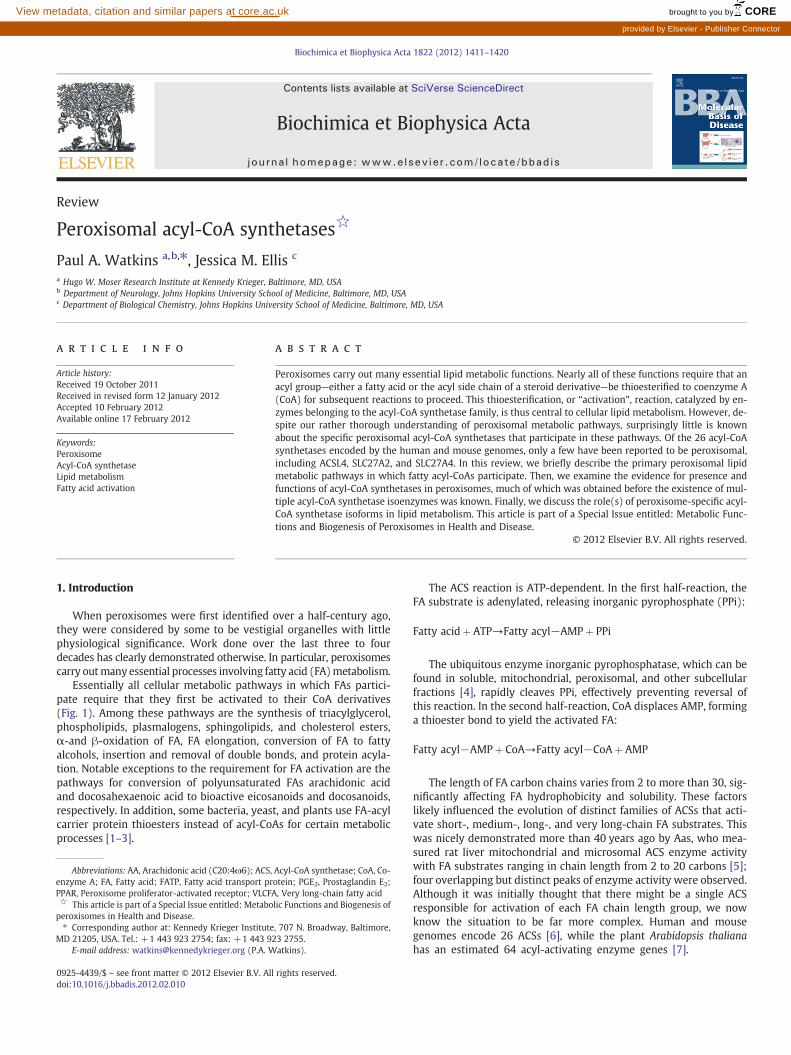
Essentially all cellular metabolic pathways in which FAs partici-pate require that they first be activated to their CoA derivatives(Fig. 1). Among these pathways are the synthesis of triacylglycerol,phospholipids, plasmalogens, sphingolipids, and cholesterol esters,α-and β-oxidation of FA, FA elongation, conversion of FA to fattyalcohols, insertion and removal of double bonds, and protein acyla-tion. Notable exceptions to the requirement for FA activation are thepathways for conversion of polyunsaturated FAs arachidonic acidand docosahexaenoic acid to bioactive eicosanoids and docosanoids,respectively. In addition, some bacteria, yeast, and plants use FA-acylcarrier protein thioesters instead of acyl-CoAs for certain metabolicprocesses [1–3].
Acyl-CoA synthetase; CoA, Co-otein; PGE2, Prostaglandin E2;A, Very long-chain fatty acidlic Functions and Biogenesis of
, 707 N. Broadway, Baltimore,23 2755.atkins).
rights reserved.
The ACS reaction is ATP-dependent. In the first half-reaction, theFA substrate is adenylated, releasing inorganic pyrophosphate (PPi):
Fatty acidþ ATP→Fatty acyl−AMPþ PPi
The ubiquitous enzyme inorganic pyrophosphatase, which can befound in soluble, mitochondrial, peroxisomal, and other subcellularfractions [4], rapidly cleaves PPi, effectively preventing reversal ofthis reaction. In the second half-reaction, CoA displaces AMP, forminga thioester bond to yield the activated FA:
Fatty acyl−AMPþ CoA→Fatty acyl−CoAþ AMP
The length of FA carbon chains varies from 2 to more than 30, sig-nificantly affecting FA hydrophobicity and solubility. These factorslikely influenced the evolution of distinct families of ACSs that acti-vate short-, medium-, long-, and very long-chain FA substrates. Thiswas nicely demonstrated more than 40 years ago by Aas, who mea-sured rat liver mitochondrial and microsomal ACS enzyme activitywith FA substrates ranging in chain length from 2 to 20 carbons [5];four overlapping but distinct peaks of enzyme activity were observed.Although it was initially thought that there might be a single ACSresponsible for activation of each FA chain length group, we nowknow the situation to be far more complex. Human and mousegenomes encode 26 ACSs [6], while the plant Arabidopsis thalianahas an estimated 64 acyl-activating enzyme genes [7].
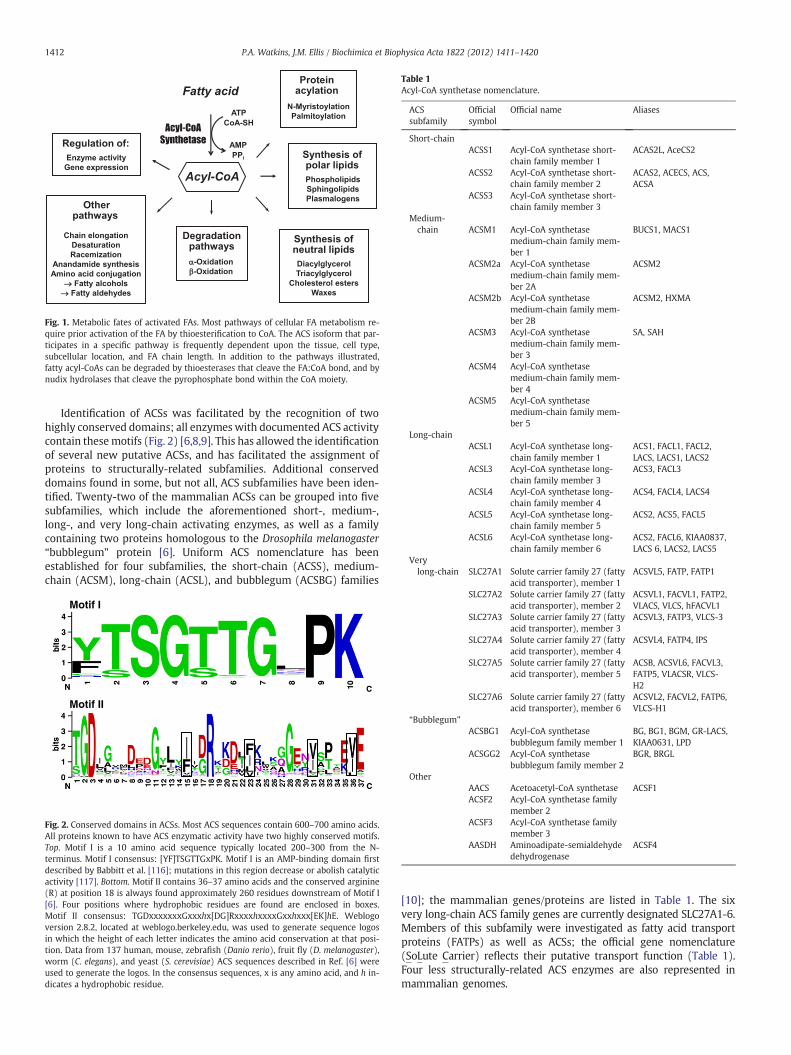
Fig. 1. Metabolic fates of activated FAs. Most pathways of cellular FA metabolism re-quire prior activation of the FA by thioesterification to CoA. The ACS isoform that par-ticipates in a specific pathway is frequently dependent upon the tissue, cell type,subcellular location, and FA chain length. In addition to the pathways illustrated,fatty acyl-CoAs can be degraded by thioesterases that cleave the FA:CoA bond, and bynudix hydrolases that cleave the pyrophosphate bond within the CoA moiety.
Table 1Acyl-CoA synthetase nomenclature.
ACSsubfamily
Officialsymbol
Official name Aliases
Short-chainACSS1 Acyl-CoA synthetase short-
chain family member 1ACAS2L, AceCS2
ACSS2 Acyl-CoA synthetase short-chain family member 2
ACAS2, ACECS, ACS,ACSA
ACSS3 Acyl-CoA synthetase short-chain family member 3
Medium-chain ACSM1 Acyl-CoA synthetase
medium-chain family mem-ber 1
BUCS1, MACS1
ACSM2a Acyl-CoA synthetasemedium-chain family mem-ber 2A
ACSM2
ACSM2b Acyl-CoA synthetasemedium-chain family mem-ber 2B
ACSM2, HXMA
ACSM3 Acyl-CoA synthetasemedium-chain family mem-ber 3
SA, SAH
ACSM4 Acyl-CoA synthetasemedium-chain family mem-ber 4
ACSM5 Acyl-CoA synthetasemedium-chain family mem-ber 5
Long-chainACSL1 Acyl-CoA synthetase long-
chain family member 1ACS1, FACL1, FACL2,LACS, LACS1, LACS2
ACSL3 Acyl-CoA synthetase long-chain family member 3
ACS3, FACL3
ACSL4 Acyl-CoA synthetase long-chain family member 4
ACS4, FACL4, LACS4
ACSL5 Acyl-CoA synthetase long-chain family member 5
ACS2, ACS5, FACL5
ACSL6 Acyl-CoA synthetase long-chain family member 6
ACS2, FACL6, KIAA0837,LACS 6, LACS2, LACS5
Verylong-chain SLC27A1 Solute carrier family 27 (fatty
acid transporter), member 1ACSVL5, FATP, FATP1
1412 P.A. Watkins, J.M. Ellis / Biochimica et Biophysica Acta 1822 (2012) 1411–1420
Identification of ACSs was facilitated by the recognition of twohighly conserved domains; all enzymes with documented ACS activitycontain thesemotifs (Fig. 2) [6,8,9]. This has allowed the identificationof several new putative ACSs, and has facilitated the assignment ofproteins to structurally-related subfamilies. Additional conserveddomains found in some, but not all, ACS subfamilies have been iden-tified. Twenty-two of the mammalian ACSs can be grouped into fivesubfamilies, which include the aforementioned short-, medium-,long-, and very long-chain activating enzymes, as well as a familycontaining two proteins homologous to the Drosophila melanogaster“bubblegum” protein [6]. Uniform ACS nomenclature has beenestablished for four subfamilies, the short-chain (ACSS), medium-chain (ACSM), long-chain (ACSL), and bubblegum (ACSBG) families
Motif I
Motif II
Fig. 2. Conserved domains in ACSs. Most ACS sequences contain 600–700 amino acids.All proteins known to have ACS enzymatic activity have two highly conserved motifs.Top. Motif I is a 10 amino acid sequence typically located 200–300 from the N-terminus. Motif I consensus: [YF]TSGTTGxPK. Motif I is an AMP-binding domain firstdescribed by Babbitt et al. [116]; mutations in this region decrease or abolish catalyticactivity [117]. Bottom. Motif II contains 36–37 amino acids and the conserved arginine(R) at position 18 is always found approximately 260 residues downstream of Motif I[6]. Four positions where hydrophobic residues are found are enclosed in boxes.Motif II consensus: TGDxxxxxxxGxxxhx[DG]RxxxxhxxxxGxxhxxx[EK]hE. Weblogoversion 2.8.2, located at weblogo.berkeley.edu, was used to generate sequence logosin which the height of each letter indicates the amino acid conservation at that posi-tion. Data from 137 human, mouse, zebrafish (Danio rerio), fruit fly (D. melanogaster),worm (C. elegans), and yeast (S. cerevisiae) ACS sequences described in Ref. [6] wereused to generate the logos. In the consensus sequences, x is any amino acid, and h in-dicates a hydrophobic residue.
SLC27A2 Solute carrier family 27 (fattyacid transporter), member 2
ACSVL1, FACVL1, FATP2,VLACS, VLCS, hFACVL1
SLC27A3 Solute carrier family 27 (fattyacid transporter), member 3
ACSVL3, FATP3, VLCS-3
SLC27A4 Solute carrier family 27 (fattyacid transporter), member 4
ACSVL4, FATP4, IPS
SLC27A5 Solute carrier family 27 (fattyacid transporter), member 5
ACSB, ACSVL6, FACVL3,FATP5, VLACSR, VLCS-H2
SLC27A6 Solute carrier family 27 (fattyacid transporter), member 6
ACSVL2, FACVL2, FATP6,VLCS-H1
“Bubblegum”
ACSBG1 Acyl-CoA synthetasebubblegum family member 1
BG, BG1, BGM, GR-LACS,KIAA0631, LPD
ACSGG2 Acyl-CoA synthetasebubblegum family member 2
BGR, BRGL
OtherAACS Acetoacetyl-CoA synthetase ACSF1ACSF2 Acyl-CoA synthetase family
member 2ACSF3 Acyl-CoA synthetase family
member 3AASDH Aminoadipate-semialdehyde
dehydrogenaseACSF4
[10]; the mammalian genes/proteins are listed in Table 1. The sixvery long-chain ACS family genes are currently designated SLC27A1-6.Members of this subfamily were investigated as fatty acid transportproteins (FATPs) as well as ACSs; the official gene nomenclature(SoLute Carrier) reflects their putative transport function (Table 1).Four less structurally-related ACS enzymes are also represented inmammalian genomes.
1413P.A. Watkins, J.M. Ellis / Biochimica et Biophysica Acta 1822 (2012) 1411–1420
In addition to FAs, other compounds containing acyl side chainsare substrates for ACSs. For example, the final step in the synthesisof bile acids requires the removal of three carbons from the cholesterolside chain, converting 27-carbon precursors to 24-carbon maturebile acids [11]. In this process, a terminal carbon of the cholesterolaliphatic side chain is oxidized to a carboxylic acid, which must beactivated to its CoA thioester before removal of a 3-carbon fragmentvia β-oxidation. Furthermore, many xenobiotic compounds, such asthe hypolipidemic drug clofibrate and the non-steroidal anti-inflammatory drug ibuprofen, are ACS substrates [12]. Enzymes ofthe ACSM family are primarily responsible for xenobiotic activation[13]. Besides providing an acyl-CoA substrate for amino acid conjugationand elimination, xenobiotic-CoAs are substrates for lipid synthesis [12].
In addition to classifying ACSs by their substrate chain lengthpreference, these enzymes have also been categorized by their sub-cellular locations. There are numerous literature references to a“mitochondrial medium-chain ACS” or a “microsomal long-chainACS”, without designating a specific enzyme. Many of these studieswere done prior to the genomic era, at a timewhen our understandingof the diversity of the ACS subfamilies was far more limited.
2. Peroxisomal pathways requiring activated FAs
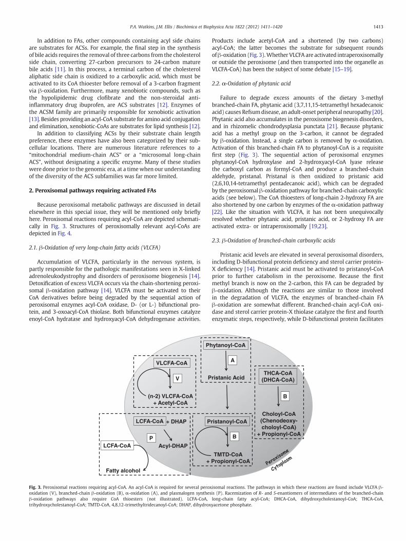
Because peroxisomal metabolic pathways are discussed in detailelsewhere in this special issue, they will be mentioned only brieflyhere. Peroxisomal reactions requiring acyl-CoA are depicted schemati-cally in Fig. 3. Structures of peroxisomally relevant acyl-CoAs aredepicted in Fig. 4.
2.1. β-Oxidation of very long-chain fatty acids (VLCFA)
Accumulation of VLCFA, particularly in the nervous system, ispartly responsible for the pathologic manifestations seen in X-linkedadrenoleukodystrophy and disorders of peroxisome biogenesis [14].Detoxification of excess VLCFA occurs via the chain-shortening peroxi-somal β-oxidation pathway [14]. VLCFA must be activated to theirCoA derivatives before being degraded by the sequential action ofperoxisomal enzymes acyl-CoA oxidase, D- (or L-) bifunctional pro-tein, and 3-oxoacyl-CoA thiolase. Both bifunctional enzymes catalyzeenoyl-CoA hydratase and hydroxyacyl-CoA dehydrogenase activities.
LCFA-CoA
Fatty alcohol
P
VLCFA-CoA
(n-2) VLCFA-CoA + Acetyl-CoA
Ph
P
+
LCFA-CoA + DHAP
Acyl-DHAP
V
P
Fig. 3. Peroxisomal reactions requiring acyl-CoA. An acyl-CoA is required for several peroxoxidation (V), branched-chain β-oxidation (B), α-oxidation (A), and plasmalogen synthesβ-oxidation pathways also require CoA thioesters (not illustrated). LCFA-CoA,trihydroxycholestanoyl-CoA; TMTD-CoA, 4,8,12-trimethyltridecanoyl-CoA; DHAP, dihydrox
Products include acetyl-CoA and a shortened (by two carbons)acyl-CoA; the latter becomes the substrate for subsequent roundsofβ-oxidation (Fig. 3).Whether VLCFA are activated intraperoxisomallyor outside the peroxisome (and then transported into the organelle asVLCFA-CoA) has been the subject of some debate [15–19].
2.2. α-Oxidation of phytanic acid
Failure to degrade excess amounts of the dietary 3-methylbranched-chain FA, phytanic acid (3,7,11,15-tetramethyl hexadecanoicacid) causes Refsumdisease, an adult-onset peripheral neuropathy [20].Phytanic acid also accumulates in the peroxisome biogenesis disorders,and in rhizomelic chondrodysplasia punctata [21]. Because phytanicacid has a methyl group on the 3-carbon, it cannot be degradedby β-oxidation. Instead, a single carbon is removed by α-oxidation.Activation of this branched-chain FA to phytanoyl-CoA is a requisitefirst step (Fig. 3). The sequential action of peroxisomal enzymesphytanoyl-CoA hydroxylase and 2-hydroxyacyl-CoA lyase releasethe carboxyl carbon as formyl-CoA and produce a branched-chainaldehyde, pristanal. Pristanal is then oxidized to pristanic acid(2,6,10,14-tetramethyl pentadecanoic acid), which can be degradedby the peroxisomal β-oxidation pathway for branched-chain carboxylicacids (see below). The CoA thioesters of long-chain 2-hydroxy FA arealso shortened by one carbon by enzymes of the α-oxidation pathway[22]. Like the situation with VLCFA, it has not been unequivocallyresolved whether phytanic acid, pristanic acid, or 2-hydroxy FA areactivated extra- or intraperoxisomally [19,23].
2.3. β-Oxidation of branched-chain carboxylic acids
Pristanic acid levels are elevated in several peroxisomal disorders,including D-bifunctional protein deficiency and sterol carrier protein-X deficiency [14]. Pristanic acid must be activated to pristanoyl-CoAprior to further catabolism in the peroxisome. Because the firstmethyl branch is now on the 2-carbon, this FA can be degraded byβ-oxidation. Although the reactions are similar to those involvedin the degradation of VLCFA, the enzymes of branched-chain FAβ-oxidation are somewhat different. Branched-chain acyl-CoA oxi-dase and sterol carrier protein-X thiolase catalyze the first and fourthenzymatic steps, respectively, while D-bifunctional protein facilitates
ristanoyl-CoA
THCA-CoA (DHCA-CoA)
ytanoyl-CoA
ristanic Acid
TMTD-CoA Propionyl-CoA
Choloyl-CoA (Chenodeoxy- choloyl-CoA)
+ Propionyl-CoA
A
B
B
isomal reactions. The pathways in which these reactions are found include VLCFA β-is (P). Racemization of R- and S-enantiomers of intermediates of the branched-chainlong-chain fatty acyl-CoA; DHCA-CoA, dihydroxycholestanoyl-CoA; THCA-CoA,yacetone phosphate.
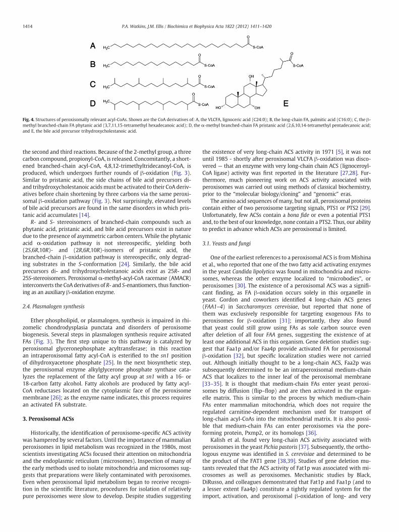
Fig. 4. Structures of peroxisomally relevant acyl-CoAs. Shown are the CoA derivatives of: A, the VLCFA, lignoceric acid (C24:0); B, the long-chain FA, palmitic acid (C16:0); C, the β-methyl branched-chain FA phytanic acid (3,7,11,15-tetramethyl hexadecanoic acid); D, the α-methyl branched-chain FA pristanic acid (2,6,10,14-tetramethyl pentadecanoic acid;and E, the bile acid precursor trihydroxycholestanoic acid.
1414 P.A. Watkins, J.M. Ellis / Biochimica et Biophysica Acta 1822 (2012) 1411–1420
the second and third reactions. Because of the 2-methyl group, a threecarbon compound, propionyl-CoA, is released. Concomitantly, a short-ened branched-chain acyl-CoA, 4,8,12-trimethyltridecanoyl-CoA, isproduced, which undergoes further rounds of β-oxidation (Fig. 3).Similar to pristanic acid, the side chains of bile acid precursors di-and trihydroxycholestanoic acidsmust be activated to their CoA deriv-atives before chain shortening by three carbons via the same peroxi-somal β-oxidation pathway (Fig. 3). Not surprisingly, elevated levelsof bile acid precursors are found in the same disorders in which pris-tanic acid accumulates [14].
R- and S- stereoisomers of branched-chain compounds such asphytanic acid, pristanic acid, and bile acid precursors exist in naturedue to the presence of asymmetric carbon centers. While the phytanicacid α-oxidation pathway is not stereospecific, yielding both(2S,6R,10R)- and (2R,6R,10R)-isomers of pristanic acid, thebranched-chain β-oxidation pathway is stereospecific, only degrad-ing substrates in the S-conformation [24]. Similarly, the bile acidprecursors di- and trihydroxycholestanoic acids exist as 25R- and25S-stereoisomers. Peroxisomal α-methyl-acyl-CoA racemase (AMACR)interconverts the CoA derivatives of R- and S-enantiomers, thus function-ing as an auxiliary β-oxidation enzyme.
2.4. Plasmalogen synthesis
Ether phospholipid, or plasmalogen, synthesis is impaired in rhi-zomelic chondrodysplasia punctata and disorders of peroxisomebiogenesis. Several steps in plasmalogen synthesis require activatedFAs (Fig. 3). The first step unique to this pathway is catalyzed byperoxisomal glyceronephosphate acyltransferase; in this reactionan intraperoxisomal fatty acyl-CoA is esterified to the sn1 positionof dihydroxyacetone phosphate [25]. In the next biosynthetic step,the peroxisomal enzyme alkylglycerone phosphate synthase cata-lyzes the replacement of the fatty acyl group at sn1 with a 16- or18-carbon fatty alcohol. Fatty alcohols are produced by fatty acyl-CoA reductases located on the cytoplasmic face of the peroxisomemembrane [26]; as the enzyme name indicates, this process requiresan activated FA substrate.
3. Peroxisomal ACSs
Historically, the identification of peroxisome-specific ACS activitywas hampered by several factors. Until the importance of mammalianperoxisomes in lipid metabolism was recognized in the 1980s, mostscientists investigating ACSs focused their attention on mitochondriaand the endoplasmic reticulum (microsomes). Inspection of many ofthe early methods used to isolate mitochondria and microsomes sug-gests that preparations were likely contaminated with peroxisomes.Even when peroxisomal lipid metabolism began to receive recogni-tion in the scientific literature, procedures for isolation of relativelypure peroxisomes were slow to develop. Despite studies suggesting
the existence of very long-chain ACS activity in 1971 [5], it was notuntil 1985 - shortly after peroxisomal VLCFA β-oxidation was disco-vered — that an enzyme with very long-chain chain ACS (lignoceroyl-CoA ligase) activity was first reported in the literature [27,28]. Fur-thermore, much pioneering work on ACS activity associated withperoxisomes was carried out using methods of classical biochemistry,prior to the “molecular biology/cloning” and “genomic” eras.
The amino acid sequences ofmany, but not all, peroxisomal proteinscontain either of two peroxisome targeting signals, PTS1 or PTS2 [29].Unfortunately, few ACSs contain a bona fide or even a potential PTS1and, to the best of our knowledge, none contain a PTS2. Thus, our abilityto predict in advance which ACSs are peroxisomal is limited.
3.1. Yeasts and fungi
One of the earliest references to a peroxisomal ACS is fromMishinaet al., who reported that one of the two fatty acid activating enzymesin the yeast Candida lipolytica was found in mitochondria and micro-somes, whereas the other enzyme localized to “microbodies”, orperoxisomes [30]. The existence of a peroxisomal ACS was a signifi-cant finding, as FA β-oxidation occurs solely in this organelle inyeast. Gordon and coworkers identified 4 long-chain ACS genes(FAA1–4) in Saccharomyces cerevisiae, but reported that none ofthem was exclusively responsible for targeting exogenous FAs toperoxisomes for β-oxidation [31]; importantly, they also foundthat yeast could still grow using FAs as sole carbon source evenafter deletion of all four FAA genes, suggesting the existence of atleast one additional ACS in this organism. Gene deletion studies sug-gest that Faa1p and/or Faa4p provide activated FA for peroxisomalβ-oxidation [32], but specific localization studies were not carriedout. Although initially thought to be a long-chain ACS, Faa2p wassubsequently determined to be an intraperoxisomal medium-chainACS that localizes to the inner leaf of the peroxisomal membrane[33–35]. It is thought that medium-chain FAs enter yeast peroxi-somes by diffusion (flip–flop) and are then activated in the organ-elle matrix. This is similar to the process by which medium-chainFAs enter mammalian mitochondria, which does not require theregulated carnitine-dependent mechanism used for transport oflong-chain acyl-CoAs into the mitochondrial matrix. It is also possi-ble that medium-chain FAs can enter peroxisomes via the pore-forming protein, Pxmp2, or its homologs [36].
Kalish et al. found very long-chain ACS activity associated withperoxisomes in the yeast Pichia pastoris [37]. Subsequently, the ortho-logous enzyme was identified in S. cerevisiae and determined to bethe product of the FAT1 gene [38,39]. Studies of gene deletion mu-tants revealed that the ACS activity of Fat1p was associated with mi-crosomes as well as peroxisomes. Mechanistic studies by Black,DiRusso, and colleagues demonstrated that Fat1p and Faa1p (and toa lesser extent Faa4p) constitute a tightly regulated system for theimport, activation, and peroxisomal β-oxidation of long- and very
1415P.A. Watkins, J.M. Ellis / Biochimica et Biophysica Acta 1822 (2012) 1411–1420
long-chain FAs [40,41]. S. cerevisiae also contains a gene, FAT2, encod-ing a protein predicted to be a peroxisomal ACS [9,42]. Fat2p containsthe two highly conserved motifs characteristic of all ACSs (Fig. 2) [6],as well as the cannonical peroxisome targeting signal 1 (PTS1), thetripeptide −SKL, at its carboxy terminus. However, the substratespecificity and metabolic function of Fat2p remain unknown.
Peroxisomal ACSs have also been found in some fungi. A survey ofthe Aspergillus nidulans genome identified six likely ACSs, one ofwhich was peroxisomal [43]. This enzyme, FaaB, was found to bethe major ACS for degradation of long-chain FAs. FaaB was requiredfor growth of A. nidulans on FAs as carbon source, and its gene was in-duced when this fungus was grown in the presence of FAs. The twofinal reactions in the synthesis of penicillin by Penicillium chryso-genum are also peroxisomal [44,45]. The ACS phenylacetyl-CoA ligasesupplies the activated substrate for acyl-CoA:6-amino penicillanicacid acyltransferase; the product of the latter reaction is penicillin G.
3.2. Plants
Plants require ACS activity for the formation of phospholipids,triacylglycerol, jasmonate, and for the oxidation of fatty acids. In2002, nine long-chain ACSs were identified in A. thaliana [46]. Twoof these long-chain ACSs, LACS6 and LACS7, were shown to be locatedin the peroxisome, where a majority of β-oxidation takes place duringplant seedling development [47]. The knockout of either A. thalianaperoxisomal ACS alone did not robustly alter plant viability; however,the knockout of both peroxisomal ACSs caused defective seed lipidmobilization [48]. The identification of plant LACS at the interfacebetween lipid bodies and peroxisomes suggested that lipids are mo-bilized from lipid bodies, activated by peroxisomal ACSs, and thendegraded by β-oxidation [49]. Thus, plant peroxisomal ACS activity,and subsequent β-oxidation, is required for normal seed germination.
Jasmonic acid is potent plant signaling molecule that plays roles inregulating plant defense mechanisms, metabolism, and development.Jasmonate is synthesized from linolenic acid (C18:3ω3) through aprocess that occurs in three cellular compartments: plastids, peroxi-somes, and cytosol. In 2005 and 2006, several 4-coumarate:CoAligase-like Arabidopsis peroxisomal proteins were found to have ACSactivity towards jasmonate precursors as well as long-chain FAs[50–52]. These data suggest that formation of jasmonic acid requiresthe ligase activity provided by peroxisomal ACS enzymes; howeverthe specific ACS required for this activity has not been identified.
3.3. Invertebrate ACSs
Firefly luciferase is a peroxisomal enzyme whose two-step reac-tion mechanism resembles that of ACSs (illustrated in the Introduc-tion). In the first ATP-requiring step, the substrate, luciferin, isadenylated with the release of PPi. The second step is the oxygen-dependent conversion of the adenylated substrate to oxyluciferinwith release of AMP and the production of light. Oba and colleaguesdemonstrated that luciferases from the North American firefly (Photi-nus pyralis) and the Japanese firefly (Luciola cruciata) also had long-chain ACS activity, suggesting that they were bifunctional enzymes[53]. In D. melanogaster, the firefly luciferase ortholog, CG6178, hasACS activity but not luciferase activity [54]. An expansive, but notcomprehensive, phylogenic analysis of the acyl-CoA synthetasesacross species suggests that CG6178 forms a clade with unknownfungi ACS genes and plant 4-coumarate:CoA ligase (4CL), and thesegenes are phylogenetically distinct from ACSL genes in mammalsand yeast [54]. Of the ACS genes phylogenetically analyzed, the lucif-erases, plant 4CLs, several fungi ACSs, and CG6178 contain a PTS1,whereas none of the vertebrate ACSL genes contain this targetingmotif, suggesting that the peroxisomal-specific nature of these ACSshas been lost in the vertebrate lineage. D. melanogaster also expressesa homolog of human ACSL4, dAcsl, early in embryonic development
[55]. Mutations in dAcsl cause defects in embryonic segmentation, al-though the mechanism by which dAcsl affects this process remainsunclear. In C. elegans, the ACSL homolog Y65B4BL.5 and the ACSVL ho-molog F28D1.9 do not lead to growth defects when knocked downusing RNA interference [56]. Thus, specific ACSL isoform activities ap-pear to be critical for fly development, but are not for worm develop-ment. Furthermore, the PTS1 found in invertebrate ACSs are notconserved in vertebrates suggesting distinct evolutionary differencesin the roles of ACSL enzymes.
3.4. Mammals: long-chain acyl-CoA synthetases (ACSL)
Activation of palmitic acid (C16:0) in peroxisome-enriched frac-tions of rat liver was reported by Shindo and Hashimoto at aboutthe same time as the discovery of ACS in Candida peroxisomes [57].This finding was soon confirmed in purified peroxisomes by otherlaboratories [58,59]. ACS activity remained associated with peroxi-somal membranes when organelles were disrupted, indicating thatit was not a matrix enzyme [59]. Most of this palmitoyl-CoA synthe-tase activity was destroyed when intact peroxisomes were treatedwith the proteinase Pronase, suggesting that it was topographicallyorientated facing the cytoplasm [59]. Measurement of ACS activityin liver subcellular fractions showed that approximately 16% of liverlong-chain ACS activity occurs in peroxisomes [60]. Peroxisomallong-chain ACSs are also thought to catalyze the activation of thebranched-chain FAs phytanate and pristanate [23,61–63].
3.4.1. Which ACSL isoform is peroxisomal?None of the five mammalian ACSL isoforms contain either PTS1 or
PTS2. To determine which ACSL isoform is expressed in the peroxi-some, Lewin et al. examined protein abundance of ACSL1, ACSL4,and ACSL5 in liver subcellular fractions using isoform specific anti-bodies [64]. Peroxisomal fractions were identified by highperoxisomal-specific catalase activity. These peroxisomal fractionswere clear of microsomal and mitochondrial contamination as evi-denced by the lack of esterase and glutamate dehydrogenase activity.ACSL4 was highly abundant in the peroxisomal fraction. This proteinwas also detected in the mitochondria-associated membrane fraction,but its abundance was nearly 10-fold higher in the peroxisomal frac-tion. Troglitazone, an inhibitor of ACSL4 but not of the other ACSL iso-forms [65,66], decreased total ACSL activity by ~30% in theperoxisomal rich fractions, suggesting that other ACS enzymes (likelyvery long-chain ACSs) contribute up to 70% of the remaining ACS ac-tivity. These data suggest that ACSL4 is highly expressed in liverperoxisomes.
Purified peroxisomal fractions have been analyzed by mass spec-trometry to identify the peroxisomal proteome. In 2003, proteomicanalysis of rat liver peroxisomes, isolated by subcellular fractionationfollowed by immunoprecipitation of organelles using an antibody tothe 70-kDa peroxisomal membrane protein (ABCD3), identified“long-chain acyl-CoA synthetase 2” (now called ACSL1) as a peroxi-somal membrane protein [67]. This was confirmed in a 2006 proteo-mic analysis of rat liver peroxisomal membrane fractions isolated byfractionation followed by incubation at high pH to release inner per-oxisomal proteins [68]. Islinger et al. investigated the hepatic peroxi-somal proteome of rats treated with the peroxisome proliferator,clofibrate, and also identified ACSL1 as a peroxisomal protein [69].In 2010, proteomic analysis of rat liver heavy peroxisome fractionwas compared to light peroxisomal fraction isolated by fractionation[70]. These authors identified ACSL1 in the mitochondria, peroxisome,and the ER while ACSL5 was found in the ER. The lack of identificationof ACSL4 and ACSL3 in any of the fractions, suggests that either com-mon fractionation methods lead to loss of substantial proteins or thatthe mass spectrometry methods employed do not allow for completeidentification of all ACSL proteins. Overall the proteomic approachstrongly suggests that ACSL1 is a peroxisomal protein in the liver.
1416 P.A. Watkins, J.M. Ellis / Biochimica et Biophysica Acta 1822 (2012) 1411–1420
An inherent problem with fractionation methods is the contami-nation of peroxisomal fractions with ER proteins; thus it is difficultto conclusively determine whether a protein identified by proteomicsfrom a peroxisomal fraction is truly peroxisomal and not ER contam-ination. Furthermore, because ACSL4 is a membrane-associated pro-tein but not a transmembrane protein, and because it was notidentified in any cell fraction from any of the above mentioned stud-ies, it is possible that ACSL4 is removed from the fraction by themethods used. Despite the identification of ACSL1 in the peroxisomeby proteomics , there are no studies that identify ACSL1 as a peroxi-somal protein using non-proteomic approaches, nor are there reportsthat elude to ACSL1 playing a functional role in the peroxisome.
3.4.2. ACSL4 effects on metabolismBecause ACSL4 is the only ACSL isoform to be identified at the per-
oxisome using specific antibodies, it is likely that ACSL4 serves a spe-cialized role at the peroxisome. Recombinant ACSL4 has a highpreference for long-chain polyunsaturated FAs, mainly arachidonicacid (C20:4ω6) (AA) and eicosapentaenoic acid (C20:5ω3). Thehigh abundance of ACSL4 in steroidogenic tissues and its preferencefor AA and eicosapentaenoic acid, the precursors for prostaglandinand leukotriene synthesis, suggests that ACSL4 plays a role in regulat-ing the availability of these fatty acids for the synthesis of eicosanoids[71]. Because eicosanoid synthesis begins with a free fatty acid, not anacyl-CoA, ACSL4 activity would be predicted to reduce the flux of fattyacids towards eicosanoid synthesis. Indeed the overexpression ofACSL4 in human smooth muscle cells leads to reduced prostaglandinE2 (PGE2) secretion [72]. Likewise, the inhibition of ACSL4 with rosi-glitazone or triacsin C increased PGE2 release from smooth musclecells [72]. These data suggest that ACSL4 plays a role in regulatingprostaglandin synthesis. While inhibition of ACSL4 with thiazolidine-diones increases eicosanoid synthesis in smooth muscle cells, theknockdown of ACSL4 in Leydig cells of the testis decreases progester-one synthesis [73]. Similarly, the knockdown of ACSL4 in cancer cellsdecreases the production of 5-, 12-, and 15-hydroxyeicosatetraenoicacids and PGE2 [74]. The high expression of ACSL4 seen in some can-cers is linked to increased invasiveness. Thus, the increased invasive-ness of tumors that highly express ACSL4 is potentially mediated byincreased steroid and eicosanoid production [75]. Alternatively, theincreased ACSL4 expression may be a mechanism of cancer cells todecrease apoptosis that is triggered by free arachidonate levels [76].Whether ACSL4s activity on AA occurs at the peroxisome or themitochondria-associated membrane has not been investigated, thusany potential link between peroxisomal ACSL4 and prostaglandinsynthesis remains unclear. Very little research has been conductedto determine the peroxisomal-specific ACSL isoform and the functionof ACSLs at the peroxisome. We conclude that further investigationinto the role of ACSL at the peroxisome is warranted.
3.5. Mammals: very long-chain acyl-CoA synthetases (ACSVL)
The discovery that peroxisomal β-oxidation of VLCFA was im-paired in peroxisome biogenesis disorders and in X-linked adrenoleu-kodystrophy prompted several laboratories to search for aperoxisomal ACS that could activate VLCFA substrates. Using methodsof classical biochemistry, Hashimoto and colleagues succeeded in pu-rifying such an enzyme from rat liver peroxisomes in 1996 [77]. Theseinvestigators then used genetic methods to clone cDNA encoding theprotein [78]. The amino acid sequence of this enzyme, which our lab-oratory subsequently designated ACSVL1 because it was the first bonafide very long-chain ACS reported [79], was similar to, but distinctfrom, that of ACSL isoforms known at that time. The sequence was,however, more similar to that of “fatty acid transport protein” (nowFATP1, or SLC27A1) (see Table 1) [80]. ACSVL1 (FATP2; SLC27A2) isfound in both peroxisomes and microsomes [19,78]. The C-terminusof the human, rat, and mouse protein ends in the tripeptide -LKL;
although this is not a recognized variant of PTS1, its similarity to theconsensus sequence –SKL suggests that it may be partially functional.ACSVL1 is primarily expressed in liver and kidney, with little expres-sion in brain [19]. In peroxisomes, the role of this enzyme is pre-sumed to be the activation of VLCFA for β-oxidation. Studies doneusing skin fibroblasts from patients with peroxisome biogenesis dis-orders and X-linked adrenoleukodystrophy suggested that forVLCFA to be β-oxidized in peroxisomes, activation had to occur inperoxisomes, not microsomes. Although its abundance in microsomesis significantly higher than in peroxisomes, ACSVL1's function in theendoplasmic reticulum is not known.
Overexpression of ACSVL1 in COS-1 cells suggests that this en-zyme can activate long-chain FAs (e.g. palmitic acid; C16:0), VLCFAs(e.g. lignoceric acid; C24:0), and branched-chain FAs phytanic acidand pristanic acid [19]. The topographic orientation of ACSVL1 inthe peroxisomal membrane has not completely been resolved [15–19]. Investigations into the function of the peroxisomal membraneprotein ABCD1, which is defective in X-linked adrenoleukodystrophy,suggests that VLCFA are activated extraperoxisomally via ACSVL1 (oranother ACS) oriented facing the cytoplasm, after which the CoA de-rivatives are transported into the organelle by ABCD1 [81]. On theother hand, orientation of the enzyme facing the matrix is appealingwith respect to branched-chain FA metabolism. When phytanic acidis degraded by the peroxisomal α-oxidation pathway, the product,pristanal, is then oxidized to pristanic acid. If the ACSVL1 active sitefaces the peroxisomal matrix, pristanic acid could then be activatedintraperoxisomally to its CoA derivative and then further degradationvia peroxisomal β-oxidation in a metabolically efficient manner(Fig. 3). Whether phytanic acid is activated primarily by ACSVL1, orby another ACS (e.g. ACSL1 or ACSL4) oriented in the peroxisomalmembrane facing the cytoplasm [23,61], remains unresolved.
The bile acid precursor, trihydroxycholestanoic acid, was also asubstrate for ACSVL1 when the enzyme was expressed in COS-1cells [82]. Trihydroxycholestanoyl-CoA undergoes one cycle of perox-isomal β-oxidation, yielding the CoA derivative of the primary bileacid, cholate (Fig. 3). Intraperoxisomal choloyl-CoA then reacts withglycine or taurine to form the conjugated bile acids glycocholateand taurocholate, respectively [83]. However, it is unclear whetherit is the peroxisomal ACSVL1 or the microsomal ACSVL1 that activatesbile acid precursors.
An ACSVL1 knockout mouse was generated by Heinzer et al. [84].Although this mouse had no obvious phenotypic abnormalities, therate of hepatic and renal VLCFA β-oxidation was reduced. Despite de-creased VLCFA degradation in these tissues, plasma levels of VLCFAwere not elevated in the ACSVL1 knockout mouse.
There is evidence that another member of the ACSVL family,FATP4 (SLC27A4; suggested name ACSVL5 [85]), partially localizesto peroxisomes. FATP4 localized to the endoplasmic reticulum whenheterologously expressed in several cell lines, including HeLa andCOS [86]. While the endogenous enzyme was found in endoplasmicreticulum, it was detected in peroxisomes, mitochondria, andmitochondria-associated membranes as well [87]. The C-terminus ofhuman, rat, and mouse FATP4 ends in the tripeptide −EKL. Like thesituation with ACSVL1, this sequence is not recognized as a PTS1 var-iant but may be partially functional. FATP4 was found to be the pri-mary VLCFA-activating enzyme in skin fibroblasts and, unlikeACSVL1, was also found in brain, adipose tissue, skeletal muscle,heart, and intestine [87]. Fibroblasts grown from the skin of FATP4-null mice had a >50% reduction in the rate of VLCFA β-oxidation, sug-gesting that FATP4 may be the primary ACSVL in tissues not expres-sing ACSVL1. In HeLa cells heterologously expressing FATP4, theprotein was oriented in the endoplasmic reticulum with its N-terminus toward the lumen [86]; the topographic orientation of en-dogenous FATP4 in peroxisomes or other organelles has not beenstudied. A role for this enzyme in peroxisomal metabolic processesother than VLCFA β-oxidation has not been evaluated.
1417P.A. Watkins, J.M. Ellis / Biochimica et Biophysica Acta 1822 (2012) 1411–1420
4. Peroxisomal ACSs and human disease
Although ACSs are clearly indispensible for several peroxisomalmetabolic pathways, there have been no direct correlations betweenperoxisomal deficiency of a specific ACS and human disease. In themid to late 1980s, it was hypothesized that deficiency of “the peroxi-somal very long-chain ACS” was the biochemical defect in X-linkedadrenoleukodystrophy [88–92]. This was proven incorrect when thedefective gene in this disease, ABCD1, was identified in 1993 by posi-tional cloning [93]. Nonetheless, results of yeast two-hybrid and surfaceplasmon resonance experiments suggest that ABCD1 and ACSVL1 phys-ically interact in peroxisomes [94]. Studies by Yamada et al. withABCD1-deficientmice also indicated that ABCD1was required for prop-er localization and functioning of ACSVL1 [95,96]. However, Smith andcolleagues separately produced ABCD1 knockout mice and reportedno effects on either expression or localization of ACSVL1 [84].
Deficiency of FATP4 causes a restrictive dermopathy in mice[97,98]. Because the lungs cannot expand normally, newborn pupsrarely survive for more than a day. Mutations in FATP4 have beenfound in humans with the autosomal recessive disorder, ichthyosisprematurity syndrome [99]. Individuals with this disease are bornprematurely and have neonatal asphyxia. Throughout life, they havenonscaly ichthyosis with atopic manifestations. Decreased verylong-chain ACS activity and reduced incorporation of VLCFA into cel-lular lipids was observed in skin fibroblasts from a patient withichthyosis prematurity syndrome [99]. Because this enzyme is foundin multiple cellular compartments, it is unclear if either peroxisomalFATP4, or metabolism of FATP4-derived VLCFA-CoA in peroxisomes,contributes to the clinical manifestations of this disease.
Several laboratories have reported changes in peroxisomal ACSsthat have potential relevance to diabetes and/or insulin resistance.Singh and coworkers measured ACS activity in peroxisomes and mi-tochondria isolated from livers of diabetic rats. They found that inperoxisomes, activation of the long-chain FA palmitate was increased2.6-fold, while in mitochondria there was a 2.1-fold increase; peroxi-somal VLCFA-CoA synthesis measured with lignoceric acid was in-creased 2.6-fold [100]. Durgan et al. studied the transcriptionalregulation of ACSL isoforms in mouse heart and found that neither di-abetes experimentally induced with Streptozotocin nor a high-fat dietinduced expression of ACSL4 [101]. However, in humans with non-alcoholic fatty liver and insulin resistance, hepatic ACSL4 mRNA wassignificantly increased [102]. In a study of 600 Swedish men, a singlenucleotide polymorphism in ACSL4 (rs7887981) was associatedwith statistically significant elevations in fasting serum insulin andtriglyceride concentrations [103]. It was mentioned previously thatthiazolidinedione insulin-sensitizing drugs such as troglitazone, rosi-glitazone, and pioglitazone used in the treatment of diabetes specifi-cally inhibited ACSL4 activity [66,104]. Thiazolidinediones are alsoPPARγ ligands, but the effects of these drugs on ACSL4 may be inde-pendent of PPARγ [66]. Whether or not inhibition of ACSL4 is respon-sible for the anti-diabetic properties of thiazolidinediones will requirefurther investigation.
Hepatic steatosis is a complication of estrogen deficiency intamoxifen-treated breast cancer patients; this has been studied in amouse model in which the gene encoding aromatase, an essential en-zyme of estrogen biosynthesis, has been knocked out [105]. In thismouse, ACSVL1 mRNA was decreased, along with peroxisomalVLCFA β-oxidation activity [105]. Treatment of aromatase-deficientmice with either the PPARα agonist, bezafibrate, or a novel statin,pitavastatin, restored ACSVL1 mRNA levels along with mRNA for sev-eral enzymes of peroxisomal β-oxidation [106,107].
5. Peroxisome proliferators, xenobiotics, and peroxisomal ACSs
Many xenobiotic compounds containing an aliphatic carboxylicacid function can serve as substrates for ACSs located in peroxisomes
and other subcellular compartments. Once activated, these com-pounds can be incorporated into complex lipids and/or be degraded.Some of these compounds (e.g. clofibrate, nafenopin, and related fib-ric acid derivatives) have also been identified as peroxisome prolif-erators in rodents and function as ligands for PPARs, primarilyPPARα. Other xenobiotics thought to be activated by peroxisomallong-chain ACS include 2-arylpropionates (e.g. ibuprofen, naproxen,and related non-steroidal anti-inflammatory drugs) and herbicides(e.g. silvex and 2,4,5-trichlorophenoxyacetate). These topics havebeen comprehensively reviewed by Knights [12].
PPARα activation by xenobiotics (or perhaps their CoA deriva-tives) induces the expression of peroxisomal long-chain ACS activity.Lewin et al. reported a 40% increase in ACSL4 mRNA abundance inlivers of rats treated with the synthetic PPARα agonist GW9578[64]. Peroxisomal very long-chain ACS activity was induced 4-fold inrats treated with the PPARα ligand ciprofibrate [108].
Peroxisomal β-oxidation is thought to be an important route fordegradation of some xenobiotics, such as ω-phenyl FAs; Yamadaand coworkers found that peroxisomal β-oxidation of a 12-carbonω-phenyl FA required activation by an ACS found in peroxisomes[109]. The CoA thioester of the anti-epileptic drug valproic acid wasreported to bemetabolized by β-oxidation in peroxisomes, suggestingthe participation of a peroxisomal ACS [110]; however, more recentwork suggests that valproate is also degraded in mitochondria [111].
Xenobiotics are not exclusively activated by peroxisomal ACSs,however. Sulfur- and sulfoxy-substituted FA analogues (e.g. tetrade-cylthioacetic acid) [112], oxa-FAs (e.g. 3,6,9-trioxadecanoic acid)[113], ciprofibrate [114], and fenoprofen [115] were activated byACSs in mitochondria and/or microsomes as well as in peroxisomes.
6. Concluding remarks
It has firmly been established that several peroxisomal metabolicpathways require the participation of one or more ACSs. These in-clude the β-oxidation of VLCFA, the α- and β-oxidation ofbranched-chain FAs and the synthesis of plasmalogens. Most, if notall, of the ACSs present in the genomes of humans, mice, and manyother species have been identified. Despite this progress over manydecades of research, rigorous assignment of a specific ACS to a specificpathway has not yet been achieved. The activation and oxidation ofbranched-chain FA, VLCFA, and xenobiotics at the peroxisome suggestan important role for peroxisomal ACSs in cellular metabolism. Theexistence of multiple ACS enzymes associated with peroxisomes sug-gests that each isoform has a specific function in peroxisomal metab-olism. Alternatively, peroxisomal ACSs may have overlappingresponsibilities because these enzymes play critical roles in cellularmetabolism. Clarification of these complex issues will require furtherinvestigation.
Acknowledgements
This work was supported in part by NIH grants NS037355 (PAW),HD024061 (PAW), and DK007751 (JME).
References
[1] A. Gangar, A.A. Karande, R. Rajasekharan, Purification and characterization ofacyl–acyl carrier protein synthetase from oleaginous yeast and its role in triacyl-glycerol biosynthesis, Biochem. J. 360 (2001) 471–479.
[2] Y. Jiang, R.M. Morgan-Kiss, J.W. Campbell, C.H. Chan, J.E. Cronan, Expression ofVibrio harveyi acyl-ACP synthetase allows efficient entry of exogenous fattyacids into the Escherichia coli fatty acid and lipid A synthetic pathways, Bio-chemistry 49 (2009) 718–726.
[3] A.J. Koo, M. Fulda, J. Browse, J.B. Ohlrogge, Identification of a plastid acyl–acylcarrier protein synthetase in Arabidopsis and its role in the activation andelongation of exogenous fatty acids, Plant J 44 (2005) 620–632.
[4] S. Shimizu, S. Ohkuma, Inorganic pyrophosphatase of clofibrate-induced rat liverperoxisomes, J. Biochem. 113 (1993) 462–466.
1418 P.A. Watkins, J.M. Ellis / Biochimica et Biophysica Acta 1822 (2012) 1411–1420
[5] M. Aas, Organ and subcellular distribution of fatty acid activating enzymes in therat, Biochim. Biophys. Acta 231 (1971) 32–47.
[6] P.A. Watkins, D. Maiguel, Z. Jia, J. Pevsner, Evidence for 26 distinct acyl-coenzymeA synthetase genes in the human genome, J. Lipid Res. 48 (2007) 2736–2750.
[7] J. Shockey, J. Browse, Genome-level and biochemical diversity of the acyl-activating enzyme superfamily in plants, Plant J 66 (2011) 143–160.
[8] P.N. Black, Q. Zhang, J.D. Weimar, C.C. DiRusso, Mutational analysis of a fattyacyl-coenzyme A synthetase signature motif identifies seven amino acidresidues that modulate fatty acid substrate specificity, J. Biol. Chem. 272(1997) 4896–4903.
[9] S.J. Steinberg, J. Morgenthaler, A.K. Heinzer, K.D. Smith, P.A. Watkins, Very long-chain acyl-CoA synthetases. Human "bubblegum" represents a new family ofproteins capable of activating very long-chain fatty acids, J. Biol. Chem. 275(2000) 35162–35169.
[10] D.G. Mashek, K.E. Bornfeldt, R.A. Coleman, J. Berger, D.A. Bernlohr, P. Black, C.C.DiRusso, S.A. Farber, W. Guo, N. Hashimoto, V. Khodiyar, F.A. Kuypers, L.J.Maltais, D.W. Nebert, A. Renieri, J.E. Schaffer, A. Stahl, P.A. Watkins, V. Vasiliou,T.T. Yamamoto, Revised nomenclature for the long chain mammalian acyl-CoAsynthetase gene family, J Lipid Res 45 (2004) 1958–1961.
[11] S. Ferdinandusse, S. Denis, P.L. Faust, R.J. Wanders, Bile acids: the role ofperoxisomes, J Lipid Res 50 (2009) 2139–2147.
[12] K.M. Knights, Role of hepatic fatty acid: coenzyme A ligases in the metabolism ofxenobiotic carboxylic acids, Clin. Exp. Pharmacol. Physiol. 25 (1998) 776–782.
[13] K.M. Knights, C.J. Drogemuller, Xenobiotic-CoA ligases: kinetic and molecularcharacterization, Curr Drug Metab 1 (2000) 49–66.
[14] R.J. Wanders, S. Ferdinandusse, P. Brites, S. Kemp, Peroxisomes, lipid metabolismand lipotoxicity, Biochim. Biophys. Acta 1801 (2010) 272–280.
[15] W. Lageweg, J.M. Tager, R.J. Wanders, Topography of very-long-chain-fatty-acid-activating activity in peroxisomes from rat liver, Biochem. J. 276 (Pt 1) (1991)53–56.
[16] O. Lazo, M. Contreras, I. Singh, Topographical localization of peroxisomalacyl-CoA ligases: differential localization of palmitoyl-CoA and lignoceroyl-CoA ligases, Biochemistry 29 (1990) 3981–3986.
[17] B.T. Smith, T.K. Sengupta, I. Singh, Intraperoxisomal localization of very-long-chain fatty acyl-CoA synthetase: implication in X-adrenoleukodystrophy, Exp.Cell Res. 254 (2000) 309–320.
[18] S.J. Steinberg, S. Kemp, L.T. Braiterman, P.A. Watkins, Role of very-long-chainacyl-coenzyme A synthetase in X-linked adrenoleukodystrophy, Ann. Neurol.46 (1999) 409–412.
[19] S.J. Steinberg, S.J. Wang, D.G. Kim, S.J. Mihalik, P.A. Watkins, Human very-long-chainacyl-CoA synthetase: cloning, topography, and relevance to branched-chain fattyacid metabolism, Biochem. Biophys. Res. Commun. 257 (1999) 615–621.
[20] R.J. Wanders, J. Komen, S. Ferdinandusse, Phytanic acid metabolism in healthand disease, Biochim. Biophys. Acta 1811 (2011) 498–507.
[21] R.J. Wanders, Metabolic and molecular basis of peroxisomal disorders: a review,Am J Med Genet 126A (2004) 355–375.
[22] V. Foulon, M. Sniekers, E. Huysmans, S. Asselberghs, V. Mahieu, G.P. Mannaerts,P.P. Van Veldhoven, M. Casteels, Breakdown of 2-hydroxylated straight chainfatty acids via peroxisomal 2-hydroxyphytanoyl-CoA lyase: a revised pathwayfor the alpha-oxidation of straight chain fatty acids, J. Biol. Chem. 280 (2005)9802–9812.
[23] K. Pahan, I. Singh, Phytanic acid oxidation: topographical localization ofphytanoyl-CoA ligase and transport of phytanic acid into human peroxisomes,J. Lipid Res. 36 (1995) 986–997.
[24] S. Ferdinandusse, H. Rusch, A.E. van Lint, G. Dacremont, R.J. Wanders, P. Vreken,Stereochemistry of the peroxisomal branched-chain fatty acid alpha- andbeta-oxidation systems in patients suffering from different peroxisomal disor-ders, J Lipid Res 43 (2002) 438–444.
[25] R.J. Wanders, H.R. Waterham, Biochemistry of mammalian peroxisomesrevisited, Annu. Rev. Biochem. 75 (2006) 295–332.
[26] J.B. Cheng, D.W. Russell, Mammalian wax biosynthesis: I. Identification of twofatty acyl-coenzyme a reductases with different substrate specificities and tissuedistributions, J. Biol. Chem. 279 (2004) 37789–37797.
[27] I. Singh, R. Singh, A. Bhushan, A.K. Singh, Lignoceroyl-CoA ligase activity in ratbrain microsomal fraction: topographical localization and effect of detergentsand alpha-cyclodextrin, Arch. Biochem. Biophys. 236 (1985) 418–426.
[28] K. Nagamatsu, S. Soeda, M.Mori, Y. Kishimoto, Lignoceroyl-coenzyme A synthetasefrom developing rat brain: partial purification, characterization and comparisonwith palmitoyl-coenzyme A synthetase activity and liver enzyme, Biochim.Biophys. Acta 836 (1985) 80–88.
[29] T. Lanyon-Hogg, S.L. Warriner, A. Baker, Getting a camel through the eye of aneedle: the import of folded proteins by peroxisomes, Biol. Cell 102 (2010)245–263.
[30] M. Mishina, T. Kamiryo, S. Tashiro, T. Hagihara, A. Tanaka, S. Fukui, M. Osumi, S.Numa, Subcellular localization of two long-chain acyl-coenzyme A synthetasesin Candida lipolytica, Eur. J. Biochem. 89 (1978) 321–328.
[31] D.R. Johnson, L.J. Knoll, D.E. Levin, J.I. Gordon, Saccharomyces cerevisiae containsfour fatty acid activation (FAA) genes: an assessment of their role in regulatingprotein N-myristoylation and cellular lipid metabolism, J. Cell Biol. 127 (1994)751–762.
[32] N.J. Faergeman, P.N. Black, X.D. Zhao, J. Knudsen, C.C. DiRusso, The Acyl-CoAsynthetases encoded within FAA1 and FAA4 in Saccharomyces cerevisiae func-tion as components of the fatty acid transport system linking import, activa-tion, and intracellular utilization, J. Biol. Chem. 276 (2001) 37051–37059.
[33] E.H. Hettema, C.W. van Roermund, B. Distel, M. van den Berg, C. Vilela, C.Rodrigues-Pousada, R.J. Wanders, H.F. Tabak, The ABC transporter proteins
Pat1 and Pat2 are required for import of long-chain fatty acids into peroxisomesof Saccharomyces cerevisiae, EMBO J. 15 (1996) 3813–3822.
[34] C.W. van Roermund, H.F. Tabak, M. van Den Berg, R.J. Wanders, E.H. Hettema,Pex11p plays a primary role in medium-chain fatty acid oxidation, a processthat affects peroxisome number and size in Saccharomyces cerevisiae, J CellBiol 150 (2000) 489–498.
[35] N. Verleur, E.H. Hettema, C.W. van Roermund, H.F. Tabak, R.J. Wanders, Transportof activated fatty acids by the peroxisomal ATP-binding- cassette transporterPxa2 in a semi-intact yeast cell system, Eur. J. Biochem. 249 (1997) 657–661.
[36] A. Rokka, V.D. Antonenkov, R. Soininen, H.L. Immonen, P.L. Pirila, U. Bergmann,R.T. Sormunen, M. Weckstrom, R. Benz, J.K. Hiltunen, Pxmp2 is a channel-forming protein in mammalian peroxisomal membrane, PLoS One 4 (2009)e5090.
[37] J.E. Kalish, C.I. Chen, S.J. Gould, P.A. Watkins, Peroxisomal activation of long- andvery long-chain fatty acids in the yeast Pichia pastoris, Biochem. Biophys. Res.Commun. 206 (1995) 335–340.
[38] J.Y. Choi, C.E. Martin, The Saccharomyces cerevisiae FAT1 gene encodes an acyl-CoA synthetase that is required for maintenance of very long chain fatty acidlevels, J. Biol. Chem. 274 (1999) 4671–4683.
[39] P.A.Watkins, J.F. Lu, S.J. Steinberg, S.J. Gould, K.D. Smith, L.T. Braiterman, Disruptionof the Saccharomyces cerevisiae FAT1 gene decreases very long-chain fatty acyl-CoAsynthetase activity and elevates intracellular very long-chain fatty acid concentra-tions, J. Biol. Chem. 273 (1998) 18210–18219.
[40] P.N. Black, C.C. DiRusso, Yeast acyl-CoA synthetases at the crossroads of fattyacid metabolism and regulation, Biochim. Biophys. Acta 1771 (2007) 286–298.
[41] Z. Zou, F. Tong, N.J. Faergeman, C. Borsting, P.N. Black, C.C. DiRusso, Vectorialacylation in Saccharomyces cerevisiae. Fat1p and fatty acyl-CoA synthetase areinteracting components of a fatty acid import complex, J. Biol. Chem. 278(2003) 16414–16422.
[42] F. Blobel, R. Erdmann, Identification of a yeast peroxisomal member of thefamily of AMP-binding proteins, Eur. J. Biochem. 240 (1996) 468–476.
[43] K. Reiser, M.A. Davis, M.J. Hynes, Aspergillus nidulans contains six possible fattyacyl-CoA synthetases with FaaB being the major synthetase for fatty acid degra-dation, Arch. Microbiol. 192 (2010) 373–382.
[44] M. Lamas-Maceiras, I. Vaca, E. Rodriguez, J. Casqueiro, J.F. Martin, Amplificationand disruption of the phenylacetyl-CoA ligase gene of Penicillium chrysogenumencoding an aryl-capping enzyme that supplies phenylacetic acid to the isopeni-cillin, N acyltransferase, Biochem. J. 395 (2006) 147–155.
[45] W.H. Meijer, L. Gidijala, S. Fekken, J.A. Kiel, M.A. van den Berg, R. Lascaris, R.A.Bovenberg, I.J. van der Klei, Peroxisomes are required for efficient penicillinbiosynthesis in Penicillium chrysogenum, Appl. Environ. Microbiol. 76 (2010)5702–5709.
[46] J.M. Shockey, M.S. Fulda, J.A. Browse, Arabidopsis contains nine long-chainacyl-coenzyme a synthetase genes that participate in fatty acid and glycerolipidmetabolism, Plant Physiol. 129 (2002) 1710–1722.
[47] M. Fulda, J. Shockey, M. Werber, F.P. Wolter, E. Heinz, Two long-chain acyl-CoAsynthetases from Arabidopsis thaliana involved in peroxisomal fatty acid beta-oxidation, Plant J 32 (2002) 93–103.
[48] M. Fulda, J. Schnurr, A. Abbadi, E. Heinz, J. Browse, Peroxisomal Acyl-CoA synthe-tase activity is essential for seedling development in Arabidopsis thaliana, PlantCell 16 (2004) 394–405.
[49] Y. Hayashi, M. Hayashi, H. Hayashi, I. Hara-Nishimura, M. Nishimura, Directinteraction between glyoxysomes and lipid bodies in cotyledons of the Arabidopsisthaliana ped1 mutant, Protoplasma 218 (2001) 83–94.
[50] A.J. Koo, H.S. Chung, Y. Kobayashi, G.A. Howe, Identification of a peroxisomalacyl-activating enzyme involved in the biosynthesis of jasmonic acid inArabidopsis, J. Biol. Chem. 281 (2006) 33511–33520.
[51] A.J. Koo, G.A. Howe, Role of peroxisomal beta-oxidation in the production ofplant signaling compounds, Plant Signal Behav 2 (2007) 20–22.
[52] K. Schneider, L. Kienow, E. Schmelzer, T. Colby, M. Bartsch, O. Miersch, C.Wasternack, E. Kombrink, H.P. Stuible, A new type of peroxisomal Acyl-coenzymeA synthetase from Arabidopsis thaliana has the catalytic capacity to activate biosyn-thetic precursors of jasmonic acid, J. Biol. Chem. 280 (2005) 13962–13972.
[53] Y. Oba, M. Ojika, S. Inouye, Firefly luciferase is a bifunctional enzyme: ATP-dependent monooxygenase and a long chain fatty acyl-CoA synthetase, FEBSLett. 540 (2003) 251–254.
[54] Y. Oba, M. Sato, M. Ojika, S. Inouye, Enzymatic and genetic characterization offirefly luciferase and Drosophila CG6178 as a fatty acyl-CoA synthetase, Biosci.Biotechnol. Biochem. 69 (2005) 819–828.
[55] Y. Zhang, Y. Gao, X. Zhao, Z. Wang, Drosophila long-chain acyl-CoA synthetaseacts like a gap gene in embryonic segmentation, Dev. Biol. 353 (2011) 259–265.
[56] O.I. Petriv, D.B. Pilgrim, R.A. Rachubinski, V.I. Titorenko, RNA interference ofperoxisome-related genes in C. elegans: a new model for human peroxisomaldisorders, Physiol Genomics 10 (2002) 79–91.
[57] Y. Shindo, T. Hashimoto, Acyl-coenzyme A synthetase and fatty acid oxidation inrat liver peroxisomes, J. Biochem. 84 (1978) 1177–1181.
[58] R.K. Berge, H. Osmundsen, A. Aarsland, M. Farstad, The existence of separateperoxisomal pools of free coenzyme a and long-chain acyl-CoA in rat liver,demonstrated by a specific high performance liquid chromatography method,Int. J. Biochem. 15 (1983) 205–209.
[59] G.P. Mannaerts, P.V.B. Van Veldhoven, A.G. Vandebroek, L.J. Debeer, Evidencethat peroxisomal acyl-CoA synthetase is located at the cytoplasmic side of theperoxisomal membrane, Biochem. J. 204 (1982) 17–23.
[60] M. Bronfman, N.C. Inestrosa, F.O. Nervi, F. Leighton, Acyl-CoA synthetase and theperoxisomal enzymes of beta-oxidation in human liver. Quantitative analysis oftheir subcellular localization, Biochem. J. 224 (1984) 709–720.
1419P.A. Watkins, J.M. Ellis / Biochimica et Biophysica Acta 1822 (2012) 1411–1420
[61] H. Singh, M. Brogan, D. Johnson, A. Poulos, Peroxisomal beta-oxidation ofbranched chain fatty acids in human skin fibroblasts, J. Lipid Res. 33 (1992)1597–1605.
[62] R.J. Wanders, S. Denis, C.W. van Roermund, C. Jakobs, H.J. ten Brink, Characteristicsand subcellular localization of pristanoyl-CoA synthetase in rat liver, Biochim.Biophys. Acta 1125 (1992) 274–279.
[63] P.A. Watkins, A.E. Howard, S.J. Gould, J. Avigan, S.J. Mihalik, Phytanic acidactivation in rat liver peroxisomes is catalyzed by long-chain acyl-CoA synthe-tase, J Lipid Res 37 (1996) 2288–2295.
[64] T.M. Lewin, C.G. Van Horn, S.K. Krisans, R.A. Coleman, Rat liver acyl-CoAsynthetase 4 is a peripheral-membrane protein located in two distinct sub-cellular organelles, peroxisomes, and mitochondrial-associated membrane,Arch. Biochem. Biophys. 404 (2002) 263–270.
[65] C.G. Van Horn, J.M. Caviglia, L.O. Li, S. Wang, D.A. Granger, R.A. Coleman, Charac-terization of recombinant long-chain rat acyl-CoA synthetase isoforms 3 and 6:identification of a novel variant of isoform 6, Biochemistry 44 (2005) 1635–1642.
[66] J.H. Kim, T.M. Lewin, R.A. Coleman, Expression and characterization of recombi-nant rat Acyl-CoA synthetases 1, 4, and 5. Selective inhibition by triacsin C andthiazolidinediones, J. Biol. Chem. 276 (2001) 24667–24673.
[67] M. Kikuchi, N. Hatano, S. Yokota, N. Shimozawa, T. Imanaka, H. Taniguchi, Prote-omic analysis of rat liver peroxisome: presence of peroxisome-specific isozymeof Lon protease, J. Biol. Chem. 279 (2004) 421–428.
[68] M. Islinger, G.H. Luers, H. Zischka, M. Ueffing, A. Volkl, Insights into the membraneproteome of rat liver peroxisomes:microsomal glutathione-S-transferase is sharedby both subcellular compartments, Proteomics 6 (2006) 804–816.
[69] M. Islinger, G.H. Luers, K.W. Li, M. Loos, A. Volkl, Rat liver peroxisomes afterfibrate treatment. A survey using quantitative mass spectrometry, J. Biol.Chem. 282 (2007) 23055–23069.
[70] M. Islinger, K.W. Li, M. Loos, S. Liebler, S. Angermuller, C. Eckerskorn, G. Weber,A. Abdolzade, A. Volkl, Peroxisomes from the heavy mitochondrial fraction: iso-lation by zonal free flow electrophoresis and quantitative mass spectrometricalcharacterization, J. Proteome. Res. 9 (2009) 113–124.
[71] M.J. Kang, T. Fujino, H. Sasano, H. Minekura, N. Yabuki, H. Nagura, H. Iijima, T.T.Yamamoto, A novel arachidonate-preferring acyl-coa synthetase is present insteroidogenic cells of the rat adrenal, ovary, and testis, Proc. Natl Acad. Sci.USA 94 (1997) 2880–2884.
[72] D.L. Golej, B. Askari, F. Kramer, S. Barnhart, A. Vivekanandan-Giri, S. Pennathur, K.E.Bornfeldt, Long-chain acyl-CoA synthetase 4 modulates prostaglandin E releasefrom human arterial smooth muscle cells, J Lipid Res 52 (2011) 782–793.
[73] F. Cornejo Maciel, P. Maloberti, I. Neuman, F. Cano, R. Castilla, F. Castillo, C. Paz,E.J. Podesta, An arachidonic acid-preferring acyl-CoA synthetase is a hormone-dependent and obligatory protein in the signal transduction pathway of ste-roidogenic hormones, J. Mol. Endocrinol. 34 (2005) 655–666.
[74] P.M.Maloberti, A.B. Duarte, U.D. Orlando,M.E. Pasqualini, A.R. Solano, C. Lopez-Otin,E.J. Podesta, Functional interaction between acyl-CoA synthetase 4, lipooxygenasesand cyclooxygenase-2 in the aggressive phenotype of breast cancer cells, PLoS One5 (2010) e15540.
[75] P. Maloberti, R. Castilla, F. Castillo, F.C. Maciel, C.F. Mendez, C. Paz, E.J. Podesta,Silencing the expression of mitochondrial acyl-CoA thioesterase I and acyl-CoAsynthetase 4 inhibits hormone-induced steroidogenesis, FEBS J. 272 (2005)1804–1814.
[76] Y. Cao, A.T. Pearman, G.A. Zimmerman, T.M. McIntyre, S.M. Prescott, Intracellularunesterified arachidonic acid signals apoptosis, Proc. Natl. Acad. Sci. U. S. A. 97(2000) 11280–11285.
[77] Y. Uchida, N. Kondo, T. Orii, T. Hashimoto, Purification and properties of rat liverperoxisomal very-long-chain acyl-CoA synthetase, J. Biochem. 119 (1996)565–571 (Tokyo).
[78] A. Uchiyama, T. Aoyama, K. Kamijo, Y. Uchida, N. Kondo, T. Orii, T. Hashimoto,Molecular cloning of cDNA encoding rat very long-chain acyl-CoA synthetase,J. Biol. Chem. 271 (1996) 30360–30365.
[79] Z. Jia, Z. Pei, Y. Li, L. Wei, K.D. Smith, P.A. Watkins, X-linked adrenoleukodys-trophy: role of very long-chain acyl-CoA synthetases, Mol. Genet. Metab. 83(2004) 117–127.
[80] J.E. Schaffer, H.F. Lodish, Expression cloning and characterization of a noveladipocyte long chain fatty acid transport protein, Cell 79 (1994) 427–436.
[81] C.W. van Roermund, W.F. Visser, L. Ijlst, A. van Cruchten, M. Boek, W. Kulik, H.R.Waterham, R.J. Wanders, The human peroxisomal ABC half transporter ALDPfunctions as a homodimer and accepts acyl-CoA esters, FASEB J. 22 (2008)4201–4208.
[82] S.J. Mihalik, S.J. Steinberg, Z. Pei, J. Park, D.G. Kim, A.K. Heinzer, G. Dacremont, R.J.Wanders, D.A. Cuebas, K.D. Smith, P.A. Watkins, Participation of two members ofthe very long-chain acyl-CoA synthetase family in bile acid synthesis and recycling,J. Biol. Chem. 277 (2002) 24771–24779.
[83] A. Pellicoro, F.A. van den Heuvel, M. Geuken, H. Moshage, P.L. Jansen, K.N. Faber,Human and rat bile acid-CoA:amino acid N-acyltransferase are liver-specificperoxisomal enzymes: implications for intracellular bile salt transport, Hepa-tology 45 (2007) 340–348.
[84] A.K. Heinzer, P.A. Watkins, J.F. Lu, S. Kemp, A.B. Moser, Y.Y. Li, S. Mihalik, J.M.Powers, K.D. Smith, A very long-chain acyl-CoA synthetase-deficient mouseand its relevance to X-linked adrenoleukodystrophy, Hum. Mol. Genet. 12(2003) 1145–1154.
[85] P.A. Watkins, Very-long-chain Acyl-CoA synthetases, J. Biol. Chem. 283 (2008)1773–1777.
[86] K. Milger, T. Herrmann, C. Becker, D. Gotthardt, J. Zickwolf, R. Ehehalt, P.A.Watkins, W. Stremmel, J. Fullekrug, Cellular uptake of fatty acids driven by theER-localized acyl-CoA synthetase FATP4, J Cell Sci 119 (2006) 4678–4688.
[87] Z. Jia, C.L. Moulson, Z. Pei, J.H. Miner, P.A. Watkins, Fatty acid transport protein 4is the principal very long chain fatty acyl-CoA synthetase in skin fibroblasts,J. Biol. Chem. 282 (2007) 20573–20583.
[88] R.J. Wanders, C.W. van Roermund, M.J. van Wijland, R.B. Schutgens, A.W.Schram, J.M. Tager, H. van den Bosch, C. Schalkwijk, X-linked adrenoleukodys-trophy: identification of the primary defect at the level of a deficient peroxisom-al very long chain fatty acyl-CoA synthetase using a newly developed methodfor the isolation of peroxisomes from skin fibroblasts, J. Inherit. Metab. Dis. 11(Suppl. 2) (1988) 173–177.
[89] R.J.A. Wanders, C.W.T. van Roermund, M.J.A. van Wijland, R.B.H. Schutgens, A.W.Schram, J.M. Tager, H. van den Bosch, C. Schalwijk, X-linked adrenoleukodystro-phy: identification of the primary defect at the level of a deficient peroxisomalvery long chain fatty acyl-CoA synthetase using a newly developed method forthe isolation of peroxisomes from skin fibroblasts, J. Inher. Metab. Dis. 11(Suppl. 2) (1988) 173–177.
[90] M. Hashmi, W. Stanley, I. Singh, Lignoceroyl-CoASH ligase: enzyme defect infatty acid beta-oxidation system in X-linked childhood adrenoleukodystrophy,FEBS Lett. 196 (1986) 247–250.
[91] O. Lazo, M. Contreras, A. Bhushan, W. Stanley, I. Singh, Adrenoleukodystrophy:impaired oxidation of fatty acids due to peroxisomal lignoceroyl-CoA ligasedeficiency, Arch. Biochem. Biophys. 270 (1989) 722–728.
[92] O. Lazo, M. Contreras, M. Hashmi, W. Stanley, C. Irazu, I. Singh, Peroxisomallignoceroyl-CoA ligase deficiency in childhood adrenoleukodystrophy andadrenomyeloneuropathy, Proc. Natl. Acad. Sci. U. S. A. 85 (1988) 7647–7651.
[93] J. Mosser, A.M. Douar, C.O. Sarde, P. Kioschis, R. Feil, H. Moser, A.M. Poustka, J.L.Mandel, P. Aubourg, Putative X-linked adrenoleukodystrophy gene sharesunexpected homology with ABC transporters, Nature 361 (1993) 726–730.
[94] R.S. Makkar, M.A. Contreras, A.S. Paintlia, B.T. Smith, E. Haq, I. Singh, Molecularorganization of peroxisomal enzymes: protein–protein interactions in the mem-brane and in the matrix, Arch. Biochem. Biophys. 451 (2006) 128–140.
[95] T. Yamada, N. Shinnoh, A. Kondo, A. Uchiyama, N. Shimozawa, J. Kira, T.Kobayashi, Very-long-chain fatty acid metabolism in adrenoleukodystrophyprotein- deficient mice, Cell Biochem Biophys 32 (2000) 239–246.
[96] T. Yamada, T. Taniwaki, N. Shinnoh, A. Uchiyama, N. Shimozawa, Y. Ohyagi, H.Asahara, J. Kira, Adrenoleukodystrophy protein enhances association of verylong-chain acyl-coenzyme A synthetase with the peroxisome, Neurology 52(1999) 614–616.
[97] C.L. Moulson, D.R. Martin, J.J. Lugus, J.E. Schaffer, A.C. Lind, J.H. Miner, Cloning ofwrinkle-free, a previously uncharacterized mouse mutation, reveals crucial rolesfor fatty acid transport protein 4 in skin and hair development, Proc. Natl. Acad.Sci. U. S. A. 100 (2003) 5274–5279.
[98] T. Herrmann, F. Van Der Hoeven, H.J. Grone, A.F. Stewart, L. Langbein, I. Kaiser, G.Liebisch, I. Gosch, F. Buchkremer, W. Drobnik, G. Schmitz, W. Stremmel, Micewith targeted disruption of the fatty acid transport protein 4 (Fatp 4, Slc27a4)gene show features of lethal restrictive dermopathy, J Cell Biol 161 (2003)1105–1115.
[99] J. Klar, M. Schweiger, R. Zimmerman, R. Zechner, H. Li, H. Torma, A. Vahlquist, B.Bouadjar, N. Dahl, J. Fischer, Mutations in the fatty acid transport protein 4 genecause the ichthyosis prematurity syndrome, Am. J. Hum. Genet. 85 (2009)248–253.
[100] K. Asayama, R. Sandhir, F.G. Sheikh, H. Hayashibe, T. Nakane, I. Singh, Increasedperoxisomal fatty acid beta-oxidation and enhanced expression of peroxisomeproliferator-activated receptor-alpha in diabetic rat liver, Mol. Cell. Biochem.194 (1999) 227–234.
[101] D.J. Durgan, J.K. Smith, M.A. Hotze, O. Egbejimi, K.D. Cuthbert, V.G. Zaha, J.R. Dyck,E.D. Abel, M.E. Young, Distinct transcriptional regulation of long-chain acyl-CoAsynthetase isoforms and cytosolic thioesterase 1 in the rodent heart by fatty acidsand insulin, Am. J. Physiol. Heart Circ. Physiol. 290 (2006) H2480–H2497.
[102] J. Westerbacka, M. Kolak, T. Kiviluoto, P. Arkkila, J. Siren, A. Hamsten, R.M. Fisher,H. Yki-Jarvinen, Genes involved in fatty acid partitioning and binding, lipolysis,monocyte/macrophage recruitment, and inflammation are overexpressed inthe human fatty liver of insulin-resistant subjects, Diabetes 56 (2007)2759–2765.
[103] A. Kotronen, H. Yki-Jarvinen, A. Aminoff, R. Bergholm, K.H. Pietilainen, J.Westerbacka, P.J. Talmud, S.E. Humphries, A. Hamsten, B. Isomaa, L. Groop, M.Orho-Melander, E. Ehrenborg, R.M. Fisher, Genetic variation in the ADIPOR2gene is associated with liver fat content and its surrogate markers in threeindependent cohorts, Eur. J. Endocrinol. 160 (2009) 593–602.
[104] B. Askari, J.E. Kanter, A.M. Sherrid, D.L. Golej, A.T. Bender, J. Liu, W.A. Hsueh, J.A.Beavo, R.A. Coleman, K.E. Bornfeldt, Rosiglitazone inhibits acyl-CoA synthetaseactivity and fatty acid partitioning to diacylglycerol and triacylglycerol via aperoxisome proliferator-activated receptor-gamma-independent mechanismin human arterial smooth muscle cells and macrophages, Diabetes 56 (2007)1143–1152.
[105] Y. Nemoto, K. Toda, M. Ono, K. Fujikawa-Adachi, T. Saibara, S. Onishi, H. Enzan, T.Okada, Y. Shizuta, Altered expression of fatty acid-metabolizing enzymes inaromatase-deficient mice, J. Clin. Invest. 105 (2000) 1819–1825.
[106] T. Egawa, K. Toda, Y. Nemoto, M. Ono, N. Akisaw, T. Saibara, Y. Hayashi, M. Hiroi,H. Enzan, S. Onishi, Pitavastatin ameliorates severe hepatic steatosis inaromatase-deficient (Ar−/−) mice, Lipids 38 (2003) 519–523.
[107] T. Yoshikawa, K. Toda, Y. Nemoto, M. Ono, S. Iwasaki, T. Maeda, T. Saibara, Y.Hayashi, E. Miyazaki, M. Hiroi, H. Enzan, Y. Shizuta, S. Onishi, Aromatase-deficient (ArKO) mice are retrieved from severe hepatic steatosis by peroxisomeproliferator administration, Hepatol. Res. 22 (2002) 278–287.
[108] Y. Yoshida, I. Singh, Effect of clofibrate on peroxisomal lignoceroyl-CoA ligaseactivity, Biochem. Med. Met. Bio. 43 (1990) 22–29.
1420 P.A. Watkins, J.M. Ellis / Biochimica et Biophysica Acta 1822 (2012) 1411–1420
[109] J. Yamada, S. Ogawa, S. Horie, T. Watanabe, T. Suga, Participation of peroxisomesin the metabolism of xenobiotic acyl compounds: comparison between peroxi-somal and mitochondrial beta-oxidation of omega-phenyl fatty acids in ratliver, Biochim. Biophys. Acta 921 (1987) 292–301.
[110] J. Vamecq, L. Vallee, M. Fontaine, D. Lambert, J. Poupaert, J.P. Nuyts, CoA esters ofvalproic acid and related metabolites are oxidized in peroxisomes through apathway distinct from peroxisomal fatty and bile Acyl-CoA beta-oxidation,FEBS Lett. 322 (1993) 95–100.
[111] M.F. Silva, J.P. Ruiter, H. Overmars, A.H. Bootsma, A.H. van Gennip, C. Jakobs, M.Duran, I. Tavares de Almeida, R.J. Wanders, Complete beta-oxidation of valproate:cleavage of 3-oxovalproyl-CoA by a mitochondrial 3-oxoacyl-CoA thiolase,Biochem. J. 362 (2002) 755–760.
[112] A. Aarsland, R.K. Berge, Peroxisome proliferating sulphur- and oxy-substitutedfatty acid analogues are activated to acyl coenzymeA thioesters, Biochem. Pharmacol.41 (1991) 53–61.
[113] S.D. Panuganti, J.M. Penn, K.H.Moore, Hepatic enzymatic synthesis andhydrolysis ofCoA esters of solvent-derived oxa acids, J. Biochem. Mol. Toxicol. 17 (2003) 76–85.
[114] L. Amigo, M.C. McElroy, M.N. Morales, M. Bronfman, Subcellular distributionand characteristics of ciprofibroyl-CoA synthetase in rat liver. Its possible iden-tity with long-chain acyl-CoA synthetase, Biochem. J. 284 (Pt 1) (1992)283–287.
[115] W. Lageweg, R.J. Wanders, Studies on the effect of fenoprofen on the activationand oxidation of long chain and very long chain fatty acids in hepatocytes andsubcellular fractions from rat liver, Biochem. Pharmacol. 46 (1993) 79–85.
[116] P.C. Babbitt, G.L. Kenyon, B.M. Martin, H. Charest, M. Slyvestre, J.D. Scholten,K.H. Chang, P.H. Liang, D. Dunawaymariano, Ancestry of the 4-chlorobenzoate dehalogenase—analysis of amino acid sequence identitiesamong families of acyl-adenyl ligases, enoyl-CoA hydratases/isomerases, andAcyl-CoA thioesterases, Biochemistry 31 (1992) 5594–5604.
[117] J.D.Weimar, C.C. DiRusso, R. Delio, P.N. Black, Functional role of fatty Acyl coenzymea synthetase in the transmembrane movement and activation of exogenous long-chain fatty acids: amino acid residues within the ATP/AMP signature motif ofFadD of Escherichia coli are required for enzyme activity and fatty acid transport,J. Biol. Chem. 277 (2002) 29369–29376.


