Embed Size (px)
Citation preview
S19© 2017 Annals of Cardiac Anaesthesia | Published by Wolters Kluwer - Medknow
IntroductionTracheostomy is one of the oldest and most commonly performed procedures in critically sick patients. Surgical tracheostomy (ST) was first described by Jackson in 1909. Its use in Intensive Care Unit (ICU) gained popularity during polio epidemic in the 1950’s. Percutaneous dilatational tracheostomy (PDT) over a guidewire was invented by Ciaglia in 1985. PDT has now become the standard of care in ICU and has replaced ST in this subset of patients to a large extent. It however remains imperative to be aware of conditions where ST may be preferable. Over the last few years, the original Ciaglia PDT technique has undergone modifications, and multiple other techniques have also evolved.[1,2]
DefinitionsTracheostomy is process of creating an opening in the anterior wall of trachea.
ST refers to placement of a tracheostomy cannula under direct vision after dissection of pretracheal tissues and incision of tracheal wall.
PDT involves blunt dissection of pretracheal tissues followed by dilatation of trachea over the guidewire and insertion of tracheal cannula using Seldinger technique.[3,4]
Address for correspondence: Dr. Yatin Mehta, Institute of Critical Care and Anesthesiology, Medanta - The Medicity, Gurgaon, Haryana, India. E-mail: [email protected]
Access this article online
Website: www.annals.in
DOI: 10.4103/0971-9784.197793
Quick Response Code:
AbstractPercutaneous dilatational tracheostomy (PDT) is a commonly performed procedure in critically sick patients. It can be safely performed bedside by intensivists.This has resulted in decline in the use of surgical tracheostomy in intensive care unit (ICU) except in few selected cases. Most common indication of tracheostomy in ICU is need for prolonged ventilation. About 10% of patients requiring at least 3 days of mechanical ventilator support get tracheostomised during ICU stay. The ideal timing of PDT remains undecided at present. Contraindications and complications become fewer with increase in experience. Various methods of performing PDT have been discovered in last two decades. Preoperative work up, patient selection and post tracheostomy care form key components of a successful PDT. Bronchoscopy and ultrasound have been found to be useful procedural adjuncts, especially in presence of unfavorable anatomy. This article gives a brief overview about the use of PDT in ICU.
Keywords: Critical care, Intensive Care Unit, percutaneous dilatational tracheostomy
Percutaneous Tracheostomy
Review Article
Chitra Mehta, Yatin MehtaFrom the Department of Critical Care and Anaesthesiology, Institute of Critical Care and Anesthesiology, Medanta - The Medicity, Gurgaon, Haryana, India
How to cite this article: Mehta C, Mehta Y.
Percutaneous tracheostomy. Ann Card Anaesth
2017;20:S19-25.
Received: December, 2016. Accepted: December, 2016.
This is an open access article distributed under the terms of the Creative Commons Attribution‑NonCommercial‑ShareAlike 3.0 License, which allows others to remix, tweak, and build upon the work non‑commercially, as long as the author is credited and the new creations are licensed under the identical terms.
For reprints contact: [email protected]
Indications for Percutaneous Dilatational TracheostomyPDT in ICU is classically indicated (1) to facilitate weaning in difficult to wean patients, (2) to aid in tracheobronchial toileting, (3) to protect airways in patients at risk of aspiration, (4) in anticipated prolonged ventilator stay, and (5) to minimize sedation requirement. PDT is generally avoided as an emergency intervention unless performed by an experienced operator. In case of difficult to intubate patients’ emergency, cricothyrotomy is considered as the procedure of choice.
The list of contraindications[5-7] shortens as the operator’s experience increases [Table 1].
Tracheostomy versus prolonged translaryngeal endotracheal intubation
Prolonged ventilatory stay is the most common indication of tracheostomy in critically ill patients. Up to 24% of mechanically ventilated patients in ICU undergo tracheostomy. Tracheostomy, however, has not shown to have any clear‑cut benefits, in terms of mortality or laryngotracheal complications, when compared to translaryngeal intubation. Tracheostomy has been conventionally
Mehta and Mehta: Tracheostomy
Annals of Cardiac Anaesthesia | Vol 20 | Special Issue 1 | January 2017S20
recommended for patients requiring ventilator for >21 days, and endotracheal (ET) intubation is recommended if ventilatory stay is <10 days. This was as per the first consensus on artificial airways published in 1989, for patients on mechanical ventilation.[8] Most of the recent guidelines have found insufficient evidence to make any concrete recommendations in this regard.[3,7] When compared to translaryngeal intubation, tracheostomy is associated with less sedation, better patient comfort, reduced work of breathing aiding in faster weaning from ventilator.
Timing of tracheostomy in Intensive Care Unit
Best of evidence, in the form of randomized controlled trial (RCT), does not show any benefit of early (<10 days of intubation) when compared to late tracheostomy (>10 days of intubation). No benefit has been observed in terms of mortality, ventilator-associated pneumonia, laryngotracheal complications, and ICU length of stay. Some benefit has been observed in the form of reduced ventilatory stay. Recent guidelines have found insufficient evidence at present for any recommendation to be made regarding this.[3,7]
Surgical versus percutaneous tracheostomy
Recent years have seen extensive use of PDT in ICU, almost supplanting ST. This is secondary to easy execution of PDT at patient’s bedside avoiding unnecessary, and at times high-risk transfers to operation theater, and last but not the least, cost-effectiveness. A morbidity of 13%–33% has been observed related to transport of critically ill patients, affecting management significantly in 25% of patients.[9,10] A meta-analysis was conducted in 2006
including 17 RCTs with total of 1212 patients. It found less incidence of wound infection of 2.3% compared from 10.7% associated with ST. Many have attributed it to minimally invasive nature of PDT.[11] Similar findings were also reported in a meta-analysis conducted by Higgins and Punthakee.[12]
A recent meta-analysis of Putensen et al. in 2014 included 14 RCTs with 973 patients. They found PDT to be associated with less incidence of stromal inflammation and infection but higher incidence of technical difficulties when compared to ST.[13] Major guidelines recommend PDT as the procedure of choice in critically sick patients.[3,7]
Scarce literature exists regarding the cost-effectiveness of bedside performed ST versus PDT. Data are insufficient to draw any conclusion regarding this.[3]
It is however worth keeping in mind that ST may be safer if anatomical landmarks are difficult to palpate, or there is a malignancy at the site of insertion, and if emergency, tracheostomy placement is required.[14]
Complications of percutaneous dilatational tracheostomy
Similar complications are seen with both ST and PDT. These have been divided into immediate or early (0–7 days of procedure) and late (beyond day 7) complications [Table 2].
There is a paucity of long-term follow-up studies regarding complications related to ST and PDT at present.[7,14,15] Proper patient selection remains the key component in preventing complications.
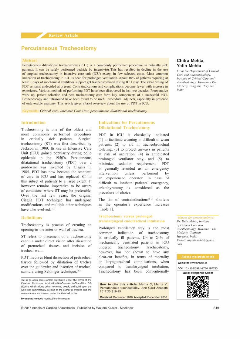
AnatomyBefore proceeding with tracheostomy, it is essential to have a thorough knowledge of neck anatomy [Figure 1]. Hyoid bone is the most stable portion of the airway and is easily felt when palpating from chin downward in the midline. Thyroid cartilage is felt next followed by cricothyroid membrane and cricoid cartilage. Tracheal rings can be palpated below the cricoid cartilage. Tracheal rings become difficult to appreciate as trachea descends into the chest. Jugular or suprasternal notch is felt as an angle at the junction of neck and chest. Cervical length of the trachea varies with spinal curvature, body build, extension‑flexion of the neck, and anteroposterior diameter of the chest. In young adults, nearly half the trachea resides in the neck region and it increases up to two-thirds with neck extension. In older adults, tracheal length in the neck may be reduced to one third. Suprasternal length of the trachea is also reduced if patients have kyphosis, limited neck mobility like in ankylosing spondylitis, and morbid obesity.
Tracheostomy is normally performed between second and third tracheal rings. Some investigators have found tracheal
Table 1: Contraindications of percutaneous dilatational tracheostomy
Absolute RelativeInfants Enlarged thyroid glandsInfection at insertion site Presence of pulsatile vessels at the
insertion siteOperator inexperience Difficult anatomy (short neck, morbid
obesity, limited neck extension, local malignancy, tracheal deviation)
Unstable cervical spine injuries
Coagulopathy
Uncontrollable coagulopathy
Close proximity to burns or surgical woundsHigh PEEP or FiO2 requirements (FiO2 >70%, PEEP >10 cm of H2O)History of cervical injury or tracheostomyHigh riding innominate arteryRadiotherapy to cervical region in last 4 weeksControlled local infection
PEEP: Positive end-expiratory pressure
Mehta and Mehta: Tracheostomy
S21Annals of Cardiac Anaesthesia | Vol 20 | Special Issue 1 | January 2017
puncture between third and fourth tracheal rings to be associated with a lower rate of injury to aberrant vessels, especially in the presence of anatomical abnormalities. Trachea is roughly 2–2.5 cm deep from the skin at the suggested insertion site, and this depth increases on moving downward to thoracic area. Tracheal slant from the vertical also increases as it descends into the chest which is more marked in older population. These anatomical changes are important to keep in mind while selecting an appropriate level for PDT, especially in older individuals.
Another important consideration is the esophagus which lies posterior to trachea in its entire course, except near carina where it is positioned slightly to the left. For all practical purposes, any injury to the posterior tracheal wall would also cause damage to the esophagus. Special attention needs to be paid to thyroid isthmus, which normally crosses second and third tracheal rings. Similarly, lateral lobes of thyroid also are closely situated. This area has a rich vascular supply and therefore predisposed to bleeding risk. Conventionally, midline is thought to be devoid of larger veins or arteries, but it is not always so. This knowledge has encouraged use of ultrasound for PDT.[16-21]
Percutaneous dilatational tracheostomy techniques
Percutaneous techniques were initially described in the mid-1980s by Ciaglia et al. Over the last two decades, it has undergone multiple modifications and some other alternatives have also come up. So far, there is no solid evidence favoring one technique over another.
Ciaglia Serial Dilatational TechniqueCiaglia et al. in 1985 carried out the first bedside PDT with the help of multiple sequentially larger dilators over the guidewire. This technique has undergone three major changes since then, in terms of level of tracheal interspace cannulation, use of concurrent bronchoscopy, and use of a single tapered dilator. The site of insertion has moved caudal from cricoid cartilage by one or two tracheal interspaces.
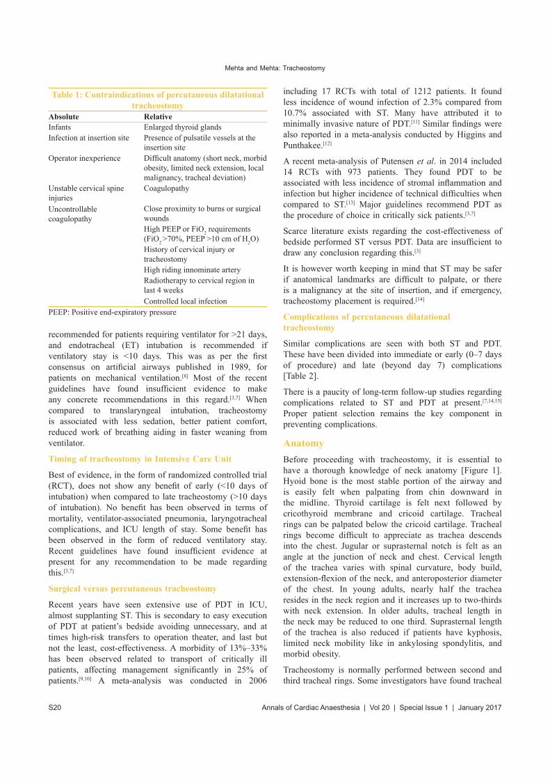
Ciaglia Single Dilator TracheostomyIt is popularly known as Ciaglia Blue Rhino (Cook Critical Care, Bloomington, IN, USA). It was introduced in 1999, more than a decade after initial description of Ciaglia technique. It is a simpler kit than the original kit and entails use of a single-beveled curved hydrophilic dilator. Use of single dilator is associated with reduced tidal volume loss during the procedure as change in dilator is not required. Similar to Blue Rhino, Portex Ultraperc single-stage dilator was developed by Smith Medical [Figure 2].
Griggs Percutaneous TechniqueThis technique was developed by Griggs et al. in 1990. It is also known as guidewire dilator forceps technique. The Portex Griggs percutaneous dilatational tracheostomy kit (Smith Medical) uses a specially designed forceps (modified Howard Kelly forceps) over the guidewire to result in single step dilatation of tissues in the pretracheal and tracheal space by spreading the forceps. The dilating forceps is designed to slide over the guidewire for the purpose. Following the dilatation, tracheostomy tube is inserted over the guidewire into the trachea. This
Table 2: Complications of percutaneous dilatational tracheostomyImmediate Early LateBleeding Tracheal ring fracture Subglottic stenosisLoss of airway Tracheal tube obstruction Unplanned decannulationHypoxia Paratracheal placement Tracheoinnominate artery bleedPneumothorax Posterior tracheal wall injury Displaced tracheal tubeFalse tract Pneumothorax/pneumomediastinum Delayed healing after decannulationPneumomediastinum Surgical emphysema Tracheoesophageal fistulaPosterior tracheal wall injury Atelectasis Stromal infectionEsophageal injury Raised intracranial pressure Scarring of the neckSurgical emphysema Swallowing difficultyNeedle damage to bronchoscope Permanent voice changesRaised intracranial pressure
Figure 1: Anatomy of neck
Mehta and Mehta: Tracheostomy
Annals of Cardiac Anaesthesia | Vol 20 | Special Issue 1 | January 2017S22
technique has lost its popularity due to higher incidence of soft tissue damage. It may be useful in places with resource constraint as forceps can be reused and no special kit is required.
Fantoni Translaryngeal TracheostomyThis was first described by Fantoni and Ripamonti in 1997. It is little cumbersome to perform and involves passage of guidewire retrogradally through the vocal cords after needle puncture of trachea. This is followed by railroading the combined dilator and tracheostomy tube over the guidewire into the larynx and out through the anterior tracheal wall. Tracheostomy tube is then separated from the dilator and rotated by 180° such that it faces the carina. This procedure requires an experienced operator.
Frova’s percutaneous tracheostomy
This involves use of a single-step screw-type dilator and is commercially available as the PercuTwist kit (Rusch, Kernen, Germany). It was first described in 2002. The dilator is rotated clockwise using a lifting motion over the guidewire. Earlier case reports had described tracheal ring fractures and posterior tracheal wall damage with this technique. However, it seems to be associated with better control over the dilating maneuver right from the start to end of procedure.
Balloon dilatational tracheostomy (Ciaglia Blue Dolphin, Cook Medical)
It is the second generation of Ciaglia technique. Inflation of a modified angioplasty balloon over a guidewire is used to dilate the trachea. The tracheal stoma is made by inflating the balloon with saline to 11 atmospheric pressure for 15 s. Then, the balloon is deflated and the tracheostomy tube insertion is done in a single step. This is an advantage over conventional single dilator PDT where the dilator needs to be removed before inserting the tracheostomy tube. There is a presumption that balloon dilation minimizes the pressure on the tracheal wall as compared to other techniques.[2,22-27]
As the experience with PDT is increasing, intensivists are venturing into performing the procedure in obese patients as well. Conventionally, it had been difficult in morbidly obese population, especially while inserting a longer
adjustable flange tube as a suitable loading dilator was not available. Now, it is smoother as two new kits with longer stoma dilation and adjustable flange armored tracheostomy tubes have been introduced recently (UniPerc, Smith Medical Kent, UIL and Expert set, TRACOE Medical GmbH, Frankfurt, Germany).[28]
Comparison of different percutaneous dilatational tracheostomy techniques
Each technique of PDT has its own set of characteristics and advantages. Many comparative studies have been conducted, but they are largely heterogeneous and with small sample size. A recent meta-analysis in 2012 had included 13 studies which compared at least two PDT techniques. No major differences were found between various techniques, except for the Fantoni technique. Fantoni technique was found to be associated with more serious complications with the need to convert to an alternative PDT technique. Ciaglia Blue Rhino technique fared better than other PDT techniques such as PercuTwist, Blue Dolphin, and Griggs technique.[29-32]
Practical aspects of percutaneous dilatational tracheostomy
The key aspect of preoperative planning is proper patient selection after thorough anatomical and physiological examination. Anatomical landmarks should be easily palpable, and there should be a space of at least 3–4 cm between the cricoid cartilage and the sternal notch. Once PDT is found to be feasible, informed consent should be taken. Special care should be taken to withhold anticoagulation if any. Patient should be nil by mouth as per common protocol. Continuous monitoring with blood pressure, electrocardiogram, pulse oximetry, and capnography is done throughout the procedure.
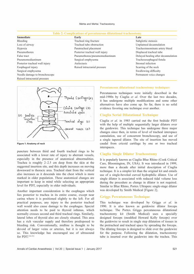
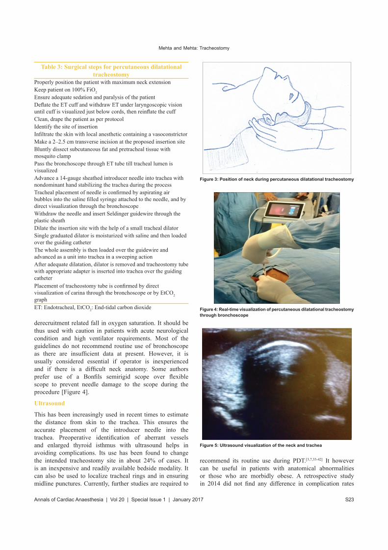
PDT can be performed under local anesthesia but is generally performed under adequate analgesia, sedation, and muscle relaxants sometimes. Most of the times, the patient is already intubated. Neck is properly positioned and adequately extended with help of a roll placed under the patient’s shoulders [Figure 3]. The incision point is typically located half way between the cricoids cartilage and sternal notch. Table 3 describes the surgical procedure in detail.
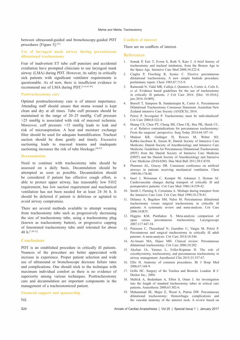
Procedural AdjunctsBronchoscopy
Use of bronchoscope during the procedure has certain obvious advantages such as real‑time confirmation of needle placement, midline position of the needle, tube placement, and avoidance of posterior tracheal wall injury. There have however been concerns regarding its routine use. It has been found to be associated with measurable increases in intracranial pressure and alveolar
Figure 2: Ultraperc and Blue Rhino set
Mehta and Mehta: Tracheostomy
S23Annals of Cardiac Anaesthesia | Vol 20 | Special Issue 1 | January 2017
derecruitment related fall in oxygen saturation. It should be thus used with caution in patients with acute neurological condition and high ventilator requirements. Most of the guidelines do not recommend routine use of bronchoscope as there are insufficient data at present. However, it is usually considered essential if operator is inexperienced and if there is a difficult neck anatomy. Some authors prefer use of a Bonfils semirigid scope over flexible scope to prevent needle damage to the scope during the procedure [Figure 4].
Ultrasound
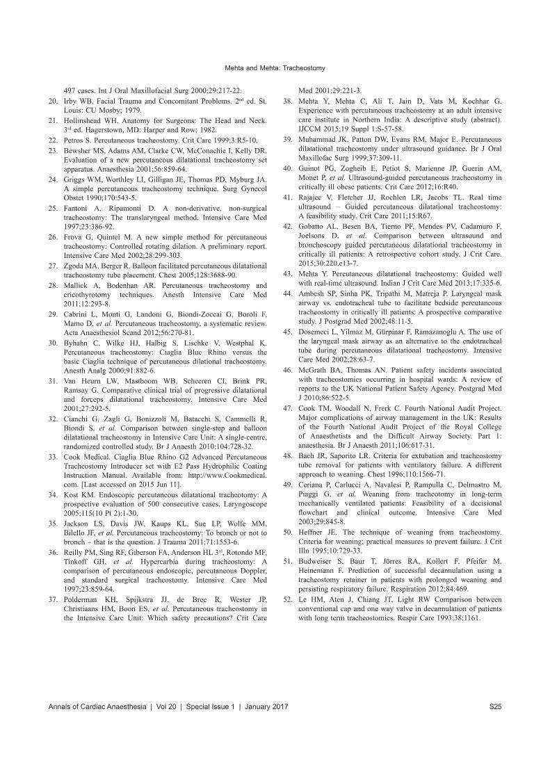
This has been increasingly used in recent times to estimate the distance from skin to the trachea. This ensures the accurate placement of the introducer needle into the trachea. Preoperative identification of aberrant vessels and enlarged thyroid isthmus with ultrasound helps in avoiding complications. Its use has been found to change the intended tracheostomy site in about 24% of cases. It is an inexpensive and readily available bedside modality. It can also be used to localize tracheal rings and in ensuring midline punctures. Currently, further studies are required to
recommend its routine use during PDT.[3,7,33-42] It however can be useful in patients with anatomical abnormalities or those who are morbidly obese. A retrospective study in 2014 did not find any difference in complication rates
Table 3: Surgical steps for percutaneous dilatational tracheostomy
Properly position the patient with maximum neck extensionKeep patient on 100% FiO2
Ensure adequate sedation and paralysis of the patientDeflate the ET cuff and withdraw ET under laryngoscopic vision until cuff is visualized just below cords, then reinflate the cuffClean, drape the patient as per protocolIdentify the site of insertionInfiltrate the skin with local anesthetic containing a vasoconstrictorMake a 2–2.5 cm transverse incision at the proposed insertion siteBluntly dissect subcutaneous fat and pretracheal tissue with mosquito clampPass the bronchoscope through ET tube till tracheal lumen is visualizedAdvance a 14-gauge sheathed introducer needle into trachea with nondominant hand stabilizing the trachea during the processTracheal placement of needle is confirmed by aspirating air bubbles into the saline filled syringe attached to the needle, and by direct visualization through the bronchoscopeWithdraw the needle and insert Seldinger guidewire through the plastic sheathDilate the insertion site with the help of a small tracheal dilatorSingle graduated dilator is moisturized with saline and then loaded over the guiding catheterThe whole assembly is then loaded over the guidewire and advanced as a unit into trachea in a sweeping actionAfter adequate dilatation, dilator is removed and tracheostomy tube with appropriate adapter is inserted into trachea over the guiding catheterPlacement of tracheostomy tube is confirmed by direct visualization of carina through the bronchoscope or by EtCO2 graphET: Endotracheal, EtCO2: End-tidal carbon dioxide
Figure 3: Position of neck during percutaneous dilatational tracheostomy
Figure 4: Real-time visualization of percutaneous dilatational tracheostomy through bronchoscope
Figure 5: Ultrasound visualization of the neck and trachea
Mehta and Mehta: Tracheostomy
Annals of Cardiac Anaesthesia | Vol 20 | Special Issue 1 | January 2017S24
between ultrasound-guided and bronchoscopy-guided PDT procedures [Figure 5].[41]
Use of laryngeal mask airway during percutaneous dilatational tracheostomy
Fear of inadvertent ET tube cuff puncture and accidental extubation have prompted clinicians to use laryngeal mask airway (LMA) during PDT. However, its safety in critically sick patients with significant ventilator requirements is questionable. As of now, there is insufficient evidence to recommend use of LMA during PDT.[3,14,43-45]
Posttracheostomy care
Optimal posttracheostomy care is of utmost importance. Attending staff should ensure that stoma wound is kept clean and dry at all times. Tube cuff pressure should be maintained in the range of 20–25 mmHg. Cuff pressure >25 mmHg is associated with risk of mucosal ischemia. Moreover, cuff pressure <15 mmHg leads to leak and risk of microaspiration. A heat and moisture exchange filter should be used for adequate humidification. Tracheal suction should be done gently and timely. Excessive suctioning leads to mucosal trauma and inadequate suctioning increases the risk of tube blockage.[46,47]
Decannulation
Need to continue with tracheostomy tube should be assessed on a daily basis. Decannulation should be attempted as soon as possible. Decannulation should be considered if patient has effective cough effort, is able to protect upper airway, has reasonably low FiO2 requirement, has low suction requirement and mechanical ventilation has not been needed for at least 24–36 h. It should be deferred if patient is delirious or agitated to avoid airway compromise.
There are several methods available to attempt weaning from tracheostomy tube such as progressively decreasing the size of tracheostomy tube, using a tracheostomy plug (known as tracheostomy button), or progressive capping of fenestrated tracheostomy tube until tolerated for about 48 h.[7,48-52]
ConclusionsPDT is an established procedure in critically ill patients. Nuances of the procedure are better appreciated with increase in experience. Proper patient selection and wide use of ultrasound or bronchoscope decrease failure rates and complications. One should stick to the technique with maximum individual comfort as there is no evidence of superiority among various techniques. Posttracheostomy care and decannulation are important components in the management of a tracheostomized patient.
Financial support and sponsorship
Nil.
Conflicts of interest
There are no conflicts of interest.
References1. Szmuk P, Ezri T, Evron S, Roth Y, Katz J. A brief history of
tracheostomy and tracheal intubation, from the Bronze Age to the Space Age. Intensive Care Med 2008;34:222-8.
2. Ciaglia P, Firsching R, Syniec C. Elective percutaneous dilatational tracheostomy. A new simple bedside procedure; preliminary report. Chest 1985;87:715-9.
3. Raimondi N, Vidal MR, Calleja J, Quintero A, Cortés A, Celis E, et al. Evidence based guidelines for the use of tracheostomy in critically ill patients. J Crit Care 2016. [Doi: 10.1016/j.jerc.2016.10.009].
4. Burrell T, Sampson B, Sundararajan K, Carter A. Percutaneous Dilatational Tracheostomy Consensus Statement. Australian New Zealand intensive Care Society (ANZICS); 2014.
5. Pelosi P, Severgnini P. Tracheostomy must be individualized! Crit Care 2004;8:322-4.
6. Huang CS, Chen PT, Cheng SH, Chen CK, Hsu PK, Hsieh CC, et al. Relative contraindications for percutaneous tracheostomy: From the surgeons’ perspective. Surg Today 2014;44:107-14.
7. Madsen KR, Guldager H, Rewers M, Weber SO, Købke-Jacobsen K, Jensen R; Danish Society of Intensive Care Medicine; Danish Society of Anesthesiology and Intensive Care Medicine. Guidelines for Percutaneous Dilatational Tracheostomy (PDT) from the Danish Society of Intensive Care Medicine (DSIT) and the Danish Society of Anesthesiology and Intensive Care Medicine (DASAIM). Dan Med Bull 2011;58:C4358.
8. Plummer AL, Gracey DR. Consensus conference on artificial airways in patients receiving mechanical ventilation. Chest 1989;96:178-80.
9. Insel J, Weissman C, Kemper M, Askanazi J, Hyman AI. Cardiovascular changes during transport of critically ill and postoperative patients. Crit Care Med 1986;14:539-42.
10. Smith I, Fleming S, Cernaianu A. Mishaps during transport from the Intensive Care Unit. Crit Care Med 1990;18:278-81.
11. Delaney A, Bagshaw SM, Nalos M. Percutaneous dilatational tracheostomy versus surgical tracheostomy in critically ill patients: A systematic review and meta-analysis. Crit Care 2006;10:R55.
12. Higgins KM, Punthakee X. Meta-analysis comparison of open versus percutaneous tracheostomy. Laryngoscope 2007;117:447-54.
13. Putensen C, Theuerkauf N, Guenther U, Vargas M, Pelosi P. Percutaneous and surgical tracheostomy in critically ill adult patients: A meta-analysis. Crit Care 2014;18:544.
14. Al-Ansari MA, Hijazi MH. Clinical review: Percutaneous dilatational tracheostomy. Crit Care 2006;10:202.
15. Akulian JA, Yarmus L, Feller-Kopman D. The role of cricothyrotomy, tracheostomy, and percutaneous tracheostomy in airway management. Anesthesiol Clin 2015;33:357-67.
16. Ellis H. Anatomy of common procedures. Br J Hosp Med 2006;67:168-9.
17. Grills HC. Surgery of the Trachea and Bronchi. London: B C Decker Inc.; 2004.
18. Mallick A, Bodenham A, Elliot S, Oram J. An investigation into the length of standard tracheostomy tubes in critical care patients. Anaesthesia 2008;63:302-6.
19. Muhammad JK, Major E, Wood A, Patton DW. Percutaneous dilatational tracheostomy: Hemorrhagic complications and the vascular anatomy of the anterior neck. A review based on
Mehta and Mehta: Tracheostomy
S25Annals of Cardiac Anaesthesia | Vol 20 | Special Issue 1 | January 2017
497 cases. Int J Oral Maxillofacial Surg 2000;29:217-22.20. Irby WB. Facial Trauma and Concomitant Problems. 2nd ed. St.
Louis: CU Mosby; 1979.21. Hollinshead WH. Anatomy for Surgeons: The Head and Neck.
3rd ed. Hagerstown, MD: Harper and Row; 1982.22. Petros S. Percutaneous tracheostomy. Crit Care 1999;3:R5-10.23. Bewsher MS, Adams AM, Clarke CW, McConachie I, Kelly DR.
Evaluation of a new percutaneous dilatational tracheostomy set apparatus. Anaesthesia 2001;56:859-64.
24. Griggs WM, Worthley LI, Gilligan JE, Thomas PD, Myburg JA. A simple percutaneous tracheostomy technique. Surg Gynecol Obstet 1990;170:543-5.
25. Fantoni A, Ripamonti D. A non-derivative, non-surgical tracheostomy: The translaryngeal method. Intensive Care Med 1997;23:386-92.
26. Frova G, Quintel M. A new simple method for percutaneous tracheostomy: Controlled rotating dilation. A preliminary report. Intensive Care Med 2002;28:299-303.
27. Zgoda MA, Berger R. Balloon facilitated percutaneous dilatational tracheostomy tube placement. Chest 2005;128:3688-90.
28. Mallick A, Bodenhan AR. Percutaneous tracheostomy and cricothyrotomy techniques. Anesth Intensive Care Med 2011;12:293-8.
29. Cabrini L, Monti G, Landoni G, Biondi‑Zoccai G, Boroli F, Mamo D, et al. Percutaneous tracheostomy, a systematic review. Acta Anaesthesiol Scand 2012;56:270-81.
30. Byhahn C, Wilke HJ, Halbig S, Lischke V, Westphal K. Percutaneous tracheostomy: Ciaglia Blue Rhino versus the basic Ciaglia technique of percutaneous dilational tracheostomy. Anesth Analg 2000;91:882-6.
31. Van Heurn LW, Mastboom WB, Scheeren CI, Brink PR, Ramsay G. Comparative clinical trial of progressive dilatational and forceps dilatational tracheostomy. Intensive Care Med 2001;27:292-5.
32. Cianchi G, Zagli G, Bonizzoli M, Batacchi S, Cammelli R, Biondi S, et al. Comparison between single-step and balloon dilatational tracheostomy in Intensive Care Unit: A single-centre, randomized controlled study. Br J Anaesth 2010;104:728-32.
33. Cook Medical. Ciaglia Blue Rhino G2 Advanced Percutaneous Tracheostomy Introducer set with E2 Pass Hydrophilic Coating Instruction Manual. Available from: http://www.Cookmedical.com. [Last accessed on 2015 Jun 11].
34. Kost KM. Endoscopic percutaneous dilatational tracheotomy: A prospective evaluation of 500 consecutive cases. Laryngoscope 2005;115(10 Pt 2):1-30.
35. Jackson LS, Davis JW, Kaups KL, Sue LP, Wolfe MM, Bilello JF, et al. Percutaneous tracheostomy: To bronch or not to bronch – that is the question. J Trauma 2011;71:1553-6.
36. Reilly PM, Sing RF, Giberson FA, Anderson HL 3rd, Rotondo MF, Tinkoff GH, et al. Hypercarbia during tracheostomy: A comparison of percutaneous endoscopic, percutaneous Doppler, and standard surgical tracheostomy. Intensive Care Med 1997;23:859-64.
37. Polderman KH, Spijkstra JJ, de Bree R, Wester JP, Christiaans HM, Boon ES, et al. Percutaneous tracheostomy in the Intensive Care Unit: Which safety precautions? Crit Care
Med 2001;29:221-3.38. Mehta Y, Mehta C, Ali T, Jain D, Vats M, Kochhar G.
Experience with percutaneous tracheostomy at an adult intensive care institute in Northern India: A descriptive study (abstract). IJCCM 2015;19 Suppl 1:S-57-58.
39. Muhammad JK, Patton DW, Evans RM, Major E. Percutaneous dilatational tracheostomy under ultrasound guidance. Br J Oral Maxillofac Surg 1999;37:309-11.
40. Guinot PG, Zogheib E, Petiot S, Marienne JP, Guerin AM, Monet P, et al. Ultrasound-guided percutaneous tracheostomy in critically ill obese patients. Crit Care 2012;16:R40.
41. Rajajee V, Fletcher JJ, Rochlen LR, Jacobs TL. Real time ultrasound – Guided percutaneous dilatational tracheostomy: A feasibility study. Crit Care 2011;15:R67.
42. Gobatto AL, Besen BA, Tierno PF, Mendes PV, Cadamuro F, Joelsons D, et al. Comparison between ultrasound and bronchoscopy guided percutaneous dilatational tracheostomy in critically ill patients: A retrospective cohort study. J Crit Care. 2015;30:220.e13-7.
43. Mehta Y. Percutaneous dilatational tracheostomy: Guided well with real-time ultrasound. Indian J Crit Care Med 2013;17:335-6.
44. Ambesh SP, Sinha PK, Tripathi M, Matreja P. Laryngeal mask airway vs. endotracheal tube to facilitate bedside percutaneous tracheostomy in critically ill patients: A prospective comparative study. J Postgrad Med 2002;48:11-5.
45. Dosemeci L, Yilmaz M, Gürpinar F, Ramazanoglu A. The use of the laryngeal mask airway as an alternative to the endotracheal tube during percutaneous dilatational tracheostomy. Intensive Care Med 2002;28:63-7.
46. McGrath BA, Thomas AN. Patient safety incidents associated with tracheostomies occurring in hospital wards: A review of reports to the UK National Patient Safety Agency. Postgrad Med J 2010;86:522-5.
47. Cook TM, Woodall N, Frerk C. Fourth National Audit Project. Major complications of airway management in the UK: Results of the Fourth National Audit Project of the Royal College of Anaesthetists and the Difficult Airway Society. Part 1: anaesthesia. Br J Anaesth 2011;106:617-31.
48. Bach JR, Saporito LR. Criteria for extubation and tracheostomy tube removal for patients with ventilatory failure. A different approach to weaning. Chest 1996;110:1566-71.
49. Ceriana P, Carlucci A, Navalesi P, Rampulla C, Delmastro M, Piaggi G, et al. Weaning from tracheotomy in long-term mechanically ventilated patients: Feasibility of a decisional flowchart and clinical outcome. Intensive Care Med 2003;29:845-8.
50. Heffner JE. The technique of weaning from tracheostomy. Criteria for weaning; practical measures to prevent failure. J Crit Illn 1995;10:729-33.
51. Budweiser S, Baur T, Jörres RA, Kollert F, Pfeifer M, Heinemann F. Prediction of successful decannulation using a tracheostomy retainer in patients with prolonged weaning and persisting respiratory failure. Respiration 2012;84:469.
52. Le HM, Aten J, Chiang JT, Light RW Comparison between conventional cap and one way valve in decannulation of patients with long term tracheostomies. Respir Care 1993;38:1161.


